Brief Report

Tear Cytokine Changes up to One Year After Allogeneic Hematopoietic Stem Cell Transplant: Effect of Daily Topical Cyclosporine-A 0.1% Emulsion

1
Corneal and External Eye Disease Service, Singapore National Eye Centre, 11 Third Hospital Avenue, Singapore 168751, Singapore
2
Ocular Surface Research Group, Singapore Eye Research Institute, 20 College Road Discovery Tower Level 6, The Academia, Singapore 169856, Singapore
3
Eye-Academic Clinical Program, Duke-National University of Singapore Medical School, 8 College Road, Singapore 169857, Singapore
4
Department of Ophthalmology, Yong Loo Lin School of Medicine, National University of Singapore, 10 Medical Dr., Singapore 117597, Singapore
5
Department of Haematology, Singapore General Hospital, 31 Third Hospital Ave, Singapore 168753, Singapore
*
Author to whom correspondence should be addressed.
Int. J. Mol. Sci. 2025, 26(12), 5915; https://doi.org/10.3390/ijms26125915
Submission received: 11 April 2025 / Revised: 3 June 2025 / Accepted: 7 June 2025 / Published: 19 June 2025
(This article belongs to the Section Molecular Pathology, Diagnostics, and Therapeutics)

Abstract

:
Purpose: To profile tear cytokine changes in Allogeneic Hematopoietic Stem Cell Transplant (HSCT) patients after instillation of daily topical cyclosporine-A 0.1% cationic emulsion. Methods: Participants in a longitudinal study were given cyclosporine eyedrops daily from 3 to 5 weeks before and 3 months, 6 months, and 12 months post-HSCT. The outcomes included tear cytokine concentration assayed by the Proximity Extension Assay O-linked target 96 platform. The patients were divided into two groups: Group 1 (n = 8 conjunctival CD4 cells responding to cyclosporine) and Group 2 (n = 5 conjunctival CD4 cells not suppressed after cyclosporine, where patients were non-compliant with cyclosporine). All participants had a standardized clinical examination, including meibomian gland evaluation and tear breakup times. Results: The levels of 38 cytokines/chemokines showed significant changes (p < 0.05) over time, and in many, the elevation was marked at one year. These include gamma-interferon, CXCL9, CCL3, and CCL4 (all p < 0.0001). For gamma-interferon, there was significant interaction between group and time at 1 year (p = 0.022), where the cytokine was significantly suppressed in Group 1. Four other cytokines showed significant group and time interaction at 1 year: FGF23, FGF5, LIFR, and Enrage (all p < 0.05). All patients had either withdrawal or a reduction in systemic immunomodulation between 6 months and 1 year. We found several cytokines to be associated with changes in tear osmolarity or symptom scores. Conclusions: HSCT induces significant elevation of 38 tear cytokines/chemokines even without the occurrence of ocular graft-versus-host disease when systemic immunosuppression is reduced within the first year. Topical daily cyclosporine eyedrops can reduce some pro-inflammatory tear cytokines.

1. Introduction

Dry eye disease (DED) is a common ocular sequela of graft-versus-host disease (GVHD) after allogeneic hematopoietic stem cell transplantation (allo-HSCT), with one study showing more than 40% of allo-HSCT patients developing chronic ocular GVHD (according to both NIH consensus criteria and the International Chronic Ocular GVHD Consensus Group) [1,2,3]. Acute GVHD happens during the first 100 days following HSCT, but chronic GVHD occurs beyond that time. Approximately 7.5 months passed on average between allo-HSCT and the start of ocular GVHD [4,5]
The pathogenesis of DED in GVHD is characterized by a T cell-mediated infiltration and inflammation of the lacrimal gland, conjunctiva, and ocular surface. This process leads to fibrosis of the lacrimal gland and conjunctiva, a reduction in the number of conjunctival goblet cells, and a decrease in tear production [6,7,8]. GVHD also causes meibomian gland obstruction and anterior and posterior blepharitis [9,10]. Previously, we showed that people who underwent HSCT displayed one of two patterns of change in conjunctival CD4 T cells, depending on their compliance with cyclosporine [11].
It is advantageous to investigate preventive measures for DED in this population, given the high incidence of DED in allo-HSCT patients. It has been observed that topical CsA is both safe and effective in treating DED linked to chronic GVHD [12,13,14]. However, there are only a few reports on topical cyclosporine-A in the prophylaxis of ocular GVHD [15,16,17].
Since cytokines in the tear drive ocular surface inflammation, we are interested in determining whether concentrations of various cytokines change with time after HSCT and whether instillation of topical cyclosporine influences these concentrations.
In this prospective study, our primary aim was to profile tear cytokine concentrations in allogeneic HSCT patients up to one year and to investigate the effect of daily topical cyclosporine-A 0.1% cationic emulsion on tear cytokines. A secondary aim was to evaluate whether clinical symptoms and signs of dry eye in the participants would significantly change when levels of tear cytokines were altered during the follow-up.

2. Methods

2.1. Design

This is an open-label single-arm interventional study performed at the Singapore National Eye Centre (SNEC) in collaboration with the Hematology Department at the Singapore General Hospital (SGH).

2.2. Study Population

Patients who underwent allo-HSCT at the Hematology Department, SGH, between November 2019 and September 2021 were recruited. Every participant signed an informed consent form. The SingHealth Centralised Institutional Review Board prospectively examined and authorized our work, which aligned with the principles of the Declaration of Helsinki on human research (reference number: 2019/2635). The Clinical Trials database registration number for the trial is NCT04636918; the URL is https://clinicaltrials.gov/ct2/show/NCT04636918?cond=ocular+Graft+Versus+Host+Disease&cntry=SG&draw=2&rank=2 (accessed on 16 June 2025).

2.3. Eligibility

The participants in the trial had undergone HSCT and were 13 years old or older, capable of providing informed consent, and able to meet study evaluation conditions. The following patients were excluded from the study: age below 13 years, the presence of other concurrent ocular or systemic disease that may interfere with study results, non-resident patients who were unable to complete follow-up, and existing users of topical cyclosporine eyedrops. During the COVID-19 pandemic, there was limited access to hospital inpatients, and patients who could not be evaluated as in the trial protocol were not included.

2.4. Study Intervention

The participants were prescribed topical cyclosporine-A 0.1% (Ikervis®, Santen Pharmaceuticals Asia Pte Ltd., Osaka, Japan) prophylaxis once daily to both eyes and preservative-free sodium hyaluronate 0.3% (Hialid Mini, Santen Pharmaceuticals Asia Pte Ltd., Osaka, Japan) lubricating eyedrop to be used pro re nata (PRN) from 3 to 5 weeks prior to allo-HSCT to 12 months after allo-HSCT. Based on the transplant protocol used, the participants received systemic GVHD therapy with systemic CsA or tacrolimus combined with a course of methotrexate or mycophenolate. Antithymocyte globulin (ATG) or post-transplant cyclophosphamide was added in cases of unrelated/sibling donor with antigen mismatch or haploidentical transplant, respectively. (Refer to our previous report for the systemic diagnoses and the regimen of systemic immunosuppression in these patients) [11].

2.5. Assessment of Tolerance/Compliance with Ikervis®

Compliance with the use of Ikervis® in this study was based on the daily drug diary [11]. Briefly, compliance was defined as usage on more than 70% of days and less than five blocks of three consecutive days of not using Ikervis® eyedrops.

2.6. Study Visits

Study visits occurred at 3–5 weeks prior to allo-HSCT (screening visit), right before allo-HSCT, and 3 months, 6 months, and 12 months post-allo-HSCT (Supplementary Table S1).

2.7. Clinical Procedures

Details of the clinical procedures were published elsewhere [11]. Briefly, at each study visit, symptoms were evaluated using the Standardized Evaluation of Eye Dryness (SPEED) questionnaire [18]. Corneal staining was evaluated on a slit lamp biomicroscopy using five zones after the addition of fluorescein dye from a wetted Floret [19,20]. The non-invasive tear breakup times [21,22] and the conjunctival redness grading [23] were performed using the Oculus Keratograph 5M [24]. Tear osmolarity was determined using a TearLab osmometer at the baseline study visit [25,26]. The number of liquid meibum-expressing glands in the lower eyelids was evaluated using a standard force meibomian gland evaluator at baseline and final visit [27].

2.8. Tear Cytokines

Schirmer strips from each eye collected over five minutes (without anesthesia) were frozen at −80 Celsius until ready for extraction.
Tear cytokines in all tear samples were analyzed using Olink® Target 96 (Thermo Fisher Scientific, Waltham, MA, USA). Schirmer strips with tear fluid samples were rolled up and transferred to a Spin-X centrifuge tube (0.22 µm Cellulose Acetate; 98231-UT-1; Costar®, Costar, Glendale, CA, USA) and submerged in 300 µL of elution buffer, consisting of 0.55 M NaCl (S3014-500G, Sigma Life Science, Darmstadt, Germany), 0.33% Tween 20 (P1379-500ML; Sigma Life Science), and 0.55% Bovine Serum Albumin (9998S; Cell Signalling Technology, Danvers, MA, USA). The samples were subjected to an incubation at 450 rpm for 10 mins at 21 °C. The eluted tears were subsequently centrifuged at 16,000× g for 10 min at 21 °C. Subsequently, the clear supernatant was collected and stored in 1.5 mL Protein Lo-bind Tubes (022431081; Eppendorf®, Eppendorf, Hamburg, Germany) at −80 °C until the day of analysis.
After the extraction of tears, cytokines were assayed by the Proximity Extension Assay O-linked target 96 platform, a very sensitive method of analysis for a panel of 96 cytokines previously reported [28]. Briefly, each oligonucleotide antibody pair contains unique DNA sequences, allowing hybridization only to each other. Subsequent proximity extension created 96 unique DNA reporter sequences (amplified by real-time PCR).
Concentrations normalized to tear volume, collected as reported in a previous paper, were estimated from the wetting of the Schirmer strip+3 mm.

2.9. Statistical Analysis

Only the right eye from each participant was analyzed to avoid the influence of the high correlation between eyes.
The patients were divided into two groups based on the impression flow cytology analysis in our previous report: Group 1 (n = 8 conjunctival CD4 cells responding to cyclosporine) and Group 2 (n = 5 conjunctival CD4 cells not suppressed after cyclosporine, where patients were non-compliant with cyclosporine). Group 2 also contained one patient who had central cornea staining prior to HSCT and continued to have some signs of DED during the visits despite being compliant with topical cyclosporine [11].
The method of analysis of the repeated measures used linear mixed model regression (Stata), where Y is the cytokine concentration. The procedure used was xtmixed y profile##time || id:, var reml, where Id is the identifier of each patient and Var is the variance of the regression (mixed linear). To obtain joint tests (multiple degrees of freedom) of the interaction and main effects, we used the contrast group##time command. Relevant time trends of specific cytokines were plotted using Stata’s margins and marginsplot commands. More details can be found in the following REF citation: Bruin, J. 2006. newtest: command to compute new test. UCLA Statistical Consulting Group. https://stats.oarc.ucla.edu/stata/ado/analysis/ (accessed on 16 June 2025).
Linear mixed-effect models (LMMs) were used to examine the association between individual clinical parameters and each of the tear cytokine concentrations. The LMMs were adjusted for the correlation between both eyes and repeated time points. Statistical significance of the beta was defined as p < 0.05.

3. Results

Please refer to our previous paper for the clinical evaluation results of these patients (Supplementary Figure S1) [11]. Briefly, no participant developed significant ocular signs and symptoms during the study visits, except for one participant who already suffered from central corneal staining prior to the HSCT. The p-values for the time effect refer to whether there is a significant alteration of a specific cytokine between different times after HSCT. The p-values for the group effect refer to whether there is a difference between the two groups (with respect to cyclosporine usage) overall. The p-values for the group*time interaction seek to reveal differences in the tear cytokine level between the two groups at specific times but not at other times.

3.1. Clinical Symptom Changes

Across all the participants, the severity of SPEED symptoms was increased when the levels of four cytokines were elevated: fibroblast growth factor (FGF)5 (beta 10.383 [95%CI 2.906–17.861], p = 0.006), interleukin (IL)2 (beta 5.897 [95%CI 1.108–10.686], p = 0.016), tumor necrosis factor receptor superfamily member (TNFRSF)9 (beta 4.283 [95%CI 1.481–7.084], p = 0.003) and glial cell line-derived neurotrophic factor (GDNF) (beta 3.824 [95%CI 0.024–7.623], p = 0.049).
The scatter plots for the tear levels of these four cytokines and SPEED symptom scores are shown in Figure 1.

3.2. Clinical Sign Changes

Across all the participants, the level of the conjunctival redness varied positively with the level of three of the cytokines: CD8A (beta 0.33 [95%CI 0.02–0.63], p = 0.04), IL2RB (beta 0.50 [95%CI 0.05–0.94], p < 0.001), and IL33 (beta 0.63 [95%CI 0.02–1.23], p = 0.04).
Increased levels of IL17A were associated with increased cornea staining (beta 0.387 [95%CI 1.301–6.334], p = 0.003).
Increased levels of one cytokine, FGF23, were associated with reduced expressivity of meibomian glands (number of liquid meibum expressed from lower eyelids), i.e., FGF23 (beta −2.259 [95%CI −4.5 to −0.018], p = 0.048), whereas IL20 was associated with increased expressivity (beta 1.999 [95%CI 0.043–3.955], p = 0.045).
Most interestingly, the tear level of fourteen cytokines was associated with baseline tear osmolarity. Nine cytokines increased with tear osmolarity levels, whereas five cytokines were inversely correlated with tear osmolarity (Table 1). Increased levels of matrix metalloproteinase (MMP) 1 and certain pro-inflammatory cytokines (CCL3, CCL4, IL 1alpha, and IL12beta) were associated with increased tear osmolarity during the study visits.

3.3. Tear Cytokine Changes

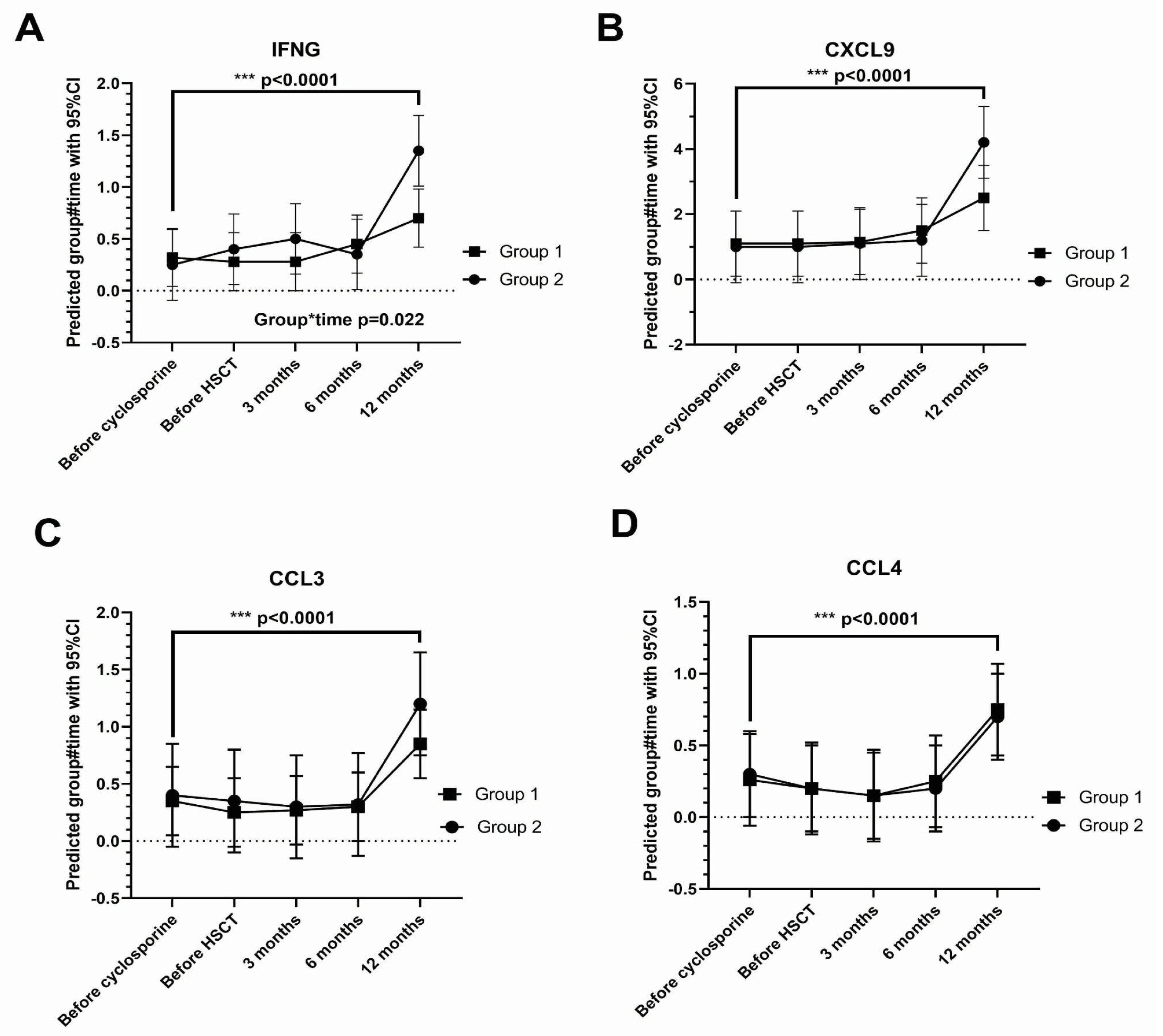
The tear concentration of 38 cytokines (Table 2) showed a significant change (p < 0.05) over time, and for many of these, the elevation was marked at the one-year visit. These include gamma-interferon (IFNG), CXC-chemokine ligand (CXCL)9, CC-chemokine ligand (CCL)3, and CCL4 (all p < 0.0001) (Figure 2).
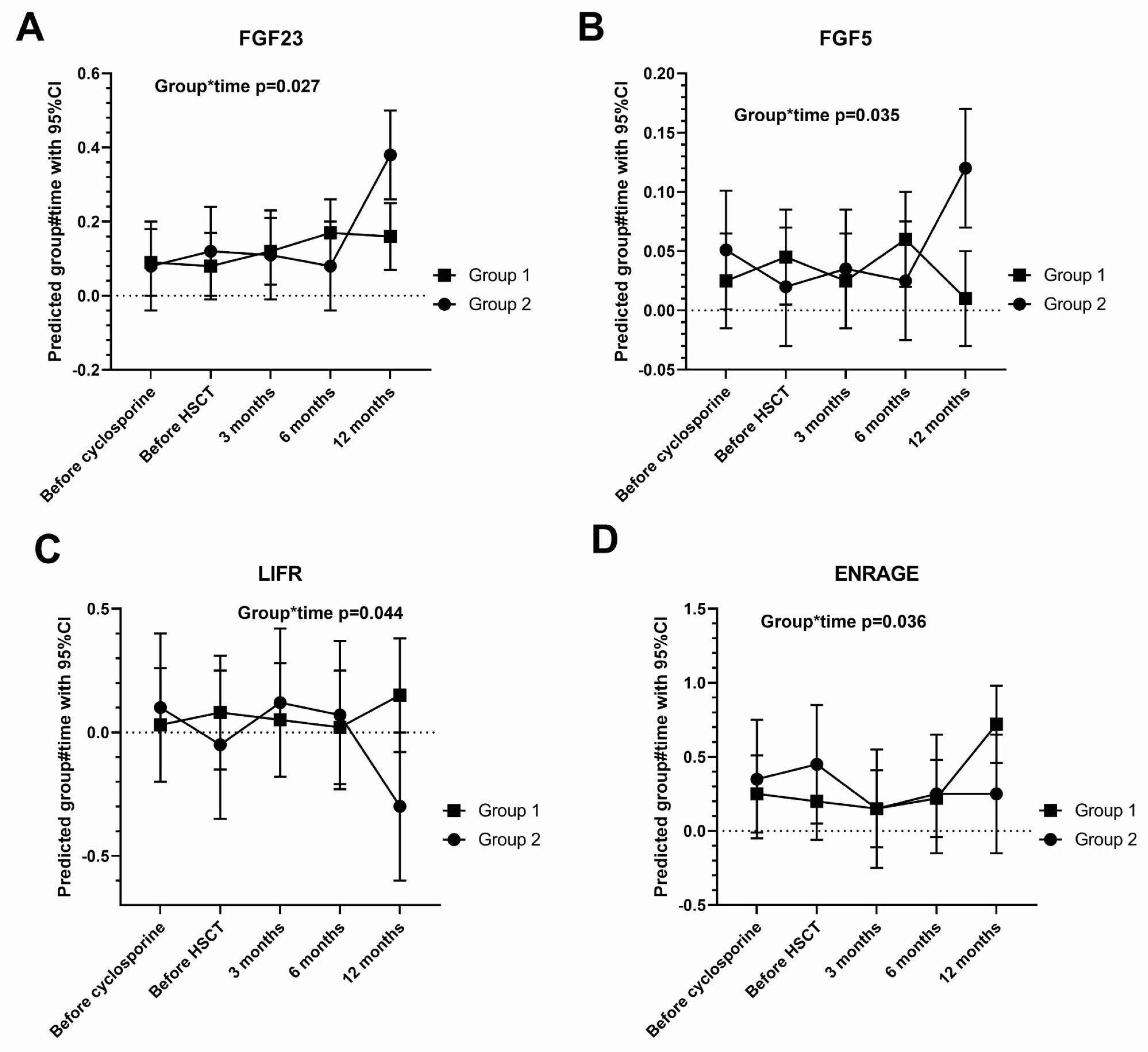
For gamma-interferon, there was significant interaction between group and time at the 1-year visit (p = 0.022), where the cytokine was significantly suppressed in Group 1. Four other cytokines showed significant group and time interaction at the last (1 year) visit: FGF23 (p = 0.027), FGF5 (p = 0.035), leukemia inhibitory factor receptor (LIFR) (p = 0.044), and advanced glycation end-products binding protein (ENRAGE) (p = 0.036). Tear FGF23 and FGF5 were suppressed by cyclosporine, whereas LIFR and ENRAGE were depressed by cyclosporine (Figure 3).
Prior to HSCT (first two visits), T cell stimulant tumor necrosis factor superfamily (TNFSF) 14 was suppressed in Group 1 (p = 0.039 by paired T test) but not in Group 2 (Figure 4).
Systemic immunosuppression, as decided by the hematologists managing the patients, tended to be reduced or withdrawn at the end of six months in all the participants (Table 3 and Table 4).

4. Discussion

This study shows that HSCT induces a significant elevation of 38 tear cytokines/chemokines, which becomes most marked at the one-year visit, even without the occurrence of ocular graft-versus-host disease. The reduction in or stoppage of the systemic immunosuppression at six months occurred at the same time as the increase in tear cytokines.
The findings of this study suggest that an elevation in tear cytokines is a phenomenon that may occur after HSCT, even though no clinical ocular GVHD occurred up to one year. It remains to be seen if tear cytokines would increase if systemic immunosuppression had not been followed in the first year. Given that the tear cytokines increased, it is unclear whether ocular GVHD will occur in these patients years or sometime after the cessation of the topical cyclosporine. These patients will continue to be monitored by the study team. This has implications for the pathophysiology of chronic ocular GVHD. It may be that immune cells or progenitor cells migrate or traffic to ocular glands and the ocular surface over a long time after HSCT.
Our study also verified that changes in the levels of tear cytokines occurred with changes in the clinical parameters of dry eye disease. This highlights the real-world significance of cytokine levels.
The fact that many cytokines increased between six months and one year after transplantation, corresponding to the period when most of the systemic immunosuppression was reduced or stopped, suggests that systemic immunosuppression may be critical for maintaining an anti-inflammatory milieu in the tear and ocular surface. Beyond the one-year period, topical cyclosporine on its own may not be sufficient to prevent further elevation in pro-inflammatory cytokines.
Cyclosporine works by inhibiting calcineurin/nuclear factor of activated T cell signaling, inducing apoptosis of T cells [29], and inhibiting immunoglobulin synthesis [30]. There are plenty of T cells in the human conjunctiva [31,32,33,34,35]. It has been shown that treatment with topical cyclosporine resulted in a reduction in T cells in the conjunctiva epithelium and stroma of people with Sjogren’s syndrome [36,37] and in people with allergic conjunctivitis [38].
IFNG is a well-known cytokine in DED, which drives the inflammatory response in the ocular surface. It is interesting that topical cyclosporine suppresses the rise in this cytokine at the one-year time point [39,40,41,42,43] and that the participants compliant with topical cyclosporine had a significant reduction in conjunctival CD4 T cells [11].
The FGF family has been implicated in multiple biological processes in mammals, such as cell proliferation and differentiation, including tissue repair. The functions of LIFR and ENRAGE in the post-HSCT context are unknown.
A previous study involving thirty healthy volunteers found a link between increased cytokines and increased tear osmolarity [44]. Tear cytokines have also been linked to the severity of dry eye disease in patients [45].
A cross-sectional study of patients with dry eye disease also reported a link between elevated IFNG and elevated tear osmolarity [42].
An interesting in vitro study on human corneal epithelial cells and a clinical study of patients with dry eye disease found an association between elevated IL33 and various parameters of dry eye disease (positive correlation with OSDI and corneal staining; negative correlation with TBUT). However, the study did not evaluate conjunctival redness. The authors suggested that the IL-33/ST2 pathway regulates ocular surface inflammation [46].
A previous study described the importance of tear cytokines in dry eye disease [47]. A study showed lowered levels of IL-1β, IL-6, and TNF-α in dry eye participants after treatment with topical cyclosporine tds [48]. Our study differs in findings because of the differences in study subjects (our study participants were HSCT patients without DED diagnosis) and the treatment formulation and regimen.
The levels of tear cytokines changed after treatment of meibomian gland dysfunction with intense pulse light, thus supporting our findings on changes in gland expressivity and cytokines [49].
We do not know why topical cyclosporine reduced tear TNFSF14 in some participants and not others prior to HSCT. More research is necessary to determine if this pre-HSCT response of TNFSF14 can predict the ability of cyclosporine to lower other tear cytokines post-HSCT.
One of the clinical implications of this study is that novel treatments with serine protease inhibitors may be possible in dry eye disease, and tear cytokine levels may be used as markers of response to treatment [2,50,51].
The strength of this study is that for all the participants, tear samples were available and analyzable at all the time points with no missing data. The availability of flow cytometric data on T cells is also an advantage because it makes the interpretation of cytokines, like IFNG, easier. An increase in IFNG corresponds to and is explainable by the observed increase in CD4 conjunctival T cells in our previous report. The suppression of the IFNG rise corresponds to participants whose CD4 T cells did not increase in the conjunctiva.
The limitation of this study is that it has a small sample size and was terminated at one year after HSCT. Previous studies have found ocular GVHD to occur many years after HSCT. The difficulty in studying patients many years after HSCT is that the DED from ocular GVHD may be indistinguishable clinically from idiopathic, environmentally induced, or age-related DED. This study was not designed to evaluate the use of topical cyclosporine versus a comparator drop, such as sodium hyaluronate. Therefore, caution should be exercised when interpreting the results concerning the two group comparisons. Although there is no universally agreed-upon best method of tear collection, the method using Schirmer’s strip strikes a good compromise between ease of collection and other factors, such as protein yield and potential for reflex tearing, and was employed successfully in our previous tear cytokine studies.
Our study shows that topical daily cyclosporine eyedrops can suppress some inflammatory cytokines, e.g., tear IFNG. It remains to be seen whether the suppression of specific cytokines up to one year can have more lasting prophylaxis beyond that time. It is neither feasible nor practical to continue the administration of cyclosporine in asymptomatic people who had HSCT beyond that time frame. The management of ocular complications of GVHD should be multidisciplinary, involving systemic immunosuppression.

5. Conclusions

In conclusion, we are able to quantify longitudinal changes in tear cytokines after HSCT using a sensitive method. These have a tendency to increase long after the transplant, corresponding to the period of reduced systemic immunosuppression, and prophylactic cyclosporine eyedrops may suppress inflammatory cytokines, like IFNG. The association of an elevation in tear cytokine levels with various clinical parameters of dry eye suggests that these may serve as markers of ocular surface inflammation.

Supplementary Materials

The following supporting information can be downloaded at: https://www.mdpi.com/article/10.3390/ijms26125915/s1.

Author Contributions

Conceptualization, L.T., L.L. and Y.C.L.; methodology, L.T., L.L. and Y.C.L.; validation, L.T., L.L. and Y.C.L.; data acquisition, L.T., L.L., Y.-C.L., S.W.J.Y., C.L., I.X.Y.L., Y.C.L., A.H., H.T., J.K.S.Q., W.Y.K.H. and F.L.W.I.L.; investigation, L.T., L.L., Y.-C.L., S.W.J.Y., C.L. and I.X.Y.L.; data curation, L.T. and L.L.; writing—original draft preparation, L.T., L.L., Y.-C.L., S.W.J.Y., C.L. and I.X.Y.L.; writing—review and editing, L.T., L.L., Y.-C.L., S.W.J.Y., C.L., I.X.Y.L., Y.-C.L., A.H., H.T., J.K.S.Q., W.Y.K.H. and F.L.W.I.L.; visualization, L.T. and L.L.; supervision, L.T. and L.L.; project administration, L.T. and L.L.; funding acquisition, L.T. and L.L. All authors have read and agreed to the published version of the manuscript.

Funding

This trial was sponsored by Santen Pharmaceutical Asia Pte. Ltd. The sponsors did not have a role in the design or conduct of the study. The journal’s Rapid Service fee was funded by Santen Pharmaceutical Asia Pte. Ltd. Manpower was supported by NMRC/MOH-CSA-SI23jan-0001, MOH-001363-00.

Institutional Review Board Statement

The study was conducted in accordance with the Declaration of Helsinki, and approved by the SingHealth Institutional Review Board (reference number: 2019/2635 and date of approval: 4 September 2019).

Informed Consent Statement

Informed consent was obtained from all subjects involved in the study.

Data Availability Statement

The datasets generated during and/or analyzed during the current study are not publicly available as the informed consent signed by the study subjects did not include public sharing of their data.

Acknowledgments

This trial was sponsored by Santen Pharmaceutical Asia Pte. Ltd. The sponsors did not have a role in the design or conduct of this study. Manpower was supported by NMRC/MOH-CSA-SI23jan-0001.

Conflicts of Interest

Li Lim received funding from Santen Pharmaceutical Asia Pte. Ltd. for this study. She has no other conflicts of interest pertaining to this study. Louis Tong, Yu-Chi Liu, Sharon Wan Jie Yeo, Chang Liu, Isabelle Xin Yu Lee, Yeh Ching Linn, Aloysius Ho, Hein Than, Jeffrey Kim Siang Quek, William Ying Khee Hwang, Francesca Lorraine Wei Inng Lim do not have any conflicts of interest pertaining to this study.

References

  1. Berchicci, L.; Miserocchi, E.; Iuliano, L.; Bandello, F.; Rabiolo, A.; Marchese, A.; Modorati, G.; Gigliotti, C. Ocular chronic graft-versus-host disease after allogeneic hematopoietic stem cell transplantation in an Italian referral center. Ocul. Surf. 2018, 16, 314–321. [Google Scholar] [CrossRef] [PubMed]
  2. Paczesny, S.; Parkman, R.; Cooke, K.R.; Lee, S.; Flowers, M.E.; Griffith, L.M.; Vogelsang, G.; Hakim, F.T.; Russek-Cohen, E.; Hansen, J.; et al. National Institutes of Health Consensus Development Project on Criteria for Clinical Trials in Chronic Graft-versus-Host Disease: III. The 2014 Biomarker Working Group Report. Biol. Blood Marrow Transpl. 2015, 21, 780–792. [Google Scholar] [CrossRef] [PubMed]
  3. Westeneng, A.C.; Verdonck, L.; Hettinga, Y.; Lokhorst, H.; van Dorp, S.; Rothova, A. Ocular graft-versus-host disease after allogeneic stem cell transplantation. Cornea 2010, 29, 758–763. [Google Scholar] [CrossRef] [PubMed]
  4. Na, K.-S.; Yoo, Y.-S.; Mok, J.W.; Lee, J.W.; Joo, C.-K. Incidence and risk factors for ocular GVHD after allogeneic hematopoietic stem cell transplantation. Bone Marrow Transpl. 2015, 50, 1459–1464. [Google Scholar] [CrossRef]
  5. Mian, S.I.; De la Parra-Colín, P.; De Melo-Franco, R.; Johnson, C.; Barrientos-Gutierrez, T. Dry Eye Disease Incidence Associated with Chronic Graft-Host Disease: Nonconcurrent Cohort Study, An American Ophthalmological Society Thesis. Trans. Am. Ophthalmol. Soc. 2015, T11, 113. [Google Scholar]
  6. Ogawa, Y.; Tsubota, K.; Yamada, M.; Wakui, M.; Mashima, Y.; Watanabe, R.; Ono, M.; Oguchi, Y.; Yoshino, M.; Yang, H.-Y. Dry eye after haematopoietic stem cell transplantation. Br. J. Ophthalmol. 1999, 83, 1125–1130. [Google Scholar] [CrossRef]
  7. Shikari, H.; Antin, J.H.; Dana, R. Ocular graft-versus-host disease: A review. Surv. Ophthalmol. 2013, 58, 233–251. [Google Scholar] [CrossRef]
  8. Ogawa, Y.; Mori, T.; Yamada, M.; Mashima, Y.; Kuwana, M.; Yamazaki, K.; Oguchi, Y.; Kawakami, Y.; Okamoto, S. Periductal area as the primary site for T-cell activation in lacrimal gland chronic graft-versus-host disease. Investig. Ophthalmol. Vis. Sci. 2003, 44, 1888–1896. [Google Scholar] [CrossRef]
  9. Townley, J.R.; Dana, R.; Jacobs, D.S. Keratoconjunctivitis sicca manifestations in ocular graft versus host disease: Pathogenesis, presentation, prevention, and treatment. Semin. Ophthalmol. 2011, 26, 251–260. [Google Scholar] [CrossRef]
  10. Munir, S.Z.; Aylward, J. A Review of Ocular Graft-Versus-Host Disease. Optom. Vis. Sci. 2017, 94, 545–555. [Google Scholar] [CrossRef]
  11. Tong, L.; Ho, A.; Linn, Y.C.; Hou, A.; Hwang, W.Y.K.; Than, H.; Yeo, S.W.J.; Quek, J.K.S.; Lim, E.W.L.; Lim, F.L.W.I.; et al. Conjunctival T Cell Profile in Allogeneic Hematopoietic Stem Cell Transplant Patients after Instilling Topical Cyclosporine-A 0.1% Cationic Emulsion. Ophthalmol Ther 2023, 12, 1547–1567. [Google Scholar] [CrossRef] [PubMed]
  12. Lelli, G.J., Jr.; Farjo, A.Q.; Nairus, T.M.; Musch, D.C.; Mian, I.S.; Gupta, A.B. Ophthalmic cyclosporine use in ocular GVHD. Cornea 2006, 25, 635–638. [Google Scholar] [CrossRef] [PubMed]
  13. Kiang, E.; Tesavibul, N.; Yee, R.; Kellaway, J.; Przepiorka, D. The use of topical cyclosporin A in ocular graft-versus-host-disease. Bone Marrow Transpl. 1998, 22, 147–151. [Google Scholar] [CrossRef] [PubMed]
  14. Wang, Y.; Okada, N.; Kawai, M.; Shimazaki, J.; Ogawa, Y.; Uchino, M.; Tatematsu, Y.; Tsubota, K.; Igarashi, A.; Dogru, M.; et al. Ocular surface and tear functions after topical cyclosporine treatment in dry eye patients with chronic graft-versus-host disease. Bone Marrow Transpl. 2008, 41, 293–302. [Google Scholar] [CrossRef]
  15. Malta, J.B.; Rhoades, W.; Sugar, A.; Shtein, R.M.; Musch, D.C.; Soong, H.K.; Mian, I.S. Treatment of ocular graft-versus-host disease with topical cyclosporine 0.05%. Cornea 2010, 29, 1392–1396. [Google Scholar] [CrossRef]
  16. Chun, Y.H.; Beak, J.U.; Kim, H.-S.; Na, K.-S. Topical Cyclosporine Pretreatment of Ocular Surface in Allogeneic Hematopoietic Stem Cell Transplant Recipients. J. Ocul. Pharmacol. Ther. 2018, 34, 628–632. [Google Scholar] [CrossRef]
  17. Cantú-Rodríguez, O.G.; Gómez-Almaguer, D.; Jaime-Pérez, J.C.; González-Llano, O.; Mancías-Guerra, C.; Herrera-Rojas, M.A.; Vázquez-Mellado, A.; León, A.G.-D.; Garza-Acosta, A.C.; González-Cantú, G.A.; et al. Cyclosporine A for the Prevention of Ocular Graft versus Host Disease in Allogeneic Hematopoietic Stem Cell Transplant Recipients Is Safe and Feasible. Acta Haematol. 2020, 143, 425–431. [Google Scholar] [CrossRef]
  18. Ngo, W.; Blackie, C.; Simpson, T.; Keir, N.; Situ, P.; Korb, D. Psychometric properties and validation of the Standard Patient Evaluation of Eye Dryness questionnaire. Cornea 2013, 32, 1204–1210. [Google Scholar] [CrossRef]
  19. Fenner, B.J.; Tong, L. Corneal staining characteristics in limited zones compared with whole cornea documentation for the detection of dry eye subtypes. Investig. Ophthalmol. Vis. Sci. 2013, 54, 8013–8019. [Google Scholar] [CrossRef]
  20. Terry, R.L.; Grant, T.; Holden, B.A.; Cornish, R.; Back, A. CCLRU standards for success of daily and extended wear contact lenses. Optom. Vis. Sci. 1993, 70, 234–243. [Google Scholar] [CrossRef]
  21. Tong, L.; Teo, C.H.Y.; Lee, R.K.J. Spatial Distribution of Noninvasive Break Up Times and Clinical Relevance in Healthy Participants and Mild Dry Eye. Transl. Vis. Sci. Technol. 2019, 8, 30. [Google Scholar] [CrossRef] [PubMed]
  22. Hong, J.; Sun, X.; Qian, T.; Wei, A.; Xu, J.; Cui, X.; Li, Y.; Wang, W.M. Assessment of tear film stability in dry eye with a newly developed keratograph. Cornea 2013, 32, 716–721. [Google Scholar] [CrossRef] [PubMed]
  23. Wu, S.; Sun, X.; Hong, J.; Xu, J.; Cui, X. Assessment of Bulbar Redness with a Newly Developed Keratograph. Optom. Vis. Sci. 2015, 92, 892–899. [Google Scholar] [CrossRef] [PubMed]
  24. Lee, R.; Yeo, S.; Aung, H.T.; Tong, L. Agreement of noninvasive tear break-up time measurement between Tomey RT-7000 Auto Refractor-Keratometer and Oculus Keratograph 5M. Clin. Ophthalmol. 2016, 10, 1785–1790. [Google Scholar]
  25. Versura, P.; Campos, E.C. TearLab® Osmolarity System for diagnosing dry eye. Expert Rev. Mol. Diagn. 2013, 13, 119–129. [Google Scholar] [CrossRef]
  26. Lemp, M.A.; Foulks, G.N.; Geffen, D.; Sullivan, B.D.; Pepose, J.S.; Tauber, J.; del Castillo, J.M.B.; Baudouin, C. Tear osmolarity in the diagnosis and management of dry eye disease. Am. J. Ophthalmol. 2011, 151, 792–798.e1. [Google Scholar] [CrossRef]
  27. Ambaw, Y.A.; Torta, F.; Ji, S.; Chao, C.; Wenk, M.R.; Raida, M. Tear eicosanoids in healthy people and ocular surface disease. Sci. Rep. 2018, 8, 11296. [Google Scholar] [CrossRef]
  28. Wik, L.; Pettersson, E.; Grundberg, I.; Assarsson, E.; Lundberg, M.; Nordberg, N.; Henriksson, S.; Broberg, J.; Björkesten, J.; Westerberg, C. Proximity Extension Assay in Combination with Next-Generation Sequencing for High-throughput Proteome-wide Analysis. Mol. Cell. Proteomics 2021, 20, 100168. [Google Scholar] [CrossRef]
  29. Eckstein, L.A.; Van Quill, K.R.; Bui, S.K.; Uusitalo, M.S.; O’Brien, J.M. Cyclosporin a inhibits calcineurin/nuclear factor of activated T-cells signaling and induces apoptosis in retinoblastoma cells. Investig. Ophthalmol. Vis. Sci. 2005, 46, 782–790. [Google Scholar] [CrossRef]
  30. Paavonen, T.; Häyry, P. Effect of cyclosporin A on T-dependent and T-independent immunoglobulin synthesis in vitro. Nature 1980, 287, 542–544. [Google Scholar] [CrossRef]
  31. Williams, G.P.; Oswal, K.S.; Tomlins, P.J.; Curnow, S.J.; Rauz, S.; Barry, R.J.; Denniston, A.K.O. The dominant human conjunctival epithelial CD8αβ+ T cell population is maintained with age but the number of CD4+ T cells increases. Age 2012, 34, 1517–1528. [Google Scholar] [CrossRef] [PubMed]
  32. Knop, N.; Knop, E. Conjunctiva-associated lymphoid tissue in the human eye. Investig. Ophthalmol. Vis. Sci. 2000, 41, 1270–1279. [Google Scholar]
  33. Bose, T.; Lee, R.; Hou, A.; Tong, L.; Chandy, K.G. Tissue resident memory T cells in the human conjunctiva and immune signatures in human dry eye disease. Sci. Rep. 2017, 7, 45312. [Google Scholar] [CrossRef] [PubMed]
  34. Arnous, R.; Cunningham, A.L.; Sandgren, K.; Carnt, N.; Arshad, S.; White, A. Tissue resident memory T cells inhabit the deep human conjunctiva. Sci. Rep. 2022, 12, 6077. [Google Scholar] [CrossRef]
  35. Hingorani, M.; Metz, D.; Lightman, S.L. Characterisation of the normal conjunctival leukocyte population. Exp. Eye Res. 1997, 64, 905–912. [Google Scholar] [CrossRef]
  36. Power, W.J.; Mullaney, P.; Farrell, M.; Collum, L.M. Effect of topical cyclosporin A on conjunctival T cells in patients with secondary Sjögren’s syndrome. Cornea 1993, 12, 507–511. [Google Scholar] [CrossRef]
  37. Kunert, K.S.; Tisdale, A.S.; Stern, M.E.; Smith, J.A.; Gipson, I.K. Analysis of topical cyclosporine treatment of patients with dry eye syndrome: Effect on conjunctival lymphocytes. Arch. Ophthalmol. 2000, 118, 1489–1496. [Google Scholar] [CrossRef]
  38. Hingorani, M.; Calder, V.L.; Buckley, R.J.; Lightman, S. The immunomodulatory effect of topical cyclosporin A in atopic keratoconjunctivitis. Investig. Ophthalmol. Vis. Sci. 1999, 40, 392–399. [Google Scholar]
  39. Zhang, X.; De Paiva, C.S.; Su, Z.; Volpe, E.A.; Li, D.-Q.; Pflugfelder, S.C. Topical interferon-gamma neutralization prevents conjunctival goblet cell loss in experimental murine dry eye. Exp. Eye Res. 2014, 118, 117–124. [Google Scholar] [CrossRef]
  40. Zhang, X.; Li, D.-Q.; Farley, W.J.; Corrales, R.M.; McClellan, A.J.; Pflugfelder, S.C.; Volpe, E.A.; Chen, W.; De Paiva, C.S. Interferon-γ exacerbates dry eye-induced apoptosis in conjunctiva through dual apoptotic pathways. Investig. Ophthalmol. Vis. Sci. 2011, 52, 6279–6285. [Google Scholar] [CrossRef]
  41. Ko, J.H.; Kim, S.; Ryu, J.S.; Song, H.J.; Oh, J.Y. Interferon-γ elicits the ocular surface pathology mimicking dry eye through direct modulation of resident corneal cells. Cell Death Discov. 2023, 9, 209. [Google Scholar] [CrossRef] [PubMed]
  42. Jackson, D.C.; Wong, C.Y.; Zeng, W.; Ang, C.-S.; Downie, L.E.; Mifsud, E.J.; Vingrys, A.J. Tear Interferon-Gamma as a Biomarker for Evaporative Dry Eye Disease. Investig. Ophthalmol. Vis. Sci. 2016, 57, 4824–4830. [Google Scholar] [CrossRef] [PubMed]
  43. Ogawa, Y.; Shimizu, E.; Tsubota, K. Interferons and Dry Eye in Sjögren’s Syndrome. Int. J. Mol. Sci. 2018, 19, 3548. [Google Scholar] [CrossRef] [PubMed]
  44. VanDerMeid, K.R.; Su, S.P.; Ward, K.W.; Zhang, J.-Z. Correlation of tear inflammatory cytokines and matrix metalloproteinases with four dry eye diagnostic tests. Investig. Ophthalmol. Vis. Sci. 2012, 53, 1512–1518. [Google Scholar] [CrossRef]
  45. Suzuki, M.; Ye, F.; Godbold, J.; Vallabhajosyula, M.; Elfassy, T.; Massingale, M.L.; Asbell, P.A. Tear osmolarity as a biomarker for dry eye disease severity. Investig. Ophthalmol. Vis. Sci. 2010, 51, 4557–4561. [Google Scholar] [CrossRef]
  46. Wang, S.; Zhang, H. Upregulation of the IL-33/ST2 pathway in dry eye. Mol. Vis. 2019, 25, 583–592. [Google Scholar]
  47. Roda, M.; Reggiani, M.L.B.; Corazza, I.; Pellegrini, M.; Giannaccare, G.; Versura, P.; Taroni, L. Dry eye disease and tear cytokine levels-A meta-analysis. Int. J. Mol. Sci. 2020, 21, 3111. [Google Scholar] [CrossRef]
  48. Wu, J.; Li, G.-J.; Niu, J.; Wen, F.; Han, L. Analyze interleukin-1β, interleukin-6, and tumor necrosis factor-α levels in dry eye and the therapeutic effect of cyclosporine A. World J. Clin. Cases 2024, 12, 5665–5672. [Google Scholar] [CrossRef]
  49. Yu, H.; Zeng, W.; Zhao, G.; Hong, J.; Feng, Y. Response of tear cytokines following intense pulsed light combined with meibomian gland expression for treating meibomian gland dysfunction-related dry eye. Front. Endocrinol. 2022, 13, 973962. [Google Scholar] [CrossRef]
  50. Joossen, C.; Baán, A.; Moreno-Cinos, C.; Joossens, J.; Cools, N.; Lanckacker, E.; Moons, L.; Lemmens, K.; Lambeir, A.M.; Fransen, E.; et al. A novel serine protease inhibitor as potential treatment for dry eye syndrome and ocular inflammation. Sci. Rep. 2020, 10, 17268. [Google Scholar] [CrossRef]
  51. Schultz, K.R.; Miklos., D.B.; Fowler, D.; Cooke, K.; Shizuru, J.; Zorn, E.; Holler, E.; Ferrara, J.; Shulman, H.; Lee, S.J.; et al. Toward biomarkers for chronic graft-versus-host disease: National Institutes of Health consensus development project on criteria for clinical trials in chronic graft-versus-host disease: III. Biomarker Working Group Report. Biol. Blood Marrow Transpl. 2006, 12, 126–137. [Google Scholar] [CrossRef]
Figure 1. Scatter plots showing cytokines that are significantly associated with standard patient evaluation of eye dryness (SPEED) scores in multivariate analyses. (A). Interleukin (IL)-2, (B). fibroblast growth factor (FGF)-5, (C). tumor necrosis factor receptor superfamily member (TNFRSF)-9, and (D). glial cell line-derived growth factor (GDNF). The dashed line represents the regression line.
Ijms 26 05915 g001
Figure 2. Cytokine changes over time. (A) (interferon gamma [IFNG]; (B) chemokine (C-X-C motif) ligand [CXCL9]; (C) chemokine (C-C motif) ligand [CCL3], and (D) CCL4). Drug-responsive Group 1 (n = 8), non-responsive Group 2 (n = 5). Group*time interaction not statistically significant (p > 0.05) except at the time point indicated. Error bars: 95% confidence intervals. The dashed line represents zero. *** p < 0.001.
Ijms 26 05915 g002
Figure 3. Cytokine changes with significant group*time interaction: Interferon gamma [IFNG] (shown in Figure 1). (A). Fibroblast growth factor [FGF23], (B). FGF5, (C). leukemia inhibitory factor receptor [LIFR], and (D). advanced glycation end-products binding protein [ENRAGE]. Drug-responsive Group 1 (n = 8); non-responsive Group 2 (n = 5). Group*time interaction not statistically significant (p > 0.05) except at the time point indicated. Error bars: 95% confidence intervals. The dashed line represents zero.
Ijms 26 05915 g003
Figure 4. Dot plots showing the concentrations of TNFSF14 in the four groups. Horizontal line: group means; error bars: 95% confidence intervals. p-values (via paired T test comparing before [pre] and after treatment [post] with cyclosporine at 2–4 weeks, prior to HSCT).
Ijms 26 05915 g004
Table 1. Relationship between changes in tear osmolarity and tear cytokine levels.
InterleukinBeta (Coefficient)p-Value95% Confidence Interval (CI)
Lower EndUpper End
BETANGF281.70.0419.71543.69
IL12B83.950.0124.33143.57
TRANCE64.580.044.07125.09
TNFSF1432.96011.3254.6
CCL324.510.017.2641.77
CCL423.070.015.4540.68
IL1α20.240.015.9534.53
TGFα13.620.031.1726.07
MMP15.830.011.3310.33
CCL11−77.520.02−140.51−14.53
SLAMF1−71.250.02−132.85−9.65
IL7−62.410.02−112.88−11.93
IL10RA−46.350.03−87.58−5.12
Table 2. p-values of cytokines with a significant time trend.
Cytokinep-Value (from Linear Mixed Model Regression)
CXCL9<0.0001
CCL4<0.0001
CCL3<0.0001
IFN-gamma<0.0001
CXCL110.0004
CXCL100.0005
TRANCE0.0014
ADA0.0018
IL12B0.0022
FGF230.0024
CD50.0024
IL20.0037
IL2RB0.0042
Trail0.0068
NRTN0.0069
TNFSF90.0072
UPA0.0083
TGFAlpha0.0086
TNFSF140.0106
IL180.0115
IL18R10.0122
IL22RA10.0133
IL80.0176
CDCP10.0182
CD400.0183
IL200.0219
SIRT20.0252
HGF0.0261
CASP80.0275
CSF10.0285
IL60.0304
STAMBP0.0308
CCL200.0312
EBP10.032
CXC110.0336
FLT310.038
FGF190.0455
IL1A0.0493
Table 3. Systemic drug used by participants in Group 1.
Participant
Number
Systemic Drug Dosage
After HSCT 3 Months Post-HSCT 6 Months Post-HSCT
1Predisolone--5 mg (EOD, 2×/wk)
Mycophenolic acid EC (Myfortic)720 mg, 360 mg180 mg-
Tacrolimusnil--
Ciclosporin (NEORAL)150 mg, 125 mg, 175 mg, 125 mg, 75 mg, 10 mg, 3 mg50 mg, 75 mg, 50 mg40 mg, 25 mg
2Tacrolimus1 mg--
3Ciclosporin (NEORAL)125 mg75 mg, 50 mg-
4Mycophenolic acid EC (Myfortic)720 mg, 540 mg, 360 mg--
Ciclosporin (NEORAL)200 mg, 225 mg, 125 mg50 mg, 25 mg-
5Predisolone50 mg, 40 mg10 mg, 25 mg-
Mycophenolic acid EC (Myfortic)720 mg--
Tacrolimus1 mg0.5 mg (4×/wk), 0.2 mg (2×/wk)-
6Dexamethasone5 mg 4 mg -
Mycophenolic acid EC (Myfortic)720 mg--
Ciclosporin (NEORAL)150 mg, 50 mg, 100 mg75 mg-
7Mycophenolic acid EC (Myfortic)360 mg180 mg, 360 mg-
Ciclosporin (NEORAL)250 mg, 100 mg, 25 mg--
8Mycophenolic acid EC (Myfortic)720 mg--
Ciclosporin (NEORAL)175 mg, 50 mg50 mg, 25 mg, 25 mg (3×/wk)-
Group 1 (n = 8 conjunctival CD4 cells responding to cyclosporine).
Table 4. Systemic drug used by participants in Group 2.
Participant
Number
Systemic DrugDosage
After HSCT3 Months Post-HSCT6 Months Post-HSCT
9Mycophenolic acid EC (Myfortic)-720 mg, 540 mg360 mg
Tacrolimus1 mg--
10Predisolone60 mg, 20 mg, 15 mg, 10 mg10 mg, 5 mg, 5 mg (EOD)10 mg, 5 mg
Mycophenolic acid EC (Myfortic)360 mg, 180 mg, 180 mg EOD, 180 mg (2·/week)--
Tacrolimus2.5 mg, 3 mg, 2 mg, 0.5 mg, 1.5 mg1 mg, 0.5 mg-
11Predisolone30 mg, 20 mg, 10 mg-10 mg, 5 mg
Tacrolimus2 mg, 1.5 mg1 mg, 0.5 mg (3×/wk), 0.5 mg (2×/wk)-
12Tacrolimus1.5 mg--
13Tacrolimus1.5 mg, 1 mg0.5 mg-
Group 2 (n = 5 conjunctival CD4 cells not suppressed after cyclosporine, where patients were non-compliant with cyclosporine).
Disclaimer/Publisher’s Note: The statements, opinions and data contained in all publications are solely those of the individual author(s) and contributor(s) and not of MDPI and/or the editor(s). MDPI and/or the editor(s) disclaim responsibility for any injury to people or property resulting from any ideas, methods, instructions or products referred to in the content.