Review

Biodegradation of Poly(ε-caprolactone): Microorganisms, Enzymes, and Mechanisms

1
Institute of Microbiology, Bulgarian Academy of Sciences, 1113 Sofia, Bulgaria
2
Institute of Chemical Engineering, Bulgarian Academy of Sciences, 1113 Sofia, Bulgaria
*
Author to whom correspondence should be addressed.
Int. J. Mol. Sci. 2025, 26(12), 5826; https://doi.org/10.3390/ijms26125826
Submission received: 21 April 2025 / Revised: 15 June 2025 / Accepted: 16 June 2025 / Published: 18 June 2025

Abstract

Poly(ε-caprolactone) (PCL) is a synthetic plastic known for its excellent physicochemical properties and a wide range of applications in packaging, coatings, foaming, and agriculture. In medicine, its versatility allows it to function as a scaffold for drug delivery, sutures, implants, tissue engineering, and 3D printing. In addition to its biocompatibility, PCL’s most notable characteristic is its biodegradability. However, this property is affected by temperature, microbial activity, and environmental conditions, which means PCL can sometimes remain in nature for long periods. This review shows that various types of microorganisms can efficiently degrade PCL, including different strains of Pseudomonas spp., Streptomyces spp., Alcaligenes faecalis, and fungi like Aspergillus oryzae, Fusarium spp., Rhizopus delemar, and Thermomyces lanuginosus. These microorganisms produce enzymes such as lipases, esterases, and cutinases that break down PCL into smaller molecules that act as substrates. The review also examines the phylogenetic diversity of organisms capable of biodegrading PCL, the biochemical pathways involved in this process, and specific aspects of the genetic framework responsible for the expression of the enzymes that facilitate degradation. Targeted research on microbial PCL biodegradation and its practical applications could significantly aid in reducing and managing plastic waste on a global ecological scale.

Graphical Abstract

1. Introduction

Poly(ε-caprolactone) (PCL) is a synthetic biodegradable polyester known for its excellent physicochemical properties, thus finding wide applications in packaging, coatings, foaming, and agriculture [1,2]. Its chemical structure closely resembles that of polylactic acid (PLA) and polyhydroxyalkanoates (PHAs) [3,4]. However, unlike these biopolymers, its monomer, ε-caprolactone (ε-CL), is not biosynthesized by microorganisms but must be obtained through chemical synthesis [5]. As a petroleum-derived polymer, the production of PCL relies on non-renewable fossil resources, raising environmental concerns such as greenhouse gas emissions and the depletion of fossil carbon during production (Figure 1). Despite these drawbacks, PCL remains a more environmentally friendly alternative to non-biodegradable plastics such as polyethylene, polypropylene, and polyvinyl chloride, significantly contributing to global plastic pollution [6].
In 2022, the global market value for PCL reached USD 485.0 million, with projections suggesting it will increase to USD 1.1 billion by 2031, driven by a compound annual growth rate (CAGR) of 9.5% [7]. PCL offers several compelling advantages over other biodegradable polymers. For instance, PLA lacks impact resistance and exhibits poor thermal properties [4], whereas PCL is characterized by its rubber-like texture, high elasticity, and excellent mechanical strength. These properties render PCL highly suitable for applications that require flexibility and structural resilience. Additionally, PCL demonstrates superior solubility in various organic solvents, good compatibility in blends [7], and ease of processing, including techniques such as 3D printing and electrospinning [8,9]. Depending on the processing method, PCL shows slow degradation, which can benefit applications that demand long-term material stability, such as medical implants or agricultural films [10].
One of PCL’s most critical environmental attributes is its biodegradability. However, PHA, PCL, and their blends are not always biodegradable or may need conditions only available in industrial-scale composting facilities [9,10]. Efficient polycaprolactone (PCL) breakdown in natural environments relies on specific external factors, including temperature, salinity, microbial activity, and other ecological conditions. When these factors are suboptimal, the polymer’s persistent nature can lead to limited biodegradability and microplastic accumulation, posing long-term environmental risks [11,12].
The present review examines the potential of microorganisms as effective biodegraders of PCL, summarizing scientific data on the isolation and enzymatic characterization of various natural isolates from the past decade. The primary search engines used were Web of Science, Scopus, and Google Scholar, with a focus on peer-reviewed studies on PCL biodegradation, enzyme characterization, and biotechnological applications. Approximately two-thirds of the articles were published between 2020 and 2025. About 60% of the articles addressed bacterial strains with PCL-degrading activity, while approximately 30% focused on fungal species. Consequently, the extensive biodiversity of taxonomic groups and their enzymatic activities, genetic basis, and mechanisms of PCL degradation are detailed. Clarifying the specificity of various classes of microorganisms emphasizes some as particularly promising and indicates that PCL is a readily available substrate for them.

2. PCL Synthesis, Structure, Physicochemical Properties, and Applications

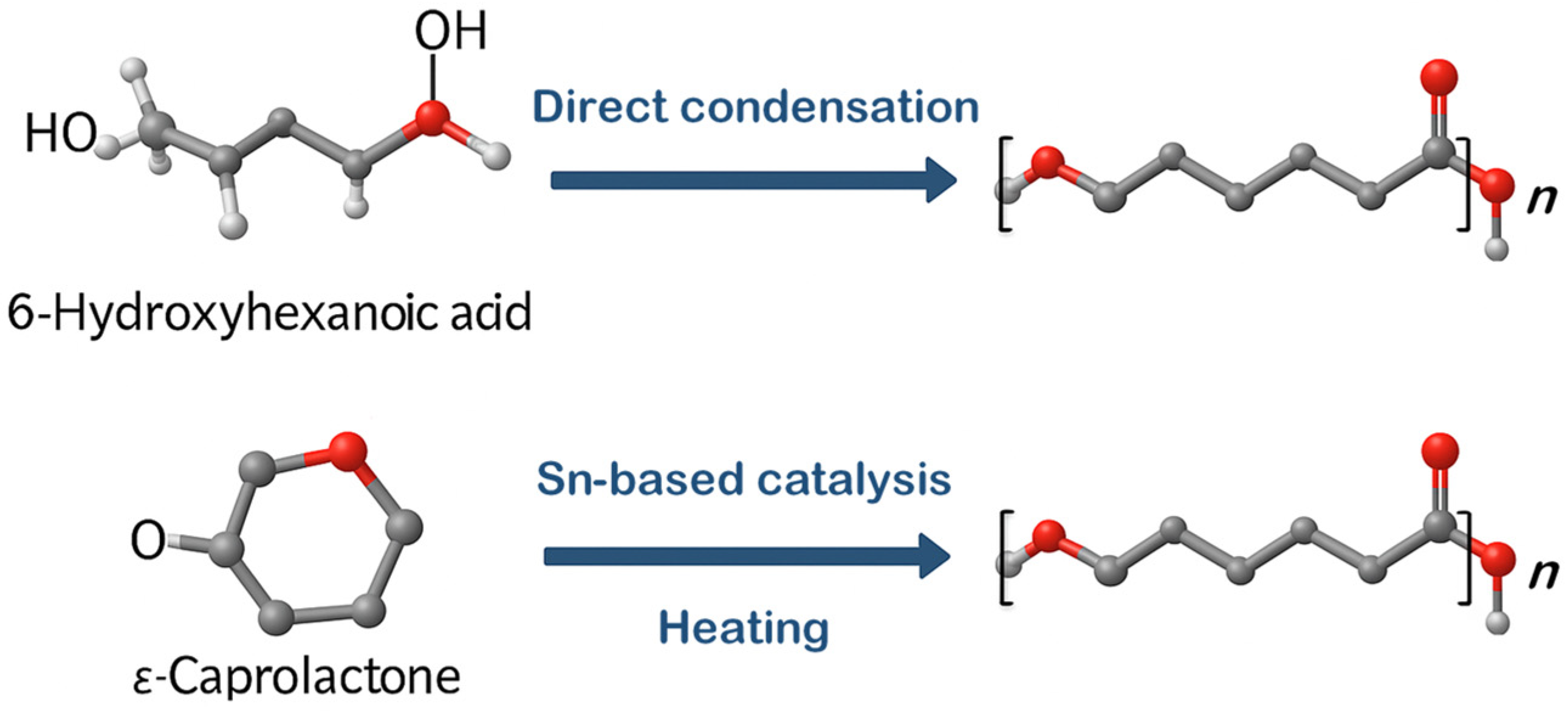
2.1. PCL Synthesis Methods

Poly(ε-caprolactone), or (1,7)-polyoxepan-2-one, can be synthesized from oil using various methods [13]. One approach involves the direct condensation of 6-hydroxyhexanoic acid (6-hydroxycaproic acid, 6-HCA), as illustrated in Figure 2 [11,14]. However, the most commonly used technique is the ring-opening polymerization (ROP) of ε-caprolactone (ε-CL) with catalysts (Figure 2). ROP is widely preferred in industrial settings for producing PCL due to its ability to yield polymers with higher molecular weights, narrower molecular weight distributions (lower polydispersity index), and better control over the polymerization process. The ring-opening polymerization (ROP) of ε-caprolactone typically utilizes one of three types of catalysts—anionic, cationic, or coordination–insertion—each producing polymers with distinct structural and molecular properties [15]. Anionic ROP relies on alkali metal salts such as potassium tert-butoxide, potassium isopropoxide, KH, phenyllithium, and lithium diisopropylamide as catalysts, where the ring-opening occurs specifically at the acyl–oxygen bond [13]. The active species in this process are alkoxide anions. A key limitation of anionic ROP is the occurrence of intramolecular transesterification, causing the formation of low-molecular-weight or cyclic polymers unless the reaction is halted beforehand. Nevertheless, this approach enables the controlled synthesis of high-molecular-weight polymers in polar solvents [16].
In cationic ROP, a positively charged intermediate is formed that is attacked by the monomer’s carbonyl oxygen, leading to ring opening via an SN2-like mechanism. This method is often challenging to control and generally results in polymers with low molecular weights or oligomers. Catalysts for this pathway include cationic aluminum complexes, trityl tetrafluoroborate (Ph3CBF4), and iron(III) chloride-based systems [17].
The coordination–insertion mechanism, or quasi-anionic ROP, involves coordinating the ε-caprolactone monomer to a metal center via its carbonyl oxygen. This reaction typically employs tin-based catalysts, including stannous (II) ethyl hexanoate, stannous (II) octoate, or stannous triflate [2,13]. The activated monomer then undergoes acyl–oxygen bond cleavage, allowing for the sequential insertion of monomers and the formation of the polymer chain. The end product can be chemically modified or blended with other materials to achieve the desired characteristics.
Sustainable PCL composites can be synthesized by initiating the ring-opening polymerization of ε-caprolactone using the surface hydroxyl groups of exfoliated oxidized biochar derived from hardwood waste biomass. This method enables neat, catalyst-assisted polymerization and produces composites with improved crystallinity, mechanical properties, and degradability compared to pure PCL [18].
Further, the possibility of producing or synthesizing ε-CL from renewable sources has also been investigated. Pyo et al. [5] developed a sustainable synthetic pathway for producing 6-HCA, adipic acid, and ε-CL by integrating biocatalysis and chemical catalysis. Bio-based 6-HCA was obtained from biomass containing glucose, fructose, or sucrose through the intermediate metabolite cyclohexanone, followed by the physicochemical conversion of 6-HCA to ε-CL using an ion exchange catalyst and a molecular sieve [5].
Alternative enzymatic methods include Baeyer–Villiger monooxygenases, such as cyclohexanone monooxygenase from Acinetobacter calcoaceticus, which catalyzes the oxidation of cyclohexanone to ε-CL in the presence of oxygen [19]. However, this enzyme is unstable and is inhibited by both the substrate and the product.
Another enzymatic method employs Candida antarctica lipase A, which facilitates the direct ring-opening oligomerization of ε-CL with up to 200 mM of cyclohexanol, yielding over 20 g/L of oligo-ε-CL [20]. However, this technique faces economic constraints due to the high cost of petroleum-derived cyclohexanol [5]. Previously, Dong et al. [21] utilized a lipase from Pseudomonas sp. to conduct ring-opening polymerization of lactones, achieving higher molecular weights and better monomer conversion compared to the condensation of the corresponding linear hydroxyesters and the copolymerization of ε-CL with lactide and cyclopentadecanolide [21].

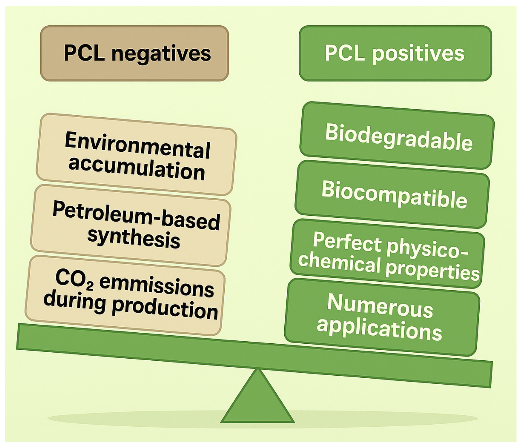
2.2. PCL Structure and Physicochemical Properties

PCL is a linear, unbranched polymer in the polyester family, composed of up to 1000 repeating units of the cyclic ester caprolactone [22]. Its typical aliphatic structure contributes to its hydrophobic nature and solubility in organic solvents [23]. PCL exhibits various physical and mechanical properties depending on its chain length and molecular weight; higher-molecular-weight variants (e.g.,~40,000 Da) are rigid yet flexible. In comparison, lower-molecular-weight types like PCL-300 (10,000 Da) and PCL-150 (5000 Da) tend to be more brittle [24]. Being hydrophobic, the polymer is highly soluble at room temperature in organic solvents such as chloroform, toluene, and benzene. However, it does not dissolve well in acetone and acetonitrile, though solubility improves when heated to 50 °C [25]. It remains insoluble in water, alcohol, and petroleum ether [26].
PCL is a semi-crystalline polyester whose crystallinity decreases as molecular weight increases [27]. Its relatively low glass transition temperature (−60 °C) and melting range (58–64 °C) facilitate easy thermal processing but reduce stability at elevated temperatures—a limitation that can be addressed through blending or cross-linking [28,29]. PCL exhibits excellent thermoformability, making it widely used in 3D printing applications, where heating and melting are common fabrication steps [30]. It also demonstrates shape memory behavior [31,32].
PCL degrades slowly, typically over 2–3 years, and its high toughness is attributed to the presence of amorphous regions in its rubbery phase [13]. Table 1 compares the key physicochemical properties of PCL and other biodegradable polymers.

2.3. PCL Applications

Polycaprolactone (PCL) is widely valued across various industries for its distinctive thermoplastic behavior, biocompatibility, and hydrophobic nature. Its applications extend to the biomedical field, cosmetics, and agriculture (Figure 3), where it enhances product functionality, improves delivery mechanisms, and supports sustainable practices.

2.3.1. Biomedical Applications

PCL has become a valuable material in the biomedical field due to its unique combination of biodegradability, ease of processing, and mechanical flexibility [33]. Given its advantageous rheological and viscoelastic behavior, the Food and Drug Administration of the USA (FDA) has approved PCL for use in long-term bioresorbable medical devices and implants, where it serves as a biodegradable alternative to traditional metallic components like screws and plates [34]. Its relatively low melting point enables thermo-responsive behaviors, making it ideal for advanced applications such as self-tightening sutures, biodegradable stents [35], and smart implants that can respond to body temperature and integrate seamlessly into surrounding tissues [36]. These characteristics contribute to developing less invasive surgical procedures and improved healing outcomes. PCL’s resistance to water further supports its use in drug delivery systems [7], bone and tissue scaffolds [37], and orthopedic fixation devices [38]. These applications leverage mechanical stability and gradual degradation over extended periods, which aligns with tissue regeneration timelines.
PCL’s excellent biocompatibility is a crucial property for various medical applications. When implanted, PCL does not elicit a strong immune or inflammatory response, allowing tissues in direct contact with it to heal wholly and swiftly [36,37]. However, the hydrophobic nature of PCL and its relatively slow degradation can restrict specific applications. As a result, researchers have concentrated on developing PCL-based blends and composites to address these limitations [7,9,12,30]. PCL composites frequently demonstrate improved hydrophilicity, mechanical strength, and tailored degradation rates, enhancing their biomedical potential [28]. For example, PCL/chitosan (CS) blends have demonstrated enhanced hydrophilicity and cytocompatibility. The double-porosity scaffolds created from PCL/CS facilitate cell adhesion and nutrient transport, making them ideal for bone and skin tissue engineering [7]. Lignin–PCL composites have also shown potential in neural tissue regeneration due to their antioxidant properties and ability to support Schwann cell proliferation [26]. Similarly, chitosan/PCL bio-inks have been optimized for lung tissue engineering through 3D printing, exhibiting excellent swelling, degradation, mechanical performance, and superior cell compatibility [37,38]. Blended PCL composites are also widely utilized in wound healing applications [39]. Their ability to support tissue regeneration, absorb exudate, and provide antibacterial properties renders them ideal for wound dressings. PCL/bacterial cellulose composites display high thermal stability and porosity, both advantageous for tissue repair [40]. Other blends, such as PCL/cellulose acetate/dextran with tetracycline, improve antibacterial activity while promoting blood clotting and cell proliferation [13]. Additional studies have indicated that incorporating Ag-containing nanoparticles into PCL scaffolds significantly enhances antimicrobial efficacy [41].
Due to its capacity for forming micro- and nanofibrous structures, PCL is a versatile platform for drug delivery. The large surface-area-to-volume ratio of electrospun PCL scaffolds enables high drug loading and controlled release [42,43].

2.3.2. Cosmetic, Agricultural, and Environmental Applications

PCL has also gained attention in the cosmetics industry. Owing to its biocompatibility and slow resorption profile, it is employed as a dermal filler [44]. Additionally, PCL-based encapsulating agents are utilized in skincare formulations to enhance active ingredients’ targeted and sustained release, thereby improving product performance [45].
PCL’s agricultural applications utilize encapsulated controlled-release fertilizers that reduce nutrient loss and enhance crop efficiency. Furthermore, PCL is employed in the development of mulch films [30,45], seed coatings [46], and eco-composites [47], all of which help minimize plastic waste and improve soil health.
In addition to biomedical and agricultural applications, PCL-based blends serve as components in biosensors and optical sensors [13]. Furthermore, modified pectin/PCL blends are being researched as flexible, compostable films for biodegradable packaging, offering an eco-friendly alternative to traditional plastics [48].

3. Biodiversity of Microorganisms Capable of PCL Biodegradation

Many microorganisms have been described in the scientific literature as capable of biodegrading PCL. Nearly all known species documented to perform such biochemical processes are either bacteria (Figure 4) or fungi (Figure 5), as archaea are rare [49].

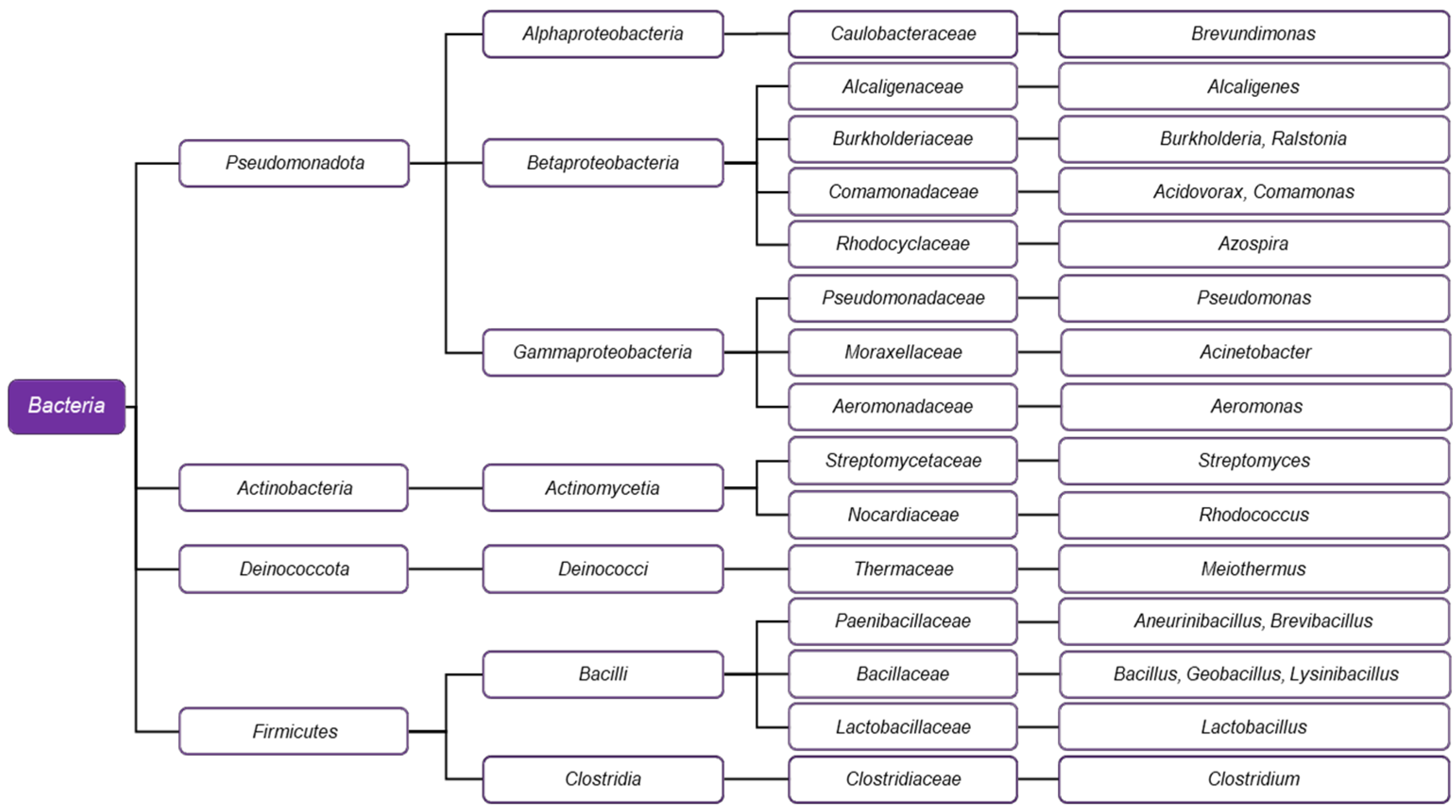
3.1. PCL-Degrading Bacteria

Various classes within the Bacteria domain are recognized for their ability to degrade PCL, primarily represented by the phyla Pseudomonadota, Actinobacteria, Deinococcota, and Firmicutes.

3.1.1. Pseudomonadota (Formerly Proteobacteria)

At least three major lineages of the formerly known phylum Proteobacteria can degrade PCL: Alphaproteobacteria, Betaproteobacteria, and Gammaproteobacteria. The Alphaproteobacteria are notable for Brevundimonas sp., a soil-isolated bacterium recognized for producing a powerful PCL depolymerase [49,50].
Among Betaproteobacteria, PCL-degrading species are found in the genera Acidovorax, Alcaligenes, Burkholderia, Comamonas, Diaphorobacter, and Ralstonia (order Burkholderiales), and in Azospira (order Rhodocyclales) (Figure 4). Alcaligenes faecalis has been found to hyperproduce the enzyme PCL depolymerase when cultured in a medium containing PCL [51]. Burkholderia cepacia has been shown to express a lipase utilized in the bioconversion of PCL within a two-phase system [52]. The genus Ralstonia, initially classified as Pseudomonas, is a close relative of Burkholderia and belongs to the same family, Burkholderiaceae [53]. Samples from a hot spring in Pakistan contained a thermophilic strain of Ralstonia sp. that effectively biodegrades PCL as a biofilm [54]. The Comamonadaceae family is notable for its genera Acidovorax, Comamonas, and Diaphorobacter, which include representatives that degrade PCL. Together with Azospira, another Betaproteobacterium, and the unrelated Ignavibacterium (Fibrobacteres-Chlorobi-Bacteroidetes superphylum) and Frateuria (Gammaproteobacteria), these microorganisms have been employed in a specialized aquaculture system, where they carry out a heterotrophic denitrification process using PCL as the sole carbon source [55].
Among Gammaproteobacteria degrading PLC are the representatives of the genera Pseudomonas and Acinetobacter, which belong to the order Pseudomonadales. The genus Aeromonas has been successfully employed in nitrification and denitrification processes, where PCL serves as a carbon source for growth, suggesting its potential application in wastewater treatment systems [56]. The effects of the Pseudomonas lipase enzyme have also been investigated. Scanning electron microscope (SEM) images of PCL–polyglycolide nanocomposites treated with this enzyme indicate visible degradation in the material’s structure [57]. In another study, a lipase from Ps. chlororaphis and Streptomyces strains grown on waste cooking oil was effective on PCL and polyhydroxyalkanoates [58]. Ps. hydrolytica has been found to express two different lipases, which have been successfully purified and characterized [59]. Compost isolates of Pseudomonas pseudoalcaligenes, currently considered a synonym of P. oleovorans, express cutinase when cultivated in a medium containing plant waste [60]. The genus Acinetobacter, a relative of Pseudomonas, is notable for a specific variety with biodegradative potential. Early studies with mixed cultures of Acinetobacter calcoaceticus var. lwoffii and the yeast Cryptococcus laurentii (syn. Papiliotrema laurentii) provide evidence that even polymeric chains with very high molecular weight are subjected to hydrolysis by the metabolic activity of the microorganisms [61]. Another member of the genus, Ac. seifertii, also demonstrates promising properties. Soil isolates, later identified as belonging to the species, exhibit a significant ability of their PCL depolymerase enzyme to degrade PCL effectively [62]. Furthermore, Acinetobacter has provided genes for 6-oxohexanoic acid (6-OHA) dehydrogenase, which have been utilized to develop a recombinant Escherichia coli strain with potential applications for eco-friendly poly-ε-caprolactone (PCL) upcycling [63].
The gammaproteobacterium Aeromonas attracts attention due to its ability to form biofilms on the surface of PCL and other plastic materials. A strain identified as Aeromonas media has been investigated in conjunction with a strain of Rhodococcus quingshengii. Both species showed the ability to degrade PCL with biofilm-forming properties and appeared resistant to a biocide introduced in cultured samples [64].

3.1.2. Actinobacteria

In addition to Proteobacteria, another major phylum—Actinobacteria—also includes genera with similar metabolic capabilities. A prominent example is the genus Streptomyces, part of the order Streptomycetales. A thermophilic isolate of Str. thermoviolaceus subsp. thermoviolaceus, recovered from Taiwanese soils, degrades PCL via extracellular depolymerase, as shown by agar plate assays [65]. Researchers isolated two depolymerases from this microorganism, one displaying chitinase activity. The second key actinomycete genus is Rhodococcus, which belongs to the Nocardiaceae order. Two species, R. erythropolis and R. opacus, have provided essential insights into plastic degradation research. The genomes of these organisms have been sequenced and analyzed for the presence of genetic sequences responsible for PCL biodegradation, and several genetic determinants responsible for the process have been reported [66].

3.1.3. Firmicutes and Deinococcota

Most members of the phylum Firmicutes that degrade PCL are limited to the class Bacilli and include the genera Aneurinibacillus, Bacillus, Brevibacillus, Geobacillus, Lactobacillus, and Lysinibacillus, with the genus Clostridium being the only exception. Aneurinibacillus thermoaerophilus and Brevibacillus thermoruber, both belonging to the family Paenibacillaceae, as well as the unrelated Meiothermus cateniformans (Deinococcota), are thermophilic bacterial species isolated from a plastic-contaminated hot spring in Bulgaria [67]. Through a series of morphological, microscopic, and enzymatic analyses, all three have demonstrated the ability to degrade PCL, with a strain of Br. thermoruber exhibiting the highest esterase activity in the collection [67]. Bacillus sp. strain NR4 degrades PCL alongside poly(butylene adipate-co-terephthalate), indicating that the genus may have a broader range of applications [68].
Another study shows that B. pumilus can also biodegrade PCL, which occurs naturally in the mangrove rhizosphere [69]. Moreover, a strain of Bacillus sp. has been utilized in developing bacterial fertilizers, where PCL serves as a material for creating an eco-friendly, biodegradable capsule [70]. Geobacillus, which belongs to the same Bacillaceae family, has been utilized as a source of lipase/esterase enzymes successfully engineered to degrade PCL with enhanced efficiency [71]. Lysinibacillus sp. also degraded PCL, as demonstrated by gel permeation chromatography [72].
PLC-degrading lactobacilli belong to the species Levilactobacillus brevis and Lactiplantibacillus plantarum. Three different lipases have been extracted from these species, as the lipase from Lpl. plantarum showed the highest activity [73]. Further investigation of this enzyme using an enzyme-embedded assay revealed significant physical changes to the plastic material resulting from the degradation process [74].
Clostridium is the most phylogenetically distinct example among the biodegraders within Firmicutes. However, at least two studies provide evidence that members of this genus exhibit this capability. Two strains of Clostridium sp. have been isolated from anaerobic sludge and evaluated for their biodegradative properties on PCL and two other plastic materials (polyhydroxybutyrate and the copolymer polyhydroxybutyrate-co-hydroxyvalerate). Following 16S rDNA analysis, the strains were identified as belonging to the species C. acetobutylicum [75]. In another study, Clostridium sp. isolated from aquaculture samples utilized PCL as a carbon source. The relatively high abundance of clostridia identified among the diverse array of microorganisms in the samples suggests that the genus may be essential in the biodegrading consortium [76].

3.1.4. Putative PCL Degradation by Other Bacterial Species

Research on PCL biodegradation by bacterial species from phyla other than Proteobacteria, Actinobacteria, and Firmicutes is limited in the scientific literature. However, the taxonomic diversity of these microorganisms may be greater than currently recognized, considering that enzymes such as lipases and esterases, which are crucial to the process, are nearly ubiquitous among bacteria [77]. Members of their enzymatic family readily act upon PCL. Additionally, polyester, a plastic material chemically related to PCL, has been found to affect soil microbial communities by increasing the presence of bacteria from the phyla Acidobacteria, Verrucomicrobia, Bacteroidetes, Nitrospirae, and Patescibacteria, suggesting that these microorganisms may play a role in plastic degradation. It is essential to conduct in vitro cultivation of species from these taxa, along with experiments on the impact of PCL on their growth, which is critical for enhancing our understanding of the potential of bacteria to improve the exploitation of biodegradable plastics.

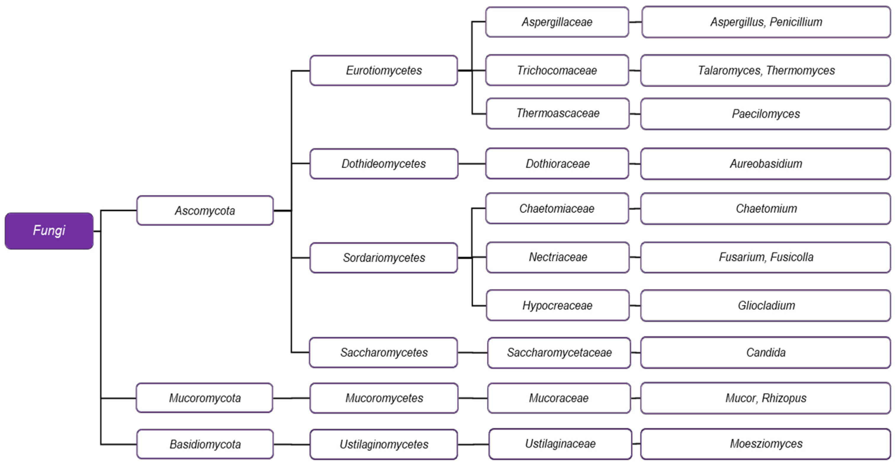
3.2. PCL-Degrading Fungi

PCL-degrading fungi show less taxonomic diversity in representative phyla than bacteria (Figure 5). Except for the basidiomycete Moesziomyces antarcticus and two members of Mucoromycota, Mucor and Rhizopus, the other studied genera—Aspergillus, Aureobasidium, Gliocladium, Paecilomyces, Penicillium, Chaetomium, Talaromyces, Thermomyces, Fusarium, Fusicolla, and Candida—are classified within Ascomycota. Both Aspergillus and Penicillium belong to the Eurotiomycetes class, and their biodegradative potential was recognized quite early, as demonstrated by the SEM of PCL, which was subjected to the action of Asp. flavus and Pen. funiculosum [78].
Benedict et al. [79] conducted plastic degradation experiments involving Asp. niger, Asp. fumigatus, Chaetomium globosum, and Fusarium sp., demonstrating that all tested fungal strains produce low-molecular-weight material from PCL [79]. These fungi also degrade a blend of PCL and polyethylene as a consortium with two other species, Gliocladium virens and Aureobasidium pullulans [80]. Recent studies have highlighted Chaetomium globosum, which can fully degrade PCL in specific samples within 28 days [81].
Considering the final products of degradation, a thermotolerant strain of Asp. fumigatus has been discovered to produce a mixture of carboxylic acids from PCL film [82], similar to the purified lipase of Asp. oryzae [83]. Another Aspergillus strain sourced from a polluted seacoast in Korea has been studied for its ability to degrade PCL, with the authors noting that it, along with another ascomycete genus, Talaromyces, demonstrates higher activity than Penicillium [84]. Researchers have assessed the ability of Aspergillus fumigatus to degrade PCL in both soil and compost environments. These experiments also examined the properties of Thermomyces lanuginosus, another member of the Eurotiomycetes class, identifying it as a quick PCL microbial degrader [85]. It is noteworthy that although Therm. lanuginosus appears less frequently in plastic-related research, the enzymatic capabilities of this fungus warrant equal attention, given that a lipase from this species has been successfully utilized in vitro for the degradation of cross-linked PCL implants [86]. Paecilomyces lilacinus, also a representative of Eurotiomycetes, shows biodegradative capacity for both PCL and poly(3-hydroxybutyrate), as evidenced by a single study involving five different strains of filamentous fungi [87]. In contrast, the abilities of Penicillium species seem to be more evident in research on plastic degradation. Besides Pen. funiculosum, at least three other species from this genus have been studied for their effects on PCL, one being Pen. oxalicum. This fungus has been subjected to UV radiation mutagenesis, resulting in an effective biodegrading strain [88].
Penicillium chrysogenum, isolated from Russian soils, has been shown to degrade PCL, PLA, and polypropylene [89]. The most recently discovered fungi displaying similar activity are Penicillium samsonianum and Fusicolla acetilerea, both isolated from terrestrial samples in Korea [90]. Fusarium, a widely distributed soil fungus, is another genus of key importance for PCL biodegradation. In addition to being part of pioneering reports on fungal microorganisms demonstrating this ability, it has significantly contributed to understanding their enzymatic properties. Fusarium is a well-known plant pathogen, and as such, it relies on the activity of cutinase to lyse cutin [91], the outermost layer of many plant tissues. An analysis comparing a wild-type F. moniliforme and a knockout mutant of F. solani revealed that the cutinase functions as PCL-depolymerase [92]. Shortly after, a second depolymerase was isolated from F. solani and identified as a lipase distinct from the previously identified catalyst, indicating that biodegradation can be approached with different enzymatic strategies [93]. Moreover, researchers have successfully expressed a cutinase from Fusarium solani in the yeast Yarrowia lipolytica [94].
Another essential study targeting cutinase as a substrate showed that an extracellular protease can inactivate it [95]. More recent research has developed an enzymatic model for the hydrolysis of PCL through the activity of cutinase from F. solani and compared it to an accompanying model on the activity of a lipase from Candida antarctica [96], now reclassified as Moesziomyces antarcticus, thereby becoming the only currently known basidiomycete capable of biodegradation. The biodegradation process carried out by M. antarcticus has also been studied, with emphasis on the molecular weight of film-based PCL. While molecular weight does not significantly impact overall biodegradability, it does influence the size and depth of the hydrolytic pores and the time needed for the reaction to proceed [97].
Interestingly, in addition to its ability to biodegrade PCL, M. antarcticus has also demonstrated the ability to synthesize it, and the effects of two significant parameters on the process—temperature and organic media—have been evaluated [98].
Members of Mucoromycotina, the subphylum of Mucoromycota—including the common bread molds Mucor and Rhizopus—have also been reported to degrade PCL [99].

4. Mechanism of Microbial PCL Biodegradation

The microbial degradation of PCL proceeds through a sequence of three stages, each characterized by specific biochemical and physiological processes that convert the polymer into metabolic waste products while storing energy. These stages are as follows: (i) surface hydrolysis, where key enzymes cleave the bonds in the polymer backbone; (ii) absorption and digestion, involving the uptake and further assimilation of monomers and oligomers generated in the previous stage; and (iii) mineralization and excretion, during which the microorganism produces and releases small molecules into its environment.

4.1. Overview of Enzymes Involved in the Process of PCL Degradation

The key to microbial hydrolysis of PCL lies in the secretion of extracellular hydrolytic enzymes. Several lipases (EC 3.1.1.3) and some carboxylesterases (EC 3.1.1.1) are effective on this aliphatic polyester. Both groups of enzymes target the ester bonds in biological molecules, and depending on the specific enzyme, they participate in a broad range of biochemical processes. The precise distinction between lipases and esterases uses various criteria, including kinetic properties, structural features, substrate specificity, and hydrolysis mechanisms. Table 2 summarizes a widely accepted classification system for lipolytic enzymes, which organizes them into eight classes based on different structural and biochemical features.

4.2. Enzymes Involved in the Process of PCL Degradation by Bacteria

All esterases and lipases in bacteria share a characteristic α/β-hydrolase fold in their active sites, defining the catalytic strategy through which these enzymes carry out their functions. Most α/β-hydrolase fold proteins consist of six α-helices surrounding a central β-sheet composed of eight β-strands, nearly all of which are oriented parallel, with the only exception being strand β2, which runs antiparallel to the others. The β-sheet presents a distinctive half-barrel structure due to spatial twisting that positions the first and last strands at roughly a 90° angle to one another. Two of the α-helices flank one side of the structure, while the other four are located on the opposite side. Access to the active site for substrates is regulated by a set of secondary structures that form various lids, caps, or flaps. These features are particularly important for the lipase class (Class I), where a lid domain is present and plays a key role in interfacial activation.
In contrast to most other classes, Class II esterases (SGNH, or GDSL) lack the traditional α/β-hydrolase fold and instead exhibit a highly divergent α/β/α sandwich architecture (Figure 6). A notable difference is the β-sheet, which consists of five parallel strands (flavodoxin-like) in SGNH enzymes, contrasting with the eight-stranded structure typical of α/β-hydrolases. The oxyanion hole and the catalytic triad are present in GDSL esterases, operating through a similar mechanism. However, the amino acid sequence is generally conserved as Asp-His-Ser. GDSL esterases display distinct Nuc-Oxy and Acid–Base zones, forming an extensive network of hydrogen bonds in the active site that provides additional stability during substrate transition. The so-called SHLink, a two-bond interaction between residues adjacent to the catalytic nucleophile and base, connects these zones.
Class VIII esterases represent the most distinct category of bacterial lipolytic enzymes. Unlike the α/β-hydrolase fold, these enzymes have a β-lactamase-like fold, named for its similarities to β-lactamases. These esterases feature a two-domain architecture. The large domain consists of a β-sheet surrounded by α-helices. The β-sheet significantly differs from the α/β-hydrolase fold, as all its strands are arranged antiparallelly. The small domain contains four α-helices and two β-sheets with antiparallel strands. The binding site is located between the two domains. Another notable difference is the unusual catalytic triad, Ser–Lys–Tyr, where Ser acts as the acid component while Tyr is proposed to function as the base. Table 3 presents the main properties of bacterial enzymes exhibiting activity towards PCL.
A common characteristic of all lipases is that they typically target water-insoluble molecules and require a hydrophobic interface (Figure 7). Hydrophobic interactions are significant, given that lipases possess a conserved hydrophobic region of amino acids in their active site (Figure 7c,d). Well-known lipase substrates include triglycerides, cholesterol, and specific vitamins, underscoring their essential role in fat metabolism.
In contrast, esterases generally act on smaller molecules that have higher water solubility [105,106]. Consequently, they often play a significant role in nucleic acid digestion, neurotransmitter inactivation, and the metabolism of xenobiotics, including drugs and toxic organic compounds. Regardless of the classification system, enzymes from the esterase and lipase classes have shown the ability to catalyze the breakdown of the polymeric PCL molecule into smaller products. This process begins when the secreted enzyme adsorbs to the material’s surface, followed by the formation of a temporary transition complex with the substrate. In the subsequent step, chain scission occurs, and the protein interacts with other molecular segments [107]. Experiments with polyethylene terephthalate, another member of the polyester family, indicate that biodegrading enzymes have a strong tendency for surface adsorption, making attempts to deliberately remove them from the plastic even more challenging [108].

4.3. Enzymes Involved in the Process of PCL Degradation by Fungi

The enzymes responsible for PCL degradation in fungi are lipases and cutinases. In addition to being subjects of scientific research, some are produced industrially (Table 4). One example is the lipase from Candida rugosa, although its PCL degradation level has been reported as low [89]. Similar results have been observed with the lipase produced by Rhizopus delemar. In contrast, the lipase from Mucor miehei has shown a sufficiently high degradation yield of the plastic, indicating that it may hold considerable significance for the practical application of the enzyme in this process [99].
Cutinases (E.C. 3.1.1.74) are serine esterases originally associated with fungal invasion of plant cells by phytopathogenic fungi. In addition to their natural substrates, cutinases show activity toward a broad range of substrates, including PCL. Their structure is highly similar among Fusarium and Aspergillus species, and Figure 8 displays the conserved regions in the enzyme molecules.

4.4. Catalytic Mechanism of Enzymatic PCL Degradation

At least two experimentally supported models of the catalytic mechanism responsible for PCL hydrolysis propose a broadly comparable concept, though with some notable differences. The first model is based on the activity of an esterase from the thermophilic archaeon Archaeoglobus fulgidus (AfEST). This highly thermostable enzyme operates optimally at 80 °C and can synthesize polyester.
Three key amino acid residues, known as the catalytic triad—specifically Nucleophile–His–Acid—are responsible for the catalytic mechanism through which the α/β-hydrolase fold operates. The first and last residues may vary depending on the enzyme, while His is almost always conserved. The nucleophilic component is usually Ser, Cys, or Asp, and it is situated in the so-called nucleophilic elbow, representing a tight turn in the fold’s structure between αC and β5. On the other hand, the acidic residue is usually either Asp or Glu, and its function is to stabilize His in its protonated form. The mechanism involves two steps. First, the nucleophile attacks the substrate during acylation. Then, the process passes through two transition states before completing a deacylation reaction that releases the final product. In the AfEST model, aspartate (Asp) or glutamate (Glu), histidine (His), and serine (Ser) lie between the characteristic α/β hydrolase fold and the protein’s cap domain. The oxyanion hole, another crucial region, comprises three hydrophobic amino acids, two glycine (Gly) residues, and one alanine (Ala) residue. This region provides chemical stability by forming hydrogen bonds with its amide groups.
As the reaction progresses, the catalytic triad undergoes a cycle, starting with a nucleophilic attack by serine, which transfers a proton to histidine and continues with the formation of three successive intermediates: the first tetrahedral intermediate (INT-1), the acyl-enzyme intermediate (also known as enzyme-activated monomer, EAM), and the second tetrahedral intermediate (INT-2). The process concludes with the conversion to INT-2, which results in the recovery of the enzyme (RC), allowing the cycle to begin anew. The process includes four identifiable transition states: T1, T2, T3, and T4, each representing a specific step in the cycle. After the formation of INT-1 in the first step, another proton transfer occurs, cleaving a C-O bond in the substrate, leading to the release of 6-hydroxycaproic acid (6-HCA) from the PCL chain and the formation of EAM. EAM is subsequently subjected to deacylation by a nucleophilic attack from a water molecule, converting it to INT-2. In the final step, water establishes an equilibrium that drives the reaction toward releasing a second molecule, 6-HCA, and regenerating the enzyme (RC), accompanied by a decrease in Gibbs free energy, indicating that this step is exergonic. In conclusion, two 6-HCA molecules are cleaved from the PCL chain for each cycle turnover, representing the final product, which moves on to the next stage of biodegradation [109].
The second model centers on the thermophilic esterase MGS0156, identified through environmental metagenomic screening and capable of degrading various plastics, including PCL. Unlike AfEST, it operates optimally at a lower temperature (35–40 °C) and features a modified α/β hydrolase fold with a lid domain and a highly hydrophobic active site suited for binding PCL. As with AfEST, this model emphasizes the critical role of three catalytic amino acid residues in the reaction mechanism, characterized as a two-step process: triad-assisted nucleophilic attack and C-O bond cleavage. The model stresses that C-O bond cleavage is the rate-determining step, supported by a Molecular Mechanics Poisson–Boltzmann Surface Area Method (MM-PBSA). It indicates that different sets of six amino acids determine the binding strength of a PCL oligomer of a given size, noting that octamers demonstrate the highest binding energy. A valuable aspect of the study is the identification of two residues, His231 and Asp237, as targets for mutagenesis, providing implications for the bioengineering of enzymes derived from MGS0156 and their potential use as improved enzymatic tools in the biodegradation of PCL and other plastic materials [110]. Researchers have also noted other products resulting from the hydrolysis of PCL in addition to 6-HCA. An experiment comparing the abiotic and biotic degradation of PCL films, with the latter involving a mixed culture of compost microorganisms, shows that this process also leads to the release of various small molecules, namely caprolactone, a cyclic dimer, and a cyclic trimer, confirmed through a coupled analysis with gas chromatography and mass spectrometry (GC-MS). These products and 6-HCA are quickly assimilated and disappear entirely after two weeks in the biotic medium [111]. Interestingly, a GC-MS analysis of the degradation products in Rhodococcus erythropolis and Rhodococcus opacus shows a variety of other molecules, such as decanoic acid, undecanoic acid, dodecanoic acid, eicosane, pentacosane, and hexacosane, among others, but does not detect the presence of 6-HCA [112]. An earlier study examining the biodegradative potential of a thermotolerant strain of Aspergillus sp. identified the presence of succinic acid, butyric acid, valeric acid, and caproic acid after exposing plastic pellets to the activity of the microorganism at 50 °C. As anticipated, considering the chemical structure of its typical substrate, cutinase expressed by a strain of Ps. pachastrellae, isolated from coastal waters, results in 16-hydroxyhexadecanoic (Juniperic) acid, the main component of cutin, in addition to 6-HCA [113].
The post-hydrolysis stages of PCL biodegradation (Figure 9)—that is, the assimilation and further metabolization of lower-weight products followed by the excretion of waste molecules such as H2O and CO2 or, for some anaerobes, CH4—remain a largely unexplored area in bioplastic research.
Potential candidates for transporting 6-HCA across the cell membrane can be found among proteins from the major facilitator superfamily (MFS), representing an evolutionarily ancient lineage of molecular transport systems in bacteria. The MFS routinely imports carboxylic acids, including hydroxy acids like lactate, sugar acids such as gluconate, and various short-chain fatty acids [114,115]. Another possibility is transport via a member of the tripartite ATP-independent periplasmic transporter (TRAP) family, a common prokaryote-specific protein group known to engage in the secondary transport of a diverse array of carboxylates, including typical examples such as fumarate and malate—both of which are dicarboxylates—as well as pyruvate, lactate, L-glutamate, and others [116]. Since 6-HCA is chemically related to the aforementioned compounds as a carboxylic acid and exhibits a relatively simple chemical structure, it seems reasonable to focus on MFS and TRAP when investigating its transport mechanism. The fate of 6-HCA after it enters the bacterial cytoplasm is another critical area for potential experimental research.
Degradation through a catabolic pathway, such as beta-oxidation, is plausible since this process is the primary means of breaking down carboxylic acids of various chain lengths in many organisms, including bacteria [117,118]. Enzymes from the acyl-CoA synthase family can catalyze the formation of a thioester bond with CoA, allowing them to target a wide range of carboxylic acid molecules for degradation, resulting in products like palmitoyl-CoA [119], stearoyl-CoA, 3-hydroxybutyryl-CoA [120,121], isovaleryl-CoA [122], isobutyryl-CoA [123], and caproyl-CoA. The latter represents the activated form of caproic acid, which contains the same carbon atoms as 6-HCA, differing only by a single hydroxyl group.
Most current knowledge about the genetic factors governing PCL biodegradation comes from a pivotal set of experiments conducted with strains of R. erythropolis and R. opacus, two closely related aerobic species of actinobacteria. Zampolli et al. [112] have effectively determined the biodegradation capacity of these microorganisms by evaluating established hydrolytic zones (halos) on agar plates and conducting coupled GC-MS analysis of products extracted from bacterial cultures supplied with PCL. They have also measured high esterase activity through spectrophotometric tests, performing enzymatic reactions with five different organic substrates. The substrates include p-nitrophenyl acetate, p-nitrophenyl butyrate, p-nitrophenyl octanoate, p-nitrophenyl laurate, and p-nitrophenyl palmitate. The genome of R. erythropolis was subsequently sequenced via Illumina MiSeq v3, annotated, and then compared with the genome of R. opacus using the Rapid Annotations using Subsystems Technology (RAST) server.
Furthermore, the transcription levels of genes potentially involved in PCL biodegradation have been assayed using reverse transcription quantitative real-time PCR (RT-qPCR). An automated annotation of the genome of R. erythropolis reveals that it contains 6165 open reading frames (ORFs) and 72 RNA genes. Sequence-based and functional comparisons indicate that the two species share a substantial degree of identity, which the authors identify as a ‘common genomic core’. Interestingly, according to the RAST server, the closest neighbors of R. erythropolis are R. jostii and Nocardia farcinica. These findings are significant as they may indicate other microorganisms with potential biodegradation capabilities and suggest that genetic tools can be employed to identify such species. The RAST server also reveals that R. erythropolis and R. opacus share the same set of most populated functional categories, specifically amino acids and derivatives, carbohydrates, fatty acids, lipids, isoprenoids, cofactors, vitamins, prosthetic groups or pigments, and protein metabolism. The authors focus on genes involved in lipid metabolism, arguing that they are likely relevant to aliphatic polyester degradation, given the chemical similarities between the two groups of molecules. Further bioinformatic analyses have shown that this category includes a set of 10 and 9 genes encoding hypothetical proteins and lipases, which are potential objects of interest. Ultimately, RT-qPCR has revealed that three key genes from the selected group are expressed during the PCL biodegradation process: P133, which encodes a hypothetical protein; P253, which encodes a lipase; and P337, which encodes another hypothetical protein [112].

5. Conclusions and Prospects

PCL biodegradation offers a promising and sustainable approach to reducing plastic pollution while paving the way for eco-friendly material use. This review shows that various microorganisms, including bacteria, fungi, and archaea, can degrade PCL. The widespread presence of esterases and lipases in microbial communities indicates that the potential for biodegradation in nature may be significantly greater than documented. Exploring microbial diversity, particularly in extreme environments and under different ecological conditions, could reveal novel strains with enhanced biodegradative abilities.
A persistent challenge in this field is the limited understanding of the complete metabolic pathways involved in the assimilation and processing of PCL degradation products. While the enzymatic mechanisms of hydrolysis are relatively well characterized, the intracellular fate of intermediates such as 6-HCA remains largely unknown. Potential transport mechanisms, including the major facilitator superfamily (MFS) and tripartite ATP-independent periplasmic (TRAP) transport systems, are still hypothetical and have not been experimentally validated. Similarly, the specific roles of metabolic routes like β-oxidation in converting PCL-derived compounds into central metabolites require further clarification. Addressing these knowledge gaps is essential for optimizing microbial strains and enhancing biodegradation efficiency. Enzymatic models, such as those derived from Archaeoglobus fulgidus esterase and the metagenomic esterase MGS0156, provide mechanistic insight into substrate binding and catalysis, offering a valuable foundation for enzyme engineering. Advances in protein engineering, particularly directed evolution and rational design, could significantly enhance enzyme specificity, thermal stability, and catalytic performance, making them more suitable for large-scale bioplastic degradation and environmental remediation applications. However, translating laboratory success into large-scale biodegradation remains a considerable challenge. Environmental applications must consider microbial community dynamics, interactions with other pollutants, and abiotic factors such as temperature, pH, and nutrient availability. Utilizing complex microbial consortia instead of isolated strains may boost degradation rates while maintaining stability across varying environmental conditions.
Despite significant progress, several critical gaps remain in our understanding of microbial PCL biodegradation. Although many microorganisms and hydrolytic enzymes have been identified, most studies emphasize extracellular depolymerization under aerobic conditions, leaving anaerobic biodegradation pathways largely unexplored. The transport and intracellular metabolism of degradation products like 6-HCA are still poorly understood, particularly regarding how they are assimilated and integrated into central metabolic pathways. Furthermore, the genetic basis for the regulation and expression of PCL-degrading enzymes in various microbial taxa has yet to be fully characterized. Although genome and metagenome sequencing have revealed numerous putative enzymes, only a small percentage have undergone functional validation. Additionally, there is insufficient data on microbial consortia in complex environmental systems where PCL is present alongside other organic and inorganic contaminants.
In conclusion, a comprehensive approach that integrates microbiology, enzymology, synthetic biology, and environmental science is essential for advancing the field of PCL biodegradation. Future research should prioritize discovering novel microbial taxa, clarifying complete catabolic pathways, and developing engineered enzymes that function more effectively under environmentally relevant conditions. Addressing gaps in the existing literature will be critical for building a bridge toward scalable solutions in plastic waste management and improving the efficiency of PCL degradation in varied ecological settings.

Author Contributions

Conceptualization, N.K. and P.P.; investigation, N.A., N.K., I.B., and P.P.; resources, K.P.; writing—original draft preparation, N.K., P.P., N.A., and K.P.; writing—review and editing, I.B. and K.P.; funding acquisition, K.P. All authors have read and agreed to the published version of the manuscript.

Funding

The authors gratefully acknowledge the financial support from the National Science Fund, Republic of Bulgaria, Grant No. KP-06-PN87/12.

Conflicts of Interest

The authors declare no conflicts of interest.

References

  1. Ramaraju, H.; Verga, A.S.; Steedley, B.J.; Kowblansky, A.P.; Green, G.E.; Hollister, S.J. Investigation of the biodegradation kinetics and associated mechanical properties of 3D-printed polycaprolactone during long-term preclinical testing. Biomaterial 2025, 321, 123257. [Google Scholar] [CrossRef] [PubMed]
  2. Richert, A.; Kalwasińska, A.; Brzezinska, M.S.; Dąbrowska, G.B. Biodegradability of Novel Polylactide and Polycaprolactone Materials with Bacteriostatic Properties Due to Embedded Birch Tar in Different Environments. Int. J. Mol. Sci. 2021, 22, 10228. [Google Scholar] [CrossRef]
  3. Injorhor, P.; Trongsatitkul, T.; Wittayakun, J.; Ruksakulpiwat, C.; Ruksakulpiwat, Y. Biodegradable Polylactic Acid-Polyhydroxyalkanoate-Based Nanocomposites with Bio-Hydroxyapatite: Preparation and Characterization. Polymers 2023, 15, 1261. [Google Scholar] [CrossRef]
  4. Naser, A.Z.; Deiab, I.; Defersha, F.; Yang, S. Expanding Poly(lactic acid) (PLA) and Polyhydroxyalkanoates (PHAs) Applications: A Review on Modifications and Effects. Polymers 2021, 13, 4271. [Google Scholar] [CrossRef]
  5. Pyo, S.-H.; Park, J.H.; Srebny, V.; Hatti-Kaul, R. A sustainable synthetic route for biobased 6-hydroxyhexanoic acid, adipic acid and ε-caprolactone by integrating bio- and chemical catalysis. Green Chem. 2020, 22, 4450–4455. [Google Scholar] [CrossRef]
  6. Shekh, M.R.; Kumar, V. Impact of plastic pollution on ecosystems: A review of adverse effects and sustainable solutions. Environ. Monit. Assess. 2025, 197, 264. [Google Scholar] [CrossRef] [PubMed]
  7. Krobot, Š.; Melčová, V.; Menčík, P.; Kontárová, S.; Rampichová, M.; Hedvičáková, V.; Mojžišová, E.; Baco, A.; Přikryl, R. Poly(3-hydroxybutyrate) (PHB) and Polycaprolactone (PCL) Based Blends for Tissue Engineering and Bone Medical Applications Processed by FDM 3D Printing. Polymers 2023, 15, 2404. [Google Scholar] [CrossRef]
  8. Hottle, T.A.; Bilec, M.M.; Landis, A.E. Biopolymer production and end of life comparisons using life cycle assessment. Resour. Conserv. Recycl. 2017, 122, 295–306. [Google Scholar] [CrossRef]
  9. Narancic, T.; Verstichel, S.; Chaganti, S.R.; Morales-Gamez, L.; Kenny, S.T.; De Wilde, B.; Padamati, R.B.; O’Connor, K.E. Biodegradable plastic blends create new possibilities for end-of-life management of plastics but they are not a panacea for plastic pollution. Environ. Sci. Technol. 2018, 52, 10441–10452. [Google Scholar] [CrossRef]
  10. Mandic, M.; Spasic, J.; Ponjavic, M.; Nikolic, M.; Cosovic, V.R.; O’Connor, K.E.; Nikodinovic-Runic, J.; Djokic, L.; Jeremic, S. Biodegradation of poly(ε-caprolactone) (PCL) and medium chain length polyhydroxyalkanoate (mcl-PHA) using whole cells and cell free protein preparations of Pseudomonas and Streptomyces strains grown on waste cooking oil. Polym. Degrad. Stab. 2019, 162, 160–168. [Google Scholar] [CrossRef]
  11. Labet, M.; Thielemans, W. Synthesis of PCL: A review. Chem. Soc. Rev. 2009, 38, 3484–3504. [Google Scholar] [CrossRef] [PubMed]
  12. Martín-González, D.; de la Fuente Tagarro, C.; De Lucas, A.; Bordel, S.; Santos-Beneit, F. Genetic Modifications in Bacteria for the Degradation of Synthetic Polymers: A Review. Int. J. Mol. Sci. 2024, 25, 5536. [Google Scholar] [CrossRef] [PubMed]
  13. Kayan, G.Ö.; Kayan, A. Polycaprolactone Composites/Blends and Their Applications Especially in Water Treatment. ChemEngineering 2023, 7, 104. [Google Scholar] [CrossRef]
  14. Woodruff, M.A.; Hutmacher, D.W. The return of a forgotten polymer—Polycaprolactone in the 21st century. Prog. Polym. Sci. 2010, 35, 1217–1256. [Google Scholar] [CrossRef]
  15. Kayan, A. Recent studies on single site metal alkoxide complexes as catalysts for ring opening polymerization of cyclic compounds. Catal. Surv. Asia 2020, 24, 87–103. [Google Scholar] [CrossRef]
  16. Grobelny, Z.; Golba, S.; Jurek-Suliga, J. Mechanism of ε-caprolactone polymerization in the presence of alkali metal salts: Investigation of initiation course and determination of polymers structure by MALDI-TOF mass spectrometry. Polym. Bull. 2019, 76, 3501–3515. [Google Scholar] [CrossRef]
  17. Xu, Y.; Wang, L.; Chen, C.; Huang, P.; Dai, H.; Jiang, W.; Zhou, Y. Living Cationic Polymerization of ε-Caprolactone Catalyzed by a Metal-free Lewis Acid of Trityl Tetrafluoroborate. Macromolecules 2023, 56, 501–509. [Google Scholar] [CrossRef]
  18. Vidal, J.L.; Yavitt, B.M.; Wheeler, M.D.; Kolwich, J.L.; Donovan, L.N.; Sit, C.S.; Kerton, F.M. Biochar as a sustainable and renewable additive for the production of Poly (ε-caprolactone) composites. Sustain. Chem. Pharm. 2022, 25, 100586. [Google Scholar] [CrossRef]
  19. Srinivasamurthy, V.S.T.; Böttcher, D.; Engel, J.; Kara, S.; Bornscheuer, U.T. A whole-cell process for the production of ε-caprolactone in aqueous media. Proc. Biochem. 2020, 88, 22–30. [Google Scholar] [CrossRef]
  20. Schmidt, S.; Scherkus, C.; Muschiol, J.; Menyes, U.; Winkler, T.; Hummel, W.; Gröger, H.; Liese, A.; Herz, H.G.; Bornscheuer, U.T. An enzyme cascade synthesis of ε-caprolactone and its oligomers. Angew. Chem. Int. Ed. Engl. 2015, 54, 2784–2787. [Google Scholar] [CrossRef]
  21. Dong, H.; Wang, H.; Cao, S.; Shen, J. Lipase-catalyzed polymerization of lactones and linear hydroxyesters. Biotechnol. Lett. 1998, 20, 905–908. [Google Scholar] [CrossRef]
  22. Yu, W.; Song, X.; Wang, Y.; Zhang, L.; Liu, Y.; Liu, Y. Enhancing the oil/water separation efficiency of polylactic acid fiber membrane via polydimethylsiloxane-PCL copolymer. J. Environ. Chem. Eng. 2024, 12, 114738. [Google Scholar] [CrossRef]
  23. Thakur, M.; Majid, I.; Hussain, S.; Nanda, V. Poly (ε-caprolactone): A potential polymer for biodegradable food packaging applications. Packag. Technol. Sci. 2021, 34, 449–461. [Google Scholar] [CrossRef]
  24. Guarino, V.; Gentile, G.; Sorrentino, L.; Ambrosio, L. Polycaprolactone: Synthesis, properties, and applications. In Encyclopedia of Polymer Science and Technology; Wiley: Hoboken, NJ, USA, 2017; pp. 1–36. [Google Scholar] [CrossRef]
  25. Goel, V.; Luthra, P.; Kapur, G.S.; Ramakumar, S.S.V. Biodegradable/bio-plastics: Myths and realities. J. Polym. Environ. 2021, 29, 3079–3104. [Google Scholar] [CrossRef]
  26. Radhakrishnan, S.; Nagarajan, S.; Belaid, H.; Farha, C.; Iatsunskyi, I.; Coy, E.; Soussan, L.; Huon, V.; Bares, J.; Belkacemi, K.; et al. Fabrication of 3D printed antimicrobial PCL scaffolds for tissue engineering applications. Mater. Sci. Eng. C 2020, 118, 111525. [Google Scholar] [CrossRef]
  27. Castro, J.I.; Araujo-Rodríguez, D.G.; Valencia-Llano, C.H.; Tenorio, D.L.; Saavedra, M.; Zapata, P.A.; Grande-Tovar, C.D. Biocompatibility Assessment of PCL/Polylactic Acid/Zinc Oxide Nanoparticle Composites under In Vivo Conditions for Biomedical Applications. Pharmaceutics 2023, 15, 2196. [Google Scholar] [CrossRef]
  28. Zhou, L.; He, H.; Li, M.-C.; Huang, S.; Mei, C.; Wu, Q. Grafting PCL diol onto cellulose nanocrystals via click chemistry: Enhancing thermal stability and hydrophobic property. Carbohydr. Polym. 2018, 189, 331–341. [Google Scholar] [CrossRef]
  29. Li, P.; Ruan, L.; Jiang, G.; Sun, Y.; Wang, R.; Gao, X.; Yunusov, K.E.; Aharodnikau, U.E.; Solomevich, S.O. Design of 3D PCL/ε-polylysine-modified chitosan fibrous scaffolds with incorporation of bioactive factors for accelerating wound healing. Acta Biomater. 2022, 152, 197–209. [Google Scholar] [CrossRef]
  30. Menossi, M.; Salcedo, F.; Rivilli, N.; Nicolini, A.T.; Alvarez, V.A.; Ludueña, L.N. Biodegradable mulch films based on Starch/Poly (Lactic Acid)/Poly (Ε-Caprolactone) ternary blends. J. Polym. Environ. 2022, 31, 2114–2137. [Google Scholar] [CrossRef]
  31. Salvekar, A.V.; Zhou, Y.; Huang, W.M.; Wong, Y.S.; Venkatraman, S.S.; Shen, Z.; Zhu, G.; Cui, H.P. Shape/temperature memory phenomena in un-crosslinked poly-ε-caprolactone (PCL). Eur. Polym. J. 2015, 72, 282–295. [Google Scholar] [CrossRef]
  32. Jing, X.; Mi, H.-Y.; Huang, H.-X.; Turng, L.-S. Shape memory thermoplastic polyurethane (TPU)/poly(ε-caprolactone) (PCL) blends as self-knotting sutures. J. Mech. Behav. Biomed. Mater. 2016, 64, 94–103. [Google Scholar] [CrossRef] [PubMed]
  33. Espinoza, S.M.; Patil, H.I.; San Martin Martinez, E.; Casañas Pimentel, R.; Ige, P.P. Poly-ε-caprolactone (PCL), a promising polymer for pharmaceutical and biomedical applications: Focus on nanomedicine in cancer. Int. J. Polym. Mater. Polym. Biomater. 2020, 69, 85–126. [Google Scholar] [CrossRef]
  34. Homaeigohar, S.; Boccaccini, A.R. Nature-Derived and Synthetic Additives to poly (ε-Caprolactone) Nanofibrous Systems for Biomedicine; an Updated Overview. Front. Chem. 2022, 9, 809676. [Google Scholar] [CrossRef]
  35. Peng, Y.; Lu, Y.; Liu, K.; Liu, S.; Fan, L.; Huang, W. Biodegradable balloon-expandable self-locking PCL stents as buckling explants for the treatment of retinal detachment: An in vitro and in vivo study. J. Biomed. Mater. Res. A 2012, 101, 167–175. [Google Scholar] [CrossRef]
  36. Silva-López, M.S.; Alcántara-Quintana, L.E. The era of biomaterials: Smart implants? ACS Appl. Bio Mater. 2023, 6, 2982–2994. [Google Scholar] [CrossRef] [PubMed]
  37. Stafin, K.; Śliwa, P.; Piątkowski, M. Towards Polycaprolactone-Based Scaffolds for Alveolar Bone Tissue Engineering: A Biomimetic Approach in a 3D Printing Technique. Int. J. Mol. Sci. 2023, 24, 16180. [Google Scholar] [CrossRef] [PubMed]
  38. Bou-Francis, A.; Piercey, M.; Al-Qatami, O.; Mazzanti, G.; Khattab, R.; Ghanem, A. PCL blends for fracture fixation in low load-bearing applications. J. Appl. Polym. Sci. 2020, 137, 48940. [Google Scholar] [CrossRef]
  39. Raina, N.; Pahwa, R.; Khosla, J.K.; Gupta, P.N.; Gupta, M. PCL-based materials in wound healing applications. Polym. Bull. 2021, 79, 7041–7063. [Google Scholar] [CrossRef]
  40. Kotcharat, P.; Chuysinuan, P.; Thanyacharoen, T.; Techasakul, S.; Ummartyotin, S. Development of bacterial cellulose and polycaprolactone (PCL) based composite for medical material. Sustain. Chem. Pharm. 2021, 20, 100404. [Google Scholar] [CrossRef]
  41. Ahmed, M.K.; Zayed, M.A.; El-Dek, S.I.; Hady, M.A.; El Sherbiny, D.H.; Uskoković, V. Nanofibrous ε-polycaprolactone scaffolds containing Ag-doped magnetite nanoparticles: Physicochemical characterization and biological testing for wound dressing applications in vitro and in vivo. Bioact. Mater. 2021, 6, 2070–2088. [Google Scholar] [CrossRef]
  42. Baptista, C.; Azagury, A.; Shin, H.; Baker, C.M.; Ly, E.; Lee, R.; Mathiowitz, E. The effect of temperature and pressure on PCL morphology. Polymer 2020, 191, 122227. [Google Scholar] [CrossRef]
  43. Musílková, J.; Beran, M.; Sedlář, A.; Slepička, P.; Bartoš, M.; Kolská, Z.; Havlíčková, Š.; Luňáčková, J.; Svobodová, L.; Froněk, M.; et al. Composite Polylactide/Polycaprolactone Foams with Hierarchical Porous Structure for Pre-Vascularized Tissue Engineering. Int. J. Mol. Sci. 2025, 26, 2974. [Google Scholar] [CrossRef]
  44. Casanova, F.; Santos, L. Encapsulation of cosmetic active ingredients for topical application—A review. J. Microencapsul. 2015, 33, 1–17. [Google Scholar] [CrossRef]
  45. Christen, M.-O.; Vercesi, F. PCL: How a Well-Known and Futuristic Polymer Has Become an Innovative Collagen-Stimulator in Esthetics. Clin. Cosm. Investig. Dermatol. 2020, 13, 31–48. [Google Scholar] [CrossRef]
  46. Qiao, D.; Li, J.; Zhang, S.; Yang, X. Controlled release fertilizer with temperature-responsive behavior coated using polyether polyol (PPG)/PCL (PCL) blend-based polyurethane performs smart nutrient release. Mater. Today Chem. 2022, 26, 101249. [Google Scholar] [CrossRef]
  47. Yang, N.; Ying, L.; Li, K.; Chen, F.; Zhao, F.; Sun, Z.; Feng, L.; Liu, J. Biodegradable mulching films based on PCL and its porous structure construction. Polymers 2022, 14, 5340. [Google Scholar] [CrossRef]
  48. Chakkalakkal, N.D.; Thomas, M.; Chittillapilly, P.S.; Sujith, A.; Anjali, P.D. Electrospun polymer nanocomposite membrane as a promising seed coat for controlled release of agrichemicals and improved germination: Towards a better agricultural prospect. J. Clean. Prod. 2022, 377, 134479. [Google Scholar] [CrossRef]
  49. Hamzah, M.S.A.; Austad, A.; Abd Razak, S.I.; Nayan, N.H.M. Tensile and wettability properties of electrospun polycaprolactone coated with pectin/polyaniline composite for drug delivery application. Int. J. Struct. Integr. 2019, 10, 704–713. [Google Scholar] [CrossRef]
  50. Almeida, B.C.; Figueiredo, P.; Carvalho, A.T.P. PCL enzymatic hydrolysis: A Mechanistic study. ACS Omega 2019, 4, 6769–6774. [Google Scholar] [CrossRef]
  51. Nawaz, A.; Hasan, F.; Shah, A.A. Degradation of poly(ε-caprolactone) (PCL) by a newly isolated Brevundimonas sp. strain MRL-AN1 from soil. FEMS Microbiol. Lett. 2015, 362, 1–7. [Google Scholar] [CrossRef]
  52. Oda, Y.; Oida, N.; Urakami, T.; Tonomura, K. PCL depolymerase produced by the bacterium Alcaligenes faecalis. FEMS Microbiol. Lett. 2006, 152, 339–343. [Google Scholar] [CrossRef] [PubMed]
  53. Chew, P.L.; Annuar, M.S.M.; Show, P.L.; Ling, T.C. Extractive bioconversion of poly-ϵ-caprolactone by Burkholderia cepacia lipase in an aqueous two-phase system. Biochem. Eng. J. 2015, 101, 9–17. [Google Scholar] [CrossRef]
  54. Zhang, Y.; Qiu, S. Phylogenomic analysis of the genus Ralstonia based on 686 single-copy genes. Antonie Leeuwenhoek 2015, 109, 71–82. [Google Scholar] [CrossRef]
  55. Shah, A.A.; Nawaz, A.; Kanwal, L.; Hasan, F.; Khan, S.; Badshah, M. Degradation of poly(ε-caprolactone) by a thermophilic bacterium Ralstonia sp. strain MRL-TL isolated from hot spring. Int. Biodeterior. Biodegrad. 2014, 98, 35–42. [Google Scholar] [CrossRef]
  56. Luo, G.; Hou, Z.; Gao, J.; Chen, X.; Tan, H.; Fan, L. Performance of PCL-based heterotrophic denitrification for recirculating aquaculture systems with varying hydraulic retention times. Desal. Water Treat. 2018, 116, 103–111. [Google Scholar] [CrossRef]
  57. Wang, X.; Wang, W.; Zhang, Y.; Sun, Z.; Zhang, J.; Chen, G.; Li, J. Simultaneous nitrification and denitrification by a novel isolated Pseudomonas sp. JQ-H3 using PCL as carbon source. Biores. Technol. 2019, 288, 121506. [Google Scholar] [CrossRef]
  58. Spearman, S.S.; Irin, F.; Ramesh, S.; Rivero, I.V.; Green, M.J.; Harrysson, O.L.A. Effect of pseudomonas lipase enzyme on the degradation of PCL/PCL-polyglycolide fiber blended nanocomposites. Int. J. Polym. Mater. 2018, 68, 360–367. [Google Scholar] [CrossRef]
  59. Li, L.; Lin, X.; Bao, J.; Xia, H.; Li, F. Two Extracellular Poly(ε-caprolactone)-Degrading Enzymes From Pseudomonas hydrolytica sp. DSWY01T: Purification, Characterization, and Gene Analysis. Front. Bioeng. Biotechnol. 2022, 10, 835847. [Google Scholar] [CrossRef] [PubMed]
  60. Inglis, G.D.; Yanke, L.J.; Selinger, L.B. Cutinolytic esterase activity of bacteria isolated from mixed-plant compost and characterization of a cutinase gene from Pseudomonas pseudoalcaligenes. Can. J. Microbiol. 2011, 57, 902–913. [Google Scholar] [CrossRef]
  61. Benedict, C.V.; Cameron, J.A.; Huang, S.J. PCL degradation by mixed and pure cultures of bacteria and a yeast. J. Appl. Polym. Sci. 1983, 28, 335–342. [Google Scholar] [CrossRef]
  62. Budkum, J.; Thammasittirong, S.N.-R.; Thammasittirong, A. High poly ε-caprolactone biodegradation activity by a new Acinetobacter seifertii isolate. Folia Microbiol. 2022, 67, 659–669. [Google Scholar] [CrossRef] [PubMed]
  63. Oh, Y.-R.; Jang, Y.-A.; Eom, G.T. Microbial production of adipic acid from 6-hydroxyhexanoic acid for biocatalytic upcycling of PCL. Enz. Microb. Technol. 2024, 181, 110521. [Google Scholar] [CrossRef]
  64. Brzezinska, M.S.; Walczak, M.; Kalwasińska, A.; Richert, A.; Świątczak, J.; Deja-Sikora, E.; Burkowska-But, A. Biofilm formation during biodegradation of polylactide, poly (3,4 hydroxybutyrate) and poly(ε-caprolactone) in activated sludge. Int. J. Biol. Macromol. 2020, 159, 539–546. [Google Scholar] [CrossRef]
  65. Chua, T.-K.; Tseng, M.; Yang, M.-K. Degradation of Poly(ε-caprolactone) by thermophilic Streptomyces thermoviolaceus subsp. thermoviolaceus 76T-2. AMB Express 2013, 3, 8. [Google Scholar] [CrossRef] [PubMed]
  66. Atanasova, N.; Stoitsova, S.; Paunova-Krasteva, T.; Kambourova, M. Plastic Degradation by Extremophilic Bacteria. Int. J. Mol. Sci. 2021, 22, 5610. [Google Scholar] [CrossRef] [PubMed]
  67. Atanasova, N.; Paunova-Krasteva, T.; Stoitsova, S.; Radchenkova, N.; Boyadzhieva, I.; Petrov, K.; Kambourova, M. Degradation of Poly(ε-caprolactone) by a Thermophilic Community and Brevibacillus thermoruber Strain 7 Isolated from Bulgarian Hot Spring. Biomolecules 2021, 11, 1488. [Google Scholar] [CrossRef]
  68. Shin, N.; Kim, S.H.; Cho, J.Y.; Hwang, J.H.; Kim, H.J.; Oh, S.J.; Park, S.-H.; Park, K.; Bhatia, S.K.; Yang, Y.-H. Fast Degradation of PCL/Poly(butylene adipate-co-terephthalate) Blends by Novel Bacillus Strain NR4 with Broad Degrading Activity. J. Polym. Environ. 2023, 32, 898–912. [Google Scholar] [CrossRef]
  69. Motiwalla, M.J.; Punyarthi, P.P.; Mehta, M.K.; D’Souza, J.S.; Kelkar-Mane, V. Studies on degradation efficiency of PCL by a naturally-occurring bacterium. J. Environ. Biol. 2013, 34, 43–49. [Google Scholar]
  70. Wu, K.-J.; Wu, C.-S.; Chang, J.-S. Biodegradability and mechanical properties of PCL composites encapsulating phosphate-solubilizing bacterium Bacillus sp. PG01. Process Biochem. 2007, 42, 669–675. [Google Scholar] [CrossRef]
  71. Malunavicius, V.; Padaiga, A.; Stankeviciute, J.; Pakalniskis, A.; Gudiukaite, R. Engineered Geobacillus lipolytic enzymes—Attractive polyesterases that degrade PCLs and simultaneously produce esters. Int. J. Biol. Macromol. 2023, 253, 127656. [Google Scholar] [CrossRef]
  72. Xu, S.; Yamaguchi, T.; Osawa, S.; Suye, S.-I. Biodegradation of Poly (ε-Caprolactone) Film in the Presence of Lysinibacillus sp. 70038 and Characterization of the Degraded Film. Biocontrol Sci. 2007, 12, 119–122. [Google Scholar] [CrossRef] [PubMed]
  73. Khan, I.; Dutta, J.R.; Ganesan, R. Lactobacillus spp. lipase mediated poly (ε-caprolactone) degradation. Int. J. Biol. Macromol. 2016, 95, 126–131. [Google Scholar] [CrossRef] [PubMed]
  74. Khan, I.; Nagarjuna, R.; Dutta, J.R.; Ganesan, R. Enzyme-Embedded Degradation of Poly(ε-caprolactone) using Lipase-Derived from Probiotic Lactobacillus plantarum. ACS Omega 2019, 4, 2844–2852. [Google Scholar] [CrossRef] [PubMed]
  75. Federle, T.W.; Barlaz, M.A.; Pettigrew, C.A.; Kerr, K.M.; Kemper, J.J.; Nuck, B.A.; Schechtman, L.A. Anaerobic Biodegradation of Aliphatic Polyesters: Poly(3-hydroxybutyrate-co-3-hydroxyoctanoate) and Poly(ε-caprolactone). Biomacromolecules 2002, 3, 813–822. [Google Scholar] [CrossRef]
  76. Lopardo, C.R.; Urakawa, H. Performance and microbial diversity of bioreactors using PCL and polyhydroxyalkanoate as carbon source and biofilm carrier in a closed recirculating aquaculture system. Aquac. Int. 2019, 27, 1251–1268. [Google Scholar] [CrossRef]
  77. Rüthi, J.; Rast, B.M.; Qi, W.; Perez-Mon, C.; Pardi-Comensoli, L.; Brunner, I.; Frey, B. The plastisphere microbiome in alpine soils alters the microbial genetic potential for plastic degradation and biogeochemical cycling. J. Hazard. Mater. 2022, 441, 129941. [Google Scholar] [CrossRef]
  78. Cook, W.J.; Cameron, J.A.; Bell, J.P.; Huang, S.J. Scanning electron microscopic visualization of biodegradation of PCLs by fungi. J. Polym. Sci. Polym. Lett. 1981, 19, 159–165. [Google Scholar] [CrossRef]
  79. Benedict, C.V.; Cook, W.J.; Jarrett, P.; Cameron, J.A.; Huang, S.J.; Bell, J.P. Fungal degradation of PCLs. J. Appl. Polym. Sci. 1983, 28, 327–334. [Google Scholar] [CrossRef]
  80. Tilstra, L.; Johnsonbaugh, D. The biodegradation of blends of PCL and polyethylene exposed to a defined consortium of fungi. J. Environ. Polym. Degrad. 1993, 1, 257–267. [Google Scholar] [CrossRef]
  81. Vivi, V.K.; Martins-Franchetti, S.M.; Attili-Angelis, D. Biodegradation of PCL and PVC: Chaetomium globosum (ATCC 16021) activity. Folia Microbiol. 2018, 64, 1–7. [Google Scholar] [CrossRef]
  82. Sanchez, J.G.; Tsuchii, A.; Tokiwa, Y. Degradation of PCL at 50 °C by a thermotolerant Aspergillus sp. Biotechnol. Lett. 2000, 22, 849–853. [Google Scholar] [CrossRef]
  83. Hermanová, S.; Omelková, J.; Voběrková, S.; Bálková, R.; Richtera, L.; Mravcová, L.; Jančář, J. The effect of processing of PCL films on degradation process initiated by Aspergillus oryzae Lipase. Int. J. Polym. Anal. Charact. 2012, 17, 465–475. [Google Scholar] [CrossRef]
  84. Kim, S.H.; Lee, J.W.; Kim, J.S.; Lee, W.; Park, M.S.; Lim, Y.W. Plastic-inhabiting fungi in marine environments and PCL degradation activity. Antonie Leeuwenhoek 2022, 115, 1379–1392. [Google Scholar] [CrossRef]
  85. Hosni, A.S.A.; Pittman, J.K.; Robson, G.D. Microbial degradation of four biodegradable polymers in soil and compost demonstrating PCL as an ideal compostable plastic. J. Waste Manag. 2019, 97, 105–114. [Google Scholar] [CrossRef] [PubMed]
  86. Yang, L.; Li, J.; Jin, Y.; Li, M.; Gu, Z. In vitro enzymatic degradation of the cross-linked poly(ε-caprolactone) implants. Polym. Degrad. Stab. 2014, 112, 10–19. [Google Scholar] [CrossRef]
  87. Oda, Y.; Asari, H.; Urakami, T.; Tonomura, K. Microbial degradation of poly(3-hydroxybutyrate) and PCL by filamentous fungi. J. Ferment. Bioeng. 1995, 80, 265–269. [Google Scholar] [CrossRef]
  88. Li, F.; Yu, D.; Lin, X.; Liu, D.; Xia, H.; Chen, S. Biodegradation of poly(ε-caprolactone) (PCL) by a new Penicillium oxalicum strain DSYD05-1. World J. Microbiol. Biotechnol. 2012, 28, 2929–2935. [Google Scholar] [CrossRef]
  89. Antipova, T.V.; Zhelifonova, V.P.; Zaitsev, K.V.; Nedorezova, P.M.; Aladyshev, A.M.; Klyamkina, A.N.; Kostyuk, S.V.; Danilogorskaya, A.A.; Kozlovsky, A.G. Biodegradation of poly-Ε-caprolactones and poly-L-lactides by fungi. J. Polym. Environ. 2018, 26, 4350–4359. [Google Scholar] [CrossRef]
  90. Lee, S.-Y.; Ten, L.N.; Das, K.; You, Y.-H.; Jung, H.-Y. Biodegradative Activities of Fungal Strains Isolated from Terrestrial Environments in Korea. Mycobiology 2021, 49, 1–9. [Google Scholar] [CrossRef]
  91. Urbanek, H. The role of cutinase and cell wall degrading enzymes produced by fusaria in pathogenesis. In Elsevier eBooks Topics in Secondary Metabolism; Elsevier: Amsterdam, The Netherlands, 1989; Volume 2, pp. 243–256. [Google Scholar] [CrossRef]
  92. Murphy, C.A.; Cameron, J.A.; Huang, S.J.; Vinopal, R.T. Fusarium PCL depolymerase is cutinase. Appl. Environ. Microbiol. 1996, 62, 456–460. [Google Scholar] [CrossRef]
  93. Murphy, C.A.; Cameron, J.A.; Huang, S.J.; Vinopal, R.T. A second PCL depolymerase from Fusarium, a lipase distinct from cutinase. Appl. Microbiol. Biotechnol. 1998, 50, 692–696. [Google Scholar] [CrossRef]
  94. Kosiorowska, K.E.; Połomska, X.; Wang, G.; Borodina, I.; Mirończuk, A.M. Efficient Biodegradation of Aliphatic Polyester by Genetically Engineered Strains of the Yeast Yarrowia lipolytica. Int. Biodeterior. Biodegrad. 2021, 161, 105232. [Google Scholar] [CrossRef]
  95. Murphy, C.A.; Cameron, J.A.; Huang, S.J.; Vinopal, R.T. Inactivation of PCL depolymerase (cutinase) in Fusarium cultures by an extracellular protease. J. Ind. Microbiol. Biotechnol. 1999, 22, 71–77. [Google Scholar] [CrossRef]
  96. Shi, K.; Jing, J.; Song, L.; Su, T.; Wang, Z. Enzymatic hydrolysis of polyester: Degradation of poly(ε-caprolactone) by Candida antarctica lipase and Fusarium solani cutinase. Int. J. Biol. Macromol. 2019, 144, 183–189. [Google Scholar] [CrossRef]
  97. Ma, Q.; Shi, K.; Su, T.; Wang, Z. Biodegradation of PCL (PCL) with Different Molecular Weights by Candida antarctica Lipase. J. Polym. Environ. 2020, 28, 2947–2955. [Google Scholar] [CrossRef]
  98. Arumugasamy, S.K.; Ahmad, Z. Candida antarctica as catalyst for PCL synthesis: Effect of temperature and solvents. Asia-Pac. J. Chem. Eng. 2011, 6, 398–405. [Google Scholar] [CrossRef]
  99. Pastorino, L.; Pioli, F.; Zilli, M.; Converti, A.; Nicolini, C. Lipase-catalyzed degradation of poly(ε-caprolactone). Enzym. Microb. Technol. 2004, 35, 321–326. [Google Scholar] [CrossRef]
  100. Arpigny, J.L.; Jaeger, K.-E. Bacterial lipolytic enzymes: Classification and properties. Biochem. J. 1999, 343, 177. [Google Scholar] [CrossRef]
  101. Holmquist, M. Alpha Beta-Hydrolase Fold enzymes structures, functions and mechanisms. Curr. Prot. Pept. Sci. 2000, 1, 209–235. [Google Scholar] [CrossRef]
  102. Denessiouk, K.; Denesyuk, A.I.; Permyakov, S.E.; Permyakov, E.A.; Johnson, M.S.; Uversky, V.N. The active site of the SGNH hydrolase-like fold proteins: Nucleophile–oxyanion (Nuc-Oxy) and Acid–Base zones. Curr. Res. Struct. Biol. 2023, 7, 100123. [Google Scholar] [CrossRef]
  103. Cea-Rama, I.; Coscolín, C.; Gonzalez-Alfonso, J.L.; Raj, J.; Vasiljević, M.; Plou, F.J.; Ferrer, M.; Sanz-Aparicio, J. Crystal structure of a family VIII β-lactamase fold hydrolase reveals the molecular mechanism for its broad substrate scope. FEBS J. 2022, 289, 6714–6730. [Google Scholar] [CrossRef] [PubMed]
  104. Fojan, P.; Jonson, P.H.; Petersen, M.T.N.; Petersen, S.B. What distinguishes an esterase from a lipase: A novel structural approach. Biochimie 2000, 82, 1033–1041. [Google Scholar] [CrossRef] [PubMed]
  105. Ali, Y.B.; Verger, R.; Abousalham, A. Lipases or esterases: Does it really matter? Toward a new Bio-Physico-Chemical classification. Methods Mol. Biol. 2012, 861, 31–51. [Google Scholar] [CrossRef]
  106. Bartnikowski, M.; Dargaville, T.R.; Ivanovski, S.; Hutmacher, D.W. Degradation mechanisms of PCL in the context of chemistry, geometry and environment. Prog. Polym. Sci. 2019, 96, 1–20. [Google Scholar] [CrossRef]
  107. Badino, S.F.; Bååth, J.A.; Borch, K.; Jensen, K.; Westh, P. Adsorption of enzymes with hydrolytic activity on polyethylene terephthalate. Enz. Microb. Technol. 2021, 152, 109937. [Google Scholar] [CrossRef] [PubMed]
  108. Jaeger, K.-E.; Ransac, S.; Dijkstra, B.W.; Colson, C.; Heuvel, M.; Misset, O. Bacterial lipases. FEMS Microbiol. Rev. 1994, 15, 29–63. [Google Scholar] [CrossRef]
  109. Hide, W.; Chan, L.; Li, W. Structure and evolution of the lipase superfamily. J. Lipid Res. 1992, 33, 167–178. [Google Scholar] [CrossRef]
  110. Drabløs, F.; Petersen, S.B. Identification of conserved residues in family of esterase and lipase sequences. Methods Enzymol. 1997, 286, 28–61. [Google Scholar] [CrossRef]
  111. Feng, S.; Yue, Y.; Chen, J.; Zhou, J.; Li, Y.; Zhang, Q. Biodegradation mechanism of PCL by a novel esterase MGS0156: A QM/MM approach. Environ. Sci. Process. Impacts 2020, 22, 2332–2344. [Google Scholar] [CrossRef]
  112. Zampolli, J.; Vezzini, D.; Brocca, S.; Di Gennaro, P. Insights into the biodegradation of polycaprolactone through genomic analysis of two plastic-degrading Rhodococcus bacteria. Front. Microbiol. 2024, 14, 1284956. [Google Scholar] [CrossRef]
  113. Suzuki, M.; Tachibana, Y.; Oba, K.; Takizawa, R.; Kasuya, K.-I. Microbial degradation of poly(ε-caprolactone) in a coastal environment. Polym. Degr. Stab. 2018, 149, 1–8. [Google Scholar] [CrossRef]
  114. Drew, D.; North, R.A.; Nagarathinam, K.; Tanabe, M. Structures and general transport mechanisms by the Major Facilitator Superfamily (MFS). Chem. Rev. 2021, 121, 5289–5335. [Google Scholar] [CrossRef] [PubMed]
  115. Mandrand-Berthelot, M.; Condemine, G.; Hugouvieux-Cotte-Pattat, N. Catabolism of hexuronides, hexuronates, aldonates, and aldarates. EcoSal Plus 2004, 1, 10–1128. [Google Scholar] [CrossRef]
  116. Mulligan, C.; Fischer, M.; Thomas, G.H. Tripartite ATP-independent periplasmic (TRAP) transporters in bacteria and archaea. FEMS Microbiol. Rev. 2010, 35, 68–86. [Google Scholar] [CrossRef]
  117. Schulz, H. Beta oxidation of fatty acids. Biochim. Biophys. Acta—Lipids Lipid Metab. 1991, 1081, 109–120. [Google Scholar] [CrossRef]
  118. Wang, Z.; Hou, X.; Shang, G.; Deng, G.; Luo, K.; Peng, M. Exploring fatty acid Β-Oxidation pathways in bacteria: From general mechanisms to DSF signaling and pathogenicity in Xanthomonas. Curr. Microbiol. 2024, 81, 336. [Google Scholar] [CrossRef]
  119. Bar–Tana, J.; Rose, G.; Brandes, R.; Shapiro, B. Palmitoyl-coenzyme A synthetase. Mechanism of reaction. Biochem. J. 1973, 131, 199–209. [Google Scholar] [CrossRef]
  120. Yaneva, N.; Schuster, J.; Schäfer, F.; Lede, V.; Przybylski, D.; Paproth, T.; Harms, H.; Müller, R.H.; Rohwerder, T. Bacterial Acyl-COA mutase specifically catalyzes coenzyme B12-dependent isomerization of 2-Hydroxyisobutyryl-COA and (S)-3-Hydroxybutyryl-COA. J. Biol. Chem. 2012, 287, 15502–15511. [Google Scholar] [CrossRef] [PubMed]
  121. Johnson, J.; Tolar, B.B.; Tosun, B.; Yoshikuni, Y.; Francis, C.A.; Wakatsuki, S.; DeMirci, H. Crystal structure of the 4-hydroxybutyryl-CoA synthetase (ADP-forming) from Nitrosopumilus maritimus. Commun. Biol. 2024, 7, 1364. [Google Scholar] [CrossRef]
  122. Mahmud, T.; Wenzel, S.C.; Wan, E.; Wen, K.W.; Bode, H.B.; Gaitatzis, N.; Müller, R. A biosynthetic pathway to Isovaleryl-COA in myxobacteria: The involvement of the mevalonate pathway. Chem. BioChem. 2004, 6, 322–330. [Google Scholar] [CrossRef]
  123. Yang, Q.; Guo, S.; Lu, Q.; Tao, Y.; Zheng, D.; Zhou, Q.; Liu, J. Butyryl/Caproyl-CoA: Acetate CoA-transferase: Cloning, expression and characterization of the key enzyme involved in medium-chain fatty acid biosynthesis. Biosci. Rep. 2021, 41, BSR20211135. [Google Scholar] [CrossRef] [PubMed]
Figure 1. Pros and cons of using PCL.
Ijms 26 05826 g001
Figure 2. Two methods for synthesizing PCL: either through the direct condensation of 6-hydroxyhexanoic acid or by opening the ε-caprolactone ring in the presence of catalysts. Designation: n, variable number of monomers.
Ijms 26 05826 g002
Figure 3. PCL applications.
Ijms 26 05826 g003
Figure 4. The phylogenetic diagram of bacterial genera capable of PCL degradation.
Ijms 26 05826 g004
Figure 5. The phylogenetic scheme of fungal genera that are capable of PCL degradation.
Ijms 26 05826 g005
Figure 6. A 3D molecular model of the GDSL-type esterase/lipase family protein from Brevibacillus thermoruber strain 7, NCBI Reference Sequence WP_029099798.1. The sequence is deduced from genome mining of the whole genome shotgun sequence of B. thermoruber 423. The gene responsible, located in Scaffold 5, encodes a protein of 262 amino acids, which corresponds to the 28 kDa protein with PLA-degrading activity isolated by Atanasova et al. [67] from Br. thermoruber strain 7. (a) Ball and stick model presentation; (b) tube model; (c) catalytic triad.
Ijms 26 05826 g006
Figure 7. (a) Molecular model (3D) of the full-length carboxyl esterase EstA (E.C. 3.1.1.1) from Pseudomonas aeruginosa ATCC 15692, illustrating the protein’s contact with the outer cell membrane; (b) 3D view of the enzyme after a 90° horizontal rotation; (c) amino acid residues located in the hydrophobic pocket (illustrated in blue and red); (d) hydrophobic interactions with a model substrate (hydroxyethyl-oxy) tri(ethyl-oxy)octane). The SWISS-MODEL tool ‘Protein-Ligand Interaction Profiler’ predicts that eight amino acids are involved in hydrophobic interactions: L25, P198, I242, L293, and P300; S24 and N157, which form hydrogen bonds; and G102, which forms water bridges. The SWISS-MODEL Workspace https://swissmodel.expasy.org/ (accessed on 11 May 2025) was used to create the model and generate the prediction.
Ijms 26 05826 g007
Figure 8. Comparison of amino acids in fungal cutinases (EC 3.1.1.74) using the free software GeneDoc v2.7. NCBI GenBank accession numbers and species: AAA33334, Fusarium solani; 1CEX_A, F. vanettenii; KAL3410561, Asp. fumigatus. Black shading indicates the conservation mode, red arrows highlight the amino acids involved in the active center, and asterisks indicate intermediate numbering of amino acids.
Ijms 26 05826 g008
Figure 9. Proposed hypothetical model of the post-hydrolysis stage of PCL biodegradation. Designations: the thicker blue arrows indicate the possible transport pathways of 6-hydroxycaproic acid (6-HCA, blue circles) into the bacterial cell; the thin arrows indicate hypothetical biochemical conversions.
Ijms 26 05826 g009
Table 1. Comparison of key physicochemical properties of PCL and other biodegradable polymers.
PropertyPCLPLAPHAReference
Chemical structureAliphatic polyesterAliphatic polyesterPolyhydroxyalkanoate[2,4]
Melting temperature (Tm °C)58–64 °C130–180 °C170–180 °C[6,10]
Glass transition temperature (Tg, °C)−60 °C55–60 °C−10 to 5 °C[11]
Crystallinity (%)45–70%35–40%30–80%[4,11]
Degradation rate
(in vivo/in vitro)
Slow
(months–years)
Moderate
(weeks–months)
Fast (depending on microbial strain)[14]
BiocompatibilityExcellentModerate to highExcellent[1,2,11]
Solubility in organic solventsHighPoorPoor[25,26]
ProcessabilityEasy (low Tm, flexible)Requires high temperatureBrittle, limited solubility[28,29]
Shape memory effectPresentAbsent or weakAbsent[31,32]
Mechanical behaviorTough, flexibleStiff, brittleVariable, often brittle[14]
Table 2. Classification system of esterases and lipases according to Arpigny and Jaeger [100], Holmquist [101], Denessiouk et al. [102], and Cea-Rama et al. [103].
ClassTypeSubstrate PreferenceNotable Features
ILipasesLong-chain triglyceridesα/β-Hydrolase fold, Interfacial activation, Lid domain
IIEsterasesBroadDivergent, α/β/α sandwich; SGNH conserved sequence, GDSL motif near N-terminus (most)
IIIEsterasesShort-chain estersPAF-AH-like, α/β-hydrolase fold
IVEsterasesShort-chain estersHSL-like, α/β-hydrolase fold
VEsterasesVariableDehalogenase and haloperoxidase-like enzymes in extremophiles
VICutinasesEstersPlant cuticle degradation
VIIEsterasesAcetylated compoundsAcetylcholinesterase-specific activity
VIIIEsterasesBroadDistinct β-lactamase-like fold, untypical catalytic triad
Table 3. Enzymes that bacteria use to hydrolyze PCL.
Species/StrainSourceEnzymePropertiesEfficiencyReference
Brevibacillus thermoruber strain 7Marikostinovo Hot Spring, BulgariaLipase28 kDa, T range 45–65 °C, Topt 55 °C, pH range 6–9, pHopt 7–8, ↑Ca2+, ↓Mg2+, Co2+, K+, Na2+, Cu2+, Mn2+, Hg2+, Zn2+, Fe3+Clear halos on 0.1% PCL-containing agar; deep surface damages on PCL after 1 week of incubation[67]
Brevundimonas sp. MRL-AN1Soil sample, PakistanPCL-depolymerase63.49 kDa, T range 30–37 °C, pH range 6.0–8.0, ↓Fe2+ and Zn2+80% of PCL film degraded in 10 days; prefers C6-C10 p-nitrophenyl acyl esters[51]
Alcaligenes faecalis B273Soil and activated sludgePCL-depolymeraseTopt 40 °C, pHopt 7.0,
Km PCL = 0.29 mg/mL
Prefers C10 p-nitrophenyl acyl esters and tributyrin; does not cleave PHB[52]
Burkholderia cepacia ST8Soil and water, MalaysiaLipase↑Ca2+
↓Cu2+ and Co2+
179 U/mL in medium with Tween 80[53]
Ralstonia sp. MRL-TLHot springEsterase, serine hydrolase family50 kDa, Topt 50 °C, pHopt 7.0, preferred substrate p-NP-caproate50% of PCL film degraded in 10 days; it also degrades PLA, PES, PHB, and PHBV[54]
Pseudomonas pachastrellaeCoastal seawater, Okinoshima Park, JapanCutinase30 kDa, optimal at 0.51 M NaCl, operates via surface hydrolysisSolid-state PCL-hydrolytic activity[104]
Pseudomonas sp.Sigma-Aldrich
(Merck KGaA)
LipaseTopt 37° C in 0.05 M phosphate buffer solution (PBS) with pH 7Visible degradation of the PCL matrix by the end of the first week[58]
Pseudomonas hydrolyticaLab-maintained strain, originally isolated from forest soil in ChinaPCLase I and PCLase IIPCLase I: 29,64 kDa; T 50 °C, pH 9; highly stable at pH 12 with 100% activity; ↑Mg2+, Ca2+, Fe3+, and Fe2+; ↓Cu2+ and Co2+ PCLase II: 33,2 kDa; T 40 °C; pH 10; ↑Mg2+ and Ca2+ at 1 Mm, ↑Co2+ and Cu2+, ↓Mg2+ and Ca2+ at 10 mM, ↓Fe2+PCL I: degrades PCL (70% weight loss in 3 days), PBS, pNP ester, tributyrin, olive oil, and cutin
PCL II: degrades PCL (75% weight loss in 8 days), PHB, PBS, pNP ester, tributyrin, and olive oil
[59]
Pseudomonas pseudoalcaligenesMixed-plant compostCutinase32 kDa, consists of 302 amino acids, cutin-inducedVariable PCL degradation; clearing zones up to 65 mm[61]
Acinetobacter seifertiiSoil samplesEsterase with PCL-depolymerase activity30–40 °C-[62]
Streptomyces thermoviolaceus ssp. thermoviolaceusSoil, TaiwanChitinase with PCL-depolymerase activity, PCL depolymeraseChitinase with PCL-depolymerase activity: 35 kDa PCL depolymerase: 55 kDa Both form bands in a polyacrylamide gel containing 0.1% PCL after incubation at 45 °C for 30 min[66]
Geobacillus sp.Lab-maintained strain, originally isolated from a Lithuanian
oil well
Lipase/esterase, used for the development of engineered polyesterases: GD-95 RM and GDEst-lip in E. coliGD-95 RM: LA 1400 U/mg, stable up to 85 °C
GDEst-lip: 98 kDa, LA 600 U/mg; active between 5 and 90 °C, pH 6–12, both resistant to organic solvents, Topt 50 °C
GD-95 RM: 264.0 mg and 280.7 mg of PCL45,000 and PCL80,000, 24 h at 30 °C
GDEst-lip: 145.5 mg PCL45,000 and 134.0 mg PCL80,000, 24 h
[71]
Levilactobacillus brevisCommercially obtained strain from IMTECHLipase26 kDa, assayed at 37 °C, pH 710-day incubation;
~2 wt.% mass loss for 1 mg/mL lipase, increases to ~10 wt.% for 4–5 mg/mL
[73]
Lactiplantibacillus plantarumCommercially obtained strain from IMTECHLipase66 kDa, assayed at T 37 °C, pH 7; also at pH 8.1 + Tween 20 in an embedded approach10-day incubation; ~10 wt % mass loss for 1 mg/mL lipase, increases to ~60 wt.% for 5 mg/mL; 73% mass loss in 8 days for 8% lipase-embedded PCL, crystallinity increases from 39% to 95%[74]
Table 4. Enzymes involved in PCL hydrolysis by filamentous fungi.
OrganismSourceEnzymePropertiesEfficiencyReference
Aspergillus oryzaeMucos Pharma 1LipaseHighest activity at Topt 37 °C, pH 7; retains stability at 55 °C 10% decrease in PCL Mw for 45 days of incubation[73]
Thermomyces lanuginosusSigma Aldrich 1LipaseTopt: 37 °C, initial pH: 6.02, decreases during reactionMass loss over 5 days: ~54.8% at 0.1 mol% and ~17.3% at 3.0 mol% cross-linking. Corresponding degradation rates: 14.5%/day and 3.5%/day[75,76]
Paecilomyces lilacinusSoil, activated sludgePCL depolymeraseT 30 °C; pH 3.5–4.5; expression inhibited by starch, glucose, and lactose10% PCL degraded in 10 days[86]
Fusarium moniliformeUniversity of Connecticut Culture CollectionCutinase with PCL depolymerase activity24 kDa, pH 9–10, induced by 16-hydroxy hexadecanoic acidZones of clearing on MM-PCL agar plates after 48 h[82]
Fusarium solani f. sp. pisi 77–102Institut fuÈr Genbiologische ForschungLipase/Cutinase 2Lipase: pH 7.8, assayed at T 30 °C
Cutinase: T range 20–70 °C, Topt 30–40 °C, pH range 3–11, pHopt 6–9
Lipase: clear MM/PCL/agar zones with Tween 80 or tributyrin;
lipase/cutinase coexpression: 0.5 g PCL degraded in 144 h
[83,84]
Candida rugosaSigma-Aldrich 1LipaseT 40 °C, assayed at pH 7.7PCL drops from 86,909 g/mol to 80,873 g/mol, and Mw remains at 1.48[89]
Mucor mieheiSigma-Aldrich 1LipaseT 40 °C, retains activity up to 60 °C, assayed at pH 7.774% PCL hydrolyzed 24 h, Mw of PCL drops from 86,909 g/mol to 24,011 g/mol, Mn/Mw increases to 2.39[89]
Rhizopus delemarFluka 1LipaseT 40 °C, assayed at pH 7.7PCL drops from 86,909 g/mol to 64,137 g/mol[89]
Moesziomyces antarcticusBeijing Cliscent Technology; Novozymes China Biotechnology 1Lipase33 kDa, 317 amino acids, T 45 °C, pH 7.287% PCL weight loss in 72 h via two-phase degradation: rapid (85%, 0–12 h) and slow (86.9%, 12–20 h), independent of PCL Mw[86,87]
1 Commercially obtained enzyme; 2 lipase with PCL depolymerase activity; cutinase, coexpressed in Y. lipolytica.
Disclaimer/Publisher’s Note: The statements, opinions and data contained in all publications are solely those of the individual author(s) and contributor(s) and not of MDPI and/or the editor(s). MDPI and/or the editor(s) disclaim responsibility for any injury to people or property resulting from any ideas, methods, instructions or products referred to in the content.

