Article

Biotype Determines Survival of Yersinia enterocolitica in Red Blood Cell Concentrates

1
Department of Food Hygiene and Consumer Health Protection, Wrocław University of Environmental and Life Sciences, Norwida Str. 31, 50-375 Wrocław, Poland
2
Department of Microbiology, Faculty of Biological Sciences, University of Wroclaw, Przybyszewskiego 63 St., 51-148 Wrocław, Poland
3
Regional Center for Blood Donation and Treatment, Czerwonego Krzyża 5/9, 50-345 Wrocław, Poland
4
Ineos Oxford Institute for Antimicrobial Research, Department of Biology, University of Oxford, Mansfield Road, Oxford OX1 3RB, UK
*
Author to whom correspondence should be addressed.
Int. J. Mol. Sci. 2025, 26(12), 5775; https://doi.org/10.3390/ijms26125775
Submission received: 30 April 2025 / Revised: 9 June 2025 / Accepted: 12 June 2025 / Published: 16 June 2025
(This article belongs to the Section Molecular Microbiology)

Abstract

Red blood cell (RBC) concentrates remain at risk of bacterial contamination during cold storage. Although infrequent, Yersinia enterocolitica poses a significant blood safety risk. This study aimed to assess Y. enterocolitica bioserotype growth in RBC concentrates, serum sensitivity, and genetic diversity including iron metabolism genes. Ten Y. enterocolitica isolates from bioserotypes 1A, 1B/O:8, 4/O:3, and 2/O:9 were incubated in RBC concentrates and counted on days 3, 7, 14, 21, and 28. After incubation, the isolates were tested in human serum (NHS). Eight genomes were sequenced, analyzed using cgMLST, and screened for iron metabolism genes. The isolates formed two clusters, with 186dz (1A) and Ye8 (1B/O:8) as singletons. After 28 days in the RBC concentrates, the bacterial counts ranged from 1.98 × 10⁵ to 1.2 × 10⁹ CFU/mL, with Ye8 (1B/O:8) achieving the highest growth and one 4/O:3 isolate showing the lowest. All isolates survived 15–30 min in NHS, but the 28s isolate did not survive at 60 min. Serum sensitivity increased in two isolates, decreased in three, and remained unchanged in five. Isolates contained 27–42 iron metabolism genes with multiple allelic variants. The iron metabolism gene content or variants may influence the growth of Y. enterocolitica in RBC.

1. Introduction

The transfusion of red blood cell (RBC) concentrates is the most common medical procedure and is often a life-saving intervention for patients [1]. RBC concentrates are not routinely tested for the presence of microorganisms [2]. The transfusion of microbiologically contaminated RBC concentrates is a rare phenomenon, but may lead to severe and even fatal complications for patients. Infections can arise from commensal skin bacteria as well as bacteria like Yersinia enterocolitica or other cold-tolerant organisms that may go undetected in donors with transient bacteremia, possibly due to recent intestinal yersiniosis [1,3]. Research by Prax et al. [4] demonstrated that even under refrigerated storage, Y. enterocolitica could proliferate extensively in RBC concentrates. Other psychrotrophic pathogens, such as L. monocytogenes, S. liquefaciens, S. marcescens, and P. fluorescens can also multiply in stored RBC concentrates. For most of these psychrotrophs, the key is an iatrogenic source of contamination such as a colonized hospital environment, blood bags, or test tubes. However, for Y. enterocolitica, the blood donor themselves can serve as a direct source of RBC infection [1,5,6,7].
Up to 46% of sepsis cases following RBC concentrate transfusion are caused by Y. enterocolitica, 25% by Pseudomonas spp., 11% by Serratia spp., and 18% by other bacteria [1,8]. An analysis of 55 cases of post-transfusion sepsis caused by Y. enterocolitica between 1975 and 2007 showed an overall mortality rate of 54.5% among RBC concentrate recipients. Among the donors responsible for these infections, 89.5% had antibodies for Y. enterocolitica, indicating prior exposure, and 51% reported gastrointestinal symptoms a month before or during blood donation [1]. These cases of post-transfusion yersiniosis typically originate from blood donors who had recently experienced a self-limited diarrhea followed by asymptomatic bacteremia.
The first documented case of transfusion-related yersiniosis was published in the Netherlands in 1975 [9]. More recent cases include a 2015 report of a woman who developed fatal septicemia caused by Y. enterocolitica serotype O:9 a few days after childbirth [7]; a 2007 case involving a 23-year-old man who developed hemorrhagic shock and multi-organ failure following an RBC concentrate transfusion for recurrent pneumothorax [5]; and a fatal case in a 71-year-old diabetic patient with anemia after heart surgery, infected by Y. enterocolitica bioserotype 4/O:3 contaminating the RBC concentrate [10]. Symptoms that appeared after RBC concentrate transfusion included fever, chills, abdominal pain, cyanosis, and acute respiratory distress syndrome, ultimately leading to death [10]. Autotransfusion of the patient’s own blood can also be a source of infection, as seen in a 13-year-old patient who had previously experienced mild diarrhea and received four units of RBC concentrate that had been collected before surgery. After transfusion, the patient developed symptoms of fever, low blood pressure, and metabolic acidosis with reduced serum bicarbonate levels [11]. This case of septic shock due to autologous RBC concentrate transfusion highlights Y. enterocolitica’s potential to cause bacteremia and subsequently proliferate in blood components [11].
To mitigate the risk of post-transfusion yersiniosis, several measures have been proposed [10]. First, blood donors should be thoroughly screened for recent gastrointestinal infections, particularly within 3.5 to 6 months before donation. However, this may result in excluding many donors, potentially limiting the blood supply. Another measure is to shorten the RBC storage duration from 42 days to 21–25 days, as older RBC units are more commonly associated with transfusion-related yersiniosis. Additionally, testing blood components for Y. enterocolitica antibodies has been suggested to identify potential infections among donors before donation, although this would increase the costs. A standard practice in many European countries is leukodepletion—removing white blood cells from blood components using special filters, which can reduce the infection risk by around 80%. Bacteriological testing of blood components is another proposed solution, although it is the most effective, it is also costly and time-consuming [10].
In Poland, the current guidelines from the Ministry of Health (30 March 2021) state that RBC concentrate preparation should be completed in a single stage, immediately after donation. This process includes centrifugation, separation, visual inspection, and removal of the buffy coat. It must follow validated procedures to minimize microbiological contamination and bacterial growth in blood components [12]. While routine bacteriological testing of blood components is not performed, validation of the process with sterile welding machines and the disinfection of donor skin before blood collection is mandatory. Donors are temporarily disqualified from donating blood for at least two weeks following recovery from diarrhea, and for 28 days after full recovery from yersiniosis [12].
Y. enterocolitica consists of six biotypes (1A, 1B, 2, 3, 4, 5) that differ in their level of pathogenicity against humans. The main source of yersiniosis infection in humans is pigs; however, wild boars (Sus scrofa) have recently become an important new reservoir for Y. enterocolitica, partly due to the increasing popularity of game meat consumption. To date, differences in the ability of Y. enterocolitica to survive in RBC concentrates of different biotypes have not been studied. In the literature, there are reports about Y. enterocolitica growth in RBC concentrates, however, the variables examined there concerned RBC concentrates, their preservation buffers, exposure time to room temperature, and type of preparation rather than specific details about the microorganisms [2,4,13].
Y. enterocolitica can grow in blood and blood components due to its resistance to human serum and its ability to multiply at refrigerated temperatures (1–6 °C) [13]. The anticoagulant-preservative buffer and additive solutions of RBC concentrates contain glucose and adenine, which are a source of carbon and energy for the growth of Y. enterocolitica. Moreover, a pH = 7.3 of RBC concentrate is optimal for the growth of Y. enterocolitica [1]. During RBC concentrate storage, iron is released from breaking down blood cells [14]. Changes in iron concentration in the environment affect the growth and cellular metabolism of bacteria. There is an assumption that the existence of specific iron acquisition-related genes may be responsible for some of the differences in the survival of Y. enterocolitica strains in RBC concentrates [15,16]. Yersiniabactin (Ybt) is the intrinsic endogenous siderophore iron uptake system in the highly virulent Y. enterocolitica 1B biotype, encoded by the ybt gene cluster (irp1-9, fyuA) on High Pathogenicity Island (HPI) [17,18]. Ybt binds Fe3+ with high affinity and the yersiniabactin–iron complex is then transported into the bacterial cell. Alternative iron-scavenging systems potentially utilized by biotypes other than 1B include the uptake of the enterochelin (enterobactin) catecholate siderophore, which is widely distributed among Enterobacteriaceae. Y. enteorocolitica possesses the genes fepBDGC, fepA, and fes, which are involved in this uptake but are unable to synthesize enterochelin itself [19]. Another iron sequestration mechanism involves the heme uptake systems (Hem and Has), encoded by hemRSTUV, hasA, and hasR, which enable the uptake of heme and hemoglobin as iron sources. The TonB-dependent OM transporter/receptor system encoded by fecA, fepA, foxA, and fcuA facilitates the acquisition of foreign siderophore–Fe3+ complexes. The Feo system, which comprises the feoABC gene cluster, mediates the transport of ferrous iron (Fe2+) [15,16,20].
Y. enterocolitica is classified into six biotypes (1A, 1B, 2, 3, 4, and 5) and approximately 70 serotypes, based on the structural variations of the lipopolysaccharide O-antigen [21]. The primary difference between Y. enterocolitica biotypes is based on distinct biochemical and phenotypic characteristics described by Wauters et al. [22]. This classification corresponds with geographical distribution and is associated with specific clinical manifestations [23]. Furthermore, biotypes can be differentiated at the genetic level, particularly through variations in virulence factors: biotypes 2, 3, 4, and 5 harbor the pYV plasmid, biotype 1B also carries the HPI, whereas biotype 1A lacks both of these virulence determinants. Only the 1B and 2–5 biotypes possess pathogenicity-related genes such as ail and yadA, which confer resistance to human serum complement and promote systemic dissemination [24]. Until recently, this classification suggested that biotype 1B is highly virulent, biotypes 2–5 are weakly pathogenic, and biotype 1A is non-pathogenic; however, recent findings indicate that some strains within biotype 1A may possess pathogenic potential, partly attributed to the acquisition of the ail gene [25,26,27,28]. However, to accurately assess the potential function of the ail gene in biotype 1A, it is necessary to analyze its sequence variants—specifically A4 to A6—which are typical of non-pathogenic biotype 1A strains and contain missense mutations [26]. Biotype 1A compensates for the absence of classical virulence factors by expressing alternative virulence-associated genes encoding the pore-forming toxins YaxA and YaxB, invA, ystB, the adhesion factor MyfA, the enterochelin utilization gene cluster (fepBDGC, fepA, fes), and, in some strains, the insecticidal toxin complex gene whose role in Y. enterocolitica pathogenesis remains unelucidated [29,30]. These observations challenge the established classification of Y. enterocolitica biotypes and the corresponding assumptions about their pathogenicity.
This study examined Y. enterocolitica isolates belonging to the strongly pathogenic bioserotype 1B/O:8, weakly pathogenic bioserotypes 4/O:3 and 2/O:9, and isolates of bioserotype 1A considered as non-pathogenic. Isolates tested in this study were genotyped using cgMLST (core-genome multilocus sequence typing) and the repertoire of iron metabolism-related genes was determined based on genomic data. Y. enterocolitica isolates were grown in RBC concentrates, then their sensitivity to exposition to human serum was tested. The aim of this study was to assess whether a bioserotype of Y. enterocolitica or iron acquisition and storage-related genes may be linked to the dynamics of bacterial growth in RBC concentrates. We also aimed to assess whether prolonged incubation in RBC concentrates affects the sensitivity of Y. enterocolitica to human blood serum, assuming that such incubation could lead to bacterial adaptation to the conditions, which would subsequently enable it to better survive in serum after transfusion to the recipient.

2. Results

2.1. Whole-Genome Sequencing of Y. enterocolitica Isolates

Whole-genome sequencing of eight Y. enterocolitica isolates resulted in obtaining genome sequences with a mean length of 4,590,698 bp (Supplementary Table S1). The number of contigs ranged from 197 to 412 and the GC (%) content was 47.13 ± 0.19. Genomic sequences of the studied isolates were deposited at the NCBI database under BioProject accession number PRJNA1144334. All contigs under 200 bp in length were manually removed.

2.2. Phylogenetic Analysis and Detection of Iron Metabolism Genes in Y. enterocolitica Isolates

The relationship between eight Y. enterocolitica isolates and two Y. enterocolitica reference strains was investigated through construction of a neighbor-joining phylogenetic tree constructed from an alignment of cgMLST genes (n = 42). Clustering by cgMLST generally matched the bioserotypes. Most of the isolates formed two clusters, while isolate 186dz and the Ye8 strain formed their own singleton branches. Isolates 28s, 205dz, and 209z belonging to the 1A bioserotype clustered together, but a fourth 1A serotype, isolate 186dz, was found to be genetically distant. In turn, all of the 4/O:3 isolates (i.e., 176z, 90z, and 3d) clustered together according to cgMLST. The 58d isolate and Ye8 reference strain, both belonging to the 1B/O:8 serotype, were more distantly related than the 58d isolate and Ye9N strains, the last classified as the 2/O:9 serotype (Figure 1).
Genomes of all sequenced isolates, along with the Ye8 and Ye9N Y. enterocolitica reference strains were screened for the presence of 42 iron metabolism-related genes. The presence of 27 to 42 genes was found in all of the tested isolates. Each of the studied genes responsible for iron metabolism occurred in numerous allelic variants (Figure 1). The content of iron metabolism related-genes was similar in isolates 186dz, 28s, 205dz, and 209dz, but the hasA gene was absent from isolate 28s. Each of the above isolates carried a specific allele repertoire of iron metabolism related-genes. Isolates 90z, 176z, and 3d harbored 27 iron metabolism related-genes of the same allelic variants, except the hasA gene, having a different variant in the 90z isolate. This group of isolates, representing the 4/O:3 biotype and being genotypically closely related, lacked 15 iron acquisition genes when compared to the remaining isolates. The Ye8 isolate harbored all 42 of the studied iron metabolism related-genes. The genomes of isolates 58d and Ye9N, classified as different bioserotypes but closely related genetically, contained 33 and 32 of the studied genes, respectively, with 12 genes sharing the same allelic variant.

2.3. Growth of Y. enterocolitica in the RBC Units

All of the investigated Y. enterocolitica isolates were able to grow in RBC concentrates (Table 1). The increase in bacterial count was observed starting from the third day after inoculation. The highest bacterial count after 28 days of storage in the RBC concentrates was observed for the reference 1B/O:8 Y. enterocolitica strain Ye8 (1.20 × 10⁹), and the lowest CFU/mL value was achieved by the 4/O:3 Y. enterocolitica isolate 176z (1.98 × 10⁵) (p < 0.05, Table 1, Figure 2). The 1A biotype isolates 209z, 186dz, and 205dz reached similar CFU/mL values after 28 days of incubation (2.15 × 10⁸, 2.67 × 10⁸, 4.47 × 10⁸ CFU/mL, respectively), while the other 1A biotype 28s isolate, genetically clustering with the isolates 205dz and 209z, reached 3.41 × 10⁶ CFU/mL at 28 days (p < 0.05, Figure 2).
The Y. enterocolitica Ye9N strain and 58d isolate produced a similar bacterial count after 28-days of RBC concentrate incubation (5.69 × 10⁹ and 2.61 × 10⁸ CFU/mL, respectively) (Figure 2).
Isolates belonging to the 4/O:3 bioserotype (i.e., 176z, 90z, and 3d), genetically clustered together, had a comparable count at 28 days of RBC concentrate incubation, being in the range of 1.98 × 10⁵ to 6.05 × 10⁵ CFU/mL) (p < 0.05, Table 1, Figure 2).
The most statistically significant differences between isolates were observed only after 21 days of storage in RBC concentrate, when the Y. enterocolitica reference 1B/O:8 strain demonstrated significantly greater growth compared with isolates 3d, 58d, 90z, 176z, 186dz, and 28s (p = 0.0004) as well as 209z (p = 0.0006). After 28 days of storage, statistically significant growth differences were noted only between the Y. enterocolitica reference 1B/O:8 strain and isolates 3d, 90z, 176z (p = 0.0008), 209z (p = 0.0314), and 28s (p = 0.0009).

2.4. Pathogenic Bioserotypes Survived Longer Exposure to NHS

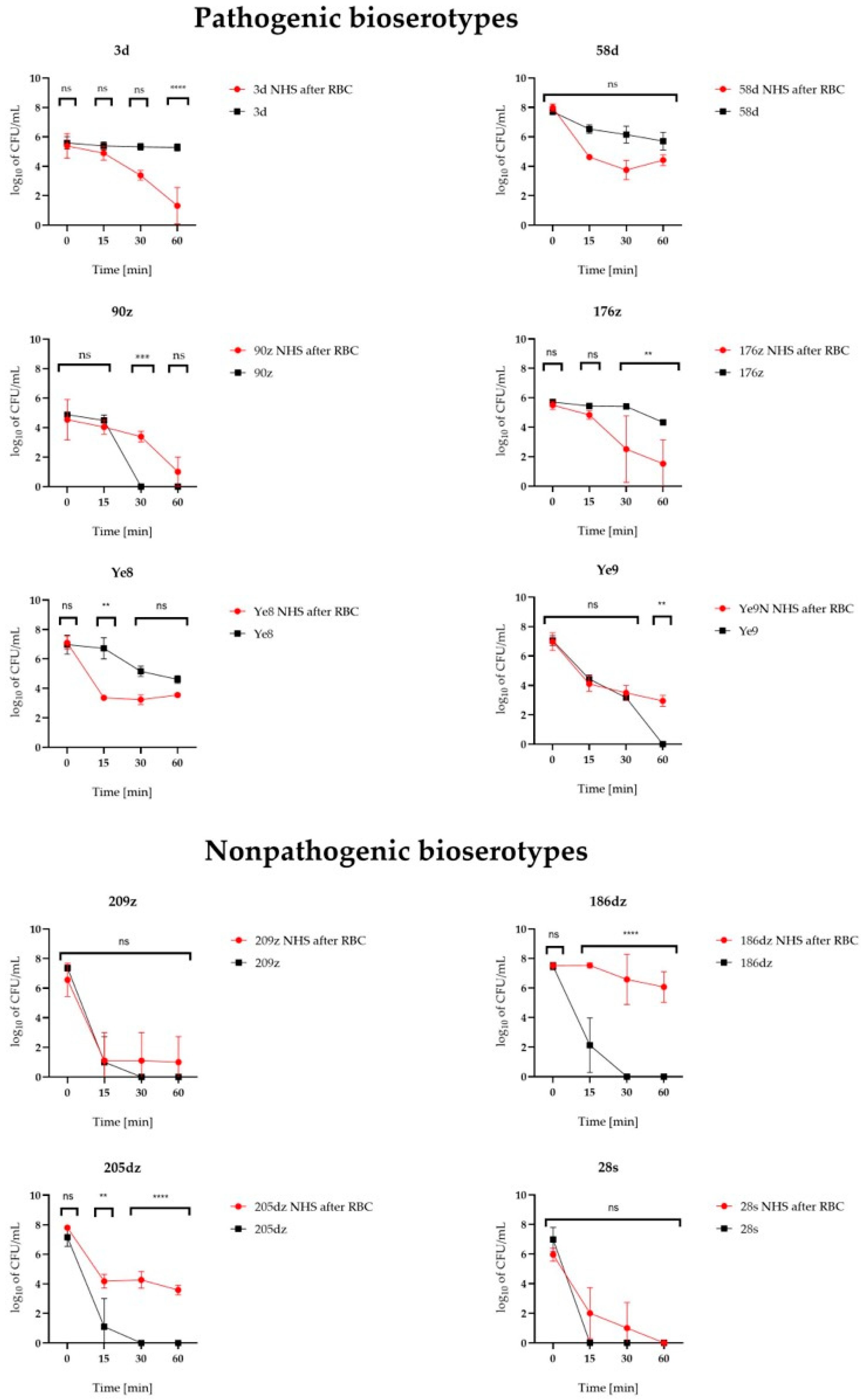
The effect of exposure to NHS on the survival of Y. enterocolitica was assessed. All of the isolates kept in control conditions (not exposed to RBC), except for the 28s Y. enterocolitica isolate, survived the 15 min exposition on NHS (p < 0.0001). Exposure of Y. enterocolitica to NHS for longer periods of 30 and 60 min led to a significant decrease in the bacterial counts (p < 0.0001). Only five and four out of the ten isolates kept in control conditions survived the 30 min (3d, 58d, 176z, Ye8, Ye9) and 60 min (3d, 58d, 176z, Ye8) NHS exposition, respectively. All of these isolates belonged to bioserotypes considered as pathogenic. None of the isolates belonging to the non-pathogenic 1A biotype survived the 30 min exposition to NHS (p < 0.0001, Figure 3, Table 2).
All strains after 28-days of RBC concentrate incubation survived the 15 min, 30 min, and 60 min exposition on NHS, however, the 28s Y. enterocolitica isolate did not survive 60 min (p < 0.0001, Figure 3, Table 2).
Comparison of NHS survival between the control and RBC-grown Y. enterocolitica isolates at 60 min indicated that the 3d and 176z isolates became more sensitive to human serum after RBC concentrate incubation (p < 0.0001, and p = 0.006, respectively). The Y. enterocolitica 3d isolate exhibited a bacterial count of 2.14 × 10⁵ CFU/mL after 60 min of incubation in NHS without prior exposure to the RBC concentrate. However, following incubation in RBC concentrate, this value decreased markedly to 1.1 × 10² CFU/mL. A similar reduction was observed for the 176z isolate, which declined from 2.24 × 10⁴ to 6.07 × 10² CFU/mL under the same conditions.
In turn, the 186dz (p < 0.0001) and 205dz (p < 0.0001) isolates and Ye9N strain (p = 0.0029) became more resistant to NHS following RBC concentrate growth when compared to the control bacteria. For these isolates, an increase in CFU/mL was observed from 0 at 60 min to 3.24 × 10⁶, 4.7 × 10³, and 1.10 × 10³, respectively.
We observed no influence of RBC concentrate incubation on the bacterial serum sensitivity level for the 28s, 90z, 209z, and 58d isolates and Ye8 strain (p > 0.5, Figure 3, Table 2).

3. Discussion

RBC concentrate transfusion is a common and often life-saving medical procedure. However, RBC concentrate preparations are not routinely screened for microorganisms. While microbiological contamination of RBC concentrates is rare, it can result in severe or fatal outcomes for patients [4]. Enteropathogenic Y. enterocolitica contamination may originate from donors with bacteremia following intestinal yersiniosis [1]. There are numerous reports on the growth of Y. enterocolitica in RBC concentrates. However, the studied factors potentially impacting the growth of Y. enterocolitica in RBC concentrates include the process of blood preparation, RBC concentrate manufacturing method, use of additive solutions, blood group, donor sex, and time of blood exposure to room temperature [4,13,14,31,32,33]. The growth of different Y. enterocolitica biotypes in RBC concentrates has not been extensively studied.
The effect of temperature and temperature changes on Y. enterocolitica growth in RBC concentrates has previously been studied. Aplin et al. [31] observed rapid bacterial declines when RBC concentrate units were exposed to refrigeration and intermittent warming to 30 °C for 30 and 60 min. However, in studies applying constant storage temperatures (2–6 °C), Y. enterocolitica exhibited steady growth, reaching levels of 108–109 CFU/mL, consistent with our findings [2,4,13,14]. In our work, a constant temperature of incubation was applied to assess the development of Y. enterocolitica in standard conditions of RBC concentrate storage. Ramirez-Arcos et al. [4] demonstrated that variables related to RBC concentrate manufacturing, rather than the additive solution type or donor sex, had a greater impact on bacterial proliferation in RBC concentrate units. The same author also confirmed that exposure to uncontrolled temperature did not affect the growth of Y. enterocolitica in RBC concentrates [13]. Prax et al. [2] confirmed Y. enterocolitica as one of the key psychrotrophic bacteria capable of growing in refrigerated RBC concentrates, demonstrating its rapid and stable growth, reaching high bacterial concentrations (over 108 CFU/mL) within 21 days. Only Graveman et al. [33] reported no Y. enterocolitica proliferation in RBC concentrates when whole blood (WB) units were spiked prior to component separation. However, their study primarily investigated the influence of whole blood manufacturing variables on bacterial proliferation, rather than the effects of prolonged incubation on bacterial growth. Our study further demonstrates that, in addition to manufacturing-related factors, intrinsic bacterial characteristics, such as biotype and genes for iron metabolism, significantly affect the growth dynamics of Y. enterocolitica in RBC concentrates. All isolates demonstrated growth in RBC concentrates over 28 days, with significant differences among bioserotypes. The highly pathogenic 1B/O:8 strain (Ye8) exhibited the highest proliferation, correlating with the presence of the yersiniobactin gene cluster (ybtAESUX, irp1, irp2) (Figure 1). Yersiniobactin, encoded on a pathogenicity island, seems to be crucial for bacterial development in RBC concentrates, playing a role as a siderophore-dependent uptake system for iron released from erythrocytes [17]. Strains lacking yersiniobactin showed lower growth potential, highlighting its importance. Interestingly, biotype 1A isolates, traditionally considered non-pathogenic, reached comparable counts and showed high allelic diversity in iron metabolism-related genes (Table 1, Figure 1). For example, the 28s strain, lacking the hasA heme acquisition gene [15], displayed reduced growth compared with other 1A isolates. This suggests that the ability to utilize heme as an iron source may be critical for optimal proliferation within the red blood cell environment for the Y. enterocolitica 1A biotype. Bioserotype 4/O:3 strains, characterized by a reduced repertoire of iron metabolism-related genes, including the absence of the fepABCDG, fes, irp1, irp2, fyuA, hasR, and ybtAESUX gene clusters, exhibited the lowest growth. This finding confirms that the lack of both enterochelin and yersiniabactin systems can severely limit bacterial proliferation, regardless of the biotype 4 pathogenic potential. Notably, isolates genetically similar to 4/O:3 but harboring additional iron metabolism genes (fepBCDG, fes), such as Ye9N and 58d, achieved higher growth, suggesting that gene content may outweigh genetic similarity in influencing RBC concentrate proliferation.
Serum resistance level is another factor influencing Y. enterocolitica survival in human blood [24]. Orozova et al. [14] reported that prolonged RBC concentrate incubation reduced the bacterial resistance to normal human serum (NHS). Our study directly examined the serum sensitivity post-RBC incubation, observing variable responses: increased resistance in three isolates including biotype 1A from wild boars (186dz and 205dz) and reference strain 2/O:9, decreased resistance in two isolates (4/O:3 3d and 176z), and no change in the remaining five isolates. The most NHS-resistant isolates belonged to biotype 1A, suggesting potential pathogenicity in strains historically considered non-pathogenic. This finding aligns with reports of wild boars as an emerging reservoir for human yersiniosis [30,34,35,36]. Notably, the 1A isolate 205dz, which carried the ail virulence gene and almost complete iron metabolism gene repertoire, supports reevaluating the pathogenic potential of certain 1A strains.
Our study faced limitations, including variability in donor ages, RBC concentrate bag volumes, and blood group types. However, previous research indicates that blood group (A, B, O) has minimal impact on Y. enterocolitica growth in RBC concentrates [14].
In conclusion, our findings demonstrate the capacity of diverse Y. enterocolitica bioserotypes to proliferate in red blood cells, influenced by iron metabolism-related genes. This emphasizes the clinical importance of Y. enterocolitica in RBC concentrate storage and highlights the need for prioritized microbiological testing when bacterial contamination is suspected. Continuous research and improved screening strategies are essential to ensure the microbiological safety of blood products. Furthermore, these findings highlight the need for a comprehensive reevaluation of the conventional biotype classification; although the biotype system may be retained, it should no longer be used as a definitive criterion for determining pathogenic potential.

4. Materials and Methods

4.1. Bacterial Strains

Eight Y. enterocolitica isolates and two Y. enterocolitica reference strains were included in this study. Table 3 contains the preliminary characteristics of the eight Y. enterocolitica isolates and Y. enterocolitica reference strains used in this study [37].

4.2. Detection of Virulence-Associated Genes of Y. enterocolitica

PCR was used to detect virulence-associated genes: irp2, ystA, 16S, ail, ystB, ymoA, yadA, ystC, irp1, ureC, virF, invA, yst, blaA, blab, myfA, myfB, myfC, chiY, ysrS, yst1M, sat, hreP, fepA, fepD, rfbC, tccC, fyuA. The PCR mixtures consisted of 1 mM MgCl2, 1 × buffer, 0.2 mMdNTP, primers (Genomed, Warszawa, Poland), used at concentration ranging from 0.15 to 1.25 µM (Supplementary Table S2), 1U of Taq DNA polymerase, and 1 µL of DNA template. PCRs were performed with an initial denaturation step at 94 °C for 3 min, 35 cycles each of denaturation, annealing and extension, as indicated in Supplementary Table S2, and a final extension of 10 min at 72 °C. Amplicons were separated in 1.5% agarose gel containing 0.5 μg/mL ethidium bromide, at 120–130 V for 1 h and documented using the GelDocXR System (Bio-Rad, Hercules, CA, USA) [38,39].

4.3. Sequencing of the Genomes

Eight Y. enterocolitica isolates were subjected to whole genome sequencing (WGS). The DNA for the genomic sequencing was extracted with the MasterPure™ Complete DNA and RNA Purification Kit (Lucigen, Middleton, WI, USA) according to the manufacturer’s instructions. De novo whole-genome was performed on an Illumina MiSeq sequencer using the Nextera XT Library Preparation Kit with standard protocols. Libraries were sequenced using 2 × 300 bp paired-end v3 Reagent Kit (Illumina), following the manufacturer’s protocols [40]. The reads were trimmed and assembled into contigs using Shovill software, version 0.9.0 (https://github.com/tseemann/shovill, accessed on 12 June 2025). The quality of the obtained sequences was checked using the Quast program [41]. Genomes of publicly available Y. enterocolitica reference strains were included in the study—Ye9N and Ye8 (8081) (accession numbers: JAALCX000000000 and AM286415, respectively).

4.4. Phylogenetic Analysis of the Isolates and Iron Metabolism Gene Detection

Nucleotide sequences of 42 genes responsible for iron metabolism were obtained from GenBank, accession numbers AM286415 and U41370. Their detection in the genomes was performed using the Genome Comparator module (version 2.8.5) in BIGSdb [42] with gene presence defined as a BLAST (version 1.6.3) match of sequence identity >70% over 50% of a locus length. cgMLST was performed according to Savin et al. [43] using the BIGSdb—Yersinia database at https://bigsdb.pasteur.fr/yersinia (accessed on 12 June 2025), employing the Yersinia cgMLST scheme with standard settings. A phylogenetic tree was created using RapidNJ 2.3.2 software [44] using the Jukes and Cantor evolution model and visualized with Phandango [45].

4.5. Red Blood Cell Concentrate Units

The research material consisted of anonymized samples of RBC concentrate, obtained from healthy volunteer donors at the Regional Center for Blood Donation and Treatment in Wrocław, Poland. The RBC concentrate units used in this study are listed in Table 4. Donors consented to the use of their blood for scientific research when completing the qualification questionnaire before blood donation. This was conducted according to the principles expressed in the Declaration of Helsinki and was approved by the Director of Regional Center for Blood Donation and Treatment in Wrocław (cooperation agreement number N0BR000.7117.IN.6/Wet/2022). Donations of whole blood were taken into sterile, non-pyrogenic bags systems with anticoagulant CPD. After collection, to prevent bacterial growth, the whole blood donations were stored at room temperature (RT, 22 °C ± 2 °C) for at least 2 h, then centrifuged to obtain blood components including RBC concentrates in SAGM (saline-adenine-glucose-mannitol) solution. The first batch of whole blood was discarded thanks to the use of a pre-donation container. Each RBC concentrate used in the experiments was subjected to leukocyte depletion. Only RBC concentrates that met the specific criteria for acceptance of the blood donors in accordance with the national guidelines of the Ministry of Health in Poland, lacking hemolysis and negative for HIV 1/2, HCV and Treponema pallidum antibodies, a negative HBs antigen test, and negative viral genome screen (HIV RNA, HBV DNA, HCV RNA) were used [12,46]. The samples were not used for diagnostic tests, but were the research medium spiked with Y. enterocolitica isolates. Bacterial growth was observed under strictly defined conditions. The research used RBC concentrates after splitting into pediatric portions that had a shorter shelf life, (i.e., those that could not be transfused into adult recipients and were still an appropriate research model for planned experiments). All RBC concentrate units were tested for sterility before Y. enterocolitica infecting.

4.6. Incubation of Y. enterocolitica in Red Blood Cells

The study protocol was based on the method described by Prax et al., with modifications [2]. The volume of RBC concentrate in the bags, ranged from 50 to 167 mL. To avoid cross-contamination, the RBC concentrate volume was not normalized. The bacterial count was adjusted to the volume of RBC concentrate in the bag to obtain the starting bacterial count of 101–102 CFU/mL. For this, Y. enterocolitica was grown overnight in 5 mL of BHI broth (Th. Geyer Polska Sp. z o.o., Warszawa, Poland). Then, the cultures were diluted with sterile 0.85% saline solution to reach the OD600 of 0.1, corresponding to a bacterial count of 1.6–3.7 × 10⁸ CFU/mL, as determined by the plating of serial dilutions of bacteria. Next, the cultures were diluted 10,000 times, and the appropriate volume of bacterial suspension was taken to obtain the final bacterial count of 101–102 CFU/mL in a given RBC concentrate unit. Bacterial suspensions were transferred to RBC concentrate units using a sterile syringe needle through the sampling site coupler (Fenwal, Bloodbankdepot, Douglasville, GA, USA). Immediately after starting the incubation of bacteria in RBC concentrate units (time 0 days), 1 mL of RBC concentrate was taken from each bag and plated on solid media to determine the starting number of bacteria.
The RBC concentrate units were mixed by inversion and incubated at 2–6 °C with no agitation to mimic standard storage conditions. The bacterial counts in RBC concentrate were determined at days 3, 7, 14, 21, and 28. The RBC concentrates were sampled from the bags using a sampling site coupler under aseptic conditions. The serial RBC concentrate dilutions were plated onto BHI agar (Th. Geyer Polska Sp. z o.o., Warszawa, Poland) in triplicate, then incubated at 25 °C for 48 h.

4.7. Normal Human Serum Bactericidal Tests

The NHS bactericidal assay was carried out with major modifications according to Orozova et al. [14]. At least 50 mL of RBC concentrate was transferred to sterile tubes and centrifuged at 770× g for 9 min. Supernatants were pipetted into fresh 15 mL sterile tubes, then subsequently centrifuged at 4000 rpm, 4 °C, 20 min; after that, the supernatants were discarded. The bacterial pellets were then suspended in 5 mL of BHI broth. The density of bacterial suspensions was adjusted to an OD600 value equal to 0.8–0.9 (Spark® Microplate Reader, Tecan, Männedorf, Switzerland). These optimized suspensions were then centrifuged again (4000 rpm, 4 °C, 20 min). The pellets were then suspended in 3 mL of NaCl (Th. Geyer Polska Sp. z o.o., Warszawa, Poland). One mL of these bacterial mixtures was refreshed in 5 mL of 0.9% NaCl, and 1 mL of that was combined with 1 mL of NHS (Sigma-Aldrich, Darmstadt, Germany). Incubation with NHS lasted up to 1 h at 37 °C. The CFU/mL value was assessed at time points 0, 15, 30, and 60 min. For this, the mixture of bacteria in NHS were plated onto BHI agar, then incubated at 25 °C for 48 h and counted. The NHS bactericidal tests were also conducted using the same bacterial isolates without RBC concentrate incubation. The overnight Y. enterocolitica cultures were diluted using BHI broth to an OD600 0.1 and then grown to reach an OD600 of 0.8–0.9. Subsequent stages of the test remained unchanged.

4.8. Statistical Methods

Y. enterocolitica growth in the RBC concentrate units was assessed in triplicate. Statistical analysis was performed with GraphPad Prism 9.1.1. The data from each NHS bactericidal assay and RBC incubation were compared by analysis of variance (two-way ANOVA) [24,47]. Bacterial survival rates were compared using Tukey’s multiple comparisons test. The p-values from these tests are shown in the text and the figures. A p < 0.05 indicates that the compared values were significantly different at a 95% confidence level.

Supplementary Materials

The following supporting information can be downloaded at: https://www.mdpi.com/article/10.3390/ijms26125775/s1. References [48,49,50,51,52,53,54,55,56] are cited in the supplementary materials.

Author Contributions

Conceptualization, K.M.; Methodology, K.M., J.K., S.K.S. and B.P.; Formal analysis, K.M.; Investigation, K.M. and S.B.; Resources, M.S., U.S. and M.S.-N.; Writing—original draft preparation, K.M. and J.B. (Jacek Bania); Writing—review and editing, K.M., J.B. (Jacek Bania), J.B. (Jarosław Bystroń), G.B.-P., M.S., M.S.-N., S.K.S. and B.P.; Funding acquisition, K.M. All authors have read and agreed to the published version of the manuscript.

Funding

This research was funded in whole by the National Science Center, Poland, grant number 2021/05/X/NZ6/01383. The APC was financed by Wrocław University of Environmental and Life Sciences.

Institutional Review Board Statement

Not applicable.

Informed Consent Statement

Not applicable.

Data Availability Statement

The original contributions presented in this study are included in the article and Supplementary Material. Further inquiries can be directed to the corresponding author.

Conflicts of Interest

The authors declare no conflicts of interest.

References

  1. Guinet, F.; Carniel, E.; Leclercq, A. Transfusion-transmitted Yersinia Enterocolitica sepsis. Clin. Infect. Dis. 2011, 53, 583–591. [Google Scholar] [CrossRef] [PubMed]
  2. Prax, M.; Spindler-Raffel, E.; McDonald, C.P.; Bearne, J.; Satake, M.; Kozakai, M.; Rojo, J.; Hanschmann, K.O.; Lambrecht, B.; Grundmann, U.; et al. Establishment of Transfusion-relevant Bacteria Reference Strains for Red Blood Cells. Vox Sang. 2021, 116, 692–701. [Google Scholar] [CrossRef]
  3. Shepel, M.; Boyd, J.; Luider, J.; Gibb, A.P. Interaction of Yersinia enterocolitica and Y. Pseudotuberculosis with Platelets. J. Med. Microbiol. 2001, 50, 1030–1038. [Google Scholar] [CrossRef] [PubMed]
  4. Ramirez-Arcos, S.; Kou, Y.; Cayer, M.; De Grandmont, M.; Girard, M.; Cloutier, M. The Impact of Red Blood Cell Manufacturing Variables on Bacterial Growth Dynamics: A Pilot Study. Vox Sang. 2019, 114, 478–486. [Google Scholar] [CrossRef]
  5. Hoelen, D.W.M.; Tjan, D.H.T.; Schouten, M.A.; Dujardin, B.C.G.; van Zanten, A.R.H. Severe Yersinia Enterocolitica Sepsis after Blood Transfusion. Neth. J. Med. 2007, 65, 301–303. [Google Scholar]
  6. Strobel, E.; Heesemann, J.; Mayer, G.; Peters, J.; Müller-Weihrich, S.; Emmerling, P. Bacteriological and Serological Findings in a Further Case of Transfusion-Mediated Yersinia enterocolitica Sepsis. J. Clin. Microbiol. 2000, 38, 2788–2790. [Google Scholar] [CrossRef] [PubMed]
  7. Frati, P.; Busardò, F.P.; Di Stefano, M.A.; Neri, M.; Sessa, F.; Fineschi, V. A Fatal Case of Post-Transfusion Sepsis Caused by Yersinia Enterocolitica after Delivery. Blood Transfus. 2015, 13, 528–531. [Google Scholar] [CrossRef]
  8. Wagner, S.J. Transfusion-transmitted Bacterial Infection: Risks, Sources and Interventions. Vox Sang. 2004, 86, 157–163. [Google Scholar] [CrossRef]
  9. Bruining, A.; De Wilde-Huizen, C.C.M. A case of contamination of donor blood by Yersinia enterocolitica type 9. Med. Ned. 1975, 4, 25–26. [Google Scholar]
  10. Leclercq, A.; Martin, L.; Vergnes, M.L.; Ounnoughene, N.; Laran, J.; Giraud, P.; Carniel, E. Fatal Yersinia enterocolitica Biotype 4 Serovar O:3 Sepsis after Red Blood Cell Transfusion. Transfusion 2005, 45, 814–818. [Google Scholar] [CrossRef]
  11. Benavides, S.; Nicol, K.; Koranyi, K.; Nahata, M.C. Yersinia Septic Shock Following an Autologous Transfusion in a Pediatric Patient. Transfus. Apher. Sci. 2003, 28, 19–23. [Google Scholar] [CrossRef] [PubMed]
  12. Dziennik Urzędowy Ministra Zdrowia. Obwieszczenie Ministra Zdrowia z dnia 30 marca 2021 r. w Sprawie Wymagań Dobrej Praktyki Pobierania Krwi i jej Składników, Badania, Preparatyki, Przechowywania, Wydawania i Transportu dla Jednostek Organizacyjnych Publicznej Służby Krwi. DZ. URZ. Min. Zdr. 2021.28. Available online: https://dziennikmz.mz.gov.pl/legalact/2021/28/ (accessed on 15 April 2025).
  13. Ramirez-Arcos, S.; Perkins, H.; Kou, Y.; Mastronardi, C.; Kumaran, D.; Taha, M.; Yi, Q.-L.; McLaughlin, N.; Kahwash, E.; Lin, Y.; et al. Bacterial Growth in Red Blood Cell Units Exposed to Uncontrolled Temperatures: Challenging the 30-minute Rule. Vox Sang. 2013, 105, 100–107. [Google Scholar] [CrossRef] [PubMed]
  14. Orozova, P.; Markova, N.; Radoucheva, T. Properties of Yersinia entetocolitica and Yersinia Pseudotuberculosis in Red Blood Cell Concentrate of Different ABO Groups during 30-Day Storage at 4 °C. Clin. Microbiol. Infect. 2001, 7, 358–361. [Google Scholar] [CrossRef]
  15. Kanaujia, P.K.; Bajaj, P.; Virdi, J.S. Analysis of Iron Acquisition and Storage-related Genes in Clinical and Non-clinical Strains of Yersinia Enterocolitica Biovar 1A. APMIS 2015, 123, 858–866. [Google Scholar] [CrossRef]
  16. Jaworska, K.; Nieckarz, M.; Ludwiczak, M.; Raczkowska, A.; Brzostek, K. OmpR-Mediated Transcriptional Regulation and Function of Two Heme Receptor Proteins of Yersinia Enterocolitica Bio-Serotype 2/O:9. Front. Cell. Infect. Microbiol. 2018, 8, 333. [Google Scholar] [CrossRef]
  17. Rakin, A.; Schneider, L.; Podladchikova, O. Hunger for Iron: The Alternative Siderophore Iron Scavenging Systems in Highly Virulent Yersinia. Front. Cell. Infect. Microbiol. 2012, 2, 151. [Google Scholar] [CrossRef] [PubMed]
  18. Carniel, E. The Yersinia High-Pathogenicity Island: An Iron-Uptake Island. Microbes Infect. 2001, 3, 561–569. [Google Scholar] [CrossRef]
  19. Schubert, S.; Fischer, D.; Heesemann, J. Ferric Enterochelin Transport in Yersinia enterocolitica: Molecular and Evolutionary Aspects. J. Bacteriol. 1999, 181, 6387–6395. [Google Scholar] [CrossRef]
  20. Jaworska, K.; Ludwiczak, M.; Murawska, E.; Raczkowska, A.; Brzostek, K. The Regulator OmpR in Yersinia enterocolitica Participates in Iron Homeostasis by Modulating Fur Level and Affecting the Expression of Genes Involved in Iron Uptake. Int. J. Mol. Sci. 2021, 22, 1475. [Google Scholar] [CrossRef]
  21. Ventola, E.; Lovari, S.; Farneti, S.; Finazzi, G.; Bilei, S.; Owczarek, S.; Delibato, E. Molecular Characterization of Yersinia enterocolitica Strains to Evaluate Virulence Associated Genes. Ann. Ist. Super. Sanita 2023, 59, 280–285. [Google Scholar] [CrossRef]
  22. Wauters, G.; Kandolo, K.; Janssens, M. Revised Biogrouping Scheme of Yersinia enterocolitica. Contrib. Microbiol. Immunol. 1987, 9, 14–21. [Google Scholar] [PubMed]
  23. Bottone, E.J. Yersinia enterocolitica: Overview and Epidemiologic Correlates. Microbes Infect. 1999, 1, 323–333. [Google Scholar] [CrossRef] [PubMed]
  24. Skorek, K.; Raczkowska, A.; Dudek, B.; Miętka, K.; Guz-Regner, K.; Pawlak, A.; Klausa, E.; Bugla-Płoskońska, G.; Brzostek, K. Regulatory Protein OmpR Influences the Serum Resistance of Yersinia Enterocolitica O:9 by Modifying the Structure of the Outer Membrane. PLoS ONE 2013, 8, e79525. [Google Scholar] [CrossRef]
  25. Platt-Samoraj, A.; Syczyło, K.; Szczerba-Turek, A.; Bancerz-Kisiel, A.; Jabłoński, A.; Łabuć, S.; Pajdak, J.; Oshakbaeva, N.; Szweda, W. Presence of ail and ystB Genes in Yersinia enterocolitica Biotype 1A Isolates from Game Animals in Poland. Vet. J. 2017, 221, 11–13. [Google Scholar] [CrossRef]
  26. Joutsen, S.; Johansson, P.; Laukkanen-Ninios, R.; Björkroth, J.; Fredriksson-Ahomaa, M. Two Copies of the ail Gene Found in Yersinia enterocolitica and Yersinia kristensenii. Vet. Microbiol. 2020, 247, 108798. [Google Scholar] [CrossRef] [PubMed]
  27. Sihvonen, L.M.; Hallanvuo, S.; Haukka, K.; Skurnik, M.; Siitonen, A. The ail Gene Is Present in Some Yersinia enterocolitica Biotype 1A Strains. Foodborne Pathog. Dis. 2011, 8, 455–457. [Google Scholar] [CrossRef]
  28. Kraushaar, B.; Dieckmann, R.; Wittwer, M.; Knabner, D.; Konietzny, A.; Mäde, D.; Strauch, E. Characterization of a Yersinia enterocolitica Biotype 1A Strain Harbouring an ail Gene. J. Appl. Microbiol. 2011, 111, 997–1005. [Google Scholar] [CrossRef]
  29. Platt-Samoraj, A. Toxigenic Properties of Yersinia enterocolitica Biotype 1A. Toxins 2022, 14, 118. [Google Scholar] [CrossRef]
  30. Batzilla, J.; Heesemann, J.; Rakin, A. The Pathogenic Potential of Yersinia Enterocolitica 1A. Int. J. Med. Microbiol. 2011, 301, 556–561. [Google Scholar] [CrossRef]
  31. Aplin, K.; Pitt, T.; Allen, J.; Roy, A.; Tidey, K.; Ball, J.; McDonald, C.P. Extending the 30-minute Rule for Red Cell Units—Investigation of the Bacterial Risk of 60-minute Exposures to Ambient Temperature. Vox Sang. 2019, 114, 189–197. [Google Scholar] [CrossRef]
  32. Ramirez-Arcos, S.; Mastronardi, C.; Perkins, H.; Kou, Y.; Turner, T.; Mastronardi, E.; Hansen, A.; Yi, Q.; McLaughlin, N.; Kahwash, E.; et al. Evaluating the 4-hour and 30-minute Rules: Effects of Room Temperature Exposure on Red Blood Cell Quality and Bacterial Growth. Transfusion 2013, 53, 851–859. [Google Scholar] [CrossRef] [PubMed]
  33. Gravemann, U.; Handke, W.; Schulze, T.J.; Seltsam, A. Growth and Distribution of Bacteria in Contaminated Whole Blood and Derived Blood Components. Transfus. Med. Hemother. 2024, 51, 75–82. [Google Scholar] [CrossRef] [PubMed]
  34. Bhagat, N.; Virdi, J.S. Distribution of Virulence-Associated Genes in Yersinia enterocolitica Biovar 1A Correlates with Clonal Groups and Not the Source of Isolation. FEMS Microbiol. Lett. 2007, 266, 177–183. [Google Scholar] [CrossRef] [PubMed]
  35. Bhagat, N.; Virdi, J.S. The Enigma of Yersinia enterocolitica Biovar 1A. Crit. Rev. Microbiol. 2011, 37, 25–39. [Google Scholar] [CrossRef]
  36. Tennant, S.M.; Grant, T.H.; Robins-Browne, R.M. Pathogenicity of Yersinia enterocolitica Biotype 1A. FEMS Immunol. Med. Microbiol. 2003, 38, 127–137. [Google Scholar] [CrossRef]
  37. Morka, K.; Bystroń, J.; Bania, J.; Korzeniowska-Kowal, A.; Korzekwa, K.; Guz-Regner, K.; Bugla-Płoskońska, G. Identification of Yersinia Enterocolitica Isolates from Humans, Pigs and Wild Boars by MALDI TOF MS. BMC Microbiol. 2018, 18, 86. [Google Scholar] [CrossRef]
  38. Wannet, W.J.B.; Reessink, M.; Brunings, H.A.; Maas, H.M.E. Detection of Pathogenic Yersinia enterocolitica by a Rapid and Sensitive Duplex PCR Assay. J. Clin. Microbiol. 2001, 39, 4483–4486. [Google Scholar] [CrossRef] [PubMed]
  39. Bancerz-Kisiel, A.; Szczerba-Turek, A.; Platt-Samoraj, A.; Socha, P.; Szweda, W. Bioserotypes and Virulence Markers of Y. Enterocolitica Strains Isolated from Roe Deer (Capreolus capreolus) and Red Deer (Cervus elaphus). Pol. J. Vet. Sci. 2014, 17, 315–319. [Google Scholar] [CrossRef]
  40. Macori, G.; Romano, A.; Adriano, D.; Razzuoli, E.; Bianchi, D.M.; Gallina, S.; Bellio, A.; Decastelli, L. Draft Genome Sequences of Four Yersinia enterocolitica Strains, Isolated from Wild Ungulate Carcasses. Genome Announc. 2017, 5, e00192-17. [Google Scholar] [CrossRef]
  41. Mikheenko, A.; Prjibelski, A.; Saveliev, V.; Antipov, D.; Gurevich, A. Versatile Genome Assembly Evaluation with QUAST-LG. Bioinformatics 2018, 34, i142–i150. [Google Scholar] [CrossRef]
  42. Jolley, K.A.; Maiden, M.C. BIGSdb: Scalable Analysis of Bacterial Genome Variation at the Population Level. BMC Bioinform. 2010, 11, 595. [Google Scholar] [CrossRef] [PubMed]
  43. Savin, C.; Criscuolo, A.; Guglielmini, J.; Le Guern, A.-S.; Carniel, E.; Pizarro-Cerdá, J.; Brisse, S. Genus-Wide Yersinia Core-Genome Multilocus Sequence Typing for Species Identification and Strain Characterization. Microb. Genom. 2019, 5, 301. [Google Scholar] [CrossRef] [PubMed]
  44. Simonsen, M.; Mailund, T.; Pedersen, C.N.S. Rapid Neighbour-Joining. In Algorithms in Bioinformatics, Proceedings of the 8th International Workshop, WABI 2008, Karlsruhe, Germany, 15–19 September 2008; Springer: Berlin/Heidelberg, Germany, 2008; Volume 5251, pp. 113–123. [Google Scholar] [CrossRef]
  45. Hadfield, J.; Croucher, N.J.; Goater, R.J.; Abudahab, K.; Aanensen, D.M.; Harris, S.R. Phandango: An Interactive Viewer for Bacterial Population Genomics. Bioinformatics 2018, 34, 292–293. [Google Scholar] [CrossRef]
  46. Doerr, P., Dr. (Ed.) Guide to the Preparation, Use and Quality Assurance of Blood Components; European Committee (Partial Agreement) on Blood Transfusion (CD-P-TS). 21. wyd; European Directorate for the Quality of Medicines & HealthCare (EDQM) Council of Europe: Strasbourg, France, 2023. [Google Scholar]
  47. Miętka, K.; Brzostek, K.; Guz-Regner, K.; Bugla-Płoskońska, G. The Mechanisms of Complement Activation in Normal Bovine Serum and Normal Horse Serum against Yersinia Enterocolitica O:9 Strains with Different Outer Membrane Proteins Content. Pol. J. Vet. Sci. 2016, 19, 99–107. [Google Scholar] [CrossRef] [PubMed]
  48. Neubauer, H.; Hensel, A.; Aleksic, S.; Meyer, H. Identification of Yersinia enterocolitica within the genus Yersinia. Syst. Appl. Microbiol. 2000, 23, 58–62. [Google Scholar] [CrossRef]
  49. Gierczyński, R. Evaluation of the usefulness of selected virulence markers for identification of virulent Yersinia enterocolitica strains. II. Genotypic markers associated with the pYV plasmid. Med. Dosw. Mikrobiol. 2000, 52, 35–49. [Google Scholar]
  50. Platt-Samoraj, A.; Ugorski, M.; Szweda, W.; Szczerba-Turek, A.; Wojciech, K.; Procajło, Z. Analysis of the presence of ail, ystA and ystB genes in Yersinia enterocolitica strains isolated from aborting sows and aborted fetuses. J. Vet. Med. B Infect. Dis. Vet. Public Health 2006, 53, 341–346. [Google Scholar] [CrossRef]
  51. Momtaz, H.; Davood Rahimian, M.; Safarpoor Dehkordi, F. Identification and characterization of Yersinia enterocolitica isolated from raw chicken meat based on molecular and biological techniques. J. Appl. Poul. Res. 2013, 22, 137–145. [Google Scholar] [CrossRef]
  52. Bancerz-Kisiel, A.; Szczerba-Turek, A.; Platt-Samoraj, A.; Szweda, W. Distribution of the ymoA and ystA genes and enterotoxins yst production by Yersinia enterocolitica strains isolated from humans and pigs. Pol. J. Vet. Sci. 2012, 15, 609–614. [Google Scholar] [CrossRef]
  53. Zacharczuk, K. Investigation of molecular virulence factors of Yersinia enterocolitica 1B/08 human clinical isolates collected in Poland in 2009. Med. Dosw. Mikrobiol. 2013, 65, 233–243. [Google Scholar]
  54. Sharma, S.; Mittal, S.; Mallik, S.; Virdi, J.S. Molecular characterization of β-lactamase genes blaA and blaB of Yersinia enterocolitica biovar 1A. FEMS Microbiol. Lett. 2006, 257, 319–327. [Google Scholar] [CrossRef] [PubMed]
  55. Thisted Lambertz, S.; Danielsson-Tham, M.L. Identification and characterization of pathogenic Yersinia enterocolitica isolates by PCR and pulsed-field gel electrophoresis. Appl. Environ. Microbiol. 2005, 71, 3674–3681. [Google Scholar] [CrossRef] [PubMed]
  56. Gierczyński, R.; Jagielski, M.; Rastawicki, W. Evaluation of usefulness for selected virulence markers for identifying pathogenic Yersinia enterocolitica strains, I.V. Genes myfA and ureC. Med. Dosw. Mikrobiol. 2002, 54, 347–355. [Google Scholar] [PubMed]
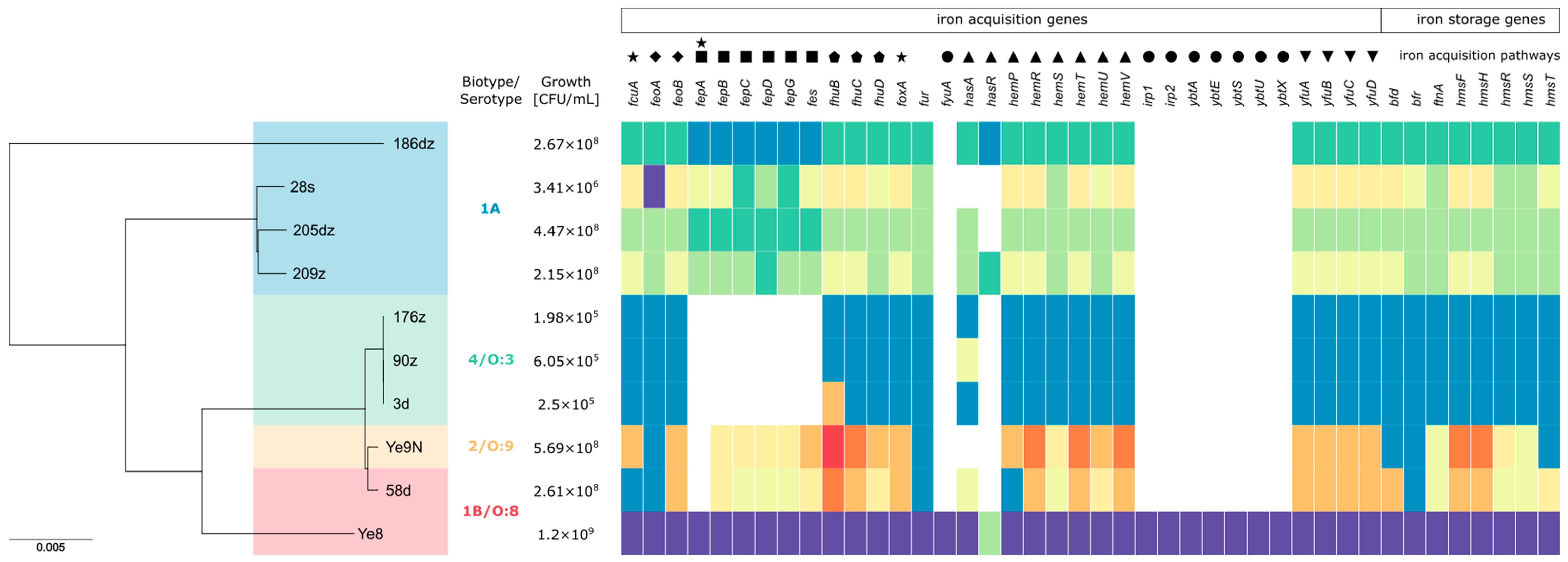
Figure 1. Phylogenetic tree of the studied Y. enterocolitica isolates, alongside the distribution of 42 iron metabolism-related genes and their growth potential in red blood cells (RBCs) after 28 days of incubation. The tree was constructed using the core genome multilocus sequence typing (cgMLST) scheme and the Jukes and Cantor evolutionary model, visualized with Phandango. Each isolate is annotated with its corresponding iron metabolism-related gene repertoire. Genes are represented by colored blocks indicating different allelic variants, while white blocks signify gene absence. Clades in the phylogenetic tree highlight genetic relationships among isolates including differences in bioserotypes, allelic repertoires, and the presence or absence of specific genes. The number of detected genes in each isolate ranged from 27 to 42, reflecting variation in their iron acquisition capabilities. Reference strains Ye8 and Ye9N are included for comparison, with Ye8 harboring all 42 genes. Clustering patterns correlate with specific genetic characteristics. Legend:●—yersiniabactin (Ybt), ∎—enterochelin catecholate siderophore, ▲—Hem uptake system, ★—TonB-dependent OM transporters, ◆—Feo system, ▼—inorganic iron ABC transport systems of Y. pestis (Yfu), ⬟—ferric hydroxamate uptake system (fhuCDB), fur—the master regulator of iron homeostasis in Y. enterocolitica, bfr—bacterioferritin, bfd—bacterioferritin-associated ferredoxin complex, ftn—ferritin, hmsFRST—hemin storage system.
Ijms 26 05775 g001
Figure 2. Bacterial growth in triplicates in RBC concentrates during 28 days of incubation at 2–4 °C. The data from each RBC incubation were compared by analysis of variance (two-way ANOVA). Bacterial survival rates were compared using the Tukey’s multiple comparisons test. The p-values from these tests are shown in the text. p < 0.05 indicates that the compared values were significantly different at a 95 % confidence level.
Ijms 26 05775 g002
Figure 3. Survival of pathogenic and nonpathogenic 1A Y. enterocolitica isolates during 60 min at 37 °C in NHS before and after RBC concentrate incubation. The asterisks indicate statistically significant differences between strains (ns p > 0.05, ** p < 0.01, *** p < 0.001, **** p < 0.0001, by two-way ANOVA with Tukey’s comparison post-test).
Ijms 26 05775 g003
Table 1. Mean and SD of Y. enterocolitica growth in the RBC concentrate units in triplicates. The data from each RBC concentrate incubation were compared by analysis of variance (two-way ANOVA).
Days/Isolate037142128
MeanSDMeanSDMeanSDMeanSDMeanSDMeanSD
3d, human, 4/O:33.50 × 1013.12 × 1011.76 × 1022.01 × 1021.11 × 1041.91 × 1041.21 × 1041.76 × 1042.50 × 1041.19 × 1042.50 × 1054.81 × 104
58d, human, 1B/O:82.00 × 1011.00 × 1012.42 × 1023.97 × 1021.40 × 1032.15 × 1038.27 × 1045.85 × 1043.03 × 1062.16 × 1062.61 × 1081.32 × 108
90z, pig, 4/O:32.53 × 1012.57 × 1013.77 × 1011.97 × 1011.88 × 1021.59 × 1014.22 × 1034.65 × 1022.21 × 1049.54 × 1026.05 × 1052.94 × 105
176z, pig, 4/O:34.89 × 1012.17 × 1011.98 × 1022.15 × 1021.07 × 1031.29 × 1035.11 × 1032.71 × 1033.79 × 1041.75 × 1041.98 × 1058.91 × 104
209z, pig, 1A5.68 × 1012.01 × 1014.01 × 1022.91 × 1027.27 × 1041.25 × 1051.97 × 1062.98 × 1062.71 × 1071.77 × 1072.15 × 1082.69 × 107
186dz, wild boar, 1A7.00 × 1017.94 × 1019.83 × 1019.09 × 1011.17 × 1031.01 × 1037.13 × 1059.07 × 1056.55 × 1062.49 × 1062.67 × 1088.42 × 107
205dz, wild boar, 1A5.67 × 1014.17 × 1014.26 × 1026.70 × 1024.42 × 1047.34 × 1041.45 × 1062.13 × 1061.49 × 1073.20 × 1064.47 × 1082.52 × 108
28s, roe deer, 1A4.67 × 1011.26 × 1011.41 × 1021.58 × 1021.12 × 1041.89 × 1048.10 × 1041.02 × 1052.04 × 1061.03 × 1063.41 × 1068.02 × 105
Ye9N, reference strain 2/O:94.00 × 1013.97 × 1013.79 × 1026.07 × 1022.14 × 1043.56 × 1044.91 × 1057.96 × 1059.22 × 1081.20 × 1095.69 × 1088.78 × 108
Ye8, reference strain 1B/O:83.00 × 1012.00 × 1011.43 × 1021.16 × 1028.49 × 1031.09 × 1047.76 × 1071.30 × 1081.24 × 1091.14 × 1091.20 × 1091.01 × 109
Table 2. Bacterial serum survival before and after 28 days of RBC concentrate incubation. The data from each tests were compared by analysis of variance (two-way ANOVA).
Isolate/
Time
[min]
Before RBCAfter RBCBefore RBCAfter RBCBefore RBCAfter RBCBefore RBCAfter RBC
0153060
MeanSDMeanSDMeanSDMeanSDMeanSDMeanSDMeanSDMeanSD
3d5.07 × 1053.33 × 1057.60 × 1051.16 × 1062.77 × 1051.55 × 1051.18 × 1051.30 × 1052.31 × 1051.05 × 1052.89 × 1031.65 × 1032.14 × 1059.83 × 1041.10 × 1021.65 × 102
58d5.15 × 1071.18 × 1051.09 × 1086.41 × 1073.95 × 1062.52 × 1064.29 × 1043.19 × 1032.21 × 1061.79 × 1069.50 × 1037.47 × 1039.13 × 1051.07 × 1063.33 × 1042.82 × 104
90z7.58 × 1048.08 × 1034.44 × 1057.59 × 1053.79 × 1042.41 × 1041.49 × 1041.10 × 104003.00 × 1032.18 × 103003.67 × 1015.51 × 101
176z5.26 × 1059.97 × 1043.71 × 1052.53 × 1052.80 × 1052.16 × 1048.15 × 1045.96 × 1042.59 × 1051.96 × 1048.60 × 1031.27 × 1042.24 × 1047.37 × 1036.07 × 1021.03 × 103
209z2.48 × 1071.06 × 1072.23 × 1073.68 × 1073.33 × 1025.77 × 1026.67 × 1021.15 × 103006.67 × 1021.15 × 103003.77 × 1025.41 × 102
186dz3.36 × 1071.59 × 1073.28 × 1076.10 × 1061.17 × 1031.26 × 1033.54 × 1071.37 × 107002.48 × 1072.31 × 107003.24 × 1063.02 × 106
205dz2.22 × 1071.69 × 1076.43 × 1071.24 × 1076.67 × 1021.15 × 1032.09 × 1041.78 × 104002.86 × 1042.49 × 104004.70 × 1033.75 × 103
28s1.98 × 1071.66 × 1071.31 × 1061.35 × 106006.67 × 1025.77 × 102003.33 × 1025.77 × 102001.33 × 1012.31 × 101
Ye81.55 × 1071.25 × 1071.62 × 1071.39 × 1079.36 × 1067.48 × 1062.34 × 1034.74 × 1021.73 × 1059.96 × 1042.00 × 1031.41 × 1034.52 × 1042.29 × 1043.68 × 1039.55 × 102
Ye9N1.35 × 1078.03 × 1061.84 × 1072.42 × 1073.09 × 1041.61 × 1041.79 × 1041.39 × 1041.56 × 1035.10 × 1024.67 × 1034.73 × 103001.10 × 1038.06 × 102
Table 3. Characteristics of Y. enterocolitica.
Y. enterocoliticaOriginBioserotypeVirulence-Associated Genes Content
3dHuman feces4/O:3yadA, ail, inv, yst, ystA, ystC, myfA, myfB, ureC, ymoA, rfbC, blaA, blaB
58d1B/O:8yadA, ail, inv, yst, ystA, ystC, myfA, myfB, ureC, ymoA, rfbC, fepD, blaA, blaB
90zPigs4/O:3yadA, ail, inv, yst, ystA, ystC, myfA, myfB, ureC, ymoA, rfbC, blaA, blaB
176z4/O:3yadA, ail, inv, yst, ystA, ystC, myfA, myfB, ureC, ymoA, rfbC, hreP, sat, blaA, blaB
209z1A/O:9inv, ystB, ureC, ymoA, fepD
186dzWild boars1A/NTinv, ureC, ymoA, fepA, fepD
205dz1A/NTail, inv, ystB, myfA, ureC, ymoA, hreP, fepD
28sRoe deer1A/NTinv, ystB, ureC, ymoA, tccC, hreP, fepA, fepD
Ye9NReference2/O:9yadA, virF, ail, inv, yst, ystA, ystC, myfA, myfB, ureC, ymoA, tccC, hreP, fepD, blaA, blaB
Ye81B/O:8yadA, ail, inv, yst, ystA, ystC, myfA, myfB, ureC, ymoA, irp1, irp2, fyuA, chiY, Yts1M, hreP, fepD, blaB, ysrS
NT—lack of serotype information.
Table 4. List of RBC concentrate units used in this study. SAGM—saline-adenine-glucose-mannitol; 0.2 J denotes a 0.2 portion of the whole RBC concentrate component for adults, which has been divided into smaller parts for pediatric use.
No.StrainBlood GroupCharacteristics of RBC Concentrate UnitsVolume [mL]Age of RBC Concentrate on the Infecting Day
13d0 RhD+SAGM/450 mL/2-6C, leukoreduced, 0.2 J5031
20 RhD+SAGM/450 mL/2-6C, irradiated, leukoreduced, 0.4 J847
3B RhD+SAGM/450 mL/2-6C, irradiated, leukoreduced, 0.4 J1009
458dA RhD+SAGM/450 mL/2-6C, leukoreduced, 0.2 J5027
5A RhD-SAGM/450 mL/2-6C, leukoreduced, 0.4 J9325
6A RhD+SAGM/450 mL/2-6C, leukoreduced, 0.4 J10313
790zA RhD+SAGM/450 mL/2-6C, leukoreduced, 0.6 J12825
8A RhD+SAGM/450 mL/2-6C, leukoreduced, 0.6 J13921
90 RhD-SAGM/450 mL/2-6C, leukoreduced, 0.6 J11912
10176dz0 RhD+SAGM/450 mL/2-6C, leukoreduced, 0.8 J16722
11B RhD+SAGM/450 mL/2-6C, leukoreduced, 0.4 J10012
12B RhD+SAGM/450 mL/2-6C, leukoreduced, 0.4 J10521
13209zA RhD+SAGM/450 mL/2-6C, leukoreduced, 0.4 J6523
14A RhD-SAGM/450 mL/2-6C, leukoreduced, 0.4 J9325
150 RhD+SAGM/450 mL/2-6C, leukoreduced, 0.4 J11628
16186dzB RhD+SAGM/450 mL/2-6C, leukoreduced, 0.4 J8618
17A RhD+SAGM/450 mL/2-6C, leukoreduced, 0.6 J14219
18AB RhD+SAGM/450 mL/2-6C, leukoreduced, 0.4 J10022
19205dz0 RhD+SAGM/450 mL/2-6C leukoreduced, 0.6 J14025
20A RhD-SAGM/450 mL/2-6C, leukoreduced, 0.4 J8225
210 RhD-SAGM/450 mL/2-6C, leukoreduced, 0.4 J10127
2228sB RhD+SAGM/450mL/2-6C, leukoreduced, 0.6 J15429
23A RhD-SAGM/450 mL/2-6C, leukoreduced, 0.6 J13415
24A RhD-SAGM/450 mL/2-6C, leukoreduced, 0.6 J13520
25Ye9NB RhD+SAGM/450 mL/2-6C, leukoreduced, 0.4 J7123
260 RhD+SAGM/450 mL/2-6C, leukoreduced, 0.6 J15023
270 RhD+SAGM/450 mL/2-6C, leukoreduced, 0.6 J15022
28Ye80 RhD+SAGM/450 mL/2-6C, leukoreduced, 0.4 J8829
29B RhD+SAGM/450 mL/2-6C, leukoreduced, 0.4 J6518
30B RhD+SAGM/450 mL/2-6C, leukoreduced, 0.4 J11821
Disclaimer/Publisher’s Note: The statements, opinions and data contained in all publications are solely those of the individual author(s) and contributor(s) and not of MDPI and/or the editor(s). MDPI and/or the editor(s) disclaim responsibility for any injury to people or property resulting from any ideas, methods, instructions or products referred to in the content.