Communication

Tryptophan Metabolite ITE Attenuates LPS-Induced MMP-9 via NF-κB/AP-1 in Monocytes

1
Immunology & Microbiology Department, Dasman Diabetes Institute, Dasman 15462, Kuwait
2
Translational Research Department, Dasman Diabetes Institute, Dasman 15462, Kuwait
*
Author to whom correspondence should be addressed.
Int. J. Mol. Sci. 2025, 26(12), 5663; https://doi.org/10.3390/ijms26125663
Submission received: 22 April 2025 / Revised: 27 May 2025 / Accepted: 4 June 2025 / Published: 13 June 2025
(This article belongs to the Section Molecular Endocrinology and Metabolism)

Abstract

Matrix metalloproteinase-9 (MMP-9) and lipopolysaccharide (LPS) levels are known to be elevated in obesity and contribute to metabolic dysfunction. 2-(1′H-indole-3′-carbonyl)-thiazole-4-carboxylic acid methyl ester (ITE), an endogenous ligand of the aryl hydrocarbon receptor (AhR), has been implicated in the regulation of inflammatory responses. This study aimed to determine whether ITE can inhibit LPS-induced MMP-9 expression in monocytic cells and to explore the underlying signaling mechanisms involved. Human monocytic THP-1 cells and primary human monocytes were treated with LPS in the presence or absence of ITE. MMP-9 mRNA and protein levels were assessed using quantitative real-time PCR and ELISA, respectively, while gelatin zymography was employed to evaluate MMP-9 enzymatic activity. Chromatin immunoprecipitation followed by qPCR (ChIP-qPCR) was performed to assess NF-κB and AP-1 binding to the MMP-9 promoter region. Our findings demonstrate that ITE significantly suppresses LPS-induced MMP-9 gene and protein expression. This suppression is associated with a marked reduction in LPS-induced NF-κB and AP-1 transcriptional activity. ChIP-qPCR confirmed that ITE attenuates the recruitment of NF-κB and AP-1 to the MMP-9 promoter, thereby inhibiting its transcription. In summary, ITE downregulates LPS-induced MMP-9 expression by interfering with NF-κB/AP-1 signaling, suggesting a potential anti-inflammatory mechanism that could be relevant in the context of MMP-9-driven inflammatory conditions.

1. Introduction

Matrix metalloprotease (MMP)-9 and lipopolysaccharide (LPS) are elevated in obesity, and both participate in the development of chronic low-grade inflammation in obese individuals [1,2]. MMP-9 is mainly secreted by neutrophils, monocytes, and macrophages and it regulates inflammation in tissues and diseases [3]. A significant elevation of MMP-9 expression has been observed in inflammatory diseases, including obesity, cardiovascular diseases, tumor invasion, and rheumatoid arthritis [4,5,6,7,8]. MMP-9 expression in monocytes and macrophages is increased by lipopolysaccharide (LPS) [9]. In mice, a high-fat diet increased plasma LPS concentrations by two to three times when compared to lean animals [10]. Similarly, obese individuals with type 2 diabetes have higher plasma LPS levels than healthy controls, whereas weight loss lowers LPS levels [1,11]. Overnutrition or high-fat diet intake results in metabolic endotoxemia via increased LPS absorption involving changes in the gut microbiota and an increased permeability of the gut epithelium [11,12,13]. LPS induces a strong immune response via its interaction with the cell surface receptor Toll-like receptor (TLR4) [14]. LPS binds to the TLR4 receptor and triggers a strong immune response, and this activation leads to an enhanced production of cytokines and chemokines via mediators such as nuclear factor-κB (NF-κB), activator protein-1 (AP-1), and interferon regulatory factors (IRFs), which contribute to the development of the chronic low-grade inflammation seen in obesity [14].
Since the overexpression and dysregulation of MMP-9 levels are associated with various diseases [4,5,6], the regulation and inhibition of MMP-9 is an important therapeutic approach for combating various diseases including cancer. So far, no selective MMP-9 inhibitors have passed clinical trials.
Aryl hydrocarbon receptors (AHRs) are found in numerous tissues throughout the body. These receptors are expressed by various immune cells [15]. AHRs are activated by dietary components such as fats and fat derivatives and gut microbial-derived metabolites [16]. Recent research has shown the significance of AHR signaling in both healthy and diseased immunological conditions [17]. The endogenous ligand and tryptophan metabolite 2-(1′H-indole-3′-carbonyl)-thiazole-4-carboxylic acid methyl ester (ITE) is an AHR agonist that suppresses inflammation [18,19]. Manuel et al. reported that ITE activates the AHR pathway and partially protects against EAP-induced prostate inflammation, pain, and bladder dysfunction by modulating inflammation-related gene expression in mice [20]. Abron et al. demonstrated that the nontoxic AhR ligand ITE reduces intestinal inflammation in a mouse model of colitis by promoting regulatory T cells, suppressing pro-inflammatory cytokines, and modulating immune cell populations, suggesting its potential as a therapeutic agent for IBD [21]. Since ITE is known to reduce inflammation, this study is the first to examine its effect on LPS-induced MMP-9 gene expression and production in monocytic cells, revealing a new mechanistic pathway involved in this process.

2. Results

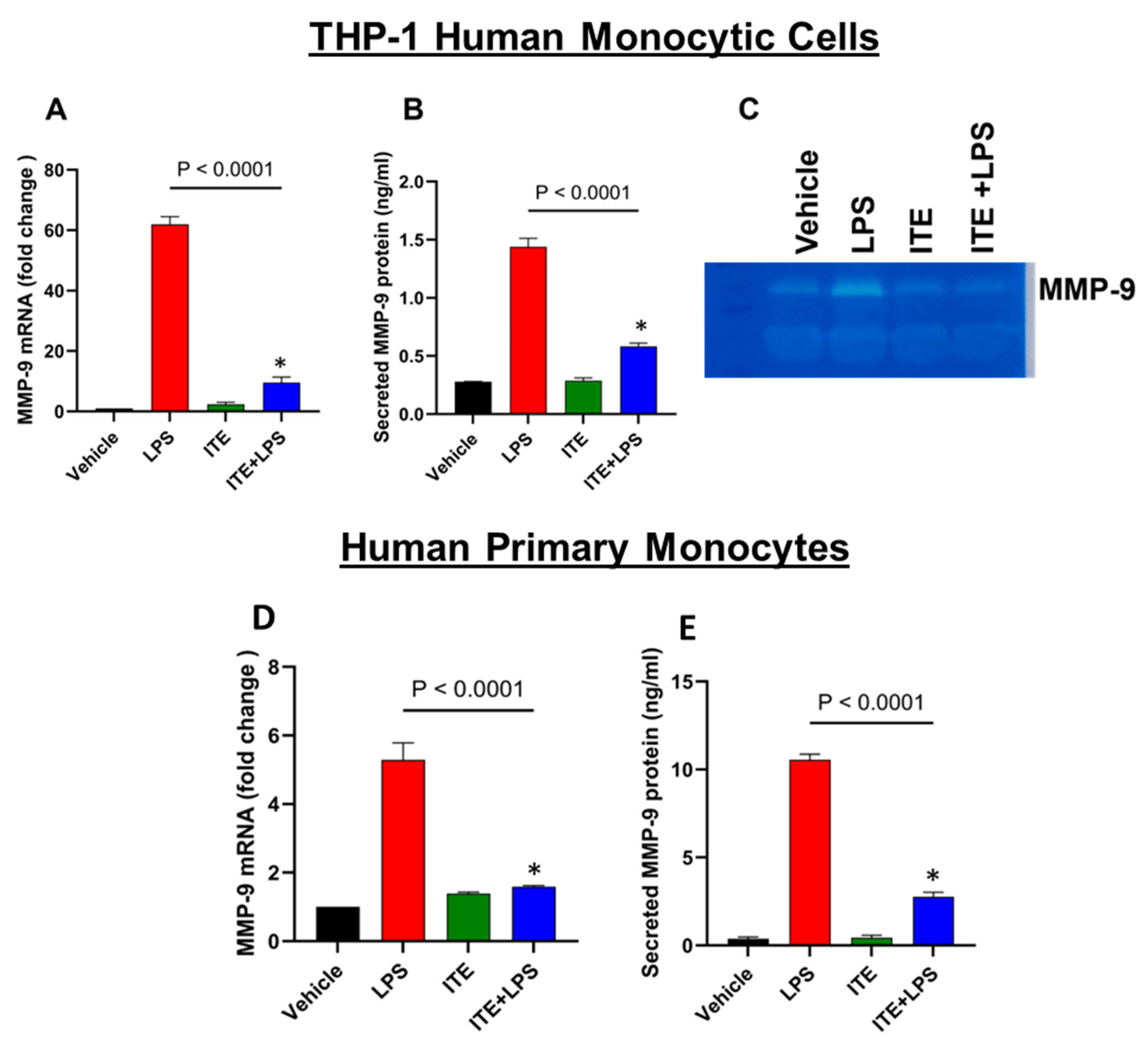
2.1. ITE Inhibits LPS-Induced MMP-9 Expression

It has been reported that ITE inhibits inflammatory responses in mice [21]. To examine whether ITE suppresses MMP-9 expression in monocytic cells, we pretreated THP-1 cells with ITE (10 µM) for 1 h before exposure to LPS (10 ng/mL) for 24 h. Our results showed that ITE significantly decreases MMP-9 expression at both the mRNA (Figure 1A) and protein (Figure 1B) levels in THP-1 cells. Furthermore, to determine whether the MMP-9 protein released in response to LPS is biologically active, we assessed gelatinolytic activity in the culture supernatants. Gelatin zymography was performed on the conditioned media obtained from the cells treated with LPS or ITE+LPS. Zymography data showed a reduced level of MMP-9 activity in the conditioned media obtained from ITE+LPS treated cells when compared to LPS (Figure 1C). Next, we determined the effect of ITE on LPS-induced MMP-9 in primary monocytes. A similar ITE mediated suppression of MMP-9 gene and protein expression was observed in primary human monocytes (Figure 1D,E).

2.2. ITE Reduces LPS-Induced NF-κB/AP-1 Activity in Monocytic Cells

LPS promotes the expression of MMP-9 via the activation of AP-1 and NF-κB transcription factors [9]. We investigated whether these transcriptions play a role in ITE’s inhibition of LPS-stimulated MMP-9 in THP-1 monocytic cells. The MMP-9 promoter has binding sites for NF-κB and AP-1 [22]. Therefore, we next investigated whether ITE suppresses LPS-induced NF-κB/AP1 activity in THP-1 reporter monocytic cells. To this effect, we used THP-1 X-Blue cells expressing the NF-κB/AP-1 response element-driven SEAP reporter activity. As expected, the response of ITE-pretreated monocytic reporter cells to LPS for the activation of NF-κB/AP-1 was significantly lower compared to that of controls (Figure 2A). This reduction in NF-κB/AP-1 activity was associated with decreased MMP-9 mRNA and protein expression in these cells (Figure 2B,C).

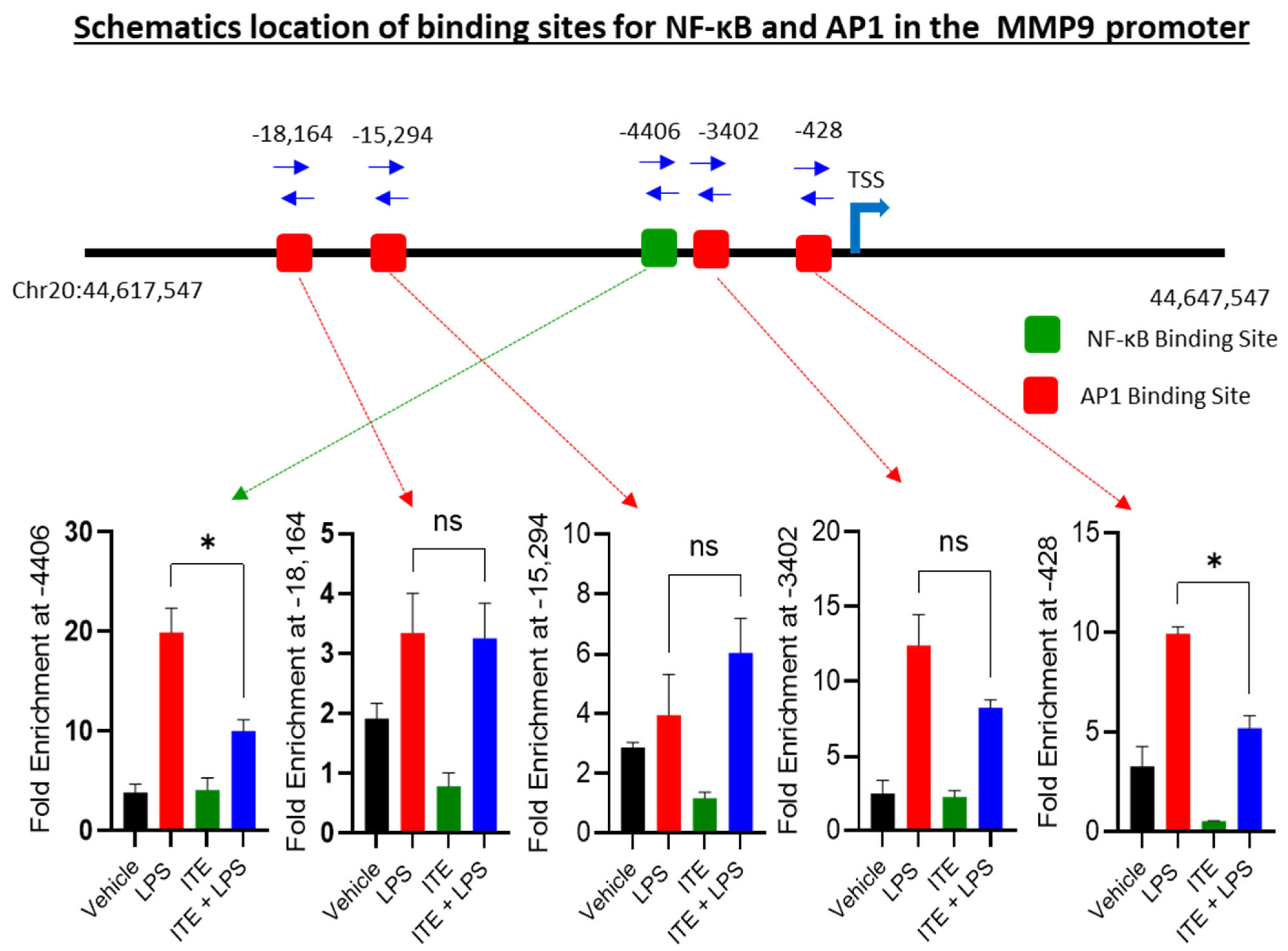
2.3. ITE Reduces the NF-κB/AP-1 Binding to the MMP-9 Promoter

Because the MMP-9 promoter is regulated by one NF-κB- and multiple AP-1-responsive sequences (Figure 3) [22], we sought to assess whether these transcription factors are involved in the inhibitory effect of ITE on LPS-induced MMP-9 expression. To verify the effect of ITE on the LPS-induced direct binding of NF-κB to the MMP-9 promoter, we performed ChIP followed by real-time quantitative PCR (qPCR). Quantitative ChIP analysis revealed that the LPS-induced recruitment of NF-κB to its known binding site was significantly inhibited in cells pretreated with ITE, specifically at the −4406 position (Figure 3).
We next assessed whether AP-1 binding to the MMP-9 promoter is affected by ITE using ChIP-PCR of c-Jun as a proxy. There are several known AP-1 binding sites on the MMP-9 promoter. Quantitative ChIP analysis revealed a significant recruitment of AP-1 to its known binding sites by LPS, specifically at the −18,164, −15,294, −3402, and −428 positions relative to the transcription start site of the MMP-9 promoter (Figure 3), although the −428 site showed no degree of inhibition by ITE. Collectively, our data suggest that ITE decreases LPS-induced NF-κB/AP-1 binding to the MMP-9 promoter and inhibits its expression.

3. Discussion

The overexpression of matrix metalloproteinase-9 (MMP-9) has been implicated in the pathogenesis of various inflammatory disorders, including arthritis, cancer metastasis, and cardiovascular diseases. MMP-9 is a proteolytic enzyme whose expression is strongly induced by lipopolysaccharide (LPS), contributing to the inflammatory response and the progression of associated diseases. Given that MMP-9 dysregulation plays a key role in cancer invasion, metastasis, and other inflammatory conditions, its inhibition represents a promising therapeutic strategy.
In the present study, we demonstrate that the aryl hydrocarbon receptor (AHR) ligand 2-(1′H-indole-3′-carbonyl)-thiazole-4-carboxylic acid methyl ester (ITE) effectively inhibits LPS-induced MMP-9 expression in monocytic cells. This observation supports the notion that AHR activation can exert anti-inflammatory effects. In agreement with our results, Domínguez-Acosta et al. reported that the activation of AHR by 2,3,7,8-tetrachlorodibenzo-p-dioxin (TCDD) led to a reduced production of pro-inflammatory cytokines such as TNF-α, IL-6, and IL-12 in murine macrophages [23]. Similarly, another study demonstrated that the exposure of human monocyte-derived dendritic cells to ITE impaired their capacity to induce pro-inflammatory Th17 cell differentiation. ITE treatment also significantly suppressed IL-1β, IL-6, and IL-23 production while promoting IL-10 secretion by dendritic cells [24]. Collectively, these findings underscore the anti-inflammatory potential of ITE. To elucidate the molecular mechanisms underlying ITE-mediated suppression of LPS-induced MMP-9 expression, we investigated its impact on the NF-κB and MAPK signaling pathways, which are well-established regulators of MMP-9 gene expression. The promoter region of the MMP-9 gene contains binding sites for both nuclear factor-κB (NF-κB) and activator protein-1 (AP-1), and activation of both transcription factors is essential for MMP-9 transcription [3,25].
Our data provide two lines of evidence supporting the inhibition of NF-κB signaling by ITE. First, LPS-induced NF-κB activation was significantly attenuated in reporter cells pretreated with ITE. Second, nuclear extracts showed diminished NF-κB DNA-binding activity upon ITE treatment, suggesting that ITE suppresses MMP-9 transcription by interfering with NF-κB activation. These findings align with previous studies indicating that AHR activation by ITE suppresses NF-κB-mediated inflammation in microglial cells [26]. Furthermore, studies using AHR-deficient human alveolar basal epithelial cells showed heightened inflammatory responses to airborne pollutants, further supporting the role of AHR as a negative regulator of inflammation through NF-κB pathway modulation [27].
We also explored the involvement of the AP-1 transcription factor in the ITE-mediated downregulation of MMP-9. Again, two key findings support this mechanism. First, LPS-induced AP-1 activation was significantly reduced in reporter assays following ITE treatment. Second, ITE attenuated the DNA-binding activity of AP-1 to the MMP-9 promoter. These results suggest that ITE interferes with AP-1 transcriptional regulation of MMP-9. AP-1 is a downstream effector of MAPK signaling pathways, including ERK, JNK, and p38, and it plays a critical role in the regulation of genes involved in inflammation and monocyte activation [28]. We found that LPS-induced AP-1 activity was inhibited in cells pretreated with ITE. AP-1 is a critical factor that regulates the expression of several inflammation-related genes, including MMP-9 [3,25]. Our findings clearly demonstrate the role of ITE in the DNA-binding and transcriptional activities of AP-1 in monocytic cells.

3.1. Study Limitations

Although the results show that ITE can reduce LPS-induced MMP-9 expression in monocytic cells, there are some limitations to consider. The experiments were only performed in vitro using THP-1 cells and primary human monocytes. While these cell models are useful, they do not fully represent the complex immune responses and cell interactions that occur in the human body. Therefore, more studies are needed to see whether these findings apply to real-life conditions, such as in animal models or in patients with obesity-related inflammation.

3.2. Future Directions

Given the demonstrated ability of ITE to suppress LPS-induced MMP-9 expression through the inhibition of NF-κB and AP-1 signaling, future studies will explore its therapeutic potential in in vivo models of obesity-associated inflammation. Chronic low-grade inflammation is a key driver of metabolic dysfunction in obesity, and targeting MMP-9 may help reduce tissue remodeling and systemic inflammation. Also, investigating the effects of ITE on adipose tissue inflammation, insulin sensitivity, and metabolic markers will be essential to assess its broader applicability. Additionally, exploring ITE’s role in other MMP-9–driven inflammatory conditions, such as cardiovascular disease and autoimmune disorders, may further validate its potential as a novel anti-inflammatory therapeutic agent.

4. Materials and Methods

4.1. Cell Culture

THP-1 cells were purchased from the American Type Culture Collection (ATCC). THP-1 cells were cultured in RPMI-1640 medium supplemented with fetal bovine serum (FBS) (10%), L-glutamine (2 mM), sodium pyruvate (1 mM), HEPES (10 mM), Normocin (100 µg/mL), and 50U penicillin/50 µg streptomycin (Gibco, USA). For in vitro experiments, the THP-1 cells were seeded at 1 × 10⁶ cells/well in 12-well plates (Gibco, Life Technologies, Grand Island, NY, USA). The cells were pretreated with ITE (10 µM) (InvivoGen, San Diego, CA, USA) for 60 min. Then, they were stimulated with LPS (10 ng/mL) (R&D Systems, Minneapolis, MN, USA) or 0.1% bovine serum albumin (BSA) as a vehicle overnight. All plates were incubated at 37 °C in 5% CO2. Then, the cells were collected for RNA extraction, and the supernatant media were used for the quantification of MMP-9 protein secretion by ELISA. NF-κB/AP-1 reporter cells (THP-1 XBlue cells) were used to assess the biological activity. The cells were cultured in a complete RPMI medium with the addition of zeocin (200 µg/mL) as a selective factor (InvivoGen, USA). Primary monocytes were isolated from PBMCs (RA 2010-003) as described earlier [29].

4.2. Measurement of NF-κB/AP-1 Activity

THP-1 XBlue cells were transfected with a reporter construct expressing SEAP under the control of a NF-κB and AP1 transcription factor-inducible promoter. As a result of cell stimulation, the activation of NF-κB and AP-1 caused SEAP to be secreted into the supernatant. The experiment was performed on THP-1 XBlue cells which were pretreated with ITE (10 µM) followed by adding LPS (10 ng/mL) for 24 h at 37 °C. SEAP levels in the supernatant media were detected after 4 h of incubation with Quanti-Blue medium (InvivoGen, San Diego, CA, USA) and analyzed by an ELISA reader at a wavelength of 650 nm; the background of vehicle (control) correction was applied to the results.

4.3. Real-Time Quantitative PCR

The RNeasy Mini Kit (Qiagen, Germantown, MD, USA) was used for RNA extraction. cDNA was synthesized using a high-capacity cDNA reverse transcription kit (Applied Biosystems, Thermo Fisher Scientific, Carlsbad, CA, USA) using 1 μg of total RNA as a template [30]. For real-time PCR, we used 50 ng of cDNA template to be amplified using inventoried TaqMan Gene Expression Assay products (MMP-9: Hs00234579_m1; GAPDH: Hs03929097_g1) containing two gene-specific primers and one TaqMan MGB probe (6-FAM dye-labeled) with a TaqMan® Gene Expression Master Mix (Applied Biosystems, Thermo Fisher Scientific, Carlsbad, CA, USA) and a 7500 Fast Real-Time PCR System (Applied Biosystems, Thermo Fisher Scientific, Carlsbad, CA, USA). For the analysis, the mRNA levels were normalized against GAPDH mRNA, and MMP-9 mRNA expression relative to the control was calculated using the 2−ΔΔCt-method [31]. Relative mRNA expression was shown as fold expression over an average of control gene expression [32]. The MMP-9 gene expression level in the control sample was taken as 1; the data were presented as mean ± SEM values, and p < 0.0001 was considered significant.

4.4. Quantification of MMP-9 Secretion

The determination of MMP-9 in the supernatants of control and pretreated THP-1 cells which were stimulated with LPS was quantified using ELISA as described previously [32] (R&D systems, USA).

4.5. Gelatin Zymography

For the measurement of the activated form of MMP-9, we used gelatin zymography of THP-1 cells, as described [32]. The cells were pretreated with ITE and stimulated by LPS (10 ng/mL). After incubation for 24 h, the culture media were collected and mixed with the zymogram sample buffer (BioRad) (62.5 mM Tris-HCl, pH 6.8, 25% glycerol, 4% SDS, and 0.01% bromophenol blue). The mixture was loaded onto gel with gelatin (10% Ready Gel® Zymogram Gel, Biorad, Hercules, CA, USA) for electrophoresis. The gel was incubated with renaturing buffer (2.5% Triton X-100, BioRad) for 1 h at room temperature and incubated with zymogram-developing buffer for 24 h at 37 °C (50 mM Tris-HCl, pH 7.5, 200 mM NaCl and 5 mM CaCl2, BioRad). The gels were stained with a staining solution (0.5% Coomassie Brilliant Blue R-250, 40% Methanol, 10% Acetic Acid) for 2 h and then destained with a destaining solution (40% Methanol, 10% Acetic Acid) until the bands showed. Clear bands against the stained gel’s dark background served as a marker of positive proteolytic activity.

4.6. Chromatin Immunoprecipitation (ChIP) Assay

The Simple ChIP Enzymatic Chromatin Immunoprecipitation Kit, purchased from Cell Signaling Technology Inc. (Danvers, MA, USA), was used as described by the manufacturer. THP1 cells were pretreated with ITE for 1 h, and after that, the cells were stimulated with LPS. Chromatin fragments obtained from human THP-1 monocytic cells were fixed with formaldehyde and lysed. The Micrococcal Nuclease was used to partially digest the chromatin followed by sonication to yield fragments ranging from 200 to 800 bp using the Covaris system. The chromatin fragments were digested and then subjected to immunoprecipitation using primary antibodies specific to the p65 subunit of NF-κB and AP-1 (Cell Signaling Technology) overnight at 4 °C and incubated with Protein G magnetic beads for 2 h at 4 °C. The chromatin was eluted from the Antibody/Protein G magnetic bead complex by incubation at 65 °C for 30 min and by magnetic separation. Proteinase K was used to reverse-crosslink chromatin for 2 h at 65 °C, and DNA was purified from the ChIP fractions using a spin column. In order to detect DNA enrichment, RT-qPCR was carried out using a Syber green mix and Epitect qPCR primers specific to the transcription factor-binding sites within the promoter region of the MMP-9 gene (Table 1).

4.7. Statistical Analysis

For statistical analysis, GraphPad Prism software 10.3.1 was used. All data were expressed as mean ± SEM values. Multiple groups were compared by one-way ANOVA followed by post hoc Tukey’s comparison test. Two groups were compared by unpaired Student’s t-test. For all analyses, a p-value < 0.05 was considered significant.

5. Conclusions

In summary, our findings provide compelling evidence that ITE suppresses LPS-induced MMP-9 expression in monocytic cells through the inhibition of both NF-κB and AP-1 transcriptional activities. This dual regulatory mechanism positions ITE as a potential therapeutic agent for controlling MMP-9-associated inflammation.

Author Contributions

Conceptualization, R.A.; methodology, F.B., N.A. and S.K.; validation, R.A.; formal analysis, F.B., N.A. and S.K.; resources, R.A. and F.A.-M.; data curation, F.B., N.A. and S.K.; writing—original draft preparation, F.B., N.A. and S.K.; writing—review and editing, F.A.-M. and R.A.; supervision, R.A.; project administration, R.A.; funding acquisition, R.A. All authors have read and agreed to the published version of the manuscript.

Funding

This research was funded by the Kuwait Foundation for the Advancement of Sciences, grant number RA AH 2023-023.

Institutional Review Board Statement

Isolated PBMCs used from previous project (RA 2010-003).

Informed Consent Statement

Not applicable.

Data Availability Statement

The data presented in this study are available on request from the corresponding author.

Conflicts of Interest

The authors declare no conflicts of interest.

References

  1. Creely, S.J.; McTernan, P.G.; Kusminski, C.M.; Fisher, f.M.; Da Silva, N.F.; Khanolkar, M.; Evans, M.; Harte, A.L.; Kumar, S. Lipopolysaccharide activates an innate immune system response in human adipose tissue in obesity and type 2 diabetes. Am. J. Physiol. Endocrinol. Metab. 2007, 292, E740–E747. [Google Scholar] [CrossRef] [PubMed]
  2. Sylus, A.M.; Nandeesha, H.; Chitra, T. Matrix metalloproteinase-9 increases and Interleukin-10 reduces with increase in body mass index in polycystic ovary syndrome: A cross-sectional study. Int. J. Reprod. Biomed. 2020, 18, 605–610. [Google Scholar] [CrossRef]
  3. Sindhu, S.; Al-Roub, A.; Koshy, M.; Thomas, R.; Ahmad, R. Palmitate-Induced MMP-9 Expression in the Human Monocytic Cells is Mediated through the TLR4-MyD88 Dependent Mechanism. Cell. Physiol. Biochem. 2016, 39, 889–900. [Google Scholar] [CrossRef]
  4. Boumiza, S.; Chahed, K.; Tabka, Z.; Jacob, M.P.; Norel, X.; Ozen, G. MMPs and TIMPs levels are correlated with anthropometric parameters, blood pressure, and endothelial function in obesity. Sci. Rep. 2021, 11, 20052. [Google Scholar] [CrossRef]
  5. Yabluchanskiy, A.; Ma, Y.; Iyer, R.P.; Hall, M.E.; Lindsey, M.L. Matrix metalloproteinase-9: Many shades of function in cardiovascular disease. Physiology 2013, 28, 391–403. [Google Scholar] [CrossRef]
  6. Xue, M.; McKelvey, K.; Shen, K.; Minhas, N.; March, L.; Park, S.Y.; Jackson, C.J. Endogenous MMP-9 and not MMP-2 promotes rheumatoid synovial fibroblast survival, inflammation and cartilage degradation. Rheumatology 2014, 53, 2270–2279. [Google Scholar] [CrossRef] [PubMed]
  7. Ganaie, I.A.; Malik, M.Z.; Naqvi, S.H.; Jain, S.K.; Wajid, S. Differential levels of Alpha-1-inhibitor III, Immunoglobulin heavy chain variable region, and Hypertrophied skeletal muscle protein GTF3 in rat mammary tumorigenesis. Biochimie 2020, 174, 57–68. [Google Scholar] [CrossRef]
  8. Sahu, D.; Bishwal, S.C.; Malik, M.Z.; Sahu, S.; Kaushik, S.R.; Sharma, S.; Saini, E.; Arya, R.; Rastogi, A.; Sharma, S.; et al. Troxerutin-Mediated Complement Pathway Inhibition is a Disease-Modifying Treatment for Inflammatory Arthritis. Front. Cell Dev. Biol. 2022, 10, 845457. [Google Scholar] [CrossRef] [PubMed]
  9. Tian, X.; Xie, G.; Ding, F.; Zhou, X. LPS-induced MMP-9 expression is mediated through the MAPKs-AP-1 dependent mechanism in BEAS-2B and U937 cells. Exp. Lung Res. 2018, 44, 217–225. [Google Scholar] [CrossRef]
  10. Cani, P.D.; Amar, J.; Iglesias, M.A.; Poggi, M.; Knauf, C.; Bastelica, D.; Neyrinck, A.M.; Fava, F.; Tuohy, K.M.; Chabo, C.; et al. Metabolic endotoxemia initiates obesity and insulin resistance. Diabetes 2007, 56, 1761–1772. [Google Scholar] [CrossRef]
  11. Monte, S.V.; Caruana, J.A.; Ghanim, H.; Sia, C.L.; Korzeniewski, K.; Schentag, J.J.; Dandona, P. Reduction in endotoxemia, oxidative and inflammatory stress, and insulin resistance after Roux-en-Y gastric bypass surgery in patients with morbid obesity and type 2 diabetes mellitus. Surgery 2012, 151, 587–593. [Google Scholar] [CrossRef] [PubMed]
  12. Ghoshal, S.; Witta, J.; Zhong, J.; de Villiers, W.; Eckhardt, E. Chylomicrons promote intestinal absorption of lipopolysaccharides. J. Lipid Res. 2009, 50, 90–97. [Google Scholar] [CrossRef] [PubMed]
  13. Cani, P.D.; Possemiers, S.; Van de Wiele, T.; Guiot, Y.; Everard, A.; Rottier, O.; Geurts, L.; Naslain, D.; Neyrinck, A.; Lambert, D.M.; et al. Changes in gut microbiota control inflammation in obese mice through a mechanism involving GLP-2-driven improvement of gut permeability. Gut 2009, 58, 1091–1103. [Google Scholar] [CrossRef] [PubMed]
  14. Schäffler, A.; Schölmerich, J. Innate immunity and adipose tissue biology. Trends Immunol. 2010, 31, 228–235. [Google Scholar] [CrossRef]
  15. Sondermann, N.C.; Faßbender, S.; Hartung, F.; Hätälä, A.M.; Rolfes, K.M.; Vogel, C.F.; Haarmann-Stemmann, T. Functions of the aryl hydrocarbon receptor (AHR) beyond the canonical AHR/ARNT signaling pathway. Biochem. Pharmacol. 2022, 208, 115371. [Google Scholar] [CrossRef]
  16. Gutiérrez-Vázquez, C.; Quintana, F.J. Regulation of the immune response by the aryl hydrocarbon receptor. Immunity 2018, 48, 19–33. [Google Scholar] [CrossRef]
  17. Riaz, F.; Pan, F.; Wei, P. Aryl hydrocarbon receptor: The master regulator of immune responses in allergic diseases. Front. Immunol. 2022, 13, 1057555. [Google Scholar] [CrossRef]
  18. Rappaport, N.; Watanabe, K. Biological BMI uncovers hidden health risks and is more responsive to lifestyle shifts. Nat. Med. 2023, 29, 801–802. [Google Scholar]
  19. Bahman, F.; Choudhry, K.; Al-Rashed, F.; Al-Mulla, F.; Sindhu, S.; Ahmad, R. Aryl hydrocarbon receptor: Current perspectives on key signaling partners and immunoregulatory role in inflammatory diseases. Front. Immunol. 2024, 15, 1421346. [Google Scholar] [CrossRef]
  20. Manuel, R.S.; Rundquist, A.; Ambrogi, M.; Scharpf, B.R.; Peterson, N.T.; Sandhu, J.K.; Chandrashekar, S.; Ridlon, M.; Crawford, L.K.; Keil-Stietz, K.P.; et al. The aryl hydrocarbon receptor agonist ITE reduces inflammation and urinary dysfunction in a mouse model of autoimmune prostatitis. Am. J. Clin. Exp. Urol. 2024, 12, 149–161. [Google Scholar] [CrossRef]
  21. Abron, J.D.; Singh, N.P.; Mishra, M.K.; Price, R.L.; Nagarkatti, M.; Nagarkatti, P.S.; Singh, U.P. An endogenous aryl hydrocarbon receptor ligand, ITE, induces regulatory T cells and ameliorates experimental colitis. Am. J. Physiol. Gastrointest. Liver Physiol. 2018, 315, G220–G230. [Google Scholar] [CrossRef]
  22. Wu, H.T.; Sie, S.S.; Kuan, T.C.; Lin, C.S. Identifying the regulative role of NF-κB binding sites within promoter region of human matrix metalloproteinase 9 (mmp-9) by TNF-α induction. Appl. Biochem. Biotechnol. 2013, 169, 438–449. [Google Scholar] [CrossRef]
  23. Domínguez-Acosta, O.; Vega, L.; Estrada-Muñiz, E.; Rodríguez, M.S.; Gonzalez, F.J.; Elizondo, G. Activation of aryl hydrocarbon receptor regulates the LPS/IFNγ-induced inflammatory response by inducing ubiquitin-proteosomal and lysosomal degradation of RelA/p65. Biochem. Pharmacol. 2018, 155, 141–149. [Google Scholar] [CrossRef]
  24. Wang, C.; Ye, Z.; Kijlstra, A.; Zhou, Y.; Yang, P. Activation of the aryl hydrocarbon receptor affects activation and function of human monocyte-derived dendritic cells. Clin. Exp. Immunol. 2014, 177, 521–530. [Google Scholar] [CrossRef]
  25. Al-Rashed, F.; Kochumon, S.; Usmani, S.; Sindhu, S.; Ahmad, R. Pam3CSK4 Induces MMP-9 Expression in Human Monocytic THP-1 Cells. Cell. Physiol. Biochem. 2017, 41, 1993–2003. [Google Scholar] [CrossRef]
  26. Yang, Y.; Wang, N.; Xu, L.; Liu, Y.; Huang, L.; Gu, M.; Wu, Y.; Guo, W.; Sun, H. Aryl hydrocarbon receptor dependent anti-inflammation and neuroprotective effects of tryptophan metabolites on retinal ischemia/reperfusion injury. Cell Death Dis. 2023, 14, 92. [Google Scholar] [CrossRef]
  27. Vázquez-Gómez, G.; Karasová, M.; Tylichová, Z.; Kabátková, M.; Hampl, A.; Matthews, J.; Neča, J.; Ciganek, M.; Machala, M.; Vondráček, J. Aryl Hydrocarbon Receptor (AhR) Limits the Inflammatory Responses in Human Lung Adenocarcinoma A549 Cells via Interference with NF-κB Signaling. Cells 2022, 11, 707. [Google Scholar] [CrossRef]
  28. Whitmarsh, A.J.; Davis, R.J. Transcription factor AP-1 regulation by mitogen-activated protein kinase signal transduction pathways. J. Mol. Med. 1996, 74, 589–607. [Google Scholar] [CrossRef]
  29. Kochumon, S.; Jacob, T.; Koshy, M.; Al-Rashed, F.; Sindhu, S.; Al-Ozairi, E.; Al-Mulla, F.; Rosen, E.D.; Ahmad, R. Palmitate Potentiates Lipopolysaccharide-Induced IL-6 Production via Coordinated Acetylation of H3K9/H3K18, p300, and RNA Polymerase II. J. Immunol. 2022, 209, 731–741. [Google Scholar] [CrossRef]
  30. Kochumon, S.; Al-Sayyar, A.; Jacob, T.; Bahman, F.; Akhter, N.; Wilson, A.; Sindhu, S.; Hannun, Y.A.; Ahmad, R.; Al-Mulla, F. TGF-β and TNF-α interaction promotes the expression of MMP-9 through H3K36 dimethylation: Implications in breast cancer metastasis. Front. Immunol. 2024, 15, 1430187. [Google Scholar] [CrossRef]
  31. Al Madhoun, A.; Haddad, D.; Kochumon, S.; Thomas, R.; Miranda, L.; George, P.; Abu-Khalaf, N.; Al-Mulla, F.; Ahmad, R. TNF-α/NF-κB mediated upregulation of Dectin-1 in hyperglycemic obesity: Implications for metabolic inflammation and diabetes. J. Transl. Med. 2025, 23, 462. [Google Scholar] [CrossRef] [PubMed]
  32. Al-Roub, A.; Akhter, N.; Al-Rashed, F.; Wilson, A.; Alzaid, F.; Al-Mulla, F.; Sindhu, S.; Ahmad, R. TNFα induces matrix metalloproteinase-9 expression in monocytic cells through ACSL1/JNK/ERK/NF-kB signaling pathways. Sci. Rep. 2023, 13, 14351. [Google Scholar] [CrossRef] [PubMed]
Figure 1. Effect of ITE on the LPS-mediated induction of MMP-9 expression. THP-1 cells were incubated with either ITE (10 µM) or a vehicle control for 60 min prior to stimulation with LPS (10 ng/mL) for 24 h. (A) Total RNA was extracted to evaluate MMP-9 gene expression via qRT-PCR. The results were normalized and presented as fold changes relative to the mean expression level in the control group, which was set to 1. (B) MMP-9 protein levels secreted into the culture medium were quantified using ELISA. (C) Gelatin zymography was conducted to assess the activated form of MMP-9 enzyme. (D,E) In a separate experiment, primary monocytes were treated with either ITE alone or a combination of ITE and LPS, followed by an analysis of MMP-9 mRNA and protein expression levels. Primary monocytes were isolated from PBMCs and THP-1 monocytic cell lines (immortalized human monocytic leukemia cell line). Data are expressed as mean ± SEM (biological replicates n = 3) from three independent experiments, and statistical comparisons were performed using one-way ANOVA followed by Tukey’s post hoc test. * p < 0.0001.
Ijms 26 05663 g001
Figure 2. ITE suppresses LPS-activated MAPK/NF-κB signaling pathways. (A) NF-κB/Ap-1 reporter cells were pretreated with ITE (10 µM) for 60 min. Then, cells were exposed to LPS (10 ng/mL). SEAP reporter activity (degree of AP-1/NF-κB activation) was determined in the media supernatants as described in the Section 4. (B,C) mRNA and protein expression of MMP-9 were determined by qPCR and ELISA, respectively. Data are expressed as mean ± SEM (biological replicates n = 3) from three independent experiments. *. Statistical analysis was performed using one-way ANOVA (Tukey’s multiple comparisons test). * p < 0.0001.
Ijms 26 05663 g002
Figure 3. Chromatin analysis of MMP-9 promoter. Schematic location of one transcription factor binding site for NF-κB (−4406) and four binding sites for AP-1 ((−18,164), (−15,294), (−3402), and (−428)) in the MMP-9 promoter. Cells were treated with vehicle, LPS, ITE, or ITE+LPS for 24 h. Chromatin was immunoprecipitated with anti NF-κB or c-Jun antibodies. Levels of NF-κB and AP-1 binding were measured using the primer for the NF-κB or AP-1 DNA binding sites in the MMP-9 promoter as shown. Data are expressed as mean ± SEM values from three replicates of each experiment, and similar data were obtained from three independent experiments; * p < 0.05 was considered statistically significant.
Ijms 26 05663 g003
Table 1. CHIP-qPCR primers used.
ChIP-qPCR Assay Cat# TF-BS Relative to MMP9 TSSTF-BS Position (NC_)
GPH1008476(-)19AAP-1 (−18,164)44619180
GPH1008476(-)16AAP-1 (−15,294)44622312
GPH1008476(-)04AAP-1 (−3402)44633814
GPH1008476(-)01AAP-1 (−428)44637547
GPH1008476(-)05ANF-kappaB (−4406)44633494
Disclaimer/Publisher’s Note: The statements, opinions and data contained in all publications are solely those of the individual author(s) and contributor(s) and not of MDPI and/or the editor(s). MDPI and/or the editor(s) disclaim responsibility for any injury to people or property resulting from any ideas, methods, instructions or products referred to in the content.

