Article

Paradoxical SERCA2a Dysregulation Contributes to Atrial Fibrillation in a Model of Diet-Induced Obesity

by
Daniela Ponce-Balbuena
1,2,†,‡,
Daniel J. Tyrrell
1,†,§,
Carlos Cruz-Cortés
1,2,†,
Guadalupe Guerrero-Serna
1,†,
Andre Monteiro Da Rocha
1,2,
Todd J. Herron
1,2,‖,
Jianrui Song
1,
Danyal S. Raza
1,2,
Justus Anumonwo
1,2,
Daniel R. Goldstein
1,3,4,¶ and
L. Michel Espinoza-Fonseca
1,2,*
1
Department of Internal Medicine, University of Michigan, Ann Arbor, MI 48109, USA
2
Center for Arrhythmia Research, University of Michigan, Ann Arbor, MI 48109, USA
3
Graduate Program in Immunology, University of Michigan, Ann Arbor, MI 48109, USA
4
Department of Microbiology and Immunology, University of Michigan, Ann Arbor, MI 48109, USA
*
Author to whom correspondence should be addressed.
These authors contributed equally to the work.
Present address: Department of Medicine, University of Wisconsin, Madison, WI 53705, USA.
§
Present address: Department of Molecular & Cellular Pathology, The University of Alabama at Birmingham, Birmingham, AL 35205, USA.
Present address: Greenstone Biosciences, Palo Alto, CA 94304, USA.
We dedicate this paper to the memory of our colleague and friend, Daniel R. Goldstein, who passed away on 21 May 2024, at the age of 56, following a battle with a rare cancer. Daniel held the Eliza Maria Mosher Endowed Chair of Internal Medicine and was a Research Professor at the Institute of Gerontology. He led the Michigan Biology of Cardiovascular Aging program and served as Editor-in-Chief of the Journal of Heart and Lung Transplantation. A renowned physician–scientist and expert in cardiovascular aging, Daniel authored more than 150 peer-reviewed publications. His research, continuously supported by the National Institutes of Health for over two decades, profoundly shaped our understanding of the link between inflammation, aging, and cardiovascular disease. Daniel was a dedicated educator and mentor, recognized for inspiring physicians and scientists at all stages of their careers. He was a tireless interdisciplinary innovator who combined analytical rigor with deep intellectual generosity. Beyond his scientific contributions, Daniel was a passionate advocate for gender equality in science and a strong voice for underrepresented communities, including trainees and early-career professionals in medicine and research. We honor his legacy by reaffirming our commitment to advancing research and education, ensuring that his enduring passion for scientific discovery continues to inform and inspire future generations in the field of cardiovascular medicine.
Int. J. Mol. Sci. 2025, 26(12), 5603; https://doi.org/10.3390/ijms26125603
Submission received: 15 April 2025 / Revised: 29 May 2025 / Accepted: 10 June 2025 / Published: 11 June 2025
(This article belongs to the Special Issue Calcium Homeostasis of Cells in Health and Disease: 2nd Edition)

Abstract

Obesity is a major risk factor for atrial fibrillation (AF), the most common serious cardiac arrhythmia, but the molecular mechanisms underlying obesity-induced AF remain unclear. In this study, we subjected mice to a chronic high-fat diet and acute sympathetic activation to investigate how obesity promotes AF. Surface electrocardiography revealed that obesity and sympathetic activation synergize during intracardiac tachypacing to induce AF. At the cellular level, this combination facilitated delayed afterdepolarizations in atrial myocytes, implicating altered Ca2+ dynamics. Interestingly, obesity did not affect the expression of key atrial Ca2+-handling proteins, including the cardiac sarcoplasmic reticulum Ca2+-ATPase (SERCA2a). However, obesity increases the proportion of inhibitory phospholamban (PLN) monomers and decreases PLN phosphorylation, suggesting reduced SERCA2a activity. Paradoxically, Ca2+ reuptake in atrial myocytes from obese mice was similar to that achieved by potent small-molecule SERCA2a activators. We found that adrenergic stimulation increased Ca2+ transient amplitude without altering Ca2+ reuptake in myocytes from obese mice. Transcriptomic analysis revealed that a high-fat diet upregulated neuronatin, a protein involved in obesity that enhances SERCA2-mediated Ca2+ reuptake in neurons. We propose that obesity enables SERCA2a activation independently of PLN regulation, while adrenergic stimulation triggers arrhythmogenic Ca2+-induced Ca2+ release, promoting AF. In conclusion, this study demonstrates that obesity causes a paradoxical dysregulation of SERCA2a in atrial myocytes, with increased activity despite higher levels of inhibitory PLN monomers and reduced PLN phosphorylation. These findings offer new insights into the cellular mechanisms of obesity-induced AF and suggest potential therapeutic targets.

1. Introduction

In the last several decades, the rate of obesity has progressively increased and is now one of the leading causes of morbidity and mortality in the world. According to the World Health Organization, one in eight people in the world are currently living with obesity; 2.2 billion adults were overweight in 2022 and 890 million were considered obese [1]. It is estimated that by 2030, about half of the world’s population will suffer from obesity, and about 11% of individuals will be morbidly obese [2]. Obesity contributes to cardiovascular risk factors, including dyslipidemia, diabetes, hypertension, and sleep disorders. Obesity also leads to the development of cardiovascular disease and cardiovascular disease mortality independently of other cardiovascular risk factors [3].
Diet-induced obesity is a major risk factor for atrial fibrillation (AF), the most common serious cardiac arrhythmia in the developed world [4,5,6,7]. AF represents the leading cause of hospitalization, affecting nearly five million patients across the United States [8]. There is evidence indicating that obese individuals have −50% increased risk compared to non-obese patients [9]. Other studies have shown that obese young men have more than a 2-fold risk of AF compared with young men of normal weight [9]. Clinical studies have associated increased body mass index with AF, where every unit increase in body mass index (BMI) increases the development of AF by up to 8% [5,10]. While obesity occurs with other risk factors for AF, including hypertension, atherosclerosis, diabetes, and sleep apnea, a recent study demonstrated that obesity alone promotes AF [11]. Moreover, obesity is associated with a higher recurrence of AF [12]. Inflammation and remodeling of ion transport have been shown to contribute to obesity-induced AF [13,14], yet the pathophysiological and molecular mechanisms associated with AF are complex, and the molecular basis for this association remains unclear. Therefore, there is an urgent need to investigate the mechanisms by which obesity increases the risk for AF, ultimately revealing new pathways that can lead to therapeutic interventions for this condition.
In this study, we developed a two-hit mouse model (chronic high-fat diet and acute sympathetic activation) to study the molecular mechanisms by which diet-induced obesity promotes AF. We found that diet-induced obesity and sympathetic activation synergize during intracardiac tachypacing to induce AF. Diet-induced obesity and acute adrenergic stimulation facilitate the formation of delayed afterdepolarizations in atrial myocytes, suggesting that altered Ca2+ dynamics are the underlying mechanism promoting AF in obese mice. Obesity does not affect the expression of major Ca2+-handling proteins, including the sarcoplasmic reticulum Ca2+-ATPase (SERCA2a), a major component of beat-to-beat Ca2+ cycling in the heart. Unexpectedly, obesity increases the proportion of inhibitory PLN monomers and decreases PLN phosphorylation, suggesting decreased SERCA2a activity. Paradoxically, Ca2+ reuptake in atrial myocytes from obese mice was similar to that achieved by potent small-molecule SERCA2a activators. Adrenergic stimulation further increases the Ca2+ transient amplitude but does not affect the Ca2+ uptake in atrial myocytes from obese mice. Transcriptomics showed that a high-fat diet upregulates neuronatin, a protein that has been implicated in obesity and is known to stimulate SERCA2a activity in neurons. Based on these findings, we propose a novel mechanism where obesity primes SERCA2a for activation independently from PLN regulation, altering Ca2+ signaling in atrial myocytes and leading to atrial fibrillation during sympathetic activation.

2. Results

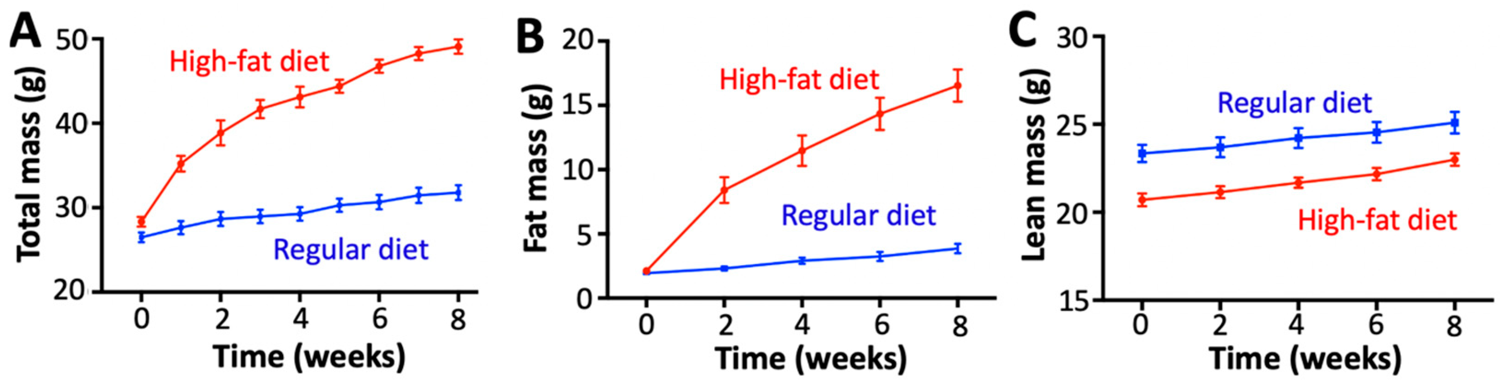
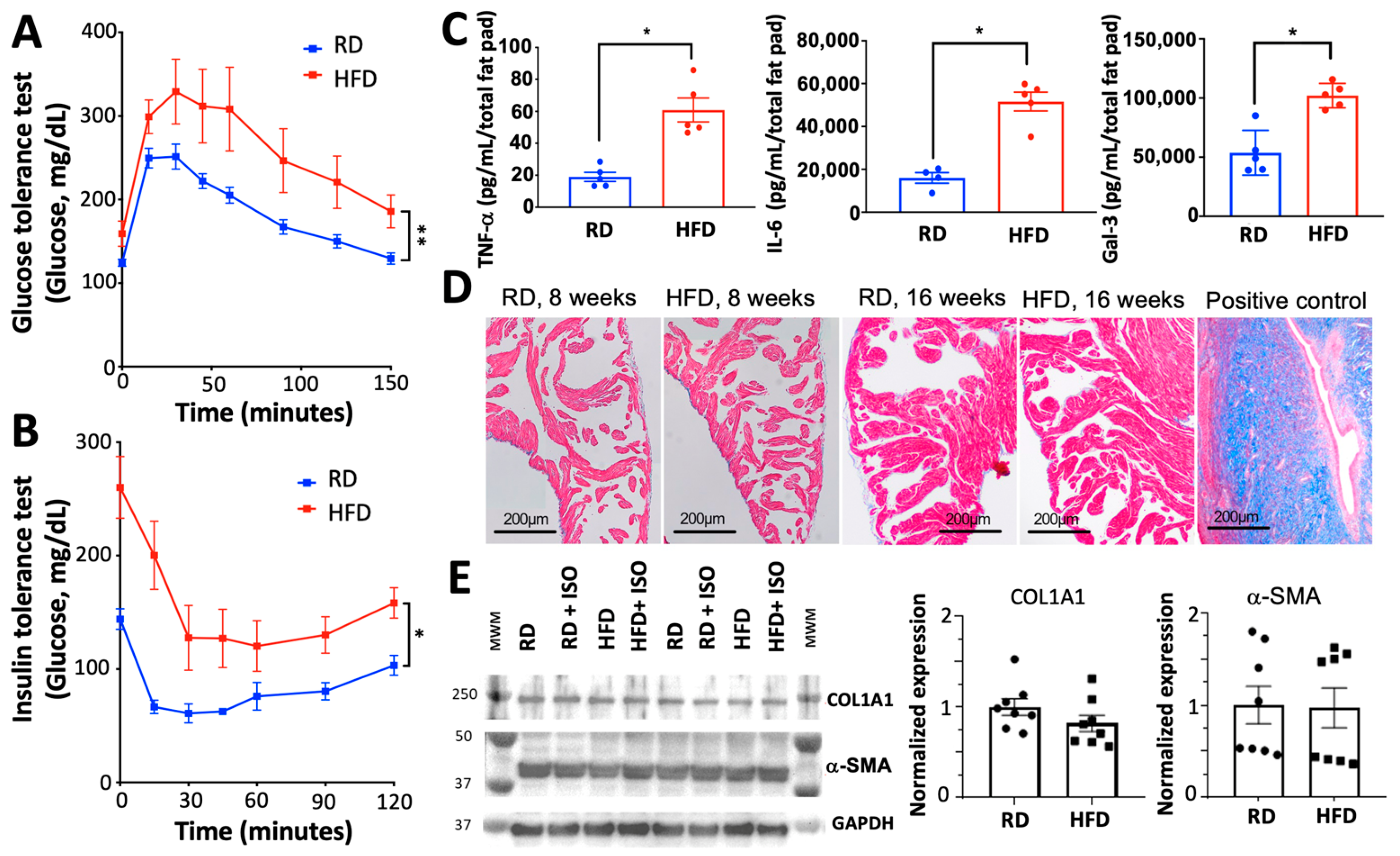
2.1. Diet-Induced Obesity Induces Metabolic and Inflammatory Imbalances Without Fibrosis or Alterations in Cardiovascular Hemodynamics

We used an established obesity model in which mice are fed a diet high in fat for 8 weeks [15] to examine the effect of obesity on the induction of AF. We used a group of mice fed a regular chow diet as a control. Mice fed a high-fat diet gained substantial weight and fat mass (Figure 1A,B) but maintained a similar lean mass over the 8-week feeding period compared to the mice fed a regular diet (Figure 1C). A high-fat diet produced both glucose intolerance and insulin resistance (Figure 2A,B), in agreement with previous studies linking a high-fat diet with metabolic remodeling [16]. We also found that diet-induced obesity increased the secretion of tumor necrosis factor-α (TNF-α), interleukin-6 (IL-6), and galectin-3 (Figure 2C). These findings agree with previous studies showing that obesity increases the levels of TNF-α and IL-6 [17,18].
A high-fat diet prompted the expected metabolic and inflammatory alterations in mice, but histological examination did not show evidence of fibrosis in the atria of mice fed a high-fat diet (Figure 2D). Extending the diet for 16 weeks did not induce fibrosis (Figure 2D); thus, the duration of the diet is not a factor for the lack of fibrosis in our model. Western blot analysis did not show significant changes in the expression of the pro-fibrotic proteins collagen and α-smooth muscle actin (α-SMA) (Figure 2E). Echocardiography studies showed that a high-fat diet does not alter heart rate, left ventricular ejection fraction, left ventricular mass, or left atrial dimensions (Table 1). Non-invasive blood pressure assessment at the end of the diet showed that there are no significant differences in either diastolic or systolic blood pressure between mice fed a high-fat diet versus a regular diet (Table 1). Collectively, these findings indicate that mice fed a high-fat diet undergo the expected inflammatory and metabolic imbalances that are typical of obesity, but without apparent fibrosis or alterations in cardiovascular hemodynamics.

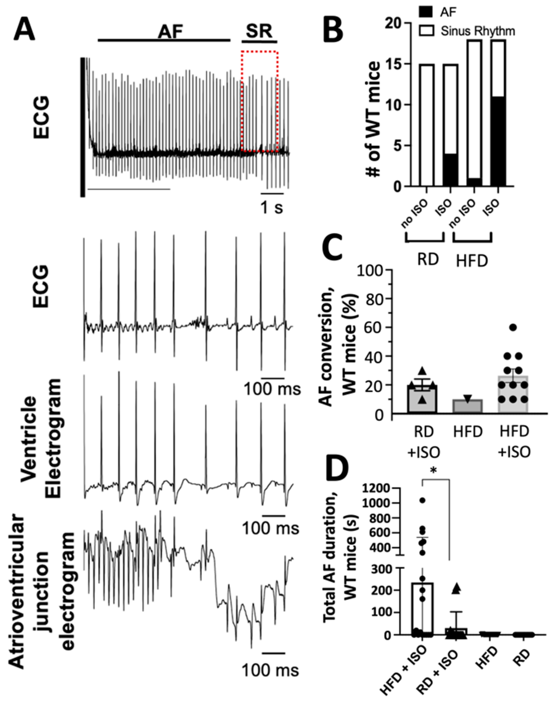
2.2. Diet-Induced Obesity and Acute Sympathetic Activation Synergize During Intracardiac Tachypacing to Induce Atrial Fibrillation

We used surface electrocardiogram (ECG) analysis to determine whether a high-fat diet alone is sufficient to increase susceptibility to atrial arrhythmias before applying intracardiac electrical stimulation. AF was determined as the occurrence of rapid and fragmented atrial electrograms (lack of regular P-waves) with irregular ventricular rhythm (irregular RR intervals) lasting at least 3 s [19]. A representative intracardiac recording of AF is shown in Figure 3A. Following an 8-week feeding period, the P-wave duration, PR interval, and QRS duration were similar between mice fed high-fat and regular diets (Table 2). However, mice fed a high-fat diet presented prolonged RR interval duration (Table 2). Alterations in the autonomic nervous system can predispose to arrhythmia, including AF [20,21]; therefore, we studied the effects of acute sympathetic activation. Upon isoproterenol administration, but before applying intracardiac electrical stimulation, mice fed a high-fat diet exhibited a significantly shorter RR interval duration compared to mice fed a regular diet (Table 3). However, obese mice did not have changes in P-wave duration, PR interval, and QRS duration (Table 3).
Alterations in the RR interval duration are a significant biomarker for AF detection [22]. Therefore, we used intracardiac electrical stimulation to determine the rate of conversion of atrial fibrillation induced by diet-induced obesity. Mice fed high-fat and regular diets were subjected to tachypacing in the right atrium to induce AF. Upon intracardiac electrical stimulation and in the absence of isoproterenol, mice fed a standard diet did not convert to AF (Figure 3B). Following tachypacing and isoproterenol administration, 26% of mice on a regular diet converted to AF (Figure 3B); however, this difference was not significant compared to mice fed a regular diet but without isoproterenol treatment (p = 0.09; Figure 3B). Conversely, 61% of mice on a high-fat diet treated acutely with isoproterenol developed AF following tachypacing; this represents a significant increase compared to obese mice without isoproterenol administration (p = 0.0009). We determined the ease of conversion and duration of AF by performing ten tachypacing attempts on mice fed both high-fat and regular diets that exhibited AF events in the presence or absence of isoproterenol treatment. Eleven mice that were fed a high-fat diet and treated with isoproterenol exhibited an increased ease of AF conversion compared to mice fed a regular diet that converted to AF after treatment without isoproterenol (Figure 3C). Furthermore, mice fed a high-fat diet exhibited a significantly longer AF duration compared to mice fed a regular diet (p = 0.02; Figure 3D). In the absence of isoproterenol, mice fed high-fat and regular diets did not exhibit AF events for more than 10 s (Figure 3D). Collectively, these findings indicate that diet-induced obesity and acute sympathetic activation synergize during intracardiac tachypacing to induce atrial fibrillation.

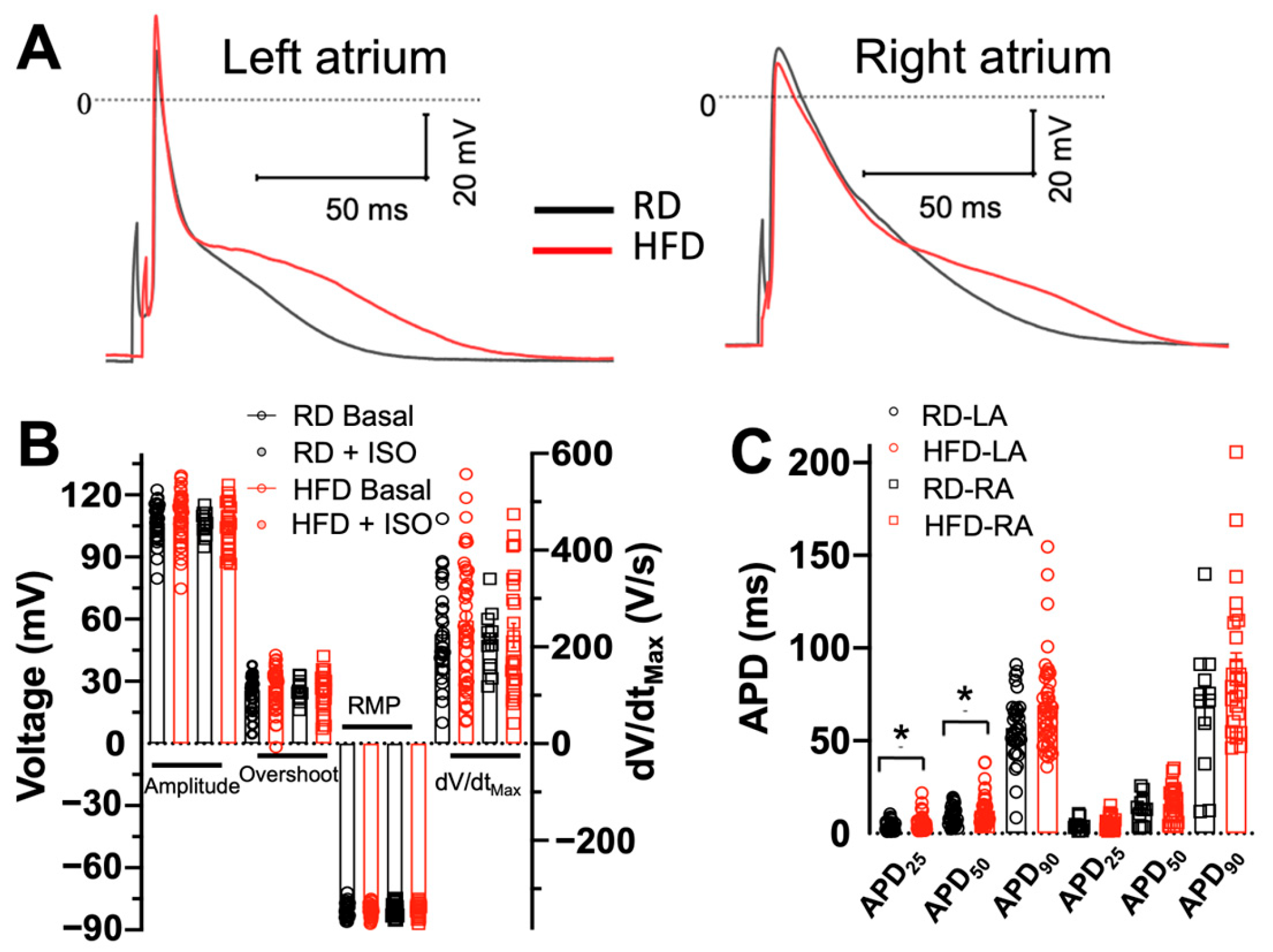
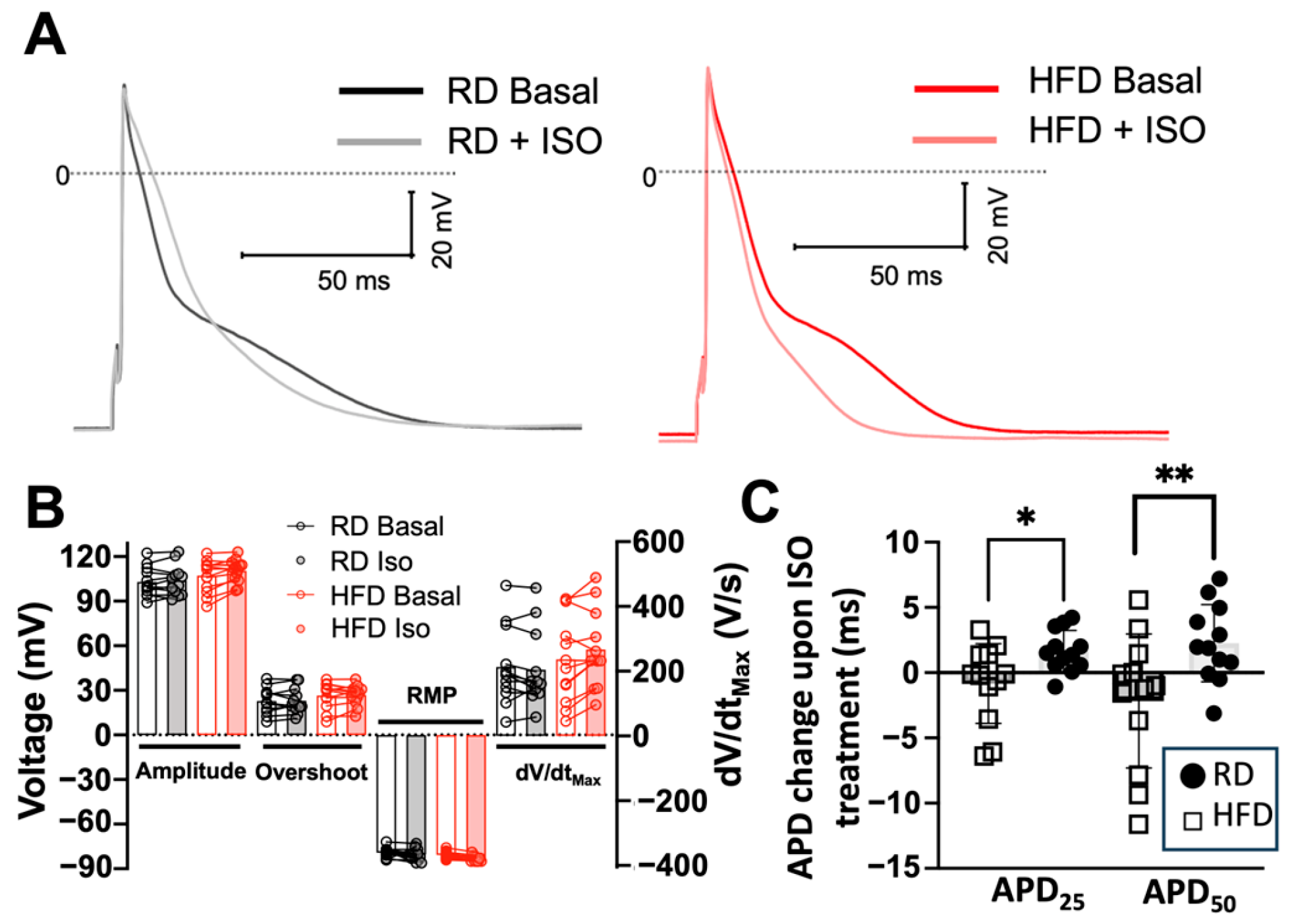
2.3. Effects of Diet-Induced Obesity on the Action Potential Characteristics of Atrial Myocytes

We performed patch-clamp electrophysiology studies to analyze the action potential (AP) characteristics of single isolated atrial myocytes from mice fed a high-fat diet and compared them with isolated atrial myocytes from animals fed a regular diet. APs were analyzed at 1-Hz stimulation; representative recordings from myocytes from the left and right atria are shown in Figure 4A. A high-fat diet does not induce significant differences in the action potential amplitude, overshoot, resting membrane potential (RPM), or maximum upstroke velocity (dV/dtmax) in myocytes isolated from either left or right atrium (Figure 4B). We also analyzed the effects of diet-induced obesity on the action potential duration (APD) at 25% (APD25), 50% (APD50), and 90% (APD90) depolarization. Compared to a regular diet, a high-fat diet does not induce significant changes in APD25, APD50, or APD90 of myocytes isolated from the right atrium (Figure 4C). However, a high-fat diet significantly increased the APD25 by −4 ms vs. a regular diet (p = 0.04) and APD50 by −8 ms vs. a regular diet (p = 0.03) in cardiomyocytes isolated from the left atrium (Figure 4C).
We next tested the effects of isoproterenol on AP characteristics in left atrial myocytes. Representative recordings are shown in Figure 5A. Isoproterenol does not induce significant differences in the action potential amplitude, overshoot, RPM, or dV/dtmax from baseline recordings (i.e., before isoproterenol treatment) in both diet groups (Figure 5B). Isoproterenol prolongs the APD25 by −30% (4.8 ± 0.6 ms to 6.4 ± 0.7 ms) and the APD50 by −18% (11 ± 1.3 ms to 13 ± 1 ms) of left atrium myocytes isolated from mice fed a regular diet. While a high-fat diet prolongs both APD25 and APD50, it also impairs the response of atrial cells to β-adrenergic stimulation. This impaired response to isoproterenol is illustrated in Figure 5C, showing a significant difference in the change in APD from baseline upon treatment with isoproterenol. These findings indicate that a high-fat diet significantly prolongs the action potential of left atrial myocytes and impairs the effects of adrenergic stimulation on APD.

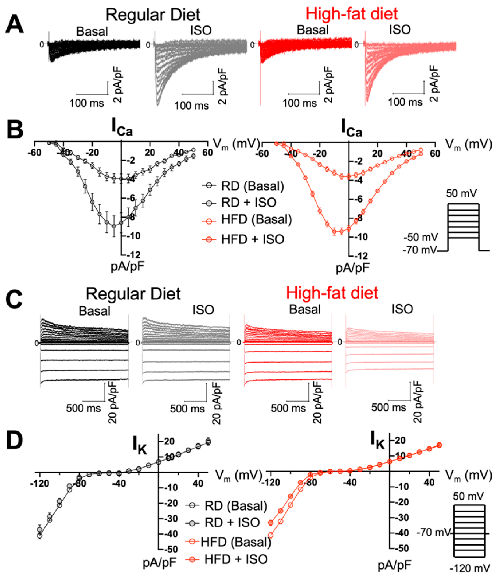
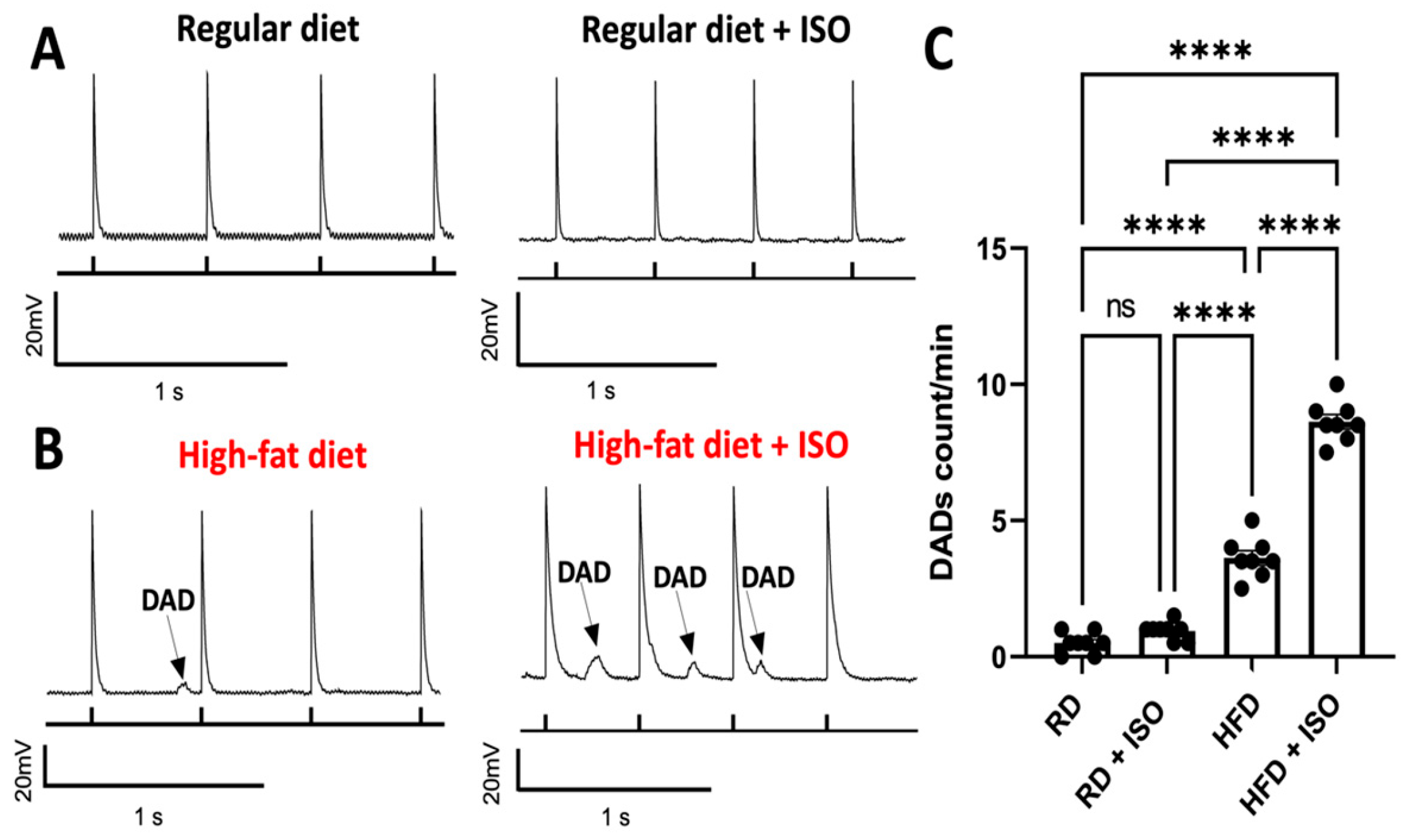
2.4. Diet-Induced Obesity Does Not Alter ICa and IK Densities but Produces Delayed Afterdepolarization Events in Atrial Myocytes

The changes in AP observed in left atrial myocytes from mice fed a high-fat diet can be produced by alterations in the balance between depolarizing inward calcium (ICa) and outward repolarizing potassium (IK) currents [23]. Therefore, we measured ICa and IK densities in atrial myocytes before and after isoproterenol treatment. Cell capacitance values were 53 ± 6 pF for myocytes isolated from mice fed a regular diet, and 53 ± 2 pF for myocytes isolated from mice fed a high-fat diet; we did not find statistically significant differences between groups (p = 0.9). Voltage-clamp studies did not show differences in ICa and IK at basal conditions in atrial myocytes from mice fed a regular vs. high-fat diet (Figure 6). Peak ICa values at 0 mV were 3.9 ± 0.5 pA/pF (regular diet) and −3.6 ± 0.2 pA/pF (high-fat diet), with no significant differences found between groups (p = 0.5; Figure 6A,B). Peak IK values at 40 mV were 16 ± 1.5 pA/pF (regular diet) vs. 14 ± 0.7 pA/pF (p = 0.29; Figure 6C,D). Overall, a high-fat diet does not induce alterations in inward depolarizing ICa or total IK outward repolarizing currents in atrial myocytes.
We performed current-clamp experiments to determine whether a high-fat diet produces delayed afterdepolarization (DAD) events in atrial myocytes. The production of DADs was measured at 2-Hz stimulation [24,25]. Representative AP traces showed that atrial myocytes from mice fed a regular diet do not show DADs in the presence or absence of isoproterenol (Figure 7A). While a high-fat diet alone is sufficient to produce a significant increase in DADs in atrial myocytes (Figure 7B,C), isoproterenol increases the formation of DADs by −150% (Figure 7C). These findings indicate that a high-fat diet produces DADs in atrial myocytes, and that isoproterenol increases the incidence of DADs induced by diet-induced obesity in mice.

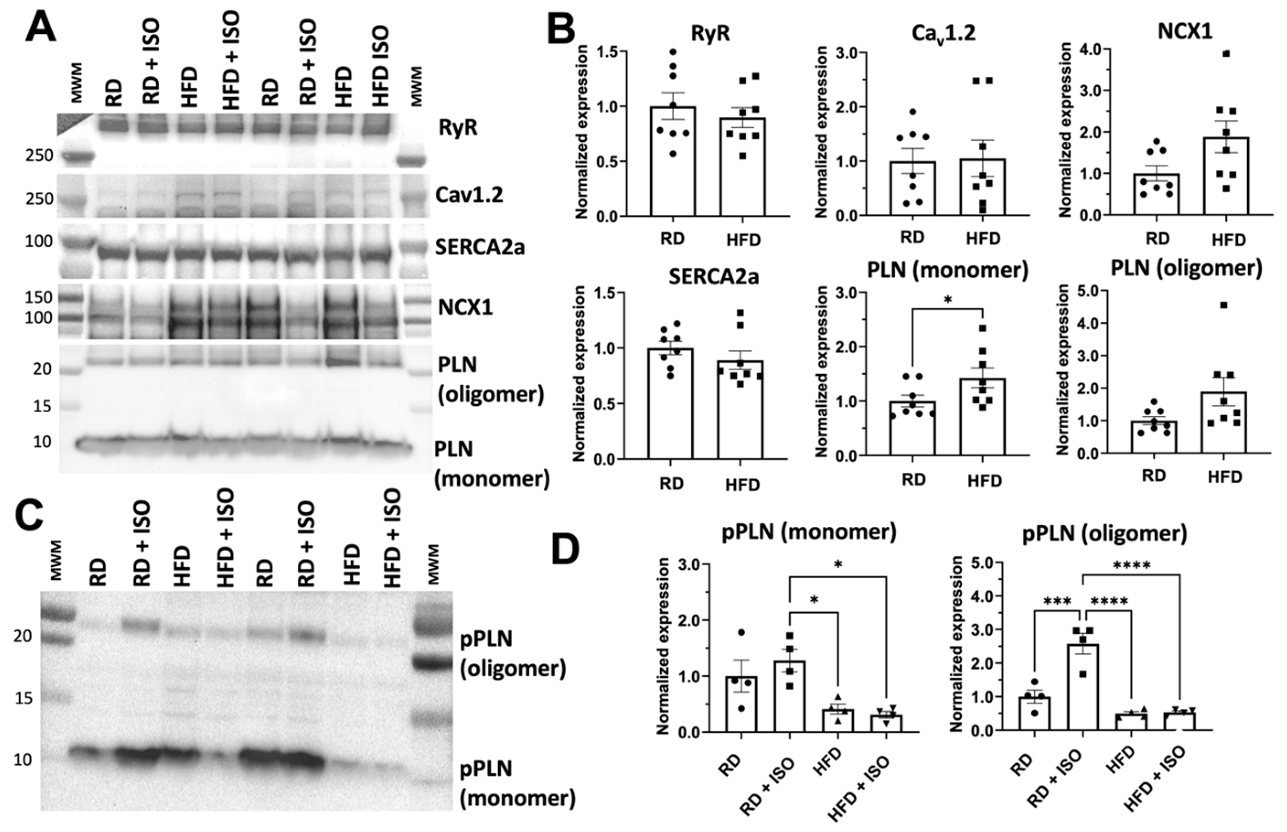
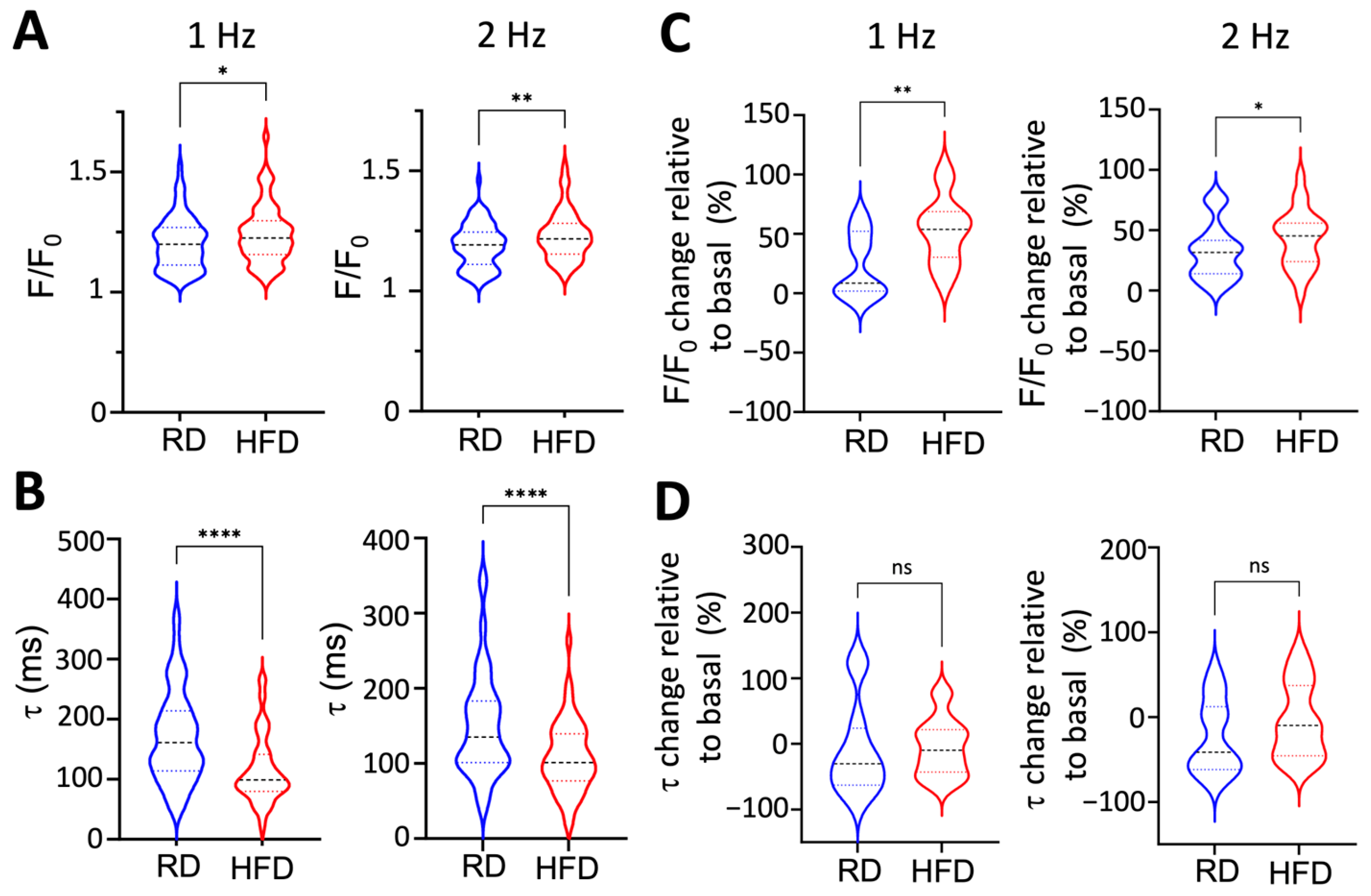
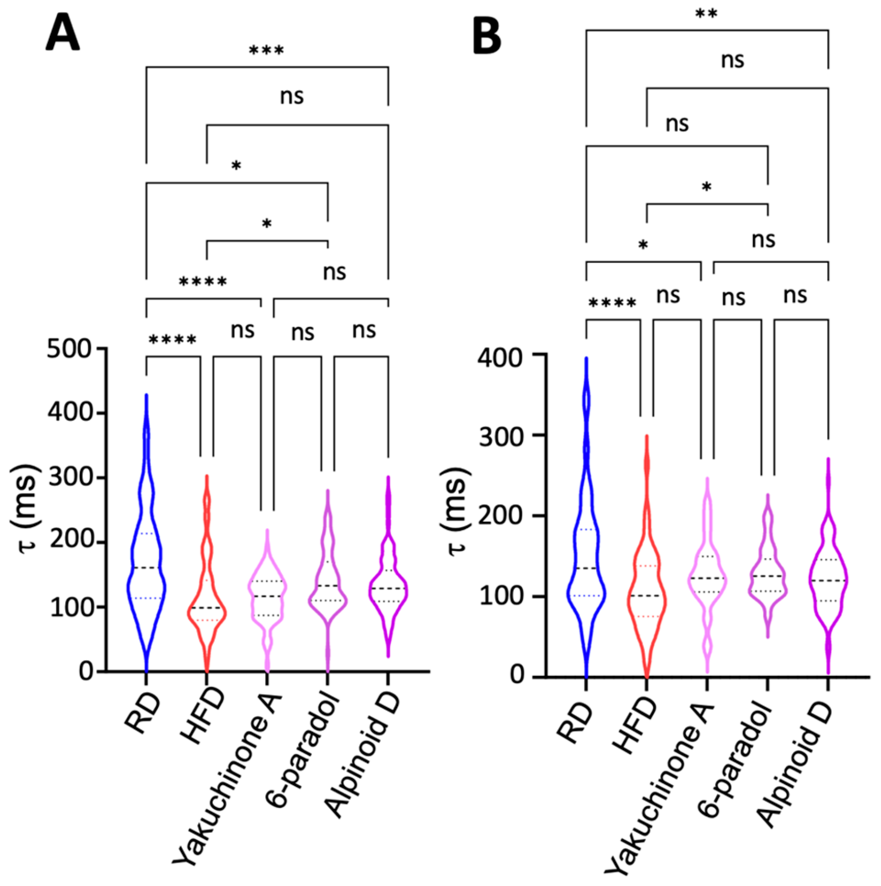
2.5. Obesity Induces Paradoxical Dysregulation of SERCA2a in Atrial Myocytes

A hallmark of DADs is the dysregulation of intracellular Ca2+ dynamics in cardiac cells [26]. Therefore, we used Western blot analysis to determine if diet-induced obesity induces changes in the expression of proteins within the atria that are involved in intracellular Ca2+ dynamics. Compared to a regular diet, a high-fat diet did not induce significant changes in the expression of the ryanodine receptor (RyR), the L-type Ca2+ channel (Cav1.2), the cardiac Na+-Ca2+ exchanger (NCX1), and SERCA2a (Figure 8A,B). We also determined changes in phospholamban (PLN) expression and phosphorylation. We note that PLN exists as monomers that directly inhibit SERCA2a activity, and as oligomers that serve either as a storage or effector of SERCA2a activity [27,28,29,30]. Hence, we also analyzed the relative abundance of PLN monomers and oligomers. Compared to mice fed a regular diet, there is a significant increase in the presence of the monomeric, but not the oligomeric, form of PLN in atria of mice fed a high-fat diet (Figure 8A,B). Atria from mice fed a high-fat diet showed significantly lower basal (i.e., before isoproterenol treatment) PLN phosphorylation and no response to isoproterenol treatment (i.e., increased PLN phosphorylation) compared to atria from mice fed a regular diet (Figure 8C,D). Our findings indicate that diet-induced obesity increases the proportion of inhibitory PLN monomers and impairs PLN phosphorylation in atrial myocytes.
It has been shown that the dephosphorylated monomeric form of PLN inhibits SERCA2a, reduces Ca2+ reuptake into the sarcoplasmic reticulum, and impairs cardiac contractility [31,32,33]. Therefore, we expect that the increased abundance of the PLN monomer and impaired PLN phosphorylation induced by obesity impair intracellular Ca2+ transport in atrial myocytes. We tested this mechanism by using single-cell Ca2+ imaging of atrial myocytes. Ca2+ transients were analyzed at 1-Hz and 2-Hz field stimulation. Surprisingly, the stimulated Ca2+ transient amplitude was significantly increased in myocytes from mice fed a high-fat diet compared to those from mice on a regular diet (Figure 9A). More importantly, the Ca2+ transient decay, τ, is significantly faster in atrial myocytes from mice fed a high-fat diet vs. a regular diet (Figure 9B). Isoproterenol treatment of atrial myocytes from mice fed a high-fat diet significantly increased Ca2+ transient amplitude (relative to baseline) compared to myocytes from mice fed a regular diet (Figure 9C). However, isoproterenol treatment does not affect the relative Ca2+ transient decay of myocytes from obese vs. nonobese mice (Figure 9D).
In mouse cardiac myocytes, SERCA2a contributes to −90% of Ca2+ clearance in mice [34]; hence, the unexpected acceleration in the Ca2+ transient suggests activation of SERCA2a in atrial myocytes from obese mice. Therefore, we used SERCA2a activators as pharmacological probes to investigate whether the effects on τ are comparable to those induced by a high-fat diet. For these experiments, we used three potent SERCA2a activators recently discovered by our group as pharmacological probes [35]. These small-molecule effectors, Yakuchinone A, 6-Paradol, and Alpinoid D, stimulate SERCA2a activity, increasing the enzyme’s maximal velocity by 19–31% at a compound concentration of 10 µM [35]. Here, we used τ as a proxy to establish SERCA2a engagement in myocytes, and experiments were performed at 1-Hz and 2-Hz field stimulation. We found that the activators significantly accelerated the Ca2+ transient decay compared to myocytes from mice fed a regular diet (Figure 10A,B). Conversely, we found that a high-fat diet has a similar effect on the Ca2+ transient decay compared to that induced by small-molecule SERCA2a activators. Interestingly, we found that in some cases, the effect of a high-fat diet on Ca2+ transient decay is even more profound than that of small-molecule SERCA2a activation, e.g., in the case of a high-fat diet vs. treatment with the SERCA2a activator 6-paradol at a pacing frequency of 1 Hz (Figure 10A). Collectively, these findings suggest that obesity induces paradoxical dysregulation of SERCA2a in atrial myocytes, stimulating SERCA2a activation despite an increase in the relative abundance of the inhibitory PLN monomer and a reduction in PLN phosphorylation.

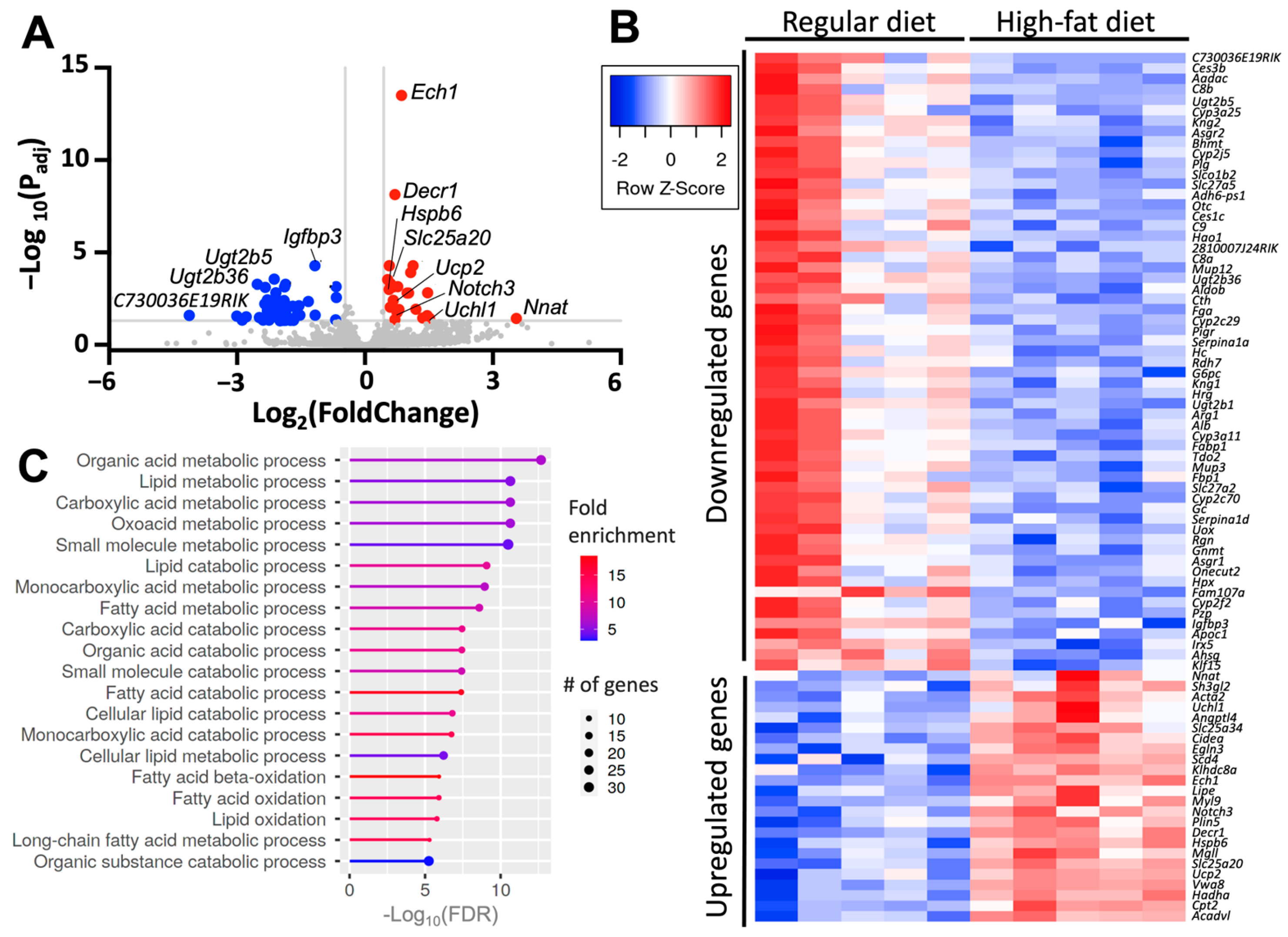
2.6. Diet-Induced Atrial Upregulates Genes Involved in Metabolism and Stress

We performed RNA sequencing to obtain the transcriptomic signature of atrial tissue in our model of diet-induced obesity, and to identify targets that may explain the paradoxical SERCA2a activation observed in atrial myocytes from obese mice. The analysis was performed on atrial tissue from five mice fed a high-fat diet, and five mice fed a regular diet as a control. After adjusting for known covariates and correcting for multiple comparisons, we found 83 differentially expressed genes between mice fed a high-fat diet and control (FDR < 0.05, Figure 11A,B). Of these genes, 24 were upregulated and 59 were downregulated (Figure 11B and Table S1 and S2 of the Supplementary Information). Gene ontology pathway analysis of the differentially expressed genes showed that a high-fat diet primarily affects the expression of genes involved in atrial metabolic processes including organic acid metabolism, lipid metabolism, lipid catabolism, fatty acid oxidation, and small-molecule catabolic processing (Figure 11C).
The transcriptomics showed the upregulation of various genes that are known to contribute to either AF or cardiac remodeling. This included Uchl1, which encodes for a ubiquitin hydrolase that is upregulated in myocytes following myocardial infarction [36]; Slc25a20, which has been independently associated with rhythm status among patients with AF [37]; and Upc2, which influences the susceptibility for Ca2+-mediated arrhythmias, modulates myocardial excitation–contraction coupling, and attenuates oxidative stress [38,39,40]. Conversely, a high-fat diet does not induce changes in gene expression of Atp2a2 and Pln, which encode for SERCA2a and PLN, respectively (Table S3 in the Supplementary Information). We also did not find changes in the gene expression of the Ca2+-handling proteins RyR, Cav1.2, NCX1, sarcolipin, and calsequestrin (Table S3 in the Supplementary Information). A high-fat diet also does not affect the gene expression of the Na+/K+-ATPase (NKA), the NKA-regulating FXYD proteins [41], and the Nav1.5 channel (Table S3 in the Supplementary Information). A high-fat diet also induces upregulation of Hspb6, a heat-shock protein that protects against remodeling in atrial myocytes upon tachypacing [42], and Notch3, a protein that inhibits cardiac fibroblast proliferation [43] (Table S1 of the Supplementary Information). We did not find changes in gene expression of the Sumo1 and Sirt1 (Table S3 in the Supplementary Information), two genes that are involved in modulating SERCA2a activity in cardiac cells [44,45]. Interestingly, Nnat is upregulated in mice fed a high-fat diet (Figure 11 and Table S1 of the Supplementary Information). Nnat encodes for neuronatin, a protein that plays a role in whole-body metabolic regulation and has been implicated in obesity [46]. This finding is significant because neuronatin is a calcium pump effector that stimulates the pump’s ATPase activity [47] and increases intracellular Ca2+ storage in neurons [48]. Although the protein expression of neuronatin was not studied here, the transcriptomic analysis provides a plausible explanation for the increase in Ca2+ uptake observed in isolated atrial myocytes (Figure 9 and Figure 10).

3. Discussion

In this study, we used a two-hit model of diet-induced obesity and adrenergic stimulation to investigate the mechanisms by which obesity increases the risk of AF. This model showed expected features of diet-induced obesity, including the increase in body mass and visceral fat and metabolic derangement. Specifically, the model is characterized by glucose intolerance and insulin resistance, in agreement with diet-induced metabolic remodeling [16]. Obesity is a triggering factor for diabetes associated with insulin resistance [49], which increases the risk of arrhythmogenesis and AF [50]. Our model also exhibited increased production of inflammatory cytokines, including TNF-α and IL-6. TNF-α and IL-6 are inflammatory mediators that are arrhythmogenic and have been linked to the pathogenesis of AF [51,52]. These findings agree with studies showing that obesity promotes and secretion of TNF-α and IL-6 by adipose tissue [17,18].
Histology and Western blot analysis did not reveal either fibrosis or changes in the expression of pro-fibrotic proteins in the atria of mice fed a high-fat diet. These findings contrast with previous studies showing that mice fed a high-fat diet for 10 weeks develop moderate, albeit significant, fibrosis [13,14]. We used a similar diet to that used in these studies, so we speculate that the diet composition and feeding duration are not variables that help explain the absence of fibrosis in our model. Instead, transcriptomic analysis showed that a high-fat diet induces upregulation of the Notch3 gene in atria. The Notch signaling pathway is involved in cellular differentiation, proliferation, and apoptosis [53]. Recent studies have shown that Notch3 inhibits cardiac fibroblast proliferation and fibroblast to myofibroblast transition while promoting cardiac fibroblast apoptosis by modulating the pro-fibrotic RhoA/ROCK/Hif1α signaling pathway [43]. Notch3 upregulation induced by a high-fat diet may explain the absence of atrial fibrosis observed in our model. Future studies are needed to clarify the role of Notch and other anti- and pro-fibrotic signaling pathways in a diet-induced obesity mouse model and their variability with experimental conditions.
Transcriptomic analysis showed that a high-fat diet induces upregulation of Hspb6, a heat-shock protein that protects against remodeling in atrial myocytes upon tachypacing [42]. The upregulation of Hspb6 in our diet-induced obesity model explains why a high-fat diet alone is insufficient to induce AF. Instead, we found that diet-induced obesity and acute sympathetic activation synergize during intracardiac tachypacing to induce AF and that this occurs in the absence of fibrosis [54]. In our model, obesity may promote AF by several mechanisms, including changes in cardiac output, anatomical remodeling of the atria, and systemic hypertension, which can further exacerbate the increase in left ventricular wall stress and left atrium pressure [55]. However, we did not find echocardiographic evidence indicating alterations in left ventricular dynamics, left atrium volume, or differences in systemic blood pressure in our model of diet-induced obesity. These findings agree with studies showing that diet-induced obesity does not affect either left ventricular ejection fraction or systolic blood pressure [56].
At the cellular level, a high-fat diet significantly prolongs the action potential of left atrial myocytes and impairs the effects of β-adrenergic stimulation on action potential duration. APD prolongation affects the balance of Ca2+ homeostasis, requiring robust compensatory mechanisms [57]. Therefore, we considered early and delayed afterdepolarizations as potential contributors to the action potential prolongation in isolated atrial myocytes. Early afterdepolarizations (EADs) are mainly driven by voltage oscillations in the repolarizing phase of the action potential. However, we did not observe significant changes in ICa and IK currents in myocytes isolated from mice fed a high-fat diet, suggesting that EAD formation is not the underlying cause for prolonged AP duration [58]. An alternative mechanism for prolongation of the AP is the formation of delayed afterdepolarizations (DADs), which are driven by spontaneous intracellular Ca2+ release during diastole [59]. A high-fat diet produces DADs in atrial myocytes, and isoproterenol significantly increases the incidence of DADs induced by diet-induced obesity in mice. These findings are significant because DADs have been related to the initiation of arrhythmias [60] and occur in pathological states including heart failure, diabetes, and ischemic heart disease [61].
Our findings indicate that dysregulation of intracellular Ca2+ dynamics contributes to AF in our two-hit model. Therefore, we analyzed the expression of proteins that are involved in Ca2+ handling in the atrium. Compared to a regular diet, a high-fat diet does not alter the expression of major proteins involved in Ca2+ handling, including RyR, Cav1.2, NCX1, and SERCA2a. We also found that PLN expression is not decreased in response to a high-fat diet. However, a high-fat diet impairs the ability of β-adrenergic stimulation to phosphorylate both monomeric and oligomeric forms of PLN. In its unphosphorylated state, PLN inhibits SERCA2a, and PLN phosphorylation reverses this inhibitory effect and reactivates SERCA2a upon adrenergic activation [62]. The defective PLN phosphorylation found in our model agrees with previous studies that have shown obesity in rats promotes the reduction of PLN phosphorylation [63]. Defective PLN phosphorylation has also been observed in animal models and patient samples of atrial fibrillation [24,64,65,66].
The increased abundance of the PLN monomer and impaired PLN phosphorylation suggests that SERCA2a-mediated Ca2+ uptake into the sarcoplasmic reticulum is reduced in our diet-induced obesity model. Unexpectedly, we found that atrial myocytes from obese mice presented a change in the Ca2+ transient decay that is similar to that induced by SERCA activators, suggesting that a high-fat diet stimulates SERCA2a activity in cardiac myocytes. Additional single-cell Ca2+ imaging studies and western blot analysis showed that stimulation of Ca2+ uptake induced by a high-fat diet is not mediated by PLN phosphorylation. Interestingly, SERCA2a stimulation exerts negative feedback on Ca2+-induced Ca2+ release [67], which explains the inability of a high-fat diet to induce AF in the absence of sympathetic stimulation. However, a combination of a high-fat diet and acute sympathetic activation increases sarcoplasmic reticulum Ca2+ overloading through SERCA2a activation and decreases sarcoplasmic reticulum Ca2+ release threshold, e.g., by RyR phosphorylation [68,69]. Overall, a high-fat diet primes SERCA2a for activation despite impaired PLN phosphorylation, and the combined effects of a high-fat diet SERCA2a activation and isoproterenol contribute to AF likely through a Ca2+-induced Ca2+ release gain in atrial myocytes [70,71]. This mechanism also agrees with a recent study showing that increased intracellular Ca2+ mobilization is sensitive to β-adrenergic activation, triggering pro-arrhythmia events in ventricular myocytes of mice fed a Western diet [72].
We used transcriptomics to identify gene expression of proteins that may explain the stimulation of Ca2+ reuptake into the sarcoplasmic reticulum. A high-fat diet does not induce changes in the expression of genes encoding for SERCA2a and PLN. Previous studies have shown SUMO1-dependent stimulation of SERCA2a [44]; however, there were no changes in the expression of the Sumo1 gene in response to a high-fat diet. Studies have also shown that sirtuin 1-mediated acetylation of SERCA2a affects the function of this pump [45], but we did not find changes in the expression of the Sirt1 gene. We found that Nnat, which encodes for neuronatin, is upregulated in mice fed a high-fat diet. Nnat upregulation agrees with previous studies showing that neuronatin levels correlate with an increase in BMI and body fat mass [73]. Neuronatin is known to activate the SERCA2 isoform in neurons [48], thus explaining the paradoxical stimulation of Ca2+ uptake in the absence of PLN phosphorylation. Therefore, Nnat upregulation induced by a high-fat diet may explain the increased Ca2+ reuptake into the sarcoplasmic reticulum despite impaired PLN phosphorylation in atrial myocytes. We note that neuronatin has been found expressed in vascular tissue and skeletal muscle [46,47,74], but to our knowledge, this is the first study reporting its gene expression in the heart. Therefore, the functional roles of neuronatin in the heart, including SERCA2a regulation, warrant further investigation.
Clinically, our findings agree with a previous study showing that SERCA2a activation is an AF substrate in patients with paroxysmal AF [52]. In that study, however, obesity was not taken as a contributing factor because the patients in both control and AF groups had a similar average BMI, with values that fall within the overweight classification [52]. The authors found that the mechanism for SERCA2a activation in these patients was PLN hyperphosphorylation. While the pattern of PLN phosphorylation is completely different between AF in patients and obese mice, it is intriguing that SERCA2a activation is the underlying mechanism facilitating AF. Our study suggests that obesity increases the risk of AF upon exposure to acute sympathetic activation and emphasizes the importance of autonomic regulation in initiating AF [75]. This could occur during the sympathetic nervous system fight-or-flight response, which is mediated by an increase in the spontaneous action potential firing rate of pacemaker cells in the sinoatrial node. These findings have potential translational implications, and pharmacological interventions to mitigate the risk of diet-induced AF may include partial SERCA2a inhibition [76].

Study Limitations

Studies in myocytes from different species have shown left–right differences in impulse properties of atrial myocardial cells, such as in response to stimulation frequency and pharmacologic challenges. Specifically, in the murine model, it was shown that APD in the left atrium is shorter than in the right atrium [77]. While we used an equal number of animals for the APD experiments, we found that cell isolation yielded more viable cells from the right chamber than from the left, presumably reflecting differences in the perfusion to the chambers, although this remains to be determined. While these differences may influence the APD recordings, we note that obesity induces a significant prolongation of APDs in cells isolated from both chambers. Yet, the differential (left atrium vs. right atrium) combined effects of sensitivity to stimulus frequency, temperature, and diet-induced Ca2+ dysregulation on myocyte APDs remain to be explored mechanistically.
The dietary intervention in this study likely induces changes in the lipid profile of mice, and consequently in the lipid composition of the sarcolemma and SR. Consequently, it is possible that a high-fat diet can influence the function of ion channels and ion-transporting ATPases. In this study, we found that the total potassium and calcium currents are similar between mice fed regular and high-fat diets. However, we did not measure specific currents that may be affected by changes in lipid composition, including the voltage-gated and inward rectifying K+ channels, which are sensitive to lipid composition, interactions, and metabolism [78,79,80]. Although not investigated in this study, it is also possible that diet-induced obesity alters the function of Nav1.5, a voltage-gated sodium channel that plays a critical role in cardiac electrical activity, particularly in the initiation of the cardiac action potential and the fast depolarization phase [81]. Obesity-induced Nav1.5 dysregulation has been associated with an increased susceptibility to arrhythmias [82], so dysregulation of this channel may contribute to the incidence of AF in our model of diet-induced obesity. Other diet-induced posttranslational modifications, such as palmitoylation [83], may also contribute to the changes in the function of Nav1.5, altering myocyte excitability and contributing to AF in our model. SERCA2a activity is influenced by factors like lipid chain length, degree of saturation, and the presence of specific lipid headgroups [84,85]. Therefore, it is possible that diet-induced obesity alters the lipid composition in the SR and contributes to the stimulation of SERCA2a activity in our model of diet-induced obesity. Future studies by our group will address the changes in SR lipid composition induced by a high-fat diet.

4. Materials and Methods

4.1. High-Fat Diet-Induced Obesity Mice Model

Male C57BL/6 mice were obtained from the Jackson Laboratories (Bar Harbor, ME, USA). Male mice were specifically chosen because the diet-induced obesity model has blunted effects in female mice [86]. Mice at 8 weeks of age were randomly assigned to be fed either a regular chow diet (13.6% fat, 57.5% carbohydrate, 28.9% protein; Research Diet, Labdiet, St. Louis, MO, USA) or a high-fat diet (60% fat, 20% carbohydrate, 20% protein; Research Diet, Labdiet) for 8 or 16 weeks. Body weights were recorded every two weeks. Blood glucose and insulin were measured as previously described [87] and measured at the indicated time points. Non-invasive hemodynamics and echocardiography studies were performed at the University of Michigan Physiology and Phenotyping Core. Mice were anesthetized with 2% isoflurane before imaging. We performed B-mode short-axis and M-mode imaging and under transthoracic echocardiography using the Vevo 2100 imaging system (FUJIFILM VisualSonics Inc., Toronto, ON, Canada). A detailed description of the histology studies is provided in the Supplementary Information.

4.2. Enzyme-Linked Immunosorbent Assay (ELISA)

Left perigonadal fat pads were isolated from HFD-fed or LFD-fed mice and incubated for 24 h in RPMI 1640 plus 10% fetal bovine serum with 1% penicillin-streptomycin solution (P4333; Sigma-Aldrich, St. Louis, MO, USA). Conditioned culture supernatants were collected and stored at −80 °C. ELISAS for IL-6 (ThermoFisher # KMC0061, Waltham, MA, USA), TNFα (ThermoFisher # BMS607-3), and galectin-3 (R&D Systems, # DY1197, Minneapolis, MN, USA) were performed according to the manufacturer’s instructions and normalized to fat pad weight.

4.3. Surface Electrocardiographic Recording

We performed surface ECG measurements at a single time point at the end of the feeding period. Mice were anesthetized with 2–3% isoflurane and maintained their body temperature between 36.5 and 37.5 °C using a thermally controlled heating pad. After 10 min of stabilization, the ECGs were obtained to establish the baseline (basal) ECG. We then performed an intracardiac surgical procedure to obtain simultaneous surface and intracardiac ECG recordings. Surface ECG analysis was performed as previously described [88].

4.4. Intracardiac Recordings

Programmed electrical stimulation was performed as described previously [89] with some modifications and following the guidelines on the assessment of arrhythmias in small animals [19]. Mice were anesthetized with 2–3% isoflurane and maintained their body temperature between 36.5 and 37.5 °C using a heating pad. An octapolar catheter (Transonic, Ithaca, NY, USA) was inserted through the jugular vein and advanced into the right atrium and ventricle. Electrical stimulation was performed by using ten atrial bursts pacing before and after intraperitoneal isoproterenol administration (1.5 mg/kg). Atrial stimulation was achieved by rectangular impulses (2 ms) delivered at twice the pacing threshold. A modified S1–S2 burst stimulation protocol was used to induce atrial arrhythmia: S1 (10 stimuli at cycle length 60ms) was followed by S2 (10 stimuli at cycle length 5 ms). This sequence of programmed electrical stimulation was repeated 10 times with a 1 min delay. We used the PONEMAH version 3 (Data Sciences International) software for the acquisition of intracardiac recording data.

4.5. Electrophysiology Studies of Isolated Atrial Myocytes

The isolation of atrial myocytes is described in the Supplementary Information. Experiments were carried out using a multi-clamp 700 B amplifier (Axon Instruments, Molecular Devices, Union City, CA, USA). Data were acquired and analyzed using the pCLAMP 10 Suite of programs (Axon Instruments). Borosilicate glass electrodes were pulled with a Brown–Flaming puller (model P-97), yielding appropriate tip resistances when filled with pipette solution to enable proper voltage control [90,91]. Action potentials were elicited using square wave pulses 30–50 pA amplitude, 5–8 ms duration, generated by a DS8000 digital stimulator (World Precision Instruments, Sarasota, FL, USA), and recorded at 37 °C with pipette solution containing 1 mM MgCl2, 1 mM EGTA, 150 mM KCl, 5 mM HEPES, 5 mM phosphocreatine, 4.46 mM K2ATP, 2 mM β-Hydroxybutyrate, adjusted to pH 7.2 with KOH. The extracellular solution contained 148 mM NaCl, 0.4 mM NaH2PO4, 1 mM MgCl2, 5.5 mM glucose, 5.4 mM KCl, 1 mM CaCl2, 15 mM HEPES, and 1 mM EGTA, pH adjusted to 7.4 with NaOH. RMP, overshoot, action potential amplitude, and APD25, APD50, and APD90 of repolarization were analyzed using custom-made software, and dV/dtmax was calculated using Origin 8.1 (Microcal, Northampton, MA, USA). We determined the formation of DADs as described previously [92]. Briefly, APs were recorded at 27 ± 5 °C with a pipette solution containing 1 mM MgCl2, 150 mM KCl, 5 mM HEPES, 5 mM phosphocreatine, 4.46 mM K2ATP, 2 mM β-Hydroxybutyrate, with pH adjusted to 7.2 with KOH. A Ca2+- and Mg2+-containing Hanks’ Balanced Salt Solution (ThermoFisher) was used as an extracellular solution. DADs were recorded before and after treatment with isoproterenol (100 nM). A detailed description of the measurement of ICa and IK currents is found in the Supplementary Information.

4.6. Western Blot Analysis

Hearts were harvested in cold 4 °C PBS, and then both right and left atria were separated from the ventricles, homogenized in 100 µL lysis buffer (ThermoFisher Scientific, catalog # 78510) with 1% protease inhibitor cocktail (Sigma, catalog # P8340) and 1% phosphatase inhibitor cocktail (Sigma, catalog # P5726). Atrial tissue lysates in b-mercaptoethanol 4× sample buffer were loaded into 4–12% NuPage precast gels (ThermoFisher, Waltham, MA, USA) and electrophoresis was carried out. The SDS-PAGE resolved proteins were transferred to iBlot® stacks with regular PVDF membranes using the Life Technologies iBlot2 system. Non-specific binding sites were blocked with 5% bovine serum albumin (BSA) in PBS-T (in mM, 3 KH2PO4, 10 Na2HPO4, 150 NaCl, 0.1% Tween 20, pH 7.2–7.4) for 30 min at room temperature. Membranes were incubated with specific primary antibodies diluted in 5% BSA in PBS-T overnight at 4 °C. After washing 3 times for 10 min, membranes were incubated with horseradish peroxidase-conjugated secondary antibodies diluted in 5% BSA in PBS-T for 1 h. The primary and secondary antibodies used in this study are listed in Table S4 of the Supplementary Materials. Protein–antibody reactions were detected by Supersignal chemiluminescence (Pierce Biotechnology Inc, Rockford, IL, USA), imaged using Image Lab software 5 (Bio-Rad, Hercules, CA, USA), and analyzed using Image Lab software version 5.

4.7. Single-Cell Ca2+ Imaging

Atrial myocytes were prepared for Ca2+ imaging as described in the Supplementary Information. The measurement of intracellular Ca2+ transients in atrial myocytes was performed using an Ionoptix recording system (Ionoptix LCC, Westwood, MA, USA). Culture media was removed, and the myocyte-plated coverslips were transferred into Tyrode solution containing 140 mM NaCl, 4 mM KCl, 1 mM MgCl2, 10 mM HEPES, 10 mM Glucose, 1 mM CaCl2; the pH was adjusted to 7.4 with NaOH. Cells were loaded with 5 µM Fura2-AM and 2.5 mM probenecid followed by 2 washouts of 10 min each for de-esterification with Tyrode solution. Coverslips were placed in an Ionoptix rapid change stimulation chamber and perfused with Tyrode solution at 37 °C; isoproterenol and thapsigargin were tested at a concentration of 200 nM and 1 µM, respectively. SERCA2a activators Yakuchinone A, 6-paradol, and Alpinoid D [35] were tested at a concentration of 10 µM. A temperature of 37 °C was maintained using a mTC3 micro temperature controller. We used an inverted Fluorescence Microscope Nikon ECLIPSE Ti Series and a 40×/1.30 oil Nikon objective for data acquisition (Nikon, Tokyo, Japan). Fura-2 AM excitation wavelengths, 340 nm, and 380 nm were generated using an IonOptix HyperSwitch (IonOptix LLC, Milton, MA, USA). Cells were paced with the IonOptix Myopacer. A minimum of 10 transients per myocyte were used for ensemble averaging and analyzed using Ionwizard software version 6.2.1.60 (Ionoptix LCC, Westwood, MA, USA).

4.8. RNA Sequencing and Differential Gene Expression

Total RNA was purified from the atria of mice using the Qiagen RNeasy kit (Qiagen, catalog # 74004, Hilden, Germany), and RNA content and quality were determined using the TapeStation System (Agilent, Santa Clara, CA, USA). Sequencing was performed at the University of Michigan Advanced Genomics Core with Lexogen QuantSeq libraries constructed and subsequently subjected to 101 single-end cycles on the NovaSeq-6000 platform (Illumina, San Diego, CA, USA). Data were pre-filtered to remove genes with less than 20 counts across all samples. Differential gene expression analysis was performed using DESeq2 version 1.48.1 [93], using a negative binomial generalized linear model with the following thresholds: linear fold change >1.5 or <−1.5, and a Benjamini–Hochberg FDR < 0.05. Genes were annotated with NCBI Entrez GeneIDs [94] and text descriptions. Candidate pathways activated or inhibited comparisons and GO-term enrichments were performed using iPathway Guide version 2012 (Advaita, Ann Arbor, MI, USA), iDEP version 0.95, and ShinyGO version 0.75 (San Diego State University) [95,96,97].

4.9. Statistical Analysis

All results are presented as mean ± SEM. Normality was determined using the Shapiro-Wilk test. Non-parametric tests were used for data that are not normally distributed. Data were analyzed using the Student’s t-test or Mann–Whitney U-test for paired experiments, or a two-way analysis of variance (ANOVA) followed by Tukey’s post hoc test to analyze differences between multiple groups. We used 95% confidence intervals around the differences between the groups for the post hoc test. Two-sided p-values were used, and α-level < 0.05 was considered significant.

5. Conclusions

In summary, this study demonstrates that obesity, when combined with acute sympathetic activation, significantly enhances the susceptibility to AF by promoting arrhythmogenic Ca2+ dynamics in atrial myocytes. We observed that during intracardiac tachypacing, obese mice exhibited a higher incidence of AF, driven in part by the facilitation of delayed afterdepolarizations. Interestingly, this occurred without changes in the expression of major atrial Ca2+-handling proteins such as SERCA2a, suggesting post-translational or alternative regulatory mechanisms at play. Specifically, obesity was associated with an increased proportion of inhibitory PLN monomers and decreased phosphorylation of PLN, which would typically reduce SERCA2a activity. Yet, Ca2+ reuptake in atrial myocytes from obese mice was maintained at levels comparable to those achieved with pharmacological SERCA2a activators, indicating a functional uncoupling of PLN-mediated inhibition. Transcriptomic analysis further revealed upregulation of neuronatin, a protein previously linked to obesity and known to enhance SERCA2a-mediated Ca2+ uptake in neurons, pointing to a novel mechanism of SERCA2a dysregulation in the obese atrium. Additionally, adrenergic stimulation increased the amplitude of Ca2+ transients without affecting reuptake rates, suggesting that sympathetic drive in obesity amplifies Ca2+-induced Ca2+ release, thereby promoting AF. Altogether, these findings provide new insights into how obesity alters Ca2+ handling in atrial cardiomyocytes through unexpected regulatory pathways and underscore the arrhythmogenic potential of obesity in the presence of sympathetic stimulation. This work identifies potential molecular targets and therapeutic strategies aimed at reducing obesity-related AF risk.

Supplementary Materials

The following supporting information can be downloaded at: https://www.mdpi.com/article/10.3390/ijms26125603/s1.

Author Contributions

Conceptualization, J.A., D.R.G. and L.M.E.-F.; investigation, D.P.-B., D.J.T., C.C.-C., G.G.-S., A.M.D.R., T.J.H., J.S., D.S.R. and J.A.; formal analysis, D.P.-B., D.J.T., C.C.-C., G.G.-S., J.A., D.R.G. and L.M.E.-F.; visualization, D.P.-B., D.J.T., C.C.-C., G.G.-S. and J.A.; writing—original draft preparation, D.R.G. and L.M.E.-F.; writing—review and editing, D.R.G. and L.M.E.-F.; supervision, J.A., D.R.G. and L.M.E.-F.; funding acquisition, D.R.G. and L.M.E.-F. All authors have read and agreed to the published version of the manuscript.

Funding

This research was funded by the National Institutes of Health grants R01GM120142, R01HL148068, and R01HL176212 (to L.M.E.-F.), R01AG028082, R35HL155169, and R01AI138347 (to D.R.G.), R00AG068309 (to D.J.T.), and an American Heart Association grant 20CDA35320040 (to D.P.-B.).

Institutional Review Board Statement

All animal studies were conducted with approval from the University of Michigan Institutional Animal Care and Use Committee (Protocol PRO00010704, approved on 8 March 2022; Protocol PRO00010664, approved on 11 March 2022).

Informed Consent Statement

Not applicable.

Data Availability Statement

All data are available in the main text and Supplementary Materials.

Acknowledgments

We thank Laura Martínez Mateu for her assistance in the surface ECG analysis, and Min Zhang for her assistance with statistical analysis. We acknowledge support from the Bioinformatics Core of the University of Michigan Medical School Biomedical Research Core Facilities. This research was supported in part through computational resources and services provided by Advanced Research Computing, a division of Information and Technology Services at the University of Michigan, Ann Arbor.

Conflicts of Interest

The authors declare no conflicts of interest.

References

  1. World Health Organization. Obesity and Overweight. Available online: https://www.who.int/news-room/fact-sheets/detail/obesity-and-overweight (accessed on 15 April 2025).
  2. Finkelstein, E.A.; Khavjou, O.A.; Thompson, H.; Trogdon, J.G.; Pan, L.; Sherry, B.; Dietz, W. Obesity and severe obesity forecasts through 2030. Am. J. Prev. Med. 2012, 42, 563–570. [Google Scholar] [CrossRef] [PubMed]
  3. Powell-Wiley, T.M.; Poirier, P.; Burke, L.E.; Despres, J.P.; Gordon-Larsen, P.; Lavie, C.J.; Lear, S.A.; Ndumele, C.E.; Neeland, I.J.; Sanders, P.; et al. Obesity and Cardiovascular Disease: A Scientific Statement From the American Heart Association. Circulation 2021, 143, e984–e1010. [Google Scholar] [CrossRef]
  4. Dublin, S.; French, B.; Glazer, N.L.; Wiggins, K.L.; Lumley, T.; Psaty, B.M.; Smith, N.L.; Heckbert, S.R. Risk of new-onset atrial fibrillation in relation to body mass index. Arch. Intern. Med. 2006, 166, 2322–2328. [Google Scholar] [CrossRef] [PubMed]
  5. Tsang, T.S.M.; Barnes, M.E.; Miyasaka, Y.; Cha, S.S.; Bailey, K.R.; Verzosa, G.C.; Seward, J.B.; Gersh, B.J. Obesity as a risk factor for the progression of paroxysmal to permanent atrial fibrillation: A longitudinal cohort study of 21 years. Eur. Heart J. 2008, 29, 2227–2233. [Google Scholar] [CrossRef] [PubMed]
  6. Pandit, S.V.; Anumonwo, J.; Jalife, J. Atrial Fibrillation Susceptibility in Obesity. An Excess Adiposity and Fibrosis Complicity? Circ. Res. 2016, 118, 1468–1471. [Google Scholar] [CrossRef]
  7. Limpitikul, W.B.; Das, S. Obesity-Related Atrial Fibrillation: Cardiac Manifestation of a Systemic Disease. J. Cardiovasc. Dev. Dis. 2023, 10, 323. [Google Scholar] [CrossRef]
  8. Brundel, B.; Ai, X.; Hills, M.T.; Kuipers, M.F.; Lip, G.Y.H.; de Groot, N.M.S. Atrial fibrillation. Nat. Rev. Dis. Primers 2022, 8, 21. [Google Scholar] [CrossRef]
  9. Shu, H.; Cheng, J.; Li, N.; Zhang, Z.; Nie, J.; Peng, Y.; Wang, Y.; Wang, D.W.; Zhou, N. Obesity and atrial fibrillation: A narrative review from arrhythmogenic mechanisms to clinical significance. Cardiovasc. Diabetol. 2023, 22, 192. [Google Scholar] [CrossRef]
  10. Wang, T.J.; Parise, H.; Levy, D.; D’Agostino, R.B., Sr.; Wolf, P.A.; Vasan, R.S.; Benjamin, E.J. Obesity and the risk of new-onset atrial fibrillation. JAMA 2004, 292, 2471–2477. [Google Scholar] [CrossRef]
  11. Abed, H.S.; Samuel, C.S.; Lau, D.H.; Kelly, D.J.; Royce, S.G.; Alasady, M.; Mahajan, R.; Kuklik, P.; Zhang, Y.; Brooks, A.G.; et al. Obesity results in progressive atrial structural and electrical remodeling: Implications for atrial fibrillation. Heart Rhythm 2012, 10, 90–100. [Google Scholar] [CrossRef]
  12. Pouwels, S.; Topal, B.; Knook, M.T.; Celik, A.; Sundbom, M.; Ribeiro, R.; Parmar, C.; Ugale, S. Interaction of obesity and atrial fibrillation: An overview of pathophysiology and clinical management. Expert Rev. Cardiovasc. Ther. 2019, 17, 209–223. [Google Scholar] [CrossRef] [PubMed]
  13. McCauley, M.D.; Hong, L.; Sridhar, A.; Menon, A.; Perike, S.; Zhang, M.; Da Silva, I.B.; Yan, J.; Bonini, M.G.; Ai, X.; et al. Ion Channel and Structural Remodeling in Obesity-Mediated Atrial Fibrillation. Circ. Arrhythm. Electrophysiol. 2020, 13, e008296. [Google Scholar] [CrossRef]
  14. Scott, L., Jr.; Fender, A.C.; Saljic, A.; Li, L.; Chen, X.; Wang, X.; Linz, D.; Lang, J.; Hohl, M.; Twomey, D.; et al. NLRP3 inflammasome is a key driver of obesity-induced atrial arrhythmias. Cardiovasc. Res. 2021, 117, 1746–1759. [Google Scholar] [CrossRef] [PubMed]
  15. Wang, C.-Y.; Liao, J.K. A Mouse Model of Diet-Induced Obesity and Insulin Resistance. In mTOR: Methods and Protocols; Weichhart, T., Ed.; Humana Press: Totowa, NJ, USA, 2012; pp. 421–433. [Google Scholar]
  16. Hancock, C.R.; Han, D.H.; Chen, M.; Terada, S.; Yasuda, T.; Wright, D.C.; Holloszy, J.O. High-fat diets cause insulin resistance despite an increase in muscle mitochondria. Proc. Natl. Acad. Sci. USA 2008, 105, 7815–7820. [Google Scholar] [CrossRef]
  17. Borst, S.E.; Conover, C.F. High-fat diet induces increased tissue expression of TNF-alpha. Life Sci. 2005, 77, 2156–2165. [Google Scholar] [CrossRef] [PubMed]
  18. Eder, K.; Baffy, N.; Falus, A.; Fulop, A.K. The major inflammatory mediator interleukin-6 and obesity. Inflamm. Res. 2009, 58, 727–736. [Google Scholar] [CrossRef]
  19. Ripplinger, C.M.; Glukhov, A.V.; Kay, M.W.; Boukens, B.J.; Chiamvimonvat, N.; Delisle, B.P.; Fabritz, L.; Hund, T.J.; Knollmann, B.C.; Li, N.; et al. Guidelines for assessment of cardiac electrophysiology and arrhythmias in small animals. Am. J. Physiol. Heart Circ. Physiol. 2022, 323, H1137–H1166. [Google Scholar] [CrossRef]
  20. Calvo, C.J.; Lozano, W.M.; Arias-Mutis, Ó.J.; Such-Miquel, L.; Such, L.; Genovés, P.; Guill, A.; Millet, J.; Chorro, F.J.; Alberola, A.; et al. Modifications of short-term intrinsic pacemaker variability in diet-induced metabolic syndrome: A study on isolated rabbit heart. J. Physiol. Biochem. 2019, 75, 173–183. [Google Scholar] [CrossRef]
  21. Albarado-Ibañez, A.; Arroyo-Carmona, R.E.; Sánchez-Hernández, R.; Ramos-Ortiz, G.; Frank, A.; García-Gudiño, D.; Torres-Jácome, J. The Role of the Autonomic Nervous System on Cardiac Rhythm during the Evolution of Diabetes Mellitus Using Heart Rate Variability as a Biomarker. J. Diabetes Res. 2019, 2019, 5157024. [Google Scholar] [CrossRef]
  22. Duan, J.; Wang, Q.; Zhang, B.; Liu, C.; Li, C.; Wang, L. Accurate detection of atrial fibrillation events with R-R intervals from ECG signals. PLoS ONE 2022, 17, e0271596. [Google Scholar] [CrossRef]
  23. Roden, D.M.; Balser, J.R.; George, A.L., Jr.; Anderson, M.E. Cardiac Ion Channels. Annu. Rev. Physiol. 2002, 64, 431–475. [Google Scholar] [CrossRef]
  24. Denham, N.C.; Pearman, C.M.; Caldwell, J.L.; Madders, G.W.P.; Eisner, D.A.; Trafford, A.W.; Dibb, K.M. Calcium in the Pathophysiology of Atrial Fibrillation and Heart Failure. Front. Physiol. 2018, 9, 1380. [Google Scholar] [CrossRef] [PubMed]
  25. Kistamás, K.; Veress, R.; Horváth, B.; Bányász, T.; Nánási, P.P.; Eisner, D.A. Calcium Handling Defects and Cardiac Arrhythmia Syndromes. Front. Pharmacol. 2020, 11, 72. [Google Scholar] [CrossRef] [PubMed]
  26. Lo, A.C.Y.; Bai, J.; Gladding, P.A.; Fedorov, V.V.; Zhao, J. Afterdepolarizations and abnormal calcium handling in atrial myocytes with modulated SERCA uptake: A sensitivity analysis of calcium handling channels. Philos. Trans. R. Soc. A Math. Phys. Eng. Sci. 2020, 378, 20190557. [Google Scholar] [CrossRef]
  27. Singh, D.R.; Dalton, M.P.; Cho, E.E.; Pribadi, M.P.; Zak, T.J.; Seflova, J.; Makarewich, C.A.; Olson, E.N.; Robia, S.L. Newly Discovered Micropeptide Regulators of SERCA Form Oligomers but Bind to the Pump as Monomers. J. Mol. Biol. 2019, 431, 4429–4443. [Google Scholar] [CrossRef] [PubMed]
  28. Glaves, J.P.; Primeau, J.O.; Espinoza-Fonseca, L.M.; Lemieux, M.J.; Young, H.S. The Phospholamban Pentamer Alters Function of the Sarcoplasmic Reticulum Calcium Pump SERCA. Biophys. J. 2019, 116, 633–647. [Google Scholar] [CrossRef]
  29. Liu, A.Y.; Aguayo-Ortiz, R.; Guerrero-Serna, G.; Wang, N.; Blin, M.G.; Goldstein, D.R.; Espinoza-Fonseca, L.M. Homologous cardiac calcium pump regulators phospholamban and sarcolipin adopt distinct oligomeric states in the membrane. Comput. Struct. Biotechnol. J. 2022, 20, 380–384. [Google Scholar] [CrossRef]
  30. Funk, F.; Kronenbitter, A.; Hackert, K.; Oebbeke, M.; Klebe, G.; Barth, M.; Koch, D.; Schmitt, J.P. Phospholamban pentamerization increases sensitivity and dynamic range of cardiac relaxation. Cardiovasc. Res. 2023, 119, 1568–1582. [Google Scholar] [CrossRef]
  31. Kimura, Y.; Kurzydlowski, K.; Tada, M.; MacLennan, D.H. Phospholamban inhibitory function is activated by depolymerization. J. Biol. Chem. 1997, 272, 15061–15064. [Google Scholar] [CrossRef]
  32. Zvaritch, E.; Backx, P.H.; Jirik, F.; Kimura, Y.; de Leon, S.; Schmidt, A.G.; Hoit, B.D.; Lester, J.W.; Kranias, E.G.; MacLennan, D.H. The transgenic expression of highly inhibitory monomeric forms of phospholamban in mouse heart impairs cardiac contractility. J. Biol. Chem. 2000, 275, 14985–14991. [Google Scholar] [CrossRef]
  33. MacLennan, D.H.; Kranias, E.G. Phospholamban: A crucial regulator of cardiac contractility. Nat. Rev. Mol. Cell Biol. 2003, 4, 566–577. [Google Scholar] [CrossRef]
  34. Li, L.; Chu, G.; Kranias, E.G.; Bers, D.M. Cardiac myocyte calcium transport in phospholamban knockout mouse: Relaxation and endogenous CaMKII effects. Am. J. Physiol. 1998, 274, H1335–H1347. [Google Scholar] [CrossRef] [PubMed]
  35. Cruz-Cortes, C.; Fernandez-de Gortari, E.; Aguayo-Ortiz, R.; Seflova, J.; Ard, A.; Clasby, M.; Anumonwo, J.; Espinoza-Fonseca, L.M. Machine Learning-Driven Discovery of Structurally Related Natural Products as Activators of the Cardiac Calcium Pump SERCA2a. ChemMedChem 2025, 20, e202400913. [Google Scholar] [CrossRef]
  36. Drobysheva, A.; Ahmad, M.; White, R.; Wang, H.W.; Leenen, F.H. Cardiac sympathetic innervation and PGP9.5 expression by cardiomyocytes after myocardial infarction: Effects of central MR blockade. Am. J. Physiol. Heart Circ. Physiol. 2013, 305, H1817–H1829. [Google Scholar] [CrossRef] [PubMed]
  37. Raman, K.; Aeschbacher, S.; Bossard, M.; Hochgruber, T.; Zimmermann, A.J.; Kaufmann, B.A.; Pumpol, K.; Rickenbacker, P.; Pare, G.; Conen, D. Whole Blood Gene Expression Differentiates between Atrial Fibrillation and Sinus Rhythm after Cardioversion. PLoS ONE 2016, 11, e0157550. [Google Scholar] [CrossRef]
  38. Larbig, R.; Reda, S.; Paar, V.; Trost, A.; Leitner, J.; Weichselbaumer, S.; Motloch, K.A.; Wernly, B.; Arrer, A.; Strauss, B.; et al. Through modulation of cardiac Ca(2+) handling, UCP2 affects cardiac electrophysiology and influences the susceptibility for Ca(2+)-mediated arrhythmias. Exp. Physiol. 2017, 102, 650–662. [Google Scholar] [CrossRef]
  39. Turner, J.D.; Gaspers, L.D.; Wang, G.; Thomas, A.P. Uncoupling protein-2 modulates myocardial excitation-contraction coupling. Circ. Res. 2010, 106, 730–738. [Google Scholar] [CrossRef] [PubMed]
  40. Tian, X.Y.; Ma, S.; Tse, G.; Wong, W.T.; Huang, Y. Uncoupling Protein 2 in Cardiovascular Health and Disease. Front. Physiol. 2018, 9, 1060. [Google Scholar] [CrossRef]
  41. Yap, J.Q.; Seflova, J.; Sweazey, R.; Artigas, P.; Robia, S.L. FXYD proteins and sodium pump regulatory mechanisms. J. Gen. Physiol. 2021, 153, e202012633. [Google Scholar] [CrossRef]
  42. Ke, L.; Meijering, R.A.; Hoogstra-Berends, F.; Mackovicova, K.; Vos, M.J.; Van Gelder, I.C.; Henning, R.H.; Kampinga, H.H.; Brundel, B.J. HSPB1, HSPB6, HSPB7 and HSPB8 protect against RhoA GTPase-induced remodeling in tachypaced atrial myocytes. PLoS ONE 2011, 6, e20395. [Google Scholar] [CrossRef]
  43. Shi, J.; Xiao, P.; Liu, X.; Chen, Y.; Xu, Y.; Fan, J.; Yin, Y. Notch3 Modulates Cardiac Fibroblast Proliferation, Apoptosis, and Fibroblast to Myofibroblast Transition via Negative Regulation of the RhoA/ROCK/Hif1alpha Axis. Front. Physiol. 2020, 11, 669. [Google Scholar] [CrossRef] [PubMed]
  44. Kho, C.; Lee, A.; Jeong, D.; Oh, J.G.; Chaanine, A.H.; Kizana, E.; Park, W.J.; Hajjar, R.J. SUMO1-dependent modulation of SERCA2a in heart failure. Nature 2011, 477, 601–605. [Google Scholar] [CrossRef]
  45. Gorski, P.A.; Jang, S.P.; Jeong, D.; Lee, A.; Lee, P.; Oh, J.G.; Chepurko, V.; Yang, D.K.; Kwak, T.H.; Eom, S.H.; et al. Role of SIRT1 in Modulating Acetylation of the Sarco-Endoplasmic Reticulum Ca(2+)-ATPase in Heart Failure. Circ. Res. 2019, 124, e63–e80. [Google Scholar] [CrossRef]
  46. Braun, J.L.; Geromella, M.S.; Hamstra, S.I.; Fajardo, V.A. Neuronatin regulates whole-body metabolism: Is thermogenesis involved? FASEB Bioadv. 2020, 2, 579–586. [Google Scholar] [CrossRef] [PubMed]
  47. Braun, J.L.; Teng, A.C.T.; Geromella, M.S.; Ryan, C.R.; Fenech, R.K.; MacPherson, R.E.K.; Gramolini, A.O.; Fajardo, V.A. Neuronatin promotes SERCA uncoupling and its expression is altered in skeletal muscles of high-fat diet-fed mice. FEBS Lett. 2021, 595, 2756–2767. [Google Scholar] [CrossRef] [PubMed]
  48. Zou, H.Y.; Guo, L.; Zhang, B.; Chen, S.; Wu, X.R.; Liu, X.D.; Xu, X.Y.; Li, B.Y.; Chen, S.; Xu, N.J.; et al. Aberrant miR-339-5p/neuronatin signaling causes prodromal neuronal calcium dyshomeostasis in mutant presenilin mice. J. Clin. Investig. 2022, 132, e149160. [Google Scholar] [CrossRef]
  49. Wondmkun, Y.T. Obesity, Insulin Resistance, and Type 2 Diabetes: Associations and Therapeutic Implications. Diabetes Metab. Syndr. Obes. 2020, 13, 3611–3616. [Google Scholar] [CrossRef]
  50. Chan, Y.H.; Chang, G.J.; Lai, Y.J.; Chen, W.J.; Chang, S.H.; Hung, L.M.; Kuo, C.T.; Yeh, Y.H. Atrial fibrillation and its arrhythmogenesis associated with insulin resistance. Cardiovasc. Diabetol. 2019, 18, 125. [Google Scholar] [CrossRef]
  51. Takemoto, Y.; Ramirez, R.J.; Yokokawa, M.; Kaur, K.; Ponce-Balbuena, D.; Sinno, M.C.; Willis, B.C.; Ghanbari, H.; Ennis, S.R.; Guerrero-Serna, G.; et al. Galectin-3 Regulates Atrial Fibrillation Remodeling and Predicts Catheter Ablation Outcomes. JACC Basic Transl. Sci. 2016, 1, 143–154. [Google Scholar] [CrossRef]
  52. Ablorh, N.A.; Dong, X.; James, Z.M.; Xiong, Q.; Zhang, J.; Thomas, D.D.; Karim, C.B. Synthetic phosphopeptides enable quantitation of the content and function of the four phosphorylation states of phospholamban in cardiac muscle. J. Biol. Chem. 2014, 289, 29397–29405. [Google Scholar] [CrossRef]
  53. MacGrogan, D.; Munch, J.; de la Pompa, J.L. Notch and interacting signalling pathways in cardiac development, disease, and regeneration. Nat. Rev. Cardiol. 2018, 15, 685–704. [Google Scholar] [CrossRef] [PubMed]
  54. Remes, J.; Van Brakel, T.J.; Bolotin, G.; Garber, C.; De Jong, M.M.; Van Der Veen, F.H.; Maessen, J.G. Persistent atrial fibrillation in a goat model of chronic left atrial overload. J. Thorac. Cardiovasc. Surg. 2008, 136, 1005–1011. [Google Scholar] [CrossRef] [PubMed]
  55. Lavie, C.J.; Pandey, A.; Lau, D.H.; Alpert, M.A.; Sanders, P. Obesity and Atrial Fibrillation Prevalence, Pathogenesis, and Prognosis: Effects of Weight Loss and Exercise. J. Am. Coll. Cardiol. 2017, 70, 2022–2035. [Google Scholar] [CrossRef]
  56. Lohr, D.; Thiele, A.; Stahnke, M.; Braun, V.; Smeir, E.; Spranger, J.; Brachs, S.; Klopfleisch, R.; Foryst-Ludwig, A.; Schreiber, L.M.; et al. Assessment of Myocardial Microstructure in a Murine Model of Obesity-Related Cardiac Dysfunction by Diffusion Tensor Magnetic Resonance Imaging at 7T. Front. Cardiovasc. Med. 2022, 9, 839714. [Google Scholar] [CrossRef]
  57. Bers, D.M. Calcium cycling and signaling in cardiac myocytes. Annu. Rev. Physiol. 2008, 70, 23–49. [Google Scholar] [CrossRef] [PubMed]
  58. Zeng, J.; Rudy, Y. Early afterdepolarizations in cardiac myocytes: Mechanism and rate dependence. Biophys. J. 1995, 68, 949–964. [Google Scholar] [CrossRef]
  59. Johnson, D.M.; Heijman, J.; Bode, E.F.; Greensmith, D.J.; van der Linde, H.; Abi-Gerges, N.; Eisner, D.A.; Trafford, A.W.; Volders, P.G. Diastolic spontaneous calcium release from the sarcoplasmic reticulum increases beat-to-beat variability of repolarization in canine ventricular myocytes after beta-adrenergic stimulation. Circ. Res. 2013, 112, 246–256. [Google Scholar] [CrossRef]
  60. Pogwizd, S.M.; Bers, D.M. Cellular basis of triggered arrhythmias in heart failure. Trends Cardiovasc. Med. 2004, 14, 61–66. [Google Scholar] [CrossRef]
  61. Varro, A.; Baczko, I. Cardiac ventricular repolarization reserve: A principle for understanding drug-related proarrhythmic risk. Br. J. Pharmacol. 2011, 164, 14–36. [Google Scholar] [CrossRef]
  62. Cleary, S.R.; Seflova, J.; Cho, E.E.; Bisht, K.; Khandelia, H.; Espinoza-Fonseca, L.M.; Robia, S.L. Phospholamban inhibits the cardiac calcium pump by interrupting an allosteric activation pathway. J. Biol. Chem. 2024, 300, 107267. [Google Scholar] [CrossRef]
  63. Lima-Leopoldo, A.P.; Leopoldo, A.S.; da Silva, D.C.; do Nascimento, A.F.; de Campos, D.H.; Luvizotto, R.A.; de Deus, A.F.; Freire, P.P.; Medeiros, A.; Okoshi, K.; et al. Long-term obesity promotes alterations in diastolic function induced by reduction of phospholamban phosphorylation at serine-16 without affecting calcium handling. J. Appl. Physiol. 2014, 117, 669–678. [Google Scholar] [CrossRef] [PubMed]
  64. Lugenbiel, P.; Wenz, F.; Govorov, K.; Schweizer, P.A.; Katus, H.A.; Thomas, D. Atrial fibrillation complicated by heart failure induces distinct remodeling of calcium cycling proteins. PLoS ONE 2015, 10, e0116395. [Google Scholar] [CrossRef] [PubMed]
  65. Voigt, N.; Heijman, J.; Wang, Q.; Chiang, D.Y.; Li, N.; Karck, M.; Wehrens, X.H.T.; Nattel, S.; Dobrev, D. Cellular and molecular mechanisms of atrial arrhythmogenesis in patients with paroxysmal atrial fibrillation. Circulation 2014, 129, 145–156. [Google Scholar] [CrossRef]
  66. Greiser, M.; Neuberger, H.R.; Harks, E.; El-Armouche, A.; Boknik, P.; de Haan, S.; Verheyen, F.; Verheule, S.; Schmitz, W.; Ravens, U.; et al. Distinct contractile and molecular differences between two goat models of atrial dysfunction: AV block-induced atrial dilatation and atrial fibrillation. J. Mol. Cell. Cardiol. 2009, 46, 385–394. [Google Scholar] [CrossRef]
  67. Fernandez-Tenorio, M.; Niggli, E. Stabilization of Ca(2+) signaling in cardiac muscle by stimulation of SERCA. J. Mol. Cell. Cardiol. 2018, 119, 87–95. [Google Scholar] [CrossRef]
  68. Tarifa, C.; Vallmitjana, A.; Jimenez-Sabado, V.; Marchena, M.; Llach, A.; Herraiz-Martinez, A.; Godoy-Marin, H.; Nolla-Colomer, C.; Ginel, A.; Vinolas, X.; et al. Spatial Distribution of Calcium Sparks Determines Their Ability to Induce Afterdepolarizations in Human Atrial Myocytes. JACC Basic Transl. Sci. 2023, 8, 1–15. [Google Scholar] [CrossRef]
  69. Dobrev, D.; Wehrens, X.H. Role of RyR2 phosphorylation in heart failure and arrhythmias: Controversies around ryanodine receptor phosphorylation in cardiac disease. Circ. Res. 2014, 114, 1311–1319; discussion 1319. [Google Scholar] [CrossRef]
  70. Bers, D.M. Cardiac excitation-contraction coupling. Nature 2002, 415, 198–205. [Google Scholar] [CrossRef] [PubMed]
  71. Liu, G.; Papa, A.; Katchman, A.N.; Zakharov, S.I.; Roybal, D.; Hennessey, J.A.; Kushner, J.; Yang, L.; Chen, B.X.; Kushnir, A.; et al. Mechanism of adrenergic Ca(V)1.2 stimulation revealed by proximity proteomics. Nature 2020, 577, 695–700. [Google Scholar] [CrossRef]
  72. Kaplan, A.D.; Boyman, L.; Ward, C.W.; Lederer, W.J.; Greiser, M. Ryanodine Receptor Stabilization Therapy Suppresses Ca2+-Based Arrhythmias in a Novel Model of Metabolic HFpEF. bioRxiv 2024. [Google Scholar] [CrossRef]
  73. Rudolph, A.; Stengel, A.; Suhs, M.; Schaper, S.; Wolk, E.; Rose, M.; Hofmann, T. Circulating Neuronatin Levels Are Positively Associated with BMI and Body Fat Mass but Not with Psychological Parameters. Nutrients 2023, 15, 3657. [Google Scholar] [CrossRef] [PubMed]
  74. Mzhavia, N.; Yu, S.; Ikeda, S.; Chu, T.T.; Goldberg, I.; Dansky, H.M. Neuronatin: A new inflammation gene expressed on the aortic endothelium of diabetic mice. Diabetes 2008, 57, 2774–2783. [Google Scholar] [CrossRef]
  75. Tsai, W.C.; Hung, T.C.; Kusayama, T.; Han, S.; Fishbein, M.C.; Chen, L.S.; Chen, P.S. Autonomic Modulation of Atrial Fibrillation. JACC Basic Transl. Sci. 2023, 8, 1398–1410. [Google Scholar] [CrossRef] [PubMed]
  76. Cruz-Cortes, C.; Velasco-Saavedra, M.A.; Fernandez-de Gortari, E.; Guerrero-Serna, G.; Aguayo-Ortiz, R.; Espinoza-Fonseca, L.M. A novel machine learning-based screening identifies statins as inhibitors of the calcium pump SERCA. J. Biol. Chem. 2023, 299, 104681. [Google Scholar] [CrossRef] [PubMed]
  77. Lomax, A.E.; Kondo, C.S.; Giles, W.R. Comparison of time- and voltage-dependent K+ currents in myocytes from left and right atria of adult mice. Am. J. Physiol. Heart Circ. Physiol. 2003, 285, H1837–H1848. [Google Scholar] [CrossRef]
  78. Kurata, H.T. Emerging complexities of lipid regulation of potassium channels. J. Gen. Physiol. 2016, 148, 201–205. [Google Scholar] [CrossRef]
  79. Takano, M.; Kuratomi, S. Regulation of cardiac inwardly rectifying potassium channels by membrane lipid metabolism. Prog. Biophys. Mol. Biol. 2003, 81, 67–79. [Google Scholar] [CrossRef]
  80. van der Cruijsen, E.A.; Nand, D.; Weingarth, M.; Prokofyev, A.; Hornig, S.; Cukkemane, A.A.; Bonvin, A.M.; Becker, S.; Hulse, R.E.; Perozo, E.; et al. Importance of lipid-pore loop interface for potassium channel structure and function. Proc. Natl. Acad. Sci. USA 2013, 110, 13008–13013. [Google Scholar] [CrossRef]
  81. Abriel, H. Roles and regulation of the cardiac sodium channel Na v 1.5: Recent insights from experimental studies. Cardiovasc. Res. 2007, 76, 381–389. [Google Scholar] [CrossRef]
  82. Dewal, R.S.; Greer-Short, A.; Lane, C.; Nirengi, S.; Manzano, P.A.; Hernandez-Saavedra, D.; Wright, K.R.; Nassal, D.; Baer, L.A.; Mohler, P.J.; et al. Phospho-ablation of cardiac sodium channel Na(v)1.5 mitigates susceptibility to atrial fibrillation and improves glucose homeostasis under conditions of diet-induced obesity. Int. J. Obes. 2021, 45, 795–807. [Google Scholar] [CrossRef]
  83. Pei, Z.; Xiao, Y.; Meng, J.; Hudmon, A.; Cummins, T.R. Cardiac sodium channel palmitoylation regulates channel availability and myocyte excitability with implications for arrhythmia generation. Nat. Commun. 2016, 7, 12035. [Google Scholar] [CrossRef] [PubMed]
  84. Gustavsson, M.; Traaseth, N.J.; Veglia, G. Activating and deactivating roles of lipid bilayers on the Ca(2+)-ATPase/phospholamban complex. Biochemistry 2011, 50, 10367–10374. [Google Scholar] [CrossRef]
  85. Li, J.; James, Z.M.; Dong, X.; Karim, C.B.; Thomas, D.D. Structural and functional dynamics of an integral membrane protein complex modulated by lipid headgroup charge. J. Mol. Biol. 2012, 418, 379–389. [Google Scholar] [CrossRef] [PubMed]
  86. Varghese, M.; Griffin, C.; McKernan, K.; Eter, L.; Abrishami, S.; Singer, K. Female adipose tissue has improved adaptability and metabolic health compared to males in aged obesity. Aging 2020, 12, 1725–1746. [Google Scholar] [CrossRef] [PubMed]
  87. Tyrrell, D.J.; Blin, M.G.; Song, J.; Wood, S.C.; Zhang, M.; Beard, D.A.; Goldstein, D.R. Age-Associated Mitochondrial Dysfunction Accelerates Atherogenesis. Circ. Res. 2020, 126, 298–314. [Google Scholar] [CrossRef]
  88. Filgueiras-Rama, D.; Vasilijevic, J.; Jalife, J.; Noujaim, S.F.; Alfonso, J.M.; Nicolas-Avila, J.A.; Gutierrez, C.; Zamarreno, N.; Hidalgo, A.; Bernabe, A.; et al. Human influenza A virus causes myocardial and cardiac-specific conduction system infections associated with early inflammation and premature death. Cardiovasc. Res. 2021, 117, 876–889. [Google Scholar] [CrossRef]
  89. Bao, Y.; Willis, B.C.; Frasier, C.R.; Lopez-Santiago, L.F.; Lin, X.; Ramos-Mondragon, R.; Auerbach, D.S.; Chen, C.; Wang, Z.; Anumonwo, J.; et al. Scn2b Deletion in Mice Results in Ventricular and Atrial Arrhythmias. Circ. Arrhythm. Electrophysiol. 2016, 9, e003923. [Google Scholar] [CrossRef]
  90. Musa, H.; Marcou, C.A.; Herron, T.J.; Makara, M.A.; Tester, D.J.; O’Connell, R.P.; Rosinski, B.; Guerrero-Serna, G.; Milstein, M.L.; Monteiro da Rocha, A.; et al. Abnormal myocardial expression of SAP97 is associated with arrhythmogenic risk. Am. J. Physiol. Heart Circ. Physiol. 2020, 318, H1357–H1370. [Google Scholar] [CrossRef] [PubMed]
  91. Ponce-Balbuena, D.; Guerrero-Serna, G.; Valdivia, C.R.; Caballero, R.; Diez-Guerra, F.J.; Jimenez-Vazquez, E.N.; Ramirez, R.J.; Monteiro da Rocha, A.; Herron, T.J.; Campbell, K.F.; et al. Cardiac Kir2.1 and NaV1.5 Channels Traffic Together to the Sarcolemma to Control Excitability. Circ. Res. 2018, 122, 1501–1516. [Google Scholar] [CrossRef]
  92. Cerrone, M.; Noujaim, S.F.; Tolkacheva, E.G.; Talkachou, A.; O’Connell, R.; Berenfeld, O.; Anumonwo, J.; Pandit, S.V.; Vikstrom, K.; Napolitano, C.; et al. Arrhythmogenic Mechanisms in a Mouse Model of Catecholaminergic Polymorphic Ventricular Tachycardia. Circ. Res. 2007, 101, 1039–1048. [Google Scholar] [CrossRef]
  93. Love, M.I.; Huber, W.; Anders, S. Moderated estimation of fold change and dispersion for RNA-seq data with DESeq2. Genome Biol. 2014, 15, 550. [Google Scholar] [CrossRef] [PubMed]
  94. Maglott, D.; Ostell, J.; Pruitt, K.D.; Tatusova, T. Entrez Gene: Gene-centered information at NCBI. Nucleic Acids Res. 2011, 39, D52–D57. [Google Scholar] [CrossRef] [PubMed]
  95. Draghici, S.; Khatri, P.; Tarca, A.L.; Amin, K.; Done, A.; Voichita, C.; Georgescu, C.; Romero, R. A systems biology approach for pathway level analysis. Genome Res. 2007, 17, 1537–1545. [Google Scholar] [CrossRef] [PubMed]
  96. Ge, S.X.; Jung, D.; Yao, R. ShinyGO: A graphical gene-set enrichment tool for animals and plants. Bioinformatics 2019, 36, 2628–2629. [Google Scholar] [CrossRef]
  97. Ge, S.X.; Son, E.W.; Yao, R. iDEP: An integrated web application for differential expression and pathway analysis of RNA-Seq data. BMC Bioinform. 2018, 19, 534. [Google Scholar] [CrossRef]
Figure 1. Changes in body weight and composition induced by a high-fat diet. We monitored (A) body mass, (B) fat mass, and (C) lean mass of mice fed regular and high-fat diets for eight weeks (N = 15 mice per group).
Ijms 26 05603 g001
Figure 2. Evaluation of metabolic and inflammatory derangements induced by diet-induced obesity in mice. (A) Glucose tolerance test of mice fed a regular high-fat diet for 8 weeks; N = 4 mice fed a regular diet, N = 5 mice fed a high-fat diet. (B) Insulin tolerance test of mice fed a regular and high-fat diet for 8 weeks; N = 3 mice fed a regular diet, N = 4 mice fed a high-fat diet. (C) ELISA-based quantification of tumor necrosis factor α (TNFα), interleukin 6 (IL-6), and galectin-3 (Gal-3) in the gonadal white adipose tissue of mice fed regular and high-fat diets; N = 4 mice for each group. (D) Masson’s trichrome staining of atria of mice fed regular and high-fat diets for 8 weeks or 16 weeks; for comparison, we show fibrosis in rat uterus as a positive control. (E) Western blot analysis of atria of mice fed either regular or high-fat diets for 8 weeks with or without acute isoproterenol (ISO) treatment. Western blot analysis of COL1A1 and α-SMA was normalized against GAPDH in all atrial tissue samples. N = 8 mice per group. RD, regular diet; HFD, high-fat diet. * p < 0.05, ** p < 0.01, two-tailed t-test.
Ijms 26 05603 g002
Figure 3. Diet-induced obesity and acute sympathetic activation synergize to induce atrial fibrillation in mice. (A) Representative recordings of lead II surface ECG with simultaneous ventricular and atrioventricular junction intracardiac electrograms. We show an event of AF induced by obesity in mice after the heart was paced, AF stopped spontaneously and was followed by a normal sinus rhythm (SR). Expanded signals show that the AF event spontaneously stops, followed by a normal SR (red box) in a mouse fed a high-fat diet. (B) The number of mice within each treatment group that exhibited AF (black area) or remained in sinus rhythm (white area). (C) The number of AF conversions; mice were given 10 tachypacing attempts to convert into AF. (D) Mice that were fed a high-fat diet and acutely administered isoproterenol (ISO) exhibited significantly longer AF episodes compared to mice fed a regular diet and treated acutely with ISO. RD, a regular diet; HFD, and a high-fat diet. * p < 0.05, two-tailed t-test.
Ijms 26 05603 g003
Figure 4. Effects of diet-induced obesity on the action potential characteristics of atrial myocytes. (A) Representative basal action potentials from left and right atrial myocytes isolated from mice fed regular and high-fat diets at 1-Hz stimulation. (B) Amplitude, overshoot, resting membrane potential (RMP), and maximum upstroke velocity (dV/dtmax) of myocytes from mice fed regular (black) and high-fat (red) diets in the presence and absence of isoproterenol (ISO) treatment. (C) Action potential duration (APD) to 25, 50, and 90% of repolarization. For these experiments, we used the left atrium of 6 animals, and the right atrium of 5 animals fed a regular diet; we used the left and right atria of 5 mice fed a high-fat diet. RD, regular diet; HFD, high-fat diet; LA, left atrium; RA, right atrium. For mice fed a regular diet, we used N = 6 mice, n = 12 cells from the left atrium, and N = 5 animals, n = 12 cells from the right atrium. For mice fed a high-fat diet, we used N = 5 animals, n = 12 cells from the left atrium, and N = 5 mice, n = 25 cells from the right atrium. * p < 0.05, two-tailed t-test.
Ijms 26 05603 g004
Figure 5. Effects of diet-induced obesity on the action potential characteristics of left atrial myocytes upon isoproterenol treatment. (A) Representative basal action potentials from left atrial myocytes isolated from mice fed regular and high-fat diets at 1-Hz stimulation; measurements are shown at basal and isoproterenol (ISO) treatment conditions. (B) Amplitude, overshoot, resting membrane potential (RMP), and maximum upstroke velocity (dV/dtmax) of myocytes isolated from left atria. (C) Changes in APD25 and APD50 in response to isoproterenol (ISO) in mice fed regular and high-fat diets. RD, regular diet; HFD, high-fat diet. N = 6 mice, n = 12 cells; * p < 0.05, ** p < 0.01, two-tailed t-test.
Ijms 26 05603 g005
Figure 6. Diet-induced obesity does not induce changes in inward Ca2+ and outward K+ currents in atrial myocytes. (A) Representative ICa traces from left atrial myocytes from mice fed regular and high-fat diets at baseline (basal) and upon isoproterenol treatment. (B) ICa current-voltage (I/V) relationship at baseline (basal) and under adrenergic stimulation; we used N = 3 mice, n = 6 cells from mice fed a regular diet, N = 3 mice, n = 10 cells from obese mice. (C) Representative IK traces from left atrial myocytes from mice fed regular and high-fat diets at baseline (basal) and after isoproterenol treatment. (D) IK current-voltage (I/V) relationship before and after isoproterenol treatment; we used N = 3 mice, n = 8 cells from mice fed a regular diet, N = 3 mice, n = 10 cells from obese mice. The inserts in panels B and D show the voltage/pulse protocols that were applied to record the total Ca2+ and K+ currents, respectively. RD, regular diet; HFD, high-fat diet.
Ijms 26 05603 g006
Figure 7. DAD incidence is enhanced in atrial myocytes isolated from obese mice. (A) Representative recordings of atrial myocytes isolated from mice fed (A) a regular diet and (B) a high-fat diet. Recordings are shown in the absence and presence of isoproterenol. (C) Quantification of the DAD formation events. Under current clamp conditions, cells were paced at a frequency of 2 Hz. The incidence of DADs was analyzed over a 2 min recording period and compared across the experimental groups. RD, regular diet; HFD, high-fat diet. RD, regular diet; HFD, high-fat diet. For each group, we recorded DADs from N = 5 mice and n = 8 cells. Analysis was performed using ANOVA with Tukey’s post hoc test; **** p < 0.0001; ns, not significant.
Ijms 26 05603 g007
Figure 8. Expression of Ca2+-handling proteins in atrial tissue induced by diet-induced obesity. (A) Western blot analysis of proteins involved in intracellular Ca2+ cycling in atria, including the ryanodine receptor (RyR), the L-type Ca2+ channel (Cav1.2), SERCA2a, the Na+/Ca2+ exchanger (NCX1) and PLN. (B) Quantification of protein expression by Western blot analysis of the Ca2+-handling proteins shown in panel A. Protein expression was normalized against GAPDH in all atrial tissue samples; N = 8 mice. (C) Western blotting of PLN phosphorylation in atrial tissue of mice fed regular and high-fat diets, with and without isoproterenol (ISO) treatment. (D) Protein quantification of phosphorylated PLN in atrial lysates; N = 4 mice. RD, regular diet; HFD, high-fat diet. Statistical differences were tested using ANOVA with Tukey post hoc test * p < 0.05, *** p < 0.001, **** p < 0.0001.
Ijms 26 05603 g008
Figure 9. Effects of a high-fat diet on the intracellular Ca2+ transient in atrial myocytes. (A) Ca2+ transient amplitude and (B) Ca2+ transient decay (τ) measured in atrial myocytes isolated from mice fed regular and high-fat diets at 1- and 2-Hz stimulation. Changes in (C) Ca2+ transient amplitude and (D) Ca2+ transient decay (τ) in response to isoproterenol for each group (regular and high-fat diets) at 1- and 2-Hz stimulation. Changes in these parameters are relative to the basal signal, i.e., before isoproterenol treatment. Data are presented as a violin plot, where dashed lines represent quartiles, full lines represent the median, and widths represent the number of individuals with the same value of the measured parameter. RD, regular diet; HFD, high-fat diet. N = 9 mice fed a regular diet, N = 8 mice fed a high-fat diet. Statistical differences were tested using the Mann–Whitney U-test; * p < 0.05, ** p < 0.01, **** p < 0.0001; ns, not significant.
Ijms 26 05603 g009
Figure 10. Effects of a high-fat diet and small-molecule allosteric SERCA2a regulators on the Ca2+ transient decay in myocytes. Ca2+ transient decay (τ) measured at (A) 1 Hz and (B) 2 Hz field stimulation in myocytes isolated from mice fed a regular and high-fat diet. We compared the changes in τ with those induced by three potent SERCA2a activators (Yakuchinone A, 6-paradol, and Alpinoid D) on myocytes isolated from non-obese mice. Data are presented as a violin plot, where dashed lines represent quartiles, full lines represent the median, and widths represent the number of individuals with the same value of the measured parameter. RD, regular diet; HFD, high-fat diet. N = 9 mice fed a regular diet, N = 8 mice fed a high-fat diet, and N = 8 mice for the small-molecule treatments. Statistical differences were tested using ANOVA followed by a Tukey post hoc test; * p < 0.05, ** p < 0.01, *** p < 0.001, **** p < 0.0001; ns, not significant.
Ijms 26 05603 g010
Figure 11. Transcriptomic analysis of atria from mice fed regular and high-fat diets. (A) Volcano plot of log2 (fold change) on the x-axis plotted against -log10 adjusted p-value on the y-axis showing upregulated (red) and downregulated (blue) genes induced by diet-induced obesity compared to a regular diet. (B) Pathway analysis showing the gene ontology biological process that is affected by a high-fat diet within the atria and sorted by the adjusted p-value. The color of the bar shows fold change, and the circle size indicates the number of differentially expressed genes in each biological process. (C) Heat map of the 59 downregulated and 24 upregulated genes within the atria of mice fed a high-fat diet; N = 5 mice per group.
Ijms 26 05603 g011
Table 1. Functional cardiac parameters for mice fed regular and high-fat diets.
Regular DietHigh-FAT Dietp-Value
LV ejection fraction (%)44.3 ± 2.6 (N = 10)44.2 ± 2.3 (N = 10)>0.9
LV mass (mg)102.4 ± 4.5 (N = 10)109.2 ± 3.0 (N = 10)0.22
LA volume index (mL/m2)4.0 ± 0.3 (N = 10)3.5 ± 0.3 (N = 10)0.25
LA length (mm)1.9 ± 0.1 (N = 10)1.9 ± 0.1 (N = 10)>0.9
Systolic pressure (mmHg)122.2 ± 3.4 (N = 4)111.5 ± 4.8 (N = 5)0.13
Diastolic pressure (mmHg)105.3 ± 1.5 (N = 4)104.2 ± 3.5 (N = 5)0.8
Table 2. Surface ECG parameters for mice fed regular and high-fat diets.
Regular DietHigh-Fat Dietp-Value
P-wave duration (ms)8.9 ± 0.2 (N = 16)9.1 ± 0.1 (N = 18)0.4
PR interval (ms)37 ± 0.7 (N = 16)38 ± 0.7 (N = 18)0.4
QRS duration (ms)11 ± 0.17 (N = 16)11 ± 0.13 (N = 18)0.3
RR interval (ms)142 ± 2.8 (N = 16)153 ± 2.4 (N = 18)0.004
Table 3. Surface ECG parameters before and after isoproterenol treatment of mice fed regular and high-fat diets.
Regular Diet, Basal (N = 10)High-Fat Diet, Basal (N = 9)p-ValueRegular Diet, ISO
(N = 10)
High-Fat Diet, ISO
(N = 9)
p-Value
P-wave duration (ms)8.5 ± 0.48.6 ± 0.40.88.1 ± 0.68.8 ± 0.30.3
PR interval (ms)37 ± 1.735 ± 0.90.335 ± 1.830 ± 1.60.07
QRS duration (ms)11 ± 0.410 ± 0.10.111 ± 0.510 ± 0.10.1
RR interval (ms)124.5 ± 3.7125.6 ± 3.10.8102 ± 1.391 ± 1.4<0.0001
Disclaimer/Publisher’s Note: The statements, opinions and data contained in all publications are solely those of the individual author(s) and contributor(s) and not of MDPI and/or the editor(s). MDPI and/or the editor(s) disclaim responsibility for any injury to people or property resulting from any ideas, methods, instructions or products referred to in the content.

