Article

Polymorphisms in Necroptosis Pathway Genes: Novel Prognostic Markers for Multiple Myeloma Treatment Outcomes

by
Marcin Sokołowski
1,2,
Piotr Łacina
3,*,
Katarzyna Bogunia-Kubik
3,
Grzegorz Mazur
4 and
Aleksandra Butrym
5,6
1
Dr Alfred Sokolowski Specialist Hospital in Walbrzych, 58-309 Wałbrzych, Poland
2
Lower Silesian Oncology, Pulmonology and Hematology Center, 53-314 Wroclaw, Poland
3
Laboratory of Clinical Immunogenetics and Pharmacogenetics, Hirszfeld Institute of Immunology and Experimental Therapy, Polish Academy of Sciences, 53-114 Wroclaw, Poland
4
Department of Emergency Medical Service, Wroclaw Medical University, 51-616 Wroclaw, Poland
5
Department of Cancer Prevention and Therapy, Wroclaw Medical University, 50-556 Wroclaw, Poland
6
Oncological Clinical Trials Support Center at Dr Alfred Sokolowski Specialist Hospital in Walbrzych, 58-309 Wałbrzych, Poland
*
Author to whom correspondence should be addressed.
Int. J. Mol. Sci. 2025, 26(11), 5237; https://doi.org/10.3390/ijms26115237
Submission received: 13 April 2025 / Revised: 15 May 2025 / Accepted: 27 May 2025 / Published: 29 May 2025
(This article belongs to the Special Issue Molecular Studies of Hematologic Malignancies)

Abstract

Multiple myeloma is a neoplastic disease characterised by the proliferation of clonal, atypical plasma cells. In cancer cells, the balance between two paths of cell death, necroptosis and apoptosis, is disrupted. The aim of this study was to analyse the occurrence of polymorphisms in genes encoding key proteins for the necroptosis process, i.e., RIPK-1, RIPK-3 and MAPKAPK2. We investigated the potential relations between the occurrence of genetic variability and the clinical course of the disease. We analysed six single-nucleotide polymorphisms in a population of patients with multiple myeloma (n = 205) and healthy volunteers (n = 100): RIPK1 rs2272990, RIPK1 rs9391981, RIPK3 rs724165, RIPK3rs3212243, MAPKAPK2, rs45514798 and MAPKAPK2 rs4073250. We found that genotypes rs9391981 CG, rs724165 CG, rs3212243 GG, and rs4073250 AA were independent predictors of overall survival, while genotype MAPKAPK2 rs4073250 AA was an independent predictor of progression-free survival. MAPKAPK2 rs45514798 AA was associated with polyneuropathy after thalidomide therapy. In conclusion, some of the SNPs tested have potential prognostic value and could be used as marker of survival in patients with multiple myeloma.

1. Introduction

Multiple myeloma (MM) is a haematological malignancy characterised by proliferation of clonal, atypical plasma cells. It can lead to renal insufficiency, bone lesions, anaemia and hypercalcaemia. In the last 5 years, 230,000 cases of MM have been diagnosed, with a standardised ratio of 1.5 per 100,000 people [1]. The average age of diagnosis is 66–70; however, as many as 37% of patients were younger than 65 [2]. MM develops as a result of multistage transformation of differentiated plasma cells, resulting in constantly multiplying myeloma cells that produce and secrete abnormal immunoglobulins known as the M protein [3]. The oldest classification system of MM was created by Salmon and Durie in 1975 based on a host of clinical parameters [4]. A new, simpler classification known as the International Staging System (ISS) was presented in 2005, and its revised version is still commonly used in clinical practice [5,6,7]. MM treatment strategies constantly change due to the introduction of new therapies, although many older efficient groups of drugs are still in use. Proteasome inhibitors are commonly employed therapeutics which include bortezomib [8] and many second-generation therapeutics such as carfilzomib [9], delanzomib, oprozomib, and marizomib [10]. Immunomodulatory drugs (thalidomide, lenalidomide, and pomalidomide) are another class of potent anti-myeloma therapeutics used for MM treatment [11]. There are multiple clinical parameters that are of prognostic value in multiple myeloma, e.g., calcium blood level, haemoglobin, platelet count. Because of this, they are used by clinicians in routine practice for prognosis and staging of multiple myeloma [4,5,6,7].
Necroptosis is a programmed cell death pathway distinct from apoptosis. It shares some characteristics with the more unregulated necrosis, as both are caspase-independent and both result in a similar morphology of the dying cell. Similarly to apoptosis, the best-known activation pathway of necroptosis starts with tumour necrosis factor alpha (TNF-α) binding to its receptor TNFR1 on the cell surface [12]. This induces the formation of Complex I, which includes receptor-interacting serine/threonine kinase 1 (RIPK1) [13,14,15]. Complex I is stable due to polyubiquitination of RIPK1, and its deubiquitination leads to the formation of Complex II [16]. This new complex is formed around a scaffold made of RIPK1 and receptor-interacting serine/threonine kinase 3 (RIPK3), two proteins that are crucial in necroptosis [17]. At this point, the presence of activated caspase 8 decides whether the cell will follow the apoptotic or necroptotic pathway [18]. Necroptosis is initiated if caspase 8 is inhibited [19,20,21]. This leads to phosphorylation of RIPK1 and RIPK3, which is followed by their recruitment and activation of mixed lineage kinase domain-like pseudokinase (MLKL) [17,22,23,24,25,26,27,28,29]. RIPK1 and RIPK3, together with activated MLKL, form a necrosome, which then leads to cell death through permeabilisation of cell membrane, either due to uncontrolled Ca2+ influx or due to fragmentation of mitochondrial membrane by PGAM5 and DNM1L [30,31,32]. The consequence is a release of the alarmins–DAMP (damage-associated nuclear pattern) proteins characteristic of necrosis [33]. Formation of Complex II can be effectively inhibited by MAP kinase-activated protein kinase 2 (MAPKAPK2)-mediated phosphorylation of RIPK1. This event suppresses RIPK1 activation and blocks necroptosis, making MAPKAPK2-mediated phosphorylation a major survival checkpoint [34,35].
The aim of the present study was to analyse selected single-nucleotide polymorphisms (SNPs) in genes encoding proteins associated with necroptosis—RIPK1, RIPK3 and MAPKAPK2. Our previous studies showed that single-nucleotide polymorphisms (SNPs) located in genes crucial for myeloma cell survival may function as markers of survival and/or treatment outcome in MM patients [36,37,38]. We hypothesise that SNPs located in RIPK1, RIPK3 and MAPKAPK2 could be associated with multiple myeloma survival, treatment outcome and clinical parameters.

2. Results

2.1. Distribution of RIPK1, RIPK3, and MAPKAPK2 Genotypes in MM Patients and Healthy Controls

The distribution of the analysed polymorphisms was compared between MM patients and the control group. We observed no statistically significant differences between the two groups. The distribution of genotypes in both groups is presented in Table 1.

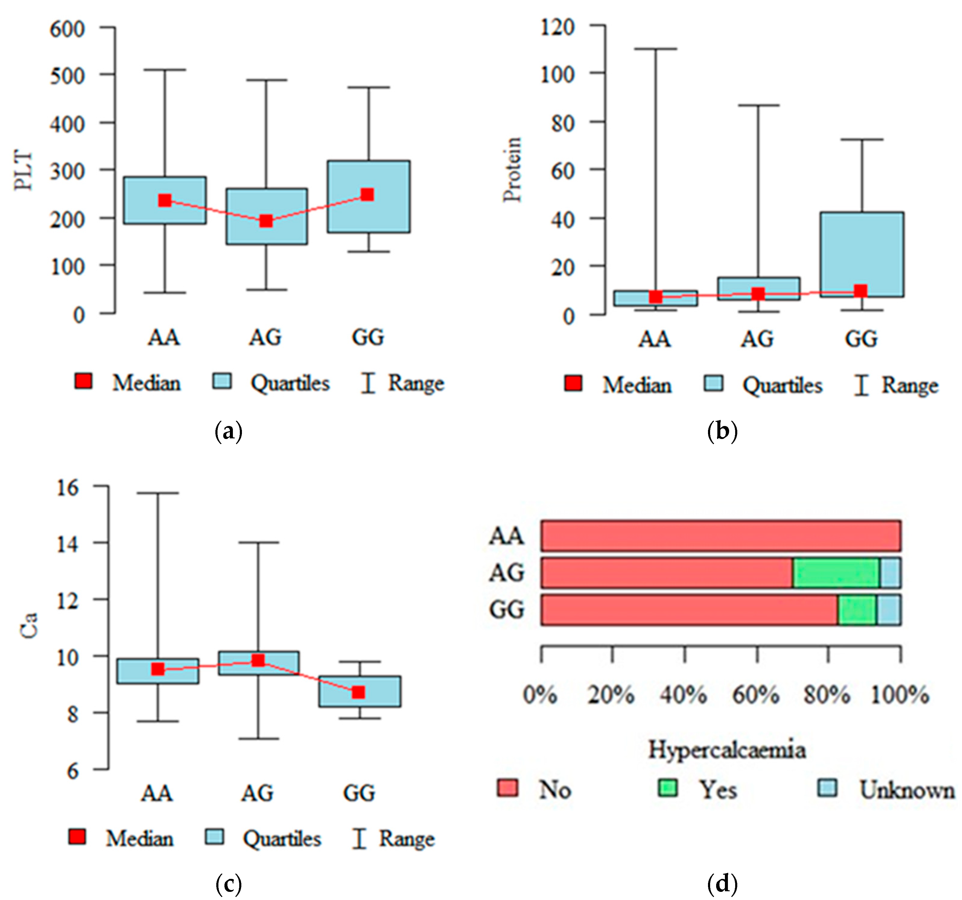
2.2. Associations with Diagnostic and Prognostic Parameters

Thrombocytopenia was observed in 18 patients (platelets < 100,103/µL), anaemia in 70 patients (haemoglobin < 10 G/L), leucocytosis in 17 patients (white blood cell count > 10,103/µL) and leukopenia in 44 patients (white blood cell count < 4 × 10³/µL). The average total protein concentration was 17.20 g/dL (median 8.40 dL), while the average concentration of beta-2-microglobulin was 5.74 g/dL (median 3.90 g/dL). The average creatinine serum level was 1.30 mg/dL (median 0.95 mg/dL). Patients with genotypes RIPK3 rs724165 GG and AA had a statistically higher platelet count than patients with genotype AG (p = 0.020, Figure 1a). However, RIPK3 rs724165 GG patients had a significantly higher level of total protein in serum compared to patients with genotype AA (p = 0.010, Figure 1b). Regarding the other SNPs, we observed that patients with genotype RIPK3 rs3212243 AG had statistically higher serum levels of calcium than patients with genotype GG (see Figure 1c). Moreover, we noted that hypercalcaemia was most common in the group of patients with variant MAPKAPK2 rs4073250 AG, and least common in patients with genotype AA (p = 0.039, Figure 1d).

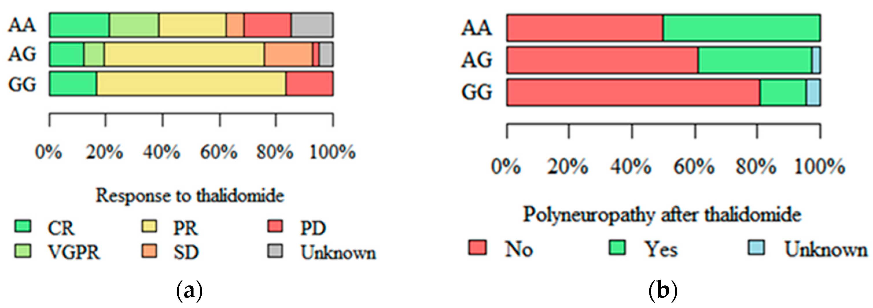
2.3. Response to Treatment and Peripheral Polyneuropathy

Differences in response to treatment with thalidomide were observed between patients with different RIPK3 rs3212243 genotypes. Both progressive disease (PD) and complete remission (CR) were significantly more common in patients with genotype GG as compared to other genotypes (p = 0.007, Figure 2a). Polyneuropathy as an adverse effect of thalidomide therapy was significantly more common among patients with MAPKAPK2 rs45514798 AA and less common in patients with genotype GG (p = 0.013, Figure 2b).

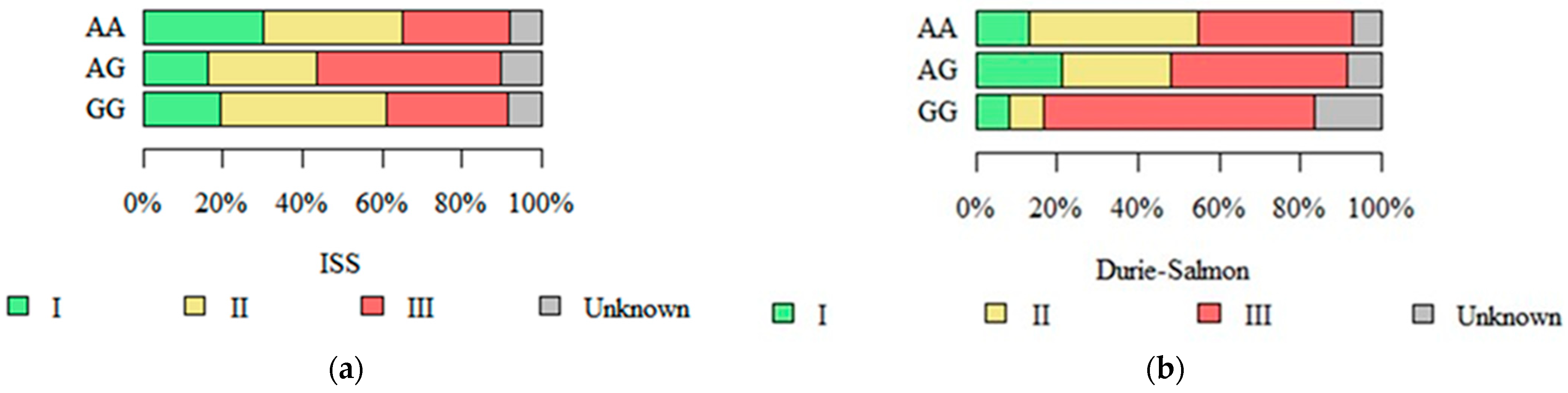
2.4. Stage in Durie–Salmon, ISS, and R-ISS Systems

We observed an association between RIPK3 rs724165 genotypes and the stage of disease at diagnosis defined by the International Staging System (ISS). The stage of disease was the highest in patients with genotype AG, and the lowest in patients with genotype AA (p = 0.038, see Figure 3a). Furthermore, an association between RIPK3 rs3212243 and the stages defined by the Durie–Salmon staging system were also observed. Patients with genotype GG had a more advanced stage at diagnosis than patients with genotypes AA and AG (p = 0.035; see Figure 3b).

2.5. Survival Analysis

2.5.1. Overall Survival

The median overall survival (OS) of all patients was 31 months. None of the analysed SNPs yielded any associations with OS in a univariate analysis. To account for the effect of potential confounding factors, we constructed a multivariate Cox proportional hazard model including various clinical parameters. The model includes RIPK1, RIPK3, and MAPKAPK2 SNPs as well as a number of clinical parameters. The analysis showed that the following genotypes are independent predictors of OS: RIPK1 rs9391981 CG, RIPK3 rs724165 CG, RIPK3 rs3212243 GG, MAP-KAPK2 rs4073250 AA (p < 0.05). Detailed results are presented in Table 2.

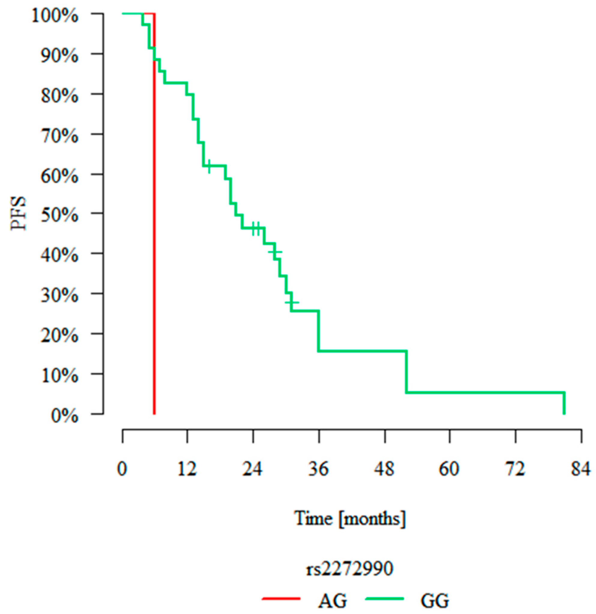
2.5.2. Progression-Free Survival

The median progression-free survival (PFS) of all patients was 12 months. None of the analysed SNPs yielded any associations with PFS in a univariate analysis when analysing the whole group, although an analysis of a subgroup of patients treated with lenalidomide showed that genotype RIPK1 rs2272990 GG was associated with longer progression-free survival than genotype AG (p = 0.003, Figure 4). Similarly to OS, as described in the previous section, we analysed the effect of SNPs on PFS using a multivariate Cox proportional hazard model. It showed that only genotype MAPKAPK2 rs4073250 AA was an independent predictor of PFS (p = 0.025, HR = 6.919). Details of that analysis are shown in Table 3.

3. Discussion

The role of necroptosis in the pathogenesis of cancer, including multiple myeloma, has been proven many times before [39,40]. Ali and Mocarski showed that proteasome inhibitors cause a biochemical blockade of necroptosis in multiple myeloma, and mentioned that the interaction between myeloma cells and carfilzomib led to apoptosis [41]. This suggests that the mechanism of activity of proteasome inhibitors could be based on promotion of apoptosis and inhibition of necroptosis. Polymorphic variants of genes encoding proteins crucial for necroptosis may affect this process. Consequently, they could affect the clinical course of multiple myeloma, especially in patients treated with proteasome inhibitors. In the present study, we showed that SNPs located in genes encoding RIPK1, RIPK3 and MAPKAPK2 may be associated with the clinical parameters of multiple myeloma.
The first SNP analysed in the present study was rs9391981 C>G, an intron variant located in the gene coding for RIPK1 [42]. It has only been described once before, and it has been shown before that the occurrence of genotypes CG and GG increases the risk of multiple myeloma [43]. Although no such association was observed in our current study, we found that genotype CG was associated with worse overall survival in a multivariate analysis. The lack of association with risk may be due to a relatively small sample size, especially given the low minor allele frequency of this SNP. Another RIPK1 SNP investigated in this study, rs2272990 A>G, is a synonymous variant situated in a coding region. While synonymous variants do not affect the amino acid sequence of a protein, they can influence mRNA structure and stability, as well as protein folding [44]. Our survival analysis of patients treated with lenalidomide showed that patients with genotype AG had a worse prognosis than patients with genotype GG. In a similar study focusing on diffuse large B cell lymphoma (DLBCL), GG homozygosity was associated with a significantly shorter PFS [45]. However, this genotype was also a marker of longer OS in patients with colorectal cancer after the resection of primary lesion. Furthermore, patients with genotype AA and AG showed a significant reduction in disease-free survival (DFS) [46]. In a study of hepatocellular cancer, genotype GG correlated with a higher stage of TMN (tumour, nodes, metastases) and metastases in lymph nodes. GG homozygosity was significantly associated with severe hepatic ischemia-reperfusion injury and decreased survival after hepatectomy [47].
Although we observed an association between RIPK1 rs2272990 and progression-free survival, no statistically significant difference in overall survival was detected in our study. This could be related to a break in the natural balance between apoptosis and necroptosis in myeloma cells. This balance is dependent on a direct interaction between RIPK1 and RIPK3 by the means of trans- and autophosphorylation [39]. Longer PFS in the group of patients with genotype GG could be caused by apoptosis being favoured as a result of lenalidomide treatment. Lenalidomide stimulates T cells and increases the production of pro-apoptotic factors such as interleukin 2, interferon γ, and decreases the secretion of TNF-alfa. Cytotoxic T cells may activate apoptosis by ligation of FasL to FasR. Interleukin 2 could also stimulate lymphocytes and NK cells [48]. Furthermore, immunomodulatory agents may induce apoptosis by activating caspase 8, favouring the ligation of FasL and decreasing the NF-kappa B concentration. Therapy might also possibly block the stimulation effect of insulin-like growth factor 1 on NF-κB [49]. Interestingly, we did not observe any association between rs2272990 and response for any type of treatment.
The role of RIPK1 and necroptosis in carcinogenesis, as well as its effect on course of disease and prognosis, has been shown in many cancers [50]. In head and neck squamous cell carcinoma, RIPK1 downregulation increased the risk of metastasis through inhibition of NF-κB-pathway-dependent TLR3 receptor and by promoting apoptosis over necrosis [51]. The overexpression of RIPK1 was also detected in pancreatic adenocarcinoma cells and increased after chemotherapy. In vitro blockade of necrosome proteins promoted proliferation and led to an aggressive phenotype of pancreatic adenocarcinoma [51]. This suggests that RIPK1 may have a significant effect on cancer development. In the case of multiple myeloma, inhibition of necroptosis may lead to worse prognosis. The mechanism behind this process is unclear. As observed by Ali and Mocarski, proteasome initiates degradation of cIAP by secretion of SMAC/DIABLO from mitochondria [41]. This leads to inhibition of pro-survival processes and protects cells from necroptosis. The accumulation of cIAP2 may have a protective effect on cancer cells and may work against SMAC-dependent apoptosis [41]. After sensitising myeloma cells with cycloheximide, the degradation of cFLIP protein was observed [41]. In our study, we did not observe any association between RIPK1 SNPs (either rs2272990 or rs9391981) and response to proteasome inhibitor therapy. Further studies may be required to establish if there is any association between RIPK1 SNPs and response to proteasome inhibitors affecting necroptosis in multiple myeloma patients.
Our study also included rs724165 G>A, an intronic variant of RIPK3 [42]. We found it to be associated with the stage of disease using the ISS system, but not Durie–Salmon. ISS has been shown to have higher predictive value than Durie–Salmon staging in patients who did not undergo auto-HSCT. In our study, no association between rs724165 and response to auto-HSCT was found. Patients with genotype GG in locus RIPK3 rs724165 had a significantly higher level of total protein in serum than patients with the AA genotype. Furthermore, the level of platelets in the group with genotype GG and AA was significantly higher than for patients with genotype AG. Low platelet counts (<100,000/µL) may be a factor leading to a reduction in PFS and OS [52]. Other diagnostic and prognostic parameters analysed in our study (haemoglobin level, calcium serum level, creatinine serum level, kappa and lambda light chains in serum, ESR, ratio of plasma cells in bone marrow, and frequency of bone lesions) did not show any significant differences in patients with different rs724165 genotypes. To the best of our knowledge, rs724165 has only been described twice before. The first study showed an association between rs724165 and time to treatment, as well as OS [53]. The other study focused on non-Hodgkin lymphoma, and found that rs724165 is not a risk factor for that disease [54]. Due to its involvement in necroptosis, RIPK3 may play a role in cancer development [5]. Decreased expression of RIPK3 was connected with poor prognosis for women with breast cancer [55], and decreased OS and PFS in patients with colorectal cancer [46], as well as accelerated leukogenesis in patients with AML [56,57]. The other RIPK3 SNP included in our study, rs3212243, is located in the flanking region of the gene. We found that among patients with genotype AA, the frequency of CR and PD after thalidomide treatment was significantly higher than among patients with other genotypes. The explanation and significance of that phenomenon is unclear. Furthermore, patients with the GG genotype are characterised by a higher Durie–Salmon stage than patients with genotypes AG and AA. We also observed that the calcium serum level was significantly higher in carriers of genotype AG than in those with genotype GG.
We also analysed two SNPs located in the gene encoding MAPKAPK2–rs45514798 and rs4073250. The former is an intronic variant, while the latter is a synonymous SNP located in the coding regions of MAPKAPK2. Little is known about these two SNPs. In the case of rs45514798, is has been analysed in relation to HDL serum level in a Uyghur population in Xinjiang, China. In a cohort consisting of men under 50, rs45514798 genotypes were found to be associated with serum level of HDL [58]. However, no association with either serum level of TNF-alpha or LDL was observed [59]. Our study is the first to analyse rs4073250. We proved that rs4073250 was associated with the calcium level in serum, with a higher calcium level being observed in AG heterozygotes. Regarding rs45514798, we showed that polyneuropathy after thalidomide therapy was most frequent in AA homozygotes. It has been shown previously that necroptosis may have a role in neuron injury and Parkinson’s disease [60,61,62]. Necroptosis may also have a role in axial degeneration of the central and peripheral nervous systems. This could partially explain the pathophysiology of neurodegenerative disease and adverse events after chemotherapy [63]. The exact mechanism of thalidomide-induced neurotoxicity is not clear. The antiangiogenic effect of thalidomide could lead to ischaemia and hypoxia of nerves [64]. Another possible mechanism is decreased expression of TNF-alfa and inhibition of NF-κB [55]. Oxidative damage of DNA is also postulated [17]. To the best of our knowledge, there are no studies describing any association between MAPKAPK2 and polyneuropathy in any disease. One study focusing on motor neuron disease showed a crucial role of SHSP1 (small heat shock protein 1), which is phosphorylated by MAPKAPK2 [20]. Mutations in the gene coding for SHSP1 (R127W and R135F) tend to form small oligomers, which increase phosphorylation of MAPKAPK2. That process could play a role in neuropathy in Charcot–Marie–Tooth disease [20].
A multivariate Cox proportional hazard model analysis proved that RIPK1 rs9391981 CG, RIPK3 rs724165 GG, RIPK3 rs3212243 GG, and MAPKAPK2 rs4073250 AA are independent prognostic factors of shorter overall survival. In the case of rs724165, this seems to be in line with other results showing that rs724165 GG is associated with high total protein at diagnosis. Likewise, it is in line with our findings that genotype rs3212243 GG is more common in patients with more advanced disease. A separate multivariate analysis also proved the significance of MAPKAPK2 rs4073250 AA as an independent prognostic factor of shorter PFS in patients with MM.
It should be acknowledged that the treatment regimens used in the studied cohort partly reflect earlier therapeutic standards, which may represent a limitation of this study in the context of current multiple myeloma treatment paradigms. However, it is important to emphasise that the fundamental mechanisms of action of the main drug classes—particularly immunomodulatory drugs (thalidomide, lenalidomide) and proteasome inhibitors (bortezomib)—remain unchanged and still form the foundation of contemporary therapeutic protocols. Moreover, the molecular mechanisms influencing necroptosis processes that we investigated are universal and independent of specific treatment regimens. The associations we observed between polymorphisms in RIPK1, RIPK3, and MAPKAPK2 genes and patient survival and treatment response reflect deeper biological relationships between genetic variants of necroptosis pathways and the pathophysiology of multiple myeloma. The value of the presented findings lies in their potential as prognostic biomarkers that could aid in patient risk stratification regardless of the treatment schemes employed. Furthermore, the identified polymorphisms may represent promising targets for future research on novel targeted therapies that could modulate necroptosis pathways to enhance treatment efficacy. In this context, our discoveries maintain significant scientific and clinical value, opening new perspectives for understanding the biological basis of multiple myeloma and personalising therapeutic approaches.
In summary, this is the first study to analyse the effect of SNPs in genes associated with necroptosis in a group of patients with multiple myeloma. Some of the SNPs have a potentially prognostic value and could be useful in clinical practice. Genotypes rs9391981 CG (RIPK1), rs724165 GG (RIPK3), rs3212243 GG (RIPK3) and rs4073250 AA (MAPKAPK2) were found to be independent prognostic factors connected with a reduction in overall survival in patients with multiple myeloma, while genotype rs4073250 AA (MAPKAPK2) also led to reduction in progression-free survival. We also proved that patients treated with lenalidomide who carried genotype rs2272990 GG (RIPK1) had a longer progression-free survival than those with AG heterozygotes. Polyneuropathy as an adverse effect of therapy with thalidomide was significantly more common in patients with genotype rs45514798 AA (MAPKAPK2) and least common in patients with genotype GG. These results suggest that SNPs in RIPK1, RIPK3 and MAPKAPK2, genes associated with necroptosis, may have an effect on multiple myeloma development and treatment. However, further studies on larger groups of patients may be required to confirm these results.

4. Materials and Methods

4.1. General Characteristic of Population

The clinical group was composed of 205 patients with multiple myeloma (102 women and 103 men) from the Alfred Sokołowski Specialist Hospital in Wałbrzych, Poland, and from the Clinical University Hospital in Wrocław, Poland. The inclusion criteria were age over 18 and a multiple myeloma diagnosis. Written informed consent was obtained from all participants. This project received a positive opinion of the Bioethical Committee of Medical University in Wrocław. This study was conducted in accordance with the ethical standards of the Declaration of Helsinki from 2013. The median age in the group of patients was 65 years (the youngest was 37 and the oldest was 86). Detailed information about the patients is included in Table 4.
Renal insufficiency was present in 55 patients, 32 patients had hypercalcaemia, and 99 had osteolytic lesions in bones. The secretion of IgG M protein was detected in 118 patients, IgA–in 45, IgD–1, IgM–1, and 1 patient secreted two classes of M protein, IgG and IgM. Non-secretory multiple myeloma was diagnosed in 23 cases and 1 participant had plasma cell leukaemia. The control group was composed of 100 healthy volunteers (58 women and 48 men), aged 27 to 76 years (median 59 years).
The treatment was as follows: 100 patients were treated with bortezomib, 46 with lenalidomide, and 124 with thalidomide in various lines of therapy. The participants received, on average, 2 lines of therapy (1 to 4). Thalidomide was given to 91 patients, bortezomib to 91 patients and lenalidomide to 62 patients. Megachemotherapy before autologous haematopoietic stem cell transplantation (auto-HSCT) was applied to 58 patients. Polyneuropathy was observed in 33 patients as a result of previous therapy (16 after thalidomide, 9 after bortezomib, 3 after lenalidomide—probably not associated with therapy—and 4 cases had unclear aetiology and could have been caused by diabetes mellitus or many lines of therapy).

4.2. Genotyping

DNA was isolated from samples of peripheral blood taken on EDTA during routine laboratory tests using Maxwell 16 Blood DNA Purification Kit (Promega Corp., Madison, WI, USA) or silica membranes (Qiagen, Hilden, Germany), following the manufacturers’ recommendations. Specific RIPK1, RIPK3, and MAPKAPK2 SNPs were selected based on previous studies [43,45,47,54,58] and the results from the SNP Function Prediction tool [42]. To detect genotypes, LightSNiP assays (TIB MOLBI-OL, Berlin, Germany) and the real-time PCR (Polimerase Chain Reaction) method with melting curve analysis were used. Reactions were performed in a LightCycler 480 II (Roche Diagnostics GmbH, Rotkreuz, Switzerland) device, according to the recommendations of the manufacturer.

4.3. Statistical Analysis

A comparison of the qualitative variables between groups as performed by the chi-squared test (with Yates correction) or Fisher’s exact test. Comparisons of quantitative variables between two groups were performed using the Mann–Whitney U test. Comparisons of three and more groups were performed by Kruskal–Wallis test and post hoc Dunn analysis if the difference was statistically significant. The multivariate analysis was performed by logistic regression. The results are presented as an odds ratio with a 95% confidence interval. A comparison of survival was performed by the long-rank test. Multivariate survival analysis was calculated using the Cox proportional hazard model. The results are presented as hazard ratios with a 95% confidence interval. p-values < 0.05 were considered statistically significant.

Author Contributions

Conceptualisation, A.B.; data curation, M.S. and P.Ł.; formal analysis, K.B.-K. and G.M.; funding acquisition, A.B.; investigation, M.S. and P.Ł.; methodology, P.Ł. and K.B.-K.; resources, M.S.; Supervision, K.B.-K., G.M. and A.B.; writing—original draft, M.S.; writing—review and editing, M.S., P.Ł., K.B.-K., G.M. and A.B. All authors have read and agreed to the published version of the manuscript.

Funding

This research was funded by the Wroclaw Medical University, grant number SUBZ.A360.24.056.

Institutional Review Board Statement

This study was conducted in accordance with the Declaration of Helsinki, and approved by the Ethics Committee of the Medical University in Wrocław (No. KB-301/2018).

Informed Consent Statement

Informed consent was obtained from all subjects involved in this study.

Data Availability Statement

The data presented in this study are available on request from the corresponding author.

Conflicts of Interest

The authors declare no conflicts of interest.

Abbreviations

The following abbreviations are used in this manuscript:
MMMultiple myeloma
ISSInternational Staging System
TNF-αTumour necrosis factor alpha
RIPK1Receptor-interacting serine/threonine kinase 1
RIPK3Receptor-interacting serine/threonine kinase 3
MLKLMixed lineage kinase domain-like pseudokinase
DAMPDamage-associated nuclear pattern
MAPKAPK2MAP kinase-activated protein kinase 2
SNPSingle-nucleotide polymorphism
CRComplete remission
PRPartial remission
VGPRVery good partial remission
SDStable disease
PDProgressive disease
OSOverall survival
PFSProgression-free survival
DLBCLDiffuse large B cell lymphoma
DFSDisease-free survival
TMNTumour, nodes, metastases
SHSP1Small heat shock protein 1
auto-HSCTAutologous haematopoietic stem cell transplantation

References

  1. Cid Ruzafa, J.; Merinopoulou, E.; Baggaley, R.F.; Leighton, P.; Werther, W.; Felici, D.; Cox, A. Patient population with multiple myeloma and transitions across different lines of therapy in the USA: An epidemiologic model. Pharmacoepidemiol. Drug Saf. 2016, 25, 871–879. [Google Scholar] [CrossRef] [PubMed]
  2. Park, S.; Zhao, D.; Hatanpaa, K.J.; Mickey, B.E.; Saha, D.; Boothman, D.A.; Story, M.D.; Wong, E.T.; Burma, S.; Georgescu, M.M.; et al. RIP1 activates PI3K-Akt via a dual mechanism involving NF-kappaB-mediated inhibition of the mTOR-S6K-IRS1 negative feedback loop and down-regulation of PTEN. Cancer Res. 2009, 69, 4107–4111. [Google Scholar] [CrossRef] [PubMed]
  3. Furukawa, Y.; Kikuchi, J. Molecular pathogenesis of multiple myeloma. Int. J. Clin. Oncol. 2015, 20, 413–422. [Google Scholar] [CrossRef]
  4. Durie, B.G.; Salmon, S.E. A clinical staging system for multiple myeloma. Correlation of measured myeloma cell mass with presenting clinical features, response to treatment, and survival. Cancer 1975, 36, 842–854. [Google Scholar] [CrossRef] [PubMed]
  5. Gong, Y.; Fan, Z.; Luo, G.; Yang, C.; Huang, Q.; Fan, K.; Cheng, H.; Jin, K.; Ni, Q.; Yu, X.; et al. The role of necroptosis in cancer biology and therapy. Mol. Cancer 2019, 18, 100. [Google Scholar] [CrossRef]
  6. Palumbo, A.; Anderson, K. Multiple myeloma. N. Engl. J. Med. 2011, 364, 1046–1060. [Google Scholar] [CrossRef]
  7. Terpos, E.; Katodritou, E.; Roussou, M.; Pouli, A.; Michalis, E.; Delimpasi, S.; Parcharidou, A.; Kartasis, Z.; Zomas, A.; Symeonidis, A.; et al. High serum lactate dehydrogenase adds prognostic value to the international staging system even in the era of novel agents. Eur. J. Haematol. 2010, 85, 114–119. [Google Scholar] [CrossRef]
  8. Adams, J. Proteasome inhibitors as new anticancer drugs. Curr. Opin. Oncol. 2002, 14, 628–634. [Google Scholar] [CrossRef]
  9. Arastu-Kapur, S.; Anderl, J.L.; Kraus, M.; Parlati, F.; Shenk, K.D.; Lee, S.J.; Muchamuel, T.; Bennett, M.K.; Driessen, C.; Ball, A.J.; et al. Nonproteasomal targets of the proteasome inhibitors bortezomib and carfilzomib: A link to clinical adverse events. Clin. Cancer Res. 2011, 17, 2734–2743. [Google Scholar] [CrossRef]
  10. Ludwig, H.; Durie, B.G.; Bolejack, V.; Turesson, I.; Kyle, R.A.; Blade, J.; Fonseca, R.; Dimopoulos, M.; Shimizu, K.; San Miguel, J.; et al. Myeloma in patients younger than age 50 years presents with more favorable features and shows better survival: An analysis of 10,549 patients from the International Myeloma Working Group. Blood 2008, 111, 4039–4047. [Google Scholar] [CrossRef]
  11. Holstein, S.A.; McCarthy, P.L. Immunomodulatory drugs in multiple myeloma: Mechanisms of action and clinical experience. Drugs 2017, 77, 505–520. [Google Scholar] [CrossRef] [PubMed]
  12. Vandenabeele, P.; Galluzzi, L.; Vanden Berghe, T.; Kroemer, G. Molecular mechanisms of necroptosis: An ordered cellular explosion. Nat. Rev. Mol. Cell Biol. 2010, 11, 700–714. [Google Scholar] [CrossRef]
  13. Ea, C.K.; Deng, L.; Xia, Z.P.; Pineda, G.; Chen, Z.J. Activation of IKK by TNFalpha requires site-specific ubiquitination of RIP1 and polyubiquitin binding by NEMO. Mol. Cell 2006, 22, 245–257. [Google Scholar] [CrossRef]
  14. Micheau, O.; Tschopp, J. Induction of TNF receptor I-mediated apoptosis via two sequential signaling complexes. Cell 2003, 114, 181–190. [Google Scholar] [CrossRef] [PubMed]
  15. Wegner, K.W.; Saleh, D.; Degterov, A. Complex Pathologic Roles of RIPK1 and RIPK3: Moving Beyond Necroptosis. Trends Pharmacol. Sci. 2017, 38, 202–225. [Google Scholar] [CrossRef]
  16. Annibaldi, A.; Wicky, J.S.; Vanden Berghe, T.; Swatek, K.N.; Ruan, J.; Liccardi, G.; Bianchi, K.; Elliott, P.R.; Choi, S.M.; Van Coillie, S.; et al. Ubiquitin-Mediated Regulation of RIPK1 Kinase Activity Independent of IKK and MK2. Mol. Cell 2018, 15, 566–580. [Google Scholar] [CrossRef] [PubMed]
  17. Wani, T.H.; Chakrabarty, A.; Shibata, N.; Yamazaki, H.; Guengerich, F.P.; Chowdhury, G. The Dihydroxy Metabolite of the Teratogen Thalidomide Causes Oxidative DNA Damage. Chem. Res. Toxicol. 2017, 30, 1622–1628. [Google Scholar] [CrossRef] [PubMed]
  18. Tummers, B.; Green, D.R. Caspase-8; regulating life and death. Immunol. Rev. 2017, 277, 76–89. [Google Scholar] [CrossRef]
  19. Weeks, S.D.; Muranova, L.K.; Heirbaut, M.; Beelen, S.; Strelkov, S.V.; Gusev, N.B. Characterization of human small heat shock protein HSPB1 α-crystallin domain localised mutants associated with hereditary motor neuron diseases. Sci. Rep. 2018, 8, 688. [Google Scholar] [CrossRef]
  20. Maliński, M.; Cichocki, M. Inhibicja aktywności proteasomu jako nowa strategia w terapii i chemioprewencji nowotworów. Postep. Hig. Med. Dosw. 2013, 67, 90–106. [Google Scholar] [CrossRef]
  21. Vercammen, D.; Brouckaert, G.; Denecker, G.; Van de Craen, M.; Declercq, W.; Fiers, W.; Vandenabeele, P. Dual signaling of the Fas receptor: Initiation of both apoptotic and necrotic cell death pathways. J. Exp. Med. 1998, 188, 919–930. [Google Scholar] [CrossRef] [PubMed]
  22. Hitomi, J.; Christofferson, D.E.; Ng, A.; Yao, J.; Degterev, A.; Xavier, R.J.; Yuan, J. Identification of a molecular signaling network that regulates a cellular necrotic cell death pathway. Cell 2008, 135, 1311–1323. [Google Scholar] [CrossRef] [PubMed]
  23. Moquin, D.M.; McQuade, T.; Chan, F.K. CYLD deubiquitinates RIP1 in the TNFα-induced necrosome to facilitate kinase activation and programmed necrosis. PLoS ONE 2013, 8, e76841. [Google Scholar] [CrossRef]
  24. O’Donnell, M.A.; Perez-Jimenez, E.; Oberst, A.; Ng, A.; Massoumi, R.; Xavier, R.; Green, D.R.; Ting, A.T. Caspase 8 inhibits programmed necrosis by processing CYLD. Nat. Cell Biol. 2011, 13, 1437–1442. [Google Scholar] [CrossRef]
  25. Li, D.; Xu, T.; Cao, Y.; Wang, H.; Li, L.; Chen, S.; Wang, X.; Shen, Z. A cytosolic heat shock protein 90 and cochaperone CDC37 complex is required for RIP3 activation during necroptosis. Proc. Natl. Acad. Sci. USA 2015, 112, 5017–5022. [Google Scholar] [CrossRef] [PubMed]
  26. Li, J.; McQuade, T.; Siemer, A.B.; Napetschnig, J.; Moriwaki, K.; Hsiao, Y.S.; Damko, E.; Moquin, D.; Walz, T.; McDermott, A.; et al. The RIP1/RIP3 necrosome forms a functional amyloid signaling complex required for programmed necrosis. Cell 2012, 150, 339–350. [Google Scholar] [CrossRef]
  27. Rodriguez, D.A.; Weinlich, R.; Brown, S.; Guy, C.; Fitzgerald, P.; Dillon, C.P.; Oberst, A.; Quarato, G.; Low, J.; Cripps, J.G.; et al. Characterization of RIPK3-mediated phosphorylation of the activation loop of MLKL during necroptosis. Cell Death Differ. 2016, 23, 76–88. [Google Scholar] [CrossRef]
  28. Chen, W.; Wu, J.; Li, L.; Zhang, Z.; Ren, J.; Liang, Y.; Chen, F.; Yang, C.; Zhou, Z.; Su, S.S.; et al. Ppm1b negatively regulates necroptosis through dephosphorylating Rip3. Nat. Cell Biol. 2015, 17, 434–444. [Google Scholar] [CrossRef]
  29. Raninga, P.V.; Di Trapani, G.; Vuckovic, S.; Tonissen, K.F. Targeted knockdown of DJ-1 induces multiple myeloma cell death via KLF6 upregulation. Apoptosis 2016, 12, 1422–1437. [Google Scholar] [CrossRef]
  30. Murphy, J.M.; Czabotar, P.E.; Hildebrand, J.M.; Lucet, I.S.; Zhang, J.G.; Alvarez-Diaz, S.; Lewis, R.; Lalaoui, N.; Metcalf, D.; Webb, A.I.; et al. The pseudokinase MLKL mediates necroptosis via a molecular switch mechanism. Immunity 2013, 39, 443–453. [Google Scholar] [CrossRef]
  31. Quarato, G.; Guy, C.S.; Grace, C.R.; Llambi, F.; Nourse, A.; Rodriguez, D.A.; Wakefield, R.; Frase, S.; Moldoveanu, T.; Green, D.R. Sequential engagement of distinct MLKL phosphatidylinositol-binding sites executes necroptosis. Mol. Cell 2016, 61, 589–601. [Google Scholar] [CrossRef] [PubMed]
  32. Lu, W.; Sun, J.; Yoon, J.S.; Zhang, Y.; Zheng, L.; Murphy, E.; Mattson, M.P.; Lenardo, M.J. Mitochondrial protein PGAM5 regulates mitophagic protection against cell necroptosis. PLoS ONE 2016, 11, e0147792. [Google Scholar] [CrossRef] [PubMed]
  33. Kaczmarek, A.; Vandenabeele, P.; Krysko, D.V. Necroptosis: The Release of Damage-Associated Molecular Patterns and Its Physiological Relevance. Immunity 2013, 38, 209–223. [Google Scholar] [CrossRef]
  34. Iwama, K.; Chihara, D.; Tsuda, K.; Ugai, T.; Sugihara, H.; Nishida, Y.; Yamakura, M.; Takeuchi, M.; Matsue, K. Normalization of free light chain kappa/lambda ratio is a robust prognostic indicator of favorable outcome in patients with multiple myeloma. Eur. J. Haematol. 2012, 90, 134–141. [Google Scholar] [CrossRef]
  35. Degterev, A.; Maki, J.L.; Yuan, J. Activity and specificity of necrostatin-1, small-molecule inhibitor of RIP1 kinase. Cell Death Differ. 2013, 20, 366. [Google Scholar] [CrossRef]
  36. Butrym, A.; Rybka, J.; Łacina, P.; Gębura, K.; Frontkiewicz, D.; Bogunia-Kubik, K.; Mazur, G. Polymorphisms within beta-catenin encoding gene affect multiple myeloma development and treatment. Leuk. Res. 2015, 39, 1462–1466. [Google Scholar] [CrossRef]
  37. Łacina, P.; Butrym, A.; Mazur, G.; Bogunia-Kubik, K. BSG and MCT1 Genetic Variants Influence Survival in Multiple Myeloma Patients. Genes 2018, 9, 226. [Google Scholar] [CrossRef] [PubMed]
  38. Dratwa, M.; Łacina, P.; Butrym, A.; Porzuczek, D.; Mazur, G.; Bogunia-Kubik, K. Telomere length and hTERT genetic variants as potential prognostic markers in multiple myeloma. Sci. Rep. 2023, 13, 15792. [Google Scholar] [CrossRef]
  39. Galluzzi, L.; Kepp, O.; Chan, F.K.; Kroemer, G. Necroptosis: Mechanisms and Relevance to Disease. Annu. Rev. Pathol. 2017, 12, 103–130. [Google Scholar] [CrossRef]
  40. Rajkumar, S.V.; Kumar, S. Multiple Myeloma: Diagnosis and Treatment. Mayo Clin. Proc. 2016, 91, 101–119. [Google Scholar] [CrossRef]
  41. Ali, M.; Mocarski, E.S. Proteasome inhibition blocks necroptosis by attenuating death complex aggregation. Cell Death Dis. 2018, 9, 346. [Google Scholar] [CrossRef] [PubMed]
  42. Xu, Z.; Taylor, J.A. SNPinfo: Integrating GWAS and candidate gene information into functional SNP selection for genetic association studies. Nucleic Acids Res. 2009, 37, W600–W605. [Google Scholar] [CrossRef]
  43. Hosgood, H.D., 3rd; Baris, D.; Zhang, Y.; Berndt, S.I.; Menashe, I.; Morton, L.M.; Lee, K.M.; Yeager, M.; Zahm, S.H.; Chanock, S.; et al. Genetic variation in cell cycle and apoptosis related genes and multiple myeloma risk. Leuk. Res. 2009, 33, 1609–1614. [Google Scholar] [CrossRef]
  44. Hunt, R.; Sauna, Z.E.; Ambudkar, S.V.; Gottesman, M.M.; Kimchi-Sarfaty, C. Silent (synonymous) SNPs: Should we care about them? Methods Mol. Biol. 2009, 578, 23–39. [Google Scholar] [PubMed]
  45. Chae, Y.S.; Kim, J.G.; Sohn, S.K.; Moon, J.H.; Kim, S.N.; Lee, S.J.; Park, T.I.; Lee, M.H. Lymphotoxin alfa and receptor-interacting protein kinase 1 gene polymorphisms may correlate with prognosis in patients with diffuse large B cell lymphoma treated with R-CHOP. Cancer Chemother. Pharmacol. 2010, 65, 571–577. [Google Scholar] [CrossRef]
  46. Feng, X.; Song, Q.; Yu, A.; Tang, H.; Peng, Z.; Wang, X. Receptor-interacting protein kinase 3 is a predictor of survival and plays a tumor suppressive role in colorectal cancer. Neoplasma 2015, 62, 592–601. [Google Scholar] [CrossRef] [PubMed]
  47. Yao, C.; Li, G.; Cai, M.; Qian, Y.; Wang, L.; Xiao, L.; Thaiss, F.; Shi, B. Expression and genetic polymorphism of necroptosis related protein RIPK1 is correlated with severe hepatic ischemia-reperfusion injury and prognosis after hepatectomy in hepatocellular carcinoma patients. Cancer Biomark. 2017, 20, 23–29. [Google Scholar] [CrossRef]
  48. Piechnik, A.; Giannopoulos, K. Mechanizmy działania leków immunomodulujących w szpiczaku plazmocytowym. Hematologia 2011, 2, 105–115. [Google Scholar]
  49. Mitsiades, N.; Mitsiades, C.S.; Poulaki, V.; Chauhan, D.; Richardson, P.G.; Hideshima, T.; Munshi, N.C.; Treon, S.P.; Anderson, K.C. Apoptotic signaling induced by immunomodulatory thalidomide analogs in human multiple myeloma cells: Therapeutic implications. Blood 2002, 99, 4525–4530. [Google Scholar] [CrossRef]
  50. McCormick, K.D.; Ghosh, A.; Trivedi, S.; Wang, L.; Coyne, C.B.; Ferris, R.L.; Sarkar, S.N. Innate immune signaling through differential RIPK1 expression promote tumor progression in head and neck squamous cell carcinoma. Carcinogenesis 2016, 37, 522–529. [Google Scholar] [CrossRef]
  51. Seifert, L.; Werba, G.; Tiwari, S.; Giao Ly, N.N.; Alothman, S.; Alqunaibit, D.; Avanzi, A.; Barilla, R.; Daley, D.; Greco, S.H.; et al. The necrosome promotes pancreatic oncogenesis via CXCL1 and Mincle-induced immune suppression. Nature 2016, 532, 245–249. [Google Scholar] [CrossRef] [PubMed]
  52. Mellors, P.W.; Binder, M.; Buadi, F.K.; Lacy, M.Q.; Gertz, M.A.; Dispenzieri, A.; Hayman, S.R.; Kapoor, P.; Gonsalves, W.I.; Hwa, Y.L.; et al. Development of thrombocytopenia during first-line treatment and survival outcomes in newly diagnosed multiple myeloma. Leuk. Lymphoma 2019, 60, 2960–2967. [Google Scholar] [CrossRef] [PubMed]
  53. Moreau, P.; San Miguel, J.; Sonneveld, P.; Mateos, M.V.; Zamagni, E.; Avet-Loiseau, H.; Hajek, R.; Dimopoulos, M.A.; Ludwig, H.; Einsele, H.; et al. Multiple myeloma: ESMO Clinical Practice Guidelines for diagnosis, treatment and follow-up. Ann. Oncol. 2017, 28, 52–61. [Google Scholar] [CrossRef] [PubMed]
  54. Cerhan, J.R.; Ansell, S.M.; Fredericksen, Z.S.; Kay, N.E.; Liebow, M.; Call, T.G.; Dogan, A.; Cunningham, J.M.; Wang, A.H.; Liu-Mares, W.; et al. Genetic variation in 1253 immune and inflammation genes and risk of non-Hodgkin lymphoma. Blood 2007, 110, 4455–4463. [Google Scholar] [CrossRef]
  55. Keifer, J.A.; Guttridge, D.C.; Ashburner, B.P.; Baldwin, A.S., Jr. Inhibition of NF-kappa B activity by thalidomide through suppression of IkappaB kinase activity. J. Biol. Chem. 2001, 276, 22382–22387. [Google Scholar] [CrossRef]
  56. Höckendorf, U.; Yabal, M.; Herold, T.; Munkhbaatar, E.; Rott, S.; Jilg, S.; Kauschinger, J.; Magnani, G.; Reisinger, F.; Heuser, M.; et al. RIPK3 restricts myeloid Leukemogenesis by promoting cell death and differentiation of leukemia initiating cells. Cancer Cell 2016, 30, 75–91. [Google Scholar] [CrossRef]
  57. Nugues, A.-L.; El Bouazzati, H.; Hétuin, D.; Berthon, C.; Loyens, A.; Bertrand, E.; Jouy, N.; Idziorek, T.; Quesnel, B. RIP3 is downregulated in human myeloid leukemia cells and modulates apoptosis and caspase-mediated p65/RelA cleavage. Cell Death Dis. 2014, 5, e1384. [Google Scholar] [CrossRef]
  58. Li, N.; Lin, N.; Yao, X.; Wang, H.; Zhou, L.; Guo, Y. Association of MK2 and ZFP36 gene polymorphisms with high density lipoprotein cholesterol in Uygur population from Hetian area of Xinjiang. Zhonghua Yi Xue Yi Chuan Xue Za Zhi 2013, 30, 345–351. [Google Scholar]
  59. Lin, N.; Li, N.F.; Yao, X.G.; Wang, H.M.; Liang, D.P.; Guo, Y.Y.; Zhou, L. Association of MK2 gene polymorphisms with low-density lipoprotein cholesterol and tumor necrosis factor-alpha in Uygur population from Hetian area of Xinjiang. Zhongguo Yi Xue Ke Xue Yuan Xue Bao 2013, 35, 52–57. [Google Scholar]
  60. Wang, X.; Wang, Y.; Ding, Z.J.; Yue, B.; Zhang, P.Z.; Chen, X.D.; Chen, X.; Chen, J.; Chen, F.Q.; Chen, Y.; et al. The role of RIP3 mediated necroptosis in ouabain-induced spiral ganglion neurons injuries. Neurosci. Lett. 2014, 578, 111–116. [Google Scholar] [CrossRef]
  61. Iannielli, A.; Bido, S.; Folladori, L.; Segnali, A.; Cancellieri, C.; Maresca, A.; Massimino, L.; Rubio, A.; Morabito, G.; Caporali, L. Pharmacological Inhibition of Necroptosis Protects from Dopaminergic Neuronal Cell Death in Parkinson’s Disease Models. Cell Rep. 2018, 22, 2066–2079. [Google Scholar] [CrossRef] [PubMed]
  62. Pu, S.; Li, S.; Xu, Y.; Wu, J.; Lv, Y.; Du, D. Role of receptor-interacting protein 1/receptor-interacting protein 3 in inflammation and necrosis following chronic constriction injury of the sciatic nerve. Neuroreport 2018, 29, 1373–1378. [Google Scholar] [CrossRef] [PubMed]
  63. Arrázola, M.S.; Saquel, C.; Catalán, R.J.; Barrientos, S.A.; Hernandez, D.E.; Martínez, N.W.; Catenaccio, A.; Court, F.A. Axonal Degeneration Is Mediated by Necroptosis Activation. J. Neurosci. 2019, 39, 3832–3844. [Google Scholar] [CrossRef] [PubMed]
  64. Tamilarasan, K.P.; Kolluru, G.K.; Rajaram, M.; Indhumathy, M.; Saranya, R.; Chatterjee, S. Thalidomide attenuates nitric oxide mediated angiogenesis by blocking migration of endothelial cells. BMC Cell Biol. 2007, 7, 17. [Google Scholar] [CrossRef]
Figure 1. Associations between various clinical parameters in patients with multiple myeloma and RIPK3/MAPKAPK2 genotypes: (a) total serum protein level and RIPK3 rs724165 genotypes; (b) platelet count and RIPK3 rs724165 genotypes; (c) serum calcium level and RIPK3 rs3212243 genotypes; (d) hypercalcaemia and MAPKAPK2 rs4073250 genotypes.
Ijms 26 05237 g001
Figure 2. Response to treatment with thalidomide in patients with different RIPK3 rs3212243 genotypes (a), and peripheral polyneuropathy after thalidomide therapy, in patients with different MAPKAPK2 rs45514798 genotypes (b). Abbreviations: CR—complete remission, PR—partial remission, VGPR—very good partial remission, SD—stable disease, PD—progressive disease.
Ijms 26 05237 g002
Figure 3. ISS stages and different RIPK3 rs724165 genotypes (a), and Durie–Salmon and different RIPK3 rs3212243 genotypes (b).
Ijms 26 05237 g003
Figure 4. Progression-free survival (PFS) Kaplan–Meier curves for a subgroup of patients treated with lenalidomide carrying different RIPK1 rs2272990 genotypes.
Ijms 26 05237 g004
Table 1. Distribution of genotypes in MM patients.
MM PatientsControl Group
RIPK1 rs2272990
GG18894
AG176
AA00
RIPK1 rs9391981
GG18992
CG168
CC00
RIPK3 rs724165
AG10652
AA6334
GG3613
RIPK3 rs3212243
AA12254
AG7144
GG122
MAPKAPK2 rs4073250
GG13058
AG6731
AA48
MAPKAPK2 rs45514798
GG13973
AG6224
AA43
Table 2. Multivariate Cox proportional hazard model analysis of overall survival. Statistically significant p-values are indicated in bold.
FeatureHR95% CIp-Value
Sexfemale1ref.
male0.4230.1051.7030.226
Age[years]1.2461.1151.392<0.001
Durie–SalmonI1ref.
II2.0770.22818.9270.517
III23.2612.007269.5900.012
ISSI1ref.
II1.6560.20113.6080.639
III0.1020.0042.5180.163
Type of light chainLambda1ref.
Kappa12.5332.01677.9190.007
Type of heavy chainA1ref.
G12.5330.0581.2020.085
Haemoglobin 0.9200.5971.4170.706
HypercalcaemiaNo1ref.
Yes0.1850.0152.2400.185
Renal insufficiencyNo1ref.
Yes1.1540.2255.9330.864
OsteolysisNo1ref.
Yes4.1860.85120.5910.078
RIPK1 rs2272990GG1ref.
AGNo deaths—impossible to calculate
RIPK1 rs9391981GG1ref.
CG71.6142.3332198.0700.014
RIPK3 rs724165AA1ref.
AG0.5670.0893.6000.548
GG<0.001<0.0010.1440.009
RIPK3 rs3212243AA1ref.
AG5.1990.67939.7950.112
GG1389.5873.493552,848.3940.018
MAPKAPK2 rs4073250GG1ref.
AG1.0290.2144.9410.971
AA30.6711.356693.5260.031
MAPKAPK2 rs45514798GG1ref.
AG2.2330.48810.2300.301
AANo deaths—impossible to calculate
Table 3. Multivariate Cox proportional hazard model analysis of progression-free survival. Statistically significant p-values are indicated in bold.
FeatureHR95% CIp-Value
Sexfemale1ref.
male1.0090.5311.9180.978
Age[years]1.0190.9851.0550.275
Durie–SalmonI1ref.
II0.6480.2441.7200.384
III0.7530.2662.1250.591
ISSI1ref.
II1.6660.6364.3660.299
III1.1390.3533.6770.828
Type of light chainLambda1ref.
Kappa1.6390.7973.3700.179
Type of heavy chainA1ref.
G1.0500.5152.1430.893
Haemoglobin 0.8970.7381.0920.279
HypercalcaemiaNo1ref.
Yes1.3730.4763.9560.557
Renal insufficiencyNo1ref.
Yes1.2260.6552.2930.524
OsteolysisNo1ref.
Yes0.8850.4671.6790.709
Response to the first line of treatmentCR1ref.
VGPR0.8720.2812.7060.813
PR1.1120.5022.4630.793
SD1.0460.4062.7000.925
PD0.3570.0851.5030.160
RIPK1 rs2272990GG1ref.
AG0.6760.0597.7320.753
RIPK1 rs9391981GG1ref.
CG1.2070.114.6110.882
RIPK3 rs724165AA1ref.
AG1.5130.6973.2850.295
GG0.6450.2181.9090.428
RIPK3 rs3212243AA1ref.
AG0.7860.3821.6190.514
GG2.2650.45311.3160.319
MAPKAPK2 rs4073250GG1ref.
AG1.1250.5482.3080.749
AA6.9191.27737.4730.025
MAPKAPK2 rs45514798GG1ref.
AG1.8840.9453.7570.072
AA3.0600.26435.460.371
Table 4. Patients’ characteristics.
N = 205
Sex
female102 (49.8%)
male103 (50.2%)
Age
median65 years
range37–88 years
Immunoglobulin secretion
IgG118 (57.6%)
IgA45 (22.0%)
IgM1 (0.5%)
IgD1 (0.5%)
IgM and IgG1 (0.5%)
Light chain15 (7.3%)
Plasma cell leukemia1 (0.5%)
Non-secretory23 (11.2%)
ISS stage
I44 (21.5%)
II67 (32.7%)
III80 (39.0%)
unknown14 (6.8%)
Durie–Salmon stage
I33 (16.1%)
II72 (35.1%)
III88 (42.9%)
unknown12 (5.9%)
Therapy
thalidomide91 (44.4%)
bortezomib91 (44.4%)
lenalidomide62 (30.2%)
auto-HSCT58 (28.3%)
Other parameters
renal insufficiency55 (26.8%)
hypercalcaemia32 (15.6%)
osteolytic lesions99 (48.3%)
Disclaimer/Publisher’s Note: The statements, opinions and data contained in all publications are solely those of the individual author(s) and contributor(s) and not of MDPI and/or the editor(s). MDPI and/or the editor(s) disclaim responsibility for any injury to people or property resulting from any ideas, methods, instructions or products referred to in the content.

