Article

Potential Influence of ADAM9 Genetic Variants and Expression Levels on the EGFR Mutation Status and Disease Progression in Patients with Lung Adenocarcinoma

by
Jer-Hwa Chang
1,2,3,†,
Tsung-Ching Lai
2,3,†,
Kuo-Hao Ho
4,
Thomas Chang-Yao Tsao
5,6,
Lun-Ching Chang
7,
Shun-Fa Yang
8,9,* and
Ming-Hsien Chien
2,10,11,12,*
1
School of Respiratory Therapy, College of Medicine, Taipei Medical University, Taipei 11031, Taiwan
2
Pulmonary Research Center, Wan Fang Hospital, Taipei Medical University, Taipei 11696, Taiwan
3
Division of Pulmonary Medicine, Department of Internal Medicine, Wan Fang Hospital, Taipei Medical University, Taipei 11696, Taiwan
4
Department of Biochemistry and Molecular Cell Biology, School of Medicine, College of Medicine, Taipei Medical University, Taipei 11031, Taiwan
5
School of Medicine, Chung Shan Medical University, Taichung 40201, Taiwan
6
Department of Internal Medicine, Chung Shan Medical University Hospital, Taichung 40201, Taiwan
7
Department of Mathematical Sciences, Florida Atlantic University, Boca Raton, FL 33431, USA
8
Institute of Medicine, Chung Shan Medical University, Taichung 40201, Taiwan
9
Department of Medical Research, Chung Shan Medical University Hospital, Taichung 40201, Taiwan
10
Graduate Institute of Clinical Medicine, College of Medicine, Taipei Medical University, Taipei 11031, Taiwan
11
Traditional Herbal Medicine Research Center, Taipei Medical University Hospital, Taipei 110301, Taiwan
12
TMU Research Center of Cancer Translational Medicine, Taipei Medical University, Taipei 11031, Taiwan
*
Authors to whom correspondence should be addressed.
These authors contributed equally to this work.
Int. J. Mol. Sci. 2025, 26(10), 4606; https://doi.org/10.3390/ijms26104606
Submission received: 28 March 2025 / Revised: 22 April 2025 / Accepted: 8 May 2025 / Published: 11 May 2025
(This article belongs to the Section Molecular Pathology, Diagnostics, and Therapeutics)

Abstract

:
Lung adenocarcinoma (LUAD) is driven by epidermal growth factor receptor (EGFR) mutations, making it a key therapeutic target. ADAM9, a member of the A disintegrin and metalloproteinase (ADAM) family, facilitates the release of growth factors and was implicated in activating the EGFR-mediated progression in several cancer types. In this study, we explored potential associations among ADAM9 single-nucleotide polymorphisms (SNPs), the EGFR mutation status, and the clinicopathological progression of LUAD in a Taiwanese population. In total, 535 LUAD patients with various EGFR statuses were enrolled, and allelic distributions of ADAM9 SNPs—located in promoter and intron regions, including rs78451751 (T/C), rs6474526 (T/G), rs7006414 (T/C), and rs10105311 (C/T)—were analyzed using a TaqMan allelic discrimination assay. We found that LUAD patients with at least one polymorphic G allele in ADAM9 rs6474526 had a lower risk of developing EGFR mutations compared to those with the wild-type (WT) TT genotype. Furthermore, G-allele carriers (TG + GG) of rs6474526 were associated with an increased likelihood of developing larger tumors (T3 or T4), particularly among patients with mutant EGFR. Conversely, in patients with WT EGFR, carriers of the T allele in rs10105311 had a lower risk of progressing to advanced stages (stage III or IV). Among females or non-smokers, G-allele carriers of rs6474526 demonstrated a higher risk of advanced tumor stages and distant metastases. In clinical data from the Genotype-Tissue Expression (GTEx) database, individuals with the polymorphic T allele in rs6474526 showed reduced ADAM9 expression in lung and whole blood tissues. Screening the genotype of rs6474526 in a set of LUAD cell lines revealed that cells carrying at least one minor G allele exhibited higher ADAM9 levels compared to those with the TT genotype. Additionally, analyses using TCGA and CPTAC databases revealed elevated ADAM9 expression in LUAD specimens compared to normal tissues. Elevated protein levels were correlated with advanced T stages, pathological stages, and worse prognoses. In summary, our results suggest that ADAM9 genetic variants of rs6474526 may affect ADAM9 expression and are associated with the EGFR mutation status. Both rs6474526 and rs10105311 were correlated with disease progression in LUAD patients. These variants could serve as potential biomarkers for predicting clinical outcomes.

1. Introduction

Lung cancer is the most common cancer worldwide and ranks among the top three leading causes of cancer-related mortality [1]. Non-small-cell lung cancer (NSCLC) accounts for about 85% of lung cancer cases, with lung adenocarcinoma (LUAD) being the most common subtype. Alterations in oncogenic driver genes are one of the important causes of NSCLC. The epidermal growth factor receptor (EGFR), a cell surface tyrosine kinase receptor, is commonly mutated in LUAD, especially in East Asians, females, and non-smokers [2,3,4]. The two most common somatic mutations in NSCLC, particularly in LUAD, are an in-frame deletion in exon 19 and the L858R point mutation in exon 21, which together account for about 85% of all EGFR mutations. These mutations enhance EGFR activity, driving tumor cell proliferation, angiogenesis, migration, and invasion, thereby accelerating tumor progression. Consequently, recent research indicated that LUAD patients with EGFR mutations are ideal candidates for treatment with EGFR tyrosine kinase inhibitors (TKIs) [4].
A genetic predisposition for lung cancer has recently garnered significant attention from thoracic oncologists and geneticists, driven by the increasing focus on lung cancer in non-smokers and the widespread adoption of somatic next-generation sequencing panels that encompass cancer susceptibility genes [5]. EGFR somatic mutations are present in 30–50% of East Asian NSCLC patients, compared to less than 20% in other ethnic groups. Notably, this high prevalence remains consistent among East Asians who migrate to other regions, suggesting that genetic factors, rather than geographic or environmental influences, play a key role in mutation development [6]. Single-nucleotide polymorphisms (SNPs), which are variations in the DNA sequence of a genome, can result in alterations in genes that impact the function of cellular proteins and enzymes [7,8]. Genome-wide association studies (GWASs) and fine-mapping analyses have identified numerous SNPs associated with LUAD susceptibility in East Asians, including rs137884934 in the PIK3CB gene, rs682888 in the DTNB gene, rs116863980 in the PALM gene, rs4268071 in the GPR37 gene, and so on [9]. Furthermore, GWASs have pinpointed SNPs potentially linked to an increased risk of EGFR mutation-positive LUAD, such as rs2179920 in the HLA-DPB1 gene and rs2495239 in the FOXP4 gene [10].
A disintegrin and metalloproteinase 9 (ADAM9) is a type I transmembrane glycoprotein that is part of the ADAM family. Recent research highlighted its association with the progression of various cancers [11,12]. In LUAD, ADAM9 is notably overexpressed and was shown to promote tumor growth in xenograft models by regulating angiogenic factors such as vascular endothelial growth factor A, interleukin-8, and angiopoietin-2 [13,14]. Additionally, increased ADAM9 levels have been recognized as an independent prognostic marker for overall survival (OS) in stage I NSCLC patients who underwent standard lobectomy and mediastinal N2 lymph node dissection [15]. The metalloprotease domain of ADAM9 is capable of cleaving the precursor of heparin-binding EGF-like growth factor (HB-EGF), producing soluble HB-EGF [16]. This, in turn, activates EGFR signaling, which drives tumor growth and angiogenesis [17]. Notably, ADAM9 knockdown was shown to reduce EGFR signaling and enhance sensitivity to EGFR inhibitors in LUAD cells [18]. Furthermore, ADAM9 was implicated in promoting LUAD cell metastasis by increasing tissue plasminogen activator activity and activating the EGFR, which stimulates the migration-promoting protein, CUB domain-containing protein 1 [18,19]. The functional roles and clinical significance of the ADAM9/EGFR axis in LUAD have been previously reported. Studies have shown a correlation between pneumonia and the development of EGFR mutations in lung cancer [20], while ADAM9 has been identified as a key player in lung acute or chronic inflammation [21]. However, the influence of ADAM9 genetic variants on LUAD, regardless of EGFR mutation status, has yet to be explored. In this study, we investigated relationships between ADAM9 SNPs and the EGFR mutation status, and their influence on the clinicopathological characteristics of LUAD in a Taiwanese population. Herein, four SNPs in the ADAM9 gene were selected: two located in the intronic region (rs6474526 and rs78451751) and two in the promoter region (rs7006414 and rs10105311). These SNPs were chosen based on previous reports indicating their association with the risk, severity, or progression of cancers or Alzheimer’s disease, as well as their influence on ADAM9 gene expression [22,23]. Our findings indicated that rs6474526 intronic SNPs are associated with susceptibility to EGFR mutations. Furthermore, both rs6474526 intronic SNPs and rs10105311 promoter SNPs show potential as predictive markers for disease progression in LUAD patients. Additionally, our results suggest that ADAM9-related pathways, such as EMT, may play a crucial role in driving LUAD progression.

2. Results

2.1. Demographic and Clinical Profiles of LUAD Patients with WT or Mutant EGFR

Table 1 presents comparisons of demographic and clinical profiles between LUAD patients with WT EGFR (219 patients) and those with mutant EGFR (316 patients). There was no significant difference in the age distribution between the two groups. However, compared to the WT EGFR group, the mutant EGFR group exhibited a higher proportion of females (66.8% vs. 38.8%, p < 0.001), a greater percentage of non-smokers (80.7% vs. 47.0%, p < 0.001), and a higher rate of well/moderately differentiated tumors (89.6% vs. 70.3%, p < 0.001). Tumor stage and TNM statuses did not significantly differ between the WT and mutant EGFR groups. Overall, the demographic and clinical profiles of LUAD patients with mutant EGFR in this study aligned with previously reported findings in Asian LUAD populations [24].

2.2. Distribution of ADAM9 Candidate SNPs (rs6474526, rs7006414, rs10105311, and rs78451751) Among LUAD Patients and Their Correlations with the EGFR Mutation Status

To investigate potential relationships between ADAM9 SNPs and the risk of developing EGFR mutations, genotypes and allelic frequencies of four SNPs [rs6474526 (intron region, T>G), rs7006414 (promoter region, T>C), rs10105311 (promoter region, C>T), and rs78451751 (intron region, T>C)] were examined in LUAD patients with either WT or mutant EGFR. The most common genotypes for these SNPs in both the WT and mutant EGFR groups were TT for rs6474526, TT for rs7006414, CC for rs10105311, and TT for rs78451751. After adjusting for age, gender, and smoking status, the TG and TG + GG genotypes of ADAM9 rs6474526 were significantly associated with a reduced likelihood of EGFR mutations (AOR: 0.477, 95% CI: 0.260–0.875, p = 0.017) (Table 2). In contrast, no significant associations were observed for rs7006414, rs10105311, or rs78451751 SNPs with the risk of EGFR mutations.

2.3. Associations Between ADAM9 Polymorphic Genotypes and Clinicopathological Features of LUAD Patients with WT or Mutant EGFR

Next, to examine the effects of ADAM9 genetic polymorphisms on the clinicopathological features of LUAD, factors including primary tumor size, cancer stage, lymph node involvement, cell differentiation, and distant metastasis were assessed. Among the four ADAM9 loci studied, LUAD patients who carried at least one minor allele (TG + GG) of rs6474526 had a significantly increased risk of being at an advanced T stage (T3 or T4) compared to those with the WT homozygote (TT) (OR = 2.046, 95% CI = 1.099–3.807; p = 0.022), as demonstrated in Table 3. However, no significant associations were observed with clinicopathological features examined for rs7006414, rs10105311, or rs78451751. Further subgroup analyses stratified LUAD patients based on the WT or mutant EGFR status to evaluate relationships between ADAM9 SNPs and clinicopathological characteristics. Among patients with mutant EGFR, carriers of the ADAM9 rs6474526 G allele (TG + GG) demonstrated a significantly increased risk of larger tumor sizes (OR = 2.939, 95% CI = 1.099–7.858; p = 0.025). In contrast, no significant associations between rs6474526 and clinicopathological statuses were found in patients with WT EGFR (Table 3). Additionally, the dominant model of ADAM9 rs10105311 was significantly associated with a reduced risk of advanced clinical stages (OR = 0.485, 95% CI = 0.249–0.946; p = 0.032) in LUAD patients with WT EGFR (Table 4).

2.4. Associations Between ADAM9 Polymorphic Genotypes and Clinicopathological Characteristics in Female LUAD Patients and Non-Smoking LUAD Patients

Considering the link between rs6474526 and the occurrence of EGFR mutations in LUAD patients, along with the high prevalence of EGFR mutations in female never-smokers with LUAD in Asian populations [25], we further analyzed correlations between variant genotypes of ADAM9 rs6474526 and clinical characteristics within female and non-smoking populations. Notably, rs6474526 variants (TG + GG) were significantly associated with larger tumor sizes and distant metastases in both the female and non-smoking groups (Table 5).

2.5. Potential Effects of ADAM9 Genetic Variants on ADAM9 Expression

We further examined relationships between ADAM9 genetic variants and ADAM9 gene expression in healthy individuals’ whole blood and lung samples using data from the GTEx database. For rs6474526, individuals with the WT homozygous TT genotype displayed the lowest ADAM9 gene expression levels in both lung tissues and whole blood compared to those carrying at least one minor allele (Figure 1A). We next investigated associations between rs6474526 genotypes and ADAM9 expression levels across various LUAD cell lines (A549, H358, H23, HCC827, and H1975). Our analysis revealed that A549, H358, and HCC827 cells carried the TG or GG mutant genotypes of rs6474526, whereas H23 and H1975 cells had the TT genotype (Figure 1B, lower panel). Using RNA expression data from the DepMap portal, we found that LUAD cell lines with at least one minor G allele (A549, H358, and HCC827) exhibited higher ADAM9 gene expression levels compared to H23 and H1975 cells with the TT genotype (Figure 1B, upper panel). We further analyzed ADAM9 protein levels in these LUAD cell lines and observed higher ADAM9 expression in A549 cells compared to H23 cells (Figure 1C, left panel). Similarly, other LUAD cell lines (H358 and HCC827) carrying at least one minor G allele exhibited higher ADAM9 protein levels than LUAD cells (H23 and H1975) with the WT genotype (Figure 1C, right panel).

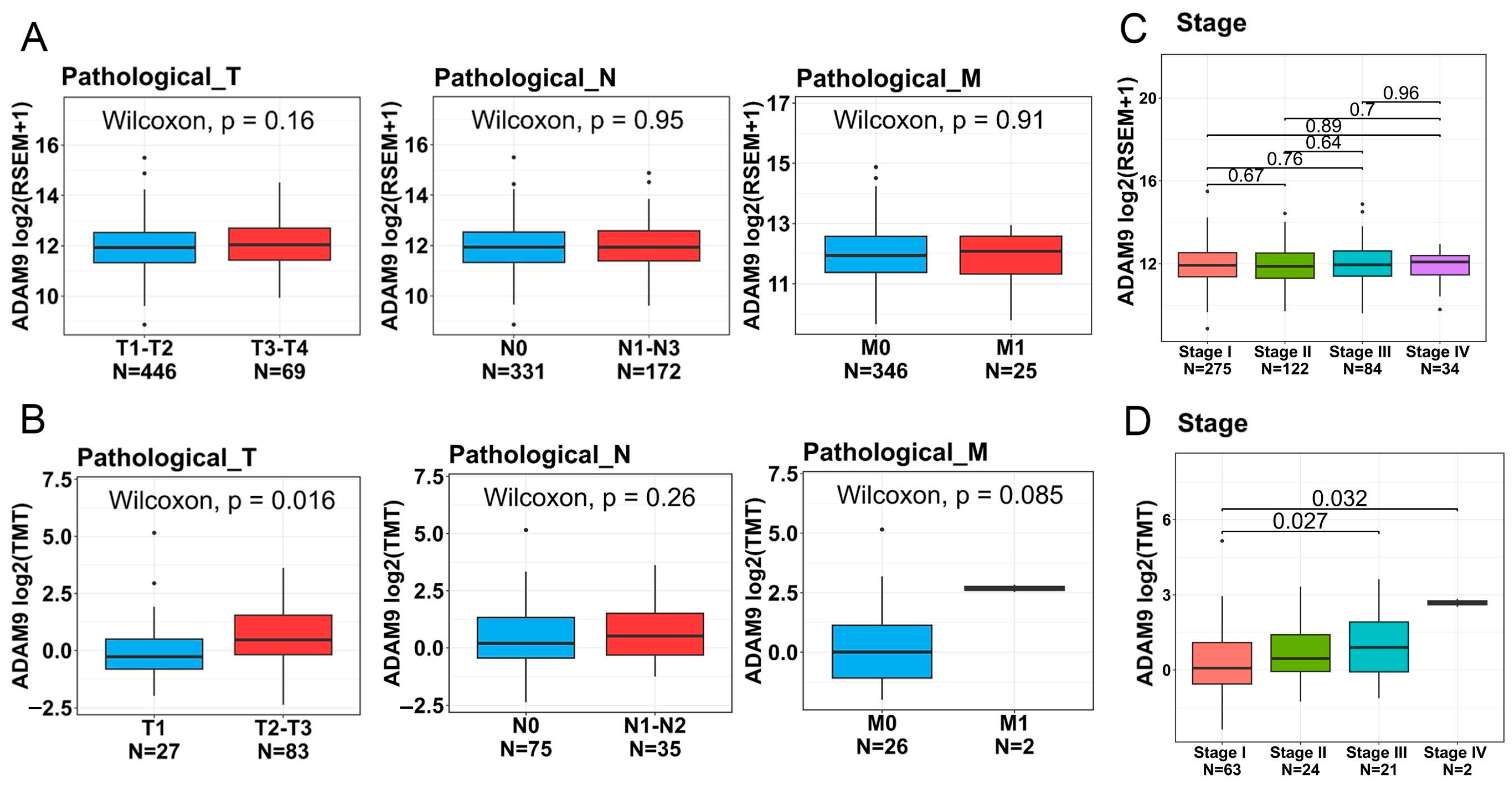
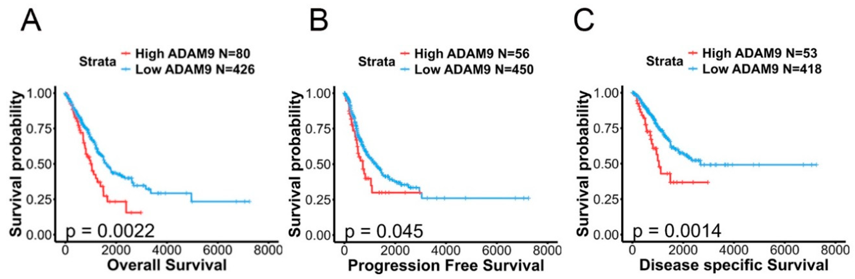
2.6. Associations Between ADAM9 Expression Levels and Clinicopathological Characteristics as Well as Prognoses of LUAD Patients

To further investigate ADAM9 expression levels in both normal and LUAD tissues and their association with disease progression, we analyzed data from TCGA and CPTAC databases. The demographic characteristics of these two datasets are presented in Supplemental Tables S1 and S2. Our findings revealed that ADAM9 messenger (m)RNA and protein expression levels were significantly higher in tumor tissues compared to normal tissues (TCGA: fold change [FC] = 1.23; CPTAC: FC = 3.28; Figure 2A,C). Elevated ADAM9 mRNA and protein levels were consistently observed in LUAD tumors, regardless of whether they harbored WT or mutant EGFR; furthermore, no significant differences in ADAM9 levels were identified between tumors with WT and mutant EGFR (TCGA: WT vs. normal: FC = 1.31; mutant EGFR vs. normal: FC = 1.41; CPTAC: WT vs. normal: FC = 3.23; mutant EGFR vs. normal: FC = 3.38; Figure 2B,D). In addition, we observed that expression levels of the ADAM9 protein, but not mRNA, were elevated in individuals with larger tumor sizes (FC = 1.47) and distant metastases (FC = 5.14) (Figure 3A,B). Additionally, ADAM9 protein expression, but not mRNA, levels were correlated with advanced pathological stages (stage III or IV) in LUAD patients (CPTAC: stage IV vs. I: FC = 5.56; stage III vs. I: FC = 1.71; Figure 3C,D). The Kaplan–Meier survival analysis further indicated that higher ADAM9 expression levels were correlated with shorter OS (Figure 4A), PFS (Figure 4B), and DSS (Figure 4C) times.

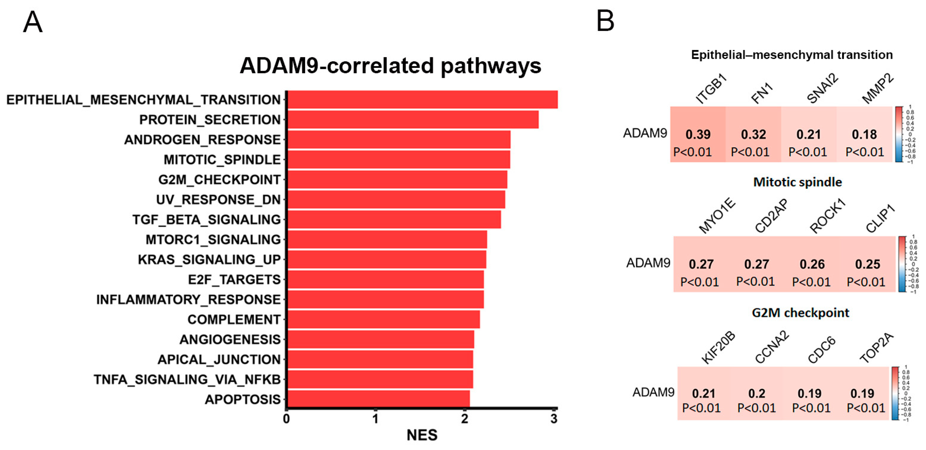
2.7. Investigation of Potential Molecular Mechanisms Regulated by ADAM9 in LUAD Progression

To uncover the mechanisms through which ADAM9 drives LUAD progression, we conducted a GSEA using TCGA-LUAD dataset. Our analysis revealed that epithelial–mesenchymal transition (EMT)-associated gene signatures were the top-ranked pathways correlated with ADAM9 expression in LUAD. In addition, several proliferation-related Hallmark gene sets, such as ‘MITOTIC_SPINDLE’ and ‘G2M_CHECKPOINT’, were also observed in patients with high levels of ADAM9 expression (Figure 5A). Analysis of human LUAD samples from TCGA using the cBioPortal platform revealed that ADAM9 expression levels were positively correlated with mesenchymal phenotype-related genes (FN1, ITGB1, SNAI2, and MMP2), mitotic-related genes (MYO1E, CD2AP, ROCK1, and CLIP1), as well as G2/M transition-related genes (KIF20B, CCNA2, CDC6, and TOP2A) (Figure 5B).

3. Discussion

Recognizing the critical role of ADAM9 as a protease with oncogenic effects in cancer progression, particularly through EGFR activation in various cancers, including LUAD [17,18,26,27], we investigated polymorphisms located on promoter and intron regions of the ADAM9 gene. These polymorphisms showed distinct distributions between LUAD patients with WT and mutant EGFR. Our findings revealed that patients carrying the mutant G allele of rs6474526 had a significantly reduced risk of developing EGFR mutations. Additionally, LUAD patients with at least one minor G allele of rs6474526 were found to have an increased likelihood of developing larger tumors, especially among EGFR-mutant, female, and non-smoking LUAD patients. Among female and non-smoking LUAD patients, a significantly increased risk of developing distant metastasis was also identified. Additionally, in patients with WT EGFR, the presence of a minor T allele in rs10105311 was linked to a decreased risk of progressing to advanced tumor stages.
The rs6474526 SNP is an intronic SNP of the ADAM9 gene. While intronic polymorphisms typically do not affect protein sequences and were once regarded as junk DNA, growing evidence now highlights the significance of introns. They play critical roles in modulating gene expressions, mRNA splicing, export, and transcription coupling, and increasing protein diversity through alternative splicing and exon shuffling [28,29,30]. Moreover, introns are frequently more prone to mutations or may act as mutational hotspots due to the presence of various essential functional elements, such as intron splice silencers, enhancers, trans-splicing elements, and other regulatory components. Beyond functional mutations, SNPs within introns can also heighten disease susceptibility, such as in cancers, and influence relationships between genotypes and phenotypes [28]. For instance, the rs12203592 intronic polymorphism in the interferon regulatory factor 4 (IRF4) gene was associated with an increased risk of acute lymphoblastic leukemia. A C-to-T substitution in this variant increases IRF4 gene expression by altering the binding affinity of the activator protein 2α transcription factor (TF) [31]. Likewise, three distinct variants (rs35054928, rs2981578, and rs45631563) within transcriptional silencer elements of the fibroblast growth factor receptor 2 (FGFR2) gene were identified. These variants boost silencer activity, reduce FGFR2 expression, and elevate the risk of breast cancer by increasing estrogen responsiveness [32]. Another example is rs72755295, an intronic SNP in the exonuclease 1 (EXO1) gene, which enhances enhancer activity, upregulates EXO1 expression, and contributes to breast cancer susceptibility [33]. In this study, we indicated the potential impact of the intronic rs6474526 SNP on ADAM9 expression in lung tissues and whole blood of healthy individuals, as observed in the GTEx database. Moreover, LUAD cells carrying at least one minor G allele displayed higher ADAM9 levels compared to those with the TT genotype. Future studies should focus on collecting both mRNA and DNA from the same LUAD patient samples to validate the impact of the rs6474526 SNPs on ADAM9 expression in LUAD patients. Additionally, further investigation is needed to understand the mechanisms through which this intronic SNP regulates ADAM9 expression.
Smoking is a significant environmental factor linked to KRAS mutations in LUAD patients [34] and increased ADAM9 expression in individuals with chronic obstructive pulmonary disease [35]. Additionally, ADAM9 was shown to stabilize mutant KRAS in pancreatic cancers [36]. Since EGFR and KRAS mutations in NSCLC are generally considered mutually exclusive, we propose that LUAD tissues with WT EGFR and mutant KRAS may exhibit higher ADAM9 expression compared to tissues with mutant EGFR. Indeed, our findings suggested that LUAD patients carrying the mutant G allele of rs6474526 tended to express higher levels of ADAM9, which was associated with a reduced risk of developing EGFR mutations.
We further investigated the influence of ADAM9 SNPs on the clinicopathological features of LUAD patients. Evidence from in vitro, in vivo, and clinical studies showed that ADAM9 facilitates lung tumor cell proliferation, adhesion to vascular endothelial cells, anoikis resistance, cell migration, tumor angiogenesis, growth, and metastasis in xenograft models. Additionally, elevated ADAM9 expression was linked to poor OS in lung cancer patients [12]. Consistent with these findings, we herein observed that ADAM9 mRNA and protein levels were significantly higher in LUAD tissues compared to noncancerous tissues. These elevated levels were correlated with advanced T stages, higher pathological stages, and worse prognostic outcomes. Furthermore, we observed that carriers of the rs6474526 G allele were significantly associated with larger tumors and distal metastases, suggesting that the rs6474526 SNPs in ADAM9 may promote tumor progression by upregulating ADAM9 expression. These findings align with previous reports highlighting the association of rs6474526 SNPs with advanced T stages in prostate cancer patients [22].
Growing evidence has demonstrated that the EMT process is closely associated with tumorigenesis, angiogenesis, metastasis, and poor prognoses in LUAD [37,38]. Although EGFR signaling has been reported to induce the EMT in cancers [39], LUAD cells harboring constitutively active EGFR mutations tend to display epithelial characteristics compared to those with WT EGFR. Additionally, LUAD patients with EGFR mutations are generally considered to have a less-aggressive disease course, partly due to the presence of epithelial traits [40]. In our study, we observed that ADAM9 expression was correlated with EMT-related gene signatures in TCGA-LUAD dataset and was associated with poor prognoses. Using the cBioPortal platform to analyze human LUAD samples further revealed a positive correlation between ADAM9 expression and mesenchymal phenotype-related genes. A previous study has demonstrated that ADAM9 can induce the upregulation of the mesenchymal marker N-cadherin in LUAD cells with WT EGFR [41]. These findings suggest that ADAM9 may drive the EGFR-mediated EMT to facilitate LUAD progression, potentially playing a more prominent role in patients with WT EGFR. However, this hypothesis requires validation in future studies.
Several limitations of this study should be acknowledged. First, we analyzed the effects of ADAM9 rs6474526 SNPs on ADAM9 gene expression in whole blood and lung tissues of healthy individuals using data from the GTEx database. To confirm the influence of ADAM9 SNPs on ADAM9 expression in LUAD patients, future studies should simultaneously collect mRNA from OCT-embedded tissues and DNA from blood samples of LUAD patients. Second, while our study provides evidence of associations between ADAM9 SNPs, tumor size, and EGFR mutation frequency in LUAD patients, we lack survival data for the recruited cohort. As a result, we are unable to assess the prognostic impact of ADAM9 SNPs in this study. Therefore, long-term survival data should be collected to further evaluate the role of ADAM9 SNPs in LUAD prognosis. Third, our analysis included lifestyle factors such as cigarette smoking status, as well as inherent factors like gender and age, as confounding variables. However, future studies should also incorporate treatment history and comorbidities into the logistic regression models for a more comprehensive analysis.

4. Materials and Methods

4.1. Study Populations and Ethics

A total of 535 patients with LUAD participated in this study at Chung Shan Medical University Hospital (Taichung, Taiwan). All patients had a minimum follow-up period of six months. Among them, 219 had wild-type EGFR, while 316 had EGFR mutations, including an exon 19 in-frame deletion and/or the L858R mutation in exon 21. LUAD specimens were collected following a protocol approved by the Institutional Review Board of Chung Shan Medical University Hospital (No. CS1-20144). The clinical data included patient gender, age, smoking history, TNM status, clinical stage, and cell differentiation.

4.2. Sequencing of EGFR from Tumor Tissues

The EGFR sequencing data of LUAD samples were processed as described in a previous report [42]. In brief, the fraction of tumor tissue for DNA extraction was macro-dissected to enrich tumor proportion from the individual OCT-embedded tissue block using a QIAamp DNA kit (Qiagen, Valencia, CA, USA). The DNA sample was first detected via a real-time polymerase chain reaction (PCR). Then, the ABI PRISM 3130XL System (Applied Biosystems, Foster City, CA, USA) was used to divide it into a wild-type EGFR or EGFR mutation group. The mutation EGFR was focused on the L858R mutation and the exon 19 in-frame deletion.

4.3. Genotyping of ADAM9 SNPs from Whole-Blood Samples

LUAD patients’ whole-blood samples were collected in tubes coated with ethylenediaminetetraacetic acid (EDTA). Following centrifugation, genomic DNA was isolated from the buffy coat using the QIAamp DNA Blood Mini Kit (Qiagen). A Nanodrop-2000 spectrophotometer (Thermo Scientific, Waltham, MA, USA) was used to determine the quality and concentration of the extracted DNA. Herein, four SNPs in the ADAM9 gene were selected: two were in the intron region (rs6474526 and rs78451751) and two in the promoter region (rs7006414 and rs10105311). Individual TaqMan SNP probes, rs6474526 (assay ID: C_449039_10), rs7006414 (assay ID: C_28949418_10), rs78451751 (assay ID: C_105349195_10), and rs10105311 (assay ID: C_26007162_10), were used to identify the genotyping of the ADAM9 SNPs using the ABI StepOnePlus™ RT-PCR System (Applied Biosystems). The results were analyzed using SDS version 3.0 software (Applied Biosystems), and the detailed DNA genotyping procedures were outlined in our previous study [43].

4.4. Bioinformatics Analysis

RNA sequencing data for LUAD patients were retrieved from The Cancer Genome Atlas (TCGA) database via UCSC Xena (https://xenabrowser.net/). Gene expression levels were normalized using RSEM and log2-transformed. Proteomics data from Clinical Proteomic Technology Assessment for Cancer (CPTAC) were obtained through LinkedOmics, with protein levels quantified using tandem mass tag (TMT) labeling and subsequent log2-transformation. For comparisons between two groups, such as tumor and normal tissues, as well as pathological TNM stages of ADAM9 gene or protein expression, the Wilcoxon rank-sum test was used. For comparisons across multiple pathological stages, the Kruskal–Wallis test was performed, followed by a post-hoc Dunn’s test for pairwise analyses. Survival analyses, including OS, progression-free survival (PFS), and disease-specific survival (DSS) of TCGA LUAD patients with respect to ADAM9, were conducted using an optimal cutoff point determined to minimize the p-value. The log-rank test was applied, and results were visualized through Kaplan–Meier plots. To evaluate signaling pathways associated with ADAM9 expression, a gene set enrichment analysis (GSEA) was conducted. Genes were ranked based on their correlation coefficients with ADAM9 expression, and the GSEA was performed using the Hallmark gene set database. The top pathways positively correlated with ADAM9 (with a false discovery rate (FDR) of <0.01) are presented in a horizontal bar plot. RNA expression levels of ADAM9 in LUAD cell lines were obtained from the DepMap portal database (DepMap public 24Q4, Broad Inst.). Values are presented as transcripts per million (TPM).

4.5. Western Blot Analysis

Protein lysates from LUAD cells were prepared and quantified as in our previous study [44]. Equal protein amounts were subjected to SDS-PAGE, transferred to PVDF membranes, and probed with primary antibodies for ADAM9 (no.2099; Cell Signaling Technology, Danvers, MA, USA) and GAPDH (60,004–1-Ig; Proteintech, Chicago, IL, USA). After three TBST washes, membranes were incubated with a secondary antibody at room temperature, and protein detection was performed using chemiluminescence.

4.6. Statistical Analysis

The chi-squared test and Student’s t-test were utilized to compare demographic characteristics between patients with WT EGFR and those with mutant EGFR. Multiple logistic regression models were applied to calculate odds ratios (ORs) and adjusted ORs (AORs), along with their 95% confidence intervals (CIs), to assess associations of genotypic frequencies and EGFR status groups with the risk of various clinicopathological characteristics. Confounding factors include lifestyle variables such as smoking and alcohol consumption, as well as inherent factors like gender and age. All statistical analyses were conducted using SAS software (version 9.1, 2005 release for Windows; SAS Institute, Cary, NC, USA), with a significance threshold set at p < 0.05.

5. Conclusions

In summary, this is the first study to explore distinct allelic effects of ADAM9 SNPs in a Taiwanese population, highlighting their impacts on EGFR mutation frequency and tumor progression in LUAD. Our findings demonstrated that rs6474526 intronic SNPs are associated with EGFR mutation susceptibility. Moreover, both the rs6474526 intronic SNPs and the rs10105311 promoter SNPs show promise as potential markers for predicting disease progression in LUAD patients. Additionally, our results suggest that ADAM9-related pathways, such as the EMT, may act as key drivers of LUAD progression. Predicting cancer risk and disease progression through SNP analysis from non-invasive biopsies offers valuable insights for personalized medicine. Our findings revealed that rs6474526 variants were significantly associated with larger tumor sizes and distant metastases of LUAD in female and non-smoking groups, suggesting that this ADAM9 SNP may serve as a useful predictor of disease progression in both LUAD populations.

Supplementary Materials

The following supporting information can be downloaded at https://www.mdpi.com/article/10.3390/ijms26104606/s1.

Author Contributions

Conceptualization, M.-H.C., J.-H.C., and S.-F.Y.; methodology, S.-F.Y. and T.-C.L.; formal analysis, T.-C.L., K.-H.H., L.-C.C., M.-H.C., and S.-F.Y., resources, T.C.-Y.T.; data curation, S.-F.Y.; writing—original draft preparation, M.-H.C. and J.-H.C.; writing—review and editing, M.-H.C. and S.-F.Y.; supervision, M.-H.C., J.-H.C., T.C.-Y.T., and S.-F.Y.; funding acquisition, T.-C.L. and M.-H.C. All authors have read and agreed to the published version of the manuscript.

Funding

This study was supported by the TMU Research Center of Cancer Translational Medicine from The Featured Areas Research Center Program within the framework of the Higher Education Sprout Project by the Ministry of Education (MOE) in Taiwan (to M.-H. Chien) and by grant no. 112-wf-eva-02 from Taipei Medical University-Wan Fang Hospital (to T.-C. Lai).

Institutional Review Board Statement

The study was approved by the Institutional Review Board of Chung Shan Medical University Hospital (IRB no. CS1-20144) and conducted in accordance with the Declaration of Helsinki.

Informed Consent Statement

All subjects involved in the study provided informed consent.

Data Availability Statement

The data from this study are available upon reasonable request to the corresponding author.

Conflicts of Interest

The authors declare that no competing interests exist.

Abbreviations

ADAM9: A disintegrin and metalloproteinase 9; AOR: adjusted odds ratio; CI: confidence interval; CPTAC: Clinical Proteomic Tumor Analysis Consortium; DSS: disease-specific survival; EGFR: epidermal growth factor receptor; EMT: epithelial–mesenchymal transition; GSEA: gene set enrichment analysis; GTEx: Genotype-Tissue Expression; GWAS: genome-wide association study; LUAD: lung adenocarcinoma; NSCLC: non-small-cell lung cancer; OS: overall survival; PFS: progression-free survival; RT-PCR: real-time polymerase chain reaction; SNP: single-nucleotide polymorphism; TCGA: The Cancer Genome Atlas; TNM: tumor/node/metastasis; WT: wild-type.

References

  1. Siegel, R.L.; Giaquinto, A.N.; Jemal, A. Cancer statistics, 2024. CA Cancer J. Clin. 2024, 74, 12–49. [Google Scholar] [CrossRef]
  2. Dearden, S.; Stevens, J.; Wu, Y.L.; Blowers, D. Mutation incidence and coincidence in non small-cell lung cancer: Meta-analyses by ethnicity and histology (mutMap). Ann. Oncol. 2013, 24, 2371–2376. [Google Scholar] [CrossRef]
  3. Midha, A.; Dearden, S.; McCormack, R. EGFR mutation incidence in non-small-cell lung cancer of adenocarcinoma histology: A systematic review and global map by ethnicity (mutMapII). Am. J. Cancer Res. 2015, 5, 2892–2911. [Google Scholar] [PubMed]
  4. Hendriks, L.E.L.; Remon, J.; Faivre-Finn, C.; Garassino, M.C.; Heymach, J.V.; Kerr, K.M.; Tan, D.S.W.; Veronesi, G.; Reck, M. Non-small-cell lung cancer. Nat. Rev. Dis. Primers 2024, 10, 71. [Google Scholar] [CrossRef] [PubMed]
  5. Benusiglio, P.R.; Fallet, V.; Sanchis-Borja, M.; Coulet, F.; Cadranel, J. Lung cancer is also a hereditary disease. Eur. Respir. Rev. 2021, 30, 210045. [Google Scholar] [CrossRef]
  6. Siegelin, M.D.; Borczuk, A.C. Epidermal growth factor receptor mutations in lung adenocarcinoma. Lab. Investig. 2014, 94, 129–137. [Google Scholar] [CrossRef] [PubMed]
  7. Chan, E.Y. Next-generation sequencing methods: Impact of sequencing accuracy on SNP discovery. Methods Mol. Biol. 2009, 578, 95–111. [Google Scholar]
  8. Hunt, R.; Sauna, Z.E.; Ambudkar, S.V.; Gottesman, M.M.; Kimchi-Sarfaty, C. Silent (synonymous) SNPs: Should we care about them? Methods Mol. Biol. 2009, 578, 23–39. [Google Scholar]
  9. Shi, J.; Shiraishi, K.; Choi, J.; Matsuo, K.; Chen, T.Y.; Dai, J.; Hung, R.J.; Chen, K.; Shu, X.O.; Kim, Y.T.; et al. Genome-wide association study of lung adenocarcinoma in East Asia and comparison with a European population. Nat. Commun. 2023, 14, 3043. [Google Scholar] [CrossRef]
  10. Seow, W.J.; Matsuo, K.; Hsiung, C.A.; Shiraishi, K.; Song, M.; Kim, H.N.; Wong, M.P.; Hong, Y.C.; Hosgood, H.D.; Wang, Z., 3rd; et al. Association between GWAS-identified lung adenocarcinoma susceptibility loci and EGFR mutations in never-smoking Asian women, and comparison with findings from Western populations. Hum. Mol. Genet. 2017, 26, 454–465. [Google Scholar]
  11. Haoyuan, M.A.; Yanshu, L.I. Structure, regulatory factors and cancer-related physiological effects of ADAM9. Cell Adh. Migr. 2020, 14, 165–181. [Google Scholar] [CrossRef] [PubMed]
  12. Chou, C.W.; Huang, Y.K.; Kuo, T.T.; Liu, J.P.; Sher, Y.P. An Overview of ADAM9: Structure, Activation, and Regulation in Human Diseases. Int. J. Mol. Sci. 2020, 21, 7790. [Google Scholar] [CrossRef]
  13. Kossmann, C.M.; Annereau, M.; Thomas-Schoemann, A.; Nicco-Overney, C.; Chéreau, C.; Batteux, F.; Alexandre, J.; Lemare, F. ADAM9 expression promotes an aggressive lung adenocarcinoma phenotype. Tumour Biol. 2017, 39, 1010428317716077. [Google Scholar] [CrossRef] [PubMed]
  14. Lin, C.Y.; Cho, C.F.; Bai, S.T.; Liu, J.P.; Kuo, T.T.; Wang, L.J.; Lin, Y.S.; Lin, C.C.; Lai, L.C.; Lu, T.P.; et al. ADAM9 promotes lung cancer progression through vascular remodeling by VEGFA, ANGPT2, and PLAT. Sci. Rep. 2017, 7, 15108. [Google Scholar] [CrossRef]
  15. Zhang, J.; Qi, J.; Chen, N.; Fu, W.; Zhou, B.; He, A. High expression of a disintegrin and metalloproteinase-9 predicts a shortened survival time in completely resected stage I non-small cell lung cancer. Oncol. Lett. 2013, 5, 1461–1466. [Google Scholar] [CrossRef] [PubMed]
  16. Izumi, Y.; Hirata, M.; Hasuwa, H.; Iwamoto, R.; Umata, T.; Miyado, K.; Tamai, Y.; Kurisaki, T.; Sehara-Fujisawa, A.; Ohno, S.; et al. A metalloprotease-disintegrin, MDC9/meltrin-gamma/ADAM9 and PKCdelta are involved in TPA-induced ectodomain shedding of membrane-anchored heparin-binding EGF-like growth factor. Embo J. 1998, 17, 7260–7272. [Google Scholar] [CrossRef]
  17. Ongusaha, P.P.; Kwak, J.C.; Zwible, A.J.; Macip, S.; Higashiyama, S.; Taniguchi, N.; Fang, L.; Lee, S.W. HB-EGF is a potent inducer of tumor growth and angiogenesis. Cancer Res. 2004, 64, 5283–5290. [Google Scholar] [CrossRef]
  18. Chiu, K.L.; Lin, Y.S.; Kuo, T.T.; Lo, C.C.; Huang, Y.K.; Chang, H.F.; Chuang, E.Y.; Lin, C.C.; Cheng, W.C.; Liu, Y.N.; et al. ADAM9 enhances CDCP1 by inhibiting miR-1 through EGFR signaling activation in lung cancer metastasis. Oncotarget 2017, 8, 47365–47378. [Google Scholar] [CrossRef]
  19. Lin, C.Y.; Chen, H.J.; Huang, C.C.; Lai, L.C.; Lu, T.P.; Tseng, G.C.; Kuo, T.T.; Kuok, Q.Y.; Hsu, J.L.; Sung, S.Y.; et al. ADAM9 promotes lung cancer metastases to brain by a plasminogen activator-based pathway. Cancer Res. 2014, 74, 5229–5243. [Google Scholar] [CrossRef]
  20. Choi, W.I.; Jeong, J.; Lee, C.W. Association between EGFR mutation and ageing, history of pneumonia and gastroesophageal reflux disease among patients with advanced lung cancer. Eur. J. Cancer 2019, 122, 101–108. [Google Scholar] [CrossRef]
  21. Dreymueller, D.; Uhlig, S.; Ludwig, A. ADAM-family metalloproteinases in lung inflammation: Potential therapeutic targets. Am. J. Physiol. Lung Cell Mol. Physiol. 2015, 308, L325–L343. [Google Scholar] [CrossRef]
  22. Lin, Y.W.; Wen, Y.C.; Lin, C.Y.; Hsiao, C.H.; Ho, K.H.; Huang, H.C.; Chang, L.C.; Wang, S.S.; Yang, S.F.; Chien, M.H. Genetic variants of ADAM9 as potential predictors for biochemical recurrence in prostate cancer patients after receiving a radical prostatectomy. Int. J. Med. Sci. 2024, 21, 2934–2942. [Google Scholar] [CrossRef] [PubMed]
  23. Cong, L.; Jia, J. Promoter polymorphisms which regulate ADAM9 transcription are protective against sporadic Alzheimer’s disease. Neurobiol. Aging 2011, 32, 54–62. [Google Scholar] [CrossRef] [PubMed]
  24. Shigematsu, H.; Lin, L.; Takahashi, T.; Nomura, M.; Suzuki, M.; Wistuba, I.I.; Fong, K.M.; Lee, H.; Toyooka, S.; Shimizu, N.; et al. Clinical and biological features associated with epidermal growth factor receptor gene mutations in lung cancers. J. Natl. Cancer Inst. 2005, 97, 339–346. [Google Scholar] [CrossRef] [PubMed]
  25. Ha, S.Y.; Choi, S.J.; Cho, J.H.; Choi, H.J.; Lee, J.; Jung, K.; Irwin, D.; Liu, X.; Lira, M.E.; Mao, M.; et al. Lung cancer in never-smoker Asian females is driven by oncogenic mutations, most often involving EGFR. Oncotarget 2015, 6, 5465–5474. [Google Scholar] [CrossRef]
  26. Liu, R.; Gu, J.; Jiang, P.; Zheng, Y.; Liu, X.; Jiang, X.; Huang, E.; Xiong, S.; Xu, F.; Liu, G.; et al. DNMT1-microRNA126 epigenetic circuit contributes to esophageal squamous cell carcinoma growth via ADAM9-EGFR-AKT signaling. Clin. Cancer Res. 2015, 21, 854–863. [Google Scholar] [CrossRef]
  27. Fu, Q.; Cheng, J.; Zhang, J.; Zhang, Y.; Chen, X.; Luo, S.; Xie, J. miR-20b reduces 5-FU resistance by suppressing the ADAM9/EGFR signaling pathway in colon cancer. Oncol. Rep. 2017, 37, 123–130. [Google Scholar] [CrossRef]
  28. Deng, N.; Zhou, H.; Fan, H.; Yuan, Y. Single nucleotide polymorphisms and cancer susceptibility. Oncotarget 2017, 8, 110635–110649. [Google Scholar] [CrossRef]
  29. Gorlova, O.; Fedorov, A.; Logothetis, C.; Amos, C.; Gorlov, I. Genes with a large intronic burden show greater evolutionary conservation on the protein level. BMC Evol. Biol. 2014, 14, 50. [Google Scholar] [CrossRef]
  30. Kalsotra, A.; Cooper, T.A. Functional consequences of developmentally regulated alternative splicing. Nat. Rev. Genet. 2011, 12, 715–729. [Google Scholar] [CrossRef]
  31. Do, T.N.; Ucisik-Akkaya, E.; Davis, C.F.; Morrison, B.A.; Dorak, M.T. An intronic polymorphism of IRF4 gene influences gene transcription in vitro and shows a risk association with childhood acute lymphoblastic leukemia in males. Biochim. Biophys. Acta 2010, 1802, 292–300. [Google Scholar] [CrossRef] [PubMed]
  32. Campbell, T.M.; Castro, M.A.A.; de Santiago, I.; Fletcher, M.N.C.; Halim, S.; Prathalingam, R.; Ponder, B.A.J.; Meyer, K.B. FGFR2 risk SNPs confer breast cancer risk by augmenting oestrogen responsiveness. Carcinogenesis 2016, 37, 741–750. [Google Scholar] [CrossRef] [PubMed]
  33. Shi, Q.; Yao, X.Y.; Wang, H.Y.; Li, Y.J.; Zhang, X.X.; Sun, C. Breast cancer-associated SNP rs72755295 is a cis-regulatory variation for human EXO1. Genet. Mol. Biol. 2022, 45, e20210420. [Google Scholar] [CrossRef] [PubMed]
  34. Ahrendt, S.A.; Decker, P.A.; Alawi, E.A.; Zhu, Y.R.; Sanchez-Cespedes, M.; Yang, S.C.; Haasler, G.B.; Kajdacsy-Balla, A.; Demeure, M.J.; Sidransky, D. Cigarette smoking is strongly associated with mutation of the K-ras gene in patients with primary adenocarcinoma of the lung. Cancer 2001, 92, 1525–1530. [Google Scholar] [CrossRef]
  35. Li, D.; Wang, T.; Ma, Q.; Zhou, L.; Le, Y.; Rao, Y.; Jin, L.; Pei, Y.; Cheng, Y.; Huang, C.; et al. IL-17A Promotes Epithelial ADAM9 Expression in Cigarette Smoke-Related COPD. Int. J. Chron. Obstruct. Pulmon. Dis. 2022, 17, 2589–2602. [Google Scholar] [CrossRef]
  36. Huang, Y.K.; Cheng, W.C.; Kuo, T.T.; Yang, J.C.; Wu, Y.C.; Wu, H.H.; Lo, C.C.; Hsieh, C.Y.; Wong, S.C.; Lu, C.H.; et al. Inhibition of ADAM9 promotes the selective degradation of KRAS and sensitizes pancreatic cancers to chemotherapy. Nat. Cancer 2024, 5, 400–419. [Google Scholar] [CrossRef]
  37. Menju, T.; Date, H. Lung cancer and epithelial-mesenchymal transition. Gen. Thorac. Cardiovasc. Surg. 2021, 69, 781–789. [Google Scholar] [CrossRef]
  38. Cui, Y.; Wang, X.; Zhang, L.; Liu, W.; Ning, J.; Gu, R.; Cui, Y.; Cai, L.; Xing, Y. A novel epithelial-mesenchymal transition (EMT)-related gene signature of predictive value for the survival outcomes in lung adenocarcinoma. Front. Oncol. 2022, 12, 974614. [Google Scholar] [CrossRef]
  39. Lo, H.W.; Hsu, S.C.; Xia, W.; Cao, X.; Shih, J.Y.; Wei, Y.; Abbruzzese, J.L.; Hortobagyi, G.N.; Hung, M.C. Epidermal growth factor receptor cooperates with signal transducer and activator of transcription 3 to induce epithelial-mesenchymal transition in cancer cells via up-regulation of TWIST gene expression. Cancer Res. 2007, 67, 9066–9076. [Google Scholar] [CrossRef]
  40. Pallier, K.; Cessot, A.; Côté, J.F.; Just, P.A.; Cazes, A.; Fabre, E.; Danel, C.; Riquet, M.; Devouassoux-Shisheboran, M.; Ansieau, S.; et al. TWIST1 a new determinant of epithelial to mesenchymal transition in EGFR mutated lung adenocarcinoma. PLoS ONE 2012, 7, e29954. [Google Scholar] [CrossRef]
  41. Sher, Y.P.; Wang, L.J.; Chuang, L.L.; Tsai, M.H.; Kuo, T.T.; Huang, C.C.; Chuang, E.Y.; Lai, L.C. ADAM9 up-regulates N-cadherin via miR-218 suppression in lung adenocarcinoma cells. PLoS ONE 2014, 9, e94065. [Google Scholar] [CrossRef] [PubMed]
  42. Wang, Y.C.; Tsao, S.M.; Li, Y.T.; Lee, C.Y.; Tsao, T.C.; Hsieh, M.J.; Yang, S.F. The Relationship between Long Noncoding RNA H19 Polymorphism and the Epidermal Growth Factor Receptor Phenotypes on the Clinicopathological Characteristics of Lung Adenocarcinoma. Int. J. Environ. Res. Public Health 2021, 18, 2862. [Google Scholar] [CrossRef] [PubMed]
  43. Lin, Y.W.; Wang, S.S.; Wen, Y.C.; Tung, M.C.; Lee, L.M.; Yang, S.F.; Chien, M.H. Genetic Variations of Melatonin Receptor Type 1A are Associated with the Clinicopathologic Development of Urothelial Cell Carcinoma. Int. J. Med. Sci. 2017, 14, 1130–1135. [Google Scholar] [CrossRef] [PubMed]
  44. Lin, Y.W.; Wen, Y.C.; Chu, C.Y.; Tung, M.C.; Yang, Y.C.; Hua, K.T.; Pan, K.F.; Hsiao, M.; Lee, W.J.; Chien, M.H. Stabilization of ADAM9 by N-α-acetyltransferase 10 protein contributes to promoting progression of androgen-independent prostate cancer. Cell Death. Dis. 2020, 11, 591. [Google Scholar] [CrossRef]
Figure 1. The effects of ADAM9 rs6474526 polymorphisms on ADAM9 expression were evaluated using validated datasets from the Genotype-Tissue Expression (GTEx) portal (https://gtexportal.org/home/, accessed on 12 Dec 2024) and the DepMap portal (https://depmap.org/portal/, accessed on 16 Jan 2025). (A) Violin plots show that the G allele of rs6474526 was associated with higher ADAM9 expression in lung tissues and whole blood. The Y-axis represents normalized ADAM9 gene expression levels (Norm. Expression), which have been adjusted to minimize technical and biological variation, allowing accurate comparison across different genotypes. The p-value on the X-axis indicates the level of statistical significance for the association between the rs6474526 SNP and ADAM9 expression. (B) Relationships between ADAM9 rs6474526 genotypes and ADAM9 expression levels were analyzed in five lung adenocarcinoma (LUAD) cell lines. The lower panel shows genotypes of ADAM9 rs6474526 in LUAD cells (A549, H23, H358, H1975, and HCC827), determined using a TaqMan SNP Genotyping Assay, while the upper panel presents ADAM9 mRNA expression levels obtained from the DepMap portal. (C) The ADAM9 protein expression levels were analyzed by the Western blot assay in LUAD cells carrying the wild-type or mutant genotype of ADAM9 rs6474526.
Ijms 26 04606 g001
Figure 2. ADAM9 mRNA and protein expression levels in lung adenocarcinoma (LUAD) patients were, respectively, analyzed using data from TCGA and CPTAC databases. (A,C) ADAM9 mRNA (A) and protein (C) levels were compared between normal and tumor tissues from LUAD patients. (B,D) LUAD tissues, regardless of the EGFR status (wild-type or mutant), showed significantly elevated ADAM9 mRNA (B) and protein (D) levels compared to normal tissues. For multiple group comparisons, brackets have been used to indicate which groups are being compared, with p < 0.05 considered significant.
Ijms 26 04606 g002
Figure 3. The clinical relevance of ADAM9 mRNA and protein levels in relation to clinicopathological features of lung adenocarcinoma (LUAD) patients was assessed using data from TCGA and CPTAC databases. (A,C) ADAM9 mRNA expression levels in LUAD samples from TCGA-LUAD were analyzed based on TNM stages (A) and pathological stages (C). (B,D) ADAM9 protein expression levels in LUAD samples from CPTAC were evaluated according to TNM stages (B) and pathological stages (D). For multiple group comparisons, brackets have been used to indicate which groups are being compared, with p < 0.05 considered significant.
Ijms 26 04606 g003
Figure 4. The prognostic impact of ADAM9 expression on lung adenocarcinoma (LUAD) patients was analyzed. (AC) Kaplan–Meier survival curves depict overall survival (A), progression-free survival (B), and disease-specific survival (C) in LUAD patients, categorized by high or low ADAM9 expression levels. The p-value expresses the statistical comparison between patients with high and low ADAM9 expression. A log-rank test p-value < 0.05 is considered significant. The LUAD dataset was retrieved from TCGA.
Ijms 26 04606 g004
Figure 5. Pathways associated with ADAM9 in lung adenocarcinoma (LUAD) patients were analyzed. (A) A horizontal bar plot displays ADAM9-related pathways, with red bars representing pathways positively correlated with ADAM9 expression. The X-axis shows the normalized enrichment scores (NESs), and the Y-axis lists the pathways identified from the Hallmark database. (B) Correlation plots illustrate the relationship between ADAM9 expression and biomarkers of the epithelial–mesenchymal transition, mitotic spindle, and G2M progression. RNA-sequencing data from The Cancer Genome Atlas-LUAD dataset were used for analysis. A Pearson correlation analysis identified these associations, with correlation coefficients and p-values provided for each square. The scale bar reflects the strength of the correlation.
Ijms 26 04606 g005
Table 1. Demographics and clinical characteristics of 535 lung adenocarcinoma patients with the EGFR mutation status.
VariableEGFR Wild-Type
(N = 219) n (%)
EGFR Mutation
(N = 316) n (%)
p-Value
Age (years)
Mean ± SD64.63 ± 12.2365.36 ± 12.51p = 0.523
Gender
Male134 (61.2%)105 (33.2%)p < 0.001
Female85 (38.8%)211 (66.8%)
Cigarette smoking status
Never-smoker103 (47.0%)255 (80.7%)p < 0.001
Ever-smoker116 (53.0%)61 (19.3%)
Stage
I or II48 (21.9%)76 (24.1%)p = 0.565
III or IV171 (78.1%)240 (75.9%)
Tumor T status
T1 or T2108 (49.3%)171 (54.1%)p = 0.275
T3 or T4111 (50.7%)145 (45.9%)
Lymph node status
Negative59 (26.9%)89 (28.2%)p = 0.756
Positive160 (73.1%)227 (71.8%)
Distant metastasis
Negative97 (44.3%)126 (39.9%)p = 0.308
Positive122 (55.7%)190 (60.1%)
Cell differentiation
Well18 (8.2%)35 (11.1%) p < 0.001
Moderately136 (62.1%)248 (78.5%)
Poorly65 (29.7%)33 (10.4%)
Table 2. Distribution frequencies of ADAM9 genotypes in lung adenocarcinoma patients and multivariate logistic regression analysis of their associations with EGFR mutations.
GenotypesEGFR
Wild-Type (N = 219)
EGFR Mutation (N = 316)AOR (95% CI)p-Value
rs6474526
TT192 (87.7%)296 (93.7%)1.000
TG27 (12.3%)20 (6.3%)0.477 (0.260~0.875)p = 0.017 *
GG0 (0.0%)0 (0.0%)------
TG + GG27 (12.3%)20 (6.3%)0.477 (0.260~0.875)p = 0.017 *
rs7006414
TT136 (62.1%)198 (62.7%)1.000
TC67 (30.6%)102 (32.3%)1.156 (0.769~1.738)p = 0.485
CC16 (7.3%)16 (5.0%)0.652 (0.302~1.412)p = 0.278
TC + CC83 (37.9%)118 (37.3%)1.051 (0.717~1.540)p = 0.799
rs10105311
CC155 (70.8%)219 (69.3%)1.000
CT56 (25.6%)83 (26.3%)1.073 (0.700~1.644)p = 0.746
TT8 (3.6%)14 (4.4%)1.017 (0.397~2.607)p = 0.972
CT + TT64 (29.2%)97 (30.7%)1.065 (0.711~1.595)p = 0.759
rs78451751
TT216 (98.6%)314 (99.4%)1.000
TC3 (1.4%)2 (0.6%)0.372 (0.058~2.408) p = 0.300
CC0 (0.0%)0 (0.0%)------
TC + CC3 (1.4%)2 (0.6%)0.372 (0.058~2.408) p = 0.300
Adjusted odds ratios (AORs) and 95% confidence intervals (CIs) were calculated using multiple logistic regression models, with adjustments for age, gender, and cigarette smoking status. * A p value of < 0.05 was defined as statistically significant.
Table 3. Clinicopathological characteristics of lung adenocarcinoma patients with wild-type or mutant EGFR, stratified by the polymorphic genotypes of ADAM9 rs6474526.
VariableAll (N = 535)EGFR Wild-Type (N = 219)EGFR Mutation (N = 316)
TT
(N = 488)
TG + GG
(N = 47)
p-ValueTT
(N = 192)
TG + GG
(N = 27)
p-ValueTT
(N = 296)
TG + GG
(N = 20)
p-Value
Stage
I or II116 (23.8%)8 (22.8%)p = 0.29542 (21.9%)6 (22.2%)p = 0.96774 (25.0%)2 (10.0%)p = 0.129
III or IV372 (76.2%)39 (83.0%) 150 (78.1%)21 (77.8%) 222 (75.0%)18 (90.0%)
Tumor T status
T1 or T2262 (53.7%)17 (36.2%)p = 0.022 a97 (50.5%)11 (40.7%)p = 0.341165 (55.7%)6 (30.0%)p = 0.025 b
T3 or T4226 (46.3%)30 (63.8%) 95 (49.5%)16 (59.3%) 131 (44.3%)14 (70.0%)
Lymph node status
Negative138 (28.3%)10 (21.3%)p = 0.30552 (27.1%)7 (25.9%)p = 0.89986 (29.1%)3 (15.0%)p = 0.176
Positive350 (71.7%)37 (78.7%) 140 (72.9%)20 (74.1%) 210 (70.9%)17 (85.0%)
Distant metastasis
Negative208 (42.6%)15 (31.9%)p = 0.15587 (45.3%)10 (37.0%)p = 0.418121 (40.9%)5 (25.0%)p = 0.160
Positive280 (57.4%)32 (68.1%) 105 (54.7%)17 (63.0%) 175 (59.1%)15 (75.0%)
Cell differentiation
Well/Moderately403 (82.6%)34 (72.3%)p = 0.083137 (71.4%)17 (63.0%)p = 0.372266 (89.9%)17 (85.0%)p = 0.491
Poorly85 (17.4%)13 (27.7%) 55 (28.6%)10 (37.0%) 30 (10.1%)3 (15.0%)
a Odds ratio (OR) (95% confidence interval (CI)): 2.046 (1.099–3.807), b OR (95% CI): 2.939 (1.099–7.858).
Table 4. Clinicopathological characteristics of lung adenocarcinoma patients with wild-type or mutant EGFR, stratified by the polymorphic genotypes of ADAM9 rs10105311.
VariableAll (N = 535)EGFR Wild-Type (N = 219)EGFR Mutation (N = 316)
CC
(N = 374)
CT + TT
(N = 161)
p-ValueCC
(N = 155)
CT + TT
(N = 64)
p-ValueCC
(N = 219)
CT + TT
(N = 97)
p-Value
Stage
I or II82 (21.9%)42 (26.1%)p = 0.29528 (18.1%)20 (31.2%)p = 0.032 a54 (24.7%)22 (22.7%)p = 0.704
III or IV292 (78.1%)119 (73.9%) 127 (81.9%)44 (68.8%) 165 (75.3%)75 (77.3%)
Tumor T status
T1 or T2190 (50.8%)89 (55.3%)p = 0.34274 (47.7%)34 (53.1%)p = 0.469116 (53.0%)55 (56.7%)p = 0.539
T3 or T4184 (49.2%)72 (44.7%) 81 (52.3%)30 (46.9%) 103 (47.0%)42 (43.3%)
Lymph node status
Negative 102 (27.3%)46 (28.6%)p = 0.75836 (23.2%)23 (35.9%)p = 0.05466 (30.1%)23 (23.7%)p = 0.241
Positive 272 (72.7%)115 (71.4%) 119 (76.8%)41 (64.1%) 153 (69.9%)74 (76.3%)
Distant metastasis
Negative149 (39.8%)74 (46.0%)p = 0.18863 (40.6%)34 (53.1%)p = 0.09186 (39.3%)40 (41.2%)p = 0.742
Positive225 (60.2%)87 (54.0%) 92 (59.4%)30 (46.9%) 133 (60.7%)57 (58.8%)
Cell differentiation
Well/Moderately306 (81.8%)131 (81.4%)p = 0.901113 (72.9%)41 (64.1%)p = 0.193193 (88.1%)90 (92.8%)p = 0.212
Poorly68 (18.2%)30 (18.6%) 42 (27.1%)23 (35.9%) 26 (11.9%)7 (7.2%)
a Odds ratio (95% confidence interval): 0.485 (0.249–0.946).
Table 5. ADAM9 rs6474526 genotype distributions and clinicopathological characteristics of lung adenocarcinoma patients who are female or non-smokers.
VariableFemale (N = 296)Non-Smoker (N = 358)
TT
(N = 278)
TG + GG
(N = 18)
OR (95% CI)p-ValueTT
(N = 336)
TG + GG
(N = 22)
OR (95% CI)p-Value
Stage
I or II73 (26.3%)2 (11.1%)1.000p = 0.15287 (25.9%)2 (9.1%)1.000p = 0.077
III or IV205 (73.7%)16 (88.9%)2.849 (0.639~12.692) 249 (74.1%)20 (90.9%)3.494 (0.800~15.256)
Tumor T status
T1 or T2156 (56.1%)4 (22.2%)1.000p = 0.005185 (55.1%)4 (18.2%)1.000p = 0.001
T3 or T4122 (43.9%)14 (77.8%)4.475 (1.437~13.940) 151 (44.9%)18 (81.8%)5.513 (1.827~16.638)
Lymph node status
Negative89 (32.0%)3 (16.7%)1.000p = 0.173101 (30.1%)4 (18.2%)1.000p = 0.236
Positive189 (68.0%)15 (83.3%)2.354 (0.665~8.342) 235 (69.9%)18 (81.8%)1.934 (0.639~5.858)
Distant metastasis
Negative118 (42.4%)3 (16.7%)1.000p = 0.031140 (41.7%)3 (13.6%)1.000p = 0.009
Positive160 (57.6%)15 (83.3%)3.688 (1.044~13.029) 196 (58.3%)19 (86.4%)4.524 (1.313~15.583)
Cell differentiation
Well/ Moderately243 (87.4%)14 (77.8%)1.000p = 0.242288 (85.7%)16 (72.7%)1.000p = 0.099
Poorly35 (12.6%)4 (22.2%)1.984 (0.618~6.368) 48 (14.3%)6 (27.3%)2.250 (0.839~6.036)
OR, odds ratio; CI, confidence interval.
Disclaimer/Publisher’s Note: The statements, opinions and data contained in all publications are solely those of the individual author(s) and contributor(s) and not of MDPI and/or the editor(s). MDPI and/or the editor(s) disclaim responsibility for any injury to people or property resulting from any ideas, methods, instructions or products referred to in the content.

