Article

mRNA-LNP COVID-19 Vaccine Lipids Induce Complement Activation and Production of Proinflammatory Cytokines: Mechanisms, Effects of Complement Inhibitors, and Relevance to Adverse Reactions

by
Tamás Bakos
1,†,
Tamás Mészáros
1,2,3,4,†,
Gergely Tibor Kozma
1,2,
Petra Berényi
1,2,
Réka Facskó
1,2,3,4,
Henriette Farkas
5,
László Dézsi
1,
Carlo Heirman
6,
Stefaan de Koker
6,
Raymond Schiffelers
7,
Kathryn Anne Glatter
8,
Tamás Radovits
3,4,
Gábor Szénási
1,‡ and
János Szebeni
1,2,9,10,*,‡
1
Nanomedicine Research and Education Center, Department of Translational Medicine, Semmelweis University, 1085 Budapest, Hungary
2
SeroScience LCC., 1089 Budapest, Hungary
3
Department of Cardiology, Heart and Vascular Center, Semmelweis University, 1122 Budapest, Hungary
4
Department of Surgical Research and Techniques, Heart and Vascular Center, Semmelweis University, 1089 Budapest, Hungary
5
Hungarian Center of Reference and Excellence, Department of Internal Medicine and Hematology, Semmelweis University, 1088 Budapest, Hungary
6
Etherna Biopharmaceuticals, 2845 Niel, Belgium
7
Division of Laboratories and Pharmacy, University Medical Center, 3584 CX Utrecht, The Netherlands
8
Department of Education, Gratz College, Philadelphia, PA 19027, USA
9
Department of Nanobiotechnology and Regenerative Medicine, Faculty of Health Sciences, Miskolc University, 3530 Miskolc, Hungary
10
Translational Nanobioscience Research Center, Sungkyunkwan University, Suwon 06351, Republic of Korea
*
Author to whom correspondence should be addressed.
These authors contributed equally to this work.
Int. J. Mol. Sci. 2024, 25(7), 3595; https://doi.org/10.3390/ijms25073595
Submission received: 8 February 2024 / Revised: 16 March 2024 / Accepted: 19 March 2024 / Published: 22 March 2024

Abstract

:
A small fraction of people vaccinated with mRNA–lipid nanoparticle (mRNA-LNP)-based COVID-19 vaccines display acute or subacute inflammatory symptoms whose mechanism has not been clarified to date. To better understand the molecular mechanism of these adverse events (AEs), here, we analyzed in vitro the vaccine-induced induction and interrelations of the following two major inflammatory processes: complement (C) activation and release of proinflammatory cytokines. Incubation of Pfizer-BioNTech’s Comirnaty and Moderna’s Spikevax with 75% human serum led to significant increases in C5a, sC5b-9, and Bb but not C4d, indicating C activation mainly via the alternative pathway. Control PEGylated liposomes (Doxebo) also induced C activation, but, on a weight basis, it was ~5 times less effective than that of Comirnaty. Viral or synthetic naked mRNAs had no C-activating effects. In peripheral blood mononuclear cell (PBMC) cultures supplemented with 20% autologous serum, besides C activation, Comirnaty induced the secretion of proinflammatory cytokines in the following order: IL-1α < IFN-γ < IL-1β < TNF-α < IL-6 < IL-8. Heat-inactivation of C in serum prevented a rise in IL-1α, IL-1β, and TNF-α, suggesting C-dependence of these cytokines’ induction, although the C5 blocker Soliris and C1 inhibitor Berinert, which effectively inhibited C activation in both systems, did not suppress the release of any cytokines. These findings suggest that the inflammatory AEs of mRNA-LNP vaccines are due, at least in part, to stimulation of both arms of the innate immune system, whereupon C activation may be causally involved in the induction of some, but not all, inflammatory cytokines. Thus, the pharmacological attenuation of inflammatory AEs may not be achieved via monotherapy with the tested C inhibitors; efficacy may require combination therapy with different C inhibitors and/or other anti-inflammatory agents.

1. Introduction

The nucleoside-modified mRNA–lipid nanoparticle (mRNA-LNP)-based COVID-19 vaccines, BNT162b2 (Comirnaty) from Pfizer-BioNTech and mRNA-1273 (Spikevax) from Moderna, are the most widely applied vaccines against SARS-CoV-2 in the United States and many other countries worldwide. These vaccines are considered effective and safe [1,2]; nevertheless, like any medical intervention, a small fraction of vaccine recipients develop acute or subacute adverse events (AEs). Some of them are severe (SAEs), leading to emergency care, hospitalization, permanent disability, or even death.
An international team of established experts (Brighton Collaboration’s Safety Platform for Emergency Vaccines) compiled a list of COVID-19 vaccine-specific AEs [3,4], whose incidence was analyzed in the Phase II/III placebo-controlled trials of mRNA-LNP vaccines. The more extensive Pfizer trial demonstrated a 36% higher risk for such “special interest” SAEs [3,4] in the vaccine group whose symptoms, importantly with respect to the present study, overlapped with those of acute and chronic COVID-19.
These findings imply that the rare AEs caused by mRNA-LNP vaccines may have the same mechanism by which the SARS-CoV-2 virus causes harm, most likely a consequence of common structural features of mRNA LNPs and the virus. In other words, beyond its mission to code for S-protein (SP) expression by antigen-presenting immune cells, mRNA-LNP vaccines share with the virus the capability to cause certain pathologic effects in a small fraction of vaccinees.
Table 1 lists the AEs reported or claimed for COVID-19 mRNA vaccines, categorized by the organ systems affected. Importantly, myocarditis/pericarditis [5,6,7,8,9,10,11,12], HSRs/anaphylaxis [13,14,15,16], autoimmune diseases [17], thrombosis, thrombocytopenia and other coagulation disorders [18,19,20,21], skin [22,23,24,25] and ocular inflammations [26,27,28], Guillain–Barré syndrome [29,30,31,32,33,34,35], and other neurologic problems [36,37] can all be linked to acute or chronic inflammatory processes that are also characteristic of SARS-CoV-2 infections.
It is known that the pathogenesis of severe COVID-19 involves complement (C) activation, a phylogenetically ancient, basic defense mechanism [38,39,40,41,42,43,44], and also the release of proinflammatory cytokines, whose most intense manifestation is cytokine storm syndrome [45]. These processes represent the activation of the humoral and cellular arms of innate immunity, respectively, and are intricately interconnected in a vicious cycle [38,45]. Consequently, there are several studies attesting to the efficacy of C inhibitors against cytokine storm in severe COVID-19 [46,47,48,49,50]. Although less well known, C activation [14,16] and release of inflammatory cytokines [51,52,53] have also been described for mRNA vaccines and their LNP models; however, in the absence of awareness of the parallelism between the innate immune stimulatory effects of the virus and the vaccine, the recommendation for using C inhibitors to prevent “vaccine injury” has remained speculative to date [13]. Obtaining proof of principle requires a better understanding of the relationship between vaccine-induced C activation and the production of inflammatory cytokines; in particular, evidence for a causal relationship between the two processes is required.
Accordingly, the main goals of the present study were to analyze the mechanisms of C activation and its role in cytokine release by Comirnaty and Spikevax and to explore the sensitivity of these processes to the clinically used C5 inhibitor Eculizumab and C1 inhibitor Berinert. We used human serum-supplemented PBMC cultures as a clinically relevant in vitro system for the simultaneous measurement of C activation and cytokine release [54,55] and 75% serum for the C-focused experiments. The latter included the analysis of activation pathways and the effects of C inhibitors and controls. Doxebo [56,57], i.e., placebo Doxil, was used as a PEGylated nanoparticle (NP) control because of the similarities in their nanostructures [56] and the wealth of information on C activation and related hypersensitivity/anaphylactoid reactions caused by Doxil [57]. Zymosan was used as the gold standard of C activation, thus enabling across-the-board conclusions.

2. Results

2.1. Complement Activation by Comirnaty in PBMC Culture

Figure 1A shows the levels of soluble terminal complex (sC5b-9) in PBMC supernatants as an endpoint of C activation after incubation with Comirnaty. In native serum-supplemented PBMC cultures (solid-colored bars), the averaged data from five different blood donors showed a significant rise in sC5b-9 at 45 min relative to the 0 min baseline both in the absence and the presence of Comirnaty, the rise being more pronounced in the presence of the vaccine. These data suggest that incubation at 37 °C for 45 min induces significant spontaneous C activation in PBMC, upon which the vaccine induced small activation superimposes. Surprisingly, we also recorded significant increases in sC5b-9 in heat-inactivated serum even at baseline, upon which the vaccine caused additional C activation relative to the vaccine-free 45 min samples. This counterintuitive observation is likely related to the liberation of heat-stable sC5b-9 upon heat treatment, which is superimposed on spontaneous C activation during incubation. This phenomenon obscured the difference between heated and native serum upon spontaneous C activation, but the Comirnaty-induced small, but significant, increase in sC5b-9 was still present. The latter data raise the possibility of incomplete decomplementation upon heat inactivation of serum, or cascade-independent (residual) C5 activation [58].
Figure 1B shows the Comirnaty-induced sC5b-9 changes in Panel A at the individual level. This presentation points to a biological, rather than a technical, cause of variation since one of the five sera (Subject #3) consistently displayed higher (up to 2-fold) sC5b-9 values compared with the other four sera under all test conditions. This donor may have had increased sensitivity towards C activation in general, exemplifying the common experience of substantial individual variation in immune response.
Taken together, these findings in 20% human serum confirm the previous data in 60% pig serum that Comirnaty can activate the C system [14]. Nevertheless, the dilution of serum reduced the sensitivity of the assay by 5-fold, thus reducing the effective dynamic range of sC5b-9 changes. This led us to test the cell-free native serum, which was diluted by 25% only with the added test agents. As shown below, this switch from 20 to 75% serum allowed us to validate the sC5b-9 assay for quantifying vaccine-induced C activation, to assess its pathways and relative potency, and to assess the effects of C inhibitors.

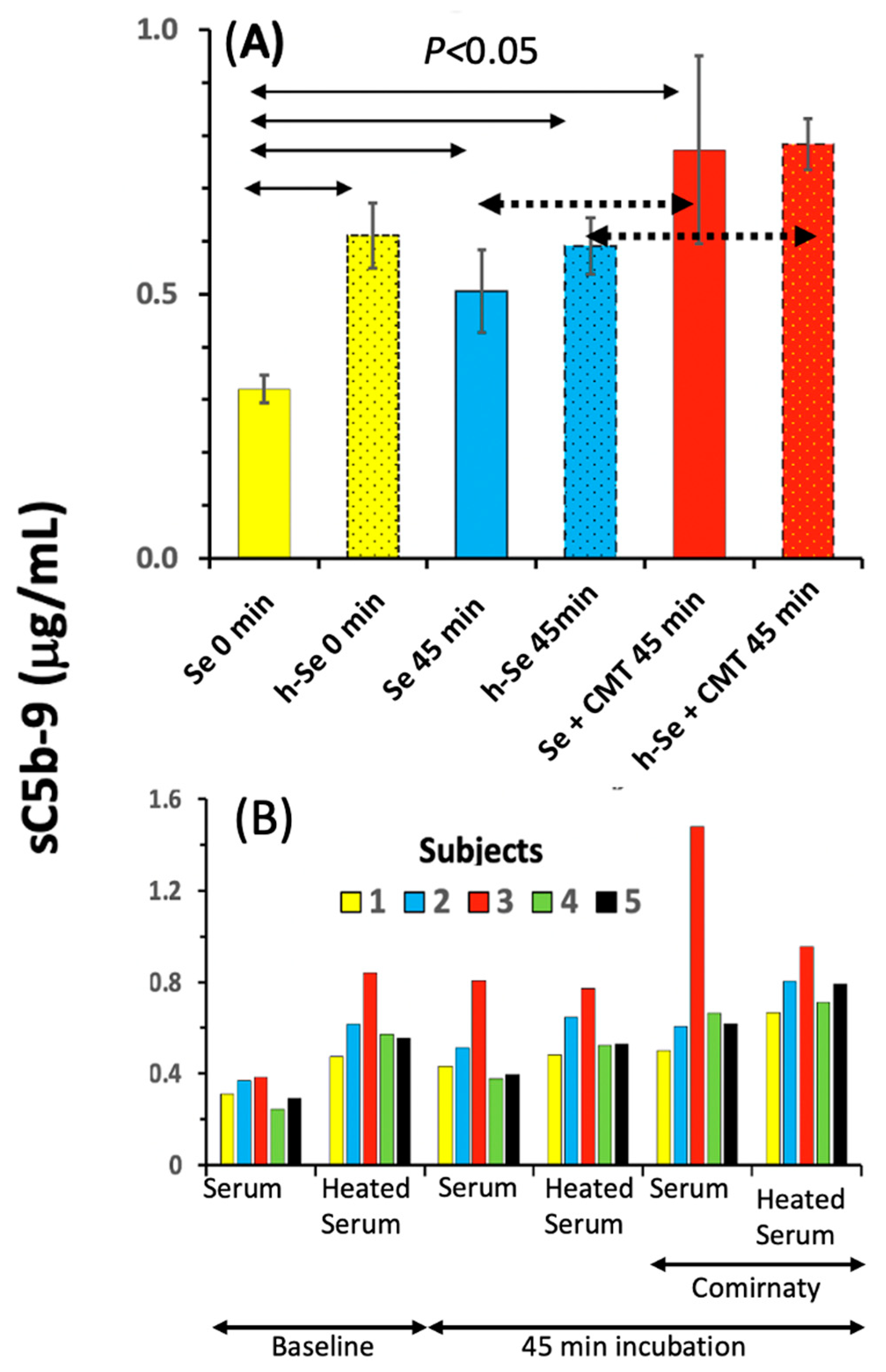
2.2. Features of C Activation by Comirnaty in 75% Human Serum

Figure 2A–C show the effects of the mRNA vaccine, Comirnaty (red bar), a PEGylated liposome, Doxebo (blue bar), and zymosan (black bar) on C activation in 75% sera of five blood donors (different donors than those shown in Figure 1), along with the time-matched PBS (baseline) values (yellow bars). To quantify C activation, we measured concurrent changes in the concentration of four reaction biomarkers including sC5b-9, a stable end product of the C cascade (Panel A); the anaphylatoxin C5a, one of the strongest proinflammatory mediators in blood (B); C4d, a C activation biomarker specific for classical and/or lectin pathway activations (C); and Bb, a biomarker specific for the alternative C pathway activation (D).
It is seen in the figure that both Comirnaty and Doxebo caused small but significant 50–220% increases in sC5b-9 (Figure 2A), C5a (B), and Bb (D) compared with the PBS baseline. In contrast, C4d was significantly increased only by Doxebo, and the Comirnaty-induced smaller increase in C4d was seen only in three of the five sera. A closer analysis of these data also reveals that the Comirnaty-induced increase in sC5b-9 in the sera of different donors on the μg mL−1 scale (A) was proportional to the rise in C5a on the ng mL−1 scale (B). Furthermore, using the MWs specified in the legend, the mean concentration increases of ~4 μg/mL sC5b-9 and ~60 ng/mL C5a correspond to 4 and 6 nM, respectively, which ratio is a realistic deviation from equimolar formation of sC5b-9 and C5a, as only a part of the de novo formed C5b-9 binds to S-protein (vitronectin) in serum [59], which is measured by the sC5b-9 kit. The above facts, taken together with the ~4-fold higher Comirnaty-induced rise in sC5b-9 in 75% serum (Figure 2A) compared with that in 20% serum (Figure 1A), attest to the proportionality of C activation with the samples’ serum content in the two types of assay systems, validating the sC5b-9 assay for the quantification of C activation under different conditions.
Further notable observations in Figure 2 are that Comirnaty and Doxebo caused far lower C activation, <1% of the activity of zymosan (Table 2), and that the Comirnaty- and Doxebo-induced rises in sC5b9, C5a, and Bb were near equal (Figure 2A,B,D), although, importantly, Comirnaty had ~6.2-fold less lipid in the samples than Doxebo (see legend).

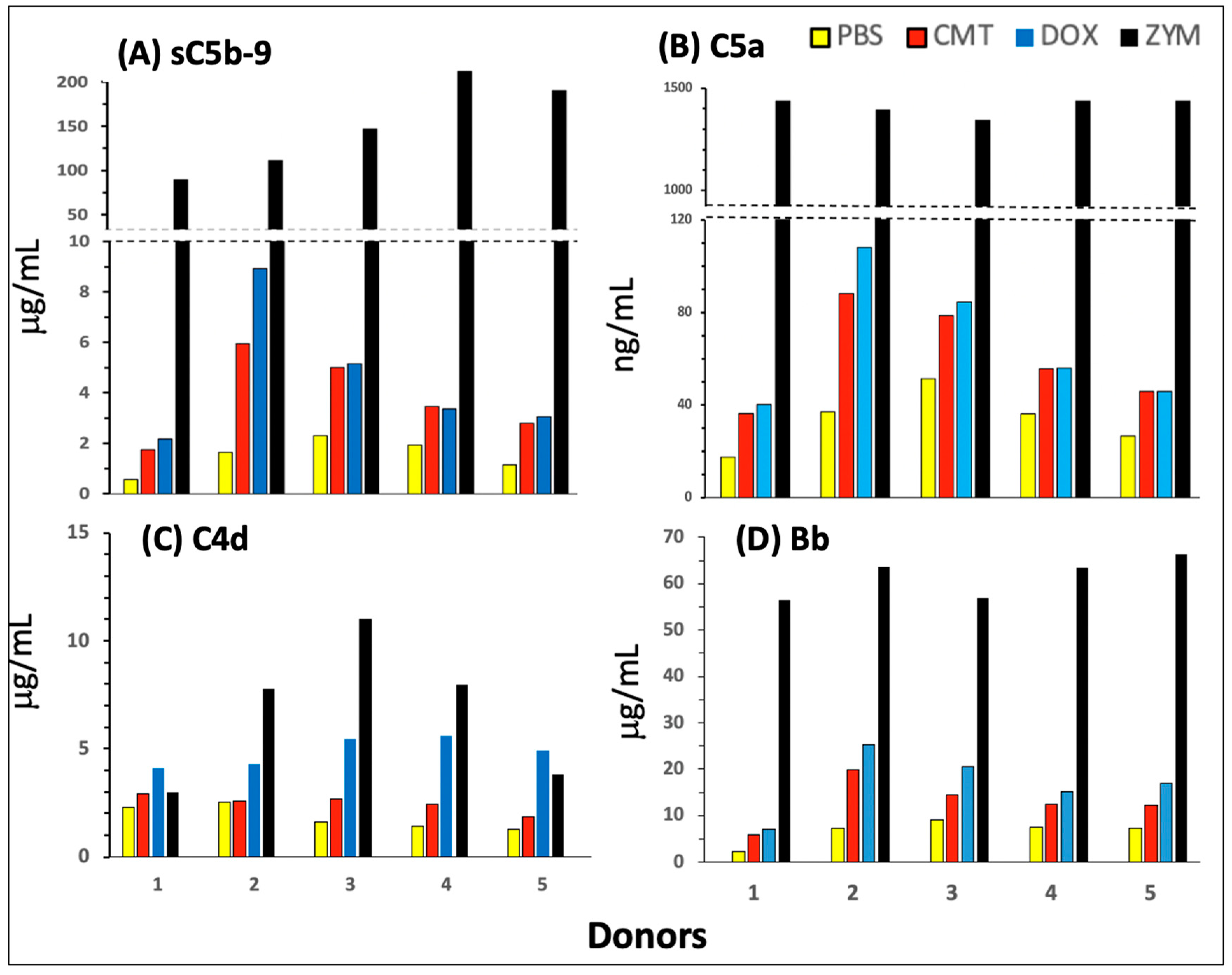
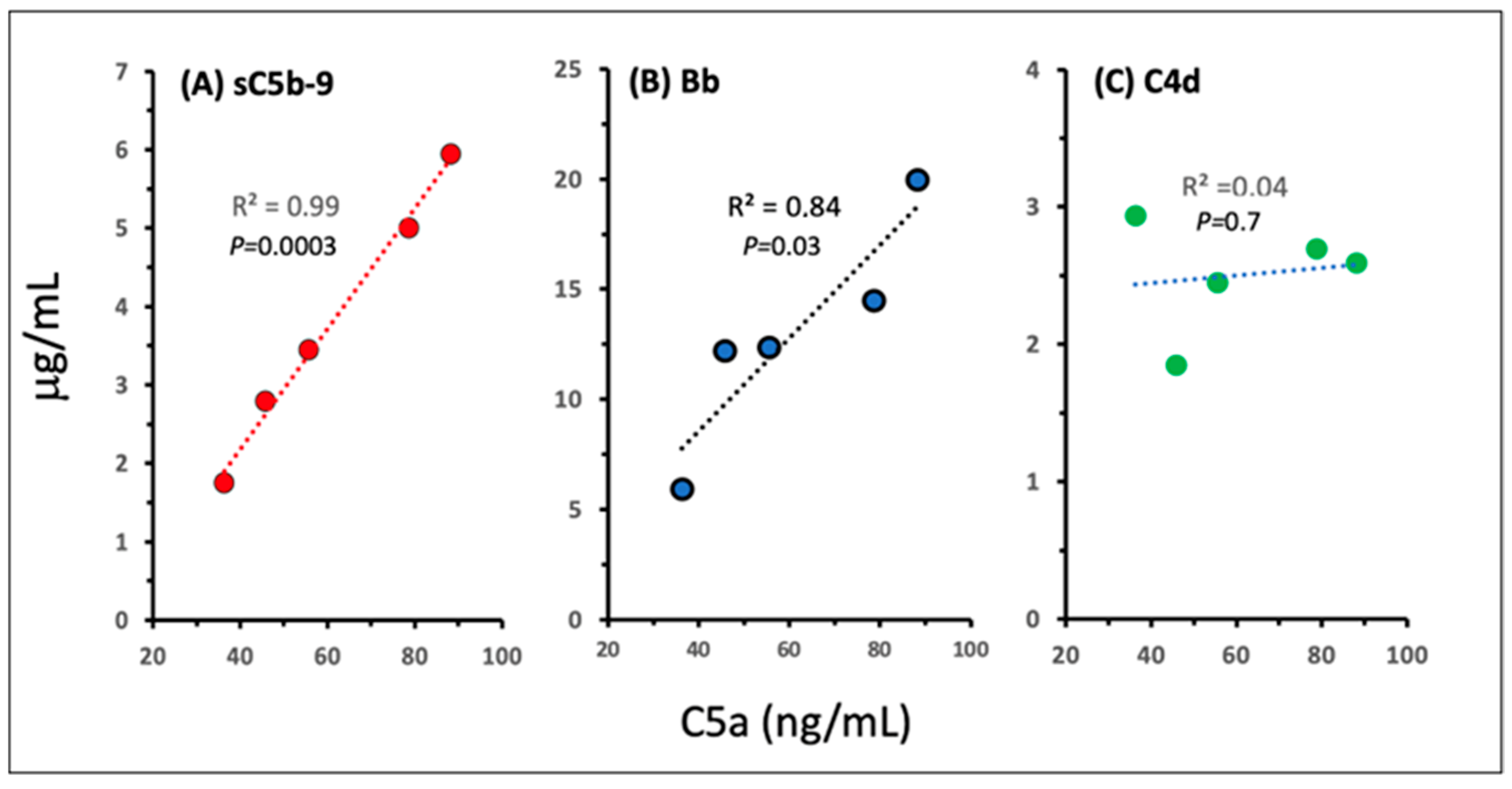
2.3. Pathways and Relative Efficacies of C Activation by Comirnaty and Doxebo

Beyond the quantification of C activation by Comirnaty and Doxebo, Figure 2 also provides information on the activation pathways of the two NPs. Notably, the finding that Doxebo caused a significantly higher increase in C4d than Comirnaty (Figure 2C) indicates that the classical pathway is more strongly involved in Doxebo- than Comirnaty-induced activation, and this difference was reduced in the Bb assay of the alternative pathway (D). The attenuation of this difference is consistent with alternative pathway amplification of classical C pathway activation, increasing the range of Bb changes to 4-fold higher concentration levels (see MWs in the legend). The proposal of primarily alternative pathway activation by Comirnaty was strongly supported by the highly significant correlation between the vaccine-induced increases in C5a and both sC5b-9 and Bb (Figure 3A,B), but not between C5a and C4d (Figure 3C).
Regarding the nearly identical increases in sC5b-9, C5a, and Bb caused by Comirnaty and Doxebo (Figure 2A,B,D), although the lipid amount was 6.2-fold lower in Comirnaty compared with Doxebo, this result raised the possibility of substantial differences in C activating efficacy. Quantifying the latter by dividing the vaccine-induced C activation, expressed as the maximum increase in sC5b-9 (sTCC), by the weight of putative C activating ingredient (i.e., lipid) (CAI), the resulting Specific C Activation (SCA) ratio was 4.6-fold higher in Comirnaty than in Doxebo (Table 2).

2.4. C Activation by Spikevax

To further dissect the individual contributions of vaccine ingredients to C activation, we also tested Spikevax, the other widely used COVID-19 mRNA vaccine, for C activation in the same 80% serum assay. The lipid composition of Comirnaty and Spikevax is different (see the legend in Figure 4); thus, if C activation was a property of a particular LNP lipid, it might be different for equivalent amounts of the two vaccines. However, as shown in Figure 4A, Comirnaty and Spikevax caused nearly identical low-level C activation in three human sera, a finding that was confirmed by another measurement performed as part of the inhibitor testing experiment (Figure 5B,C). These data suggest that independent of the core lipids, the PEGylated lipid membrane coating, a generic feature of mRNA-LNP vaccines, is one contributing factor to C activation.

2.5. Lack of C Activation by Naked mRNAs

Regarding the role of mRNA in the C activation by Comirnaty and Spikevax, we also measured and found negligible or no effect of SARS-CoV-2 mRNA in the experiment presented in Figure 4A (green bars). This result was confirmed in another experiment shown in Table S2, suggesting that mRNA plays no role in the C activation by the two vaccines. However, it must be considered that the SARS-CoV-2 full mRNA contains 7-fold more nucleotides than the SP-encoding vaccine mRNA (29,903 vs. 4284 nucleotides) [56,60], and due to a lack of information on the structure–function relationship regarding nucleic acid-induced C activation, it cannot be a priori excluded that smaller naked mRNAs have different activating potentials. Therefore, we tested the C-activating effects of three other well-characterized, virus-independent mRNAs that were even smaller than the vaccines’ SP-mRNA. The experiment shown in Figure 4B indicates small, but statistically significant increases in sC5b-9 in two of the three mRNA samples tested, but only at a 100-fold higher mRNA level than present in the vaccine. Thus, these data strengthened the argument against an active role of mRNA in C activation by the vaccines. Nevertheless, there is a further theoretical limitation of the conclusion on the immune inertness of mRNA in the vaccine, as delineated in the discussion.

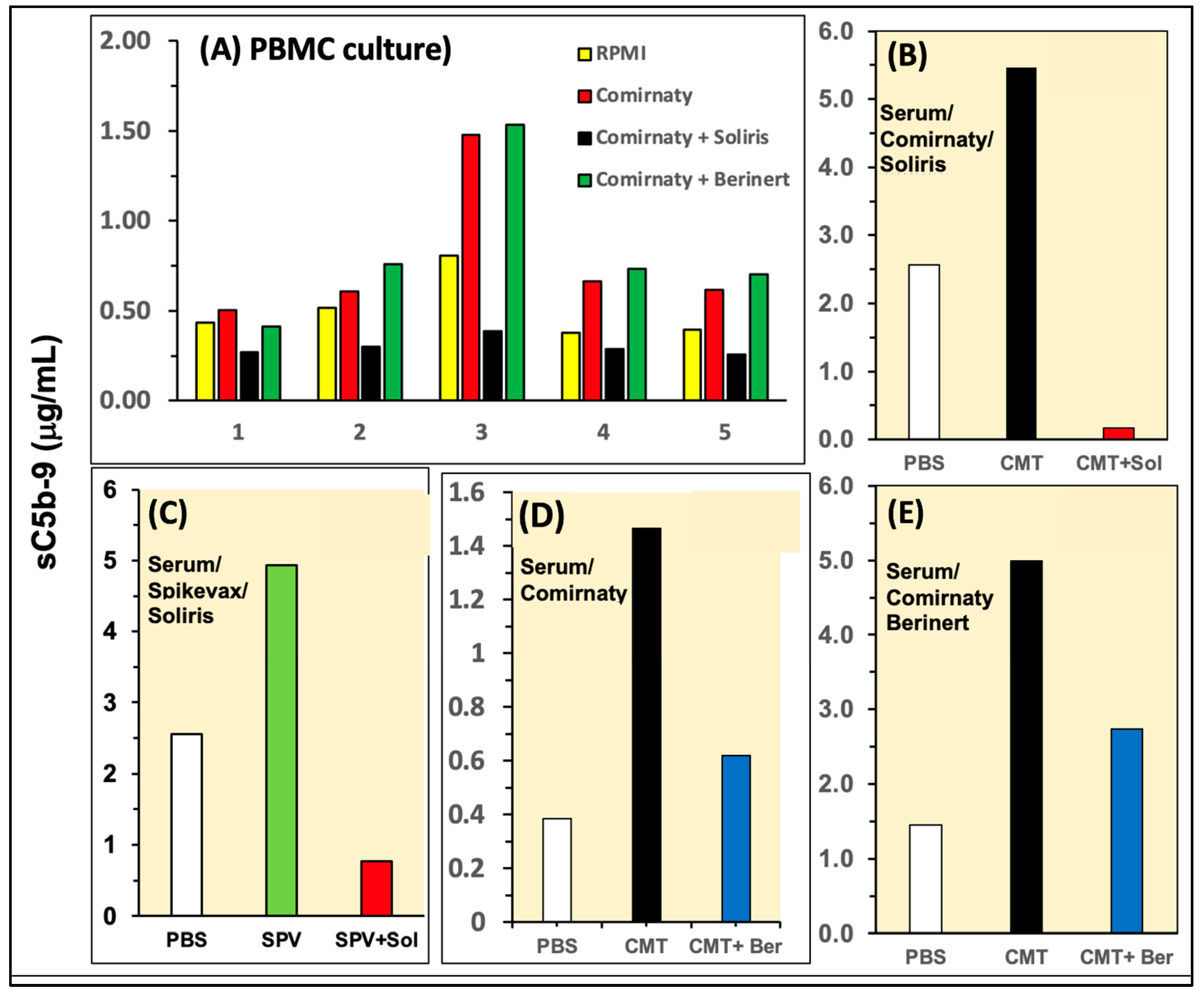
2.6. Effects of C Inhibitors on C Activation in PBMC Culture and 75% Sera

Beyond the analysis of C activation by Comirnaty, a major goal of our study was to explore possibilities for the inhibition of this adverse immune reaction. To this end, we tested the efficacies of two clinically available C inhibitors including the C5 blocker Soliris and the C1 inhibitor Berinert. Figure 5A–E show two types of experiments iterating the test systems and different variables. In PBMC culture, Soliris, but not Berinert, exerted the complete inhibition of sC5b-9 formation in all five tested sera (Figure 5A). In the case of Soliris, this effect was reproduced in 75% serum for both Comirnaty- (Figure 5B) and Spikevax-induced (Figure 5C) C activation, while Berinert exerted partial inhibition of Comirnaty-induced C activation in two independent measurements of serum (Figure 5D,E). These data provide evidence for the efficacy of both C inhibitors against mRNA vaccine-induced C activation, Soliris being more effective than Berinert, at least at the tested concentrations.

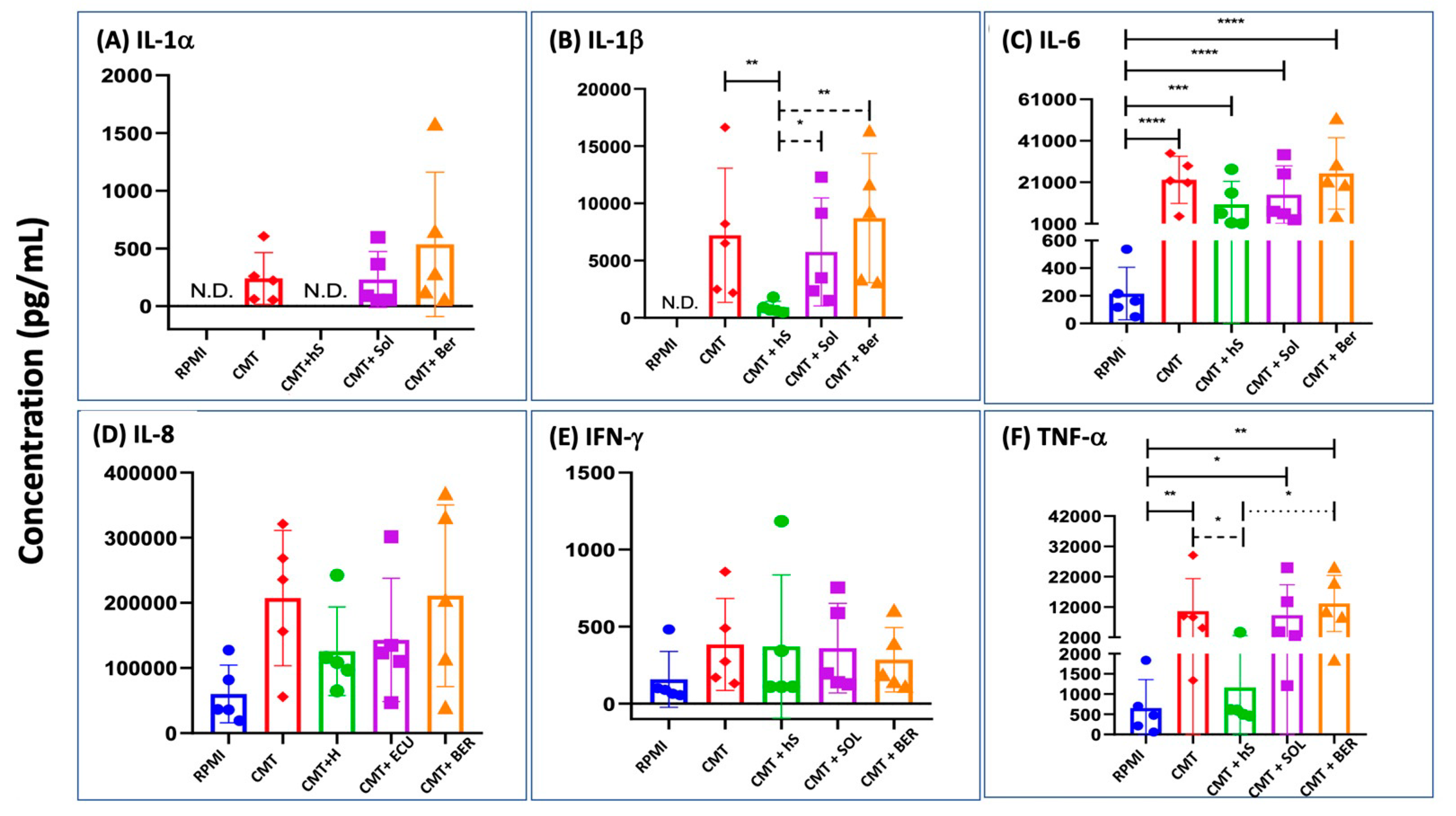
2.7. Cytokine Production in Comirnaty-Exposed, Serum-Substituted PBMC: The Effects of Heat Inactivation and Inhibition of C Activation

Figure 6A–F show significant induction by Comirnaty of the production of IL-1α, IL-1β, IL-6, IL-8, IFN-γ, and TNF-α, respectively, in the same heated or native 20% autologous serum-supplemented PBMC cultures that were tested for C activation in Figure 1B. In contrast, IL-2, IL-4, and IL-10 did not display any response to the vaccine. The secreted amounts of responder cytokines increased in the following order: IL-1α < IFN-γ < IL-1β < TNF-α < IL-6 < IL-8. Importantly, the increases in IL-1α, IL-1β, and TNF-α were absent in the heat-inactivated serum-supplemented samples, while those of IL-6 and IFN-γ were entirely serum-independent. The most abundantly produced IL-8, a chemokine that responds to most lipid- and polymer-based NPs [54], showed partial serum dependence, at least in four of the five individuals.
Since the heat inactivation of serum in the PCMB cultures did not reduce the production of IL-6 and IFN-γ (Figure 6C,E), cell starvation was unlikely to play a major role in the reductions in IL-1α, IL-1β, and TNF-α in the cultures supplemented with heated (C-depleted) serum. These data, therefore, suggest that the vaccine-induced production of at least the above three, but possibly some more yet untested, cytokines is C-dependent. To obtain alternative evidence for this conclusion and explore the utility of C inhibitors in inhibiting the C-dependent release of IL-1α, IL-1β, and TNF-α, we also tested the effects of Soliris and Berinert on cytokine induction at doses that inhibited C activation (Figure 5). However, unexpectedly, they did not reduce the production of any of the tested cytokines (Figure 6A–F). An interpretation of these data is in the Discussion below.

3. Discussion

Prompted by the rise in unusual (“special interest”) AEs of COVID-19 mRNA-LNP vaccines and consequent scientific and public debate over their safety, this study investigated the molecular mechanisms and relationship between two innate immune-stimulating effects of these vaccines that are assumed to jointly contribute to many independent SAEs of the vaccine (Table 1) including complement activation and production of proinflammatory cytokines. The in vitro data led to several new insights into the molecular mechanism of these phenomena, whose specifics and clinical relevance are discussed below.

3.1. Complement Activation by Comirnaty: Novel Findings and Mechanism

Complement activation by liposomes and other therapeutic or diagnostic NPs has been known for decades, but the fact that mRNA-LNPs can also have such activity has not obtained much attention in the mainstream literature on COVID-19 vaccines. We have previously reported C activation caused by Comirnaty in pig serum in vitro [14] and in pig blood in vivo [16], and the present reproduction of the reaction in human serum implies that it is a species-independent phenomenon, fitting into the concept that C activation is an intrinsic property of PEGylated NPs due to their resemblance to pathogenic viruses [61].
Beyond some technical novelties, such as the stability of sC5b-9 during the heat treatment of serum and the lack of cellular damage in tissue culture supplemented with intact serum, the novel findings in the present study include (i) the dominance of alternative pathway activation, (ii) the increased strength of C activation relative to corresponding PEGylated liposomes, and (iii) the absence of C activation by naked mRNAs.
Regarding the occurrence of C activation by the vaccine, it was recently pointed out [13] that in theory, almost all components of Comirnaty have the capability to promote the activation cascade for different reasons. Thus, membrane-forming PEGylated lipids may activate C due to the binding of anti-PEG antibodies [16,62], DSPC, as a long, saturated fatty acid-containing unnatural phospholipid that rigidify, and cholesterol, which stabilizes the membrane coat for antibody and/or C3b binding. ALC-0315 may activate C because of its positive charge, enabling complexation with the mRNA63 and amphiphile character, leading to micelle and cluster formation [63], and, possibly, distribution into lipoproteins.
The proposal of C activation by mRNA-ALC-0315 complexes is based on a previous study by Plank et al. [64] showing that DNA complexes with certain cationic lipids were strong C activators. The above study by Rissanou et al. also presented evidence for cluster formation from positively charged lipids63 which, in analogy to positively charged liposomes [65], could be another source of C activation via the alternative pathway. It is not excluded, either, that the liberated ALC-0315 associates with plasma lipoproteins entailing C3-binding and alternative pathway C activation, in a manner like that observed with Cremophor EL [66].
Regarding the ~5-fold higher “specific” result, i.e., weight-normalized C reactivity of Comirnaty vs. Doxebo (Table 2), the finding implies that the vaccine has a C-activating feature that Doxebo does not have. The crucial difference is the presence of mRNA and positively charged lipids in the vaccine, so the multiple possibilities by which the mRNA and core lipids could activate C could account for the increased C reactivity of the vaccine despite the lack of C activation by naked mRNAs. However, to contribute to C activation, the internal content of the vaccine needs to be released into the blood. Meet this precondition can also be predicted based on the mentioned recent structural analysis of Comirnaty [56], claiming that the vaccine consists of soft, fragile NPs prone to break up in water [56]. In fact, the latter study showed electron microscopic images of lipid fragments and snake-like twisted nanostructures in disintegrated Comirnaty samples, which were tentatively identified as mRNA-lipid complexes [56]. It should also be mentioned that the vaccine-induced significant increase in Bb (Figure 2D) and its correlation with the rises in C5a and sC5b-9 (Figure 3) indicate the predominance of alternative pathway C activation, which can most easily be rationalized by exposure of the Comirnaty’s core components to serum, i.e., rapid disintegration.

3.2. Inflammatory Cytokine Production by Comirnaty: Mechanism and Relationship with C Activation

The other Comirnaty-induced proinflammatory process analyzed in the present study as a possible contributing mechanism to the vaccine-induced SAEs was the induction of inflammatory cytokines and its dependence on C activation. A crosstalk between these two arms of the innate immune system has already been described in many previous studies [55,67,68,69,70], but not for mRNA-LNP vaccine-induced immune stimulation. In theory, Comirnaty can enhance cytokine release both via direct interaction with immune cells and their indirect stimulation via anaphylatoxin and other C receptors, and our PBMC data (Figure 6) gave an indication of the operation of both mechanisms. The clinically relevant, novel finding in our study was the identification of intact serum-dependence of the production of IL-1β, IL-6, TNF-α, and IF-γ, which provides indirect evidence for the C-dependence of the release of these cytokines. The resultant proposal that C activation plays a causal role in this process is supported by an earlier PBMC study showing the suppression of zymosan-induced IL-6 release by the alternative pathway C inhibitor, mini-Factor H [55].

3.3. Broader Implications of In Vitro Data on C Activation and Cytokine Induction by mRNA-LNP Vaccines

3.3.1. The Complement Test as an In Vitro Model for Vaccine-Induced Anaphylaxis

The detection of C activation by the vaccine supports the concept that anaphylaxis, listed among the most frequent SAEs of Comirnaty [71], represents C activation-related pseudoallergy (CARPA) [13]. Although Comirnaty is injected i.m. at a much lower lipid dose than applied upon CARPAgenic i.v. liposome therapies [61], there are considerations that reconcile the differences in administration. First, the vaccine can rapidly exit from the deltoid muscle into the blood via several routes [13]; thus, similarly to i.v. liposomes, it may activate C in blood. Second, the weight-based in vitro C reactivity of Comirnaty was ~5 times stronger than that of Doxebo, whose reactivity is close to that of Doxil in causing CARPA [57]. Consequently, the threshold concentration to cause CARPA in reactive individuals may be significantly lower for Comirnaty. Third, the incidence of mRNA-LNP vaccine-induced anaphylaxis (<0.04%) [15] is comparable or lower than that estimated for Doxil (~1% of <11% ≤ 0.1%) [72], a realistic proportionality between anaphylaxis incidence and estimated C activation in vivo.

3.3.2. Use of the PBMC-Based Pan-Innate Immune Stimulation Assay for Finding Effective Pharmacological Prevention of mRNA-Vaccine-Induced SAEs

We found that serum inactivation, assumably because of C depletion, does not necessarily prevent cytokine release; for some cytokines, it was effective, and for others, it was not. We therefore tested the effects of Soliris and Berinert, two clinically used C inhibitors, as an alternative approach to modify C activity. Although they inhibited C activation, they did not inhibit the production of IL-1α, IL-1β, or TNF-α, thus contradicting the causal relationship between C activation and secretion of these “serum-dependent” cytokines. Although we did not analyze further the possible reasons for these negative results, one explanation may be the mentioned fact that Comirnaty may enhance cytokine release by the simultaneous activation of multiple surface receptors, in which case, the inhibition of classical pathway activation by Berinert, or C5a production by Soliris, may remain sub-threshold for cytokine induction. Thus, the single use of either of these inhibitors may not be enough to attenuate the inflammatory reaction; effective prevention may require simultaneous inhibition of the C cascade at multiple checkpoints, for which there are numerous C inhibitor drugs and drug candidates available [73,74,75,76,77]. The double-endpoint PBMC assay applied in the present study therefore offers a simple in vitro model for testing the efficacy of C inhibitors against inflammatory SAEs.

3.3.3. Public Health Implications

By showing the occurrence of two essential proinflammatory effects by mRNA-LNP vaccines, the present study attempts to give a scientific explanation for the “special interest” SAEs that these vaccines rarely cause, yet that induce substantial press and media attention with top-level public debates in the U.S. Senate and U.K. Parliament in the context of vaccine safety and excess death. The number of publications on different COVID-19 vaccine-induced SAEs has been on the rise [5,6,7,8,9,10,11,12,13,14,15,16,17,18,19,20,21,22,23,24,25,26,27,28,29,30,31,32,33,34,35,36,37], and once the real scale and mechanism of these AEs are better understood, the focus can be redirected to the prevention and, hence, further extension of mRNA-LNP technology for the development of new vaccines and gene therapies [78,79,80]. The present study contributes to the latter goal.

4. Materials and Methods

4.1. Materials

Mammalian cell culture medium (RPMI-1640, R5), non-essential amino acid solution (0.1 mM), pyruvate solution (1 mM), penicillin–streptomycin antibiotics solution and β-mercaptoethanol (50 µM) Ficoll-Paque, ethylenediaminetetraacetic acid (EDTA), Dulbecco’s phosphate buffered saline (D-PBS), and trypan blue dye were purchased from Merck Life Science Ltd. (Budapest, Hungary). R5 medium was from ThermoFisher Scientific. Comirnaty and Spikevax vaccines used against the delta variant of the SARS-CoV-2 virus (lot numbers: ET7205 and FA 5829) were obtained from Semmelweis University’s Pharmacy. They were stored as instructed by the manufacturers and immediately used after opening the vials within the expiration date. The C inhibitor anti-C5 monoclonal antibody (mAb), Eculizumab (Soliris®), was from Alexion Pharmaceuticals (Boston, MA, USA), today owned by Astra Zeneca (Cambridge, U.K.). The C1-esterase inhibitor (Berinert®) was from CSL Behring GmbH (Marburg, Germany), and 5-methoxyuridine-modified SARS-CoV-2 spike protein mRNA (E484K, N501Y) was from OZ Biosciences SAS (Marseille, France).

4.2. Ethical Permission, Donors, and Blood Collection

The Scientific and Research Ethics Committee of the Medical Research Council of Hungary granted ethical approval for this research project (TUKEB 15576/2018/EKU). After obtaining informed consent from all white Caucasian, COVID-19 vaccinated, healthy adult volunteers of this study (total n = 23) their blood was withdrawn by a phlebotomist.

4.3. Separation of Serum and PBMC

The serum was separated from whole blood after coagulating the blood (approx. 25 min) at room temperature in a Greiner VACUETTE Z Serum Sep Clot Activator (8 mL) blood collector tube. The tubes were centrifuged (2000× g, 4 °C, 15 min), and the supernatants (serum) were collected. PBMCs were separated from uncoagulated whole blood collected in Greiner VACUETTE K2E K2EDTA (9 mL) blood collector tubes and diluted in a 1:1 ratio with 6 mM EDTA containing D-PBS using Ficoll-Paque density gradient centrifugation (500× g, 25 °C, 30 min). Cells that formed a white-colored ring were then suspended in 6 mM EDTA containing D-PBS to ensure the removal of the remaining substances from the previous steps and were centrifuged again (500× g, 25 °C, 30 min). To remove thrombocytes and residual EDTA, cells were washed with cold, EDTA-free, D-PBS and were centrifuged again (500× g, 4 °C, 15 min). Following the centrifugation, the cells were divided into three portions and were suspended in 3 culture media as follows: (1) medium only (R5); (2) R5 containing 20% autologous human serum, representing the in vivo conditions with 1/5 the amount of C proteins and other serum components exposed to the cells; (3) R5 containing 20% autologous human serum inactivated by heat, representing in vivo conditions without activable C proteins. To inactivate sera, also known as decomplementation, a portion of the innate serum from each donor was incubated at 56 °C for 30 min. The 20% serum ratio was chosen based on preliminary studies showing no significant cell toxicity over 18 h, as determined by trypan blue exclusion. The suspensions were centrifuged (500× g, 4 °C, 15 min) and subsequently re-suspended in the culture media specified above.

4.4. Preparation of mRNAs

To investigate the C-activating effects of naked mRNAs, we applied N1-Methyl Pseudouridine-modified, 5′ cap1 cleancapped mRNAs prepared by Etherna Biopharmaceuticals (Niel, Belgium), as described earlier [81]. The samples coding for the proteins CD40L (29.3 kDa), Cre-recombinase (38.5 kDa), and luciferase (60.7 kDa) contained 1200, 1450, and 2086 nt, respectively. Their sequence and other details are shown in the Supplemental Data Section.

4.5. PBMC Studies

Following separation, the PBMCs were suspended in R5 medium supplemented with 20% native or heat-inactivated autologous serum. The suspension was then placed into the inner wells of 96-well microtiter plates, with each well containing approximately 5 × 10⁵ cells. The plates were placed in a CO2 incubator (5% CO2) at 37 °C. Aliquots were collected at 45 min and 18 h after the start of the experiment. Additionally, baseline samples (“0 min samples”) were collected before the incubation. After collection, the samples were centrifuged (2500× g, 4 °C, 10 min), and the supernatants were stored at –80 °C until the C and cytokine assays were performed. Soluble terminal C complex (sTCC, sC5b-9) was measured with an ELISA kit from Svar Life Science AB (Malmö, Sweden), and for the measurement of cytokines, we used a Q-View™ LS chemiluminescent imager paired with Q-View™ Software, https://www.quansysbio.com/products-and-services/imaging/q-view-imager-ls/ (accessed on 18 March 2023), obtained from Quansys Biosciences (West Logan, UT, USA). The cytokine panel supplied by the company was the Human Cytokine Inflammation Panel 1 multiplex ELISA, measuring IL-1α, IL-1β, IL-2, IL-4, IL-6, IL-8, IL-10, IFN-γ, and TNF-α. All procedures followed the manufacturer’s instructions.

4.6. Serum Studies

The vaccine and other test samples were mixed with native serum at a 1:3 volume ratio, followed by incubation at 37 °C for 30 or 45 min, as stated in the text. The incubation was stopped by dilution with the kit’s sample diluent supplemented with 10 mM EDTA. Soluble TCC, C5a, C4d, and Bb were measured with Svar’s C5b-9 kit (Svar Life Sci., Malmö, Sveden) and TECOmedical’s C5a, C4d, and Bb ELISA kits (TECOmedical AG, Sissach, Switzerland).

4.7. Statistical Analysis

All data reported are means ± SEM. Statistical comparisons were made using paired t-tests or one-way ANOVA and Dunnetts’ or Tukey’s multiple comparisons post hoc tests, as specified in the text.

Supplementary Materials

The supporting information can be downloaded at: https://www.mdpi.com/article/10.3390/ijms25073595/s1.

Author Contributions

Conceptualization: T.B., G.S. and J.S.; experimentation: T.B., T.M., L.D., G.S., R.F., C.H. and P.B.; data curation: H.F., S.d.K., R.S., K.A.G., T.R., T.B., T.M., L.D., G.S., R.F. and P.B.; formal analysis: T.M., G.T.K. and S.d.K.; writing: J.S., G.S. and T.B.; review and editing: T.B., G.S., K.A.G., T.M., R.F., T.R. and P.B.; supervision: G.S., J.S., R.S. and T.R. All authors have read and agreed to the published version of the manuscript.

Funding

This study was supported by the European Union Horizon 2020 project 825828 (Expert) and grants from the National Research, Development, and Innovation Office (NKFIH) of Hungary (2020-1.1.6-JÖVŐ-2021-00013 and 2020-1.1.6-JÖVŐ-2021-00010). T.B. acknowledges financial support for Project no. KDP-14-3/PALY-2021, Ministry of Culture and Innovation of Hungary, National Research, Development, and Innovation Fund, financed under the KDP-2020 funding scheme. H.F. received research grants from CSL Behring, Takeda, and Pharming.

Institutional Review Board Statement

The Scientific and Research Ethics Committee of the Medical Research Council of Hungary granted ethical approval for this research project (TUKEB 15576/2018/EKU).

Informed Consent Statement

Informed consent was obtained from all subjects involved in this study.

Data Availability Statement

Data will be made available upon request.

Acknowledgments

Sincere thanks are due to Marina Dobrovolskaia (NCL, NCI, NIH, Frederick, USA) and Joshua Jackman (Sungkyunkwan University, Suwon, Korea) for their advice on this manuscript, and Zoltan and Miklos Szebeni for grammar corrections. J.S. acknowledges support from the Applied Materials and Nanotechnology Center of Excellence, Miskolc University, Miskolc, Hungary.

Conflicts of Interest

The authors affiliated with SeroScience LLC are involved in the company’s contract research service activity providing studies that were applied here. H.F. is an advisor for the companies Behring, Takeda, Pharming, Kalvista, Ono, Pharvaris, Astria, Intellia, and Biocryst. The remaining authors declare that the research was conducted in the absence of any commercial or financial relationships that could be construed as a potential conflict of interest.

References

  1. WHO: COVID-19 Advice for the Public: Getting Vaccinated. Available online: https://www.who.int/emergencies/diseases/novel-coronavirus-2019/covid-19-vaccines/advice (accessed on 18 March 2024).
  2. CDC: Updated COVID-19 Vaccine Recommendations are Now Available. 2023. Available online: https://www.cdc.gov/ncird/whats-new/covid-vaccine-recommendations-9-12-2023.html (accessed on 18 March 2024).
  3. Law, B. SO2-D2.1.2 Priority List of COVID-19 Adverse Events of Special Interest: Quarterly Update December 2020. 2020. Available online: https://brightoncollaboration.us/wp-content/uploads/2023/05/SO2_D2.1.2_V1.2_COVID-19_AESI-update_V1.3.pdf (accessed on 18 March 2024).
  4. Fraiman, J.; Erviti, J.; Jones, M.; Greenland, S.; Whelan, P.; Kaplan, R.M.; Doshi, P. Serious adverse events of special interest following mRNA COVID-19 vaccination in randomized trials in adults. Vaccine 2022, 40, 5798–5805. [Google Scholar] [CrossRef]
  5. Bozkurt, B. Shedding Light on Mechanisms of Myocarditis with COVID-19 mRNA Vaccines. Circulation 2023, 147, 877–880. [Google Scholar] [CrossRef]
  6. Patone, M.; Mei, X.W.; Handunnetthi, L.; Dixon, S.; Zaccardi, F.; Shankar-Hari, M.; Watkinson, P.; Khunti, K.; Harnden, A.; Coupland, C.A.C.; et al. Risk of Myocarditis After Sequential Doses of COVID-19 Vaccine and SARS-CoV-2 Infection by Age and Sex. Circulation 2022, 146, 743–754. [Google Scholar] [CrossRef]
  7. Luo, J.; Hur, K.; Salone, C.; Huang, N.; Burk, M.; Pandey, L.; Thakkar, B.; Donahue, M.; Cunningham, F. Incidence Rates and Clinical Characteristics of Patients With Confirmed Myocarditis or Pericarditis Following COVID-19 mRNA Vaccination: Experience of the Veterans Health Administration Through 9 October 2022. Open Forum Infect. Dis. 2023, 10, ofad268. [Google Scholar] [CrossRef] [PubMed]
  8. Mungmunpuntipantip, R.; Wiwanitkit, V. Cardiac inflammation associated with COVID-19 mRNA vaccination and previous myocarditis. Minerva Cardiol. Angiol. 2023, 71, 242–248. [Google Scholar] [CrossRef] [PubMed]
  9. Schroth, D.; Garg, R.; Bocova, X.; Hansmann, J.; Haass, M.; Yan, A.; Fernando, C.; Chacko, B.; Oikonomou, A.; White, J.; et al. Predictors of persistent symptoms after mRNA SARS-CoV-2 vaccine-related myocarditis (myovacc registry). Front. Cardiovasc. Med. 2023, 10, 1204232. [Google Scholar] [CrossRef]
  10. Paredes-Vazquez, J.G.; Rubio-Infante, N.; Lopez-de la Garza, H.; Brunck, M.E.G.; Guajardo-Lozano, J.A.; Ramos, M.R.; Vazquez-Garza, E.; Torre-Amione, G.; Garcia-Rivas, G.; Jerjes-Sanchez, C. Soluble factors in COVID-19 mRNA vaccine-induced myocarditis causes cardiomyoblast hypertrophy and cell injury: A case report. Virol. J. 2023, 20, 203. [Google Scholar] [CrossRef] [PubMed]
  11. Nakahara, T.; Iwabuchi, Y.; Miyazawa, R.; Tonda, K.; Shiga, T.; Strauss, H.W.; Antoniades, C.; Narula, J.; Jinzaki, M. Assessment of Myocardial (18)F-FDG Uptake at PET/CT in Asymptomatic SARS-CoV-2-vaccinated and Nonvaccinated Patients. Radiology 2023, 308, e230743. [Google Scholar] [CrossRef] [PubMed]
  12. Hviid, A.; Nieminen, T.A.; Pihlström, N.; Gunnes, N.; Dahl, J.; Karlstad, Ø.; Gulseth, H.L.; Sundström, A.; Husby, A.; Hansen, J.V.; et al. Booster vaccination with SARS-CoV-2 mRNA vaccines and myocarditis in adolescents and young adults: A Nordic cohort study. Eur. Heart J. 2024, ehae056. [Google Scholar] [CrossRef] [PubMed]
  13. Szebeni, J.; Storm, G.; Ljubimova, J.Y.; Castells, M.; Phillips, E.J.; Turjeman, K.; Barenholz, Y.; Crommelin, D.J.A.; Dobrovolskaia, M.A. Applying lessons learned from nanomedicines to understand rare hypersensitivity reactions to mRNA-based SARS-CoV-2 vaccines. Nat. Nanotechnol. 2022, 17, 337–346. [Google Scholar] [CrossRef]
  14. Dezsi, L.; Meszaros, T.; Kozma, G.; H-Velkei, M.; Olah, C.Z.; Szabo, M.; Patko, Z.; Fulop, T.; Hennies, M.; Szebeni, M.; et al. A naturally hypersensitive porcine model may help understand the mechanism of COVID-19 mRNA vaccine-induced rare (pseudo) allergic reactions: Complement activation as a possible contributing factor. Geroscience 2022, 44, 597–618. [Google Scholar] [CrossRef] [PubMed]
  15. Kozma, G.T.; Meszaros, T.; Berenyi, P.; Facsko, R.; Patko, Z.; Olah, C.Z.; Nagy, A.; Fulop, T.G.; Glatter, K.A.; Radovits, T.; et al. Role of anti-polyethylene glycol (PEG) antibodies in the allergic reactions to PEG-containing COVID-19 vaccines: Evidence for immunogenicity of PEG. Vaccine 2023, 41, 4561–4570. [Google Scholar] [CrossRef] [PubMed]
  16. Barta, B.A.; Radovits, T.; Dobos, A.B.; Kozma, G.T.; Mészáros, T.; Berényi, P.; Facskó, R.; Fülöp, T.G.; Merkely, B.; Szebeni, J. The COVID-19 mRNA vaccine Comirnaty induces anaphylactic shock in an anti-PEG hyperimmune large animal model: Role of complement activation in cardiovascular, hematological and inflammatory mediator changes. bioRxiv 2023, bioRxiv:2023.05.19.541479. [Google Scholar] [CrossRef]
  17. Guo, M.; Liu, X.; Chen, X.; Li, Q. Insights into new-onset autoimmune diseases after COVID-19 vaccination. Autoimmun. Rev. 2023, 22, 103340. [Google Scholar] [CrossRef]
  18. Root-Bernstein, R. COVID-19 coagulopathies: Human blood proteins mimic SARS-CoV-2 virus, vaccine proteins and bacterial co-infections inducing autoimmunity: Combinations of bacteria and SARS-CoV-2 synergize to induce autoantibodies targeting cardiolipin, cardiolipin-binding proteins, platelet factor 4, prothrombin, and coagulation factors. Bioessays 2021, 43, e2100158. [Google Scholar] [CrossRef]
  19. Brambilla, M.; Canzano, P.; Valle, P.D.; Becchetti, A.; Conti, M.; Alberti, M.; Galotta, A.; Biondi, M.L.; Lonati, P.A.; Veglia, F.; et al. Head-to-head comparison of four COVID-19 vaccines on platelet activation, coagulation and inflammation. The TREASURE study. Thromb. Res. 2023, 223, 24–33. [Google Scholar] [CrossRef] [PubMed]
  20. Ostrowski, S.R.; Sogaard, O.S.; Tolstrup, M.; Staerke, N.B.; Lundgren, J.; Ostergaard, L.; Hvas, A.M. Inflammation and Platelet Activation After COVID-19 Vaccines—Possible Mechanisms Behind Vaccine-Induced Immune Thrombocytopenia and Thrombosis. Front. Immunol. 2021, 12, 779453. [Google Scholar] [CrossRef]
  21. Lorente, E. Idiopathic Ipsilateral External Jugular Vein Thrombophlebitis After Coronavirus Disease (COVID-19) Vaccination. AJR Am. J. Roentgenol. 2021, 217, 767. [Google Scholar] [CrossRef]
  22. Lopatynsky-Reyes, E.Z.; Acosta-Lazo, H.; Ulloa-Gutierrez, R.; Avila-Aguero, M.L.; Chacon-Cruz, E. BCG Scar Local Skin Inflammation as a Novel Reaction Following mRNA COVID-19 Vaccines in Two International Healthcare Workers. Cureus 2021, 13, e14453. [Google Scholar] [CrossRef]
  23. Ben-Fredj, N.; Chahed, F.; Ben-Fadhel, N.; Mansour, K.; Ben-Romdhane, H.; Mabrouk, R.S.E.; Chadli, Z.; Ghedira, D.; Belhadjali, H.; Chaabane, A.; et al. Case series of chronic spontaneous urticaria following COVID-19 vaccines: An unusual skin manifestation. Eur. J. Clin. Pharmacol. 2022, 78, 1959–1964. [Google Scholar] [CrossRef]
  24. Grieco, T.; Ambrosio, L.; Trovato, F.; Vitiello, M.; Demofonte, I.; Fanto, M.; Paolino, G.; Pellacani, G. Effects of Vaccination against COVID-19 in Chronic Spontaneous and Inducible Urticaria (CSU/CIU) Patients: A Monocentric Study. J. Clin. Med. 2022, 11, 1822. [Google Scholar] [CrossRef] [PubMed]
  25. Magen, E.; Yakov, A.; Green, I.; Israel, A.; Vinker, S.; Merzon, E. Chronic spontaneous urticaria after BNT162b2 mRNA (Pfizer-BioNTech) vaccination against SARS-CoV-2. Allergy Asthma Proc. 2022, 43, 30–36. [Google Scholar] [CrossRef]
  26. Ang, T.; Tong, J.Y.; Patel, S.; Khong, J.J.; Selva, D. Orbital inflammation following COVID-19 vaccination: A case series and literature review. Int. Ophthalmol. 2023, 43, 3391–3401. [Google Scholar] [CrossRef]
  27. Li, S.; Ho, M.; Mak, A.; Lai, F.; Brelen, M.; Chong, K.; Young, A. Intraocular inflammation following COVID-19 vaccination: The clinical presentations. Int. Ophthalmol. 2023, 43, 2971–2981. [Google Scholar] [CrossRef] [PubMed]
  28. Yasaka, Y.; Hasegawa, E.; Keino, H.; Usui, Y.; Maruyama, K.; Yamamoto, Y.; Kaburaki, T.; Iwata, D.; Takeuchi, M.; Kusuhara, S.; et al. A multicenter study of ocular inflammation after COVID-19 vaccination. Jpn. J. Ophthalmol. 2023, 67, 14–21. [Google Scholar] [CrossRef] [PubMed]
  29. Le Vu, S.; Bertrand, M.; Botton, J.; Jabagi, M.J.; Drouin, J.; Semenzato, L.; Weill, A.; Dray-Spira, R.; Zureik, M. Risk of Guillain-Barre Syndrome Following COVID-19 Vaccines: A Nationwide Self-Controlled Case Series Study. Neurology 2023, 101, e2094–e2102. [Google Scholar] [CrossRef]
  30. Lee, H.; Kwon, D.; Park, S.; Park, S.R.; Chung, D.; Ha, J. Temporal association between the age-specific incidence of Guillain-Barre syndrome and SARS-CoV-2 vaccination in Republic of Korea: A nationwide time-series correlation study. Osong Public Health Res. Perspect. 2023, 14, 224–231. [Google Scholar] [CrossRef] [PubMed]
  31. Reddy, Y.M.; Murthy, J.M.; Osman, S.; Jaiswal, S.K.; Gattu, A.K.; Pidaparthi, L.; Boorgu, S.K.; Chavan, R.; Ramakrishnan, B.; Yeduguri, S.R. Guillain-Barre syndrome associated with SARS-CoV-2 vaccination: How is it different? a systematic review and individual participant data meta-analysis. Clin. Exp. Vaccine Res. 2023, 12, 143–155. [Google Scholar] [CrossRef]
  32. Algahtani, H.A.; Shirah, B.H.; Albeladi, Y.K.; Albeladi, R.K. Guillain-Barre Syndrome Following the BNT162b2 mRNA COVID-19 Vaccine. Acta Neurol. Taiwan. 2023, 32, 82–85. [Google Scholar]
  33. Ogunjimi, O.B.; Tsalamandris, G.; Paladini, A.; Varrassi, G.; Zis, P. Guillain-Barre Syndrome Induced by Vaccination Against COVID-19: A Systematic Review and Meta-Analysis. Cureus 2023, 15, e37578. [Google Scholar] [CrossRef]
  34. Ha, J.; Park, S.; Kang, H.; Kyung, T.; Kim, N.; Kim, D.K.; Kim, H.; Bae, K.; Song, M.C.; Lee, K.J.; et al. Real-world data on the incidence and risk of Guillain-Barre syndrome following SARS-CoV-2 vaccination: A prospective surveillance study. Sci. Rep. 2023, 13, 3773. [Google Scholar] [CrossRef]
  35. Abara, W.E.; Gee, J.; Marquez, P.; Woo, J.; Myers, T.R.; DeSantis, A.; Baumblatt, J.A.G.; Woo, E.J.; Thompson, D.; Nair, N.; et al. Reports of Guillain-Barre Syndrome After COVID-19 Vaccination in the United States. JAMA Netw. Open 2023, 6, e2253845. [Google Scholar] [CrossRef]
  36. Walter, A.; Kraemer, M. A neurologist’s rhombencephalitis after comirnaty vaccination. A change of perspective. Neurol. Res. Pract. 2021, 3, 56. [Google Scholar] [CrossRef] [PubMed]
  37. Hosseini, R.; Askari, N. A review of neurological side effects of COVID-19 vaccination. Eur. J. Med. Res. 2023, 28, 102. [Google Scholar] [CrossRef] [PubMed]
  38. Alosaimi, B.; Mubarak, A.; Hamed, M.E.; Almutairi, A.Z.; Alrashed, A.A.; AlJuryyan, A.; Enani, M.; Alenzi, F.Q.; Alturaiki, W. Complement Anaphylatoxins and Inflammatory Cytokines as Prognostic Markers for COVID-19 Severity and In-Hospital Mortality. Front. Immunol. 2021, 12, 668725. [Google Scholar] [CrossRef] [PubMed]
  39. Bruni, F.; Charitos, P.; Lampart, M.; Moser, S.; Siegemund, M.; Bingisser, R.; Osswald, S.; Bassetti, S.; Twerenbold, R.; Trendelenburg, M.; et al. Complement and endothelial cell activation in COVID-19 patients compared to controls with suspected SARS-CoV-2 infection: A prospective cohort study. Front. Immunol. 2022, 13, 941742. [Google Scholar] [CrossRef] [PubMed]
  40. Hurler, L.; Szilagyi, A.; Mescia, F.; Bergamaschi, L.; Mezo, B.; Sinkovits, G.; Reti, M.; Muller, V.; Ivanyi, Z.; Gal, J.; et al. Complement lectin pathway activation is associated with COVID-19 disease severity, independent of MBL2 genotype subgroups. Front. Immunol. 2023, 14, 1162171. [Google Scholar] [CrossRef] [PubMed]
  41. Meroni, P.L.; Croci, S.; Lonati, P.A.; Pregnolato, F.; Spaggiari, L.; Besutti, G.; Bonacini, M.; Ferrigno, I.; Rossi, A.; Hetland, G.; et al. Complement activation predicts negative outcomes in COVID-19: The experience from Northen Italian patients. Autoimmun. Rev. 2023, 22, 103232. [Google Scholar] [CrossRef] [PubMed]
  42. Pires, B.G.; Calado, R.T. Hyper-inflammation and complement in COVID-19. Am. J. Hematol. 2023, 98 (Suppl. S4), S74–S81. [Google Scholar] [CrossRef] [PubMed]
  43. Ruggeri, T.; De Wit, Y.; Scharz, N.; van Mierlo, G.; Angelillo-Scherrer, A.; Brodard, J.; Schefold, J.; Hirzel, C.; Jongerius, I.; Zeerleder, S. Immunothrombosis and complement activation contribute to disease severity and adverse outcome in COVID-19. J. Innate Immun. 2023, 15, 850–864. [Google Scholar] [CrossRef]
  44. Tsai, C.L.; Lai, C.C.; Chen, C.Y.; Lee, H.S. The efficacy and safety of complement C5a inhibitors for patients with severe COVID-19: A systematic review and meta-analysis. Expert. Rev. Anti Infect. Ther. 2023, 21, 77–86. [Google Scholar] [CrossRef] [PubMed]
  45. Fujimura, Y.; Holland, L.Z. COVID-19 microthrombosis: Unusually large VWF multimers are a platform for activation of the alternative complement pathway under cytokine storm. Int. J. Hematol. 2022, 115, 457–469. [Google Scholar] [CrossRef] [PubMed]
  46. Laurence, J.; Mulvey, J.J.; Seshadri, M.; Racanelli, A.; Harp, J.; Schenck, E.J.; Zappetti, D.; Horn, E.M.; Magro, C.M. Anti-complement C5 therapy with eculizumab in three cases of critical COVID-19. Clin. Immunol. 2020, 219, 108555. [Google Scholar] [CrossRef] [PubMed]
  47. Annane, D.; Heming, N.; Grimaldi-Bensouda, L.; Fremeaux-Bacchi, V.; Vigan, M.; Roux, A.L.; Marchal, A.; Michelon, H.; Rottman, M.; Moine, P.; et al. Eculizumab as an emergency treatment for adult patients with severe COVID-19 in the intensive care unit: A proof-of-concept study. EClinicalMedicine 2020, 28, 100590. [Google Scholar] [CrossRef] [PubMed]
  48. Mastellos, D.C.; Pires da Silva, B.G.P.; Fonseca, B.A.L.; Fonseca, N.P.; Auxiliadora-Martins, M.; Mastaglio, S.; Ruggeri, A.; Sironi, M.; Radermacher, P.; Chrysanthopoulou, A.; et al. Complement C3 vs C5 inhibition in severe COVID-19: Early clinical findings reveal differential biological efficacy. Clin. Immunol. 2020, 220, 108598. [Google Scholar] [CrossRef] [PubMed]
  49. Fodil, S.; Annane, D. Complement Inhibition and COVID-19: The Story so Far. ImmunoTargets Ther. 2021, 10, 273–284. [Google Scholar] [CrossRef]
  50. Lim, E.H.T.; van Amstel, R.B.E.; de Boer, V.V.; van Vught, L.A.; de Bruin, S.; Brouwer, M.C.; Vlaar, A.P.J.; van de Beek, D. Complement activation in COVID-19 and targeted therapeutic options: A scoping review. Blood Rev. 2023, 57, 100995. [Google Scholar] [CrossRef]
  51. Estape Senti, M.; de Jongh, C.A.; Dijkxhoorn, K.; Verhoef, J.J.F.; Szebeni, J.; Storm, G.; Hack, C.E.; Schiffelers, R.M.; Fens, M.H.; Boross, P. Anti-PEG antibodies compromise the integrity of PEGylated lipid-based nanoparticles via complement. J. Control. Release 2022, 341, 475–486. [Google Scholar] [CrossRef]
  52. Alameh, M.G.; Tombacz, I.; Bettini, E.; Lederer, K.; Sittplangkoon, C.; Wilmore, J.R.; Gaudette, B.T.; Soliman, O.Y.; Pine, M.; Hicks, P.; et al. Lipid nanoparticles enhance the efficacy of mRNA and protein subunit vaccines by inducing robust T follicular helper cell and humoral responses. Immunity 2021, 54, 2877–2892.e2877. [Google Scholar] [CrossRef]
  53. Tahtinen, S.; Tong, A.J.; Himmels, P.; Oh, J.; Paler-Martinez, A.; Kim, L.; Wichner, S.; Oei, Y.; McCarron, M.J.; Freund, E.C.; et al. IL-1 and IL-1ra are key regulators of the inflammatory response to RNA vaccines. Nat. Immunol. 2022, 23, 532–542. [Google Scholar] [CrossRef]
  54. Dobrovolskaia, M.A.; Afonin, K.A. Use of human peripheral blood mononuclear cells to define immunological properties of nucleic acid nanoparticles. Nat. Protoc. 2020, 15, 3678–3698. [Google Scholar] [CrossRef]
  55. Kozma, G.T.; Meszaros, T.; Bakos, T.; Hennies, M.; Bencze, D.; Uzonyi, B.; Gyorffy, B.; Cedrone, E.; Dobrovolskaia, M.A.; Jozsi, M.; et al. Mini-Factor H Modulates Complement-Dependent IL-6 and IL-10 Release in an Immune Cell Culture (PBMC) Model: Potential Benefits Against Cytokine Storm. Front. Immunol. 2021, 12, 642860. [Google Scholar] [CrossRef]
  56. Szebeni, J.; Kiss, B.; Bozo, T.; Turjeman, K.; Levi-Kalisman, Y.; Barenholz, Y.; Kellermayer, M. Insights into the Structure of Comirnaty Covid-19 Vaccine: A Theory on Soft, Partially Bilayer-Covered Nanoparticles with Hydrogen Bond-Stabilized mRNA-Lipid Complexes. ACS Nano 2023, 17, 13147–13157. [Google Scholar] [CrossRef]
  57. Szebeni, J.; Bedocs, P.; Urbanics, R.; Bunger, R.; Rosivall, L.; Toth, M.; Barenholz, Y. Prevention of infusion reactions to PEGylated liposomal doxorubicin via tachyphylaxis induction by placebo vesicles: A porcine model. J. Control. Release 2012, 160, 382–387. [Google Scholar] [CrossRef]
  58. Harder, M.J.; Kuhn, N.; Schrezenmeier, H.; Hochsmann, B.; von Zabern, I.; Weinstock, C.; Simmet, T.; Ricklin, D.; Lambris, J.D.; Skerra, A.; et al. Incomplete inhibition by eculizumab: Mechanistic evidence for residual C5 activity during strong complement activation. Blood 2017, 129, 970–980. [Google Scholar] [CrossRef]
  59. Preissner, K.P.; Podack, E.R.; Muller-Eberhard, H.J. SC5b-7, SC5b-8 and SC5b-9 complexes of complement: Ultrastructure and localization of the S-protein (vitronectin) within the macromolecules. Eur. J. Immunol. 1989, 19, 69–75. [Google Scholar] [CrossRef]
  60. Wu, F.; Zhao, S.; Yu, B.; Chen, Y.M.; Wang, W.; Song, Z.G.; Hu, Y.; Tao, Z.W.; Tian, J.H.; Pei, Y.Y.; et al. Author Correction: A new coronavirus associated with human respiratory disease in China. Nature 2020, 580, E7. [Google Scholar] [CrossRef]
  61. Szebeni, J.; Simberg, D.; Gonzalez-Fernandez, A.; Barenholz, Y.; Dobrovolskaia, M.A. Roadmap and strategy for overcoming infusion reactions to nanomedicines. Nat. Nanotechnol. 2018, 13, 1100–1108. [Google Scholar] [CrossRef] [PubMed]
  62. Kozma, G.T.; Meszaros, T.; Vashegyi, I.; Fulop, T.; Orfi, E.; Dezsi, L.; Rosivall, L.; Bavli, Y.; Urbanics, R.; Mollnes, T.E.; et al. Pseudo-anaphylaxis to Polyethylene Glycol (PEG)-Coated Liposomes: Roles of Anti-PEG IgM and Complement Activation in a Porcine Model of Human Infusion Reactions. ACS Nano 2019, 13, 9315–9324. [Google Scholar] [CrossRef] [PubMed]
  63. Rissanou, A.N.; Ouranidis, A.; Karatasos, K. Complexation of single stranded RNA with an ionizable lipid: An all-atom molecular dynamics simulation study. Soft Matter 2020, 16, 6993–7005. [Google Scholar] [CrossRef] [PubMed]
  64. Plank, C.; Mechtler, K.; Szoka, F.C.; Wagner, E. Activation of the Complement System by Synthetic DNA Complexes: A Potential Barrier for Intravenous Gene Delivery. Hum. Gene Ther. 1996, 7, 1437–1446. [Google Scholar] [CrossRef]
  65. Chonn, A.; Cullis, P.R.; Devine, D.V. The role of surface charge in the activation of the classical and alternative pathways of complement by liposomes. J. Immunol. 1991, 146, 4234–4241. [Google Scholar] [CrossRef]
  66. Szebeni, J.; Alving, C.R.; Savay, S.; Barenholz, Y.; Priev, A.; Danino, D.; Talmon, Y. Formation of complement-activating particles in aqueous solutions of Taxol: Possible role in hypersensitivity reactions. Intern. Immunopharm. 2001, 1, 721–735. [Google Scholar] [CrossRef]
  67. Zhu, X.L.; Pacheco, N.D.; Dick, E.J.; Rollwagen, F.M. Differentially increased IL-6 mRNA expression in liver and spleen following injection of liposome-encapsulated haemoglobin. Cytokine 1999, 11, 696–703. [Google Scholar] [CrossRef]
  68. O’Barr, S.; Cooper, N.R. The C5a complement activation peptide increases IL-1beta and IL-6 release from amyloid-beta primed human monocytes: Implications for Alzheimer’s disease. J. Neuroimmunol. 2000, 109, 87–94. [Google Scholar] [CrossRef]
  69. Yang, J.; Wise, L.; Fukuchi, K.I. TLR4 Cross-Talk With NLRP3 Inflammasome and Complement Signaling Pathways in Alzheimer’s Disease. Front. Immunol. 2020, 11, 724. [Google Scholar] [CrossRef] [PubMed]
  70. Kokeny, G.; Bakos, T.; Barta, B.A.; Nagy, G.V.; Meszaros, T.; Kozma, G.T.; Szabo, A.; Szebeni, J.; Merkely, B.; Radovits, T. Zymosan Particle-Induced Hemodynamic, Cytokine and Blood Cell Changes in Pigs: An Innate Immune Stimulation Model with Relevance to Cytokine Storm Syndrome and Severe COVID-19. Int. J. Mol. Sci. 2023, 24, 1138. [Google Scholar] [CrossRef]
  71. Pfizer; Biontech. COMIRNATY® (COVID-19 Vaccine, mRNA) Suspension for Injection, for Intramuscular Use 2023–2024 Formula. 2023. Available online: https://labeling.pfizer.com/ShowLabeling.aspx?id=19542 (accessed on 18 March 2024).
  72. Prescribing Information, DOXIL® (Doxorubicin Hydrochloride Liposome Injection) 2022. Available online: https://www.accessdata.fda.gov/drugsatfda_docs/label/2022/050718Orig1s060lbl.pdf (accessed on 18 March 2024).
  73. Ikeda, Z.; Kamei, T.; Sasaki, Y.; Reynolds, M.; Sakai, N.; Yoshikawa, M.; Tawada, M.; Morishita, N.; Dougan, D.R.; Chen, C.H.; et al. Discovery of a Novel Series of Potent, Selective, Orally Available, and Brain-Penetrable C1s Inhibitors for Modulation of the Complement Pathway. J. Med. Chem. 2023, 66, 6354–6371. [Google Scholar] [CrossRef]
  74. Girmenia, C.; Barcellini, W.; Bianchi, P.; Di Bona, E.; Iori, A.P.; Notaro, R.; Sica, S.; Zanella, A.; De Vivo, A.; Barosi, G.; et al. Management of infection in PNH patients treated with eculizumab or other complement inhibitors: Unmet clinical needs. Blood Rev. 2023, 58, 101013. [Google Scholar] [CrossRef] [PubMed]
  75. Wooden, B.; Tarragon, B.; Navarro-Torres, M.; Bomback, A.S. Complement inhibitors for kidney disease. Nephrol. Dial. Transplant. 2023, 38 (Suppl. S2), ii29–ii39. [Google Scholar] [CrossRef] [PubMed]
  76. Goury, A.; Mourvillier, B. Treatment of severe COVID-19: A role for JAK and complement inhibitors? Lancet Respir. Med. 2023, 11, 1036–1037. [Google Scholar] [CrossRef]
  77. Schubart, A.; Flohr, S.; Junt, T.; Eder, J. Low-molecular weight inhibitors of the alternative complement pathway. Immunol. Rev. 2023, 313, 339–357. [Google Scholar] [CrossRef] [PubMed]
  78. Cullis, P.R.; Hope, M.J. Lipid Nanoparticle Systems for Enabling Gene Therapies. Mol. Ther. 2017, 25, 1467–1475. [Google Scholar] [CrossRef] [PubMed]
  79. Hogan, M.J.; Pardi, N. mRNA Vaccines in the COVID-19 Pandemic and Beyond. Annu. Rev. Med. 2022, 73, 17–39. [Google Scholar] [CrossRef] [PubMed]
  80. Kariko, K.; Whitehead, K.; van der Meel, R. What does the success of mRNA vaccines tell us about the future of biological therapeutics? Cell Syst. 2021, 12, 757–758. [Google Scholar] [CrossRef]
  81. Bevers, S.; Kooijmans, S.A.A.; Van de Velde, E.; Evers, M.J.W.; Seghers, S.; Gitz-Francois, J.; van Kronenburg, N.C.H.; Fens, M.; Mastrobattista, E.; Hassler, L.; et al. mRNA-LNP vaccines tuned for systemic immunization induce strong antitumor immunity by engaging splenic immune cells. Mol. Ther. 2022, 30, 3078–3094. [Google Scholar] [CrossRef]
Figure 1. Complement activation by Comirnaty in PBMC cultures in R5 medium supplemented with 20% native (solid-colored bars) or heat-inactivated autologous serum (dash-framed dotted bars). The final concentration of the vaccine was 16 μg mRNA/mL. Bars in (A) show averages (±SEM) from 5 donors and yellow, blue, and red colors indicate baseline and 45 min incubation in the absence and presence of Comirnaty, respectively. Statistical comparisons were performed using repeated measures ANOVA followed by Dunnett’s multiple comparisons post hoc tests. Panel (B) shows the same data at the individual level. The incubation in a CO2 incubator at 37 °C lasted for 18 h, while aliquots were taken for the sC5b-9 ELISA after 45 min. The arrows indicate the significance of differences, dotted arrows pair the bars that show vaccine-induced C activation either in native or heat-inactivated serum-supplemented cultures. Se, serum; h-Se, heated serum, 0 min: before incubation, after adding the serum; 45 min, after 45 min incubation; CMT, Comirnaty.
Ijms 25 03595 g001
Figure 2. Complement activation by Comirnaty (CMT), control PEGylated liposomes (Doxebo, DOX), and zymosan (ZYM) in 75% serum of 5 blood donors who were different from those in Figure 1. The total lipid in the Comirnaty and Doxebo samples were 0.64 and 3.99 mg/mL, respectively (Table S1), ZYM was applied at 0.3 mg/mL, and the mRNA concentration in Comirnaty was 25 μg mRNA/mL. The above lipid contents were based on equal, 4-fold dilution of vaccine and liposome stocks in serum and imply 6.2-fold higher lipid concentrations in Doxebo than that in Comirnaty. Bars with different colors represent different reaction triggers, as defined in the key. The individual rises relative to baseline were significant for all 4 reaction markers (AD) by paired 2 sample t-tests at p < 0.05 or <0.01. Conversion of the average Comirnaty-induced increases in C4d (~1 μg/mL, (C)) and Bb (~10 μg/mL, (D)) to molar concentrations (the MWs of C4d and Bb were 47 and 60 kDa, respectively) gave a C4d/Bb ratio of 1:7.
Ijms 25 03595 g002
Figure 3. Correlations between C5a release and other reaction markers during Comirnaty-induced C activation in 75% human serum. These data were derived from the experiment also shown in Figure 2. The R2 and p-values near the regression lines indicate the significance of the correlation.
Ijms 25 03595 g003
Figure 4. Complement activation by Comirnaty, Spikevax, and SARS-CoV-2 mRNA (A) and synthetic, virus-irrelevant mRNAs (B) in human serum. The experiments were similar to those in Figure 2A, except that the blood donors were different. In A, the final mRNA concentrations in the Comirnaty, Spikevax, and SARS-CoV-2 samples were 20 μg/mL. The lipids in Comirnaty are (((4-hydroxybutyl) azanediyl) bis (hexane-6,1-diyl)bis(2-hexyldecanoate))(ALC-0315), (2-((polyethylene glycol)-2000)-N,N-ditetradecylacetamide) (ALC-0159), 1,2-distearoyl-sn-glycero-3-phosphocholine (DSPC), and cholesterol (total lipids: 2.57 mg mL−1). The relative amounts of these Comirnaty ingredients are specified in Table S1. The lipids in Spikevax are heptadecan-9-yl 8-((2-hydroxyethyl) (6-oxo-6-(undecyloxy)hexyl)amino) octanoate (SM-102), polyethylene glycol-2000-dimyristoyl glycerol (PEG-DMG), DSPC, and cholesterol (total lipids: 3.86 mg mL−1). In B, the mRNAs tested were codings for the proteins CD40L, Luciferase, and Cre-recombinase, transcribed from the moCD40L, rstsFluc, and Cre-recombinase genes. Their sequences are shown in Tables S3–S5, and the nucleotide numbers are on top of the bars. The abscissa shows the concentrations of these mRNAs added to serum: ZYM, zymosan. Mean ± SEM, n = 5 different sera. *, **, significant differences compared with PBS at p < 0.05 and p < 0.01 (ANOVA followed by Dunnetts’ post hoc test).
Ijms 25 03595 g004
Figure 5. (A) Inhibition of Comirnaty-induced C activation by Soliris in PBMC cultures. The experiment is similar to that in Figure 1B. (BE) Experiments in 75% serum using the C activators and inhibitors specified beside the bars. Yellow bars in (A) and empty bars in (BE) show the serum baselines.
Ijms 25 03595 g005
Figure 6. Cytokine production by Comirnaty in PBMC culture supplemented with 20% autologous serum with or without heat inactivation and C inhibitors. Differently colored symbols from 5 blood donors distinguish the treatments. The concentrations of Comirnaty, Soliris (SOL), and Berinert (BER) were 16 μg/mL (mRNA), 1 mM, and 1 mM, respectively. Bars show the mean along with the individual points. Statistical comparisons were performed using one-way ANOVA followed by Tukey’s post hoc tests. *, **, ***, and **** mean p < 0.5, 0.1, 0.05, and 0.01, respectively. Other experimental conditions are described in the Methods. N.D., non-detectable.
Ijms 25 03595 g006
Table 1. Recognized and claimed adverse effects of mRNA-LNP vaccines *.
SymptomsCardiovascularCoagulationEnteralImmuneNeuralRespiratorySkin
ST elevation/AMIcerebral venous sinus thrombosishepatitisanaphylaxisencephalitis/
myelitis/
encephalomyelitis
ARDSacute urticaria
tachy- or bradycardia, arrhythmiasdisseminated intravascular coagulationcholecystitishypersensitivity reactionsBell’s palsypulmonary embolismchronic urticaria
vascular inflammation (Kawasaki disease)immune thrombocytopeniacolitislymphadenopathy (Kawasaki disease)Guillain–Barré syndromestridor, hoarsenessskin graphia, dermatographia
myocarditis/
pericarditis
pulmonary embolismsenteritisautoimmune glomerulonephritisnarcolepsy/
catalepsy
dyspneadermatographic urticaria
hypo/
hypertension
stroke (hemorrhagic/
ischemic)
diarrheaautoimmune rheumatic diseaseseizures/
convulsions/
epilepsy
coughingrash
stroke (hemorrhagic/
ischemic)
thrombosis with thrombocytopenia (VITT)appendicitisautoimmune hepatitistransverse myelitis ocular/orbital inflammation
arteriosclerosisvenous thrombo-embolism CARPAdelirium
chest/back painamenorrhea/
dysmenorrhea/
oligomenorrhea
akathisa (psychomotor restlessness)
other forms of cardiac injury thrombocytopenic purpura multiple sclerosis
lip, tongue, face edemaintracerebral hemorrhage
deathfibrous white clots
* Compilation of AEs addressed in the literature; refs. [5,6,7,8,9,10,11,12,13,14,15,16,17,18,19,20,21,22,23,24,25,26,27,28,29,30,31,32,33,34,35,36,37] or claimed in public media and still under debate. Abbreviation: ST, ST segment in the ECG; AMI, acute myocardial infarction; WITT, vaccine-induced immune thrombocytopenia and thrombosis; CARPA, complement activation-related pseudoallergy; Thrombus, formed by the aggregation of platelets, fibrin and, possibly, amyloid peptides. A debated AE.
Table 2. Comparison of C activation by Comirnaty and Doxebo in 75% human serum.
Test AgentsTCC (sC5b-9, μg/mL) *CAISCA (TCC/CAI) ***Zym % #CMT/Dox ##
MeanSEM (n = 5)(mg/mL) **
Comirnaty2.30.60.54.60.94.6
Doxebo 3.11.13.21.00.2
Zymosan 148.422.90.30494.7100.0
The experiments were similar to those shown in Figure 2 and Figure 3, except that the 5 serum donors were different. * sTCC, mean ± S.E.M. of baseline-corrected sC5b-9 readings after incubation with the test agents (Column 1) for 30 min; **, CAI, final concentration of C-Activating Ingredient (mg lipid/mL), which is the sum of different lipid concentrations in Comirnaty and Doxebo vials (vial stock concentrations are in Table S1), and the dry weight/mL, in the case of zymosan; ***, SCA, Specific C Activation (mean sTCC/CAI); # Zym%, SCA related to Zym; ## CMT/Dox SCA ratio, where CMT is Comirnaty and Dox is Doxebo.
Disclaimer/Publisher’s Note: The statements, opinions and data contained in all publications are solely those of the individual author(s) and contributor(s) and not of MDPI and/or the editor(s). MDPI and/or the editor(s) disclaim responsibility for any injury to people or property resulting from any ideas, methods, instructions or products referred to in the content.

