Review

An Overview of Current Detection Methods for RNA Methylation

by
Buket Sağlam
and
Bünyamin Akgül
*
Noncoding RNA Laboratory, Department of Molecular Biology and Genetics, İzmir Institute of Technology, Urla, 35430 İzmir, Turkey
*
Author to whom correspondence should be addressed.
Int. J. Mol. Sci. 2024, 25(6), 3098; https://doi.org/10.3390/ijms25063098
Submission received: 18 February 2024 / Revised: 3 March 2024 / Accepted: 4 March 2024 / Published: 7 March 2024
(This article belongs to the Special Issue RNA Function and Structure)

Abstract

:
Epitranscriptomic mechanisms, which constitute an important layer in post-transcriptional gene regulation, are involved in numerous cellular processes under health and disease such as stem cell development or cancer. Among various such mechanisms, RNA methylation is considered to have vital roles in eukaryotes primarily due to its dynamic and reversible nature. There are numerous RNA methylations that include, but are not limited to, 2’-O-dimethyladenosine (m6Am), N7-methylguanosine (m7G), N6-methyladenosine (m6A) and N1-methyladenosine (m1A). These biochemical modifications modulate the fate of RNA by affecting the processes such as translation, target site determination, RNA processing, polyadenylation, splicing, structure, editing and stability. Thus, it is highly important to quantitatively measure the changes in RNA methylation marks to gain insight into cellular processes under health and disease. Although there are complicating challenges in identifying certain methylation marks genome wide, various methods have been developed recently to facilitate the quantitative measurement of methylated RNAs. To this end, the detection methods for RNA methylation can be classified in five categories such as antibody-based, digestion-based, ligation-based, hybridization-based or direct RNA-based methods. In this review, we have aimed to summarize our current understanding of the detection methods for RNA methylation, highlighting their advantages and disadvantages, along with the current challenges in the field.

1. Epitranscriptomics

Ribonucleic acid (RNA) serves as an important link between DNA and proteins during transmission of the genetic code based on the central dogma of molecular biology. However, the transmitted message does not always result in a corresponding change in protein abundance, suggesting that post-transcriptional processes contribute to RNA fate as well [1]. In this regard, the fate of RNA may be dictated by epitranscriptomic processes, which are all biochemical modifications in an RNA molecule that impact the fate of RNA without changing the ribonucleotide sequence. To a certain extent, there is an analogy between epigenetics and epitranscriptomics in that neither involves any change in the nucleotide sequence [2]. Such biochemical modifications include pseudouridylation [2], acetylation [3], phosphorylation [4], glycosylation, methylation [2,5] and editing [6]. Recently, epitranscriptomics has drawn a great deal of interest among researchers due to the impactful functional consequences of such modifications [5,7]. In this review, we will solely focus on RNA methylations and refer the readers to excellent reviews on RNA editing and other biochemical modifications [8,9,10].
Among over RNA 170 modifications, methylation has attracted the vast majority of attention due to its influence on critical cellular processes such as cell cycle and survival [2,11]. Thus far, approximately 10 different methylation marks have been reported on different bases of RNA, all of which are reversibly regulated by various proteins. N6-methyladenosine (m6A), N6,2′-O-dimethyladenosine (m6Am), N1-methyladenosine (m1A), N7-methylguanosine (m7G) and 5-methylcytosine (m5C) represent some of the most common types of methylation [12] (Figure 1). RNA methylation marks have a great impact on RNA stability [13], mRNA translation [14,15], alternative splicing [16], liquid–liquid phase separation [17] and nuclear export of RNA [16,18]. Consequently, these marks are overwhelmingly effective on the cellular fate such as stem cell fate determination and embryonic development [19,20,21]. Any derailment in RNA methylation perturbs the physiological cellular location and fate of transcripts, leading to cell death or diseases [22,23,24,25,26].

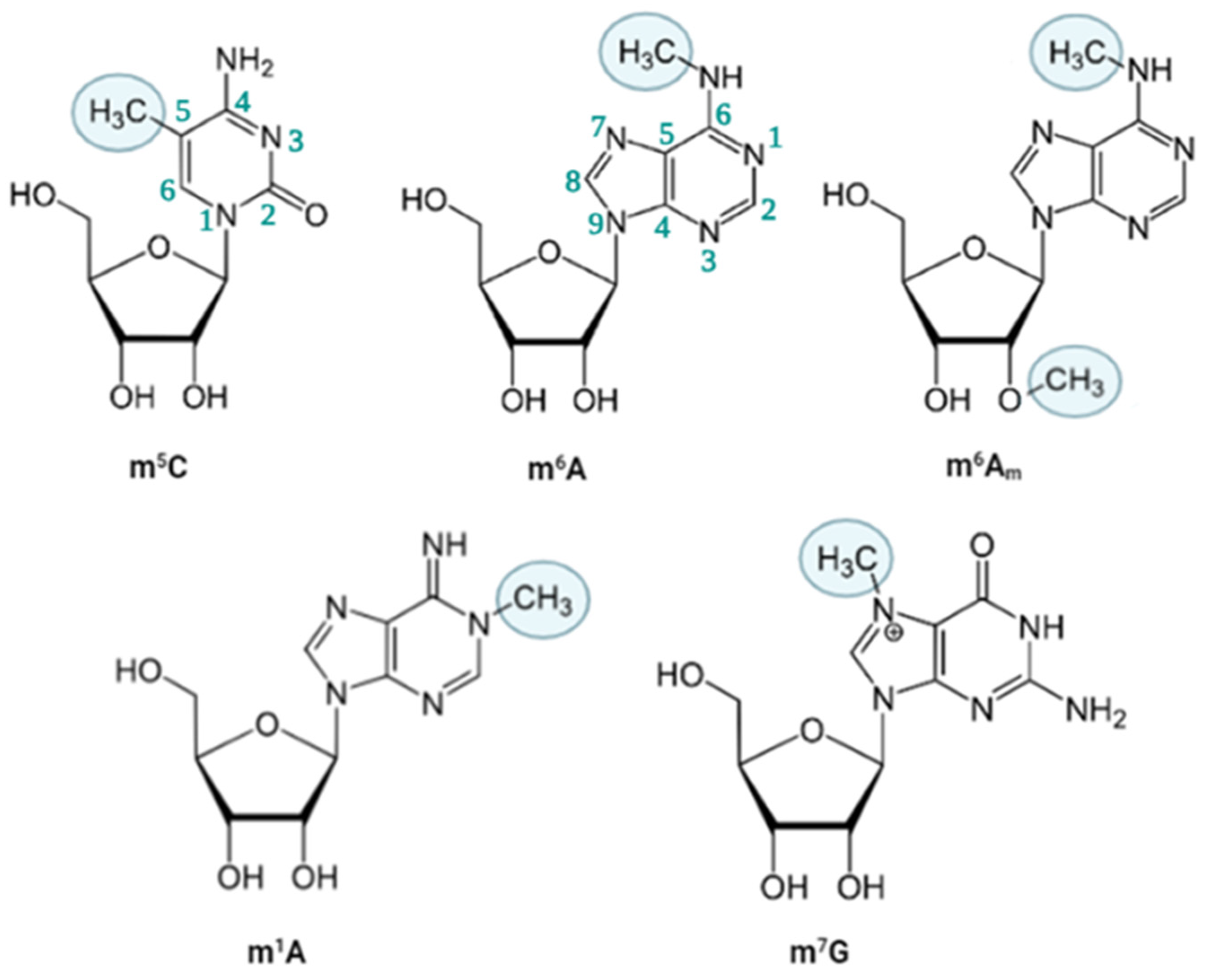
2. Types of RNA Methylation

A variety of ribonucleotides are subject to methylation. In addition to 2-O-methylation, adenosine, guanosine and cytosine nucleotides can be methylated at different sites, the examples of which include, but are not limited to, m6A, m6Am, m1A, m7G as the cap at the 5’ end and m5C [9]. Although methylations on tRNAs and rRNAs have been historically recognized as instrumental biochemical modifications for their functionality, recent reports underline the significance of these marks on mRNAs as well [27]. The deposition, recognition and removal of RNA methylations are performed by specific modifiers, leading to a reversible and dynamic RNA methylation pattern in a cell- and condition-specific manner [28]. The addition of methylation is carried out by methyltransferase enzymes called “writers” while methyl groups are removed by demethylases called “erasers” [27,29]. The destiny of methylated RNA is primarily dictated by an RNA-binding protein (RBP) called “reader”, which recognizes the methylated residue directly or the secondary structure of RNA formed as a consequence of methylation [30]. In the following sections, we will describe the most common types of RNA methylation marks followed by site-specific or genome-wide detection methods. We deeply apologize for being unable to cover all RNA methylations and other excellent studies due to space limitations.

2.1. m6A

m6A is an adenosine nucleotide whose N6 position is biochemically modified through the attachment of a methyl moiety and ranks as the most abundant modification on mRNAs (Figure 1) [31]. Although mammalian cells were reported to possess m6A methylated RNAs as early as 1970s [32], it has taken nearly 30 years to eradicate the doubts about the presence of internal m6A residues [33,34]. m6A marks have been reported in numerous species such as yeast, Arabidopsis, Drosophila and mammals [35]. The m6A deposition is performed by a writer complex composed of methyltransferase-like protein 3 (METTL3), methyltransferase-like protein 14 (METTL14), Wilms’ tumor 1-associated protein (WTAP), RNA-binding motif protein 15 (RBM15) and other methyl-transferase proteins. The actual catalysis is carried out by METTL3, and METTL14 functions as an allosteric activator for METTL3 stability [36,37]. METTL3 and METTL14 are responsible for 99% of m6A deposition on mRNAs [38]. All other writer proteins are also involved in facilitative tasks for m6A addition, such as localization and stability on target RNA site [39,40,41]. In human and mouse, m6A-specific RNA immunoprecipitation studies coupled with high throughput sequencing studies have identified the consensus motif RR(m6A)CH in which R represents A or G, and H represents A, C or U [42,43]. Minor variations have been reported in other species. Additionally, a second motif, m6ACA, has been reported, which is sensitive to digestion by the MazF RNAse if the adenosine is methylated [44]. This feature has been exploited to study m6A RNA methylations in a site-specific or genome-wide manner. Thus far, merely two eraser proteins have been identified that serve as m6A demethylases, namely fat mass and obesity-associated protein (FTO) and alpha-ketoglutarate-dependent dioxygenase homolog (ALKBH) [18,45].
m6A methylated RNAs are recognized by a class of RNA-binding proteins, readers, that can be divided into two groups based on their mode of recognition: (1) direct readers recognize the methyl moiety to coordinate the downstream events; (2) indirect readers recognize and fine-tune the secondary structure of the methylated RNA to modulate the interaction between the methylated RNA and other proteins. Direct readers include YTH domain-containing proteins (YTHDC), YTH domain-containing family proteins (YTHDF) and eukaryotic initiation factor 3 (elF3) [46,47]. Among indirect readers are heterogenous nuclear ribonucleoprotein C (HNRNPC), heterogenous nuclear ribonucleoprotein G (HNRNPG), insulin-like growth factor 2 mRNA-binding protein (IGF2BP) and Fragile X mental retardation protein (FMRP) [48]. Readers can be located either in the cytoplasm or nucleus to perform m6A-mediated cellular processes. In the nucleus, YTHDC1 modulates splicing and transport of mRNA into the cytoplasm, where YTHDC2 dictates the post-transcriptional fate of cytoplasmic mRNAs by regulating their stability and translational efficiency. Both readers recognize the m6A portion on RNAs [48,49]. Indirect readers HNRNPC and HNRNPG proteins modulate splicing by interacting with noncoding RNAs such as miRNA (microRNA), snRNA (small nuclear RNA), snoRNA (small nucleolar RNA) [50,51,52] while IGF2BP1–3 proteins bind weakly to the m6A moiety, supporting the stability of the methyl-adenine interaction. In addition, FMRP interacts with m6A-bound YTHDF2 and indirectly facilitates m6A stability [53].

2.2. m6Am

N6, 2′-O-dimethyladenosine, often abbreviated as m6Am, is a type of methylation that carries two methyl moieties, one at the N6 position of the adenine ring and another at the 2’ position of the ribose sugar. This m6Am modification influences the fate of RNAs through splicing, stability and translation. It also has implications in transcriptional processes through the modulation of chromatin interactions [54,55]. Therefore, it has been implicated in several developmental disorders as well as diseases such as cancer, obesity and neurodegenerative diseases [56,57,58].
Unlike other methylation marks, m6Am does not have a distinct sequence motif on RNA. However, the BCA motif, where B represents C, U or G, has been explored as being part of the RR(m6A)CH consensus motif at the 5’ cap structure [59,60]. Additionally, m6Am can be deposited at the 5′ cap structure, 7-methylguanosine (m7G), that can be further modified with the aid of cap-specific adenosine-N6-MTase (CAPAM) protein, also called Phosphorylated CTD-Interacting Factor 1 (PCIF1) to m6Am. In this case, the methyl group is deposited to the N6 position of the adenine base. Thus far, PCIF1 protein has been reported as the sole writer protein for m6Am addition [61]. On the other hand, FTO demethylase catalyzes the removal of m6Am modification. AlkB family member 5 (ALKBH5) is incapable of removing m6Am marks [62,63].

2.3. m1A

m1A carries a methyl moiety on a nitrogen attached to the first carbon atom of adenosine (Figure 1) [64]. Its stoichiometry is very low compared to the m6A methylation due to its location on adenosine. m1A differs from m6A in that m1A generates a positively charged nucleotide due to its location, the Watson–Crick interface [65]. Consequently, the resulting positive charge modulates the nature of RNA-protein interactions as well as the RNA secondary structure. Thus far, more than 2500 m1A sites have been identified in human despite the fact that more attention has been directed towards RNA m6A methylation [66]. m1A marks are generally located in the 5′ untranslated region (5′ UTR) of mRNAs, but mostly in rRNA and tRNA of eukaryotes, as well as mitochondrial RNA [67].
The writer complex of m1A methylation is composed of tRNA methyltransferases located in the cytoplasm, namely TRM61, TRMT6, TRMT10C and TRMT61B [68]. The TRMT6-TRMT61A complex utilizes the GUUCRA consensus motif for addition of m1A [69,70]. AlkB homolog 1, histone H2A dioxygenase or alpha-ketoglutarate-dependent dioxygenase ABH1 (ALKBH1) and AlkB homolog 3, alpha-ketoglutarate-dependent dioxygenase (ALKBH3) serve as eraser proteins of m1A [67]. In addition, FTO has been reported to remove m1A marks on tRNA [71]. m1A reader proteins include YTHDF1, YTHDF2, YTHDF3 and YTHDC1. It is worth noting that the number of m1A marks on mRNA and noncoding RNAs (ncRNAs) is relatively scarce [67,72,73].

2.4. m7G

m7G is another modification of guanosine nucleoside located at the seventh nitrogen where the 5′ cap of RNA is established. This methylation is involved in mRNA capping, which has crucial roles in mRNA stabilization, translation initiation, nuclear export and internal modifications in other types of RNAs such as rRNA, tRNA, and some miRNAs [74]. The addition of m7G modification is facilitated co-transcriptionally by guanylyl-transferase methyltransferase (GTase). On mRNAs, the methylase adds the m7G at the 5′ cap, whereas the deposition site on tRNAs is characterized by a consensus sequence “RGGUY’’. The writer protein complex of m7G for tRNA is composed of methyltransferase 1, also called tRNA (guanine-N(7)-)-methyltransferase (METTL1), and tRNA (guanine-N(7)-)-methyltransferase subunit WDR4 (WDR4) writer proteins in mammals and their orthologs Trm8/Trm82 complex in yeast. Additionally, the RNA guanine-7 methyltransferase and RNMT-activating mini protein (RNMT-RAM) complex functions in the deposition of m7G at the mRNA caps by transferring a methyl group from S-adenosylmethionine (SAM). Additional regulators have been reported in m7G modification of rRNAs [75]. Quaking (QKI) protein has been reported as the first reader protein of m7G, including three isoforms, QKI5, QKI6 and QKI7 [76]. However, no demethylases for m7G have been reported yet. It is important to note that this modification has crucial roles in various diseases such as genetic disorders, cancer and viral infectious [77,78,79].

2.5. m5C

m5C is a type of methylation in which a methyl moiety is covalently attached to the fifth carbon of cytosine (Figure 1). The existence of m5C in RNAs was reported nearly 50 years ago [32]. However, exploiting bisulfite treatment that is typically used to examine DNA methylation, m5C marks were shown to exist internally in mRNAs and lncRNAs as well [80]. The components of m5C biogenesis have been linked to various diseases. For instance, m5C writer protein NOP2/Sun RNA methyltransferase 2 (NSUN2) has been reported to promote tumor progression [81]. most m5C sites on RNAs are recognized by proteins that possess an S-adenosyl methionine (AdoMet)-binding region, a prevalent catalytic domain [81,82,83,84,85,86]. Thus far, tRNA methyltransferase 1 (TRDMT1), also known as DNA methyltransferase homolog DNMT2, has been shown to be a m5C methyltransferase just like NSUN2 [87]. Recently, NSUN1 and NSUN3–7 proteins have also been sifted out as m5C writer proteins [86,88,89,90]. Although an exact consensus sequence for m5C is still uncertain, a few potential sequences, namely, HACCR, CWUCUUC and CCDCCR, have been reported in Arabidopsis thaliana [91]. Erasers of m5C include TET family of enzymes and ALKBH1 [92,93,94]. Lastly, two reader proteins have been reported, namely Aly/REF export factor (ALYREF), an oncogenic factor, and Y-box-binding protein 1 (YBX1) [95,96]. Compared to m1A and m6A marks, m5C marks have been characterized relatively poorly. Thus, more work is required to elucidate the contribution of m5C marks in health and disease [86,94].

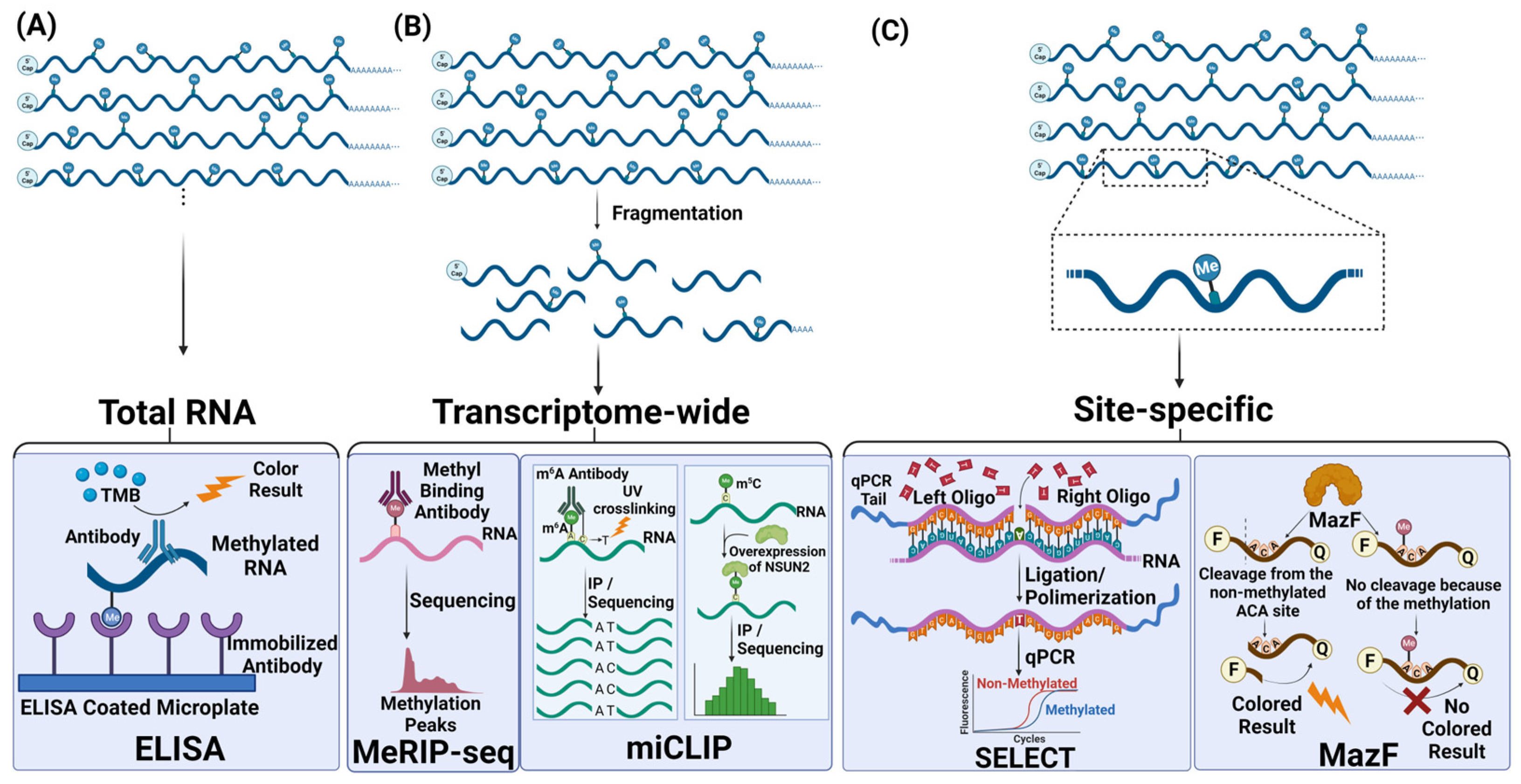
3. Detection Technologies for RNA Methylation

Various methods have been developed to evaluate the extent of RNA methylation (Figure 2). The method of choice is primarily dictated by the coverage and resolution desired in addition to cost, simplicity and expertise required (Table 1). Perhaps, the initial choice in most studies is the global assessment of fluctuations in total RNA methylation upon a stimulus. Since the global assessment lacks resolution, approaches have been developed to examine the methylation status of specific sites, especially if existing data point to the significance of a specific site. On the other hand, genome-wide approaches are exploited to cover the whole transcriptome in an unbiased manner. As such, these methods involve the use of methylation-specific antibodies, digestion of specific sequences, ligation of methylated sites, hybridization of corresponding RNAs or labelling. In addition to the coverage and resolution, cost, efficiency and the required technical infrastructure are other criteria that dictate the type of detection method to be used.

3.1. Approaches to Measure Global Changes in RNA Methylation

Global detection methods refer to the overall measurement of methylation, either total methylation or a specific type of methylation. One application of this approach is to measure the amount of total methylation without considering the transcripts carrying it or the site of methylation. This type of analysis facilitates the direct detection of RNA methylation status using total RNA isolated from any species or cells under a certain cellular condition. Typically, this approach is used to establish a link between the change in methylation abundance and a phenotype of interest. Enzyme-linked immunosorbent assays (ELISA)-based colorimetric kits are commercially available. Basically, the intensity of the signal originating from the sample RNAs is compared to a standard curve obtained from known methylated and nonmethylated control RNAs to assess the extent of methylation in test samples [97]. The major drawback of this approach is that it is impossible to deduce which transcripts or which nucleotide residues are affected from differential methylation.
Mass spectroscopy (MS) is another method utilized to analyze the total amount of RNA methylation based on their polarity and charge. This technique involves enzymatic digestion of RNAs into nucleosides, followed by mass spectrometry analysis to identify modified nucleosides. Based on the mass-to-charge ratio the identification and quantification of individual molecules are determined. Perhaps the most classical method for the analysis of modified RNAs is one-dimensional (1D) or two-dimensional (2D) thin layer chromatography (TLC). This approach takes advantage of modified nucleotides having a different net charge, hydrophobicity or polarity compared to their nonmodified counterparts. Particularly, 5′- or 3′-nucleoside monophosphates can be analyzed using this relatively simple system [98]. The comparative quantification of methylation is put into practice by forward methods such as liquid chromatography–tandem mass spectrometry (LC-MS/MS) or 2D-TLC [99,100,101]. LC/MS methodology requires cleavage of a single nucleotide with the aid of RNases and UV detection of desired methylation by using its physico-chemical properties. Although this quantitative method has straightforward steps, the localization or sequence information cannot be derived and any contamination by foreign RNA artifacts can affect the downstream analysis [99,100]. Aforementioned 2D-TLC method is a deep-rooted approach for separation of molecules in a sample depending on its size and charge. To be able to tune this method to methylation detection, cellulose substrate is used to disperse RNA based on properties changed by the effect of methylation: charge and hydrophobicity. The fragmentation of RNA is performed based on the methylation type of interest [102]. For example, RNase T1, which recognizes GAC motifs, is utilized for detection of m6A and normalized to the total adenosine level. The observation is performed with ultraviolet light or by γ-32P-ATP radioisotopes for 5′-end labeling to increase the sensitivity. Albeit with high accuracy, 2D-TLC can only detect m6A on GAC context but cannot catch AAC sites present in rRNAs [98,101,102].
A combined approach, site-specific cleavage and radioactive labelling followed by ligation-assisted extraction and TLC (SCARLET), exploits RNase H site-specific cleavage if there is a putative target methylation site. After the site-specific cleavage, splinted ligation is utilized to ligate the corresponding nucleotide to a DNA oligo, preventing it from digestion by RNase T1/A. This process is followed by thin-layer chromatography for evaluation of m6A [103,104].
Dot blot, also called slot blot, is a relatively basic method to examine different types of RNA methylations. It is performed by using a membrane coated with specific antibodies or molecules that recognize the desired methylation. The detected methylations are spotted on the membrane by signals performed with the aid of a fluorescence or chemiluminescence molecule after subjecting a vacuum process [105]. Although it is often used for m6A, it can be modified to entertain the detection of other types of methylations such as 5hmC [106]. As a negative side of this technique, it can only be used to verify the presence of methylation and to compare the changes in the global abundance of methylation among different samples. The dot blot approach is incapable of pinpointing the precise site of methylation mark. Of importance is the urgency to eliminate potential DNA contamination while analyzing 5hmC marks on RNA [105,107].
As a novel technique to identify and quantify the genome-wide methylation in RNA, DART-Seq (Deamination adjacent to RNA Modification Targets) was improved at the single-nucleotide level. This method is an antibody-free approach for m6A detection of RNA taking advantage of apolipoprotein B mRNA editing enzyme catalytic subunit 1 (APOBEC1). The method involves five steps, namely RNA isolation, in vitro deamination, library preparation, deep sequencing and data analysis. The in vitro deamination step involves APOBEC1, a chimeric protein engineered by fusing the m6A-binding YTH domain, which deaminates the cytosine nucleotide adjacent to m6A to uracil. This targeted deamination strategy offers a high specificity for m6A sites. In the last step, the abundance of reads stemming from unconverted cytosine nucleotides reflects the level of m6A modification at that specific site. DART-Seq method can be employed to quantitatively measure the extent of m6A RNA methylation in a transcriptome by coupling the analysis with RNA-seq [108].

3.2. Transcriptomic Detection Methods

Most genome-wide approaches in current use employ second generation sequencing (NGS) in which an amplification step is required to generate a cluster of templates for detectable sequencing signals in an unbiased manner. With a low error rate, NGS has been the method of choice despite its shorter read of a couple of hundreds of nucleotides at the most. Single-molecule direct sequencing protocols have been developed to overcome the size limitation. Currently, there are two direct RNA-based detection methods, namely nanopore and single-molecule real-time (SMRT) applications [35,109,110]. Initially, nanopore technology employed a technique for determination of characteristic current blockade difference based on DNA and RNA structure changes while passing through nanopores. This method was applied to the analysis of m6A and m5C marks. These modifications have been tested by sequencing of methylated and nonmethylated synthetic RNAs. The resulting difference is calculated and used to locate the methylated region of RNA [109]. The other approach, SMRT, takes advantage of labelled nucleotides during SMRT DNA library preparation followed by LC-MS observation. Although this procedure assures determination of methylation-related isoform or transcript changes and the quantity of methylation sites per isoform, it is not a sensitive enough protocol as the margin of error is relatively much higher [7,110,111].
The genome-wide analysis involves the sequencing of the precipitated RNAs, whereas qPCR can be employed, with a proper set of primers, to examine the precipitation efficiency, thus the existence of a mark, of a single target RNA. For instance, m6A-seq/MeRIP-seq is an RNA immunoprecipitation method that involves the use of an m6A-specific antibody and RNAs fragmented into approximately 100 nt in size [34]. The sequencing of precipitated RNA provides valuable information about the enrichment of a fragment of RNA as an indicator of the presence of a methyl moiety. Alternatively, a pair of primer can be designed for qPCR analysis of a target RNA to check for its enrichment in the immuno-precipitate. To compare the relative change in the extent of methylation upon a treatment (for example, control versus drug treatment or healthy versus cancer), RNAs in each condition must be sequenced first to determine the abundance of individual RNAs as treatments may lead to an increase in the RNA abundance independent from differential methylation. Subsequently, the relative enrichment of target RNAs or fragments of target RNAs must be calculated to find out the fold of differential RNA methylation. There are various library preparation and sequencing strategies for detection of modified nucleotides in RNAs [112]. MeRIP-seq is a primary choice of method since it can be adapted to examine any modification as long as a specific antibody is available [33,34]. However, cross-reactivity among antibodies is an important issue that should be always taken into consideration in antibody-based approaches as cross-reactivity may lead to false-positive signals. For example, it is highly challenging to distinguish m6A from m6Am due to nonspecific interactions between antibodies and methylation marks. Although adaptability to the analysis of different methylation marks makes MeRIP-seq an attractive choice, a major drawback of this method is its low resolution. Post-precipitation, RNA fragments of 100–200 nt in size are subjected to sequencing, making it difficult to pinpoint the exact site of methylation especially if multiple methylation motifs exist in such fragments [33,34,111].
m6A individual-nucleotide resolution UV crosslinking and immunoprecipitation (miCLIP) [59] and its improved version, enhanced CLIP (eCLIP) [113], is another antibody-based approach to map m6A sites at the single-nucleotide resolution. This approach identifies m6A marks with high confidence as the UV-crosslinked nucleotide nearby the methyl mark serves as an excellent indicator for the presence of a methyl moiety. UV crosslinking leads to a C-to-T mutation signature that ensures the partition of multiple m6A signals within the same peak. More importantly, this approach makes it possible to distinguish m6A from m6Am. Although miCLIP can overcome all the drawbacks of MeRIP-seq, it requires a higher quantity of input material and cannot provide stochiometric information [111,114,115].
MeRIP and miCLIP can be utilized for both transcriptome-wide and site-specific mapping of m6A, m1A, m6Am and m5C marks. Dynamic changes in the abundance of these types of RNA methylation marks can be examined by exploiting relatively strong interactions between specific methylation marks and antibodies that specifically recognize these marks [35]. Following the incubation of RNA samples with antibodies, the fragments of methylated RNAs are immune-precipitated with an antibody. Additionally, the comparison of the relative amount of site-specific methylation changes can be observed following site-directed mutagenesis or RT-PCR (real-time PCR) reaction by specific primers after immunoprecipitation [116].
Table 1. The summary of commonly used methylation detection methods.
MethodsAdvantagesDisadvantagesReferences
Global RNA Methylation Detection Methods
ELISA
  • Various methylation detection based on antibody presence
  • Low amount of input RNA requirement
  • Standard curve convenience
  • Easy preparation steps with commercial product
  • Contamination risk during preparation
  • Transport and storage conditions of commercial kit require attention
  • Instability of RNA and antibodies provided by commercial kit require attention
  • Long standby times during protocol and labor intensive
[97]
2D-TLC
  • Conventional method
  • Increased accuracy by radioisotopes
  • Increased specificity of m6A in mRNA by cleavage of GAC context
  • Low amount of input RNA required
  • High stoichiometric information
  • Adaptable to all types of modifications
  • Inability to detect AAC sites of m6A in rRNA
  • Difficulty of using radioactive substance
  • Low resolution
[98,101,102]
LC-MS
  • Standardized technique
  • High accuracy in quantification
  • Easy to prepare
  • Useful for all types of modifications
  • Risk of contamination
  • No sequence information
  • Labor intensive
  • Necessity for specialized equipment
  • Require complicated computational analysis
[99,100]
SCARLET
  • High accuracy
  • Site-specific determination
  • Low amount of input RNA required
  • No need for specialized equipment
  • Only one site per transcript determination at once
  • Low throughput
  • High quantity of input material
  • Used for only m6A methylation
[103,104]
Dot Blot
  • Simple
  • Relatively inexpensive
  • No requirement for fragmentation
  • Markedly saves time since no need chromatography, gel electrophoresis or complex gel blocking procedures
  • Need to use antibody or other molecule for fluorescence or chemiluminescence
  • Cannot determine the quantitation
  • Unable to determine precise location
[105,106,107]
DART-Seq
  • High sensitivity for m6A sites at the single-nucleotide level
  • An antibody-free approach to eliminate commercial assays
  • Low input RNA requirement with as little as 10 ng of total RNA as input
  • High-throughput sequencing
  • Long-read compatibility
  • Potential off-target deamination due to specificity of APOBEC1
  • Requires careful consideration during data analysis due to specificity of APOBEC1
  • Computational challenges as robust computational pipelines for accuracy
[108]
Transcriptome-wide RNA Methylation Detection Methods
Nanopore
  • Possibility of methylation-related isoforms and transcripts examination
  • Validation of potential methylation stoichiometry
  • Facility in abundance of methylation per isoform determination
  • Library preparation is not required
  • No need for PCR or qPCR equipment
  • Newly adopted to methylation detections
  • Depend on change in the current which can be hard to differentiate
  • Fewer studies with RNA isolated from cells
  • High level error rate in base assignment
  • Prone to statistical problems
[109]
SMRT
  • Possibility of methylation-related isoforms and transcripts examination
  • Detection methylation level per isoform
  • Low level of sensitivity
  • High level of error in base assignment
  • Prone to statistical problems
[110,111]
MeRIP-seq
  • Transcriptome-wide mapping is provided
  • Low quantity of RNA input is required
  • Easy steps for library construction
  • Adaption to various methylations (m6A, m1A and m5C) based on antibody availability
  • Well-studied
  • Necessity of RNA sequencing
  • Inability in discrimination of m6A from m6Am
  • Insufficient single-nucleotide resolution
  • Insufficient to distinguish multiple methylation sites in a peak
  • Stoichiometric information not provided
[33,34,112]
miCLIP
  • Transcriptome-wide mapping is provided
  • Increased specificity by a C-to-T mutation signature for m6A and NSUN2 overexpression for m5C
  • Ability in discrimination of m6A from m6Am
  • Sufficient to single-nucleotide resolution
  • Sufficient for distinguish multiple methylation sites in a peak
  • Adaptable to all types of methylations
  • Complicated steps for RNA library construction
  • Stoichiometric information not provided
  • High abundance of input material is needed
  • Requirement to special equipment
[59,113]
Site-specific RNA Methylation Detection Methods
Reverse transcriptase based-qPCR assay
  • Specific-site detection using related oligomers
  • Stoichiometric approach by melting properties
  • Useful for site-specific detection in rRNA and snRNA as well as mRNA
  • Useful for various modifications
  • Low RNA input required
  • Straightforward method
  • Low sensitivity level
  • Based on reverse-transcriptase enzyme flexibility
[117,118,119]
HRM
  • Simple
  • Specific location of m6A modification residues with high-throughput measurement
  • Commercially available kits
  • Necessity of positive and negative control to evaluate methylation abundance
  • Relatively detection of methylation in percentage
[120,121]
MazF
  • Site-specific determination of m6A using ACA sequence cleavage by MazF enzyme
  • Transcriptome-wide mapping available with further processes (MAZTER-seq)
  • Detection of m6A profile only in ACA content
  • Insufficient to distinguish adjacent ACA sites in MAZTER-seq
[122]
T3/T4 DNA ligase-qPCR
  • Methylation stoichiometry can be observed
  • Site-specific detection
  • Easy protocol steps
  • Efficiency of the ligase is crucial
  • Low throughput
[123]
SELECT
  • Feasibility of evaluation of methylation stoichiometry
  • Provided site-specific detection
  • Easy to prepare
  • Able to adapt for various types of methylations (m1A, Am (2′-O methyladenosine))
  • Based on two different enzyme efficiencies: Bst polymerase and Splint R ligase
  • Possibility of false-positive outcomes
  • Rough process of oligomer designing
  • Low throughput
[124]

3.3. Site-Specific Detection Methods

Although transcriptomic approaches provide valuable information about changes in methylation marks, its cost, requirement for sophisticated devices and expertise and potential cross-reactivity of antibodies necessitate the use of site-specific detection methods for validation of transcriptomic data. Typically, pre-existing data, such as those obtained from a transcriptomic study, is used to select a site for examination. The existing site-specific methylation detection methods employ hybridization-based, digestion-based and ligation-based approaches.
Hybridization-based approaches take advantage of the incredible mechanism of retrotranscription by nucleic acid polymerases Thermus thermophiles (Tth) and Bacillus stearothermophilus (BstI) [117]. The template RNA is retrotranscribed by these polymerases with the aid of an adjacent primer, leading to RNA-directed DNA synthesis. The key point here is the flexibility of polymerases during the elongation process and constructional access of the enzyme to the template with the primer. In principle, methylated nucleotides would impede polymerization. Therefore, RNA templates with methylation would be amplified less efficiently compared to nonmethylated RNA templates. Furthermore, polyacrylamide gel electrophoresis or RT-qPCR is performed to determine the relative level of methylation based on the selective processivity of the enzyme. It is important to note that these approaches were primarily developed to examine m6A marks [117,118,119]. Hybridization-based methods provide relative information about the methylation stoichiometry at specific RNA sites and can be used for rRNA and snRNA specific sites. However, this approach suffers from low sensitivity [111]. High-Resolution Melting (HRM), an alternative hybridization-based approach, has been improved to detect m6A methylations in a site-specific manner. This method exploits the plots of purely methylated and un-methylated RNA sequences of interest. The RNA region of interest is then compared to these melting curves and gives the result in percentage in a high-throughput manner. Methylation causes a change in the melting curves of 100% methylated and 100% unmethylated RNAs. Therefore, it is a necessity to determine the methylated region on the RNA for processing the melting curve standards [120,121].
A unique methodology of digestion-based method involves the MazF RNA for site-specific detection of methylation marks. MazF is an RNase that digests the nonmethylated ACA motifs and these fragments can be aligned to the genome to uncover the site of methylation. Single-base resolution can be accomplished by MazF by taking advantage of Förster resonance energy transfer (FRET) technique as a fluorescent biosensor of m6A by a light production after cutting ACA motif and transfer of energy from the reporter to the quencher in the absence of methylation [44,101,122].
Ligation-based detection methods have been initially utilized to analyze m6A marks by comparing the ligation efficiency of two primers complementary to the sequences upstream and downstream from the methylated nucleotides. Liu et al. employed a T3 DNA ligase-based qPCR technique to examine m6A marks in a specific region in RNA [123]. Subsequently, Xiao et al. improved this approach by using a single-base elongation- and ligation-based qPCR amplification method (SELECT), in which T3 DNA ligase was replaced by Bst 2.0 DNA polymerase and SplintR ligase [124]. In this technique, two probes, namely forward and reverse oligomers, are designed immediately upstream and downstream from the methylated sites. Two additional oligomers are designed +2/−6 nucleotides close to the methylated nucleotide to be used as negative controls. These probes are then efficiently ligated by dNTP and SplintR ligase if the target adenosine is nonmethylated. Consequently, Taq DNA polymerase can efficiently amplify the ligated product upon the conversion of the ligated RNA into cDNA. On the other hand, the presence of a methyl moiety impairs the ligation efficiency, resulting in the amplification of relatively less PCR product. SELECT is a desirable choice for the analysis of site-specific single methylation marks on a target RNA. Despite its low cost and simplicity, it has low throughput and a high false-positive rate based on the efficiency of two distinct steps: elongation by Bst 2.0 DNA polymerase and ligation by SplintR ligase [123,124].
SELECT is a well-defined technique with two main steps: the single-base elongation activity and the nick ligation efficiency of DNA polymerases and ligases, respectively. Based on our existing experience, we suggest considering the following points in executing SELECT for site-specific assessment of methylation marks: (1) there should be a single nucleotide between the up and down probes; (2) diluting the enzymes in Diluent A facilitates their long-term storage, lowering costs as opposed to freshly preparing the enzymes each time as suggested by the manufacturers; (3) SplintR ligase in small quantities (0.5 unit) is sufficient in ligation reactions as opposed to higher amounts of T3 DNA ligase (12.5 unit); (4) the negative control, an unmethylated nucleotide near the desired methylated site, must be in the region in between −6 and +2 except for ±1. So, the up and down probes must be designed at one of these sites individually; (5) dTTP instead of dNTP appears to be slightly more efficient; and (6) it is important to design a qPCR adapter with a melting temperature above 50 °C at the two sides of the complementary strand of an RNA template.

4. Conclusions

A number of advances have been made in the field of RNA methylation, which has shifted its interest from the analysis of tRNA and rRNA to mRNA and other ncRNAs. In particular, the discovery of novel approaches that have enabled researchers to examine a variety of methylation at a site-specific or genome-wide manner has paved the way to uncover changes in the abundance of methylation marks in health and disease. Simultaneously, writers, erasers and readers of these biochemical modifications have been uncovered, which have been associated with numerous diseases. However, genome-wide approaches in particular can be applied to the analysis of a small fraction of biochemical modifications, necessitating the development of novel protocols. Along this line, third-generation sequencing technologies hold great promise as they do not require cDNA construction and permit direct detection of modifications with a much longer read. Additionally, existing sequencing-based approaches are still highly expensive, further requiring the development of protocols that reduce sequencing costs. Since each detection method has both advantages and limitations, the choice of method depends on the abundance of RNA modification, the type of RNA, the amount of starting material, cost, simplicity and the availability of proper infrastructure.
RNA methylation is one of the most dynamic research topics of recent times and clearly holds great promise for translation into clinic. Although it is a matter of debate whether this dynamic mechanism can be exploited to predict the progression of various diseases such as cancer, we believe that the identification of disease-specific methylation marks, e.g., RNA methylation signatures, should pave the way for its use in the clinic. Certainly, to facilitate this aim, cheaper and more convenient methods should be developed to speed up the research in this field. Alternatively, existing methods can be improved to lower analysis costs, leading to greater accessibility.

Author Contributions

B.S. and B.A. wrote, read and approved the manuscript. All authors have read and agreed to the published version of the manuscript.

Funding

This research was funded by the Scientific and Technological Research Council of Türkiye, grant number 217Z234 and The APC was funded by Bünyamin Akgül.

Acknowledgments

The authors would like to thank A. Batuhan Dondurur for technical help.

Conflicts of Interest

The authors declare no conflicts of interest.

References

  1. Pierrat, O.A.; Mikitova, V.; Bush, M.S.; Browning, K.S.; Doonan, J.H. Control of Protein Translation by Phosphorylation of the MRNA 5-Cap-Binding Complex. Biochem. Soc. Trans. 2007, 35, 1634–1637. [Google Scholar] [CrossRef]
  2. Wiener, D.; Schwartz, S. The Epitranscriptome beyond m6A. Nat. Rev. Genet. 2021, 22, 119–131. [Google Scholar] [CrossRef]
  3. Arango, D.; Sturgill, D.; Yang, R.; Kanai, T.; Bauer, P.; Roy, J.; Wang, Z.; Hosogane, M.; Schiffers, S.; Oberdoerffer, S. Direct Epitranscriptomic Regulation of Mammalian Translation Initiation through N4-Acetylcytidine. Mol. Cell 2022, 82, 2797–2814.e11. [Google Scholar] [CrossRef]
  4. Castello, A.; Fischer, B.; Frese, C.K.; Horos, R.; Alleaume, A.M.; Foehr, S.; Curk, T.; Krijgsveld, J.; Hentze, M.W. Comprehensive Identification of RNA-Binding Domains in Human Cells. Mol. Cell 2016, 63, 696–710. [Google Scholar] [CrossRef]
  5. Zhu, Z.M.; Huo, F.C.; Pei, D.S. Function and Evolution of RNA N6-Methyladenosine Modification. Int. J. Biol. Sci. 2020, 16, 1929–1940. [Google Scholar] [CrossRef]
  6. Bazak, L.; Haviv, A.; Barak, M.; Jacob-Hirsch, J.; Deng, P.; Zhang, R.; Isaacs, F.J.; Rechavi, G.; Li, J.B.; Eisenberg, E.; et al. A-to-I RNA Editing Occurs at over a Hundred Million Genomic Sites, Located in a Majority of Human Genes. Genome Res. 2014, 24, 365–376. [Google Scholar] [CrossRef] [PubMed]
  7. Saletore, Y.; Meyer, K.; Korlach, J.; Vilfan, I.D.; Jaffrey, S.; Mason, C.E. The Birth of the Epitranscriptome: Deciphering the Function of RNA Modifications. Genome Biol. 2012, 13, 175. [Google Scholar] [CrossRef] [PubMed]
  8. Gilbert, W.V.; Nachtergaele, S. mRNA Regulation by RNA Modifications. Annu. Rev. Biochem. 2023, 92, 175–198. [Google Scholar] [CrossRef] [PubMed]
  9. Khosravi, H.M.; Jantsch, M.F. Site-Directed RNA Editing: Recent Advances and Open Challenges. RNA Biol. 2021, 18, 41–50. [Google Scholar] [CrossRef] [PubMed]
  10. Understanding RNA Editing and Its Use in Gene Editing. Gene Genome Ed. 2022, 3–4, 100021. [CrossRef]
  11. Boccaletto, P.; Stefaniak, F.; Ray, A.; Cappannini, A.; Mukherjee, S.; Purta, E.; Kurkowska, M.; Shirvanizadeh, N.; Destefanis, E.; Groza, P.; et al. MODOMICS: A Database of RNA Modification Pathways. 2021 Update. Nucleic Acids Res. 2022, 50, D231–D235. [Google Scholar] [CrossRef]
  12. Farooqi, A.A.; Fayyaz, S.; Poltronieri, P.; Calin, G.; Mallardo, M. Epigenetic Deregulation in Cancer: Enzyme Players and Non-Coding RNAs. Semin. Cancer Biol. 2022, 83, 197–207. [Google Scholar] [CrossRef] [PubMed]
  13. Chen, Z.; Huang, L.; Wang, K.; Zhang, L.; Zhong, X.; Yan, Z.; Liu, B.; Zhu, P. RtcisE2F Promotes the Self-Renewal and Metastasis of Liver Tumor-Initiating Cells via N6-Methyladenosine-Dependent E2F3/E2F6 MRNA Stability. Sci. China Life Sci. 2022, 65, 1840–1854. [Google Scholar] [CrossRef] [PubMed]
  14. Zheng, Z.H.; Zhang, G.L.; Jiang, R.F.; Hong, Y.Q.; Zhang, Q.Y.; He, J.P.; Liu, X.R.; Yang, Z.S.; Yang, L.; Jiang, X.; et al. METTL3 Is Essential for Normal Progesterone Signaling during Embryo Implantation via m6A-Mediated Translation Control of Progesterone Receptor. Proc. Natl. Acad. Sci. USA 2023, 120, e2214684120. [Google Scholar] [CrossRef]
  15. Lin, S.; Choe, J.; Du, P.; Triboulet, R.; Gregory, R.I. The m6A Methyltransferase METTL3 Promotes Translation in Human Cancer Cells. Mol. Cell 2016, 62, 335–345. [Google Scholar] [CrossRef] [PubMed]
  16. Qiao, Y.; Sun, Q.; Chen, X.; He, L.; Wang, D.; Su, R.; Xue, Y.; Sun, H.; Wang, H. Nuclear m6A Reader YTHDC1 Promotes Muscle Stem Cell Activation/Proliferation by Regulating MRNA Splicing and Nuclear Export. eLife 2023, 12, e82703. [Google Scholar] [CrossRef] [PubMed]
  17. Ries, R.J.; Zaccara, S.; Klein, P.; Olarerin-George, A.; Namkoong, S.; Pickering, B.F.; Patil, D.P.; Kwak, H.; Lee, J.H.; Jaffrey, S.R. m6A Enhances the Phase Separation Potential of MRNA. Nature 2019, 571, 424–428. [Google Scholar] [CrossRef] [PubMed]
  18. Zheng, G.; Dahl, J.A.; Niu, Y.; Fedorcsak, P.; Huang, C.M.; Li, C.J.; Vågbø, C.B.; Shi, Y.; Wang, W.L.; Song, S.H.; et al. ALKBH5 Is a Mammalian RNA Demethylase That Impacts RNA Metabolism and Mouse Fertility. Mol. Cell 2013, 49, 18–29. [Google Scholar] [CrossRef]
  19. Wang, Y.; Li, Y.; Toth, J.I.; Petroski, M.D.; Zhang, Z.; Zhao, J.C. N6-Methyladenosine Modification Destabilizes Developmental Regulators in Embryonic Stem Cells. Nat. Cell Biol. 2014, 16, 191–198. [Google Scholar] [CrossRef]
  20. Liu, L.; Song, B.; Ma, J.; Song, Y.; Zhang, S.Y.; Tang, Y.; Wu, X.; Wei, Z.; Chen, K.; Su, J.; et al. Bioinformatics Approaches for Deciphering the Epitranscriptome: Recent Progress and Emerging Topics. Comput. Struct. Biotechnol. J. 2020, 18, 1587–1604. [Google Scholar] [CrossRef]
  21. Zhao, C.; Xu, G.; Zhang, X.; Ye, Y.; Cai, W.; Shao, Q. RNA m6A Modification Orchestrates the Rhythm of Immune Cell Development from Hematopoietic Stem Cells to T and B Cells. Front. Immunol. 2022, 3, 839291. [Google Scholar] [CrossRef] [PubMed]
  22. Li, J.; Xie, H.; Ying, Y.; Chen, H.; Yan, H.; He, L.; Xu, M.; Xu, X.; Liang, Z.; Liu, B.; et al. YTHDF2 Mediates the MRNA Degradation of the Tumor Suppressors to Induce AKT Phosphorylation in N6-Methyladenosine-Dependent Way in Prostate Cancer. Mol. Cancer 2020, 19, 152. [Google Scholar] [CrossRef] [PubMed]
  23. Alasar, A.A.; Tüncel, Ö.; Gelmez, A.B.; Sağlam, B.; Vatansever, İ.E.; Akgül, B. Genomewide m6A Mapping Uncovers Dynamic Changes in the m6A Epitranscriptome of Cisplatin-Treated Apoptotic HeLa Cells. Cells 2022, 11, 3905. [Google Scholar] [CrossRef] [PubMed]
  24. Liu, L.; Wang, J.; Sun, G.; Wu, Q.; Ma, J.; Zhang, X.; Huang, N.; Bian, Z.; Gu, S.; Xu, M.; et al. m6A MRNA Methylation Regulates CTNNB1 to Promote the Proliferation of Hepatoblastoma. Mol. Cancer 2019, 18, 188. [Google Scholar] [CrossRef] [PubMed]
  25. Zhao, F.; Xu, Y.; Gao, S.; Qin, L.; Austria, Q.; Siedlak, S.L.; Pajdzik, K.; Dai, Q.; He, C.; Wang, W.; et al. METTL3-Dependent RNA m6A Dysregulation Contributes to Neurodegeneration in Alzheimer’s Disease through Aberrant Cell Cycle Events. Mol. Neurodegener. 2021, 16, 70. [Google Scholar] [CrossRef] [PubMed]
  26. Akçaöz-Alasar, A.; Tüncel, Ö.; Sağlam, B.; Gazaloğlu, Y.; Atbinek, M.; Cagiral, U.; Iscan, E.; Ozhan, G.; Akgül, B. Epitranscriptomics m6A Analyses Reveal Distinct m6A Marks under Tumor Necrosis Factor α (TNF-α)-induced Apoptotic Conditions in HeLa Cells. J. Cell Physiol. 2024; in press. [Google Scholar] [CrossRef]
  27. Xie, S.; Chen, W.; Chen, K.; Chang, Y.; Yang, F.; Lin, A.; Shu, Q.; Zhou, T.; Yan, X. Emerging Roles of RNA Methylation in Gastrointestinal Cancers. Cancer Cell Int. 2020, 20, 585. [Google Scholar] [CrossRef]
  28. Dominissini, D.; Nachtergaele, S.; Moshitch-Moshkovitz, S.; Peer, E.; Kol, N.; Ben-Haim, M.S.; Dai, Q.; Di Segni, A.; Salmon-Divon, M.; Clark, W.C.; et al. The Dynamic N1-Methyladenosine Methylome in Eukaryotic Messenger RNA. Nature 2016, 530, 441–446. [Google Scholar] [CrossRef]
  29. Esteller, M.; Pandolfi, P.P. The Epitranscriptome of Noncoding RNAs in Cancer. Cancer Discov. 2017, 7, 359–368. [Google Scholar] [CrossRef]
  30. Squires, J.E.; Patel, H.R.; Nousch, M.; Sibbritt, T.; Humphreys, D.T.; Parker, B.J.; Suter, C.M.; Preiss, T. Widespread Occurrence of 5-Methylcytosine in Human Coding and Non-Coding RNA. Nucleic Acids Res. 2012, 40, 5023–5033. [Google Scholar] [CrossRef]
  31. Motorin, Y.; Helm, M. RNA Nucleotide Methylation. Wiley Interdiscip. Rev. RNA 2011, 2, 611–631. [Google Scholar] [CrossRef]
  32. Desrosiers, R.; Friderici, K.; Rottman, F. Identification of Methylated Nucleosides in Messenger RNA from Novikoff Hepatoma Cells (RNA Methylation/RNA Processing/Methylnucleoside Composition). Proc. Natl. Acad. Sci. USA 1974, 71, 3971–3975. [Google Scholar] [CrossRef] [PubMed]
  33. Meyer, K.D.; Saletore, Y.; Zumbo, P.; Elemento, O.; Mason, C.E.; Jaffrey, S.R. Comprehensive Analysis of MRNA Methylation Reveals Enrichment in 3′ UTRs and near Stop Codons. Cell 2012, 149, 1635–1646. [Google Scholar] [CrossRef] [PubMed]
  34. Dominissini, D.; Moshitch-Moshkovitz, S.; Schwartz, S.; Salmon-Divon, M.; Ungar, L.; Osenberg, S.; Cesarkas, K.; Jacob-Hirsch, J.; Amariglio, N.; Kupiec, M.; et al. Topology of the Human and Mouse m6A RNA Methylomes Revealed by m6A-Seq. Nature 2012, 485, 201–206. [Google Scholar] [CrossRef] [PubMed]
  35. Jonkhout, N.; Tran, J.; Smith, M.A.; Schonrock, N.; Mattick, J.S.; Novoa, E.M. The RNA Modification Landscape in Human Disease. RNA 2017, 23, 1754–1769. [Google Scholar] [CrossRef] [PubMed]
  36. Bokar, J.A.; Shambaugh, M.E.; Polayes, D.; Matera, A.G.; Pottman, F.M. Purification and CDNA Cloning of the AdoMet-Binding Subunit of the Human MRNA (N6-Adenosine)-Methyltransferase. RNA 1997, 3, 1233–1247. [Google Scholar] [PubMed]
  37. Wang, X.; Huang, J.; Zou, T.; Yin, P. Human m6A Writers: Two Subunits, 2 Roles. RNA Biol. 2017, 14, 300–304. [Google Scholar] [CrossRef] [PubMed]
  38. Geula, S.; Moshitch-Moshkovitz, S.; Dominissini, D.; AlFatah Mansour, A.; Kol, N.; Salmon-Divon, M.; Hershkovitz, V.; Peer, E.; Mor, N.; Manor, Y.S.; et al. m6A MRNA Methylation Facilitates Resolution of Naïve Pluripotency toward Differentiation. Science 2015, 347, 1002–1006. [Google Scholar] [CrossRef] [PubMed]
  39. Jinek, M.; Śledz, P. Structural Insights into the Molecular Mechanism of the m 6 A Writer Complex. eLife 2016, 5, e18434. [Google Scholar] [CrossRef]
  40. Knuckles, P.; Lence, T.; Haussmann, I.U.; Jacob, D.; Kreim, N.; Carl, S.H.; Masiello, I.; Hares, T.; Villaseñor, R.; Hess, D.; et al. Zc3h13/Flacc Is Required for Adenosine Methylation by Bridging the MRNA-Binding Factor RbM15/Spenito to the m6A Machinery Component Wtap/Fl(2)d. Genes Dev. 2018, 32, 415–429. [Google Scholar] [CrossRef]
  41. Ping, X.L.; Sun, B.F.; Wang, L.; Xiao, W.; Yang, X.; Wang, W.J.; Adhikari, S.; Shi, Y.; Lv, Y.; Chen, Y.S.; et al. Mammalian WTAP Is a Regulatory Subunit of the RNA N6-Methyladenosine Methyltransferase. Cell Res. 2014, 24, 177–189. [Google Scholar] [CrossRef]
  42. Gao, Y.; Pei, G.; Li, D.; Li, R.; Shao, Y.; Zhang, Q.C.; Li, P. Multivalent m6A Motifs Promote Phase Separation of YTHDF Proteins. Cell Res. 2019, 29, 767–769. [Google Scholar] [CrossRef]
  43. Zhang, H.; Shi, X.; Huang, T.; Zhao, X.; Chen, W.; Gu, N.; Zhang, R. Dynamic Landscape and Evolution of m6A Methylation in Human. Nucleic Acids Res. 2020, 48, 6251–6264. [Google Scholar] [CrossRef]
  44. Song, J.; Yi, C. Reading Chemical Modifications in the Transcriptome. J. Mol. Biol. 2020, 432, 1824–1839. [Google Scholar] [CrossRef]
  45. Jia, G.; Fu, Y.; Zhao, X.; Dai, Q.; Zheng, G.; Yang, Y.; Yi, C.; Lindahl, T.; Pan, T.; Yang, Y.G.; et al. N6-Methyladenosine in Nuclear RNA Is a Major Substrate of the Obesity-Associated FTO. Nat. Chem. Biol. 2011, 7, 885–887. [Google Scholar] [CrossRef]
  46. Patil, D.P.; Chen, C.K.; Pickering, B.F.; Chow, A.; Jackson, C.; Guttman, M.; Jaffrey, S.R. m6A RNA Methylation Promotes XIST-Mediated Transcriptional Repression. Nature 2016, 537, 369–373. [Google Scholar] [CrossRef]
  47. Wojtas, M.N.; Pandey, R.R.; Mendel, M.; Homolka, D.; Sachidanandam, R.; Pillai, R.S. Regulation of m6A Transcripts by the 3′→5′ RNA Helicase YTHDC2 Is Essential for a Successful Meiotic Program in the Mammalian Germline. Mol. Cell 2017, 68, 374–387.e12. [Google Scholar] [CrossRef] [PubMed]
  48. Wang, S.; Lv, W.; Li, T.; Zhang, S.; Wang, H.; Li, X.; Wang, L.; Ma, D.; Zang, Y.; Shen, J.; et al. Dynamic Regulation and Functions of MRNA m6A Modification. Cancer Cell Int. 2022, 22, 48. [Google Scholar] [CrossRef] [PubMed]
  49. Luo, S.; Tong, L. Molecular Basis for the Recognition of Methylated Adenines in RNA by the Eukaryotic YTH Domain. Proc. Natl. Acad. Sci. USA 2014, 111, 13834–13839. [Google Scholar] [CrossRef] [PubMed]
  50. Xiang, Z.; Lv, Q.; Zhang, Y.; Chen, X.; Guo, R.; Liu, S.; Peng, X. Long Non-Coding RNA DDX11-AS1 Promotes the Proliferation and Migration of Glioma Cells by Combining with HNRNPC. Mol. Ther. Nucleic Acids 2022, 28, 601–612. [Google Scholar] [CrossRef] [PubMed]
  51. Zhou, K.I.; Parisien, M.; Dai, Q.; Liu, N.; Diatchenko, L.; Sachleben, J.R.; Pan, T. N6-Methyladenosine Modification in a Long Noncoding RNA Hairpin Predisposes Its Conformation to Protein Binding. J. Mol. Biol. 2016, 428, 822–833. [Google Scholar] [CrossRef] [PubMed]
  52. Coker, H.; Wei, G.; Brockdorff, N. m6A Modification of Non-Coding RNA and the Control of Mammalian Gene Expression. Biochim. Biophys. Acta Gene Regul. Mech. 2019, 1862, 310–318. [Google Scholar] [CrossRef] [PubMed]
  53. Zhang, F.; Kang, Y.; Wang, M.; Li, Y.; Xu, T.; Yang, W.; Song, H.; Wu, H.; Shu, Q.; Jin, P. Fragile X Mental Retardation Protein Modulates the Stability of Its m6A-Marked Messenger RNA Targets. Hum. Mol. Genet. 2018, 27, 3936–3950. [Google Scholar] [CrossRef] [PubMed]
  54. Zhang, M.; Sun, H.; Li, K.; Xiao, Y.; Yi, C. m6Am RNA Modification Detection by m6Am-Seq. Methods 2022, 203, 242–248. [Google Scholar] [CrossRef]
  55. Cesaro, B.; Tarullo, M.; Fatica, A. Regulation of Gene Expression by m6Am RNA Modification. Int. J. Mol. Sci. 2023, 3, 2277. [Google Scholar] [CrossRef] [PubMed]
  56. Nabeel-Shah, S.; Pu, S.; Burke, G.L.; Ahmed, N.; Braunschweig, U.; Farhangmehr, S.; Lee, H.; Wu, M.; Ni, Z.; Tang, H.; et al. Recruitment of the m6A/Am Demethylase FTO to Target RNAs by the Telomeric Zinc Finger Protein ZBTB48. bioRxiv 2024. [Google Scholar] [CrossRef]
  57. Ben-Haim, M.S.; Pinto, Y.; Moshitch-Moshkovitz, S.; Hershkovitz, V.; Kol, N.; Diamant-Levi, T.; Beeri, M.S.; Amariglio, N.; Cohen, H.Y.; Rechavi, G. Dynamic Regulation of N6,2′-O-Dimethyladenosine (m6Am) in Obesity. Nat. Commun. 2021, 12, 7185. [Google Scholar] [CrossRef]
  58. Cao, X.; Wang, M.; Huang, Y.; Zhang, M.; Zheng, F.; Zhang, G.; Su, J.; Yuan, Y.; Guo, C. Determination of Dimethylated Nucleosides in Serum from Colorectal Cancer Patients by Hydrophilic Interaction Liquid Chromatography-Tandem Mass Spectrometry. J. Chromatogr. B 2024, 1232, 123973. [Google Scholar] [CrossRef]
  59. Linder, B.; Grozhik, A.V.; Olarerin-George, A.O.; Meydan, C.; Mason, C.E.; Jaffrey, S.R. Single-Nucleotide-Resolution Mapping of m6A and m6Am throughout the Transcriptome. Nat. Methods 2015, 12, 767–772. [Google Scholar] [CrossRef]
  60. Boulias, K.; Toczydłowska-Socha, D.; Hawley, B.R.; Liberman, N.; Takashima, K.; Zaccara, S.; Guez, T.; Vasseur, J.J.; Debart, F.; Aravind, L.; et al. Identification of the m6Am Methyltransferase PCIF1 Reveals the Location and Functions of m6Am in the Transcriptome. Mol. Cell 2019, 75, 631–643.e8. [Google Scholar] [CrossRef]
  61. Akichika, S.; Hirano, S.; Shichino, Y.; Suzuki, T.; Nishimasu, H.; Ishitani, R.; Sugita, A.; Hirose, Y.; Iwasaki, S.; Nureki, O.; et al. Cap-Specific Terminal N6-Methylation of RNA by an RNA Polymerase II–Associated Methyltransferase. Science 2019, 363, 6423. [Google Scholar] [CrossRef] [PubMed]
  62. Mauer, J.; Luo, X.; Blanjoie, A.; Jiao, X.; Grozhik, A.V.; Patil, D.P.; Linder, B.; Pickering, B.F.; Vasseur, J.J.; Chen, Q.; et al. Reversible Methylation of m6Am in the 5′ Cap Controls MRNA Stability. Nature 2017, 541, 371–375. [Google Scholar] [CrossRef] [PubMed]
  63. Mauer, J.; Jaffrey, S.R. FTO, m6Am, and the hypothesis of reversible epitranscriptomic mRNA modifications. FEBS Lett. 2018, 592, 2012–2022. [Google Scholar] [CrossRef] [PubMed]
  64. Xu, Y.; Zhang, M.; Zhang, Q.; Yu, X.; Sun, Z.; He, Y.; Guo, W. Role of Main RNA Methylation in Hepatocellular Carcinoma: N6-Methyladenosine, 5-Methylcytosine, and N1-Methyladenosine. Front. Cell Dev. Biol. 2021, 9, 767668. [Google Scholar] [CrossRef] [PubMed]
  65. Roundtree, I.A.; Evans, M.E.; Pan, T.; He, C. Dynamic RNA Modifications in Gene Expression Regulation. Cell 2017, 169, 1187–1200. [Google Scholar] [CrossRef]
  66. Richter, U.; Evans, M.E.; Clark, W.C.; Marttinen, P.; Shoubridge, E.A.; Suomalainen, A.; Wredenberg, A.; Wedell, A.; Pan, T.; Battersby, B.J. RNA Modification Landscape of the Human Mitochondrial TRNALys Regulates Protein Synthesis. Nat. Commun. 2018, 9, 3966. [Google Scholar] [CrossRef]
  67. Li, X.; Xiong, X.; Wang, K.; Wang, L.; Shu, X.; Ma, S.; Yi, C. Transcriptome-Wide Mapping Reveals Reversible and Dynamic N1-Methyladenosine Methylome. Nat. Chem. Biol. 2016, 12, 311–316. [Google Scholar] [CrossRef]
  68. Chujo, T.; Suzuki, T. Trmt61B Is a Methyltransferase Responsible for 1-Methyladenosine at Position 58 of Human Mitochondrial TRNAs. RNA 2012, 18, 2269–2276. [Google Scholar] [CrossRef]
  69. Safra, M.; Sas-Chen, A.; Nir, R.; Winkler, R.; Nachshon, A.; Bar-Yaacov, D.; Erlacher, M.; Rossmanith, W.; Stern-Ginossar, N.; Schwartz, S. The M1A Landscape on Cytosolic and Mitochondrial MRNA at Single-Base Resolution. Nature 2017, 551, 251–255. [Google Scholar] [CrossRef]
  70. Li, X.; Xiong, X.; Zhang, M.; Wang, K.; Chen, Y.; Zhou, J.; Mao, Y.; Lv, J.; Yi, D.; Chen, X.W.; et al. Base-Resolution Mapping Reveals Distinct m1A Methylome in Nuclear- and Mitochondrial-Encoded Transcripts. Mol. Cell 2017, 68, 993–1005.e9. [Google Scholar] [CrossRef]
  71. Wei, J.; Liu, F.; Lu, Z.; Fei, Q.; Ai, Y.; He, P.C.; Shi, H.; Cui, X.; Su, R.; Klungland, A.; et al. Differential m6A, m6Am, and m1A Demethylation Mediated by FTO in the Cell Nucleus and Cytoplasm. Mol. Cell 2018, 71, 973–985.e5. [Google Scholar] [CrossRef]
  72. Yang, B.; Wang, J.Q.; Tan, Y.; Yuan, R.; Chen, Z.S.; Zou, C. RNA Methylation and Cancer Treatment. Pharmacol. Res. 2021, 174, 105937. [Google Scholar] [CrossRef]
  73. Zheng, Q.; Gan, H.; Yang, F.; Yao, Y.; Hao, F.; Hong, L.; Jin, L. Cytoplasmic M1A Reader YTHDF3 Inhibits Trophoblast Invasion by Downregulation of m1A-Methylated IGF1R. Cell Discov. 2020, 6, 12. [Google Scholar] [CrossRef]
  74. Tomikawa, C. 7-Methylguanosine Modifications in Transfer RNA (TRNA). Int. J. Mol. Sci. 2018, 19, 4080. [Google Scholar] [CrossRef]
  75. Wang, M.K.; Gao, C.C.; Yang, Y.G. Emerging Roles of RNA Methylation in Development. Acc. Chem. Res. 2023, 56, 3417–3427. [Google Scholar] [CrossRef]
  76. Zhao, Z.; Qing, Y.; Deng, X.; Su, R.; Chen, J. The Roles and Therapeutic Implications of Messenger RNA Internal N7-methylguanosine and N6-methyladenosine Modifications in Chemoresistance. Clin. Transl. Med. 2023, 13, e1400. [Google Scholar] [CrossRef]
  77. Lin, S.; Liu, Q.; Lelyveld, V.S.; Choe, J.; Szostak, J.W.; Gregory, R.I. Mettl1/Wdr4-Mediated m7G TRNA Methylome Is Required for Normal MRNA Translation and Embryonic Stem Cell Self-Renewal and Differentiation. Mol. Cell 2018, 71, 244–255.e5. [Google Scholar] [CrossRef]
  78. Luo, Y.; Yao, Y.; Wu, P.; Zi, X.; Sun, N.; He, J. The Potential Role of N7-Methylguanosine (M7G) in Cancer. J. Hematol. Oncol. 2022, 15, 63. [Google Scholar] [CrossRef]
  79. Martin, S.A.; Moss, B. Modification of RNA by MRNA Guanylyltransferase and MRNA (Guanine 7) Methyltransferase from Vaccinia Virions. J. Biol. Chem. 1975, 250, 9330–9335. [Google Scholar] [CrossRef]
  80. Schaefer, M.; Pollex, T.; Hanna, K.; Lyko, F. RNA Cytosine Methylation Analysis by Bisulfite Sequencing. Nucleic Acids Res. 2009, 37, e12. [Google Scholar] [CrossRef]
  81. Gao, Y.; Wang, Z.; Zhu, Y.; Zhu, Q.; Yang, Y.; Jin, Y.; Zhang, F.; Jiang, L.; Ye, Y.; Li, H.; et al. NOP2/Sun RNA Methyltransferase 2 Promotes Tumor Progression via Its Interacting Partner RPL6 in Gallbladder Carcinoma. Cancer Sci. 2019, 110, 3510–3519. [Google Scholar] [CrossRef] [PubMed]
  82. García-Vílchez, R.; Sevilla, A.; Blanco, S. Post-Transcriptional Regulation by Cytosine-5 Methylation of RNA. Biochim. Biophys. Acta Gene Regul. Mech. 2019, 1862, 240–252. [Google Scholar] [CrossRef]
  83. Gao, Y.; Fang, J. RNA 5-Methylcytosine Modification and Its Emerging Role as an Epitranscriptomic Mark. RNA Biol. 2021, 18, 117–127. [Google Scholar] [CrossRef] [PubMed]
  84. Chi, L.; Delgado-Olguín, P. Expression of NOL1/NOP2/Sun Domain (Nsun) RNA Methyltransferase Family Genes in Early Mouse Embryogenesis. Gene Expr. Patterns 2013, 13, 319–327. [Google Scholar] [CrossRef]
  85. Genenncher, B.; Durdevic, Z.; Hanna, K.; Zinkl, D.; Mobin, M.B.; Senturk, N.; Da Silva, B.; Legrand, C.; Carré, C.; Lyko, F.; et al. Mutations in Cytosine-5 TRNA Methyltransferases Impact Mobile Element Expression and Genome Stability at Specific DNA Repeats. Cell Rep. 2018, 22, 1861–1874. [Google Scholar] [CrossRef]
  86. Bohnsack, K.E.; Höbartner, C.; Bohnsack, M.T. Eukaryotic 5-Methylcytosine (m5C) RNA Methyltransferases: Mechanisms, Cellular Functions, and Links to Disease. Genes 2019, 10, 102. [Google Scholar] [CrossRef]
  87. Li, H.; Zhu, D.; Wu, J.; Ma, Y.; Cai, C.; Chen, Y.; Qin, M.; Dai, H. New Substrates and Determinants for TRNA Recognition of RNA Methyltransferase DNMT2/TRDMT1. RNA Biol. 2021, 18, 2531–2545. [Google Scholar] [CrossRef] [PubMed]
  88. Van Haute, L.; Dietmann, S.; Kremer, L.; Hussain, S.; Pearce, S.F.; Powell, C.A.; Rorbach, J.; Lantaff, R.; Blanco, S.; Sauer, S.; et al. Deficient Methylation and Formylation of Mt-TRNAMet Wobble Cytosine in a Patient Carrying Mutations in NSUN3. Nat. Commun. 2016, 7, 12039. [Google Scholar] [CrossRef]
  89. Alagia, A.; Gullerova, M. The Methylation Game: Epigenetic and Epitranscriptomic Dynamics of 5-Methylcytosine. Front. Cell Dev. Biol. 2022, 10, 915685. [Google Scholar] [CrossRef]
  90. Cheng, J.X.; Chen, L.; Li, Y.; Cloe, A.; Yue, M.; Wei, J.; Watanabe, K.A.; Shammo, J.M.; Anastasi, J.; Shen, Q.J.; et al. RNA Cytosine Methylation and Methyltransferases Mediate Chromatin Organization and 5-Azacytidine Response and Resistance in Leukaemia. Nat. Commun. 2018, 9, 1163. [Google Scholar] [CrossRef]
  91. He, Z.; Xu, J.; Shi, H.; Wu, S. M5CRegpred: Epitranscriptome Target Prediction of 5-Methylcytosine (m5C) Regulators Based on Sequencing Features. Genes 2022, 13, 677. [Google Scholar] [CrossRef]
  92. Swan, B.K.; Martinez-Garcia, M.; Preston, C.M.; Sczyrba, A.; Woyke, T.; Lamy, D.; Reinthaler, T.; Poulton, N.J.; Masland, E.D.P.; Gomez, M.L.; et al. Potential for Chemolithoautotrophy among Ubiquitous Bacteria Lineages in the Dark Ocean. Science 2011, 333, 1296–1300. [Google Scholar] [CrossRef]
  93. Lan, J.; Rajan, N.; Bizet, M.; Penning, A.; Singh, N.K.; Guallar, D.; Calonne, E.; Li Greci, A.; Bonvin, E.; Deplus, R.; et al. Functional Role of Tet-Mediated RNA Hydroxymethylcytosine in Mouse ES Cells and during Differentiation. Nat. Commun. 2020, 11, 4956. [Google Scholar] [CrossRef]
  94. Song, H.; Zhang, J.; Liu, B.; Xu, J.; Cai, B.; Yang, H.; Straube, J.; Yu, X.; Ma, T. Biological Roles of RNA m5C Modification and Its Implications in Cancer Immunotherapy. Biomark. Res. 2022, 10, 15. [Google Scholar] [CrossRef] [PubMed]
  95. Yang, X.; Yang, Y.; Sun, B.F.; Chen, Y.S.; Xu, J.W.; Lai, W.Y.; Li, A.; Wang, X.; Bhattarai, D.P.; Xiao, W.; et al. 5-Methylcytosine Promotes MRNA Export-NSUN2 as the Methyltransferase and ALYREF as an m5C Reader. Cell Res. 2017, 27, 606–625. [Google Scholar] [CrossRef]
  96. Chen, X.; Li, A.; Sun, B.F.; Yang, Y.; Han, Y.N.; Yuan, X.; Chen, R.X.; Wei, W.S.; Liu, Y.; Gao, C.C.; et al. 5-Methylcytosine Promotes Pathogenesis of Bladder Cancer through Stabilizing MRNAs. Nat. Cell Biol. 2019, 21, 978–990. [Google Scholar] [CrossRef] [PubMed]
  97. Shinde, H.; Dudhate, A.; Kadam, U.S.; Hong, J.C. RNA Methylation in Plants: An Overview. Front. Plant Sci. 2023, 14, 1132959. [Google Scholar] [CrossRef] [PubMed]
  98. Keith, G. Mobilities of Modified Ribonucleotides on Two-Dimensiona|Cellulose Thin-Layer Chromatography. Biochimie 1995, 77, 142–144. [Google Scholar] [CrossRef]
  99. Liu, J.; Yue, Y.; Han, D.; Wang, X.; Fu, Y.; Zhang, L.; Jia, G.; Yu, M.; Lu, Z.; Deng, X.; et al. A METTL3-METTL14 Complex Mediates Mammalian Nuclear RNA N6-Adenosine Methylation. Nat. Chem. Biol. 2014, 10, 93–95. [Google Scholar] [CrossRef] [PubMed]
  100. Thüring, K.; Schmid, K.; Keller, P.; Helm, M. LC-MS Analysis of Methylated RNA. In Methods in Molecular Biology; Humana Press Inc.: Totowa, NJ, USA, 2017; Volume 1562, pp. 3–18. [Google Scholar] [CrossRef]
  101. Mongan, N.P.; Emes, R.D.; Archer, N. Detection and Analysis of RNA Methylation. F1000Research 2019, 8, 559. [Google Scholar] [CrossRef]
  102. Bodi, Z.; Fray, R.G. Detection and Quantification of N6-Methyladenosine in Messenger RNA by TLC. In Methods in Molecular Biology; Humana Press Inc.: Totowa, NJ, USA, 2017; Volume 1562, pp. 79–87. [Google Scholar] [CrossRef]
  103. Liu, N.; Parisien, M.; Dai, Q.; Zheng, G.; He, C.; Pan, T. Probing N6-Methyladenosine RNA Modification Status at Single Nucleotide Resolution in MRNA and Long Noncoding RNA. RNA 2013, 19, 1848–1856. [Google Scholar] [CrossRef]
  104. Liu, N.; Pan, T. Probing RNA Modification Status at Single-Nucleotide Resolution in Total RNA. Methods Enzymol. 2015, 560, 149–159. [Google Scholar] [CrossRef]
  105. Nagarajan, A.; Janostiak, R.; Wajapeyee, N. Dot Blot Analysis for Measuring Global N6-Methyladenosine Modification of RNA. In Methods in Molecular Biology; Humana Press Inc.: Totowa, NJ, USA, 2019; Volume 1870, pp. 263–271. [Google Scholar] [CrossRef]
  106. Miao, Z.; Xin, N.; Wei, B.; Hua, X.; Zhang, G.; Leng, C.; Zhao, C.; Wu, D.; Li, J.; Ge, W.; et al. 5-Hydroxymethylcytosine Is Detected in RNA from Mouse Brain Tissues. Brain Res. 2016, 1642, 546–552. [Google Scholar] [CrossRef]
  107. Róna, G.; Scheer, I.; Nagy, K.; Pálinkás, H.L.; Tihanyi, G.; Borsos, M.; Békési, A.; Vértessy, B.G. Detection of Uracil within DNA Using a Sensitive Labeling Method for in Vitro and Cellular Applications. Nucleic Acids Res. 2016, 44, e28. [Google Scholar] [CrossRef] [PubMed]
  108. Meyer, K.D. DART-Seq: An Antibody-Free Method for Global m6A Detection. Nat. Methods 2019, 16, 1275–1280. [Google Scholar] [CrossRef] [PubMed]
  109. Garalde, D.R.; Snell, E.A.; Jachimowicz, D.; Sipos, B.; Lloyd, J.H.; Bruce, M.; Pantic, N.; Admassu, T.; James, P.; Warland, A.; et al. Highly Parallel Direct RN A Sequencing on an Array of Nanopores. Nat. Methods 2018, 15, 201–206. [Google Scholar] [CrossRef] [PubMed]
  110. Chen, Y.; Wu, Y.; Liu, L.; Feng, J.; Zhang, T.; Qin, S.; Zhao, X.; Wang, C.; Li, D.; Han, W.; et al. Study of the Whole Genome, Methylome and Transcriptome of Cordyceps Militaris. Sci. Rep. 2019, 9, 898. [Google Scholar] [CrossRef] [PubMed]
  111. Zaccara, S.; Ries, R.J.; Jaffrey, S.R. Reading, Writing and Erasing MRNA Methylation. Nat. Rev. Mol. Cell Biol. 2019, 20, 608–624. [Google Scholar] [CrossRef] [PubMed]
  112. Schwartz, S.; Motorin, Y. Next-Generation Sequencing Technologies for Detection of Modified Nucleotides in RNAs. RNA Biol. 2017, 14, 1124–1137. [Google Scholar] [CrossRef]
  113. Van Nostrand, E.L.; Pratt, G.A.; Shishkin, A.A.; Gelboin-Burkhart, C.; Fang, M.Y.; Sundararaman, B.; Blue, S.M.; Nguyen, T.B.; Surka, C.; Elkins, K.; et al. Robust Transcriptome-Wide Discovery of RNA-Binding Protein Binding Sites with Enhanced CLIP (ECLIP). Nat. Methods 2016, 13, 508–514. [Google Scholar] [CrossRef] [PubMed]
  114. Ke, S.; Alemu, E.A.; Mertens, C.; Gantman, E.C.; Fak, J.J.; Mele, A.; Haripal, B.; Zucker-Scharff, I.; Moore, M.J.; Park, C.Y.; et al. A Majority of m6A Residues Are in the Last Exons, Allowing the Potential for 3′ UTR Regulation. Genes Dev. 2015, 29, 2037–2053. [Google Scholar] [CrossRef] [PubMed]
  115. Bringmann, P.; Lührmann, R. Antibodies Specific for N6-Methyladenosine React with Intact SnRNPs U2 and U4/U6. Biochem. Biophys. Res. Commun. 1987, 213, 309–315. [Google Scholar] [CrossRef] [PubMed]
  116. Selmi, T.; Hussain, S.; DIetmann, S.; Heiß, M.; Borland, K.; Flad, S.; Carter, J.M.; Dennison, R.; Huang, Y.L.; Kellner, S.; et al. Sequence- And Structure-Specific Cytosine-5 MRNA Methylation by NSUN6. Nucleic Acids Res. 2021, 49, 1006–1022. [Google Scholar] [CrossRef] [PubMed]
  117. Castellanos-Rubio, A.; Santin, I.; Olazagoitia-Garmendia, A.; Romero-Garmendia, I.; Jauregi-Miguel, A.; Legarda, M.; Bilbao, J.R. A Novel RT-QPCR-Based Assay for the Relative Quantification of Residue Specific m6A RNA Methylation. Sci. Rep. 2019, 9, 4220. [Google Scholar] [CrossRef] [PubMed]
  118. Harcourt, E.M.; Ehrenschwender, T.; Batista, P.J.; Chang, H.Y.; Kool, E.T. Identification of a Selective Polymerase Enables Detection of N6-Methyladenosine in RNA. J. Am. Chem. Soc. 2013, 135, 19079–19082. [Google Scholar] [CrossRef] [PubMed]
  119. Wang, S.; Wang, J.; Zhang, X.; Fu, B.; Song, Y.; Ma, P.; Gu, K.; Zhou, X.; Zhang, X.; Tian, T.; et al. N6-Methyladenine Hinders RNA- and DNA-Directed DNA Synthesis: Application in Human RRNA Methylation Analysis of Clinical Specimens. Chem. Sci. 2016, 7, 1440–1446. [Google Scholar] [CrossRef] [PubMed]
  120. Golovina, A.Y.; Dzama, M.M.; Petriukov, K.S.; Zatsepin, T.S.; Sergiev, P.V.; Bogdanov, A.A.; Dontsova, O.A. Method for Site-Specific Detection of m6A Nucleoside Presence in RNA Based on High-Resolution Melting (HRM) Analysis. Nucleic Acids Res. 2014, 42, e27. [Google Scholar] [CrossRef]
  121. Wojdacz, T.K.; Dobrovic, A. Methylation-Sensitive High Resolution Melting (MS-HRM): A New Approach for Sensitive and High-Throughput Assessment of Methylation. Nucleic Acids Res. 2007, 35, e41. [Google Scholar] [CrossRef]
  122. Imanishi, M.; Tsuji, S.; Suda, A.; Futaki, S. Detection of: N6-Methyladenosine Based on the Methyl-Sensitivity of MazF RNA Endonuclease. Chem. Commun. 2017, 53, 12930–12933. [Google Scholar] [CrossRef]
  123. Liu, W.; Yan, J.; Zhang, Z.; Pian, H.; Liu, C.; Li, Z. Identification of a Selective DNA Ligase for Accurate Recognition and Ultrasensitive Quantification of: N6-Methyladenosine in RNA at One-Nucleotide Resolution. Chem. Sci. 2018, 9, 3354–3359. [Google Scholar] [CrossRef]
  124. Xiao, Y.; Wang, Y.; Tang, Q.; Wei, L.; Zhang, X.; Jia, G. An Elongation- and Ligation-Based QPCR Amplification Method for the Radiolabeling-Free Detection of Locus-Specific N6-Methyladenosine Modification. Angew. Chem. 2018, 130, 16227–16232. [Google Scholar] [CrossRef]
Figure 1. The chemical structures of methylated nucleotides. 5-methylcytosine (m5C), N6-methyladenosine (m6A), 2’-O-dimethyladenosine (m6Am), N1-methyladenosine (m1A), and N7-methylguanosine (m7G).
Ijms 25 03098 g001
Figure 2. Different approaches for mapping methylation marks. (A) The approaches for detection of total RNA methylation abundance by ELISA with specialized antibody binding and colorimetric measurements (B) Transcriptome-wide mapping analysis methods for methylation-site detections by using fragmentation and further immunoprecipitation techniques; MeRIP-seq and miCLIP (C) Site-specific abundance of methylation detection approaches for desired methylation types based on ligation and cleavage of their specialized motifs by methods of SELECT and MazF, respectively. Created with BioRender.com.
Ijms 25 03098 g002
Disclaimer/Publisher’s Note: The statements, opinions and data contained in all publications are solely those of the individual author(s) and contributor(s) and not of MDPI and/or the editor(s). MDPI and/or the editor(s) disclaim responsibility for any injury to people or property resulting from any ideas, methods, instructions or products referred to in the content.

