Review

Clinical Insights into MicroRNAs in Depression: Bridging Molecular Discoveries and Therapeutic Potential

by
Lalit Kaurani
Department for Epigenetics and Systems Medicine in Neurodegenerative Diseases, German Center for Neurodegenerative Diseases (DZNE), 37075 Göttingen, Germany
Int. J. Mol. Sci. 2024, 25(5), 2866; https://doi.org/10.3390/ijms25052866
Submission received: 31 January 2024 / Revised: 26 February 2024 / Accepted: 27 February 2024 / Published: 1 March 2024

Abstract

:
Depression is a major contributor to the overall global burden of disease. The discovery of biomarkers for diagnosis or prediction of treatment responses and as therapeutic agents is a current priority. Previous studies have demonstrated the importance of short RNA molecules in the etiology of depression. The most extensively researched of these are microRNAs, a major component of cellular gene regulation and function. MicroRNAs function in a temporal and tissue-specific manner to regulate and modify the post-transcriptional expression of target mRNAs. They can also be shuttled as cargo of extracellular vesicles between the brain and the blood, thus informing about relevant mechanisms in the CNS through the periphery. In fact, studies have already shown that microRNAs identified peripherally are dysregulated in the pathological phenotypes seen in depression. Our article aims to review the existing evidence on microRNA dysregulation in depression and to summarize and evaluate the growing body of evidence for the use of microRNAs as a target for diagnostics and RNA-based therapies.

1. Introduction

1.1. General Introduction to Depression

Depression, an overarching term encompassing a spectrum of mood disturbances, stands as a predominant global health concern, currently recognized as a principal contributor to disability across the world. An estimated 5% of the global adult population is impacted by depression, a number witnessing an alarming ascent each year [1,2,3]. Cumulative lifetime prevalence statistics, when evaluated on an international scale, suggest that about one in seven individuals will grapple with a depressive episode at some juncture in their lives, with rates ranging between 11.1% and 14.6% [4]. Notably, while depressive episodes can manifest at various life stages, spanning early adolescence to adulthood, the median onset age gravitates around the mid-20s [3,5]. Gender disparities are also evident; women exhibit nearly double the susceptibility to experiencing depression during their lifetimes compared to their male counterparts, though this ratio demonstrates considerable cross-national variation [6,7].
The etiology of depression is multifaceted, interwoven with both environmental contributors such as socioeconomic conditions and biological determinants, including genetic predispositions [3,8]. The interplay between genes and the environment is further modulated by epigenetic mechanisms, adding another layer to its complexity [9]. Among the various forms of depression, major depressive disorder (MDD) is particularly well-researched, although its pathophysiology is yet to be fully deciphered. Available evidence points towards molecular disruptions, including neurotransmitter imbalances and augmented inflammatory cytokine levels, underscoring their intricate molecular foundation [10]. This comprehensive review focuses on the clinical evidence and provides a nuanced understanding of miRNAs in the context of depression, offering insights into their diagnostic and therapeutic potential in both MDD and the wider spectrum of depressive disorders.

1.2. Current Therapeutic Approaches

The main goal of treatment for depression is an accurate diagnosis and the remission of symptoms. Depression is clinically identified as ‘major depression’ when it meets the symptomatic criteria outlined in the Diagnostic and Statistical Manual of Mental Disorders, Fourth Edition, 2000 (DSM-V) [11]. Diagnostic measures are based on clinical observation, although no objective biological markers currently exist. The main approaches to treatment can be separated into four broad categories: generic psychosocial interventions, formulation-based interventions of psychological therapy, pharmacotherapy, and electroconvulsive therapy (ECT) [8]. In mild cases of depression, psychosocial and psychological intervention may be sufficient, while in more severe cases, pharmacotherapy is also required. Most pharmacological interventions for depression enhance the transmission of monoamines through different routes, with the most common first-line treatments being selective serotonin reuptake inhibitors and serotonin–norepinephrine reuptake inhibitors. Despite the range of psychological and pharmacological therapies available, many patients do not achieve remission [12,13].

1.3. General Introduction to miRNAs

MicroRNAs are small non-coding RNAs of approximately 19–22 nucleotides that function to inhibit the level of target genes within discrete regulatory networks [14]. Over 60% of human protein-coding genes contain conserved miRNA target sites [15]. The interaction networks of miRNAs are complex and vast; one miRNA can target many mRNAs, and one mRNA can be regulated by many different miRNAs [16,17]. Their identification in many different organisms also suggests their regulatory mechanisms are evolutionarily conserved [18]. According to recent work by the Functional Annotation of the Mammalian Genome (FANTOM5) consortium, approximately half of miRNAs show cell type specificity, and one-quarter are more broadly expressed [19].
In the central nervous system, miRNAs play a role in neurodevelopment, synaptic plasticity, and stress responses, thus providing a molecular link to psychiatric conditions [20,21,22]. Several lines of evidence have already shown their involvement in other neurological and mental health disorders [22].
In fact, miRNAs are differentially expressed in depression, as identified by a multitude of recent studies, pointing to their involvement here as well [23]. miRNAs have emerged as key players in depression due to their role as post-transcriptional regulators of gene expression and their ability to pass the blood–brain barrier [24]. Excitingly, they offer new avenues for understanding the intricate molecular underpinnings of depression. Their capacity to modulate multiple gene networks associated with neuroplasticity and stress response, as well as their shuttling between the blood and the brain, makes miRNAs compelling targets for both diagnostic and therapeutic interventions in depressive disorders.

2. Role of miRNAs in Neurobiology

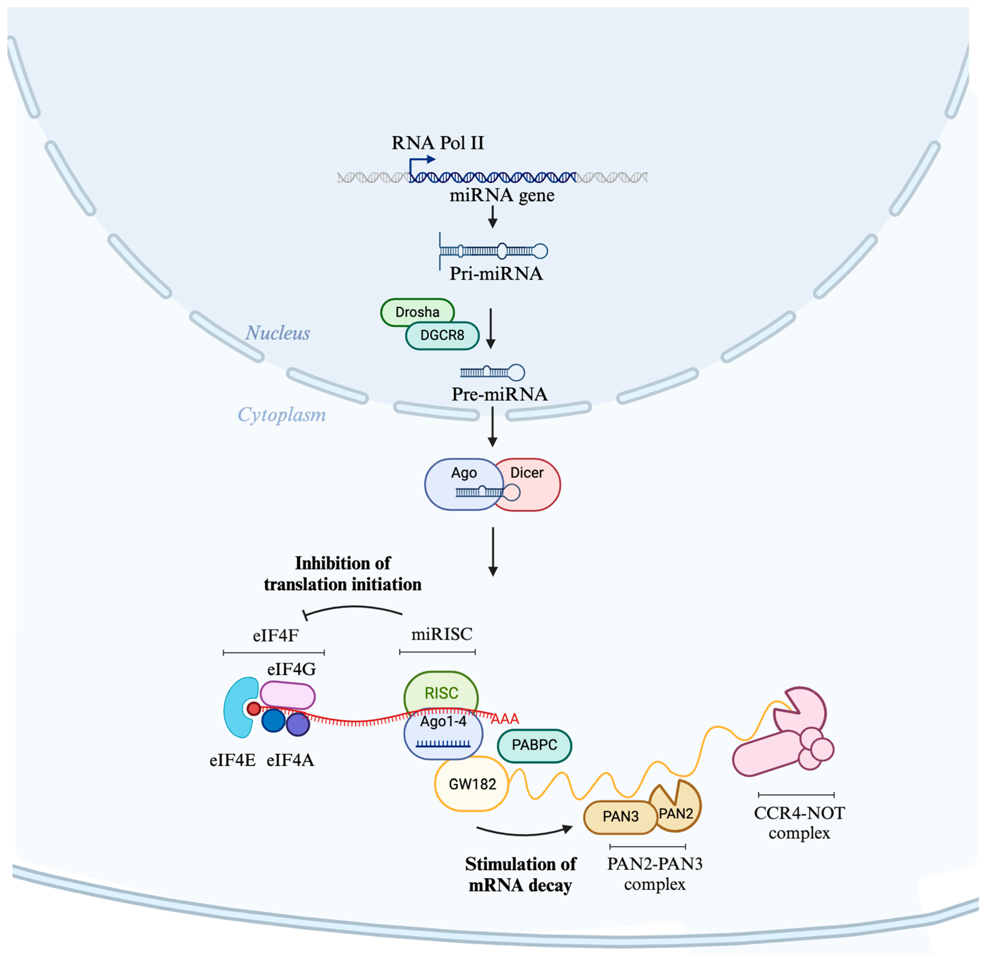
2.1. Biogenesis of miRNAs

Biogenesis of miRNAs begins when genomic regions encoding miRNAs are transcribed by RNA polymerase II into primary miRNAs [25]. miRNAs are transcribed largely from intergenic regions, but many are also found within intronic regions [26]. These primary transcripts are first processed by the microprocessor complex consisting of the ribonuclease III enzyme Drosha and the RNA-binding protein DGCR8 to yield a ~70 nucleotide pre-miRNA [27]. The pre-miRNA is then exported to the cytoplasm by exportin-5. Exportin-5 recognizes the 3′ overhang of the pre-miRNA hairpin, binding to it and facilitating its transport through the nuclear pore complex, and it is there processed by Dicer to yield a mature miRNA duplex (Figure 1). Within this duplex, one miRNA is from the 3′ and one is from the 5′ strand of the precursor; however, one is often a ‘guide’ strand, predominant in amount and biological activity [28].

2.2. Mode of Action of miRNAs

miRNAs function as part of the miRNA-induced silencing complex (miRISC), composed of a ‘guide’ strand of the final duplex loaded onto an Argonaute (AGO) protein. The complex binds usually within the 3′ untranslated region of target RNAs through base pairing at the seed sequence, with the strongest interactions being with sites that complement miRNA nucleotides 2–8 [28,29]. miRISC binding to its target results in gene silencing by inhibiting translation at the initiation step and mediating mRNA decay (Figure 1) [16,17,30,31]. The inhibition of translation initiation is caused by interfering with eIF4A-I and eIF4A-II functions by inducing their dissociation from mRNAs and consequent inhibition of assembly of the eIF4F translation initiation complex [32,33]. mRNA decay involves the recruitment of a member of the glycine-tryptophan protein of the 182 kDa (GW182) protein family, which interacts with polyadenylate-binding protein (PABPC), promoting mRNA deadenylation through PAN2–PAN3 and CCR4–NOT complexes and consequent degradation by exonucleases [34,35]. Although miRNAs function intracellularly as part of the miRISC to mediate gene silencing, their influence is not restricted to the intracellular milieu. Their ability to be secreted and circulated systemically expands their potential roles and implications, particularly in the nervous system. This brings us to their extracellular journey, a phenomenon essential for understanding their utility as diagnostic markers and therapeutic agents.

2.3. Circulation of miRNAs

Emerging evidence underscores that miRNAs are not confined to the cellular environment but can be actively released into extracellular fluids, where they circulate systemically to modulate distant target cells [36]. A portion of these circulating miRNAs associate with proteins such as AGO2, which exists in a free-floating form. However, their primary extracellular transport mechanism involves exosomes, a specialized subtype of extracellular vesicles (EVs).
EVs are lipid bilayer-enclosed particles secreted by a plethora of cell types, encapsulating diverse cargo, including but not limited to nucleic acids such as mRNA and miRNA [37,38,39]. These vesicles are categorized chiefly into ectosomes and exosomes based on their cellular origin: ectosomes are formed by the outward budding of the plasma membrane, while exosomes result from the fusion of multivesicular endosomes with the plasma membrane [38,40].
The intricacies of vesicle biogenesis and cargo loading are dictated by a complex interplay involving the Endosomal Sorting Complex Required for Transport (ESCRT), syntenin-Alix pathway, tetraspanins, various lipids, and the arrestin domain-containing protein 1 [41,42,43,44,45]. Once secreted, EVs navigate through the extracellular matrix and vascular barriers, eventually merging into the circulatory and integumentary systems [40]. In the realm of neuroscience, exosomes are of unique significance for their nanoscale dimensions (30–200 nm), which enable them to cross the blood-brain barrier [37]. While the exact mechanisms of this transport are still under investigation, transcytosis appears to be a plausible route [46]. Upon reaching the target site, the vesicle’s cargo is internalized by the recipient cell through endocytosis and subsequently released into the cytosol via fusion with the endosomal or lysosomal membrane, often in an acidification-dependent manner [47].

2.4. Role of miRNAs in Neural Function: From Synaptic Architecture to Plasticity

2.4.1. Synaptic Localization and Functional Roles of miRNAs in Neuronal Cells

miRNAs have been identified as vital regulatory entities across numerous physiological systems, with their role being especially profound in the nervous system. A significant majority of miRNAs, approximately 70%, are expressed in the brain, where they are implicated in multiple neural functions such as neurogenesis and dendritic spine formation [48,49].
The synaptic network of the brain is marked by complex dendritic interconnections and far-reaching axons, posing a challenge for the swift and effective transport of molecules to remote synaptic sites. Neurons have adapted to this by synthesizing proteins locally at synapse sites, enabling precise regulatory control. Studies have revealed the presence of miRNAs, mRNAs, and polyribosomes within various neural components such as dendrites, axons, and dendritic spines [50]. Notably, certain miRNAs are more concentrated in synaptic fractions compared to whole-cell lysates, suggesting specialized roles at these sites. This was further substantiated by Lugli G et al., who, through subcellular fractionation methods, discovered a rich presence of miRNAs in synaptoneurosomes, in pre- and post-synaptic densities of dendritic spines. These studies also highlighted a specific group of miRNAs enriched in synaptic fractions in contrast to entire cell lysates [50,51].
Regarding the origin of mature miRNAs in neuronal segments, it is either via local conversion of pre-miRNAs or through transport from the cell body. The presence of miRNA precursors and the Dicer enzyme, crucial for RNA processing, has been noted in dendritic and axonal areas [50,52,53,54]. The co-existence of pre-miRNAs and Dicer in these areas implies a mechanism that ensures miRNA specificity and rapid conversion to mature forms as required [55,56]. Emerging research further hints that synapses may actively participate in transforming precursor miRNAs into mature forms, possibly influenced by NMDA receptor activity [52,57,58]. A hypothesis that synaptic activation might be instrumental in this conversion process has been proposed [59,60], though it requires further exploration. The mechanisms governing the delivery and localization of miRNAs to distal synapses are a subject of ongoing research. Specific binding mechanisms, such as the interaction between DEAH-box helicase DHX36 and pre-miR-134, aid in the transport of miRNAs [52].
Functionally, miRNAs play nuanced roles at the synapse. Certain miRNAs, such as miR-132, miR-134, and miR-9-3p, have been identified as key players in normal synaptic functioning and neuropathologies associated with synaptic plasticity, including Alzheimer’s disease [61,62,63]. Their functional roles are also deeply intertwined with activity-dependent translation processes. Examples include the intricate interactions of miR-138 with Lypla1 mRNA or the competition between the RNA-binding protein HuD and miR-129 [64,65]. The interplay of miRNAs, mRNAs, and their associated proteins shapes synaptic functions, such as the onset of long-term synaptic depression (LTD) [66,67]. These findings underscore the nuanced role of miRNAs in synaptic dynamics and neurological health.

2.4.2. Regulatory Influence of miRNAs on Synaptic Transmission and Neural Plasticity

miRNAs exert influence on synaptic structure by modulating the components of the post and pre-synaptic terminal. Specific studies highlight the role of miRNAs such as miR-125b, miR-223, miR-137, and miR-146a-5p in regulating postsynaptic glutamate receptors. For instance, hippocampal neuron dynamics reveal that miR-125b manages the GluN2A component of the NMDA receptor [68], while miR-223 exerts control over the GluN2B NMDA receptor subunit and the AMPA receptor’s GluA2 subunit [69]. Furthermore, miR-137 has associations with the AMPA receptor, particularly its GluA1 subunit [70], and intriguingly, miR-146a-5p, while not directly linked to glutamate receptors, influences them via its interaction with the microtubule-associated protein 1B [71]. Not limited to postsynaptic interactions, miRNAs also target presynaptic components and signaling proteins. As an example, miR-188’s association with the semaphorin-3F receptor Nrp-2 impacts the abundance of dendritic spines in hippocampal neurons [72]. Meanwhile, synaptic glutamate release in Drosophila is influenced by miR-1000’s regulation of the vesicular glutamate transporter [73]. Additionally, miR-137’s reach extends beyond the AMPA receptor, affecting proteins vital for synaptic vesicle discharge [70].
On a broader scale of synaptic plasticity dependent on protein synthesis, miRNAs, including miR-26a and miR-384-5p, have shown alterations in concentration in response to synaptic triggers. Their interactions during NMDA receptor-dependent LTP have consequences for the ribosomal S6 kinase 3 (RSK3) translational regulator [74]. In various synaptic plasticity forms, miR-124 and miR-22 act through distinct targets, with miR-124, for example, influencing NMDA receptor-dependent LTP via Zif268 [75] and miR-137 engaging with mGluR-dependent LTD in hippocampal processes [70].

3. MicroRNAs and Depression

3.1. miRNAs in the Pathophysiology of Depression

Emerging research underscores noticeable shifts in miRNA levels in both the brain and periphery of individuals with depression. Generally, the predominant research paradigms encompass the following: (a) a comparative study of miRNA profiles between patients and controls; (b) an exploration of miRNA modulations subsequent to antidepressant interventions. In the former approach, the prefrontal cortex emerges as a primary brain region of interest. In the periphery, researchers often rely on whole blood, plasma, or serum samples. The second methodology is predominantly engaged to discern miRNA shifts following therapeutic regimens. A less conventional, albeit more invasive, medium is cerebrospinal fluid to probe miRNA dysregulation. Downstream analysis usually involves bioinformatics-driven approaches to identify miRNA–mRNA interactions, facilitating an understanding of the broader regulatory networks at play. Furthermore, to decode the functional dynamics of miRNA regulation, in vitro evaluations and animal model studies are occasionally integrated, though they are less prevalent.
Some of the first studies to identify the role of microRNAs in the pathophysiology of MDD have examined the postmortem human brain. In one example study, the levels of miR-182 expression were disrupted in dentate gyrus granule cells of individuals with depression [76,77]. In the anterior cingulate cortex, a brain region critical for emotional and mood regulation, a depression-specific miRNA expression profile was identified compared to controls [78]. In a similar approach, miR-1202 was found to be downregulated in the prefrontal cortex of individuals with depression compared to controls [79]. This microRNA was found to be enriched in the brain ad negatively correlated with GRM4 expression, a metabotropic glutamate receptor. Importantly, this study also showed that in individuals who had attained remission, levels of miR-1202 were increased.
While the postmortem brain remains a key system for investigating the molecular mechanisms of MDD, a drawback remains that microRNAome profiling in brain tissue at the time of death or sample collection may not be reflective of the disease course [22]. In recent years, miRNAs have been assessed in the periphery of individuals with depression. An advantage of this approach is capturing real-time miRNA differences between patient and control populations and quantifying miRNA changes in patients in response to treatment. One of the first studies to take this approach showed decreased miR-135a levels in patients with depression compared to controls, which had increased after 3 months of selective serotonin reuptake inhibitor treatment or cognitive behavioral therapy [80]. Another study by Belzeaux and colleagues identified a distinct set of miRNAs that are dysregulated in patients with depression, which could predict clinical improvement and treatment response [81]. A further study showed that with electroconvulsive therapy for treatment-resistant depression, levels of miR-223-3p could predict treatment response [82].
Despite numerous studies, there remains a substantial lack of consensus on key miRNAs and their functions in depression. To address this, recent advancements in the field have developed methods for the isolation of cell-type-specific microRNAs in the periphery. For example, microRNAs originating in different brain cell types can now be selected. Pioneering work taking this approach has examined miRNAs within neuron-derived extracellular vesicles in the periphery and has found decreased miR-93 levels in individuals with depression [83]. In another study comparing the neuron-derived extracellular vesicle microRNAome before and after treatment with escitalopram, a signature of miR-21-5p, miR-30d-5p, and miR-486-5p was found to be associated with treatment response [84].
An aggregated list of studies characterizing miRNA variations in depression highlighted in this review can be found in Table 1.

3.2. miRNAs as Diagnostic Biomarkers

The search for non-invasive peripheral biomarkers that mirror changes in the brain is a high priority in research on depression. Such biomarkers could substantially impact current clinical diagnosis practices and, if capable of predicting antidepressant response, the assignment of efficacious treatments. In this regard, circulating miRNAs in blood and other bodily fluids have emerged as promising candidates. Owing to their remarkable stability in circulation and the detectability of brain-specific miRNAs such as miR-9 and miR-223-3p [82,88], miRNAs offer a new avenue in biomarker research.

3.2.1. Blood-Derived miRNAs: Refining the Complex Landscape of Depression Biomarkers

Differential expression of miRNAs in the periphery has been consistently identified in depression and following treatment with antidepressants. Thus, it is clear that the etiology and therapeutic intervention in depression rely at least in part on regulation by miRNAs.
One seminal study highlighted the plasticity of miRNA profiles following treatment, showing that a twelve-week therapeutic regimen of escitalopram promoted the upregulation of 28 microRNAs while concomitantly downregulating miR-34c-5p and miR-770-5p in the blood of individuals with depression [87,89] (see Table 2). In another study, an eight-week citalopram treatment normalized blood concentrations of two deregulated miRNAs, miRNA-132 and miR-124 [90]. While a four-week intervention with citalopram normalized the levels of 10 previously deregulated miRNAs [91]. A particularly noteworthy observation common to these therapeutic interventions was the consistent upregulation of miR-335 [89,91], previously shown to regulate glutamate metabotropic receptor 4 (GRM4), implicated with anxiety-associated behaviors [91]. Another study observed that blood levels of miR-1202 decreased in individuals with depression, a trend that was rectified following an eight-week treatment course of either escitalopram or desvenlafaxine [79,92]. Notably, both miR-1202 and miR-335 have been implicated in regulating GRM4 signaling pathways, thereby influencing stress-related behaviors [79]. Further adding to the complexity, a different study indicated that treatment responses to escitalopram, desvenlafaxine, and duloxetine were associated with changes in miR-1202 and miR-16-5p levels [93].
In parallel to the analysis of discrete microRNAs, several studies have opted for a more comprehensive approach employing small RNA sequencing (sRNA-seq). Using this approach, Belzeaux et al. showed that miR-3688 and miR-5695 served as predictive markers for exacerbated suicidal ideation in individuals subjected to an eight-week course of duloxetine or a placebo [94]. Similarly, Yrondi et al. employed next-generation sequencing (NGS) and observed that pharmacological interventions with escitalopram affected levels of 45 microRNAs, with miR-185-5p being particularly correlated with the reduced incidence of nausea, a common side effect associated with selective serotonin reuptake inhibitors (SSRIs) [95]. In another study, Lopez et al. utilized small RNA sequencing to investigate miRNAs as potential biomarkers for antidepressant responses in MDD within a randomized placebo-controlled trial of duloxetine. The research, conducted before and 8 weeks post-treatment, identified that miR-146a-5p, miR-146b-5p, miR-425-3p, and miR-24-3p exhibited differential expression linked to treatment outcomes. Furthermore, these miRNAs were shown to regulate the MAPK/Wnt signaling pathways, suggesting their significant role in the antidepressant response [86].
In sum, these studies substantiate the proposed use of circulating miRNAs in blood as biomarkers for depressive disorders and treatment responses. Moreover, the findings highlight the complex molecular interplay underlying depression and pharmacological interventions, setting the stage for future research that can enable more targeted and efficacious therapeutic strategies.

3.2.2. Fluid-Based miRNA Landscapes: Serum, Plasma, and Cerebrospinal Fluid

The body of evidence linking specific miRNA profiles to depression extends beyond blood to encompass serum, plasma, and cerebrospinal fluid (CSF). For instance, two studies reported elevated serum and plasma levels of miR-182 and miR-132 in MDD, with both miRNAs found to modulate BDNF expression in neuronal cells [77,90]. Key studies show that inhibiting miR-182-5p alleviates depression-like behaviors in mouse models, suggesting its potential as a therapeutic target [98]. The upregulation of miR-182 reduces BDNF in the hippocampus, further underlining its role in depression pathophysiology [85]. A separate investigation by Wang et al. revealed that diminished plasma levels of miR-144-5p were correlated with depressive symptoms and reversed following eight weeks of personalized antidepressant treatment [96]. Rodent models also imply a role for miR-144-5p in hippocampal functioning, specifically through the regulation of the PTP1B protein, a known modulator of BDNF/TrkB signaling [99]. Several studies reported that many miRNAs, including miR-1202, miR-135a-5p, miR-184, let-7g-5p, miR-103a-3p, miR-107, miR-16-5p, miR-34a-5p, miR-451a, miR-221-3p, and let-7d-3p, found in CSF and serum, were dysregulated in depression and responded to antidepressant treatments [97,100,101,102,103].

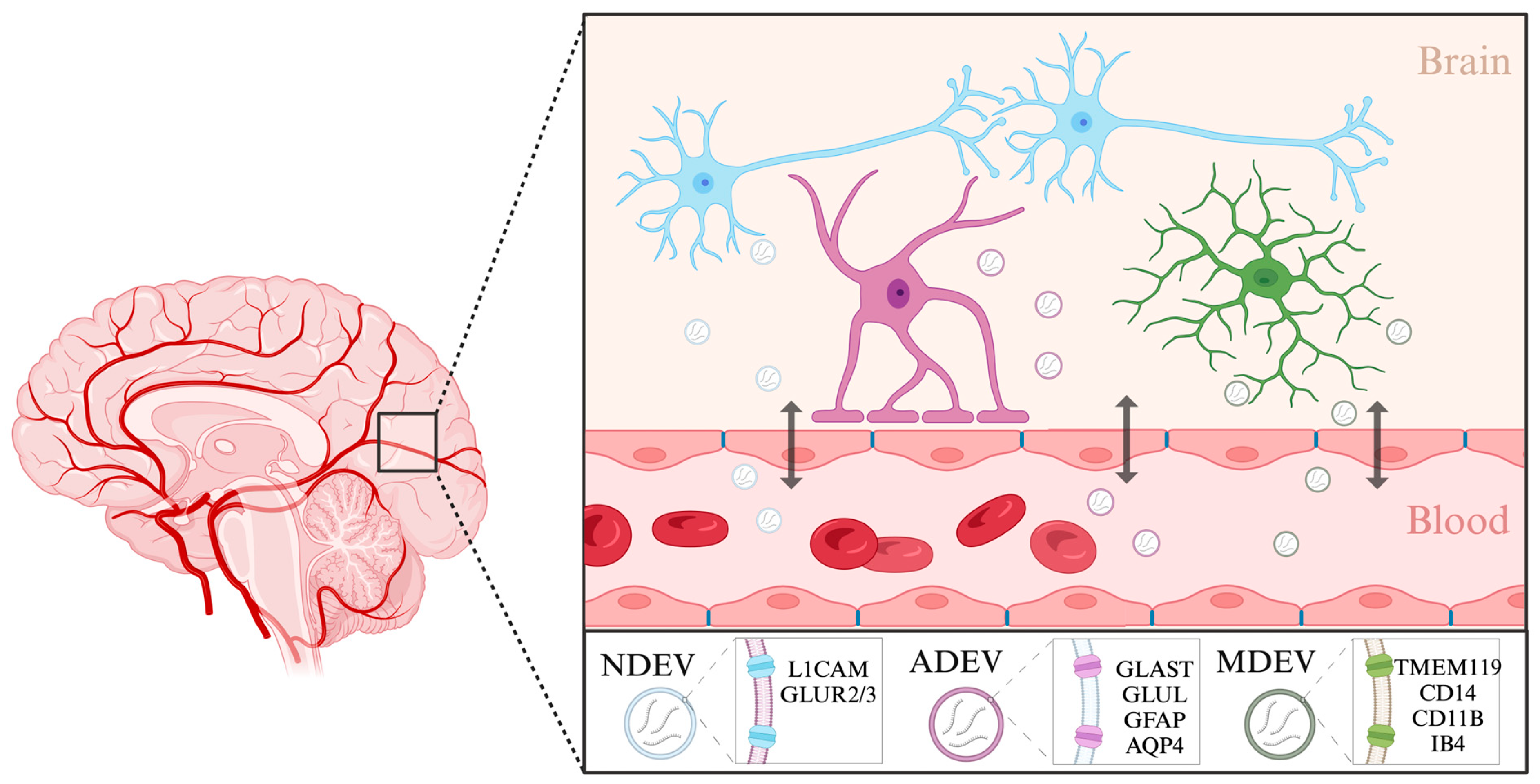
3.2.3. Extracellular Vesicles: Emerging Protagonists in the Biomarker Landscape of Depression

In the exploration of biomarkers for depression, extracellular vesicles (EVs) have recently come under investigation [104]. As previously described, EVs serve as a robust reservoir of miRNAs, facilitating their circulation between the brain and peripheral blood, and vice versa. EVs have been implicated in inflammatory disorders [105,106], cancer [107], and neurodegenerative diseases [108], with their role in psychiatric conditions gaining increasing interest [109]. For example, high-throughput miRNA sequencing identified differentially expressed miRNAs in the serum exosomes of patients with depressive symptoms [24]. Upregulated miRNAs were linked to the modulation of neurotrophic signaling pathways, while downregulated ones were associated with the regulation of apoptosis and immune functions [24]. Notably, the levels of miR-139-5p in serum exosomes distinguished individuals with MDD from healthy controls and could also induce depressive-like behaviors in murine models [110].
Of particular interest is the potential for isolating brain-derived extracellular vesicles (BDEVs) from peripheral blood, which offers a window into central nervous system-specific molecular landscapes [111,112]. BDEVs can originate from many different cell types in the brain, including neurons, astrocytes, and microglia (Figure 2). Neuron-derived EVs have been isolated previously using a biotinylated antibody to L1 cell adhesion molecule (L1CAM), a transmembrane protein specific to neurons [113]. They could also be labeled using antibodies against the GluR2/3 subunits of AMPA receptors [114]. Using this approach, studies have already begun to assess the roles of neuron-derived EVs in depression. Elevated levels of miR-17-5p in neuron-derived EVs correlated with subthreshold depressive symptoms [112]. Moreover, neuronal EVs isolated from individuals with depression undergoing 8-week escitalopram treatment displayed brain-specific proteins and miRNAs (such as miR-30d-5p and miR-486-5p) and also exhibited size alterations reversible by the antidepressant therapy. Changes in miRNA cargo in these neuron-derived EVs also reflected varying responses to antidepressant interventions [84].
The isolation of astrocyte and microglia-derived EVs is also starting to be explored. One of the main forms of communication between neurons and astrocytes is through EVs, where they can have both neuroprotective and pathological properties, highlighting their importance [115]. Astrocyte-derived EVs (ADEVs) have previously been isolated from human blood using a biotinylated antibody to glutamine aspartate transporter (GLAST), an astrocyte-specific membrane protein, glutamine synthetase (GLUL) [116], as well as using glial fibrillary acidic protein (GFAP) and aquaporin 4 (AQP4) [117]. Using this approach, ADEVs have been implicated in modulating immune responses during stress and depression [118,119]. It has been shown that individuals with depression have larger numbers of GFAP and AQP4/GFAP-positive EVs in the blood than healthy controls, suggesting increased leakage of astrocyte-derived EVs through the blood–brain barrier. In a recent study, the miRNA content of ADEVs from individuals with depression was found to have significantly deregulated miR-9 levels, suggesting a specific involvement of this miRNA in the disease’s pathophysiology [120].
Similar to astrocytes, microglia also rely on EVs for cell-to-cell communication within the brain. Largely, the isolation of microglia-derived EVs from complex fluids such as blood samples has so far also been conducted using antibodies or lectins against microglial surface markers [121]. The heterogenous nature of microglial cells is inevitably reflected in their surface protein content and cargo and should thus be used to inform isolation protocols. For instance, in a stroke experimental model, EVs from microglia were isolated using markers characteristic of their specific activation state, namely TMEM119 and CD14 [122]. In another example, small EVs of microglial origin were isolated using CD11B, a more general marker of microglia [123]. Microglia or macrophage-derived EVs have also been isolated using myeloid marker IB4 [124]. Examining the contents of cell-type specific EVs enables a more targeted understanding of disease mechanisms. Further efforts to characterize extracellular vesicles and their components which originate in different brain cell types will improve our understanding of disease mechanisms.

4. Therapeutic Potentials of miRNAs in Depression

miRNAs have emerged as promising molecular targets in the quest to develop innovative interventions for depression. By understanding their intricate regulatory roles, novel therapeutic strategies harnessing the power of miRNAs may revolutionize the treatment landscape for this debilitating condition.

4.1. Current Therapeutic Strategies

Recent studies highlight the therapeutic potential of miRNA-based treatments in a range of conditions, including depression [125,126,127,128]. Although the majority of this research remains in the preclinical phase and, to the best of our understanding, there have been no clinical trials targeting depression using miRNA-based therapies. It is evident that substantial endeavors are still required to make headway in this domain.
miRNA therapeutic strategies primarily modulate the levels of target miRNAs, either by amplifying or attenuating their levels, using oligonucleotides to emulate or curtail miRNA activity, or utilizing small molecules to modulate miRNA functionality [129]. These compounds are introduced into cells using methods such as viral vectors that encode miRNA mimics or antagonists, nanoparticle carriers including liposomes, or cellular vesicles such as exosomes and microvesicles [130,131,132]. Effective in vivo delivery of miRNAs is crucial, aiming to prevent RNase-mediated degradation, enhance targeting precision, and minimize unwanted immune responses. Historically, intravenous and localized approaches have been the primary delivery techniques for in vivo miRNA applications. However, recent investigations are also considering oral and intranasal methods for administration [130,131].

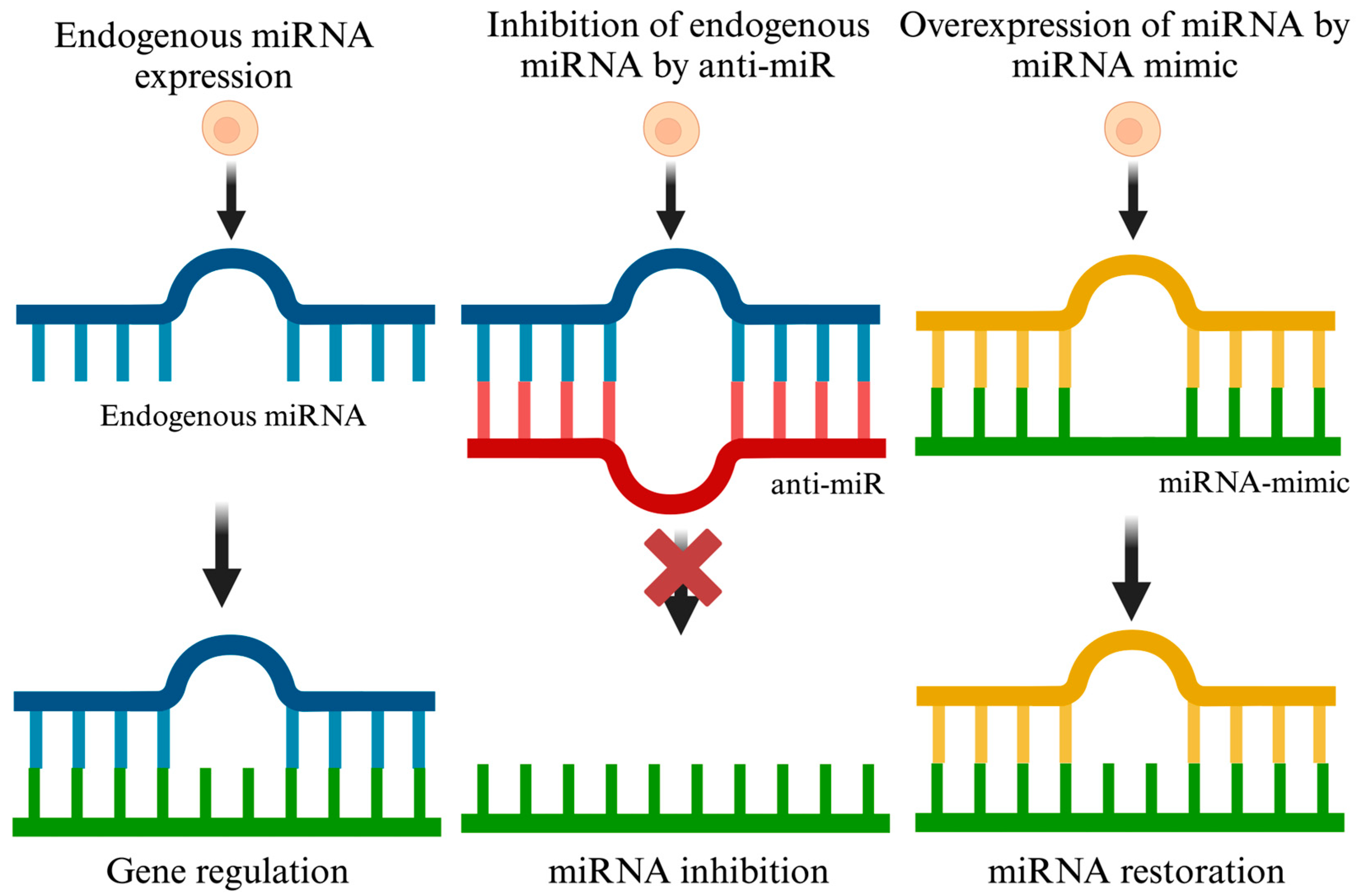
4.1.1. miRNA Inhibition Therapy

The purpose of miRNA inhibition therapy is to counterbalance miRNAs, which have a heightened presence in depressive conditions. The goal is to recalibrate the expression and operations of genes to their natural state. Various methods, including miRNA inhibitors such as antisense miRNA oligonucleotides, locked nucleic acid (LNA) antisense, miRNA antagomirs, and miRNA sponges, have been explored to reduce miRNA expression and counter upregulated miRNAs [133,134,135]. In this review, we refer to all these approaches collectively as anti-miR, which are designed to decrease target miRNA expression. Anti-miRs typically comprise 17–22 nucleotides and are tailored to be complementary to the target miRNA, effectively reducing their expression by binding to them [134]. When anti-miRs bind to their respective target miRNAs, they form double-stranded molecules with the miRNA of interest, preventing these miRNAs from interacting with the Ago complex and their target mRNA. This interruption halts the translational inhibition caused by dysregulated miRNAs in disease conditions (Figure 3).
Anti-miRs that include LNA are engineered for enhanced stability and stronger binding affinity to their target miRNAs. Furthermore, studies have shown that LNA-containing anti-miRs exhibit increased solubility in water and culture media, making them more suitable for in vivo applications [136,137,138]. For example, the research by Islam et al. demonstrated that LNA-based anti-miRs (targeting miR-181a-5p, miR-146a-3p, and miR-148a-5p) effectively silenced these overexpressed miRNAs in the CA region in mice, leading to improved cognition and reduced inflammation [139]. Apart from LNAs, miRNA sponges are also utilized to inhibit upregulated miRNAs. These RNA constructs, equipped with multiple artificial miRNA binding sites, function by competing with cellular miRNAs for their targets [140,141].
Recent advancements in animal-based research have been instrumental in identifying novel targets for mitigating the symptoms of depression. A promising area of study involves the modulation of Notch 1, aimed at improving neural connectivity. Inhibition using miR-9 has been explored as a potential therapeutic strategy [142,143,144]. Furthermore, investigations using chronic unpredictable mild stress (CUMS) have revealed its role in triggering major depression through the downregulation of excitatory synapses, particularly affecting their presynaptic targets on GABAergic neurons in the nucleus accumbens [145]. This disruption in GABAergic activity leads to an imbalance between excitatory and inhibitory signals in the brain, undermining the integrity of neural circuits. Such imbalances are key contributors to the onset of MDD and other stress-related conditions [146]. Notably, research has shown that inhibiting miR-15b-5p using anti-miR in the nucleus accumbens can substantially decrease the occurrence of CUMS-induced depression and restore the reduced levels of excitatory synapses [145].
Another key brain region involved in depression is the hippocampus. Under the regulatory control of miR-18a-5p is hippocampal serotonergic balance, and anti-miRs may be a strategic move to promote resiliency to depression through glucocorticoid signaling [147]. Another explored target is miR-132, which is a class of miRNAs that works in the neuro-immune compartment and has been linked to elevated BNDF levels in in vivo settings [148,149]. Similarly, elevated miR-202-3p levels in the hippocampus have been linked to diminished BDNF expression in a CUMS-induced depression model. The inhibition of miR-202-3p in the hippocampus has demonstrated an antidepressant-like effect, significantly reducing depressive behaviors in the CUMS model, highlighting the therapeutic potential of miRNA modulation in depression treatment strategies [150].
Strategies such as miR-134-5p inhibition have also yielded positive neuronal outcomes and reduced depression-like behaviors in specific models [151]. A noteworthy development in the field is the identification of red blood cell-specific miR-144-3p as a potential biomarker. This biomarker not only assists in the diagnosis of depression but also predicts the response to ketamine treatment in stress-susceptible mice and MDD patients. Intriguingly, the use of an anti-miR to miR-144-3p, administered subcutaneously, has led to a marked reduction in depression-related phenotypes in stress-prone mice [152]. Similarly, the increased expression of miR-96 has led to a decrease in the synaptic vesicle glycoprotein 2 family protein which is associated with depression-like behavior, while inhibiting miR-96 using anti-miR decreased depression-like behavior and memory impairment in mice [153].

4.1.2. miRNA Restoration Therapy

Within the sphere of depression research, ‘miRNA mimics’ are designed as a double-stranded RNA with a guide strand that reflects the sequence of the associated endogenous miRNA. Using these small RNA mimics, the levels of select miRNAs can be transiently overexpressed, helping researchers understand the consequent biological effects. Focusing on miRNA restoration therapy, this approach aims to reverse miRNA deficiencies associated with depressive disorders. It involves using synthetic miRNA mimics or employing viral vectors that express pertinent miRNAs (Figure 3). These mimics, serving as substitutes for natural miRNAs, are incorporated into the miRISC, leading to alterations in the activity of downstream mRNA targets [154]. The success of this miRNA restoration method has been validated through a range of cell-based and in vivo experiments. Additionally, the field of miRNA restoration is exploring the use of miRNA expression vectors, including adenoviral, lentiviral, and retroviral vectors, as another promising avenue.
A key study by Kota et al. provides insight into this strategy, demonstrating an example where the levels of miR-26 were significantly lower under specific conditions, in contrast to their normal abundance [155]. Reintroducing miR-26 into these cells slowed their cell cycle progression. Remarkably, treating cells with a recombinant adenovirus carrying miR-26 resulted in enhanced cell recovery and a decrease in cell proliferation, without any adverse effects observed.
In the context of depression, notable miRNAs which may have therapeutic implications or serve as viable targets include let-7, miR-144, miR-9, miR-16, miR-30a, miR-133b, miR-26a-2, and several others. Recent studies have focused on the potential therapeutic modulation by the let-7 family, examining the effects of physical activity on its epigenetic regulation [156]. Advances in lentiviral technologies have been harnessed to overexpress let-7d, showing promise for anxiolytic and antidepressant outcomes [157]. Another noteworthy study observed a decrease in miR-139-5p levels in CUMS-induced mice, correlating with depression-like symptoms. The increasing level of miR-139-5p expression in the hippocampus through AAV technology resulted in the activation of the cAMP/PKA signaling pathway, demonstrating antidepressant-like effects in these mice [158].
A similar methodology showed that increasing the levels of miR-144 lead to observed antidepressant effects in the CUMS rat model [99]. It is essential to recognize the potential of miR-144, which is typically reduced in MDD patients [159]. Animal models have confirmed that levels of miR-144 are upregulated in response to treatment with mood stabilizers [160]. Complementing these findings is the interesting case of miR-124-3p in depression. Studies have shown a notable upregulation of miR-124-3p in the hippocampus of CUMS mice. This upregulation plays a crucial role in reducing proinflammatory cytokines and in the deactivation of microglia [161]. Highlighting its significance, recent research by Ge et al. identified the presence of miR-124-3p within microglial EV miRNA cargos. This presence is linked to the alleviation of neurodegenerative processes and the improvement of cognitive functions, marking miR-124-3p as a potential therapeutic intervention in depression [162].
Strikingly, certain miRNAs exhibit inherent antidepressant properties in various animal models, making them also an appealing target for testing as miRNA mimic therapies. Examples include miR-26a-2, which acts on serotonergic neurons [125,163], and miR-101, which reverses specific depressive behaviors [164]. Notably, combining miR-135a with traditional antidepressants resulted in decreased inflammatory markers in certain models [80].

4.2. Future Directions

miRNAs have emerged as a key player in the complex molecular mechanisms underpinning depression. Their ability to regulate a myriad of genes post-transcriptionally places them in a unique position to be therapeutic targets. As we progress into an era emphasizing precision medicine, the potential to measure and manipulate miRNAs in minimally invasive ways offers promising avenues for treating depression. This section explores the emerging targets, innovative drug delivery methods, and potential improvements to current strategies.

4.2.1. Emerging Targets in the miRNA Landscape

As miRNA research gains momentum, novel targets are constantly being identified. Recent studies have highlighted miRNAs such as miR-16, miR-135a, miR-144, miR-26a-2, and miR-1202 as potential biomarkers in MDD [165]. Their differential expression levels in individuals with depression have been consistently reported. Moreover, their roles have been intricately linked with serotonin pathways, neuroinflammation, and neurogenesis, key pathways in brain functioning. As more miRNAs are identified, it is imperative to validate their potential therapeutic roles, understand their mechanistic pathways, and explore their interactions with other miRNAs or mRNA targets. Integrative omics approaches, coupling miRNA profiling, transcriptomics, proteomics, and network analysis, will be essential in this endeavor. Figure 4 in the manuscript visually encapsulates the methodology of a study design aimed at analyzing these miRNA candidates in plasma or serum samples from individuals with MDD compared to healthy controls, thereby providing a foundation for the biological interpretation of their roles in the disease pathology.

4.2.2. Drug Delivery—Crossing the Biobarriers

The therapeutic application of miRNAs is hindered by the challenges posed by their stability, specificity, and delivery. However, recent advances promise to address these hurdles. miRNA delivery is categorized into the following methods.
Viral-based and non-viral-based: Viral vectors, such as adenoviruses (Ads), have historically been used in gene therapy. First-generation Ads faced challenges due to their immunogenic nature, but subsequent iterations proved more promising. Ad-based miRNA delivery has been notably successful in mitigating cardiomyocyte hypertrophy in mice [166,167]. In contrast, adeno-associated viruses (AAVs) are recognized for being non-pathogenic and exhibiting reduced immunogenicity. Notably, AAV9 vectors demonstrated the capability to cross the blood–brain barrier, promising potential in treating central nervous system diseases [168]. Numerous studies have employed AAVs to modulate miRNA levels in vivo. A compelling example of this is the research conducted by Huang et al., where AAV technology was used to elevate miR-139-5p levels in the hippocampus of mice [158]. On the other hand, Herpes simplex viruses (HSVs) possess a natural inclination for nerve cells, and their application in delivering miR-124 resulted in the modification of neuron-specific alternative splicing [169]. Nevertheless, concerns about the vector’s size and possible cytotoxicity remain.
On the flip side, non-viral vectors offer a range of options. A type of lipid-based vectors, liposomes are spherical vesicles encapsulating an aqueous core. The cationic nature of some liposomes facilitates the binding of negatively charged miRNAs, safeguarding them from degradation [170,171,172]. Complexes of liposomes and nucleic acids, termed lipoplexes, have been employed to transport miRNAs into cells [130,173]. Inorganic nanoparticles such as gold nanoparticles are also under exploration for miRNA delivery, given their biocompatibility and modifiability [174,175]. Silica-based nanoparticles have also been employed to co-deliver anti-miR-221, although this approach has mostly only been used in the cancer field [176,177].
Exosomes: These naturally occurring vesicles can be engineered to carry miRNAs, ensuring effective delivery across the blood–brain barrier (BBB). They possess several features that make them particularly attractive for the delivery of biomolecules, including miRNAs: natural biocompatibility, low immunogenicity, and the inherent ability to cross biological barriers such as the BBB [178]. Exosomes can encapsulate and protect miRNA molecules from degradation in the extracellular environment, ensuring their safe passage to target cells. Once they reach their destination, exosomes can release their miRNA cargo into different cell types in the brain, modulating gene levels and potentially ameliorating molecular pathways contributing to depressive symptoms [179,180].
Several studies have demonstrated the feasibility and efficacy of exosome-mediated miRNA delivery in neurological conditions. For example, exosomes loaded with miRNA-124, known for its neuroprotective properties, facilitated recovery from ischemic conditions in a mouse model [181]. Such successes hint at the potential of a similar approach in depression therapy. Additionally, the source of exosomes can play a role in enhancing therapeutic outcomes. Exosomes derived from mesenchymal stem cells (MSCs), for instance, have shown promise due to their anti-inflammatory properties [182,183], which could be particularly beneficial in conditions such as depression, where neuroinflammation is a known contributor.
However, while the potential is immense, there are challenges. Optimizing exosome loading with desired miRNAs, ensuring target specificity, and scaling up production for clinical applications are areas that need focused research. Moreover, understanding the long-term effects of exosome-mediated miRNA delivery and ensuring minimal off-target effects is crucial.
Peptide-based delivery: Peptide-based delivery systems have emerged as a promising approach for the targeted delivery of therapeutic molecules, including miRNAs, to specific cells or tissues. This stems from the versatility, biocompatibility, and low toxicity of peptides, which can be designed to interact specifically with cell surface receptors or to traverse cell membranes efficiently [184,185]. In the context of depression, the central nervous system (CNS) remains a challenging target due to the restrictive nature of the BBB. The effective delivery of miRNA molecules to the brain is imperative for therapeutic efficacy. Peptides, with their ability to be tailored for specificity, offer a potential solution.
Certain peptides, such as those derived from rabies virus glycoprotein (RVG), have shown promise in specific targeting of neuronal cells [186,187]. By conjugating these peptides with miRNAs, one can achieve efficient transport across the BBB and subsequent delivery to neuronal cells. Such delivery ensures that therapeutic miRNAs reach their targets, maximizing therapeutic potential while minimizing off-target effects.
Another advantage of peptide-based delivery is the potential for co-delivery of multiple therapeutic agents. Peptides can be designed to encapsulate or bind to both miRNAs and other therapeutic molecules, allowing for a synergistic approach to treatment. This is especially relevant in conditions such as depression, where a multifactorial etiology might necessitate a combination of therapeutic interventions. Moreover, the ease of synthesis and modification of peptides allows for rapid optimization of delivery vectors. This adaptability means that peptide-based delivery systems can be quickly refined based on in vitro and in vivo feedback, accelerating the translational potential of miRNA therapies for depression.
However, as with all therapeutic approaches, challenges persist. Ensuring the stability of the peptide-miRNA complex in systemic circulation, avoiding rapid degradation, and preventing immune responses are areas that warrant attention. Additionally, understanding the long-term effects of repeated administration and potential peptide-related toxicity is essential for clinical translation.

4.2.3. Augmenting Existing Strategies

While the therapeutic potential of miRNAs in depression is evident, the following can help refine current strategies.
Combination Therapies: Co-administering miRNA modulators with conventional antidepressants can potentiate therapeutic effects and mitigate potential side effects. Illustratively, in one experiment, miR-135a was combined with standard antidepressant treatments, leading to a notable decrease in inflammatory markers in a mouse model exhibiting depression-like behaviors.
Bioinformatics and Personalized Medicine: Leveraging bioinformatics tools and machine learning algorithms can help predict miRNA-disease associations and individual miRNA profiles. This facilitates a personalized medicine approach, where treatments are tailored based on an individual’s unique genetic makeup.

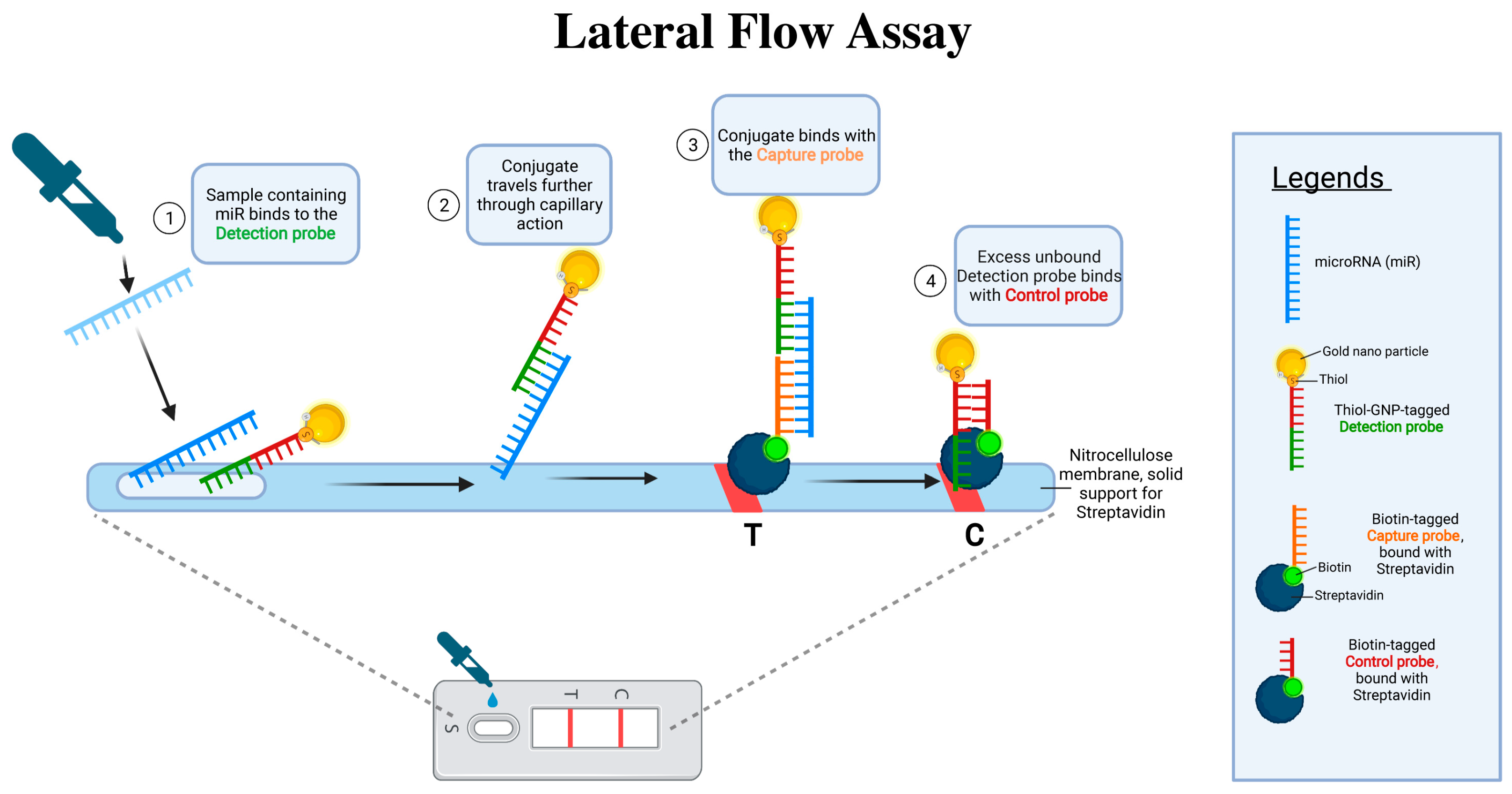
4.2.4. Lateral Flow Assay for miRNA Detection

One of the challenges in harnessing miRNAs as therapeutic targets is their accurate and rapid detection. The lateral flow assay (LFA), similar to the mechanism of a home pregnancy test, provides a quick, user-friendly, and cost-effective platform to detect specific biomolecules, including miRNAs. By using tailored probes that can bind to miRNAs associated with depression, LFA can serve as a diagnostic tool to determine miRNA deregulation in blood samples [188,189]. Such a tool is invaluable, not just for initial diagnosis but also for monitoring the efficacy of any miRNA-based therapy, allowing for dynamic treatment strategies tailored to individual patient needs (see Figure 5).
The key advantages of using LFA for detecting miRNAs in depression include speed, simplicity, and the potential for point-of-care testing. It does not require sophisticated laboratory equipment, making it feasible for routine clinical settings or even potential home use. However, there are limitations. The sensitivity and specificity of the LFA need to be rigorously validated against gold-standard miRNA detection methods. Additionally, while changes in specific blood miRNA levels may correlate with depressive states, the biological underpinnings of these changes and their direct causal relationship to depression remain to be fully elucidated.

5. Challenges and Limitations

The therapeutic potential of miRNAs in depression is a promising yet challenging frontier in molecular psychiatry. Advancements in miRNA profiling have not yet overcome the difficulty of capturing the full spectrum of miRNA interactions and their complex regulatory networks. This limitation is compounded by inconsistencies in results across similar studies, likely due to variations in sample processing, sequencing methodologies, and bioinformatic analyses. Advocating for standardized procedures is critical for achieving reliable outcomes.
Another significant hurdle is the efficient delivery of miRNA-based therapeutics across the blood–brain barrier. Despite the emergence of innovative methods such as nanoparticles and viral vectors, issues with delivery specificity and efficiency remain unresolved. Moreover, the broad regulatory roles of miRNAs raise concerns about off-target effects and unintended physiological changes, which could worsen depressive symptoms or introduce new clinical problems. This risk is exacerbated by the challenge of targeting specific miRNAs without inadvertently influencing other pathways, given the promiscuous nature of miRNA–mRNA interactions.
Finally, as with other treatments used for depression, miRNA therapeutics face the complexity of patient-specific responses, as depression’s heterogeneity suggests variability in miRNA profiles among individuals. This necessitates a more personalized approach to therapy. Additionally, the long-term effects of miRNA modulation, in terms of both therapeutic benefits and potential risks, are yet to be fully explored.

6. Conclusions

This comprehensive review illuminates the pivotal role of miRNAs in the pathogenesis and treatment of depression, highlighting their potential as biomarkers and therapeutic targets. The intricate regulatory mechanisms of miRNAs in neural development, synaptic plasticity, and stress responses underscore their significance in mental health. The exploration of miRNAs in depression reveals their influence on gene expression and their ability to cross the blood–brain barrier, making them promising candidates for diagnostic and therapeutic purposes. The dysregulation of specific miRNAs in MDD patients suggests a profound connection between miRNA expression patterns and the pathophysiology of depression. For instance, miR-132 and miR-124 have been consistently reported to be altered in MDD, affecting neuroplasticity and stress response mechanisms.
The identification of miRNA signatures specific to MDD opens new avenues for diagnosis. Certain miRNAs, including miR-1202, miR-135a-5p, miR-184, let-7g-5p, miR-103a-3p, miR-107, miR-16-5p, miR-34a-5p, miR-451a, miR-221-3p, and let-7d-3p, have been identified in CSF and serum as dysregulated in depression. This dysregulation suggests a novel approach to diagnosing MDD. Contrasting with conventional diagnostic methods, miRNA profiling offers a quick and non-invasive alternative, potentially employing technologies such as lateral flow assays in the future to measure miRNA concentrations in peripheral blood or serum/plasma. This innovative technique not only aims to improve diagnostic precision but also to enable the early identification of MDD, potentially even before the clinical symptoms manifest.
Moreover, miRNAs hold promise in predicting treatment efficacy. Variations in miRNA expression levels have been associated with patients’ responses to antidepressants, suggesting that miRNA profiling could guide personalized treatment strategies. For example, the differential expression of miR-16 in response to selective serotonin reuptake inhibitors (SSRIs) highlights its potential role in mediating treatment outcomes. Changes in miR-223-3p expression levels at baseline are also predictive of patients’ responses to electroconvulsive therapy. The exploration of miRNAs in MDD underscores a forward-looking approach to diagnosing and managing depression, emphasizing the need for robust research to harness their full diagnostic and therapeutic potential. The challenges in miRNA research, such as inconsistencies between studies, technological limitations, drug delivery complexities, and the need for personalized therapies, emphasize the necessity for further research and standardization in this field. The future of depression treatment may significantly benefit from advancements in miRNA-based strategies, provided these challenges are effectively addressed through continued interdisciplinary collaboration and innovative research.

Funding

This research received no external funding.

Data Availability Statement

This study did not report any data.

Conflicts of Interest

The author declares no conflicts of interest.

Abbreviations

MDD: Major Depressive Disorder; miRNA: microRNA; DSM-V: Diagnostic and Statistical Manual of Mental Disorders, Fifth Edition, 2000; ECT: Electroconvulsive therapy; EVs: Extracellular vesicles; miRISC: miRNA-induced silencing complex; AGO: Argonaute; ESCRT: Endosomal Sorting Complex Required for Transport; LTD: Long-term synaptic depression; RSK3: Ribosomal S6 kinase 3; GRM4: Glutamate metabotropic receptor 4; SSRIs: Selective serotonin reuptake inhibitors; CSF: Cerebrospinal fluid; BDEVs: Brain-derived extracellular vesicles; L1CAM: L1 cell adhesion molecule; NDEVs: Neuron-derived EVs; GLAST: Glutamine aspartate transporter; GLUL: Glutamine synthetase; GFAP: Glial fibrillary acidic protein; ADEVs: Astrocyte-derived EVs; AQP4: Aquaporin 4; LNA: Locked nucleic acid; CUMS: Chronic unpredictable mild stress; AAVs: Adeno-associated viruses; Ads: Adenoviruses; BBB: Blood–brain barrier; RVG: Rabies virus glycoprotein; LFA: Lateral flow assay; GNP: Gold nanoparticle.

References

  1. Institute of Health Metrics and Evaluation. Global Health Data Exchange (GHDx); Institute of Health Metrics and Evaluation: Seattle, WA, USA, 2021. [Google Scholar]
  2. James, S.L.; Abate, D.; Abate, K.H.; Abay, S.M.; Abbafati, C.; Abbasi, N.; Abbastabar, H.; Abd-Allah, F.; Abdela, J.; Abdelalim, A.; et al. Global, Regional, and National Incidence, Prevalence, and Years Lived with Disability for 354 Diseases and Injuries for 195 Countries and Territories, 1990–2017: A Systematic Analysis for the Global Burden of Disease Study 2017. Lancet 2018, 392, 1789–1858. [Google Scholar] [CrossRef]
  3. Kessler, R.C.; Bromet, E.J. The Epidemiology of Depression across Cultures. Annu. Rev. Public Health 2013, 34, 119–138. [Google Scholar] [CrossRef] [PubMed]
  4. Bromet, E.; Andrade, L.H.; Hwang, I.; Sampson, N.A.; Alonso, J.; de Girolamo, G.; de Graaf, R.; Demyttenaere, K.; Hu, C.; Iwata, N.; et al. Cross-National Epidemiology of DSM-IV Major Depressive Episode. BMC Med. 2011, 9, 90. [Google Scholar] [CrossRef] [PubMed]
  5. Nihalani, N.; Simionescu, M.; Dunlop, B.W. Depression: Phenomenology, Epidemiology, and Pathophysiology. In Depression; CRC Press: Boca Raton, FL, USA, 2010; ISBN 978-0-429-24993-8. [Google Scholar]
  6. Kuehner, C. Why Is Depression More Common among Women than among Men? Lancet Psychiatry 2017, 4, 146–158. [Google Scholar] [CrossRef] [PubMed]
  7. Van de Velde, S.; Bracke, P.; Levecque, K. Gender Differences in Depression in 23 European Countries. Cross-National Variation in the Gender Gap in Depression. Soc. Sci. Med. 2010, 71, 305–313. [Google Scholar] [CrossRef] [PubMed]
  8. Malhi, G.S.; Mann, J.J. Depression. Lancet 2018, 392, 2299–2312. [Google Scholar] [CrossRef]
  9. Heim, C.; Binder, E.B. Current Research Trends in Early Life Stress and Depression: Review of Human Studies on Sensitive Periods, Gene–Environment Interactions, and Epigenetics. Exp. Neurol. 2012, 233, 102–111. [Google Scholar] [CrossRef]
  10. Belmaker, R.H.; Agam, G. Major Depressive Disorder. N. Engl. J. Med. 2008, 358, 55–68. [Google Scholar] [CrossRef]
  11. Hasin, D.S.; Sarvet, A.L.; Meyers, J.L.; Saha, T.D.; Ruan, W.J.; Stohl, M.; Grant, B.F. Epidemiology of Adult DSM-5 Major Depressive Disorder and Its Specifiers in the United States. JAMA Psychiatry 2018, 75, 336–346. [Google Scholar] [CrossRef]
  12. Cuijpers, P.; Noma, H.; Karyotaki, E.; Vinkers, C.H.; Cipriani, A.; Furukawa, T.A. A Network Meta-Analysis of the Effects of Psychotherapies, Pharmacotherapies and Their Combination in the Treatment of Adult Depression. World Psychiatry 2020, 19, 92–107. [Google Scholar] [CrossRef]
  13. Cuijpers, P.; Stringaris, A.; Wolpert, M. Treatment Outcomes for Depression: Challenges and Opportunities. Lancet Psychiatry 2020, 7, 925–927. [Google Scholar] [CrossRef] [PubMed]
  14. Gurtan, A.M.; Sharp, P.A. The Role of miRNAs in Regulating Gene Expression Networks. J. Mol. Biol. 2013, 425, 3582. [Google Scholar] [CrossRef] [PubMed]
  15. Friedman, R.C.; Farh, K.K.-H.; Burge, C.B.; Bartel, D.P. Most Mammalian mRNAs Are Conserved Targets of microRNAs. Genome Res. 2009, 19, 92–105. [Google Scholar] [CrossRef]
  16. Gebert, L.F.R.; MacRae, I.J. Regulation of microRNA Function in Animals. Nat. Rev. Mol. Cell Biol. 2019, 20, 21–37. [Google Scholar] [CrossRef]
  17. Selbach, M.; Schwanhäusser, B.; Thierfelder, N.; Fang, Z.; Khanin, R.; Rajewsky, N. Widespread Changes in Protein Synthesis Induced by microRNAs. Nature 2008, 455, 58–63. [Google Scholar] [CrossRef]
  18. Griffiths-Jones, S.; Grocock, R.J.; van Dongen, S.; Bateman, A.; Enright, A.J. miRBase: MicroRNA Sequences, Targets and Gene Nomenclature. Nucleic Acids Res. 2006, 34, D140–D144. [Google Scholar] [CrossRef]
  19. de Rie, D.; Abugessaisa, I.; Alam, T.; Arner, E.; Arner, P.; Ashoor, H.; Åström, G.; Babina, M.; Bertin, N.; Burroughs, A.M.; et al. An Integrated Expression Atlas of miRNAs and Their Promoters in Human and Mouse. Nat. Biotechnol. 2017, 35, 872–878. [Google Scholar] [CrossRef] [PubMed]
  20. Smalheiser, N.R.; Lugli, G.; Zhang, H.; Rizavi, H.; Cook, E.H.; Dwivedi, Y. Expression of microRNAs and Other Small RNAs in Prefrontal Cortex in Schizophrenia, Bipolar Disorder and Depressed Subjects. PLoS ONE 2014, 9, e86469. [Google Scholar] [CrossRef]
  21. Wang, W.; Kwon, E.J.; Tsai, L.-H. MicroRNAs in Learning, Memory, and Neurological Diseases. Learn. Mem. 2012, 19, 359–368. [Google Scholar] [CrossRef]
  22. Issler, O.; Chen, A. Determining the Role of microRNAs in Psychiatric Disorders. Nat. Rev. Neurosci. 2015, 16, 201–212. [Google Scholar] [CrossRef]
  23. Dwivedi, Y. MicroRNAs in Depression and Suicide: Recent Insights and Future Perspectives. J. Affect. Disord. 2018, 240, 146–154. [Google Scholar] [CrossRef] [PubMed]
  24. Zhang, Y.; Zhao, Y.; Tian, C.; Wang, J.; Li, W.; Zhong, C. Differential Exosomal microRNA Profile in the Serum of a Patient with Depression. Eur. J. Psychiatry 2018, 32, 105–112. [Google Scholar] [CrossRef]
  25. Krol, J.; Loedige, I.; Filipowicz, W. The Widespread Regulation of microRNA Biogenesis, Function and Decay. Nat. Rev. Genet. 2010, 11, 597–610. [Google Scholar] [CrossRef] [PubMed]
  26. Chiang, H.R.; Schoenfeld, L.W.; Ruby, J.G.; Auyeung, V.C.; Spies, N.; Baek, D.; Johnston, W.K.; Russ, C.; Luo, S.; Babiarz, J.E.; et al. Mammalian microRNAs: Experimental Evaluation of Novel and Previously Annotated Genes. Genes. Dev. 2010, 24, 992–1009. [Google Scholar] [CrossRef]
  27. Lee, Y.; Ahn, C.; Han, J.; Choi, H.; Kim, J.; Yim, J.; Lee, J.; Provost, P.; Rådmark, O.; Kim, S.; et al. The Nuclear RNase III Drosha Initiates microRNA Processing. Nature 2003, 425, 415–419. [Google Scholar] [CrossRef]
  28. Ha, M.; Kim, V.N. Regulation of microRNA Biogenesis. Nat. Rev. Mol. Cell Biol. 2014, 15, 509–524. [Google Scholar] [CrossRef]
  29. Agarwal, V.; Bell, G.W.; Nam, J.-W.; Bartel, D.P. Predicting Effective microRNA Target Sites in Mammalian mRNAs. eLife 2015, 4, e05005. [Google Scholar] [CrossRef]
  30. Jonas, S.; Izaurralde, E. Towards a Molecular Understanding of microRNA-Mediated Gene Silencing. Nat. Rev. Genet. 2015, 16, 421–433. [Google Scholar] [CrossRef] [PubMed]
  31. Guo, H.; Ingolia, N.T.; Weissman, J.S.; Bartel, D.P. Mammalian microRNAs Predominantly Act to Decrease Target mRNA Levels. Nature 2010, 466, 835–840. [Google Scholar] [CrossRef] [PubMed]
  32. Fukaya, T.; Iwakawa, H.-O.; Tomari, Y. MicroRNAs Block Assembly of eIF4F Translation Initiation Complex in Drosophila. Mol. Cell 2014, 56, 67–78. [Google Scholar] [CrossRef]
  33. Fukao, A.; Mishima, Y.; Takizawa, N.; Oka, S.; Imataka, H.; Pelletier, J.; Sonenberg, N.; Thoma, C.; Fujiwara, T. MicroRNAs Trigger Dissociation of eIF4AI and eIF4AII from Target mRNAs in Humans. Mol. Cell 2014, 56, 79–89. [Google Scholar] [CrossRef]
  34. Liu, J.; Rivas, F.V.; Wohlschlegel, J.; Yates, J.R.; Parker, R.; Hannon, G.J. A Role for the P-Body Component GW182 in microRNA Function. Nat. Cell Biol. 2005, 7, 1261–1266. [Google Scholar] [CrossRef] [PubMed]
  35. Braun, J.E.; Huntzinger, E.; Fauser, M.; Izaurralde, E. GW182 Proteins Directly Recruit Cytoplasmic Deadenylase Complexes to miRNA Targets. Mol. Cell 2011, 44, 120–133. [Google Scholar] [CrossRef] [PubMed]
  36. Gallo, A.; Tandon, M.; Alevizos, I.; Illei, G.G. The Majority of MicroRNAs Detectable in Serum and Saliva Is Concentrated in Exosomes. PLoS ONE 2012, 7, e30679. [Google Scholar] [CrossRef]
  37. Alvarez-Erviti, L.; Seow, Y.; Yin, H.; Betts, C.; Lakhal, S.; Wood, M.J.A. Delivery of siRNA to the Mouse Brain by Systemic Injection of Targeted Exosomes. Nat. Biotechnol. 2011, 29, 341–345. [Google Scholar] [CrossRef] [PubMed]
  38. Lee, Y.; EL Andaloussi, S.; Wood, M.J.A. Exosomes and Microvesicles: Extracellular Vesicles for Genetic Information Transfer and Gene Therapy. Hum. Mol. Genet. 2012, 21, R125–R134. [Google Scholar] [CrossRef] [PubMed]
  39. Valadi, H.; Ekström, K.; Bossios, A.; Sjöstrand, M.; Lee, J.J.; Lötvall, J.O. Exosome-Mediated Transfer of mRNAs and microRNAs Is a Novel Mechanism of Genetic Exchange between Cells. Nat. Cell Biol. 2007, 9, 654–659. [Google Scholar] [CrossRef]
  40. van Niel, G.; Carter, D.R.F.; Clayton, A.; Lambert, D.W.; Raposo, G.; Vader, P. Challenges and Directions in Studying Cell–Cell Communication by Extracellular Vesicles. Nat. Rev. Mol. Cell Biol. 2022, 23, 369–382. [Google Scholar] [CrossRef]
  41. Anand, S.; Foot, N.; Ang, C.-S.; Gembus, K.M.; Keerthikumar, S.; Adda, C.G.; Mathivanan, S.; Kumar, S. Arrestin-Domain Containing Protein 1 (Arrdc1) Regulates the Protein Cargo and Release of Extracellular Vesicles. Proteomics 2018, 18, e1800266. [Google Scholar] [CrossRef]
  42. Baietti, M.F.; Zhang, Z.; Mortier, E.; Melchior, A.; Degeest, G.; Geeraerts, A.; Ivarsson, Y.; Depoortere, F.; Coomans, C.; Vermeiren, E.; et al. Syndecan-Syntenin-ALIX Regulates the Biogenesis of Exosomes. Nat. Cell Biol. 2012, 14, 677–685. [Google Scholar] [CrossRef]
  43. Colombo, M.; Moita, C.; van Niel, G.; Kowal, J.; Vigneron, J.; Benaroch, P.; Manel, N.; Moita, L.F.; Théry, C.; Raposo, G. Analysis of ESCRT Functions in Exosome Biogenesis, Composition and Secretion Highlights the Heterogeneity of Extracellular Vesicles. J. Cell Sci. 2013, 126, 5553–5565. [Google Scholar] [CrossRef] [PubMed]
  44. Trajkovic, K.; Hsu, C.; Chiantia, S.; Rajendran, L.; Wenzel, D.; Wieland, F.; Schwille, P.; Brügger, B.; Simons, M. Ceramide Triggers Budding of Exosome Vesicles into Multivesicular Endosomes. Science 2008, 319, 1244–1247. [Google Scholar] [CrossRef] [PubMed]
  45. van Niel, G.; Charrin, S.; Simoes, S.; Romao, M.; Rochin, L.; Saftig, P.; Marks, M.S.; Rubinstein, E.; Raposo, G. The Tetraspanin CD63 Regulates ESCRT-Independent and -Dependent Endosomal Sorting during Melanogenesis. Dev. Cell 2011, 21, 708–721. [Google Scholar] [CrossRef] [PubMed]
  46. Saint-Pol, J.; Gosselet, F.; Duban-Deweer, S.; Pottiez, G.; Karamanos, Y. Targeting and Crossing the Blood-Brain Barrier with Extracellular Vesicles. Cells 2020, 9, 851. [Google Scholar] [CrossRef]
  47. Joshi, B.S.; de Beer, M.A.; Giepmans, B.N.G.; Zuhorn, I.S. Endocytosis of Extracellular Vesicles and Release of Their Cargo from Endosomes. ACS Nano 2020, 14, 4444–4455. [Google Scholar] [CrossRef]
  48. Cao, L.; Jiao, X.; Zuzga, D.S.; Liu, Y.; Fong, D.M.; Young, D.; During, M.J. VEGF Links Hippocampal Activity with Neurogenesis, Learning and Memory. Nat. Genet. 2004, 36, 827–835. [Google Scholar] [CrossRef]
  49. Esteller, M. Non-Coding RNAs in Human Disease. Nat. Rev. Genet. 2011, 12, 861–874. [Google Scholar] [CrossRef]
  50. Lugli, G.; Torvik, V.I.; Larson, J.; Smalheiser, N.R. Expression of microRNAs and Their Precursors in Synaptic Fractions of Adult Mouse Forebrain. J. Neurochem. 2008, 106, 650–661. [Google Scholar] [CrossRef]
  51. Hu, Z.; Li, Z. miRNAs in Synapse Development and Synaptic Plasticity. Curr. Opin. Neurobiol. 2017, 45, 24–31. [Google Scholar] [CrossRef] [PubMed]
  52. Bicker, S.; Khudayberdiev, S.; Weiß, K.; Zocher, K.; Baumeister, S.; Schratt, G. The DEAH-Box Helicase DHX36 Mediates Dendritic Localization of the Neuronal Precursor-microRNA-134. Genes. Dev. 2013, 27, 991–996. [Google Scholar] [CrossRef] [PubMed]
  53. Kim, H.H.; Kim, P.; Phay, M.; Yoo, S. Identification of Precursor microRNAs within Distal Axons of Sensory Neuron. J. Neurochem. 2015, 134, 193–199. [Google Scholar] [CrossRef]
  54. Lugli, G.; Larson, J.; Martone, M.E.; Jones, Y.; Smalheiser, N.R. Dicer and eIF2c Are Enriched at Postsynaptic Densities in Adult Mouse Brain and Are Modified by Neuronal Activity in a Calpain-Dependent Manner. J. Neurochem. 2005, 94, 896–905. [Google Scholar] [CrossRef]
  55. Kumar, S.; Orlov, E.; Gowda, P.; Bose, C.; Swerdlow, R.H.; Lahiri, D.K.; Reddy, P.H. Synaptosome microRNAs Regulate Synapse Functions in Alzheimer’s Disease. NPJ Genom. Med. 2022, 7, 47. [Google Scholar] [CrossRef] [PubMed]
  56. Mohammadi, A.H.; Seyedmoalemi, S.; Moghanlou, M.; Akhlagh, S.A.; Talaei Zavareh, S.A.; Hamblin, M.R.; Jafari, A.; Mirzaei, H. MicroRNAs and Synaptic Plasticity: From Their Molecular Roles to Response to Therapy. Mol. Neurobiol. 2022, 59, 5084–5102. [Google Scholar] [CrossRef] [PubMed]
  57. Hu, Z.; Yu, D.; Gu, Q.; Yang, Y.; Tu, K.; Zhu, J.; Li, Z. miR-191 and miR-135 Are Required for Long-Lasting Spine Remodelling Associated with Synaptic Long-Term Depression. Nat. Commun. 2014, 5, 3263. [Google Scholar] [CrossRef] [PubMed]
  58. Hu, Z.; Zhao, J.; Hu, T.; Luo, Y.; Zhu, J.; Li, Z. miR-501-3p Mediates the Activity-Dependent Regulation of the Expression of AMPA Receptor Subunit GluA1. J. Cell Biol. 2015, 208, 949–959. [Google Scholar] [CrossRef] [PubMed]
  59. Aksoy-Aksel, A.; Zampa, F.; Schratt, G. MicroRNAs and Synaptic Plasticity—A Mutual Relationship. Philos. Trans. R. Soc. Lond. B Biol. Sci. 2014, 369, 20130515. [Google Scholar] [CrossRef]
  60. Natera-Naranjo, O.; Aschrafi, A.; Gioio, A.E.; Kaplan, B.B. Identification and Quantitative Analyses of microRNAs Located in the Distal Axons of Sympathetic Neurons. RNA 2010, 16, 1516–1529. [Google Scholar] [CrossRef] [PubMed]
  61. Epple, R.; Krüger, D.; Berulava, T.; Brehm, G.; Ninov, M.; Islam, R.; Köster, S.; Fischer, A. The Coding and Small Non-Coding Hippocampal Synaptic RNAome. Mol. Neurobiol. 2021, 58, 2940–2953. [Google Scholar] [CrossRef] [PubMed]
  62. Ye, Y.; Xu, H.; Su, X.; He, X. Role of MicroRNA in Governing Synaptic Plasticity. Neural Plast. 2016, 2016, 4959523. [Google Scholar] [CrossRef]
  63. Zhou, L.; Zhu, Y.; Chen, W.; Tang, Y. Emerging Role of microRNAs in Major Depressive Disorder and Its Implication on Diagnosis and Therapeutic Response. J. Affect. Disord. 2021, 286, 80–86. [Google Scholar] [CrossRef]
  64. Banerjee, S.; Neveu, P.; Kosik, K.S. A Coordinated Local Translational Control Point at the Synapse Involving Relief from Silencing and MOV10 Degradation. Neuron 2009, 64, 871–884. [Google Scholar] [CrossRef]
  65. Sosanya, N.M.; Huang, P.P.C.; Cacheaux, L.P.; Chen, C.J.; Nguyen, K.; Perrone-Bizzozero, N.I.; Raab-Graham, K.F. Degradation of High Affinity HuD Targets Releases Kv1.1 mRNA from miR-129 Repression by mTORC1. J. Cell Biol. 2013, 202, 53–69. [Google Scholar] [CrossRef]
  66. Gao, Y.-N.; Zhang, Y.-Q.; Wang, H.; Deng, Y.-L.; Li, N.-M. A New Player in Depression: MiRNAs as Modulators of Altered Synaptic Plasticity. Int. J. Mol. Sci. 2022, 23, 4555. [Google Scholar] [CrossRef] [PubMed]
  67. McNeill, E.; Van Vactor, D. MicroRNAs Shape the Neuronal Landscape. Neuron 2012, 75, 363–379. [Google Scholar] [CrossRef] [PubMed]
  68. Edbauer, D.; Neilson, J.R.; Foster, K.A.; Wang, C.-F.; Seeburg, D.P.; Batterton, M.N.; Tada, T.; Dolan, B.M.; Sharp, P.A.; Sheng, M. Regulation of Synaptic Structure and Function by FMRP-Associated MicroRNAs miR-125b and miR-132. Neuron 2010, 65, 373–384. [Google Scholar] [CrossRef] [PubMed]
  69. Harraz, M.M.; Eacker, S.M.; Wang, X.; Dawson, T.M.; Dawson, V.L. MicroRNA-223 Is Neuroprotective by Targeting Glutamate Receptors. Proc. Natl. Acad. Sci. USA 2012, 109, 18962–18967. [Google Scholar] [CrossRef] [PubMed]
  70. Olde Loohuis, N.F.M.; Ba, W.; Stoerchel, P.H.; Kos, A.; Jager, A.; Schratt, G.; Martens, G.J.M.; van Bokhoven, H.; Nadif Kasri, N.; Aschrafi, A. MicroRNA-137 Controls AMPA-Receptor-Mediated Transmission and mGluR-Dependent LTD. Cell Rep. 2015, 11, 1876–1884. [Google Scholar] [CrossRef] [PubMed]
  71. Chen, Y.-L.; Shen, C.-K.J. Modulation of mGluR-Dependent MAP1B Translation and AMPA Receptor Endocytosis by microRNA miR-146a-5p. J. Neurosci. 2013, 33, 9013–9020. [Google Scholar] [CrossRef]
  72. Lee, K.; Kim, J.-H.; Kwon, O.-B.; An, K.; Ryu, J.; Cho, K.; Suh, Y.-H.; Kim, H.-S. An Activity-Regulated microRNA, miR-188, Controls Dendritic Plasticity and Synaptic Transmission by Downregulating Neuropilin-2. J. Neurosci. 2012, 32, 5678–5687. [Google Scholar] [CrossRef]
  73. Verma, P.; Augustine, G.J.; Ammar, M.-R.; Tashiro, A.; Cohen, S.M. A Neuroprotective Role for microRNA miR-1000 Mediated by Limiting Glutamate Excitotoxicity. Nat. Neurosci. 2015, 18, 379–385. [Google Scholar] [CrossRef] [PubMed]
  74. Gu, Q.-H.; Yu, D.; Hu, Z.; Liu, X.; Yang, Y.; Luo, Y.; Zhu, J.; Li, Z. miR-26a and miR-384-5p Are Required for LTP Maintenance and Spine Enlargement. Nat. Commun. 2015, 6, 6789. [Google Scholar] [CrossRef] [PubMed]
  75. Yang, Y.; Shu, X.; Liu, D.; Shang, Y.; Wu, Y.; Pei, L.; Xu, X.; Tian, Q.; Zhang, J.; Qian, K.; et al. EPAC Null Mutation Impairs Learning and Social Interactions via Aberrant Regulation of miR-124 and Zif268 Translation. Neuron 2012, 73, 774–788. [Google Scholar] [CrossRef] [PubMed]
  76. Kohen, R.; Dobra, A.; Tracy, J.H.; Haugen, E. Transcriptome Profiling of Human Hippocampus Dentate Gyrus Granule Cells in Mental Illness. Transl. Psychiatry 2014, 4, e366. [Google Scholar] [CrossRef] [PubMed]
  77. Li, Y.-J.; Xu, M.; Gao, Z.-H.; Wang, Y.-Q.; Yue, Z.; Zhang, Y.-X.; Li, X.-X.; Zhang, C.; Xie, S.-Y.; Wang, P.-Y. Alterations of Serum Levels of BDNF-Related miRNAs in Patients with Depression. PLoS ONE 2013, 8, e63648. [Google Scholar] [CrossRef] [PubMed]
  78. Yuta, Y.; Roy, B.; Dwivedi, Y. Altered miRNA Landscape of the Anterior Cingulate Cortex Is Associated with Potential Loss of Key Neuronal Functions in Depressed Brain. Eur. Neuropsychopharmacol. J. Eur. Coll. Neuropsychopharmacol. 2020, 40, 70. [Google Scholar] [CrossRef]
  79. Lopez, J.P.; Lim, R.; Cruceanu, C.; Crapper, L.; Fasano, C.; Labonte, B.; Maussion, G.; Yang, J.P.; Yerko, V.; Vigneault, E.; et al. miR-1202 Is a Primate-Specific and Brain-Enriched microRNA Involved in Major Depression and Antidepressant Treatment. Nat. Med. 2014, 20, 764–768. [Google Scholar] [CrossRef] [PubMed]
  80. Issler, O.; Haramati, S.; Paul, E.D.; Maeno, H.; Navon, I.; Zwang, R.; Gil, S.; Mayberg, H.S.; Dunlop, B.W.; Menke, A.; et al. MicroRNA 135 Is Essential for Chronic Stress Resiliency, Antidepressant Efficacy, and Intact Serotonergic Activity. Neuron 2014, 83, 344–360. [Google Scholar] [CrossRef]
  81. Belzeaux, R.; Bergon, A.; Jeanjean, V.; Loriod, B.; Formisano-Tréziny, C.; Verrier, L.; Loundou, A.; Baumstarck-Barrau, K.; Boyer, L.; Gall, V.; et al. Responder and Nonresponder Patients Exhibit Different Peripheral Transcriptional Signatures during Major Depressive Episode. Transl. Psychiatry 2012, 2, e185. [Google Scholar] [CrossRef]
  82. Kaurani, L.; Besse, M.; Methfessel, I.; Methi, A.; Zhou, J.; Pradhan, R.; Burkhardt, S.; Kranaster, L.; Sartorius, A.; Habel, U.; et al. Baseline Levels of miR-223-3p Correlate with the Effectiveness of Electroconvulsive Therapy in Patients with Major Depression. Transl. Psychiatry 2023, 13, 294. [Google Scholar] [CrossRef]
  83. Burrows, K.; Figueroa-Hall, L.; Stewart, J.; Alarbi, A.; Kuplicki, R.; Hannafon, B.; Tan, C.; Risbrough, V.; McKinney, B.; Ramesh, R.; et al. Exploring the Role of Neuronal-Enriched Extracellular Vesicle miR-93 and Interoception in Major Depressive Disorder. Res. Sq. 2023; preprint. [Google Scholar] [CrossRef]
  84. Saeedi, S.; Nagy, C.; Ibrahim, P.; Théroux, J.-F.; Wakid, M.; Fiori, L.M.; Yang, J.; Rotzinger, S.; Foster, J.A.; Mechawar, N.; et al. Neuron-Derived Extracellular Vesicles Enriched from Plasma Show Altered Size and miRNA Cargo as a Function of Antidepressant Drug Response. Mol. Psychiatry 2021, 26, 7417–7424. [Google Scholar] [CrossRef] [PubMed]
  85. Li, Y.; Li, S.; Yan, J.; Wang, D.; Yin, R.; Zhao, L.; Zhu, Y.; Zhu, X. miR-182 (microRNA-182) Suppression in the Hippocampus Evokes Antidepressant-like Effects in Rats. Prog. Neuropsychopharmacol. Biol. Psychiatry 2016, 65, 96–103. [Google Scholar] [CrossRef] [PubMed]
  86. Lopez, J.P.; Fiori, L.M.; Cruceanu, C.; Lin, R.; Labonte, B.; Cates, H.M.; Heller, E.A.; Vialou, V.; Ku, S.M.; Gerald, C.; et al. MicroRNAs 146a/b-5 and 425-3p and 24-3p Are Markers of Antidepressant Response and Regulate MAPK/Wnt-System Genes. Nat. Commun. 2017, 8, 15497. [Google Scholar] [CrossRef] [PubMed]
  87. Żurawek, D.; Turecki, G. The miRNome of Depression. Int. J. Mol. Sci. 2021, 22, 11312. [Google Scholar] [CrossRef]
  88. He, C.; Bai, Y.; Wang, Z.; Fan, D.; Wang, Q.; Liu, X.; Zhang, H.; Zhang, H.; Zhang, Z.; Yao, H.; et al. Identification of microRNA-9 Linking the Effects of Childhood Maltreatment on Depression Using Amygdala Connectivity. Neuroimage 2021, 224, 117428. [Google Scholar] [CrossRef]
  89. Bocchio-Chiavetto, L.; Maffioletti, E.; Bettinsoli, P.; Giovannini, C.; Bignotti, S.; Tardito, D.; Corrada, D.; Milanesi, L.; Gennarelli, M. Blood microRNA Changes in Depressed Patients during Antidepressant Treatment. Eur. Neuropsychopharmacol. 2013, 23, 602–611. [Google Scholar] [CrossRef]
  90. Fang, Y.; Qiu, Q.; Zhang, S.; Sun, L.; Li, G.; Xiao, S.; Li, X. Changes in miRNA-132 and miR-124 Levels in Non-Treated and Citalopram-Treated Patients with Depression. J. Affect. Disord. 2018, 227, 745–751. [Google Scholar] [CrossRef]
  91. Li, J.; Meng, H.; Cao, W.; Qiu, T. MiR-335 Is Involved in Major Depression Disorder and Antidepressant Treatment through Targeting GRM4. Neurosci. Lett. 2015, 606, 167–172. [Google Scholar] [CrossRef]
  92. Fiori, L.M.; Lopez, J.P.; Richard-Devantoy, S.; Berlim, M.; Chachamovich, E.; Jollant, F.; Foster, J.; Rotzinger, S.; Kennedy, S.H.; Turecki, G. Investigation of miR-1202, miR-135a, and miR-16 in Major Depressive Disorder and Antidepressant Response. Int. J. Neuropsychopharmacol. 2017, 20, 619–623. [Google Scholar] [CrossRef]
  93. Funatsuki, T.; Ogata, H.; Tahara, H.; Shimamoto, A.; Takekita, Y.; Koshikawa, Y.; Nonen, S.; Higasa, K.; Kinoshita, T.; Kato, M. Changes in Multiple microRNA Levels with Antidepressant Treatment Are Associated with Remission and Interact with Key Pathways: A Comprehensive microRNA Analysis. Int. J. Mol. Sci. 2023, 24, 12199. [Google Scholar] [CrossRef] [PubMed]
  94. Belzeaux, R.; Fiori, L.M.; Lopez, J.P.; Boucekine, M.; Boyer, L.; Blier, P.; Farzan, F.; Frey, B.N.; Giacobbe, P.; Lam, R.W.; et al. Predicting Worsening Suicidal Ideation With Clinical Features and Peripheral Expression of Messenger RNA and MicroRNA During Antidepressant Treatment. J. Clin. Psychiatry 2019, 80, 18m12556. [Google Scholar] [CrossRef] [PubMed]
  95. Yrondi, A.; Fiori, L.M.; Frey, B.N.; Lam, R.W.; MacQueen, G.M.; Milev, R.; Müller, D.J.; Foster, J.A.; Kennedy, S.H.; Turecki, G. Association Between Side Effects and Blood microRNA Expression Levels and Their Targeted Pathways in Patients With Major Depressive Disorder Treated by a Selective Serotonin Reuptake Inhibitor, Escitalopram: A CAN-BIND-1 Report. Int. J. Neuropsychopharmacol. 2020, 23, 88–95. [Google Scholar] [CrossRef] [PubMed]
  96. Wang, X.; Sundquist, K.; Hedelius, A.; Palmér, K.; Memon, A.A.; Sundquist, J. Circulating microRNA-144-5p Is Associated with Depressive Disorders. Clin. Epigenet. 2015, 7, 69. [Google Scholar] [CrossRef] [PubMed]
  97. Kuang, W.-H.; Dong, Z.-Q.; Tian, L.-T.; Li, J. MicroRNA-451a, microRNA-34a-5p, and microRNA-221-3p as Predictors of Response to Antidepressant Treatment. Braz. J. Med. Biol. Res. 2018, 51, e7212. [Google Scholar] [CrossRef] [PubMed]
  98. Zheng, Y.-B.; Sheng, X.-M.; Jin, X.; Guan, W. MiR-182-5p: A Novel Biomarker in the Treatment of Depression in CSDS-Induced Mice. Int. J. Neuropsychopharmacol. 2023, 27, pyad064. [Google Scholar] [CrossRef]
  99. Li, Y.; Wang, N.; Pan, J.; Wang, X.; Zhao, Y.; Guo, Z. Hippocampal miRNA-144 Modulates Depressive-Like Behaviors in Rats by Targeting PTP1B. Neuropsychiatr. Dis. Treat. 2021, 17, 389–399. [Google Scholar] [CrossRef]
  100. Gheysarzadeh, A.; Sadeghifard, N.; Afraidooni, L.; Pooyan, F.; Mofid, M.R.; Valadbeigi, H.; Bakhtiari, H.; Keikhavani, S. Serum-Based microRNA Biomarkers for Major Depression: MiR-16, miR-135a, and miR-1202. J. Res. Med. Sci. 2018, 23, 69. [Google Scholar] [CrossRef]
  101. Roy, B.; Ochi, S.; Dwivedi, Y. Potential of Circulating miRNAs as Molecular Markers in Mood Disorders and Associated Suicidal Behavior. Int. J. Mol. Sci. 2023, 24, 4664. [Google Scholar] [CrossRef]
  102. Shi, Y.; Wang, Q.; Song, R.; Kong, Y.; Zhang, Z. Non-Coding RNAs in Depression: Promising Diagnostic and Therapeutic Biomarkers. eBioMedicine 2021, 71, 103569. [Google Scholar] [CrossRef]
  103. Wan, Y.; Liu, Y.; Wang, X.; Wu, J.; Liu, K.; Zhou, J.; Liu, L.; Zhang, C. Identification of Differential microRNAs in Cerebrospinal Fluid and Serum of Patients with Major Depressive Disorder. PLoS ONE 2015, 10, e0121975. [Google Scholar] [CrossRef] [PubMed]
  104. Saeedi, S.; Israel, S.; Nagy, C.; Turecki, G. The Emerging Role of Exosomes in Mental Disorders. Transl. Psychiatry 2019, 9, 122. [Google Scholar] [CrossRef] [PubMed]
  105. Buzas, E.I.; György, B.; Nagy, G.; Falus, A.; Gay, S. Emerging Role of Extracellular Vesicles in Inflammatory Diseases. Nat. Rev. Rheumatol. 2014, 10, 356–364. [Google Scholar] [CrossRef] [PubMed]
  106. Hussain, M.T.; Iqbal, A.J.; Norling, L.V. The Role and Impact of Extracellular Vesicles in the Modulation and Delivery of Cytokines during Autoimmunity. Int. J. Mol. Sci. 2020, 21, 7096. [Google Scholar] [CrossRef] [PubMed]
  107. Kalluri, R.; McAndrews, K.M. The Role of Extracellular Vesicles in Cancer. Cell 2023, 186, 1610–1626. [Google Scholar] [CrossRef]
  108. Raghav, A.; Singh, M.; Jeong, G.-B.; Giri, R.; Agarwal, S.; Kala, S.; Gautam, K.A. Extracellular Vesicles in Neurodegenerative Diseases: A Systematic Review. Front. Mol. Neurosci. 2022, 15, 1061076. [Google Scholar] [CrossRef]
  109. Kong, L.; Zhang, D.; Huang, S.; Lai, J.; Lu, L.; Zhang, J.; Hu, S. Extracellular Vesicles in Mental Disorders: A State-of-Art Review. Int. J. Biol. Sci. 2023, 19, 1094–1109. [Google Scholar] [CrossRef]
  110. Wei, Z.-X.; Xie, G.-J.; Mao, X.; Zou, X.-P.; Liao, Y.-J.; Liu, Q.-S.; Wang, H.; Cheng, Y. Exosomes from Patients with Major Depression Cause Depressive-like Behaviors in Mice with Involvement of miR-139-5p-Regulated Neurogenesis. Neuropsychopharmacology 2020, 45, 1050–1058. [Google Scholar] [CrossRef]
  111. Levine, A.; Strawn, J.R. Blood Tests of Brain Function: Neuronal Extracellular Vesicles. Biomark. Neuropsychiatry 2022, 7, 100058. [Google Scholar] [CrossRef]
  112. Mizohata, Y.; Toda, H.; Koga, M.; Saito, T.; Fujita, M.; Kobayashi, T.; Hatakeyama, S.; Morimoto, Y. Neural Extracellular Vesicle-Derived miR-17 in Blood as a Potential Biomarker of Subthreshold Depression. Hum. Cell 2021, 34, 1087–1092. [Google Scholar] [CrossRef]
  113. Sun, B.; Dalvi, P.; Abadjian, L.; Tang, N.; Pulliam, L. Blood Neuron-Derived Exosomes as Biomarkers of Cognitive Impairment in HIV. AIDS 2017, 31, F9–F17. [Google Scholar] [CrossRef]
  114. Lachenal, G.; Pernet-Gallay, K.; Chivet, M.; Hemming, F.J.; Belly, A.; Bodon, G.; Blot, B.; Haase, G.; Goldberg, Y.; Sadoul, R. Release of Exosomes from Differentiated Neurons and Its Regulation by Synaptic Glutamatergic Activity. Mol. Cell Neurosci. 2011, 46, 409–418. [Google Scholar] [CrossRef] [PubMed]
  115. Datta Chaudhuri, A.; Dasgheyb, R.M.; DeVine, L.R.; Bi, H.; Cole, R.N.; Haughey, N.J. Stimulus-Dependent Modifications in Astrocyte-Derived Extracellular Vesicle Cargo Regulate Neuronal Excitability. Glia 2020, 68, 128–144. [Google Scholar] [CrossRef] [PubMed]
  116. Goetzl, E.J.; Mustapic, M.; Kapogiannis, D.; Eitan, E.; Lobach, I.V.; Goetzl, L.; Schwartz, J.B.; Miller, B.L. Cargo Proteins of Plasma Astrocyte-Derived Exosomes in Alzheimer’s Disease. FASEB J. 2016, 30, 3853–3859. [Google Scholar] [CrossRef] [PubMed]
  117. Wallensten, J.; Nager, A.; Åsberg, M.; Borg, K.; Beser, A.; Wilczek, A.; Mobarrez, F. Leakage of Astrocyte-Derived Extracellular Vesicles in Stress-Induced Exhaustion Disorder: A Cross-Sectional Study. Sci. Rep. 2021, 11, 2009. [Google Scholar] [CrossRef]
  118. Han, J.; Cho, H.-J.; Park, D.; Han, S. DICAM in the Extracellular Vesicles from Astrocytes Attenuates Microglia Activation and Neuroinflammation. Cells 2022, 11, 2977. [Google Scholar] [CrossRef]
  119. Long, X.; Yao, X.; Jiang, Q.; Yang, Y.; He, X.; Tian, W.; Zhao, K.; Zhang, H. Astrocyte-Derived Exosomes Enriched with miR-873a-5p Inhibit Neuroinflammation via Microglia Phenotype Modulation after Traumatic Brain Injury. J. Neuroinflamm. 2020, 17, 89. [Google Scholar] [CrossRef] [PubMed]
  120. Luarte, A.; Nardocci, G.; Chakraborty, A.; Batiz, L.F.; Pino-Lagos, K.; Wyneken, Ú. Astrocyte-Derived Extracellular Vesicles in Stress-Associated Mood Disorders. Does the Immune System Get Astrocytic? Pharmacol. Res. 2023, 194, 106833. [Google Scholar] [CrossRef]
  121. Gabrielli, M.; Raffaele, S.; Fumagalli, M.; Verderio, C. The Multiple Faces of Extracellular Vesicles Released by Microglia: Where Are We 10 Years After? Front. Cell. Neurosci. 2022, 16, 984690. [Google Scholar] [CrossRef]
  122. Roseborough, A.D.; Myers, S.J.; Khazaee, R.; Zhu, Y.; Zhao, L.; Iorio, E.; Elahi, F.M.; Pasternak, S.H.; Whitehead, S.N. Plasma Derived Extracellular Vesicle Biomarkers of Microglia Activation in an Experimental Stroke Model. J. Neuroinflamm. 2023, 20, 20. [Google Scholar] [CrossRef]
  123. Cohn, W.; Melnik, M.; Huang, C.; Teter, B.; Chandra, S.; Zhu, C.; McIntire, L.B.; John, V.; Gylys, K.H.; Bilousova, T. Multi-Omics Analysis of Microglial Extracellular Vesicles From Human Alzheimer’s Disease Brain Tissue Reveals Disease-Associated Signatures. Front. Pharmacol. 2021, 12, 766082. [Google Scholar] [CrossRef] [PubMed]
  124. Scaroni, F.; Visconte, C.; Serpente, M.; Golia, M.T.; Gabrielli, M.; Huiskamp, M.; Hulst, H.E.; Carandini, T.; De Riz, M.; Pietroboni, A.; et al. miR-150-5p and Let-7b-5p in Blood Myeloid Extracellular Vesicles Track Cognitive Symptoms in Patients with Multiple Sclerosis. Cells 2022, 11, 1551. [Google Scholar] [CrossRef] [PubMed]
  125. Li, Y.; Fan, C.; Wang, L.; Lan, T.; Gao, R.; Wang, W.; Yu, S.Y. MicroRNA-26a-3p Rescues Depression-like Behaviors in Male Rats via Preventing Hippocampal Neuronal Anomalies. J. Clin. Investig. 2021, 131, e148853. [Google Scholar] [CrossRef] [PubMed]
  126. Ortega, M.A.; Alvarez-Mon, M.A.; García-Montero, C.; Fraile-Martinez, O.; Lahera, G.; Monserrat, J.; Muñoz-Merida, L.; Mora, F.; Rodríguez-Jiménez, R.; Fernandez-Rojo, S.; et al. MicroRNAs as Critical Biomarkers of Major Depressive Disorder: A Comprehensive Perspective. Biomedicines 2021, 9, 1659. [Google Scholar] [CrossRef] [PubMed]
  127. Seo, H.A.; Moeng, S.; Sim, S.; Kuh, H.J.; Choi, S.Y.; Park, J.K. MicroRNA-Based Combinatorial Cancer Therapy: Effects of MicroRNAs on the Efficacy of Anti-Cancer Therapies. Cells 2019, 9, 29. [Google Scholar] [CrossRef] [PubMed]
  128. Xu, Y.-Y.; Xia, Q.-H.; Xia, Q.-R.; Zhang, X.-L.; Liang, J. MicroRNA-Based Biomarkers in the Diagnosis and Monitoring of Therapeutic Response in Patients with Depression. Neuropsychiatr. Dis. Treat. 2019, 15, 3583–3597. [Google Scholar] [CrossRef] [PubMed]
  129. Diener, C.; Keller, A.; Meese, E. Emerging Concepts of miRNA Therapeutics: From Cells to Clinic. Trends Genet. 2022, 38, 613–626. [Google Scholar] [CrossRef] [PubMed]
  130. Dasgupta, I.; Chatterjee, A. Recent Advances in miRNA Delivery Systems. Methods Protoc. 2021, 4, 10. [Google Scholar] [CrossRef]
  131. Fu, Y.; Chen, J.; Huang, Z. Recent Progress in microRNA-Based Delivery Systems for the Treatment of Human Disease. ExRNA 2019, 1, 24. [Google Scholar] [CrossRef]
  132. Xia, S.; Xu, C.; Liu, F.; Chen, G. Development of microRNA-Based Therapeutics for Central Nervous System Diseases. Eur. J. Pharmacol. 2023, 956, 175956. [Google Scholar] [CrossRef]
  133. Bajan, S.; Hutvagner, G. RNA-Based Therapeutics: From Antisense Oligonucleotides to miRNAs. Cells 2020, 9, 137. [Google Scholar] [CrossRef] [PubMed]
  134. Lima, J.F.; Cerqueira, L.; Figueiredo, C.; Oliveira, C.; Azevedo, N.F. Anti-miRNA Oligonucleotides: A Comprehensive Guide for Design. RNA Biol. 2018, 15, 338–352. [Google Scholar] [CrossRef] [PubMed]
  135. Morris, G.; O’Brien, D.; Henshall, D.C. Opportunities and Challenges for microRNA-Targeting Therapeutics for Epilepsy. Trends Pharmacol. Sci. 2021, 42, 605–616. [Google Scholar] [CrossRef] [PubMed]
  136. Elmén, J.; Lindow, M.; Schütz, S.; Lawrence, M.; Petri, A.; Obad, S.; Lindholm, M.; Hedtjärn, M.; Hansen, H.F.; Berger, U.; et al. LNA-Mediated microRNA Silencing in Non-Human Primates. Nature 2008, 452, 896–899. [Google Scholar] [CrossRef]
  137. Tassone, P.; Di Martino, M.T.; Arbitrio, M.; Fiorillo, L.; Staropoli, N.; Ciliberto, D.; Cordua, A.; Scionti, F.; Bertucci, B.; Salvino, A.; et al. Safety and Activity of the First-in-Class Locked Nucleic Acid (LNA) miR-221 Selective Inhibitor in Refractory Advanced Cancer Patients: A First-in-Human, Phase 1, Open-Label, Dose-Escalation Study. J. Hematol. Oncol. 2023, 16, 68. [Google Scholar] [CrossRef]
  138. Vester, B.; Wengel, J. LNA (Locked Nucleic Acid): High-Affinity Targeting of Complementary RNA and DNA. Biochemistry 2004, 43, 13233–13241. [Google Scholar] [CrossRef]
  139. Islam, M.R.; Kaurani, L.; Berulava, T.; Heilbronner, U.; Budde, M.; Centeno, T.P.; Elerdashvili, V.; Zafieriou, M.-P.; Benito, E.; Sertel, S.M.; et al. A microRNA Signature That Correlates with Cognition and Is a Target against Cognitive Decline. EMBO Mol. Med. 2021, 13, e13659. [Google Scholar] [CrossRef]
  140. Ebert, M.S.; Sharp, P.A. MicroRNA Sponges: Progress and Possibilities. RNA 2010, 16, 2043–2050. [Google Scholar] [CrossRef]
  141. Nguyen, D.-D.; Chang, S. Development of Novel Therapeutic Agents by Inhibition of Oncogenic MicroRNAs. Int. J. Mol. Sci. 2017, 19, 65. [Google Scholar] [CrossRef]
  142. Alberi, L.; Hoey, S.E.; Brai, E.; Scotti, A.L.; Marathe, S. Notch Signaling in the Brain: In Good and Bad Times. Ageing Res. Rev. 2013, 12, 801–814. [Google Scholar] [CrossRef] [PubMed]
  143. Xiao, P.; Zhang, X.; Li, Y.; Ma, Z.; Si, S.; Gao, X. miR-9 Inhibition of Neuronal Apoptosis and Expression Levels of Apoptosis Genes Bcl-2 and Bax in Depression Model Rats through Notch Pathway. Exp. Ther. Med. 2020, 19, 551–556. [Google Scholar] [CrossRef]
  144. You, J.; Sun, L.; Wang, J.; Sun, F.; Wang, W.; Wang, D.; Fan, X.; Liu, D.; Xu, Z.; Qiu, C.; et al. Role of Adiponectin-Notch Pathway in Cognitive Dysfunction Associated with Depression and in the Therapeutic Effect of Physical Exercise. Aging Cell 2021, 20, e13387. [Google Scholar] [CrossRef] [PubMed]
  145. Guo, L.; Zhu, Z.; Wang, G.; Cui, S.; Shen, M.; Song, Z.; Wang, J.-H. microRNA-15b Contributes to Depression-like Behavior in Mice by Affecting Synaptic Protein Levels and Function in the Nucleus Accumbens. J. Biol. Chem. 2020, 295, 6831–6848. [Google Scholar] [CrossRef] [PubMed]
  146. Fogaça, M.V.; Duman, R.S. Cortical GABAergic Dysfunction in Stress and Depression: New Insights for Therapeutic Interventions. Front. Cell Neurosci. 2019, 13, 87. [Google Scholar] [CrossRef] [PubMed]
  147. Zurawek, D.; Gruca, P.; Antkiewicz-Michaluk, L.; Dziedzicka-Wasylewska, M. Resilient Phenotype in Chronic Mild Stress Paradigm Is Associated with Altered Expression Levels of miR-18a-5p and Serotonin 5-HT1a Receptor in Dorsal Part of the Hippocampus. Mol. Neurobiol. 2019, 56, 7680–7693. [Google Scholar] [CrossRef] [PubMed]
  148. Tong, L.; Li, M.-D.; Nie, P.-Y.; Chen, Y.; Chen, Y.-L.; Ji, L.-L. miR-132 Downregulation Alleviates Behavioral Impairment of Rats Exposed to Single Prolonged Stress, Reduces the Level of Apoptosis in PFC, and Upregulates the Expression of MeCP2 and BDNF. Neurobiol. Stress 2021, 14, 100311. [Google Scholar] [CrossRef]
  149. Wanet, A.; Tacheny, A.; Arnould, T.; Renard, P. miR-212/132 Expression and Functions: Within and beyond the Neuronal Compartment. Nucleic Acids Res. 2012, 40, 4742–4753. [Google Scholar] [CrossRef] [PubMed]
  150. Xin, C.; Xia, J.; Liu, Y.; Zhang, Y. MicroRNA-202-3p Targets Brain-Derived Neurotrophic Factor and Is Involved in Depression-Like Behaviors. Neuropsychiatr. Dis. Treat. 2020, 16, 1073–1083. [Google Scholar] [CrossRef]
  151. Jiang, T.; Hu, S.; Dai, S.; Yi, Y.; Wang, T.; Li, X.; Luo, M.; Li, K.; Chen, L.; Wang, H.; et al. Programming Changes of Hippocampal miR-134-5p/SOX2 Signal Mediate the Susceptibility to Depression in Prenatal Dexamethasone-Exposed Female Offspring. Cell Biol. Toxicol. 2022, 38, 69–86. [Google Scholar] [CrossRef]
  152. van der Zee, Y.Y.; Eijssen, L.M.T.; Mews, P.; Ramakrishnan, A.; Alvarez, K.; Lardner, C.K.; Cates, H.M.; Walker, D.M.; Torres-Berrío, A.; Browne, C.J.; et al. Blood miR-144-3p: A Novel Diagnostic and Therapeutic Tool for Depression. Mol. Psychiatry 2022, 27, 4536–4549. [Google Scholar] [CrossRef]
  153. Sun, L.; Bai, D.; Lin, M.; Eerdenidalai; Zhang, L.; Wang, F.; Jin, S. miR-96 Inhibits SV2C to Promote Depression-Like Behavior and Memory Disorders in Mice. Front. Behav. Neurosci. 2020, 14, 575345. [Google Scholar] [CrossRef] [PubMed]
  154. Ho, P.T.B.; Clark, I.M.; Le, L.T.T. MicroRNA-Based Diagnosis and Therapy. Int. J. Mol. Sci. 2022, 23, 7167. [Google Scholar] [CrossRef] [PubMed]
  155. Kota, J.; Chivukula, R.R.; O’Donnell, K.A.; Wentzel, E.A.; Montgomery, C.L.; Hwang, H.-W.; Chang, T.-C.; Vivekanandan, P.; Torbenson, M.; Clark, K.R.; et al. Therapeutic microRNA Delivery Suppresses Tumorigenesis in a Murine Liver Cancer Model. Cell 2009, 137, 1005–1017. [Google Scholar] [CrossRef] [PubMed]
  156. Wei, Y.B.; Liu, J.J.; Villaescusa, J.C.; Åberg, E.; Brené, S.; Wegener, G.; Mathé, A.A.; Lavebratt, C. Elevation of Il6 Is Associated with Disturbed Let-7 Biogenesis in a Genetic Model of Depression. Transl. Psychiatry 2016, 6, e869. [Google Scholar] [CrossRef]
  157. Bahi, A.; Dreyer, J.-L. Lentiviral-Mediated Let-7d microRNA Overexpression Induced Anxiolytic- and Anti-Depressant-like Behaviors and Impaired Dopamine D3 Receptor Expression. Eur. Neuropsychopharmacol. 2018, 28, 1394–1404. [Google Scholar] [CrossRef]
  158. Huang, P.; Wei, S.; Luo, M.; Tang, Z.; Lin, Q.; Wang, X.; Luo, M.; He, Y.; Wang, C.; Wei, D.; et al. MiR-139-5p Has an Antidepressant-like Effect by Targeting Phosphodiesterase 4D to Activate the cAMP/PKA/CREB Signaling Pathway. Ann. Transl. Med. 2021, 9, 1594. [Google Scholar] [CrossRef]
  159. Wu, X.; Zhang, Y.; Wang, P.; Li, X.; Song, Z.; Wei, C.; Zhang, Q.; Luo, B.; Liu, Z.; Yang, Y.; et al. Clinical and Preclinical Evaluation of miR-144-5p as a Key Target for Major Depressive Disorder. CNS Neurosci. Ther. 2023, 29, 3598–3611. [Google Scholar] [CrossRef] [PubMed]
  160. Zhou, R.; Yuan, P.; Wang, Y.; Hunsberger, J.G.; Elkahloun, A.; Wei, Y.; Damschroder-Williams, P.; Du, J.; Chen, G.; Manji, H.K. Evidence for Selective microRNAs and Their Effectors as Common Long-Term Targets for the Actions of Mood Stabilizers. Neuropsychopharmacology 2009, 34, 1395–1405. [Google Scholar] [CrossRef]
  161. Lou, D.; Wang, J.; Wang, X. miR-124 Ameliorates Depressive-like Behavior by Targeting STAT3 to Regulate Microglial Activation. Mol. Cell Probes 2019, 48, 101470. [Google Scholar] [CrossRef]
  162. Ge, X.; Guo, M.; Hu, T.; Li, W.; Huang, S.; Yin, Z.; Li, Y.; Chen, F.; Zhu, L.; Kang, C.; et al. Increased Microglial Exosomal miR-124-3p Alleviates Neurodegeneration and Improves Cognitive Outcome after rmTBI. Mol. Ther. 2020, 28, 503–522. [Google Scholar] [CrossRef]
  163. Xie, L.; Chen, J.; Ding, Y.-M.; Gui, X.-W.; Wu, L.-X.; Tian, S.; Wu, W. MicroRNA-26a-2 Maintains Stress Resiliency and Antidepressant Efficacy by Targeting the Serotonergic Autoreceptor HTR1A. Biochem. Biophys. Res. Commun. 2019, 511, 440–446. [Google Scholar] [CrossRef] [PubMed]
  164. Zhao, Y.; Wang, S.; Chu, Z.; Dang, Y.; Zhu, J.; Su, X. MicroRNA-101 in the Ventrolateral Orbital Cortex (VLO) Modulates Depressive-like Behaviors in Rats and Targets Dual-Specificity Phosphatase 1 (DUSP1). Brain Res. 2017, 1669, 55–62. [Google Scholar] [CrossRef] [PubMed]
  165. Miao, C.; Chang, J. The Important Roles of microRNAs in Depression: New Research Progress and Future Prospects. J. Mol. Med. 2021, 99, 619–636. [Google Scholar] [CrossRef] [PubMed]
  166. Zhou, H.; Tang, W.; Yang, J.; Peng, J.; Guo, J.; Fan, C. MicroRNA-Related Strategies to Improve Cardiac Function in Heart Failure. Front. Cardiovasc. Med. 2021, 8, 773083. [Google Scholar] [CrossRef] [PubMed]
  167. Wang, S.; Wu, J.; You, J.; Shi, H.; Xue, X.; Huang, J.; Xu, L.; Jiang, G.; Yuan, L.; Gong, X.; et al. HSF1 Deficiency Accelerates the Transition from Pressure Overload-Induced Cardiac Hypertrophy to Heart Failure through Endothelial miR-195a-3p-Mediated Impairment of Cardiac Angiogenesis. J. Mol. Cell Cardiol. 2018, 118, 193–207. [Google Scholar] [CrossRef] [PubMed]
  168. Liu, D.; Zhu, M.; Zhang, Y.; Diao, Y. Crossing the Blood-Brain Barrier with AAV Vectors. Metab. Brain Dis. 2021, 36, 45–52. [Google Scholar] [CrossRef]
  169. Chen, Y.-C.; Li, H.; Martin-Caraballo, M.; Hsia, S.V. Establishing a Herpesvirus Quiescent Infection in Differentiated Human Dorsal Root Ganglion Neuronal Cell Line Mediated by Micro-RNA Overexpression. Pathogens 2022, 11, 803. [Google Scholar] [CrossRef]
  170. Rachev, E.; Nalbansky, B.; Kolarov, G.; Agrosì, M. Efficacy and Safety of Phospholipid Liposomes in the Treatment of Neuropsychological Disorders Associated with the Menopause: A Double-Blind, Randomised, Placebo-Controlled Study. Curr. Med. Res. Opin. 2001, 17, 105–110. [Google Scholar] [CrossRef]
  171. Alberto, M.; Paiva-Santos, A.C.; Veiga, F.; Pires, P.C. Lipid and Polymeric Nanoparticles: Successful Strategies for Nose-to-Brain Drug Delivery in the Treatment of Depression and Anxiety Disorders. Pharmaceutics 2022, 14, 2742. [Google Scholar] [CrossRef] [PubMed]
  172. Kahana, M.; Weizman, A.; Gabay, M.; Loboda, Y.; Segal-Gavish, H.; Gavish, A.; Barhum, Y.; Offen, D.; Finberg, J.; Allon, N.; et al. Liposome-Based Targeting of Dopamine to the Brain: A Novel Approach for the Treatment of Parkinson’s Disease. Mol. Psychiatry 2021, 26, 2626–2632. [Google Scholar] [CrossRef] [PubMed]
  173. Moraes, F.C.; Pichon, C.; Letourneur, D.; Chaubet, F. miRNA Delivery by Nanosystems: State of the Art and Perspectives. Pharmaceutics 2021, 13, 1901. [Google Scholar] [CrossRef]
  174. Crew, E.; Tessel, M.A.; Rahman, S.; Razzak-Jaffar, A.; Mott, D.; Kamundi, M.; Yu, G.; Tchah, N.; Lee, J.; Bellavia, M.; et al. MicroRNA Conjugated Gold Nanoparticles and Cell Transfection. Anal. Chem. 2012, 84, 26–29. [Google Scholar] [CrossRef]
  175. Ghafouri-Fard, S.; Shoorei, H.; Noferesti, L.; Hussen, B.M.; Moghadam, M.H.B.; Taheri, M.; Rashnoo, F. Nanoparticle-Mediated Delivery of microRNAs-Based Therapies for Treatment of Disorders. Pathol. Res. Pract. 2023, 248, 154667. [Google Scholar] [CrossRef]
  176. O’neill, C.P.; Dwyer, R.M. Nanoparticle-Based Delivery of Tumor Suppressor microRNA for Cancer Therapy. Cells 2020, 9, 521. [Google Scholar] [CrossRef]
  177. Khaliq, N.U.; Lee, J.; Kim, J.; Kim, Y.; Yu, S.; Kim, J.; Kim, S.; Sung, D.; Kim, H. Mesoporous Silica Nanoparticles as a Gene Delivery Platform for Cancer Therapy. Pharmaceutics 2023, 15, 1432. [Google Scholar] [CrossRef]
  178. O’Brien, K.; Breyne, K.; Ughetto, S.; Laurent, L.C.; Breakefield, X.O. RNA Delivery by Extracellular Vesicles in Mammalian Cells and Its Applications. Nat. Rev. Mol. Cell Biol. 2020, 21, 585–606. [Google Scholar] [CrossRef]
  179. Mathiyalagan, P.; Sahoo, S. Exosomes-Based Gene Therapy for MicroRNA Delivery. Methods Mol. Biol. 2017, 1521, 139–152. [Google Scholar] [CrossRef] [PubMed]
  180. Sanz-Rubio, D.; Martin-Burriel, I.; Gil, A.; Cubero, P.; Forner, M.; Khalyfa, A.; Marin, J.M. Stability of Circulating Exosomal miRNAs in Healthy Subjects. Sci. Rep. 2018, 8, 10306. [Google Scholar] [CrossRef] [PubMed]
  181. Yang, J.; Zhang, X.; Chen, X.; Wang, L.; Yang, G. Exosome Mediated Delivery of miR-124 Promotes Neurogenesis after Ischemia. Mol. Ther. Nucleic Acids 2017, 7, 278–287. [Google Scholar] [CrossRef]
  182. Kandeel, M.; Morsy, M.A.; Alkhodair, K.M.; Alhojaily, S. Mesenchymal Stem Cell-Derived Extracellular Vesicles: An Emerging Diagnostic and Therapeutic Biomolecules for Neurodegenerative Disabilities. Biomolecules 2023, 13, 1250. [Google Scholar] [CrossRef] [PubMed]
  183. Kou, M.; Huang, L.; Yang, J.; Chiang, Z.; Chen, S.; Liu, J.; Guo, L.; Zhang, X.; Zhou, X.; Xu, X.; et al. Mesenchymal Stem Cell-Derived Extracellular Vesicles for Immunomodulation and Regeneration: A next Generation Therapeutic Tool? Cell Death Dis. 2022, 13, 580. [Google Scholar] [CrossRef] [PubMed]
  184. Hade, M.D.; Suire, C.N.; Suo, Z. An Effective Peptide-Based Platform for Efficient Exosomal Loading and Cellular Delivery of a microRNA. ACS Appl. Mater. Interfaces 2023, 15, 3851–3866. [Google Scholar] [CrossRef] [PubMed]
  185. Ruseska, I.; Zimmer, A. Cellular Uptake and Trafficking of Peptide-Based Drug Delivery Systems for miRNA. Eur. J. Pharm. Biopharm. 2023, 191, 189–204. [Google Scholar] [CrossRef]
  186. Dos Santos Rodrigues, B.; Arora, S.; Kanekiyo, T.; Singh, J. Efficient Neuronal Targeting and Transfection Using RVG and Transferrin-Conjugated Liposomes. Brain Res. 2020, 1734, 146738. [Google Scholar] [CrossRef]
  187. Hao, R.; Sun, B.; Yang, L.; Ma, C.; Li, S. RVG29-Modified microRNA-Loaded Nanoparticles Improve Ischemic Brain Injury by Nasal Delivery. Drug Deliv. 2020, 27, 772–781. [Google Scholar] [CrossRef]
  188. Koczula, K.M.; Gallotta, A. Lateral Flow Assays. Essays Biochem. 2016, 60, 111–120. [Google Scholar] [CrossRef] [PubMed]
  189. Wang, N.; Zhang, J.; Xiao, B.; Sun, X.; Xie, R.; Chen, A. Recent Advances in the Rapid Detection of microRNA with Lateral Flow Assays. Biosens. Bioelectron. 2022, 211, 114345. [Google Scholar] [CrossRef] [PubMed]
Figure 1. Biogenesis and mechanisms of miRNA-mediated gene regulation. This figure illustrates the miRNA gene expression pathway and subsequent miRNA-mediated gene silencing mechanisms. The process begins in the nucleus, where the miRNA gene is transcribed by RNA polymerase II into a primary miRNA (pri-miRNA). The pri-miRNA is then processed by the Drosha-DGCR8 complex into a precursor miRNA (pre-miRNA), which is exported to the cytoplasm. In the cytoplasm, the pre-miRNA is further cleaved by the Dicer enzyme, with the assistance of the Argonaute (Ago) proteins, to form a mature miRNA. This miRNA is incorporated into the RNA-induced silencing complex (miRISC), which includes Ago1-4 proteins. The miRISC complex can inhibit translation initiation through its interaction with eukaryotic initiation factors (eIFs), particularly eIF4F and eIF4A, or stimulate mRNA decay by recruiting the GW182 protein, which in turn associates with the PAN2-PAN3 and CCR4-NOT deadenylation complexes, leading to the shortening of the poly(A) tail and degradation of the target mRNA. Created with BioRender.com. (Retrieved from https://app.biorender.com/biorender-templates, accessed on 30 January 2024).
Ijms 25 02866 g001
Figure 2. Characterization of brain-derived extracellular vesicles. Brain-derived extracellular vesicles enable cell-to-cell communication between different cell types in the brain as well as in the periphery. Extracellular vesicles can originate in neurons (NDEVs), astrocytes (ADEVs), and microglia (MDEVs). These vesicles can shuttle across the blood–brain barrier and enter the body’s circulation. Listed are the markers specific to each cell type that have been used to isolate extracellular vesicles from solution. Created with BioRender.com. (Retrieved from https://app.biorender.com/biorender-templates, accessed on 30 January 2024).
Ijms 25 02866 g002
Figure 3. Modulation of miRNA function: endogenous expression, inhibition, and restoration strategies. This figure delineates three key approaches to modulating miRNA activity. On the left, ‘Endogenous miRNA expression’ illustrates a native miRNA being transcribed and subsequently leading to gene regulation through interaction with target mRNA. The middle panel, titled ‘Inhibition of endogenous miRNA by anti-miR’, depicts the process of an anti-miR molecule binding to and inhibiting an endogenous miRNA, thereby preventing it from exerting its gene regulatory function. The rightmost panel, ‘Overexpression of miRNA by miRNA mimic’, shows a miRNA mimic being introduced to overexpress a specific miRNA, leading to gene regulation. This schematic representation highlights the potential of manipulating miRNA levels and activity, either by inhibiting their function to upregulate gene expression or by mimicking their activity to downregulate gene expression in various therapeutic contexts. Created with BioRender.com.
Ijms 25 02866 g003
Figure 4. Methodological framework for miRNA biomarker discovery in depression. This figure delineates a structured approach for the identification and analysis of circulating miRNAs as potential biomarkers in the context of MDD. The process begins with the collection of plasma/serum samples from both patients diagnosed with MDD and healthy control subjects. Step 1 involves the isolation of circulatory RNA from these samples, which is then subjected to microRNA sequencing (Step 2), a critical phase where the miRNAs are identified and quantified. In Step 3, the sequencing data undergo rigorous analysis. Step 4 involves statistical analysis to detect miRNAs whose expression levels significantly differ between the two groups. Biomarker identification (Step 5) is achieved through the comparison of miRNA expression profiles, leading to the selection of candidate miRNAs for further validation. Pathway analysis (Step 6) is then conducted to understand the biological pathways affected by the dysregulated miRNAs, followed by a detailed biological interpretation (Step 7) to elucidate their potential roles in the pathophysiology of MDD. This comprehensive protocol aims to enhance the understanding of miRNA functions in MDD and support the development of new therapeutic strategies. C = Control/healthy individuals; P = Patients. Created with BioRender.com.
Ijms 25 02866 g004
Figure 5. miRNA detection using a gold nanoparticle-based lateral flow assay. This figure presents the sequential steps of a lateral flow assay (LFA) for the detection of microRNA (miRNA). Step 1: A sample (blood, plasma/serum) containing miRNA is applied to the assay, where the miRNA binds to a thiol-GNP-tagged detection probe. Step 2: The conjugate of miRNA and detection probe migrates along the nitrocellulose membrane through capillary action. Step 3: Upon reaching the test line (T), the conjugate encounters a biotin-tagged capture probe bound to streptavidin, facilitating binding and indicating a positive result. Step 4: Any excess detection probe that does not bind to the miRNA continues to migrate to the control line (C), where it is captured by a biotin-tagged control probe bound to streptavidin, confirming the assay’s functionality. The presence of two distinct lines indicates a successful assay, with the test line corresponding to miRNA detection and the control line serving as a procedural control. The legends section defines the visual symbols used in the figure, such as the miRNA, gold nanoparticle, thiol, thiol-GNP-tagged detection probe, biotin, streptavidin, and the biotin-tagged capture and control probes. Created with BioRender.com.
Ijms 25 02866 g005
Table 1. List of studies comparing miRNA expression data between depressed and healthy subjects.
Refs.SampleData SourcemiRNA (Upregulated)miRNA (Downregulated)Gene TargetEffect on Target
[76,77,85]Dentate gyrus granule cellsPostmortemhsa-miR-182-BDNFBDNF downregulation
[78]Anterior cingulate cortexPostmortemhsa-miR-6077, hsa-miR-4632-3p, hsa-miR-6789-3p, hsa-miR-648, hsa-miR-4498, hsa-miR-6084, hsa-miR-4433a-3p, hsa-miR-4638-5p, hsa-miR-4258, hsa-miR-6850-5p, hsa-miR-3651, hsa-miR-4497, hsa-miR-668-5p, hsa-miR-7108-3p, hsa-miR-4761-3p, hsa-miR-4426, hsa-miR-572, hsa-miR-1470, hsa-miR-520f-3p, hsa-miR-4746-3p, hsa-miR-1193, hsa-miR-6075, hsa-miR-6737-5p, hsa-miR-6879-3p, hsa-miR-4701-3phsa-miR-367-3p, hsa-miR-5590-5p, hsa-miR-20b-3p, hsa-miR-548ar-3p, hsa-miR-3689a-5p
[79]Prefrontal cortexPostmortem-hsa-miR-1202GRM4GRM4 upregulation
[80]BloodMDD vs. controls-hsa-miR-135aSerotonin transporter
MDD 0 weeks vs. MDD 12 weekshsa-miR-135a- Increased expression of related genes after selective serotonin reuptake inhibitors
[81,86]Peripheral blood mononuclear cellsMDD vs. controlshsa-miR-589, hsa-miR-579, hsa-miR-941, hsa-miR-133a, hsa-miR-494, hsa-miR-107, hsa-miR-148a, hsa-miR-652, hsa-miR-425-3phsa-miR-517b, hsa-miR-636, hsa-miR-1243, hsa-miR-381,
hsa-miR-200c
hsa-miR-425-3p targets MAPK/Wnt signaling pathway
MDD 0 weeks vs. MDD 8 weekshsa-miR-20b-3p, hsa-miR-433, hsa-miR-409-3p, hsa-miR-410, hsa-miR-485-3p, hsa-miR-133a, hsa-miR-145hsa-miR-331-5p
[82]BloodMDD 0 weeks vs. MDD 4 weeks (responders vs. non-responders)-hsa-miR-223-3pIL-6, IL-1b, NLRP3 and TNF-αUpregulation of these genes in ECT responders at baseline
[83]Plasma NDEVMDD vs. controls-hsa-miR-93
[84,87]Plasma NDEVMDD 0 weeks vs. MDD 8 weeks
(responders vs. non-responders)
hsa-miR-30d-5p, hsa-miR-486-5phsa-miR-21-5pNR3C1, SIRT1, SERPINE1, RPS6KB1, ATF6, PSEN1
Abbreviations: MDD, major depressive disorder; NDEV, neuron-derived extracellular vesicles; ECT, electroconvulsive therapy.
Table 2. Comparative studies on peripheral miRNA expression in depression: pre- and post-antidepressant treatment effects.
Refs.Tissue SourcemiRNAsStatus (Patient vs. Control)Antidepressant TreatmentRegulation by TreatmentmiRNAs
[79,92]Whole bloodmiR-1202DownCitalopramUpmiR-1202
[87,89]Whole blood--Escitalopram
(12 weeks)
UpmiR-130b, miR-505, miR-29b-2, miR-26b, miR-22, miR-26a, miR-664, miR-494, let-7d, let-7g, let-7e, let-7f, miR-629, miR-106b, miR-103, miR-191, miR-128, miR-502-3p, miR-374b, miR-132, miR-30d, miR-500, miR-589, miR-183, miR-574-3p, miR-140-3p, miR-335, miR-361-5p
DownmiR-34c-5p, miR-770-5p
[90]Whole bloodmiR-132-3p, miR-124-3pUpCitalopram
(8 weeks)
UpmiR-124
DownmiR-132-3p
[94]Whole blood--Duloxetine
(8 weeks)
UpmiR-3688, miR-5695
[95]Whole blood--Escitalopram
(2 weeks)
UpmiR-103a-3p, miR-103b, miR-106a-5p, miR-106b-3p, miR-140-3p, miR-145-5p, miR-148b-3p, miR-151a-5p, miR-15a-5p, miR-15b-5p, miR-17-5p, miR-182-5p, miR-185-3p, miR-185-5p, miR-186-5p, miR-20a-5p, miR-20b-5p, miR-210-3p, miR-25-3p, miR-30a-5p, miR-30b-5p, miR-3158-3p, miR-3158-5p, miR-324-5p, miR-331-5p, miR-500a-3p, miR-502-3p, miR-532-5p, miR-550a-3p, miR-584-5p, miR-589-5p, miR-660-5p, miR-93-5p
DownmiR-1301-3p, miR-191-3p, miR-200b-3p, miR-222-3p, miR-25-5p, miR-27a-3p, miR-30c-1-3p, miR-3168, miR-328-3p, miR-505-5p, miR-744-5p, miR-92a-1-5p
[96]PlasmamiR-144-5pDownPersonalized
(8 weeks)
UpmiR-144-5p
[97]SerummiRNA-34a-5p, miRNA-221-3pUpParoxetine
(8 weeks)
DownmiRNA-34a-5p, miRNA-221-3p
miRNA-451aDownUpmiRNA-451a
[84]Neuron-derived EV--Escitalopram
(8 weeks)
Up
(responders)
miR-30d-5p and miR-486-5p
Disclaimer/Publisher’s Note: The statements, opinions and data contained in all publications are solely those of the individual author(s) and contributor(s) and not of MDPI and/or the editor(s). MDPI and/or the editor(s) disclaim responsibility for any injury to people or property resulting from any ideas, methods, instructions or products referred to in the content.

