Article

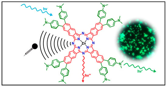
Dual Emissive Zn(II) Naphthalocyanines: Synthesis, Structural and Photophysical Characterization with Theory-Supported Insights towards Soluble Coordination Compounds with Visible and Near-Infrared Emission

by
Sidharth Thulaseedharan Nair Sailaja
1,2,
Iván Maisuls
1,2,
Alexander Hepp
1,
Dana Brünink
3,
Nikos L. Doltsinis
3,
Andreas Faust
4,5,
Sven Hermann
4,5 and
Cristian A. Strassert
1,2,*
1
Institut für Anorganische und Analytische Chemie, Universität Münster, Corrensstraße 28/30, 48149 Münster, Germany
2
CeNTech, CiMIC, SoN, Universität Münster, Heisenbergstraße 11, 48149 Münster, Germany
3
Institute for Solid State Theory and Center for Multiscale Theory and Computation, Universität Münster, Wilhelm-Klemm-Straße 10, 48149 Münster, Germany
4
European Institute for Molecular Imaging, Universität Münster, Röntgenstraße 16, 48149 Münster, Germany
5
Department of Nuclear Medicine, University Hospital Münster, Albert-Schweitzer-Campus 1, 48149 Münster, Germany
*
Author to whom correspondence should be addressed.
Int. J. Mol. Sci. 2024, 25(5), 2605; https://doi.org/10.3390/ijms25052605
Submission received: 31 December 2023 / Revised: 9 February 2024 / Accepted: 14 February 2024 / Published: 23 February 2024
(This article belongs to the Special Issue Feature Papers in 'Physical Chemistry and Chemical Physics' 2024)

Abstract

:
Metal phthalocyaninates and their higher homologues are recognized as deep-red luminophores emitting from their lowest excited singlet state. Herein, we report on the design, synthesis, and in-depth characterization of a new class of dual-emissive (visible and NIR) metal naphthalocyaninates. A 4-N,N-dimethylaminophen-4-yl-substituted naphthalocyaninato zinc(II) complex (Zn-NMe2Nc) and the derived water-soluble coordination compound (Zn-NMe3Nc) exhibit a near-infrared fluorescence from the lowest ligand-centered state, along with a unique push–pull-supported luminescence in the visible region of the electromagnetic spectrum. An unprecedentedly broad structural (2D-NMR spectroscopy and mass spectrometry) as well as photophysical characterization (steady-state state and time-resolved photoluminescence spectroscopy) is presented. The unique dual emission was assigned to two independent sets of singlet states related to the intrinsic Q-band of the macrocycle and to the push–pull substituents in the molecular periphery, respectively, as predicted by TD-DFT calculations. In general, the elusive chemical aspects of these macrocyclic compounds are addressed, involving both reaction conditions, thorough purification, and in-depth characterization. Besides the fundamental aspects that are investigated herein, the photoacoustic properties were exemplarily examined using phantom gels to assess their tomographic imaging capabilities. Finally, the robust luminescence in the visible range arising from the push–pull character of the peripheral moieties demonstrated a notable independence from aggregation and was exemplarily implemented for optical imaging (FLIM) through time-resolved multiphoton micro(spectro)scopy.

Graphical Abstract

1. Introduction

Over the past decade, there has been a rising interest in the design and synthesis of compounds exhibiting dual emission (DE) [1]. While most organic molecules typically manifest a single characteristic fluorescence, certain distinctive luminophores, wherein electron donor and electron acceptor moieties are connected by a single bond, may exhibit a dual emission [2]. The phenomenon of dual fluorescence (DF) was initially observed for 4-(dimethylamino)benzonitrile (DMABN) by Lippert et al. [3]. It arises from two distinct conformations of the same molecule in the first excited singlet state (S1), specifically, the locally excited state (LE) and the intramolecular charge-transfer state (ICT) [4]. Subsequently, several other systems were designed incorporating a strong electron-donating or -withdrawing group [5,6,7,8,9,10,11]. Owing to their nonlinear optical properties, these compounds have been extensively explored as part of the development of organic materials, serving various applications including their use as electrooptical switches, chemical sensors, and fluorescent markers [12,13,14,15].
Generally, due to their broad application range as functional materials for semiconductors, gas sensors, nonlinear optical limiters, liquid crystals and as sensitizers for photodynamic therapy (PDT) in cancer treatments, among many others, metal phthalocyanines (Pcs) are counted among the most extensively investigated dyes in history, besides others such as cyanine colorants and BODIPY derivatives [16,17,18]. Metal naphthalocyanines (Ncs) constitute expanded analogues of their Pc counterparts that possess a linearly annulated benzene ring in the periphery of the macrocyclic core that leads to an about 100 nm red shift of the absorption band in the red if compared with Pcs. This shift, known as the rule of “100 nm”, results from the destabilization of the HOMO (highest occupied molecular orbital) and the associated decrease in the HOMO-LUMO (lowest unoccupied molecular orbital) gap [19]. Thus, Ncs are able to act as versatile tetradentate chelators for various metals and show a strong absorption in the near-infrared region (750–900 nm) with high molar absorption coefficients. The intense absorption and fluorescence bands of naphthalocyanines in the near-infrared offers interesting opportunities such as the fabrication of high-performance NIR photodiodes and bioimaging [20,21], and the complexation of open-shell or late-transition element cations can add further functionalities related to spin–spin or spin–orbit coupling, respectively.
The structural flexibility of Ncs has been extensively illustrated through a diverse range of metal complexes and a wide array of substituents that can be attached to the periphery of the core or as axial ligands [19,22,23,24,25,26,27]. This chemical modification induces specific alterations in the electronic structure of the macrocyclic core, providing precise control over the physicochemical characteristics. However, despite their ability to absorb in the far red/near-infrared region, Ncs have been relatively neglected, mainly because of their intricate synthesis, with difficult purification, poor solubility, and a tendency to form inactive aggregates, owing to strong van der Waals interactions upon stacking [28,29]. As a consequence of their high tendency to form aggregates, a drop of fluorescence and singlet oxygen quantum yields is observed [30,31]. There have been several structural variations introduced to overcome the solubility and aggregation issues of (na)phthalocyanines, including the insertion of bulky substituents in the peripheral positions of the macrocycles [25,32,33,34,35], the addition of axial ligands to the central atom [28,30,36,37,38,39], or the encapsulation of the dye in colloidal particles [40,41]. Furthermore, peripheral functional groups in Ncs macrocycles may be utilized to further tailor intra- and intermolecular interactions as well as optical, photochemical, and electrochemical properties; particularly, aromatic groups increase intermolecular π-π interactions [19,34]. However, all these derivatives still exhibit a strong tendency to aggregate in aqueous media, which can significantly reduce their performance due to self-quenching [42]. In the case of Pcs, these issues were solved by the introduction of several hydrophilic and amphiphilic groups (e.g., carboxylates, sulfonates, phosphonates, PEG chains) [43,44,45,46,47,48], as well as bulky axial ligands (e.g., −OSiMe2(CH2)3NMe2 and −OCH(CH2NMe2)2) [49,50,51,52,53] in the periphery of the macrocycle or at the central atom, respectively. Among these strategies, positively charged macrocycles are of particular interest, as they might target highly vulnerable intracellular sites and produce effective DNA damage in the context of phototherapy [42]. Moreover, positively charged Pcs have been successfully employed for the photoinactivation of both Gram-negative and Gram-positive bacteria [54,55,56]. However, in the case of water-soluble Ncs, the majority of the reported exemplars are based on silicon(IV)-based coordination compounds, mainly due to the presence of two axial positions that can be readily modified to reduce aggregation while improving solubility in aqueous media [27,57]. While peripherally substituted water-soluble Pcs have been widely discussed in the literature [30,58], their Ncs counterparts with aqueous solubility have remained vastly unexplored [29,59]. Despite exhibiting NIR absorption and emission, Ncs exhibiting dual fluorescence (vis/NIR) have not been reported so far.
With this background in mind, we herein report on an unprecedented design pattern for single-component yet dual-emissive zinc(II) naphthalocyaninates, including a detailed synthesis as well as a structural and photophysical characterization. In order to suppress the intermolecular interactions and to improve their solubility, we aimed at the tailored substitution on the γ-position of the macrocyclic ligand to introduce eight 4-N,N-dimethylaminophen-4-yl moieties, which were further quaternized with methyl iodide to enhance their hydrophilicity. Moreover, substitution with 4-N,N-dimethylaminophen-4-yl moieties opens up new possibilities for push–pull-based two-photon excitability and aggregation-independent luminescence in the visible region of the electromagnetic spectrum. To assess the charge-transfer nature of the excited state that was provided by the dimethylamino substitutents, an analogous 4-methoxyphen-4-yl-substituted zinc(II) naphthalocyaninate was synthesized. The assignment of the emissive states was carried out by TD-DFT calculations while rationalizing the electronic transitions dominating the absorption and emission spectra of these complexes. Thus, we anticipate that this strategy for preparing dual-emissive (visible and NIR) naphthalocyanines will pave the way for future innovation and accelerated developments in a broad range of application fields, including optoelectronics, energy conversion, and as contrast agents that are able to provide two orthogonal readouts for multiscale–multimodal imaging.

2. Results and Discussion

2.1. Design, Synthesis, Purification, and Structural Characterization

The peripheral functionalization was achieved by means of a Suzuki–Miyaura coupling reaction between 6,7-dibromo-2,3-dicyanonaphthalene (1) and the corresponding boronic acids, following our previously reported methodology [60]. This substitution pattern was chosen in accordance with our recent report showing that the push–pull character and perpendicular-to-plane rigidified arrangement of the two phenyl moieties can provide strong luminescence in aggregates as well as in solution. The detailed synthetic procedure is shown in Section 3 (vide infra). The substituted naphthalonitriles NMe2 and OMe were converted into the corresponding naphthalocyaninato zinc(II) complexes Zn-OMeNc and Zn-NMe2Nc by refluxing them in isoamyl alcohol (i-amOH) in the presence of 1,8-diazabicycloundec-7-ene (DBU) in catalytic amounts (Figure 1).
The reaction was performed under inert conditions in order to prevent the oxidation of the macrocycle by atmospheric oxygen at high temperatures. The obtained products contained some fluorescent impurities (most likely low-molecular-weight condensates preceding the cyclotetramerization); attempts to use column chromatography with silica or alumina as stationary phases in combination with classical organic solvent gradients failed, as the polar components of the reaction mixture strongly bind to both materials. Reverse-phase silica was also explored for purification; also in this case, it was observed that the product strongly binds to the stationary phase. Thus, the crude reaction mixture was subjected to multiple Soxhlet extractions to remove bulk impurities. The obtained products were further purified by size exclusion chromatography (Sephadex LH-20, Amersham Pharmacia Biotech AB, Uppsala, Sweden) in N,N-dimethylformamide (DMF). Finally, the water-soluble octa-cationic zinc(II) naphthalocyaninate Zn-NMe3Nc was obtained by treating Zn-NMe2Nc with an excess of methyl iodide in DMF (Figure 2). Besides UV-vis absorption spectroscopy, the Ncs were structurally characterized by means of mass spectrometry and NMR spectroscopy. Due to their intrinsic aggregation tendency, attaining well-resolved 1H- and 13C-NMR spectra proved to be particularly challenging. Nonetheless, we were able to obtain precise 2D-NMR spectra (including an unambiguous assignment for all peaks).
In order to obtain a better resolution in the 1H-NMR spectra of Zn-OMeNc, Zn-NMe2Nc, and Zn-NMe3Nc, DMSO was used as the solvent in NMR spectroscopy, as it suppresses aggregation caused by stacking. Due to the de-shielding effect of the porphyrazinato core, a clear downfield shift of Hα protons compared to Hβ was observed [34]. The interpretation of all signals in the 1H-NMR spectra of the Ncs Zn-OMeNc, Zn-NMe2Nc, and Zn-NMe3Nc were enabled by 1H-1H-COSY, 1H-13C-HSQC, 1H-13C-HMBC, and 1H-1H-ROESY spectroscopies, where all signals were unambiguously assigned (also for the naphthalonitriles, see SI, Figures S1–S26). From the 1H-NMR spectra of Zn-OMeNc and Zn-NMe2Nc, it was clear that these two coordination compounds aggregate at the required concentrations, whereas the eight positive charges in the periphery of Zn-NMe3Nc suppressed aggregation and yielded well-resolved 1H-NMR spectra (Figure S21). Hence, the proton signals from Zn-NMe3Nc were better resolved than those from Zn-OMeNc and Zn-NMe2Nc. In the 1H-1H-ROESY spectra, cross-peaks were observed between the signals of Hα and Hβ protons and between the protons of the phenyl groups and signals corresponding to Hβ (see SI, Figures S14, S20 and S26). From 1H-1H-COSY, additional groups of cross-peaks were found between the protons of the phenyl groups (see SI, Figures S11, S17 and S23). The mass spectra of Zn-NMe3Nc were obtained by using ESI-EM, whereas for Zn-OMeNc and Zn-NMe2Nc, MALDI-TOF had to be employed. The MS demonstrated a good agreement between the expected and observed peaks (see SI, Figures S27–S31).

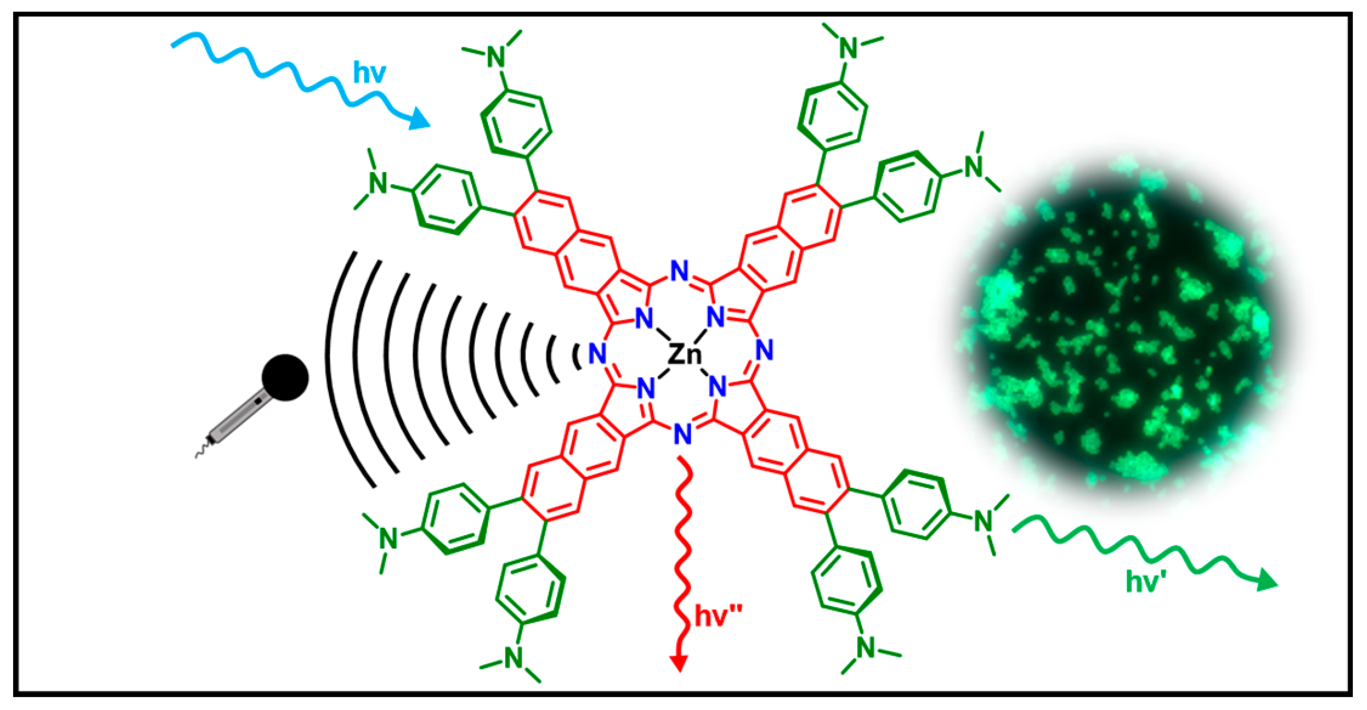
2.2. UV-Vis Absorption, Steady-State, and Time-Resolved Photoluminescence Spectroscopies

In agreement with the characteristic spectroscopic features of metal (na)phthalocyaninates, the UV-vis absorption spectra of the herein reported complexes feature a Soret-band (or B-band) in the UV range and Q-band in the NIR region [25]. Figure 3 depicts the absorption spectra of Zn-OMeNc, Zn-NMe2Nc, and Zn-NMe3Nc in DMSO and in water. A red shift of the Q-band was observed when going from Zn-OMeNc (794 nm) to Zn-NMe2Nc (800 nm), implying that the energy gap between the HOMO and the LUMO is reduced when introducing a stronger electron π-donor (i.e., replacement of methoxy by dimethylamino moieties) [61,62]. Consistently, in the case of Zn-NMe3Nc, a blue shift was observed (781 nm) in DMSO, due to the reduced electron-donating capacity of the cationic trimethylammonium groups (Table 1).
As shown in Figure 3a,b, the Q-band appears to be less intense than the B-band, which is surprising when considering that the latter is usually weaker in metal (na)phthalocyaninates. This observation can be attributed to the strong absorption in the blue and in the UV related to the eight methoxy- or dimethylamino-substituted moieties, which actually exhibit a sizeable push–pull character (in fact, this blue-shifted band is drastically reduced upon quaternization, as seen by the comparison of Zn-NMe2Nc with its derivative Zn-NMe3Nc; vide infra regarding the additional influence of aggregation phenomena on the relative intensities in the red and in the blue region of the spectrum). Similar trends were previously reported for Zn(II) phthalocyanines [63,64]. The formation of red-shifted aggregates plays a role in the case of Zn-OMeNc, in agreement with the broadened signals that are observed in NMR spectroscopy (see SI, Figure S9); hence, the low molar absorption coefficient (ε) at λmax = 794 nm (ε = 4.4 × 10⁴ M−1 cm−1) and the rather weak absorption band at around 876 nm point towards J-aggregation. Similarly, in the case of Zn-NMe2Nc, the intensity of the Q-band appears to be reduced if compared with the B-band, an observation that is reinforced by aggregation; therefore, a relatively low molar absorption coefficient at λmax = 800 nm (ε = 5.3 × 10⁴ M−1 cm−1) is measured. Typically, the values for Q-bands in metal (na)phthalocyaninates are of the order of 105 M−1 cm−1. Thus, by comparison of the three compounds presented herein, it is clear that the relatively high intensity of the B-band is prominently enhanced in the cases of Zn-OMeNc and Zn-NMe2Nc, due to the charge-transfer absorption of the eight aromatic electron-donating moieties (methoxy- or dimethylaminophenyl, respectively), which overlap with the B-band and disappear upon quaternization (vide infra); in addition, aggregation compromises the intensity of the Q-band.
Nonetheless, the eight positive charges of Zn-NMe3Nc suppress aggregation in DMSO, and the relative intensity of the Q-band vs. the B-band fall in the expected range for metal (na)phthalocyaninates [25,65]. However, the absorption spectrum of Zn-NMe3Nc in water showed clear differences when compared to its spectrum in DMSO. In water, Zn-NMe3Nc still exhibits H-aggregation, as evidenced by the presence of two non-vibrational shoulders in the Q-band region (Figure 3d). The lower energy (red-shifted) band at about 784 nm can be attributed to the monomeric species, whereas the higher energy (blue-shifted) band at 732 nm suggests the presence of H-aggregates, as it was previously reported for comparable (na)phthalocyanines [29,30,31,66]. The formation of H-aggregates in water is more evident when comparing the molar absorption coefficients (ε) of Zn-NMe3Nc in DMSO vs. water. In DMSO, the H-aggregates are disrupted, and Zn-NMe3Nc shows a characteristically high ε value at λmax = 781 nm (ε = 5.5 × 10⁵ M−1 cm−1). This contrasts with the spectrum in water, where Zn-NMe3Nc peaks at λmax = 732 nm (ε = 2.2 × 10⁵ M−1 cm−1), and λmax = 784 nm (ε = 1.8 × 10⁵ M−1 cm−1) (Table 1). In water, H-aggregation reduces the Q-band intensity with respect to the B-band, and the molar absorption coefficient in the red drops by a factor of roughly two, which points toward dimerization. Despite the clear aggregation phenomena discussed above, the Lambert–Beer law was obeyed within the experimental range for all these compounds (see SI, Figures S35–S47) [67]. In general, the molar absorption coefficients were obtained from the slope of the absorbance versus the concentration of the samples (the latter were determined by total reflection X-ray fluorescence (TXRF) addressing the concentration of Zn).
The UV-vis absorption, steady-state photoluminescence emission and excitation spectra of Zn-OMeNc, Zn-NMe2Nc, and Zn-NMe3Nc were measured in DMSO (Figure 4). In the case of Zn-NMe3Nc in water, a clear narrowing of the excitation spectrum was observed compared with the absorption spectrum, as only the emissive monomers were addressed, whereas all species (even aggregates) in a solution contribute to the broadened absorption. The proximity of the Q-band-related emission with respect to the Q-band excitation and absorption for Zn-OMeNc, Zn-NMe2Nc, and Zn-NMe3Nc in DMSO indicate that the nuclear configurations of the ground and excited states are comparable and therefore practically not affected by the excitation wavelength.
In agreement with previous reports on metal (na)phthalocyaninates, the photoluminescence emission spectra of Zn-OMeNc, Zn-NMe2Nc, and Zn-NMe3Nc mirror the excitation spectra with a typically small Stokes shift (Figure 4), if referring to the red-shifted Q-band. As can be observed in the full-range emission spectra (recorded while exciting in the UV), the compounds feature a surprisingly prominent dual emission, which will be discussed below (vide infra). This phenomenon was initially noticed while measuring the emission spectra of Zn-NMe2Nc in DMSO at different excitation wavelengths (λex). After varying λex, a broad emission band with a λem = 560 nm (λex = 335 nm) and a sharp near-infrared emission with λem = 811 nm (λex = 750 nm) was observed (Figure 5c,d, Table 2). In order to understand this rather unique dual emission, the analogous Zn-OMeNc was also designed, prepared, and studied. Interestingly, Zn-OMeNc also exhibited two clear emission bands, with a λem = 508 nm (λex = 335 nm) and λem = 804 nm (λex = 750 nm) in DMSO (Figure 5c,d, Table 2). It is important to mention that when using λex = 335 nm and screening the emission from the blue to the NIR region to acquire the full-range fluorescence spectra, both bands are visible in the case of Zn-OMeNc and Zn-NMe2Nc (Figure 5a,b, Table 2).
A similar trend was observed for Zn-NMe3Nc when measured in water and in DMSO. In DMSO, Zn-NMe3Nc exhibited two emission bands, with a λem = 550 nm (λex = 335 nm) and λem = 801 nm (λex = 750 nm) (Figure 6c,d, Table 2). In contrast, the emission bands measured in water were observed at λem = 432 nm (λex = 335 nm) and λem = 804 nm (λex = 750 nm) (Figure 6c,d, Table 2). When a full-range emission spectrum for Zn-NMe3Nc in DMSO was measured (from blue to the NIR region using λex = 335 nm), a relatively faint emission band with λem = 550 nm and a high-intensity luminescence with λem = 800 nm were observed (Figure 6a). This can be attributed to the reduction in the push–pull character upon quaternization of the peripheral 4-N,N-dimethylaminophen-4-yl groups and the monomeric nature of Zn-NMe3Nc in DMSO, yielding a comparably strong NIR fluorescence and a weak visible emission. Thus, it is inferred that the monomers of Zn-NMe3Nc in DMSO yield an intense NIR emission and a comparatively weak luminescence between 400 nm and 500 nm. In contrast, the full-range emission spectrum of Zn-NMe3Nc in water (from blue to the NIR region using λex = 335 nm) exhibited a substantially stronger emission band, peaking at λem = 400 nm, when compared with the NIR fluorescence band at λem = 801 nm (Figure 6b); this can be understood when taking into account that in water, H-aggregation suppresses the fluorescence in the NIR, and a comparatively strong residual emission remains in the visible range. The dual emission of Zn-OMeNc, Zn-NMe2Nc, and Zn-NMe3Nc was interpreted and assigned by TD-DFT calculations (vide infra).
The dual fluorescence can be explained by two sets of singlet states (with orthogonal charge-transfer vs. main-ligand-centered character). The near infrared fluorescence originates from the S1/2 → S0 transition, and the visible emission stems from the S3 → S0 transition. The doubly degenerate S1/2 state has a ππ* character, whereas the S3 states have a charge-transfer (nπ*) character involving the electron-rich methoxy- or dimethylamino-substituted phenyl moieties. The energy gap between S1/2 and S3 states is relatively large; the lack of excited state geometry distortion (i.e., parallel potential hypersurfaces or “nested states”) hamper the non-adiabatic crossover, leading to relatively slow internal conversion processes. Hence, in the case of Zn-OMeNc, Zn-NMe2Nc, and Zn-NMe3Nc, both an emission in the visible region and an NIR fluorescence are observed. In the case of Zn-NMe3Nc in water, the S1/2 state is quenched due to aggregation, the intensity of the NIR emission is low, and the push–pull character is reduced due to quaternization.
Taking into account that the relative intensities of the two emission bands remain invariant when comparing independently synthesized batches, and considering the thorough purification that was carried out with no traces of impurities in the NMR spectra, we can confidently exclude spurious emission from side products or other contamination (photodecomposition is ruled out, as the absorption and emission spectra do not significantly vary during the photophysical characterization). Moreover, TD-DFT calculations support the assignment (vide infra).

2.3. TD-DFT Calculations

In order to assign the transitions that were observed in the absorption and fluorescence spectra, we optimized the structures of Zn-OMeNc, Zn-NMe2Nc, and Zn-NMe3Nc in their electronic ground states (for computational details, see Section 3.4). All three complexes exhibited a highly planar core, which was marginally influenced by the peripheral substituents (see Figure 7). At the optimized ground state geometries, the UV-vis absorption spectra were calculated using TD-DFT/B3LYP.
As can be seen from Figure 8, the theoretical spectra generally agree well with the experimental data. Due to the degeneracy of the LUMO and LUMO + 1 orbitals, the S1 and S2 states are also doubly degenerate, as they can be described by HOMO → LUMO and HOMO → LUMO+1 excitations, respectively. The band corresponding to the S0 → S1/2 transition—located at 805 nm, 756 nm, and 740 nm for Zn-NMe2Nc, Zn-OMeNc, and Zn-NMe3Nc, respectively—shows a progressive blue shift with decreasing electron donor ability, in good agreement with the experimental data (and also qualitatively with respect to the relative intensities of the B- and Q-bands). The high intensity of this NIR maximum can be explained by the fact that it represents a local excitation within the 2,3-naphthalocyaninato ligand (the black part of the structure depicted in Figure 1, see Figure 9 for a visualization of the relevant frontier orbitals).
In addition, we calculated the 0-0 fluorescence wavelengths from the S1/2 and S3 states by optimizing the respective excited state geometries and subtracting the optimized ground state energy. While the S1/2 → S0 de-excitation involved a local LUMO/LUMO + 1 → HOMO configuration change (Figure 9), which was reasonably well described by all functionals that were tested herein, the S3 → S0 de-excitation turned out to be much more problematic, as it involved a pronounced charge-transfer character for the relevant excited state—at least for Zn-OMeNc and Zn-NMe2Nc (see Figure 10). For Zn-NMe2Nc, geometry optimization with the B3LYP functional leads to a collapse of the S3 state onto the S1/2 state, yielding spurious emission wavelengths. The long-range corrected functional CAM-B3LYP, on the other hand, significantly overestimated the S3 energy (see SI, Table S1). In fact, the PBE0 functional emerged as the best compromise for all three compounds, considering both emission lines in the visible and the NIR (see Figure 11). It is able to reproduce the position of the experimental 0-0 peaks accurately, including the slight blue shift when going from Zn-NMe2Nc to Zn-NMe3Nc via Zn-OMeNc. For Zn-OMeNc, the S3 peak was also described very well, while for Zn-NMe2Nc, the agreement with the experiment was particularly poor, with the PBE0 peak being red-shifted by about 150 nm.
Presumably, this is due to the stronger charge-transfer character in the case of Zn-NMe2Nc, with a notable amount of charge density being located at the NMe2 substituent in the HOMO-1. In addition, the S3 state contains a 7% contribution of the local HOMO → LUMO + 1 excitation. The use of the M06-2X functional leads to a considerable improvement for Zn-NMe2Nc (see Figure 11), but not for the other two compounds. PBE0 predicts the S3 state to be governed by a HOMO-1 → LUMO excitation in the case of Zn-NMe2Nc and Zn-OMeNc, whereas for Zn-NMe3Nc, the S3 state is dominated by a HOMO → LUMO + 2 excitation (Figure 10).

2.4. MSOT Imaging in Gel Phantoms

In recent studies, it has been demonstrated that naphthalocyaninato complexes (Ncs) hold significant potential as photoacoustic contrast agents [27,68,69]. In comparison with other organic compounds, such as cyanine dyes and nanoparticles, naphthalocyanines exhibit higher molar absorption coefficients in the NIR region, featuring sharp maxima between 770 nm and 850 nm [16]. The well-defined NIR absorption peaks mentioned above contribute to more precise spectral unmixing, distinguishing them from other exogenous contrast agents [70]. As a result, Zn-NMe2Nc and Zn-NMe3Nc were employed as representative examples for generating photoacoustic (PA) images utilizing multi-spectral optoacoustic tomography (MSOT). These measurements were conducted within tissue-mimicking gel phantoms, as depicted in Figure 12 [69]. The experiments were carried out at different concentrations, and the obtained photoacoustic profiles qualitatively agree with the absorption spectra regarding the wavelengths that induce the highest photoacoustic response. The phantom studies were performed by dissolving Zn-NMe2Nc in DMSO, Zn-NMe3Nc in DMSO, and Zn-NMe3Nc in water, respectively.
Among these, Zn-NMe3Nc in DMSO provided the highest photoacoustic signal even at a low concentration (1 μM), with Zn-NMe2Nc in DMSO and Zn-NMe3Nc in water showing similar yet slightly lower PA signals (Figure 13). The observed difference in signal intensity of Zn-NMe3Nc in DMSO and water can be attributed to the monomerization in DMSO and aggregation in water. Also, when comparing the photoacoustic (Figure 13) and absorption spectra (Figure 3c) of Zn-NMe3Nc in DMSO at λmax = 781 nm, the relative intensities of the vibronic features appear distorted. For the photoacoustic spectra of Zn-NMe3Nc in DMSO, the inner filter effect limits the excitation at the main maxima, even at very low concentrations (1 μM), while causing a saturation plateau between λmax = 781 nm and λmax = 700 nm. As Zn-NMe3Nc in DMSO provides the best photoacoustic signals at a low concentration, it is clear that the suppression of aggregation enhances the photoacoustic output. In the future, this could be further improved with vanadyl- or silicon-naphthalocyanines, where the axial ligands could prevent aggregation [30,69,71]; the insertion of vanadyl centers could further shift the absorption maxima to longer wavelengths in the infrared region [72,73,74].

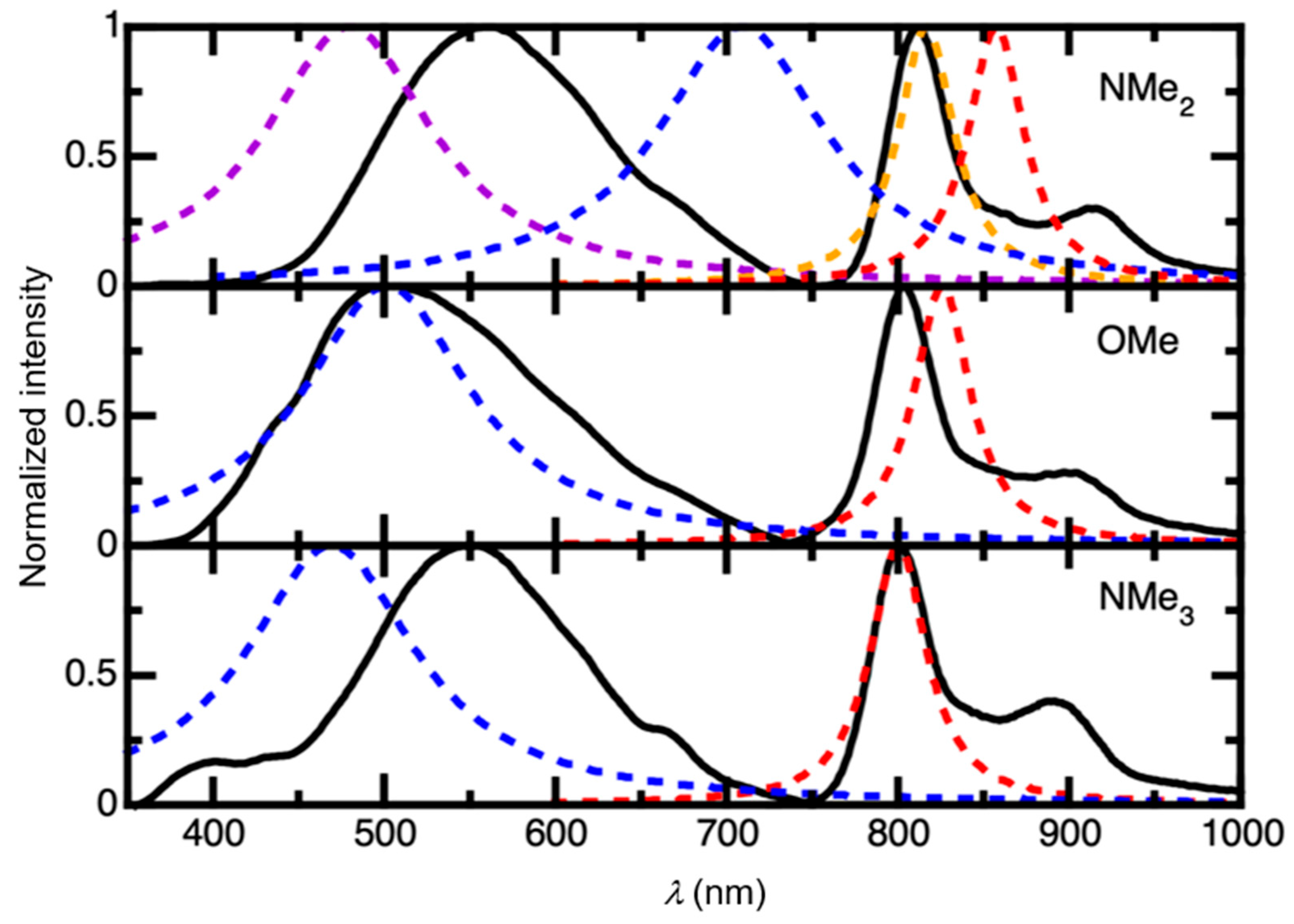
2.5. Time-Resolved Multiphoton Micro(spectro)scopy

In order to demonstrate the ability to image structural features by time-resolved multiphoton micro(spectro)scopy, we employed Zn-NMe2Nc to stain 1 μm sized aminated polystyrene microparticles (PSMPs) by a swelling–diffusion processes [75], as these are well-established models for microstructured samples (Figure 14b). As depicted in Figure 14, the photoluminescence emission spectra of Zn-NMe2Nc-loaded PSMPs (Zn-NMe2Nc@PSMP) in water show a broad emission band in the green (λmax ≈ 470 nm), but no emission was observed in the near-infrared region (Figure 14a); this points towards aggregation-caused quenching of the fluorescence from the macrocycle, as it was previously reported for other (na)phthalocyanines [76]. However, the distinct emission in the visible region, originating from the push–pull character of the excited side groups, can be utilized as an additional optical readout, despite the lack of intrinsic luminescence from the macrocycle. In fact, the push–pull side groups have been previously introduced in our recent work as robust luminophores, both in aggregated and in monomeric forms [60]. The excited state lifetime of the stained microparticles in liquid water was determined, and a τav_amp = 5.1 ns was obtained (Figure S56).
Further experiments were carried out to assess the photophysical properties of discrete Zn-NMe2Nc@PSMP. Briefly, Zn-NMe2Nc@PSMP were placed on a microscope slide and analyzed using single-photon excitation (SPE) to carry out fluorescence lifetime imaging microscopy (FLIM). As depicted in Figure 15, the particles were homogeneously stained with the push–pull luminophore, confirming the observation that is shown in Figure 14, while possessing a homogeneously distributed amplitude-weighted average lifetime of τ ≈ 3.9 ns (Figure 15 and Figure S57). In addition, employing a spectrophotometer coupled by an optical fiber to the confocal microscope, the emission spectra of discrete Zn-NMe2Nc@PSMP entities were obtained as dry samples while consistently reproducing the results for Zn-NMe2Nc@PSMP that was suspended in liquid water (Figure 15). In contrast to single-photon excitation (SPE), two-photon excitation (TPE) laser scanning microscopy is often anticipated to increase cell survival and tissue penetration [77]. Thus, we explored herein a way to increase the detection sensitivity due to an improved sample penetration employing a near-infrared TPE laser; we therefore performed comparable FLIM experiments using a high-intensity Ti:Sa oscillator as the two-photon excitation source (Figure 15), where comparable photoluminescence lifetime maps were obtained. In addition, the emission spectra of discrete Zn-NMe2Nc@PSMP were also measured by TPE. These experiments demonstrate that no significant differences are to be expected when using SPE and TPE (both in lifetimes (Figures S57 and S58) and in the emission spectra) for optical imaging, while providing a second readout for microscopic imaging.

3. Materials and Methods

All chemicals were purchased with the maximum quality available from Sigma Aldrich (Taufkirchen, Germany) or from TCI Chemicals (Eschborn, Germany) and used without further purification. Reactions were carried out using dried solvents (99.9% purity) under argon atmosphere. They were monitored by thin-layer chromatography (TLC), which was performed on 0.2 mm Macherey-Nagel ALUGRAM (Eupen, Belgium) precoated silica gel aluminum sheets. Spots were visualized by a UV handlamp (254 and 365 nm). Silica gel 60 (0.063–0.200 mm, Merck, Darmstadt, Germany) was used for column chromatography, if not otherwise stated. Fresh spectroscopic-grade solvents (Uvasol®, Merck, Darmstadt, Germany) were utilized for the spectroscopic studies.

3.1. Synthesis and Characterization of 6,7-Disubstituted Naphthalene-2,3-dicarbonitriles and Zinc(II) Naphthalocyanines

3.1.1. General Procedure for the Synthesis of 6,7-Disubstituted Naphthalene-2,3-dicarbonitriles

6,7-dibromonaphthalene-2,3-dicarbonitrile (1) was synthesized according to a published procedure [23,78,79]. The 6,7-disubstituted naphthalene-2,3-dicarbonitriles were obtained by Suzuki–Miyaura reactions from 1 with the corresponding boronic acids (Scheme 1) [60]. Briefly, a mixture of 1 (1 equiv.), the corresponding p-substituted-phenylboronic acid (4 equiv.), and a saturated aqueous solution of K2CO3 (5 mL) were stirred in 50 mL of a boiling mixture of 1,4-dioxane:acetonitrile (8:3 v/v) under argon. Afterwards, palladium(II)bis(triphenylphosphane) dichloride (0.01 equiv.) was added to the boiling mixture. The reaction was stirred for 6 h (TLC control: Al2O3, F254, ethyl acetate:hexane, 1:4). After consumption of the starting materials, the reaction mixture was cooled down to room temperature and water was added. The product was collected by extraction with dichloromethane, and the organic layer was dried with anhydrous MgSO4. The residue was treated by flash chromatography on silica (ethyl acetate:hexane, 1:2) to remove impurities. The resulting compound was additionally purified by column chromatography on silica using DCM as the eluent to obtain the corresponding p-phenyl-6,7-disubstituted naphthalene-2,3-dicarbonitrile.

3.1.2. Synthesis of 6,7-Bis(4-methoxyphenyl)naphthalene-2,3-dicarbonitrile (OMe)

OMe was synthesized according to the general procedure using 1 (50 mg, 0.15 mmol), 4-methoxyphenylboronic acid (91 mg, 0.6 mmol), and a catalytic amount of palladium(II)bis(triphenylphosphane) dichloride (1 mg, 0.0015 mmol). Molecular formula: C26H18N2O2 (pale yellow solid). Yield: 51% (29 mg, 0.07 mmol).
1H-NMR (500 MHz, DCM-d2): δ (ppm) = 8.37 (s, 2H, 3-H), 7.96 (s, 2H, 5-H), 7.18–7.12 (m, 4H, 8-H), 6.86–6.82 (m, 4H, 9-H), 3.80 (s, 6H, 12-H).
13C-{1H}-NMR (126 MHz, DCM-d2): δ (ppm) = 159.76 (C-10), 144.47 (C-6), 136.01 (C-3), 132.72 (C-4), 132.54 (C-7), 131.34 (C-8), 130.13 (C-5), 116.69 (C-1), 114.09 (C-9), 110.16 (C-2), 55.65 (C-12).
MS-ESI-EM (CH2Cl2, M = C26H18N2O2): m/z calc. for [M + H]+ = 390.13628; found 390.13624 for [M + H]+.

3.1.3. Synthesis of 6,7-Bis(4-(dimethylamino)phenyl)naphthalene-2,3-dicarbonitrile (NMe2)

NMe2 was synthesized according to the general procedure using 1 (50 mg, 0.15 mmol), 4-methoxyphenylboronic acid (98 mg, 0.6 mmol), and a catalytic amount of palladium(II)bis(triphenylphosphane) dichloride (1 mg, 0.0015 mmol). Molecular formula: C28H24N4 (yellow solid). Yield: 63% (39 mg, 0.09 mmol).
1H-NMR (400 MHz, DCM-d2): δ (ppm) = 8.30 (s, 2H, 3-H), 7.88 (s, 2H, 5-H), 7.15–7.09 (m, 4H, 8-H), 6.69–6.61 (m, 4H, 9-H), 2.96 (s, 12H, 12-H).
13C-{1H}-NMR (101 MHz, DCM-d2): δ (ppm) = 150.36 (C-10), 145.08 (C-6), 135.81 (C-3), 132.55 (C-4), 130.88 (C-8), 129.70 (C-5), 128.09 (C-7), 116.90 (C-1), 112.19 (C-9), 109.49 (C-2), 40.49 (C-12).
MS-ESI-EM (CH2Cl2, M = C28H24N4): m/z calcd. for [M + H]+ = 417.20737; found 417.20710 for [M + H]+.

3.1.4. General Procedure for the Synthesis of Zinc(II) Naphthalocyanines

A mixture of the corresponding 6,7-disubstituted naphthalene-2,3-dicarbonitrile (1 equiv.) and Zn(OAc)2·2H2O (0.5 equiv.) was refluxed in 10 mL i-amOH (isoamyl alcohol) for 6 h with catalytic amounts of DBU (Figure 1). The reaction mixture was cooled to room temperature, and a MeOH:H2O mixture (20:1 v/v) was added. The precipitate was filtered and washed with a MeOH:H2O (10:1 v/v) mixture. However, the obtained precipitate contained some impurities, which were removed by a Soxhlet extraction with successive solvents of increasing polarity (from diethyl ether to hexane and ethyl acetate). The product was further purified by size exclusion chromatography using a resin (Sephadex-LH20, Amersham Pharmacia Biotech AB, Uppsala, Sweden) in N,N-dimethylformamide to obtain the corresponding zinc(II) naphthalocyanines.

3.1.5. Synthesis of 3,4,12,13,21,22,30,31-Octakis(4-methoxyphenyl) Naphthalocyaninato Zinc(II) (Zn-OMeNc)

Zn-OMeNc was prepared by treating OMe (0.1 g, 0.25 mmol) with Zn(OAc)2·2H2O (0.028 g, 0.127 mmol) and using DBU as the catalyst in 10 mL i-amOH. The obtained product was precipitated with a MeOH:H2O (10:1 v/v) mixture and filtered. The purification of the obtained products involved a sequential two-step approach. Initially, a Soxhlet extraction was employed using solvents of increasing polarity (from diethyl ether to hexane and ethyl acetate). Subsequently, the resulting product underwent a final purification using size exclusion chromatography with a resin (Sephadex-LH20) in N,N-dimethylformamide. The collected fractions were concentrated to obtain the Zn-OMeNc. Molecular formula: C104H72N8O8Zn (brown solid). Yield: 21% (90 mg, 0.05 mmol).
1H-NMR (400 MHz, DMSO-d6): δ (ppm) = 8.50–8.26 (br, m, 8H, 3-H), 8.24–7.97 (br, m, 8H, 5-H), 7.15 (m, 16H, 8-H), 6.90 (m, 16H, 9-H), 3.76 (s, 24H, 11-H).
13C-{1H}-NMR (125 MHz, DMSO-d6): δ (ppm) = 168.80 (C-1), 158.40 (C-10), 141.00 (C-6), 134.10 (C-4), 132.50 (C-7), 131.25 (C-5), 130.70 (C-8), 128.70 (C-2), 123.90 (C-3), 113.54 (C-9), 55.00 (C-11).
MALDI-TOF-MS (CHCl3, M = C104H72N8O8Zn): m/z calcd. for [M]+ = 1624.48; found 1777.51 for [M + 2 Ph]+, 1624.40 for [M]+, and 1400.36 for [M-2 OMePh]+.

3.1.6. Synthesis of 3,4,12,13,21,22,30,31-Octakis(4-(N,N-dimethylamino)phenyl) Naphthalocyaninato Zinc(II) (Zn-NMe2Nc)

Zn-NMe2Nc was prepared by treating NMe2 (0.100 g, 0.24 mmol) with Zn(OAc)2·2H2O (0.026 g, 0.120 mmol) and using DBU as the catalyst in 10 mL i-amOH. The obtained product was precipitated with a MeOH:H2O (10:1 v/v) mixture and filtered. The purification of the obtained products involved a sequential two-step approach. Initially, a Soxhlet extraction was performed using solvents of increasing polarity (from diethyl ether to hexane and ethyl acetate). Subsequently, the resulting product underwent final purification using size exclusion chromatography with a resin (Sephadex-LH20) in N,N-dimethylformamide. The collected fractions were concentrated to obtain the complex Zn-NMe2Nc. Molecular formula: C112H96N16Zn (brown solid). Yield: 25% (102.50 mg, 0.06 mmol).
1H-NMR (400 MHz, DMSO-d6): δ (ppm) = 8.43–8.22 (m, 8H, 3-H), 8.14–7.89 (m, 8H, 5-H), 7.08 (m, 16H, 8-H), 6.65 (m, 16H, 9-H), 2.91 (s, 48H, 11-H).
13C-{1H}-NMR (125 MHz, DMSO-d6): δ (ppm) = 168.80 (C-1), 149.10 (C-10), 141.70 (C-6), 133.9 (C-4), 130.90 (C-5), 130.10 (C-8), 128.00 (C-7), 128.0 (C-2), 123.80 (C-3), 111.70 (C-9), 39.90 (C-11).
MALDI-TOF-MS (CHCl3, M = C112H96N16Zn): m/z calcd. for [M]+ = 1728.73; found 1766.79 for [M + 2H2O]+, 1728.80 for [M]+, 1646.71 for [M-NMe2Ph + 2H2O]+, and 1610.74 for [M-NMe2Ph]+.

3.1.7. Synthesis of 3,4,12,13,21,22,30,31-Octakis(4-(N,N,N-trimethylammonium)phenyl) naphthalocyaninato Zinc(II) Octaiodide (Zn-NMe3Nc)

Zn-NMe3Nc was prepared by treating Zn-NMe2Nc (0.030 g, 0.017 mmol) with methyl iodide (excess) in N,N-dimethylformamide for 3 days to quarternize the amino groups (Figure 2). Once the reaction was completed, the desired product was precipitated using acetone and then dried. The compound was further purified with a size exclusion resin (Sephadex-G25, Amersham Pharmacia Biotech AB, Uppsala, Sweden) using water as the eluent. The collected fractions were subjected to lyophilization to obtain Zn-NMe3Nc. Molecular formula: [C120H120N16Zn]I8 (greenish brown solid). Yield: 87% (28 mg, 0.015 mmol).
1H-NMR (400 MHz, DMSO-d6): δ (ppm) = 10.08 (s, 8H, 3-H), 8.87 (s, 8H, 5-H), 8.04 (d, J = 8.6 Hz, 16H, 9-H), 7.70 (d, J = 8.4 Hz, 16H, 8-H), 3.70 (s, 72H, 11-H).
13C-{1H}-NMR (100 MHz, DMSO-d6): δ (ppm) = 153.48 (C-1), 146.16 (C-10), 142.19 (C-7), 137.35 (C-6), 136.11 (C-2), 132.67 (C-4), 131.59 (C-5), 131.33 (C-8), 121.97 (C-3), 120.31 (C-9), 56.56 (C-11).
MS-ESI-EM (H2O, M = C120H120N16Zn8+): m/z calcd. for [M]8+ = 231.24; found 231.23987 for [M]8+.

3.1.8. ZnNMe2Nc Loaded onto Polystyrene Microparticles (PSMPs)

1 μm sized aminated polystyrene microparticles (PSMPs) were bought from Micromod GmbH (Rostock, Germany). The highly hydrophobic complex was encapsulated in the 1 μm sized aminated polystyrene microparticles (PSMPs) via a simple one-step staining procedure as follows: First, 50 mg/mL of PS microparticles were diluted in deionized water to 2.5 mg/mL (1.2 mL). A 0.8 mM solution of the ZnNMe2Nc complex in tetrahydrofuran (THF) was prepared. Then, 200 μL of this solution in THF was added to the PSMP dispersion in water, and the sample was subsequently shaken for 40 min. Shrinkage of the particles was induced by adding 200 μL of deionized water to the dispersion, thereby encapsulating the complex in the microparticles. The particles were then centrifuged at 16.000 rpm for 5 min (Megafuge™ 16 from Thermo Scientific, Schwerte, Germany). The precipitate was washed three times with ethanol/water mixtures (volume ratios of 40/60, 30/70, and 20/80) and once with deionized water to remove the excess of dye that was adsorbed onto the particles’ surface. Finally, the suspension containing the PSMPs loaded with the ZnNMe2Nc complex was diluted to 5 mg/mL (600 μL).

3.2. NMR Spectroscopy and Mass Spectrometry

NMR spectra were obtained at the Institut für Anorganische und Analytische Chemie (Universität Münster), using Bruker Avance Neo 500 and Bruker Avance III 400. All measurements were performed at room temperature unless otherwise specified. The 1H-NMR and 13C-NMR chemical shifts (δ) of the signals are given in parts per million and are referenced to the residual proton signal in the deuterated solvent DCM-d2 (1H: 5.32 ppm/13C: 54.0 ppm) or DMSO-d6 (1H: 2.50 ppm/13C: 39.52 ppm). The signal multiplicities are abbreviated as follows: s, singlet; d, doublet; t, triplet; q, quartet; br, broad; m, multiplet.
Exact mass (EM) determination by mass spectrometry (MS) was carried out at the Organisch-Chemisches Institut (Universität Münster) using a LTQ Orbitap LTQ XL (Thermo-Fisher Scientific, Bremen, Germany) with electrospray ionization (ESI). MALDI-TOF mass spectra were taken on Autoflex Speed MALDI-TOF spectrometer with 2-[(2E)-3-(4-tert-butylphenyl)-2-methylprop-2-enylidene]malonitrile (DCTB) as the matrix.

3.3. Absorption and Fluorescence Spectroscopy

Absorption spectra were measured using quartz Hellma® (Müllheim, Germany) square cuvettes on a Shimadzu UV-3600i Plus UV-vis-NIR spectrophotometer (Shimadzu, Kyoto, Japan) and baseline-corrected.
Steady-state excitation and emission spectra were recorded on a FluoTime 300 spectrometer from PicoQuant (Berlin, Germany), equipped with a 300 W ozone-free Xe lamp (250–900 nm), a 10 W Xe flash lamp (250–900 nm, pulse width < 10 µs) with repetition rates of 0.1–300 Hz, a single-grating excitation monochromator (Czerny-Turner 2.7 nm/mm dispersion, 1200 grooves/mm, blazed at 300 nm), diode lasers (pulse width < 80 ps) operated by a computer-controlled laser driver PDL-820 (repetition rate up to 80 MHz, burst mode for slow and weak decays), two single-grating emission monochromators (Czerny-Turner, selectable gratings blazed at 500 nm with 2.7 nm/mm dispersion and 1200 grooves/mm, or blazed at 1250 nm with 5.4 nm/mm dispersion and 600 grooves/mm), Glan–Thompson polarizers for excitation (Xe-lamps) and emission, a Peltier-thermostatized sample holder from Quantum Northwest (Washington, DC, USA) (−40–105 °C), and two detectors, namely, a PMA Hybrid 40 (transit time spread FWHM < 120 ps, 300–720 nm) and a R5509-42 NIR photomultiplier tube (transit time spread FWHM 1.5 ns, 300–1400 nm) with external cooling (−80 °C) from Hamamatsu (Shizuoka, Japan). Steady-state and fluorescence lifetimes were recorded in the TCSPC mode by a PicoHarp 300 (minimum base resolution 4 ps) from PicoQuant (Berlin, Germany). Emission and excitation spectra were corrected for source intensity (lamp and grating) by standard correction curves. Lifetime analysis was performed using the commercial EasyTau software (version 2.2) package from PicoQuant (Berlin, Germany). The quality of the fit was assessed by minimizing the reduced chi squared function (χ2) and through visual inspection of the weighted residuals and their autocorrelation. At least three independent batches of the compounds were synthesized and measured, with the samples undergoing multiple measurements to ensure data consistency and to mitigate measurement errors. Across these different batches, the results were consistently identical, underscoring the reliability of our findings.

3.4. Computational Details

All density functional theory (DFT) and time-dependent density functional theory (TD-DFT) calculations were performed using the quantum chemistry package Gaussian 09 Rev. D.01 [80]. Geometry optimizations in the ground state S0 were carried out with DFT, while TD-DFT was employed to obtain the optimized S1 and S3 excited state structures. The solvent dimethyl sulfoxide (DMSO) was modelled by the polarizable continuum model (PCM) in an integral equation formalism framework [81] with atomic radii from the universal force field model (UFF) [82]. In all cases, the spectral properties were calculated using the same functional as employed for geometry optimization.
UV-vis absorption spectra were calculated using the B3LYP functional [83], along with Grimme’s D3 dispersion correction with Becke–Johnson damping (BJ) [84] and the SDD basis set, which applies an effective core potential for the Zn atom [85] and the D95 basis set for H, C, N, and O atoms [86]. For each absorption spectrum, the 120 lowest excited singlet states were considered. A Lorentzian broadening with a half width of half maximum (HWHM) of 30 nm was used for each transition.
The emission wavelengths were obtained from the difference in energy between the optimized ground state and the corresponding excited state geometries (0-0). In addition to the B3LYP functional, emission spectra were also calculated with the CAM-B3LYP [87], PBE0 [88], and M06-2X [89] functionals due to the delicate balance between local and charge-transfer excitations. A Lorentzian broadening with a half width of half maximum (HWHM) of 20 nm was used for the S1 → S0 transition, while a value of 60 nm was applied for the S3 → S0 transition to match the experimental line shape.

3.5. MSOT Photoacoustic Imaging

Phantom photoacoustic imaging experiments were performed using a Multispectral Optoacoustic Tomography system (MSOT inVision 512-echo, iThera Medical, Munich, Germany). The phantom consists of a cylinder of 1.5% agarose with 2% of 20% soybean cream as scattering medium and 0.75% 2.5 mM Nigrosin, which was cast, including 2 straws as placeholders. Once hardened, these placeholders were removed and replaced with sealed straws, which had been filled with test solution and a control containing only the solvent. Multispectral photoacoustic imaging of phantoms containing the probe and control samples were performed tomographically for the length of the straw using an excitation wavelength ranging from 680 to 900 nm in steps of 5 nm. Images were loaded into an in-house developed image processing software, MEDgical (Version 0.9.9, EIMI, Münster, Germany). Spectra were exported and processed to create the presented plots.

3.6. Fluorescence Lifetime Imaging Microscopy (FLIM) by Time-Resolved Multiphoton Micro(spectro)scopy

Fluorescence lifetime imaging microscopy (FLIM) was recorded on a fluorescence microscope IX 73 from Olympus (Shinjuku, Japan) with a complete confocal system and a laser-combining unit (LCU), an inverted microscope body, and a multichannel detection unit, namely a MicroTime 200 from PicoQuant (Berlin, Germany) equipped with diode lasers (providing adjustable output power and repetition rates up to 80 MHz inside a compact fiber couple unit with wavelengths between 375 and 900 nm). For beam diagnostics, a charge-coupled device (CCD) camera and a photodiode were available in the main optical unit (MOU) of the microscope. The MOU was equipped with two detectors, namely, a hybrid photomultiplier-based single photon counting module (PMA Hybrid 40, PicoQuant) and an SPAD-based photon-counting module (SPCM-AQR-14, Perkin-Elmer, Hopkinton, MA, USA). Different band-pass (BP) and low-pass (LP) filters were placed before these detectors on demand to acquire lifetime maps. Data acquisition was based on the unique time-tagged time-resolved (TTTR) measurement mode, where simultaneous data acquisition on two channels is possible. Data were processed and analyzed with the SymphoTime 64 (PicoQuant) software. In order to couple the MicroTime 200 and the FluoTime 300 instruments (both from PicoQuant, Berlin, Germany), a fiber coupler was employed. In this way, the spectrometer can be used to record either steady-state or time-resolved luminescence spectra and decays from a sample mounted on the microscope. Luminescence micrographs were acquired using the same microscope mentioned above, equipped with an X-CiteQ Lamp module (Excelitas Technologies, Waltham, MA, USA) as excitation source and a UI-5580SE (IDS) digital camera. Different band-pass (BP) and low-pass (LP) cubes were using accordingly.
Additionally, a two-photon (Mai Tai® from SpectraPhysics, Darmstadt, Germany) Ti:Sapphire laser (Ti:Sa oscillator) with a pulse with <100 fs and tuning range of 690–1040 nm, connected to the MOU, can be used as excitation source. In order to reduce the repetition rate, the Ti:Sapphire laser was connected to a double pulse picker (A.P.E.®, Berlin, Germany).

4. Conclusions

In conclusion, we report on a set of novel Zn(II) naphthalocyaninates with a prominent dual fluorescence encompassing the visible and the NIR portion of the electromagnetic spectrum. A proper synthesis, purification, and full characterization of the peripherally octa-substituted zinc(II) naphthalocyaninato complexes Zn-OMeNc, Zn-NMe2Nc, and Zn-NMe3Nc was undertaken for the first time; hence, a detailed structural elucidation based on NMR studies is provided. The introduction of a push–pull system on the macrocycle enabled the integration of two orthogonal chromophores with unprecedented dual emission, which was assigned by means of TD-DFT calculations. Upon quaternization of Zn-NMe2Nc, we produced the first to-date reported octa-cationic water-soluble zinc(II) naphthalocyaninate, namely, Zn-NMe3Nc. The quaternized species, Zn-NMe3Nc, exhibited excellent solubility in water; even though aggregation phenomena can be traced in aqueous media, stacking is suppressed in DMSO. Furthermore, due to its good absorption in the near-infrared region, we also investigated the photoacoustic imaging capability of Zn-NMe2Nc and Zn-NMe3Nc. The photoacoustic profiles of these Ncs, studied in gel phantoms, show a high intensity and red-shifted photoacoustic maxima in a broad concentration range. Among Zn-NMe2Nc and Zn-NMe3Nc, the best photoacoustic performance was observed for Zn-NMe3Nc in DMSO, particularly in terms of its detectability at lower concentrations. Staining microparticles with Zn-NMe2Nc revealed that the additional readout in the visible range stemming from the side groups enables time-resolved multiphoton micro(spectro)scopy. This holds true even when aggregation quenches the intrinsic fluorescence of the macrocycle. Since well-defined purification and characterization techniques for Ncs are sparsely found in the literature, this work could expand the possibilities of water-soluble dyes for PDT and diagnostics. More generally, the unprecedented dual emission (visible and NIR) unlocks the prospective use of naphthalocyanines for multiscale–multimodal bioimaging and for applications in sensing or optoelectronics.

Supplementary Materials

The following supporting information can be downloaded at: https://www.mdpi.com/article/10.3390/ijms25052605/s1.

Author Contributions

Conceptualization, S.T.N.S., I.M., A.F. and C.A.S.; methodology, S.T.N.S., I.M., N.L.D. and C.A.S.; validation, S.T.N.S., I.M., A.F., N.L.D. and C.A.S.; formal analysis, S.T.N.S., I.M., A.F., D.B., N.L.D., S.H. and C.A.S.; investigation, S.T.N.S., I.M., D.B., N.L.D. and S.H.; resources, N.L.D. and C.A.S.; NMR-measurements, A.H.; NMR-structure analysis, A.H. and S.T.N.S.; data curation, S.T.N.S. and I.M.; writing—original draft preparation, S.T.N.S. and I.M.; writing—review and editing, S.T.N.S., I.M., A.F., D.B., N.L.D. and C.A.S.; visualization, S.T.N.S., I.M., D.B. and N.L.D.; supervision, A.F., N.L.D. and C.A.S.; project administration, N.L.D. and C.A.S.; funding acquisition, N.L.D. and C.A.S. All authors have read and agreed to the published version of the manuscript.

Funding

C.A.S. and A.F. gratefully acknowledge the CRC 1450-431,460,824 (SFB “inSight”, projects A02 and A03), both funded by the DFG. C.A.S. would also like to acknowledge the DFG (EXC 1003 Cluster of Excellence “Cells in Motion”), as well as DFG/Land NRW (INST 211/915-1 FUGG: “Integrated confocal luminescence spectrometer with spatiotemporal resolution and multiphoton excitation”), for generous financial support. S.T.N.S. thank the generous research funding from DAAD (Deutscher Akademischer Austauschdienst) for the doctoral fellowship. N.L.D thanks DFG for funding (DO 768/5-1).

Institutional Review Board Statement

Not applicable.

Informed Consent Statement

Not applicable.

Data Availability Statement

The data that support the findings of this study are available in the Supplementary Material of this article.

Acknowledgments

The authors would like to thank Uwe Karst and Michael Holtkamp for assistance in determining the concentration of Zn by TXRF and Sarah Köster for MSOT photoacoustic imaging, supported by the Interdisciplinary Center for Clinical Research (IZKF core unit PIX), Münster, Germany.

Conflicts of Interest

The authors declare no conflict of interest.

References

  1. Kukhta, N.A.; Bryce, M.R. Dual Emission in Purely Organic Materials for Optoelectronic Applications. Mater. Horiz. 2021, 8, 33–55. [Google Scholar] [CrossRef] [PubMed]
  2. Sung, R.; Sung, K. TICT Excited States of o- and p-N,N-Dimethylamino Analogues of GFP Chromophore. J. Phys. Org. Chem. 2018, 31, e3791. [Google Scholar] [CrossRef]
  3. Lippert, E.; Lüder, W.; Moll, F.; Nägele, W.; Boos, H.; Prigge, H.; Seibold-Blankenstein, I. Umwandlung von Elektronenanregungsenergie. Angew. Chem. 1961, 73, 695–706. [Google Scholar] [CrossRef]
  4. Atsbeha, T.; Mohammed, A.M.; Redi-Abshiro, M. Excitation Wavelength Dependence of Dual Fluorescence of DMABN in Polar Solvents. J. Fluoresc. 2010, 20, 1241–1248. [Google Scholar] [CrossRef]
  5. Chou, P.-T.; Pu, S.-C.; Cheng, Y.-M.; Yu, W.-S.; Yu, Y.-C.; Hung, F.-T.; Hu, W.-P. Femtosecond Dynamics on Excited-State Proton/Charge-Transfer Reaction in 4’-N,N-Diethylamino-3-Hydroxyflavone. The Role of Dipolar Vectors in Constructing a Rational Mechanism. J. Phys. Chem. A 2005, 109, 3777–3787. [Google Scholar] [CrossRef] [PubMed]
  6. Ma, L.-H.; Chen, Z.-B.; Jiang, Y.-B. Intramolecular Charge Transfer with 4-(N-Phenylamino)Benzoic Acid. The N-Phenyl Amino Conjugation Effect. Chem. Phys. Lett. 2003, 372, 104–113. [Google Scholar] [CrossRef]
  7. Murali, S.; Rettig, W. TICT Formation in Para- and Meta-Derivatives of N-Phenylpyrrole. J. Phys. Chem. A 2006, 110, 28–37. [Google Scholar] [CrossRef]
  8. Pereira, R.V.; Garcia Ferreira, A.P.; Gehlen, M.H. Excited-State Intramolecular Charge Transfer in 9-Aminoacridine Derivative. J. Phys. Chem. A 2005, 109, 5978–5983. [Google Scholar] [CrossRef]
  9. Bag, S.S.; Kundu, R. Sensing of Micellar Microenvironment with Dual Fluorescent Probe, Triazolylpyrene (TNDMBPy). J. Fluoresc. 2013, 23, 929–938. [Google Scholar] [CrossRef]
  10. Etaiw, S.E.-D.H.; Fayed, T.A.; Saleh, N.Z. Photophysics of Benzazole Derived Push–Pull Butadienes: A Highly Sensitive Fluorescence Probes. J. Photochem. Photobiol. A Chem. 2006, 177, 238–247. [Google Scholar] [CrossRef]
  11. Chen, K.-Y.; Tsai, H.-Y. Synthesis, X-Ray Structure, Spectroscopic Properties and DFT Studies of a Novel Schiff Base. Int. J. Mol. Sci. 2014, 15, 18706–18724. [Google Scholar] [CrossRef]
  12. Gómez, I.; Mercier, Y.; Reguero, M. Theoretical Investigation of Luminescence Behavior as a Function of Alkyl Chain Size in 4-Aminobenzonitrile Alicyclic Derivatives. J. Phys. Chem. A 2006, 110, 11455–11461. [Google Scholar] [CrossRef]
  13. Lippert, E.; Lüder, W.; Boos, H. Advances in Molecular Spectroscopy. In Proceedings of the European Conference on Molecular Spectroscopy, Bologna, Italy, 1959; Macmillan: New York, NY, USA, 1962; Volume 2. [Google Scholar]
  14. Bosshard, C. Organic Nonlinear Optical Materials; CRC Press: Boca Raton, FL, USA, 2020; ISBN 0429114095. [Google Scholar]
  15. Kippelen, B.; Lackritz, H.S.; Claus, R.O. Organic Nonlinear Optical Materials and Devices: Symposium Held 6–9 April 1999, San Francisco, CA, USA; Materials Research Society: Warrendale, PA, USA, 1999; ISBN 1558994688. [Google Scholar]
  16. Zhang, Y.; Lovell, J.F. Recent Applications of Phthalocyanines and Naphthalocyanines for Imaging and Therapy. WIREs Nanomed. Nanobiotechnol. 2017, 9, e1420. [Google Scholar] [CrossRef]
  17. Santos, K.L.M.; Barros, R.M.; da Silva Lima, D.P.; Nunes, A.M.A.; Sato, M.R.; Faccio, R.; de Lima Damasceno, B.P.G.; Oshiro-Junior, J.A. Prospective Application of Phthalocyanines in the Photodynamic Therapy against Microorganisms and Tumor Cells: A Mini-Review. Photodiagn. Photodyn. Ther. 2020, 32, 102032. [Google Scholar] [CrossRef]
  18. Schmidt, A.M.; Calvete, M.J.F. Phthalocyanines: An Old Dog Can Still Have New (Photo)Tricks! Molecules 2021, 26, 2823. [Google Scholar] [CrossRef] [PubMed]
  19. Dubinina, T.V.; Moiseeva, E.O.; Astvatsaturov, D.A.; Borisova, N.E.; Tarakanov, P.A.; Trashin, S.A.; De Wael, K.; Tomilova, L.G. Novel 2-Naphthyl Substituted Zinc Naphthalocyanine: Synthesis, Optical, Electrochemical and Spectroelectrochemical Properties. New J. Chem. 2020, 44, 7849–7857. [Google Scholar] [CrossRef]
  20. Leem, D.-S.; Lee, K.-H.; Li, N.; Park, B.W.; Choi, T.; Ro, T.; Kwon, O.K.; Kwon, Y.-N.; Ng, T.N.; Kim, S. Highly Responsive and Thermally Reliable Near-Infrared Organic Photodiodes Utilizing Naphthalocyanine Molecules Tuned with Axial Ligands. Adv. Opt. Mater. 2021, 9, 2001682. [Google Scholar] [CrossRef]
  21. Taratula, O.; Doddapaneni, B.S.; Schumann, C.; Li, X.; Bracha, S.; Milovancev, M.; Alani, A.W.G.; Taratula, O. Naphthalocyanine-Based Biodegradable Polymeric Nanoparticles for Image-Guided Combinatorial Phototherapy. Chem. Mater. 2015, 27, 6155–6165. [Google Scholar] [CrossRef]
  22. Huang, H.; Wang, D.; Zhang, Y.; Zhou, Y.; Geng, J.; Chitgupi, U.; Cook, T.R.; Xia, J.; Lovell, J.F. Axial PEGylation of Tin Octabutoxy Naphthalocyanine Extends Blood Circulation for Photoacoustic Vascular Imaging. Bioconjug. Chem. 2016, 27, 1574–1578. [Google Scholar] [CrossRef]
  23. Dubinina, T.V.; Trashin, S.A.; Borisova, N.E.; Boginskaya, I.A.; Tomilova, L.G.; Zefirov, N.S. Phenyl-Substituted Planar Binuclear Phthalo- and Naphthalocyanines: Synthesis and Investigation of Physicochemical Properties. Dye. Pigment. 2012, 93, 1471–1480. [Google Scholar] [CrossRef]
  24. Dubinina, T.V.; Kosov, A.D.; Petrusevich, E.F.; Borisova, N.E.; Trigub, A.L.; Mamin, G.V.; Gilmutdinov, I.F.; Masitov, A.A.; Tokarev, S.V.; Pushkarev, V.E.; et al. Sandwich Double-Decker Er(III) and Yb(III) Complexes Containing Naphthalocyanine Moiety: Synthesis and Investigation of the Effect of a Paramagnetic Metal Center. Dalton Trans. 2019, 48, 13413–13422. [Google Scholar] [CrossRef]
  25. Dubinina, T.V.; Tychinsky, P.I.; Borisova, N.E.; Maklakov, S.S.; Sedova, M.V.; Kosov, A.D.; Tomilova, L.G.; Zefirov, N.S. Zinc Complexes of 3-(Ethylthio)Phenyl-Substituted Phthalocyanines and Naphthalocyanine: Synthesis and Investigation of Physicochemical Properties. Dye. Pigment. 2017, 144, 41–47. [Google Scholar] [CrossRef]
  26. Dubinina, T.V.; Paramonova, K.V.; Trashin, S.A.; Borisova, N.E.; Tomilova, L.G.; Zefirov, N.S. Novel Near-IR Absorbing Phenyl-Substituted Phthalo- and Naphthalocyanine Complexes of Lanthanide(III): Synthesis and Spectral and Electrochemical Properties. Dalton Trans. 2014, 43, 2799–2809. [Google Scholar] [CrossRef] [PubMed]
  27. Saad, M.A.; Pawle, R.; Selfridge, S.; Contreras, L.; Xavierselvan, M.; Nguyen, C.D.; Mallidi, S.; Hasan, T. Optimizing Axial and Peripheral Substitutions in Si-Centered Naphthalocyanine Dyes for Enhancing Aqueous Solubility and Photoacoustic Signal Intensity. Int. J. Mol. Sci. 2023, 24, 2241. [Google Scholar] [CrossRef] [PubMed]
  28. Vagin, S.; Hanack, M. Synthesis and Characterization of Soluble Octaaryl- and Octaaryloxy-Substituted Metal-Naphthalocyanines. Eur. J. Org. Chem. 2003, 2003, 2661–2669. [Google Scholar] [CrossRef]
  29. Luan, L.; Ding, L.; Zhang, W.; Shi, J.; Yu, X.; Liu, W. A Naphthalocyanine Based Near-Infrared Photosensitizer: Synthesis and in vitro Photodynamic Activities. Bioorg. Med. Chem. Lett. 2013, 23, 3775–3779. [Google Scholar] [CrossRef] [PubMed]
  30. Mitra, K.; Hartman, M.C.T. Silicon Phthalocyanines: Synthesis and Resurgent Applications. Org. Biomol. Chem. 2021, 19, 1168–1190. [Google Scholar] [CrossRef]
  31. Kakade, S.; Ghosh, R.; Palit, D.K. Excited State Dynamics of Zinc–Phthalocyanine Nanoaggregates in Strong Hydrogen Bonding Solvents. J. Phys. Chem. C 2012, 116, 15155–15166. [Google Scholar] [CrossRef]
  32. Safonova, E.A.; Polovkova, M.A.; Martynov, A.G.; Gorbunova, Y.G.; Tsivadze, A.Y. Crown-Substituted Naphthalocyanines: Synthesis and Supramolecular Control over Aggregation and Photophysical Properties. Dalton Trans. 2018, 47, 15226–15231. [Google Scholar] [CrossRef]
  33. de Souza, T.; Antonio, F.; Zanotto, M.; Homem-de-Mello, P.; Ribeiro, A. Photophysical and Photochemical Properties and Aggregation Behavior of Phthalocyanine and Naphthalocyanine Derivatives. J. Braz. Chem. Soc. 2017, 29, 1199–1209. [Google Scholar] [CrossRef]
  34. Dubinina, T.V.; Maklakov, S.S.; Petrusevich, E.F.; Borisova, N.E.; Trashin, S.A.; De Wael, K.; Tomilova, L.G. Photoactive Layers for Photovoltaics Based on Near-Infrared Absorbing Aryl-Substituted Naphthalocyanine Complexes: Preparation and Investigation of Properties. New J. Chem. 2021, 45, 14815–14821. [Google Scholar] [CrossRef]
  35. Dubinina, T.V.; Borisova, N.E.; Paramonova, K.V.; Tomilova, L.G. Synthesis and Spectral Properties of Dodecaphenyl-Substituted Planar Binuclear Naphthalocyanine Magnesium Complex Sharing a Common Benzene Ring. Mendeleev Commun. 2011, 21, 165–167. [Google Scholar] [CrossRef]
  36. Keleş, T.; Barut, B.; Özel, A.; Biyiklioglu, Z. Design, Synthesis and Biological Evaluation of Water Soluble and Non-Aggregated Silicon Phthalocyanines, Naphthalocyanines against A549, SNU-398, SK-MEL128, DU-145, BT-20 and HFC Cell Lines as Potential Anticancer Agents. Bioorg. Chem. 2021, 107, 104637. [Google Scholar] [CrossRef] [PubMed]
  37. Biyiklioglu, Z.; Alp, H. Synthesis and Electropolymerization Studies of Non-Aggregated (4-{3-[3-(Dimethylamino,diethylamino)phenoxy]propoxy}phenyl)propanoxy Substituted Silicon Naphthalocyanines. J. Coord. Chem. 2017, 70, 2359–2370. [Google Scholar] [CrossRef]
  38. Wheeler, B.L.; Nagasubramanian, G.; Bard, A.J.; Schechtman, L.A.; Kenney, M.E. A Silicon Phthalocyanine and a Silicon Naphthalocyanine: Synthesis, Electrochemistry, and Electrogenerated Chemiluminescence. J. Am. Chem. Soc. 1984, 106, 7404–7410. [Google Scholar] [CrossRef]
  39. Sato, K.; Ando, K.; Okuyama, S.; Moriguchi, S.; Ogura, T.; Totoki, S.; Hanaoka, H.; Nagaya, T.; Kokawa, R.; Takakura, H.; et al. Photoinduced Ligand Release from a Silicon Phthalocyanine Dye Conjugated with Monoclonal Antibodies: A Mechanism of Cancer Cell Cytotoxicity after Near-Infrared Photoimmunotherapy. ACS Cent. Sci. 2018, 4, 1559–1569. [Google Scholar] [CrossRef] [PubMed]
  40. Choi, H.; Choi, W.; Kim, J.; Kong, W.H.; Kim, K.S.; Kim, C.; Hahn, S.K. Multifunctional Nanodroplets Encapsulating Naphthalocyanine and Perfluorohexane for Bimodal Image-Guided Therapy. Biomacromolecules 2019, 20, 3767–3777. [Google Scholar] [CrossRef] [PubMed]
  41. Marchenko, I.V.; Borodina, T.N.; Trushina, D.B.; Nabatov, B.V.; Logachev, V.V.; Plotnikov, G.S.; Baranov, A.N.; Saletskii, A.M.; Ryabova, A.V.; Bukreeva, T.V. Incorporation of Naphthalocyanine into Shells of Polyelectrolyte Capsules and Their Disruption under Laser Radiation. Colloid J. 2018, 80, 399–406. [Google Scholar] [CrossRef]
  42. Li, H.; Jensen, T.J.; Fronczek, F.R.; Vicente, M.G.H. Syntheses and Properties of a Series of Cationic Water-Soluble Phthalocyanines. J. Med. Chem. 2008, 51, 502–511. [Google Scholar] [CrossRef]
  43. Huang, J.-D.; Wang, S.; Lo, P.-C.; Fong, W.-P.; Ko, W.-H.; Ng, D.K.P. Halogenated Silicon(IV) Phthalocyanines with Axial Poly(Ethylene Glycol) Chains. Synthesis, Spectroscopic Properties, Complexation with Bovine Serum Albumin and in vitro Photodynamic Activities. New J. Chem. 2004, 28, 348–354. [Google Scholar] [CrossRef]
  44. Lo, P.-C.; Huang, J.-D.; Cheng, D.Y.Y.; Chan, E.Y.M.; Fong, W.-P.; Ko, W.-H.; Ng, D.K.P. New Amphiphilic Silicon(IV) Phthalocyanines as Efficient Photosensitizers for Photodynamic Therapy: Synthesis, Photophysical Properties, and in vitro Photodynamic Activities. Chem.—A Eur. J. 2004, 10, 4831–4838. [Google Scholar] [CrossRef]
  45. Lee, P.P.S.; Lo, P.-C.; Chan, E.Y.M.; Fong, W.-P.; Ko, W.-H.; Ng, D.K.P. Synthesis and in vitro Photodynamic Activity of Novel Galactose-Containing Phthalocyanines. Tetrahedron Lett. 2005, 46, 1551–1554. [Google Scholar] [CrossRef]
  46. Huang, J.-D.; Lo, P.-C.; Chen, Y.-M.; Lai, J.C.; Fong, W.-P.; Ng, D.K.P. Preparation and in vitro Photodynamic Activity of Novel Silicon(IV) Phthalocyanines Conjugated to Serum Albumins. J. Inorg. Biochem. 2006, 100, 946–951. [Google Scholar] [CrossRef]
  47. Liu, W.; Jensen, T.J.; Fronczek, F.R.; Hammer, R.P.; Smith, K.M.; Vicente, M.G.H. Synthesis and Cellular Studies of Nonaggregated Water-Soluble Phthalocyanines. J. Med. Chem. 2005, 48, 1033–1041. [Google Scholar] [CrossRef]
  48. Yang, Y.C.; Ward, J.R.; Seiders, R.P. Dimerization of Cobalt(II) Tetrasulfonated Phthalocyanine in Water and Aqueous Alcoholic Solutions. Inorg. Chem. 1985, 24, 1765–1769. [Google Scholar] [CrossRef]
  49. Oleinick, N.L.; Antunez, A.R.; Clay, M.E.; Rihter, B.D.; Kenney, M.E. New Phthalocyanine Photosensitizers for Photodynamic Therapy. Photochem. Photobiol. 1993, 57, 242–247. [Google Scholar] [CrossRef]
  50. Li, Y.-S.; Zaidi, S.I.A.; Rodgers, M.A.J.; Mukhtar, H.; Kenney, M.E.; Oleinick, N.L.; He, J.; Larkin, H.E.; Rihter, B.D. The Synthesis, Photophysical and Photobiological Properties and in vitro Structure-Activity Relationships of a Set of Silicon Phthalocyanine PDT Photosensitizers. Photochem. Photobiol. 1997, 65, 581–586. [Google Scholar] [CrossRef]
  51. Lee, P.P.S.; Ngai, T.; Huang, J.-D.; Wu, C.; Fong, W.-P.; Ng, D.K.P. Synthesis, Characterization, Biodegradation, and in vitro Photodynamic Activities of Silicon(IV) Phthalocyanines Conjugated Axially with Poly(ε-Caprolactone). Macromolecules 2003, 36, 7527–7533. [Google Scholar] [CrossRef]
  52. Lee, P.P.S.; Ngai, T.; Yang, C.; Wu, C.; Ng, D.K.P. Synthesis, Characterization, and Degradation of Silicon(IV) Phthalocyanines Conjugated Axially with Poly(sebacic anhydride). J. Polym. Sci. Part A Polym. Chem. 2005, 43, 837–843. [Google Scholar] [CrossRef]
  53. Zhu, Y.-J.; Huang, J.-D.; Jiang, X.-J.; Sun, J.-C. Novel Silicon Phthalocyanines Axially Modified by Morpholine: Synthesis, Complexation with Serum Protein and in vitro Photodynamic Activity. Inorg. Chem. Commun. 2006, 9, 473–477. [Google Scholar] [CrossRef]
  54. Wöhrle, D.; Iskander, N.; Graschew, G.; Sinn, H.; Friedrich, E.A.; Maier-Borst, W.; Stern, J.; Schlag, P. Synthesis of Positively Charged Phthalocyanines and Their Activity in the Photodynamic Therapy of Cancer Cells. Photochem. Photobiol. 1990, 51, 351–356. [Google Scholar] [CrossRef]
  55. Michelsen, U.; Schnurpfeil, G.; Sobbi, A.K.; Wöhrle, D.; Kliesch, H. Unsymmetrically Substituted Benzonaphthoporphyrazines: A New Class of Cationic Photosensitizers for the Photodynamic Therapy of Cancer*. Photochem. Photobiol. 1996, 64, 694–701. [Google Scholar] [CrossRef]
  56. Sobbi, A.K.; Wöhrle, D.; Schlettwein, D. Photochemical Stability of Various Porphyrins in Solution and as Thin Film Electrodes. J. Chem. Soc. Perkin Trans. 1993, 2, 481–488. [Google Scholar] [CrossRef]
  57. Brewis, M.; Helliwell, M.; McKeown, N.B. Phthalocyanine-Centred and Naphthalocyanine-Centred Aryl Ether Dendrimers with Oligo(Ethyleneoxy) Surface Groups. Tetrahedron 2003, 59, 3863–3872. [Google Scholar] [CrossRef]
  58. Lioret, V.; Saou, S.; Berrou, A.; Lernerman, L.; Arnould, C.; Decréau, R.A. Water Soluble Octa-Imidazolium Zinc Phthalocyanine for Nucleus/Nucleolus Cell Fluorescence Microscopy and Photodynamic Therapy. Photochem. Photobiol. Sci. 2023, 22, 303–309. [Google Scholar] [CrossRef]
  59. Iqbal, Z.; Lyubimtsev, A.; Hanack, M.; Ziegler, T. Anomerically Glycosylated Zinc(II) Naphthalocyanines. Tetrahedron Lett. 2009, 50, 5681–5685. [Google Scholar] [CrossRef]
  60. Thulaseedharan Nair Sailaja, S.; Maisuls, I.; Kösters, J.; Hepp, A.; Faust, A.; Voskuhl, J.; Strassert, C.A. Naphthalonitriles Featuring Efficient Emission in Solution and in the Solid State. Beilstein J. Org. Chem. 2020, 16, 2960–2970. [Google Scholar] [CrossRef]
  61. Esenpınar, A.A.; Bulut, M. Synthesis and Characterization of Novel α- or β-Tetra[6,7-dihexyloxy-3-(4-oxyphenyl)coumarin]-Substituted Metal-Free and Metallo Phthalocyanines. Polyhedron 2009, 28, 3129–3137. [Google Scholar] [CrossRef]
  62. Bıyıklıoğlu, Z.; Kantekin, H. Synthesis and Spectroscopic Properties of a Series of Octacationic Water-Soluble Phthalocyanines. Synth. Met. 2011, 161, 943–948. [Google Scholar] [CrossRef]
  63. Ghazal, B.; Azizi, K.; Ewies, E.F.; Youssef, A.S.A.; Mwalukuku, V.M.; Demadrille, R.; Torres, T.; Makhseed, S. Push–Pull Zinc Phthalocyanine Bearing Hexa-Tertiary Substituted Carbazolyl Donor Groups for Dye-Sensitized Solar Cells. Molecules 2020, 25, 1692. [Google Scholar] [CrossRef] [PubMed]
  64. Ince, M.; Cardinali, F.; Yum, J.-H.; Martínez-Díaz, M.V.; Nazeeruddin, M.K.; Grätzel, M.; Torres, T. Convergent Synthesis of Near-Infrared Absorbing, “Push–Pull”, Bisthiophene-Substituted, Zinc(II) Phthalocyanines and Their Application in Dye-Sensitized Solar Cells. Chem.—A Eur. J. 2012, 18, 6343–6348. [Google Scholar] [CrossRef]
  65. Kazak, A.V.; Marchenkova, M.A.; Dubinina, T.V.; Smirnova, A.I.; Tomilova, L.G.; Rogachev, A.V.; Chausov, D.N.; Stsiapanau, A.A.; Usol’tseva, N.V. Self-Organization of Octa-Phenyl-2,3-Naphthalocyaninato Zinc Floating Layers. New J. Chem. 2020, 44, 3833–3837. [Google Scholar] [CrossRef]
  66. Kutlu, Ö.D.; Avcil, D.; Erdoğmuş, A. New Water-Soluble Cationic Indium (III) Phthalocyanine Bearing Thioquinoline Moiety; Synthesis, Photophysical and Photochemical Studies with High Singlet Oxygen Yield. Main Group Chem. 2019, 18, 139–151. [Google Scholar] [CrossRef]
  67. Strassert, C.A.; Bilmes, G.M.; Awruch, J.; Dicelio, L.E. Comparative Photophysical Investigation of Oxygen and Sulfur as Covalent Linkers on Octaalkylamino Substituted Zinc(II) Phthalocyanines. Photochem. Photobiol. Sci. 2008, 7, 738–747. [Google Scholar] [CrossRef] [PubMed]
  68. Bézière, N.; Ntziachristos, V. Optoacoustic Imaging of Naphthalocyanine: Potential for Contrast Enhancement and Therapy Monitoring. J. Nucl. Med. 2015, 56, 323–328. [Google Scholar] [CrossRef] [PubMed]
  69. Duffy, M.J.; Planas, O.; Faust, A.; Vogl, T.; Hermann, S.; Schäfers, M.; Nonell, S.; Strassert, C.A. Towards Optimized Naphthalocyanines as Sonochromes for Photoacoustic Imaging in vivo. Photoacoustics 2018, 9, 49–61. [Google Scholar] [CrossRef] [PubMed]
  70. Liu, N.; Gujrati, V.; Malekzadeh-Najafabadi, J.; Werner, J.P.F.; Klemm, U.; Tang, L.; Chen, Z.; Prakash, J.; Huang, Y.; Stiel, A.; et al. Croconaine-Based Nanoparticles Enable Efficient Optoacoustic Imaging of Murine Brain Tumors. Photoacoustics 2021, 22, 100263. [Google Scholar] [CrossRef] [PubMed]
  71. Biyiklioglu, Z.; Bas, H.; Alp, H. Non-Aggregated Axially Disubstituted Silicon Phthalocyanines Bearing Electropolymerizable Ligands and Their Aggregation, Electropolymerizaton and Thermal Properties. Dalton Trans. 2015, 44, 14054–14062. [Google Scholar] [CrossRef] [PubMed]
  72. Mbambisa, G.; Nyokong, T. Synthesis and Electrochemical Characterisation of a near Infrared Absorbing Oxo Vanadium(IV) Octapentylthio-Phthalocyanine. Polyhedron 2008, 27, 2799–2804. [Google Scholar] [CrossRef]
  73. Christie, R.M. Colour and Constitution Relationships in Organic Pigments. Part 5: The Influence of Solvents, the Central Metal Atom and Substituents on the Electronic Spectra of Phthalocyanines. Dye. Pigment. 1995, 27, 35–43. [Google Scholar] [CrossRef]
  74. Tian, M.; Zhang, Y.; Wada, T.; Sasabe, H. Syntheses of Novel Non-Aggregated Binuclear Phthalocyaninato Vanadyl Complexes Using a Palladium-Catalyzed Cross-Coupling Reaction. Dye. Pigment. 2003, 58, 135–143. [Google Scholar] [CrossRef]
  75. Lee, J.-H.; Gomez, I.J.; Sitterle, V.B.; Meredith, J.C. Dye-Labeled Polystyrene Latex Microspheres Prepared via a Combined Swelling-Diffusion Technique. J. Colloid Interface Sci. 2011, 363, 137–144. [Google Scholar] [CrossRef] [PubMed]
  76. Gvozdev, D.A.; Solovchenko, A.E.; Martynov, A.G.; Yagodin, A.V.; Strakhovskaya, M.G.; Gorbunova, Y.G.; Maksimov, E.G. Fluorescence Quenching of Carboxy-Substituted Phthalocyanines Conjugated with Nanoparticles under High Stoichiometric Ratios. Photonics 2022, 9, 668. [Google Scholar] [CrossRef]
  77. Bush, P.G.; Wokosin, D.L.; Hall, A.C. Two-versus One Photon Excitation Laser Scanning Microscopy: Critical Importance of Excitation Wavelength. Front. Biosci. 2007, 12, 2646–2657. [Google Scholar] [CrossRef]
  78. Dubinina, T.V.; Ivanov, A.V.; Borisova, N.E.; Trashin, S.A.; Gurskiy, S.I.; Tomilova, L.G.; Zefirov, N.S. Synthesis and Investigation of Spectral and Electrochemical Properties of Alkyl-Substituted Planar Binuclear Phthalocyanine Complexes Sharing a Common Naphthalene Ring. Inorg. Chim. Acta 2010, 363, 1869–1878. [Google Scholar] [CrossRef]
  79. Song, C.J.; Jang, C.K.; Yao, W.; Jaung, J.Y. Synthesis and Spectral Characterisation of Dicyanopyrazine-Related Cyanoheterocycles. J. Chem. Res. 2013, 37, 268–272. [Google Scholar] [CrossRef]
  80. Frisch, M.J.; Trucks, G.W.; Schlegel, H.B.; Scuseria, G.E.; Robb, M.A.; Cheeseman, J.R.; Scalmani, G.; Barone, V.; Mennucci, B.; Petersson, G.A. Gaussian 09, Revision, D. 01, Programme; Gaussian Inc.: Wallingford, CT, USA, 2009. [Google Scholar]
  81. Tomasi, J.; Mennucci, B.; Cammi, R. Quantum Mechanical Continuum Solvation Models. Chem. Rev. 2005, 105, 2999–3094. [Google Scholar] [CrossRef]
  82. Rappe, A.K.; Casewit, C.J.; Colwell, K.S.; Goddard, W.A.; Skiff, W.M. UFF, a Full Periodic Table Force Field for Molecular Mechanics and Molecular Dynamics Simulations. J. Am. Chem. Soc. 1992, 114, 10024–10035. [Google Scholar] [CrossRef]
  83. Becke, A.D. Density-functional Thermochemistry. III. The Role of Exact Exchange. J. Chem. Phys. 1993, 98, 5648–5652. [Google Scholar] [CrossRef]
  84. Grimme, S.; Ehrlich, S.; Goerigk, L. Effect of the Damping Function in Dispersion Corrected Density Functional Theory. J. Comput. Chem. 2011, 32, 1456–1465. [Google Scholar] [CrossRef] [PubMed]
  85. Andrae, D.; Häußermann, U.; Dolg, M.; Stoll, H.; Preuß, H. Energy-Adjustedab Initio Pseudopotentials for the Second and Third Row Transition Elements. Theor. Chim. Acta 1990, 77, 123–141. [Google Scholar] [CrossRef]
  86. Dunning, T.H.; Hay, P.J. Modern Theoretical Chemistry; Plenum: New York, NY, USA, 1977; Volume 3, pp. 1–28. [Google Scholar]
  87. Yanai, T.; Tew, D.P.; Handy, N.C. A New Hybrid Exchange–Correlation Functional Using the Coulomb-Attenuating Method (CAM-B3LYP). Chem. Phys. Lett. 2004, 393, 51–57. [Google Scholar] [CrossRef]
  88. Adamo, C.; Barone, V. Toward Reliable Density Functional Methods without Adjustable Parameters: The PBE0 Model. J. Chem. Phys. 1999, 110, 6158–6170. [Google Scholar] [CrossRef]
  89. Zhao, Y.; Truhlar, D.G. The M06 Suite of Density Functionals for Main Group Thermochemistry, Thermochemical Kinetics, Noncovalent Interactions, Excited States, and Transition Elements: Two New Functionals and Systematic Testing of Four M06-Class Functionals and 12 Other Function. Theor. Chem. Acc. 2008, 120, 215–241. [Google Scholar] [CrossRef]
Figure 1. Synthesis of zinc(II) naphthalocyaninates.
Ijms 25 02605 g001
Figure 2. Synthesis of 3,4,12,13,21,22,30,31-octakis(4-(N,N,N-trimethylammonium)phenyl)-2,3-naphthalocyaninato zinc(II) (Zn-NMe3Nc).
Ijms 25 02605 g002
Figure 3. UV-vis absorption spectra normalized at the corresponding λmax for Zn-OMeNc (a), Zn-NMe2Nc (b), and Zn-NMe3Nc (c) in DMSO, as well as for Zn-NMe3Nc in water (d).
Ijms 25 02605 g003
Figure 4. Absorption (red), excitation (black, λem = 920 nm), and emission (blue, λex = 750 nm) spectra of Zn-OMeNc (a), Zn-NMe2Nc (b), Zn-NMe3Nc in DMSO (c) and Zn-NMe3Nc in water (d).
Ijms 25 02605 g004
Figure 5. Fluorescence in DMSO: (a) full-range emission spectrum of Zn-OMeNc (blue), λex = 335 nm; (b) full-range spectrum of Zn-NMe2Nc (red), λex = 335 nm; (c) normalized emission spectra of Zn-OMeNc (blue) and Zn-NMe2Nc (red) in the visible region, λex = 335 nm; (d) normalized emission spectra of Zn-OMeNc (blue) and Zn-NMe2Nc (red) in the near-infrared region, λex = 750 nm. In the full-range emission spectra (a,b), the bands may appear slightly different (if compared with (c,d), both in shape and intensity), due to the different instrumental settings and filters employed (for details, see Section 3.3).
Ijms 25 02605 g005
Figure 6. Fluorescence in DMSO and in water: (a) full-range emission spectrum of Zn-NMe3Nc (blue) in DMSO, λex = 335 nm; (b) full-range emission spectrum of Zn-NMe3Nc (red) in water, λex = 335 nm; (c) normalized emission spectra of Zn-NMe3Nc in DMSO (blue) and water (red) in the visible region, λex = 335 nm; (d) normalized emission spectra of Zn-NMe3Nc in DMSO (blue) and water (red) in the near-infrared region, λex = 750 nm. In the full-range emission spectra (a,b), the bands may appear slightly different (if compared with (c,d), both in shape and intensity) due to the different instrumental settings and filters employed (for details see Section 3.3).
Ijms 25 02605 g006
Figure 7. Optimized ground state structures of Zn-OMeNc, Zn-NMe2Nc, and Zn-NMe3Nc, obtained with DFT.
Ijms 25 02605 g007
Figure 8. Calculated UV-vis absorption spectra of Zn-OMeNc, Zn-NMe2Nc, and Zn-NMe3Nc in DMSO (red lines), together with the corresponding experimental data (black lines).
Ijms 25 02605 g008
Figure 9. Isosurface plots (isovalue = 0.02) of the molecular orbitals of Zn-OMeNc, Zn-NMe2Nc, and Zn-NMe3Nc, with the highest contributions describing the S1/2 → S0 de-excitation.
Ijms 25 02605 g009
Figure 10. Isosurface plots (isovalue = 0.02) of the molecular orbitals of Zn-OMeNc, Zn-NMe2Nc, and Zn-NMe3Nc, with the highest contributions describing the S3 → S0 de-excitation.
Ijms 25 02605 g010
Figure 11. Calculated 0-0 emission wavelengths (dashed lines) from the S1/2 (red) and S3 (blue) states, obtained with the PBE0 functional, compared to experimental fluorescence spectra (black lines) of Zn-OMeNc, Zn-NMe2Nc, and Zn-NMe3Nc in DMSO. For Zn-NMe2Nc, additional results using the M06-2X functional are shown (S1/2 in orange, S3 in purple).
Ijms 25 02605 g011
Figure 12. MSOT photoacoustic images of gel phantoms at different concentrations: (a) Zn-Nme2Nc in DMSO, (b) Zn-Nme3Nc in DMSO, and (c) Zn-NMe3Nc in water (λex = 745 nm).
Ijms 25 02605 g012
Figure 13. Photoacoustic spectra of Zn-NMe2Nc in DMSO (red) and Zn-NMe3Nc in DMSO (green) and water (blue) at different concentrations, obtained from reconstructed MSOT photoacoustic images. Spectra at concentration of 5 μM (a), 2.5 μM (b), and 1 μM (c).
Ijms 25 02605 g013
Figure 14. Left: emission spectrum of the Zn-NMe2Nc@PSMP, suspended in H2O (λex = 335 nm). Right: luminescence image of the Zn-NMe2Nc@PSMP, observed under an optical microscope (scale bar = 10 μm; λex = 355 ± 20 nm).
Ijms 25 02605 g014
Figure 15. Upper row: Fluorescence micrograph of Zn-NMe2Nc@PSMP (left); fluorescence lifetime map measured with single-photon excitation (SPE, λex = 375 nm, center); emission spectrum of a discrete particle using SPE (λex = 375 nm, right). Lower row: Fluorescence micrograph of Zn-NMe2Nc@PSMP (left); fluorescence lifetime map measured with two-photon excitation (TPE, λex = 810 nm, center); emission spectrum of a discrete particle using TPE (λex = 810 nm, right). Raw photoluminescence decays and further details can be found in Figures S57 and S58.
Ijms 25 02605 g015
Scheme 1. Synthesis of 6,7-disubstituted naphthalene-2,3-dicarbonitriles.
Ijms 25 02605 sch001
Table 1. Q- and B-bands observed in the absorption spectra. The corresponding molar absorption coefficient (ε) for each band is also listed in square brackets.
Compound/
Solvent
Q-Band (λmax/nm)
[ε/104 M−1 cm−1]
B-Band (λmax/nm)
[ε/105 M−1 cm−1]
Zn-OMeNc/DMSO794
[4.4]
293
[1.9]
Zn-NMe2Nc/DMSO800
[5.3]
350
[2.0]
Zn-NMe3Nc/DMSO781
[55]
350
[1.8]
Zn-NMe3Nc/H2O732, 784
[22], [18]
340
[2.7]
Table 2. Photophysical properties of the compounds (λmax and τ). P1 and P2 are the peaks in the visible and NIR regions in the emission spectra, respectively. For the bi-exponential photoluminescence decays, the amplitude-weighted average lifetimes (τav_amp) are shown. The τ values are rounded to match the significant figures of the uncertainties; raw time-resolved photoluminescence decays and fitting parameters are shown in the SI, Figures S48–S55.
Compound/
Solvent
λmax/nm
P1/P2
τ/ns
[P1]
τ/ns
[P2]
Zn-OMeNc/DMSO508/804τ1 = 8.2 ± 0.3 (21%)
τ2 = 2.7 ± 0.1 (79%)
τav_amp = 3.8 ± 0.1
τ1 = 2.05 ± 0.03 (68%)
τ2 = 1.0 ± 0.1 (32%)
τav_amp = 1.7 ± 0.1
Zn-NMe2Nc/DMSO560/811τ1 = 8.4 ± 0.2 (34%)
τ2 = 2.3 ± 0.2 (61%)
τav_amp = 4.6 ± 0.1
τ1 = 1.80 ± 0.08 (21%)
τ2 = 0.61 ± 0.03 (79%)
τav_amp = 0.86 ± 0.04
Zn-NMe3Nc/DMSO550/801τ1 = 9.1 ± 0.2 (55%)
τ2 = 3.4 ± 0.4 (45%)
τav_amp = 6.6 ± 0.2
τ = 5.69 ± 0.02
Zn-NMe3Nc/H2O432/804τ1 = 8.5 ± 0.4 (9%)
τ2 = 5.2 ± 0.1 (91%)
τav_amp = 5.51 ± 0.03
τ1 = 5.43 ± 0.05 (28%)
τ2 = 1.20 ± 0.08 (72%)
τav_amp = 2.4 ± 0.1
Disclaimer/Publisher’s Note: The statements, opinions and data contained in all publications are solely those of the individual author(s) and contributor(s) and not of MDPI and/or the editor(s). MDPI and/or the editor(s) disclaim responsibility for any injury to people or property resulting from any ideas, methods, instructions or products referred to in the content.

