Review

P53 and Rb Aberrations in Small Cell Lung Cancer (SCLC): From Molecular Mechanisms to Therapeutic Modulation

by
Kostas A. Papavassiliou
1,*,
Amalia A. Sofianidi
2,†,
Vassiliki A. Gogou
1,†,
Nektarios Anagnostopoulos
1 and
Athanasios G. Papavassiliou
2,*
1
First University Department of Respiratory Medicine, ‘Sotiria’ Hospital, Medical School, National and Kapodistrian University of Athens, 11527 Athens, Greece
2
Department of Biological Chemistry, Medical School, National and Kapodistrian University of Athens, 11527 Athens, Greece
*
Authors to whom correspondence should be addressed.
These authors contributed equally to this work.
Int. J. Mol. Sci. 2024, 25(5), 2479; https://doi.org/10.3390/ijms25052479
Submission received: 17 January 2024 / Revised: 13 February 2024 / Accepted: 20 February 2024 / Published: 20 February 2024

Abstract

:
The genes coding for the tumor suppressors p53 and retinoblastoma (Rb) are inactivated in the vast majority of small cell lung cancer (SCLC) tumors. Data support the notion that these two deleterious genetic events represent the initial steps in the development of SCLC, making them essential for a lung epithelial cell to progress toward the acquisition of a malignant phenotype. With the loss of TP53 and RB1, their broad tumor suppressive functions are eliminated and a normal cell is able to proliferate indefinitely, escape entering into cellular senescence, and evade death, no matter the damage it has experienced. Within this setting, lung epithelial cells accumulate further oncogenic mutations and are well on their way to becoming SCLC cells. Understanding the molecular mechanisms of these genetic lesions and their effects within lung epithelial cells is of paramount importance, in order to tackle this aggressive and deadly lung cancer. The present review summarizes the current knowledge on p53 and Rb aberrations, their biological significance, and their prospective therapeutic potential, highlighting completed and ongoing clinical trials with agents that target downstream pathways.

1. Introduction

Cancer development and progression are multifaceted processes that entail several steps and rely on the persistent expression/silencing or activation/inactivation of transcription factors to facilitate the malignant growth and survival of cancer cells [1]. Transcription factors are DNA-binding proteins that attach to specific promoter or enhancer DNA sequences to orchestrate gene transcription by physically interacting with RNA polymerase and transcriptional co-regulators, including co-activators and co-repressors. Small cell lung cancer (SCLC) is a highly aggressive and lethal neuroendocrine lung cancer characterized by transcriptional deregulation, with mutations in the well-known transcriptional regulators p53 and retinoblastoma (Rb) being critical for its development [2].
The TP53 and RB1 tumor suppressor genes, responsible for encoding the transcriptional regulators p53 and Rb, respectively, are almost universally inactivated in SCLC cells and are considered the genomic hallmarks of SCLC [3]. This review summarizes recent advances in our understanding of p53 and Rb aberrations, their functional significance in the pathobiology of SCLC development and progression, and their potential clinical application, emphasizing completed and ongoing clinical trials with various therapeutic agents that target dysregulated downstream signaling pathways.

2. P53 and Rb Inactivation in SCLC

Loss of p53 and Rb protein function is strongly associated with cancer biology, occurring in a plethora of cancers [4,5,6,7]. p53 is a major tumor suppressive transcription factor that responds to several cellular stresses, including DNA damage, oxidative stress, hypoxia, lack of nutrients, and hyperproliferative stimuli. Physiologically, the protein levels of p53 are under the control of mouse double minute 2 homolog (MDM2), a nuclear-localized E3 ubiquitin ligase, which triggers the breakdown of p53 through the addition of ubiquitin tags, i.e., ubiquitylation, and subsequently, proteasomal degradation. p53 and MDM2 form a negative feedback loop, whereby p53 upregulates the protein expression of MDM2 by binding to the latter’s promoter, hence inducing p53 protein breakdown and keeping low levels of p53 within the cell [8]. When cellular stresses are present, p53 becomes phosphorylated and acetylated, leading to its dissociation from MDM2, its stabilization, and accumulation in the cell nucleus, where it is potentiated and initiates the transcriptional activation or repression of its target genes that are responsible for halting the proliferation of genomically damaged cells [9]. The transcriptional activity of p53 upregulates the expression of numerous genes that participate in DNA repair mechanisms, cell cycle arrest, cell apoptosis, autophagy, and cellular senescence, all of which aim to maintain the stability of the cellular genome and suppress cancer development [10,11,12]. Therefore, cells that harbor loss-of-function mutations in the gene TP53 are unable to repair DNA damage and acquire potentially oncogenic genetic alterations, cannot enter senescence and undergo apoptosis in response to oncogenic signals, and proliferate excessively and uncontrollably.
Rb is another important transcriptional regulator with tumor suppressive functions that is involved in the regulation of the progression of the cell cycle [13,14]. It exerts its regulatory function via forming protein complexes with the E2F family of transcription factors and binding to the promoters of genes that trigger progression through the S phase of the cell cycle and cell proliferation. The physical interaction of Rb with E2F transcription factors attracts transcriptional co-repressors to these gene promoters or impedes the binding of transcriptional co-activators, either way resulting in downregulation of the protein expression of the cell cycle-related genes and blocking the transition from the G1 phase to the S phase of the cell cycle. When upstream mitogenic signals are present, cyclin-dependent kinases (CDKs), including CDK2, CDK4, and CDK6, associate with cyclins, including cyclin D and cyclin E, and once activated they go on to phosphorylate Rb [15,16]. The addition of multiple phosphate groups generates a hyperphosphorylated Rb protein that releases E2F transcription factors and allows the recruitment of transcriptional co-activators, thus promoting the transcription of S phase-related genes, which was previously repressed by Rb. As the cell progresses through the S phase of the cell cycle, the activity of CDKs is reduced and enzymes that remove phosphate groups, such as protein phosphatase 1, dephosphorylate Rb, returning the latter to a state where it can reform the Rb–E2F complexes that downregulate the protein expression of DNA synthesis and cell cycle progression genes [17]. Given the central role of Rb in the negative regulation of the cell cycle, it becomes evident why loss-of-function mutations in the gene RB1 renders cells capable of transforming into cancer cells. In most cancers, mutations affect the RB1 gene or genes coding for components of the Rb signaling pathway, for example, CDK4, CDK6, cyclin D, or the inhibitor of CDKs protein p16 [18].
Regarding SCLC biology, comprehensive analysis of the genomes of SCLC tumors derived from patients has revealed that most recurrent genetic alterations occur in components of the transcriptional machinery, such as transcription factors and chromatin remodeling complexes. Besides p53 and Rb aberrations, studies have identified recurrent mutations in the Rb-related tumor suppressor genes Rb transcriptional corepressor like 1 (RBL1) and Rb transcriptional corepressor like 2 (RBL2); cyclin-dependent kinase inhibitor 2A (CDKN2A), the histone-modifying enzymes E1A binding protein p300 (EP300), CREB binding protein (CREBBP), and mixed-lineage leukemia (MLL); amplification of B-cell lymphoma 2 (BCL2); members of the Myc family of proto-oncogenes (MYC, MYCN, MYCL1); SRY-box transcription factor 2 (SOX2); fibroblast growth factor receptor 1 (FGFR1); KIT; insulin receptor substrate 2 (IRS2); nuclear factor I B (NFIB); components of the mTOR signaling pathway, including phosphatidylinositol-4,5-bisphosphate 3-kinase catalytic subunit alpha (PIK3CA), regulatory associated protein of mTOR complex 1 (RPTOR), RPTOR independent companion of mTOR complex 2 (RICTOR), TSC complex subunit 2 (TSC2), and phosphatase and tensin homolog (PTEN); mutations leading to N-terminal dominant-negative truncations in the tumor protein 73 (TP73); and inactivating mutations in the members of the Notch family of receptors; as well as in Myc-regulatory factors, such as Myc-associated factor X (MAX), Max gene-associated protein (MGA), and Brahma-related gene-1 (BRG1) (Figure 1) [3,19]. Lesions in the genes encoding the transcriptional regulators p53 and Rb are present in the majority of SCLC tumors, reaching up to 100% and 93%, respectively [19]. Although other SCLC genomic analyses provide somewhat different mutational frequencies, probably as a result of a different sample size and sequencing technique, all studies have suggested that the loss of both tumor suppressors p53 and Rb is catalytic for the development of SCLC. Missense mutations, chromosomal deletions, and truncating mutations comprise the most frequent genetic aberrations of TP53 and RB1 in SCLC. As far as TP53 is concerned, missense mutations affected the DNA sequence responsible for encoding the DNA-binding protein domain. With respect to RB1, several mutations occurred in the DNA sequences located at intron–exon boundaries, thus generating splice-variants that were translated into non-functioning Rb proteins [19].
The fact that both TP53 and RB1 genes are ubiquitously inactivated via genetic aberrations in SCLC tumors suggests that elimination of their extensive tumor suppressive functions is an essential step in the development of these aggressive lung cancers. A valuable research tool that has allowed cancer researchers to further understand SCLC biology was the generation and study of a genetically engineered mouse model (GEMM) with somatic inactivation of both p53 and Rb. Meuwissen et al. generated the original SCLC GEMM by lung-specific compound deletion of the TP53 and RB1 genes and showed that complete loss of these genes led to the development of SCLC tumors, resembling and recapitulating many characteristics of human SCLC tumors [20]. Interestingly, the authors observed that these TP53/RB1-mutated mice exhibited a long tumor latent period, lasting approximately 9–12 months after the engineered deletion of TP53 and RB1. This indicates that secondary oncogenic alterations are required for TP53/RB1-deficient neuroendocrine cells in the lung epithelium to become malignant, an idea which has been validated by the generation of SCLC GEMMs that, in addition to the Cre-mediated deletion of TP53 and RB1, also harbor further genetic aberrations and demonstrate a shorter tumor latent period [21,22,23,24,25,26,27,28]. Furthermore, in corroboration of the above, when TP53/RB1-null lung epithelial cells were isolated from a SCLC GEMM one month after genetic ablation of both TP53 and RB1 genes and cultured in vitro, they only continued to proliferate, without transforming into malignant cells [29]. Based on these findings, we could perhaps consider lung epithelial cells with inactivated p53 and Rb as precancerous SCLC cells that are able to proliferate indefinitely, while simultaneously escaping entrance into cellular senescence and apoptosis. Additionally, loss of Rb seems to confer an additional attribute to these cells, namely the capability of following a neuroendocrine differentiation, since conditional knockout of only the RB1 gene in lung epithelial cells caused increased proliferation of neuroendocrine cells [30].
In a recent study, Sivakumar et al. performed an integrative genomic analysis of the largest real-world cohort of SCLC tumors evaluated to date, encompassing tumor samples from 3600 SCLC cases [31]. Their data revealed that the most frequent genomic alterations occurred in the TP53 and RB1 genes and were present in 91.6% and 73.5% of SCLC cases, respectively. TP53 inactivation resulted from the generation of non-functioning short variants, due to base substitutions and insertions or deletions (98%), while RB1 inactivation was brought about mainly via short variants (85%) and to a lesser extent via focal copy-number loss (14%). Gene rearrangements were also found to be responsible for inactivation of both genes, reported in 31 tumor specimens for RB1 and 8 tumor specimens for TP53 from a total of 338 tumors with gene rearrangements. An interesting finding of this study, which could potentially be harnessed in the future for the stratification and management of SCLC patients, was the identification of SCLC tumors without genomic inactivation of TP53 and/or RB1. Specifically, the authors observed that in a cohort of 3590 SCLC tumors, 96 were wild-type TP53 (2.7%), 747 were wild-type RB1 (20.8%), and 197 were wild-type for both TP53 and RB1 (5.5%). In addition, SCLC tumors with TP53 wild-type or RB1 wild-type presented a statistically significant correlation with a low tumor mutational burden status, whereas SCLC tumors with both wild-type TP53 and RB1 displayed the lowest tumor mutational burden status. The authors also evaluated the co-occurrence and mutual exclusivity of other genetic alterations in SCLC tumors with wild-type TP53 and/or RB1 in comparison with TP53- and RB1-mutant SCLC tumors, and found that loss-of-function mutations in CDKN2A, encoding the positive modulator of Rb, namely p16, as well as gain-of-function mutations CCND1, encoding the negative modulator of Rb, namely cyclin D1, showed mutual exclusivity with genomic aberrations in RB1. In a similar fashion, gaining multiple copies of MDM2, whose protein product functions as a negative p53 regulator, was mutually exclusive with TP53 inactivation. Another observation of this study was the finding that wild-type TP53 and RB1 SCLC tumors were enriched for mutations in genes frequently associated with non-small cell lung cancer (NSCLC), including Kirsten rat sarcoma virus (KRAS), BRAF, Kelch-like ECH-associated protein 1 (KEAP1), and fibroblast growth factor receptor 1 (FGFR1). This implies that such SCLC tumors may represent NSCLC that has been transformed into SCLC. From a clinical perspective, in terms of median overall survival, the authors did not find any statistically significant difference between wild-type TP53/RB1 SCLC tumors and TP53/RB1-mutant SCLC tumors. Similar data were reported for wild-type RB1 SCLC tumors and RB1-mutated SCLC tumors. Nevertheless, SCLC tumors harboring loss-of-function TP53 mutations exhibited a somewhat lower median overall survival in comparison with SCLC tumors expressing a functioning p53 protein (8.0 vs. 8.8 months, hazard ratio (HR) = 1.6 (1.1–2.5), p = 0.03). Finally, a low frequency of genomic alterations in TP53 or RB1 was correlated with African genetic ancestry, and TP53 and RB1 aberrations were more frequent in older patients compared with younger patients. This study provided valuable data that will trigger the generation of further preclinical and clinical studies, in order to expand our understanding of the molecular underpinnings of SCLC pathobiology and translate this knowledge into better clinical outcomes in patients with SCLC. For example, given that we are now aware of the existence of a small percentage of SCLC patients that carry wild-type copies of RB1, together with the fact that SCLC cells with a functional Rb protein may be amenable to CDK4/6 inhibition and more responsive to immune checkpoint inhibitors compared with SCLC tumors with mutated RB1, we can design clinical trials with SCLC patients stratified for wild-type RB1 and evaluate the administration of CDK4/6 inhibitors and immune checkpoint inhibitors individually or in combination with currently available approved drugs for SCLC [32,33].

3. Aberrant p53 and Rb Signaling in SCLC

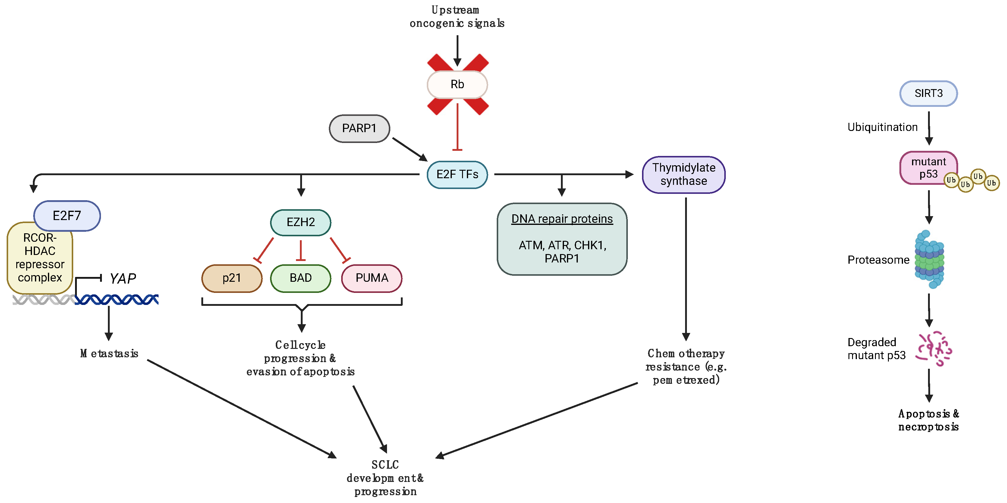
As a result of TP53 and RB1 inactivation in the vast majority of SCLCs, the upstream and downstream pathways associated with p53 and Rb are disrupted, thereby eliminating their tumor suppressive effects and promoting SCLC development and progression instead (Figure 2). Several preclinical studies have emerged that uncovered the molecular mechanisms of these oncogenic processes and, importantly, suggested novel potential targets that could be utilized for the development of drugs to tackle SCLC via different strategies.
A recent study by Hubaux et al. investigated the functional role of enhancer of zeste homolog 2 (EZH2), a histone-lysine N-methyltransferase that methylates histone proteins and induces transcriptional repression, in the biology of SCLC [34]. By targeting EZH2 with shRNA, the authors probed the effects of EZH2 silencing on the cell cycle and cell death. Their data show that EZH2 blocks apoptosis via downregulating the protein expression of the pro-apoptotic factors p53 upregulated modulator of apoptosis (PUMA) and BCL2 associated agonist of cell death (BAD), as well as promoting cell cycle progression through downregulating the expression of the cell cycle inhibitor p21 [34]. Byers et al. demonstrated that SCLC cells have an increased expression of the transcription factor E2F1 and its target genes, including EZH2, thymidylate synthase, mediators of apoptosis, and DNA repair proteins, such as poly [ADP-ribose] polymerase 1 (PARP1), checkpoint kinase 1 (CHK1), ataxia-telangiectasia mutated (ATM), and ataxia telangiectasia and Rad3-related protein (ATR) [35]. In the setting of NSCLC, thymidylate synthase renders cancer cells resistant to the chemotherapeutic drug pemetrexed, while in patients with SCLC, it may, in part, be responsible for the lower efficacy of the drug combination pemetrexed and carboplatin in comparison with the chemotherapeutic regimen of etoposide plus carboplatin [36,37,38]. In another study, Tang et al. explored the functional relationship between mutant p53 and the NAD-dependent deacetylase sirtuin-3 (SIRT3) in human SCLC cells. They found that SIRT3 triggers the ubiquitination and proteasomal degradation of mutant p53, resulting in the promotion of apoptosis and necroptosis of SCLC cells [39]. Recently, Wu et al. discovered that inactivation of RB1 in SCLC leads to the repression of yes-associated protein (YAP) transcription, which, in turn, enhances the metastatic potential of SCLC cells [40]. Mechanistically, loss of RB1 allows the transcription factor E2F7 to recruit the repressive REST Corepressor 1 (RCOR1/CoREST1)-lysine-specific demethylase 1A (LSD1, KDM1A)-histone deacetylases 1 and 2 (HDAC1/2) complex to the gene promoter of YAP and to downregulate its expression. They also found that entinostat, a benzamide histone deacetylase inhibitor, prolonged the survival of a SCLC xenograft model.

4. Targeted Therapies in p53 and RB Deficient SCLC Cells

The purpose of unraveling the molecular pathways involved in p53 and RB aberrations in SCLC is to establish effective targeted therapies that will prolong the progression-free survival (PFS) and overall survival (OS) of these patients. Neither TP53 nor RB1 have been therapeutically targetable to date. Thus, efforts are preclinically and clinically oriented towards finding therapies targeting the molecular pathways dysregulated in p53- and RB-deficient SCLC cells (Table 1 and Table 2).

4.1. AURK A/B Inhibitors

Overexpression of Aurora kinases (AURKs) is a prevalent protumorigenic pathway in numerous cancer types, including SCLC [41]. In particular, in p53-deficient cells, the protumorigenic effects of the AURKs are even augmented [42]. About 10 years ago, it was found that blocking AURK A or B halts the proliferation and growth of both in vitro and in vivo SCLC models [43]. Alisertib is a selective, small molecule, and orally administered AURK A inhibitor [44]. Its safety and antitumor activity were explored in a prior phase II clinical trial (NCT01045421) of patients with SCLC, breast cancer, NSCLC, head and neck squamous cell carcinoma, and gastroesophageal adenocarcinoma. The encouraging findings provided a rationale for the development of the phase II trial PUMA-ALI-4201 (NCT06095505), which will explore the efficacy of alisertib monotherapy towards extensive stage SCLC patients who have progressed after first-line platinum-based chemotherapy and immunotherapy. Chiauranib is an AURK B inhibitor currently being evaluated in multiple ongoing clinical trials in SCLC patients. A phase III clinical trial (NCT04830813) will explore chiauranib monotherapy in patients who have received at least two different systemic chemotherapy regimens (contained platinum-based regimen) and have progressed or relapsed extensive stage (ES)-SCLC.
Multiple attempts have been made to combine AURK inhibitors with chemotherapy as a treatment option for SCLC. The most recent data are from a randomized phase II trial combining alisertib with paclitaxel as second line therapy in patients with cMYC-positive SCLC. A total of 178 patients with relapsed or refractory SCLC were enrolled, stratified by relapse type (sensitive versus (vs.) refractory/resistant) and the presence of brain metastases. They were randomized 1:1 to alisertib/paclitaxel or placebo plus paclitaxel (89 patients in each group). The promising activity of alisertib/paclitaxel in relapsed or refractory SCLC was confirmed by the 3.2 months median progression-free survival (mPFS) for alisertib/paclitaxel compared to 2.17 months for placebo/paclitaxel (hazard ratio (HR) = 0.77) [45]. Preliminary clinical trials revealed significant indications of AURK inhibitors’ effectiveness, especially when combined with taxanes [41], but these findings require validation through phase III randomized trials.
Recently, there have also been attempts to combine AURK inhibitors with immunotherapy in SCLC. Preclinically, AURK A inhibition blocks SCLC cells in mitosis with restored interferon signaling, promoting T-lymphocyte infiltration [46]. Li et al. demonstrated that AURK A inhibition combined with PD-L1 immunotherapy has long-lasting efficacy in SCLC mouse models [46].

4.2. CDK7 Inhibitors

CDK7, a crucial controller of cell-cycle progression, is the best known cell-cycle regulator in SCLC. CDK7 serves as the catalytic core of the CDK-activating kinase (CAK) complex, becoming activated through binding with Cyclin H and Mat1. The trimeric CAK complex activates various core cell-cycle CDKs through phosphorylation [47]. In p53 and RB deficient SCLC murine models, a selective CDK7 inhibitor, YKL-5-12, was tested in association with anti-topoisomerase I, topotecan, and immune checkpoint inhibitors (ICIs). CDK7 inhibition impairs cell cycle and DNA replication and induces genome instability in SCLC cells, also enhancing immune response signaling. In this context, combining YKL-5-124 with anti-PD-1 showed a significant preclinical survival benefit [48]. There is an active phase I study (NCT04247126) on SY 5609, an oral and selective CDK7 inhibitor, in adult patients with advanced solid tumors for which standard curative or palliative measures do not exist or are no longer effective.

4.3. DDR Inhibitors

Nearly all cases of SCLC exhibit either homozygous loss or inactivation of RB1, a key regulator of the G1-S cell cycle checkpoint, and TP53, which is essential for multiple DNA Damage Response (DDR) pathways. As a result, SCLC cells exhibit high expression levels of DDR proteins [19,49]. On this basis, numerous inhibitors of DNA repair have recently been created and subjected to assessment in both preclinical models and clinical trials as potential candidates for treating SCLC.

4.3.1. CHK1 Inhibitors

SCLC cell lines possess an elevated level of both CHK1 gene and protein expression [35]. Preclinical efficacy was evoked by targeting the overexpressed cell-cycle checkpoint kinase CHK1 in SCLC cell lines [50]. Prexasertib, a selective CHK1 inhibitor, was explored as a single agent in a phase II trial (NCT02735980) involving patients with previously treated ES-SCLC, but it did not show favorable activity [51]. The inhibitor was given every 14 days to patients who had experienced progression after no more than two prior lines of therapy and with platinum-sensitive (Cohort 1) or platinum-resistant/platinum-refractory (Cohort 2) disease. In Cohort 1 (n = 58), the overall response rate (ORR) was 5.2%, while in Cohort 2 (n = 60), ORR was 0%. Prexasertib did not exhibit sufficient activity to be considered for further development as a monotherapy in ES-SCLC [51].

4.3.2. PARP Inhibitors

PARP1 is highly expressed in SCLC and was initially identified as a potential therapeutic target in SCLC through preclinical models explored by Byers et al. [35]. Several attempts have been made since to exploit the antitumor effects of PARP inhibitors (PARPi) as a monotherapy or in combination with other treatments. Monotherapy with the PARPi olaparib was evaluated as a maintenance treatment for patients with chemoresponsive SCLC in a phase II trial (ISRCTN 73164486, EudraCT 2010-021165-76). However, there was no notable distinction in either PFS or OS between olaparib and placebo [52]. The combination of PARPi with chemotherapy in ES-SCLC was explored by Owonikoko et al. in a phase I/II randomized trial ECOG-ACRIN 2511 (NCT01642251). The first arm received cisplatin-etoposide (CE) along with the PARPi veliparib (CE + V), and the second arm received CE along with placebo (CE + P). The ORR was 71.9% vs. 65.6% for CE + V and CE + P, respectively. The incorporation of veliparib into frontline chemotherapy showed effectiveness in patients with ES-SCLC, and the study achieved its pre-specified endpoint [53]. Another phase II randomized trial (NCT02289690) investigated veliparib plus carboplatin and etoposide in patients with treatment-naïve ES-SCLC. The randomization was 1:1:1: veliparib plus chemotherapy with carboplatin plus etoposide followed by veliparib maintenance, veliparib plus carboplatin plus etoposide followed by placebo, or placebo plus carboplatin plus etoposide followed by placebo (control arm). The combination in the first arm demonstrated an enhancement in PFS compared to the control arm (HR 0.67; 80% confidence interval (CI) 0.50–0.88; p = 0.059); however, the difference was not clinically significant (median PFS 5.8 versus 5.6 months), with a trend observed in SLFN11 positive patients (HR 0.6; 80% CI: 0.36–0.97). Additionally, there was no notable advantage of OS [54]. Another PARPi, niraparib, was evaluated as a first-line maintenance therapy in Chinese patients with platinum-responsive ES-SCLC in a phase III randomized trial ZL-2306-005. Although the study did not meet its primary endpoint, niraparib as a maintenance therapy was shown to modestly improve PFS in patients with platinum-responsive ES-SCLC (HR 0.66; 95% CI: 0.46–0.95; p = 0.0242), even though not being clinically significant (mPFS 1.54 vs. 1.36 months) [55].
PARP inhibition has also been evaluated in combination with the alkylating agent temozolamide (TMZ). A phase II, randomized, double-blind study (NCT01638546) evaluated whether addition of the PARPi veliparib to TMZ improved 4-month PFS. There was no significant difference in 4-month PFS between TMZ/veliparib (36%) and TMZ/placebo (27%; p = 0.19); median OS was also not improved significantly with TMZ/veliparib (8.2 months; 95% CI, 6.4 to 12.2 months; vs. 7.0 months; 95% CI, 5.3 to 9.5 months; p = 0.50). Interestingly, ORR was significantly higher in patients receiving TMZ/veliparib compared with TMZ/placebo (39% vs. 14%; p = 0.016) [56]. In another phase I/II trial (NCT02446704), TMZ was also combined with olaparib in patients with relapsed SCLC. After a median follow-up of 7.1 months, across all dose levels, the mPFS was 4.2 months (95% CI, 2.8–5.7) and the median OS was 8.5 months (95% CI, 5.1–11.3) [57]. An ongoing phase II study is investigating continuous talazoparib in combination with intermittent low-dose TMZ (NCT03672773) in relapsed/refractory SCLC.
Immunotherapy has also been studied in combination with PARP inhibition. Preclinical findings indicated that combining immune checkpoint blockade with PARP inhibition could be a promising therapeutic approach [58]. PARP inhibition has been shown to enhance the antitumor impact of PD-L1 blockade and increase cytotoxic T-cell infiltration in various immunocompetent SCLC models [59]. Moreover, growing evidence suggests that the induction of PD-L1 expression can occur through mechanisms involving DNA double-strand breaks and cytosolic DNA, including the stimulation of interferon gene-mediated innate immune response [60,61,62]. A single-arm, phase II trial (NCT02484404) evaluated olaparib and durvalumab in patients with relapsed SCLC. Partial or complete responses (ORR 10.5%) were observed in 2 out of 19 evaluable patients, including a patient with EGFR-transformed SCLC [63]. The MEDIOLA phase I/II multicenter open-label and single-arm trial also evaluated olaparib plus durvalumab in relapsed SCLC. Forty patients received olaparib monotherapy (300 mg twice daily) for four weeks, followed by a combination of olaparib (300 mg twice daily) and durvalumab 1500 mg intravenously (IV) administered every four weeks. The observed ORR was 10.5% (95% CI: 2.9–24.8). Despite the study not meeting the primary endpoint of a disease control rate at 12 weeks (28.9%), one patient attained a complete response, and three exhibited a partial response [64]. There are currently several ongoing clinical trials evaluating PARPi along with immunotherapy, as presented in Table 2.

4.3.3. ATM/ATR Inhibitors

ATM and ATR inhibitors have recently been combined with standard chemotherapy regimens for the treatment of SCLC. An interesting approach has been the combination of ATM/ATR inhibitors along with the topoisomerase I inhibitor topotecan, as it exacerbates replication stress in SCLC cells. A phase II trial (NCT02487095) combined M6620, an ATR inhibitor, and topotecan in SCLC patients who had relapsed after at least one prior chemotherapy. Overall, 9 out of 25 patients (36%, 95% CI: 18.0–57.5) achieved a confirmed partial response, meeting the primary endpoint for response. Most patients (17/25 patients; 68%) experienced tumor regressions [65]. Recently, it was preclinically observed that the RNA Pol-II inhibitor lurbinectedin, which induces DNA damage, exhibited strong synergy with the ATR inhibitor berzosertib in SCLC cell lines. Synergy was reduced with high p21 expression, as p21 causes G1 arrest [66]. It could be possible that SCLC cells with p53 aberrations, and thus reduced levels of the protein p21, would respond better to the combination of berzosertib and lurbinectedin. On this basis, an ongoing phase I/II clinical trial (NCT04802174) is examining the combination of lurbinectedin and berzosertib in relapsed SCLC. Several studies are also ongoing regarding SCLC patients with ATM deficiencies (Table 2).

5. Conclusions

SCLC cancer is a highly aggressive and lethal lung cancer that is characterized by genetic inactivation of the tumor suppressive genes TP53 and RB1. Preclinical research on SCLC cells and SCLC GEMMs, as well as genomic analyses of SCLC tumors derived from patients, reveal that these two genes are the most frequently mutated and support the notion that the inactivation of p53 and Rb is the catalytic step in the development of SCLC. Aberrations in TP53 and RB1 form the setting for further oncogenic events to occur and transform precancerous cells into malignant cells. Although we do not yet possess drugs that are able to revive mutated p53 and Rb, targeting the effects of their loss is a promising therapeutic strategy that is currently being exploited in clinical trials. A major consequence of the loss of TP53 and RB1 is the upregulation of the DNA damage repair pathway, and most drug development efforts are now focusing on the inhibition of multiple DNA repair proteins, such as PARP1 and CHK1. PARP inhibitors seem to be a promising therapeutic strategy, as data from preclinical and clinical studies indicate that these agents not only block tumor growth when used as a monotherapy but can also enhance the response to treatment in SCLC by making cancer cells more sensitive to currently available treatment modalities, including chemotherapy, radiotherapy, targeted therapy, and immunotherapy. When combined with other therapies, PARP inhibitors leave very few options, if any at all, for SCLC cells to repair the ongoing molecular damage elicited by other drugs. In order to maximize their activity and realize their full potential in the clinic, future research needs to identify predictive biomarkers for PARP inhibitors and, thus, optimize the selection of patients that will benefit the most from these novel therapies. If we are to fully grasp the functional significance of p53 and Rb aberrations, we need to further elucidate the molecular underpinnings of their intricate pathobiology during SCLC development, which will assist us in developing therapeutic strategies that may halt this deadly malignancy at its initial steps.

Author Contributions

Conceptualization, K.A.P. and A.G.P.; writing—original draft preparation, K.A.P., A.A.S. and V.A.G.; literature search and preparation of all references, A.A.S., V.A.G. and N.A.; supervision, A.G.P.; writing—review and editing, K.A.P. and A.G.P. All authors have read and agreed to the published version of the manuscript.

Funding

This research received no external funding.

Data Availability Statement

Data are contained within the article.

Conflicts of Interest

The authors declare no conflicts of interest.

References

  1. Hasan, A.; Khan, N.A.; Uddin, S.; Khan, A.Q.; Steinhoff, M. Deregulated transcription factors in the emerging cancer hallmarks. Semin. Cancer Biol. 2023, 18, 31–50. [Google Scholar] [CrossRef]
  2. Kim, D.W.; Kim, K.C.; Kim, K.B.; Dunn, C.T.; Park, K.S. Transcriptional deregulation underlying the pathogenesis of small cell lung cancer. Transl. Lung Cancer Res. 2018, 7, 4–20. [Google Scholar] [CrossRef] [PubMed]
  3. Drapkin, B.J.; Rudin, C.M. Advances in Small-Cell Lung Cancer (SCLC) Translational Research. Cold Spring Harb. Perspect. Med. 2021, 11, a038240. [Google Scholar] [CrossRef] [PubMed]
  4. Hernández Borrero, L.J.; El-Deiry, W.S. Tumor suppressor p53: Biology, signaling pathways, and therapeutic targeting. Biochim. Biophys. Acta Rev. Cancer 2021, 1876, 188556. [Google Scholar] [CrossRef] [PubMed]
  5. Olivier, M.; Hollstein, M.; Hainaut, P. TP53 mutations in human cancers: Origins, consequences, and clinical use. Cold Spring Harb. Perspect. Biol. 2010, 2, a001008. [Google Scholar] [CrossRef]
  6. Di Fiore, R.; D’Anneo, A.; Tesoriere, G.; Vento, R. RB1 in cancer: Different mechanisms of RB1 inactivation and alterations of pRb pathway in tumorigenesis. J. Cell Physiol. 2013, 228, 1676–1687. [Google Scholar] [CrossRef] [PubMed]
  7. Knudsen, E.S.; Knudsen, K.E. Tailoring to RB: Tumour suppressor status and therapeutic response. Nat. Rev. Cancer 2008, 8, 714–724. [Google Scholar] [CrossRef] [PubMed]
  8. Haupt, Y.; Maya, R.; Kazaz, A.; Oren, M. Mdm2 promotes the rapid degradation of p53. Nature 1997, 387, 296–299. [Google Scholar] [CrossRef] [PubMed]
  9. Hafner, A.; Bulyk, M.L.; Jambhekar, A.; Lahav, G. The multiple mechanisms that regulate p53 activity and cell fate. Nat. Rev. Mol. Cell Biol. 2019, 20, 199–210. [Google Scholar] [CrossRef]
  10. Kastenhuber, E.R.; Lowe, S.W. Putting p53 in context. Cell 2017, 170, 1062–1078. [Google Scholar] [CrossRef]
  11. Bykov, V.J.N.; Eriksson, S.E.; Bianchi, J.; Wiman, K.G. Targeting mutant p53 for efficient cancer therapy. Nat. Rev. Cancer 2018, 18, 89–102. [Google Scholar] [CrossRef]
  12. Sullivan, K.D.; Galbraith, M.D.; Andrysik, Z.; Espinosa, J.M. Mechanisms of transcriptional regulation by p53. Cell Death Differ. 2018, 25, 133–143. [Google Scholar] [CrossRef]
  13. Dick, F.A.; Rubin, S.M. Molecular mechanisms underlying RB protein function. Nat. Rev. Mol. Cell Biol. 2013, 14, 297–306. [Google Scholar] [CrossRef]
  14. Van Den Heuvel, S.; Dyson, N.J. Conserved functions of the pRB and E2F families. Nat. Rev. Mol. Cell Biol. 2008, 9, 713–724. [Google Scholar] [CrossRef]
  15. Burke, J.R.; Deshong, A.J.; Pelton, J.G.; Rubin, S.M. Phosphorylation-induced conformational changes in the Retinoblastoma Protein Inhibit E2F Transactivation Domain Binding. J. Biol. Chem. 2010, 285, 16286–16293. [Google Scholar] [CrossRef]
  16. Rubin, S.M.; Sage, J.; Skotheim, J.M. Integrating Old and New Paradigms of G1/S Control. Mol. Cell 2020, 80, 183–192. [Google Scholar] [CrossRef] [PubMed]
  17. Kolupaeva, V.; Janssens, V. PP1 and PP2A phosphatases–cooperating partners in modulating retinoblastoma protein activation. FEBS J. 2013, 280, 627–643. [Google Scholar] [CrossRef] [PubMed]
  18. Sherr, C.J.; McCormick, F. The RB and p53 pathways in cancer. Cancer Cell 2002, 2, 103–112. [Google Scholar] [CrossRef] [PubMed]
  19. George, J.; Lim, J.S.; Jang, S.J.; Cun, Y.; Ozretić, L.; Kong, G.; Leenders, F.; Lu, X.; Fernández-Cuesta, L.; Bosco, G.; et al. Comprehensive genomic profiles of small cell lung cancer. Nature 2015, 524, 47–53. [Google Scholar] [CrossRef]
  20. Meuwissen, R.; Linn, S.C.; Linnoila, R.I.; Zevenhoven, J.; Mooi, W.J.; Berns, A. Induction of small cell lung cancer by somatic inactivation of both Trp53 and Rb1 in a conditional mouse model. Cancer Cell 2003, 4, 181–189. [Google Scholar] [CrossRef]
  21. McFadden, D.G.; Papagiannakopoulos, T.; Taylor-Weiner, A.; Stewart, C.; Carter, S.L.; Cibulskis, K.; Bhutkar, A.; McKenna, A.; Dooley, A.; Vernon, A.; et al. Genetic and clonal dissection of murine small cell lung carcinoma progression by genome sequencing. Cell 2014, 156, 1298–1311. [Google Scholar] [CrossRef]
  22. Schaffer, B.E.; Park, K.S.; Yiu, G.; Conklin, J.F.; Lin, C.; Burkhart, D.L.; Karnezis, A.N.; Sweet-Cordero, E.A.; Sage, J. Loss of p130 accelerates tumor development in a mouse model for human small-cell lung carcinoma. Cancer Res. 2010, 70, 3877–3883. [Google Scholar] [CrossRef]
  23. Gazdar, A.F.; Savage, T.K.; Johnson, J.E.; Berns, A.; Sage, J.; Linnoila, R.I.; MacPherson, D.; McFadden, D.G.; Farago, A.; Jacks, T.; et al. The comparative pathology of genetically engineered mouse models for neuroendocrine carcinomas of the lung. J. Thorac. Oncol. 2015, 10, 553–564. [Google Scholar] [CrossRef]
  24. Kwon, M.C.; Berns, A. Mouse models for lung cancer. Mol. Oncol. 2013, 7, 165–177. [Google Scholar] [CrossRef]
  25. Lázaro, S.; Pérez-Crespo, M.; Enguita, A.B.; Hernández, P.; Martínez-Palacio, J.; Oteo, M.; Sage, J.; Paramio, J.M.; Santos, M. Ablating all three retinoblastoma family members in mouse lung leads to neuroendocrine tumor formation. Oncotarget 2017, 8, 4373–4386. [Google Scholar] [CrossRef]
  26. Semenova, E.A.; Kwon, M.C.; Monkhorst, K.; Song, J.Y.; Bhaskaran, R.; Krijgsman, O.; Kuilman, T.; Peters, D.; Buikhuisen, W.A.; Smit, E.F.; et al. Transcription Factor NFIB Is a Driver of Small Cell Lung Cancer Progression in Mice and Marks Metastatic Disease in Patients. Cell Rep. 2016, 16, 631–643. [Google Scholar] [CrossRef]
  27. Mollaoglu, G.; Guthrie, M.R.; Böhm, S.; Brägelmann, J.; Can, I.; Ballieu, P.M.; Marx, A.; George, J.; Heinen, C.; Chalishazar, M.D.; et al. MYC Drives Progression of Small Cell Lung Cancer to a Variant Neuroendocrine Subtype with Vulnerability to Aurora Kinase Inhibition. Cancer Cell 2017, 31, 270–285. [Google Scholar] [CrossRef] [PubMed]
  28. Cui, M.; Augert, A.; Rongione, M.; Conkrite, K.; Parazzoli, S.; Nikitin, A.Y.; Ingolia, N.; MacPherson, D. PTEN is a potent suppressor of small cell lung cancer. Mol. Cancer Res. 2014, 12, 654–659. [Google Scholar] [CrossRef] [PubMed]
  29. Kim, D.W.; Wu, N.; Kim, Y.C.; Cheng, P.F.; Basom, R.; Kim, D.; Dunn, C.T.; Lee, A.Y.; Kim, K.; Lee, C.S.; et al. Genetic requirement for Mycl and efficacy of RNA Pol I inhibition in mouse models of small cell lung cancer. Genes Dev. 2016, 30, 1289–1299. [Google Scholar] [CrossRef]
  30. Wikenheiser-Brokamp, K.A. Rb family proteins differentially regulate distinct cell lineages during epithelial development. Development 2004, 131, 4299–4310. [Google Scholar] [CrossRef] [PubMed]
  31. Sivakumar, S.; Moore, J.A.; Montesion, M.; Sharaf, R.; Lin, D.I.; Colón, C.I.; Fleishmann, Z.; Ebot, E.M.; Newberg, J.Y.; Mills, J.M.; et al. Integrative Analysis of a Large Real-World Cohort of Small Cell Lung Cancer Identifies Distinct Genetic Subtypes and Insights into Histologic Transformation. Cancer Discov. 2023, 13, 1572–1591. [Google Scholar] [CrossRef]
  32. McColl, K.; Wildey, G.; Sakre, N.; Lipka, M.B.; Behtaj, M.; Kresak, A.; Chen, Y.; Yang, M.; Velcheti, V.; Fu, P.; et al. Reciprocal expression of INSM1 and YAP1 defines subgroups in small cell lung cancer. Oncotarget 2017, 8, 73745–73756. [Google Scholar] [CrossRef]
  33. Dowlati, A.; Abbas, A.; Chan, T.; Henick, B.; Wang, X.; Doshi, P.; Fu, P.; Patel, J.; Kuo, F.; Chang, H.; et al. Immune Checkpoint Blockade Outcome in Small-Cell Lung Cancer and Its Relationship with Retinoblastoma Mutation Status and Function. JCO Precis. Oncol. 2022, 6, e2200257. [Google Scholar] [CrossRef]
  34. Hubaux, R.; Thu, K.L.; Coe, B.P.; MacAulay, C.; Lam, S.; Lam, W.L. EZH2 promotes E2F-driven SCLC tumorigenesis through modulation of apoptosis and cell-cycle regulation. J. Thorac. Oncol. 2013, 8, 1102–1106. [Google Scholar] [CrossRef]
  35. Byers, L.A.; Wang, J.; Nilsson, M.B.; Fujimoto, J.; Saintigny, P.; Yordy, J.; Giri, U.; Peyton, M.; Fan, Y.H.; Diao, L.; et al. Proteomic profiling identifies dysregulated pathways in small cell lung cancer and novel therapeutic targets including PARP1. Cancer Discov. 2012, 2, 798–811. [Google Scholar] [CrossRef]
  36. Shintani, Y.; Ohta, M.; Hirabayashi, H.; Tanaka, H.; Iuchi, K.; Nakagawa, K.; Maeda, H.; Kido, T.; Miyoshi, S.; Matsuda, H. Thymidylate synthase and dihydropyrimidine dehydrogenase mRNA levels in tumor tissues and the efficacy of 5- fluorouracil in patients with non-small-cell lung cancer. Lung Cancer 2004, 45, 189–196. [Google Scholar] [CrossRef]
  37. Huang, C.L.; Liu, D.; Nakano, J.; Yokomise, H.; Ueno, M.; Kadota, K.; Wada, H. E2F1 overexpression correlates with thymidylate synthase and survivin gene expressions and tumor proliferation in non small-cell lung cancer. Clin. Cancer Res. 2007, 13, 6938–6946. [Google Scholar] [CrossRef] [PubMed]
  38. Socinski, M.A.; Smit, E.F.; Lorigan, P.; Konduri, K.; Reck, M.; Szczesna, A.; Blakely, J.; Serwatowski, P.; Karaseva, N.A.; Ciuleanu, T.; et al. Phase III study of pemetrexed plus carboplatin compared with etoposide plus carboplatin in chemotherapy-naive patients with extensive-stage small-cell lung cancer. J. Clin. Oncol. 2009, 27, 4787–4792. [Google Scholar] [CrossRef] [PubMed]
  39. Tang, X.; Li, Y.; Liu, L.; Guo, R.; Zhang, P.; Zhang, Y.; Zhang, Y.; Zhao, J.; Su, J.; Sun, L.; et al. Sirtuin 3 induces apoptosis and necroptosis by regulating mutant p53 expression in small-cell lung cancer. Oncol. Rep. 2020, 43, 591–600. [Google Scholar] [CrossRef] [PubMed]
  40. Wu, Z.; Su, J.; Li, F.L.; Chen, T.; Mayner, J.; Engler, A.; Ma, S.; Li, Q.; Guan, K.L. YAP silencing by RB1 mutation is essential for small-cell lung cancer metastasis. Nat. Commun. 2023, 14, 5916. [Google Scholar] [CrossRef]
  41. Stefani, A.; Piro, G.; Schietroma, F.; Strusi, A.; Vita, E.; Fiorani, S.; Barone, D.; Monaca, F.; Sparagna, I.; Valente, G.; et al. Unweaving the Mitotic Spindle: A Focus on Aurora Kinase Inhibitors in Lung Cancer. Front. Oncol. 2022, 12, 1026020. [Google Scholar] [CrossRef]
  42. Meraldi, P. Aurora-A Overexpression Reveals Tetraploidization as a Major Route to Centrosome Amplification in P53−/− Cells. EMBO J. 2002, 21, 483–492. [Google Scholar] [CrossRef]
  43. Lu, Y.; Liu, Y.; Jiang, J.; Xi, Z.; Zhong, N.; Shi, S.; Wang, J.; Wei, X. Knocking down the Expression of Aurora-A Gene Inhibits Cell Proliferation and Induces G2/M Phase Arrest in Human Small Cell Lung Cancer Cells. Oncol. Rep. 2014, 32, 243–249. [Google Scholar] [CrossRef]
  44. Liewer, S.; Huddleston, A. Alisertib: A Review of Pharmacokinetics, Efficacy and Toxicity in Patients with Hematologic Malignancies and Solid Tumors. Expert Opin. Investig. Drugs 2018, 27, 105–112. [Google Scholar] [CrossRef]
  45. Owonikoko, T.K.; Niu, H.; Nackaerts, K.; Csoszi, T.; Ostoros, G.; Mark, Z.; Baik, C.; Joy, A.A.; Chouaid, C.; Jaime, J.C.; et al. Randomized Phase II Study of Paclitaxel plus Alisertib versus Paclitaxel plus Placebo as Second-Line Therapy for SCLC: Primary and Correlative Biomarker Analyses. J. Thorac. Oncol. 2020, 15, 274–287. [Google Scholar] [CrossRef] [PubMed]
  46. Li, Y.; Mahadevan, N.R.; Duplaquet, L.; Hong, D.; Durmaz, Y.T.; Jones, K.L.; Cho, H.; Morrow, M.; Protti, A.; Poitras, M.J.; et al. Aurora A Kinase Inhibition Induces Accumulation of SCLC Tumor Cells in Mitosis with Restored Interferon Signaling to Increase Response to PD-L1. Cell Rep. Med. 2023, 4, 101282. [Google Scholar] [CrossRef]
  47. Schachter, M.M.; Fisher, R.P. The CDK-Activating Kinase Cdk7: Taking Yes for an Answer. Cell Cycle 2013, 12, 3239–3240. [Google Scholar] [CrossRef] [PubMed]
  48. Zhang, H.; Christensen, C.L.; Dries, R.; Oser, M.G.; Deng, J.; Diskin, B.; Li, F.; Pan, Y.; Zhang, X.; Yin, Y.; et al. CDK7 Inhibition Potentiates Genome Instability Triggering Anti-Tumor Immunity in Small Cell Lung Cancer. Cancer Cell 2020, 37, 37–54.e9. [Google Scholar] [CrossRef]
  49. Venugopala, K.N. Targeting the DNA Damage Response Machinery for Lung Cancer Treatment. Pharmaceuticals 2022, 15, 1475. [Google Scholar] [CrossRef] [PubMed]
  50. Sen, T.; Tong, P.; Stewart, C.A.; Cristea, S.; Valliani, A.; Shames, D.S.; Redwood, A.B.; Fan, Y.H.; Li, L.; Glisson, B.S.; et al. CHK1 Inhibition in Small-Cell Lung Cancer Produces Single-Agent Activity in Biomarker-Defined Disease Subsets and Combination Activity with Cisplatin or Olaparib. Cancer Res. 2017, 77, 3870–3884. [Google Scholar] [CrossRef] [PubMed]
  51. Byers, L.A.; Navarro, A.; Schaefer, E.; Johnson, M.; Özgüroğlu, M.; Han, J.-Y.; Bondarenko, I.; Cicin, I.; Dragnev, K.H.; Abel, A.; et al. A Phase II Trial of Prexasertib (LY2606368) in Patients with Extensive-Stage Small-Cell Lung Cancer. Clin. Lung Cancer 2021, 22, 531–540. [Google Scholar] [CrossRef]
  52. Woll, P.; Gaunt, P.; Danson, S.; Steele, N.; Ahmed, S.; Mulatero, C.; Shah, R.; Bhosle, J.; Hodgkinson, E.; Watkins, B.; et al. Olaparib as Maintenance Treatment in Patients with Chemosensitive Small Cell Lung Cancer (STOMP): A Randomised, Double-Blind, Placebo-Controlled Phase II Trial. Lung Cancer 2022, 171, 26–33. [Google Scholar] [CrossRef]
  53. Owonikoko, T.K.; Dahlberg, S.E.; Sica, G.L.; Wagner, L.I.; Wade, J.L.; Srkalovic, G.; Lash, B.W.; Leach, J.W.; Leal, T.B.; Aggarwal, C.; et al. Randomized Phase II Trial of Cisplatin and Etoposide in Combination with Veliparib or Placebo for Extensive-Stage Small-Cell Lung Cancer: ECOG-ACRIN 2511 Study. J. Clin. Oncol. Off. J. Am. Soc. Clin. Oncol. 2019, 37, 222–229. [Google Scholar] [CrossRef]
  54. Byers, L.A.; Bentsion, D.; Gans, S.; Penkov, K.; Son, C.; Sibille, A.; Owonikoko, T.K.; Groen, H.J.M.; Gay, C.M.; Fujimoto, J.; et al. Veliparib in Combination with Carboplatin and Etoposide in Patients with Treatment-Naïve Extensive-Stage Small Cell Lung Cancer: A Phase 2 Randomized Study. Clin. Cancer Res. Off. J. Am. Assoc. Cancer Res. 2021, 27, 3884–3895. [Google Scholar] [CrossRef]
  55. Ai, X.; Pan, Y.; Shi, J.; Yang, N.; Liu, C.; Zhou, J.; Zhang, X.; Dong, X.; He, J.; Li, X.; et al. Efficacy and Safety of Niraparib as Maintenance Treatment in Patients with Extensive-Stage SCLC After First-Line Chemotherapy: A Randomized, Double-Blind, Phase 3 Study. J. Thorac. Oncol. 2021, 16, 1403–1414. [Google Scholar] [CrossRef]
  56. Pietanza, M.C.; Waqar, S.N.; Krug, L.M.; Dowlati, A.; Hann, C.L.; Chiappori, A.; Owonikoko, T.K.; Woo, K.M.; Cardnell, R.J.; Fujimoto, J.; et al. Randomized, Double-Blind, Phase II Study of Temozolomide in Combination with Either Veliparib or Placebo in Patients with Relapsed-Sensitive or Refractory Small-Cell Lung Cancer. J. Clin. Oncol. 2018, 36, 2386–2394. [Google Scholar] [CrossRef]
  57. Farago, A.F.; Yeap, B.Y.; Stanzione, M.; Hung, Y.P.; Heist, R.S.; Marcoux, J.P.; Zhong, J.; Rangachari, D.; Barbie, D.A.; Phat, S.; et al. Combination Olaparib and Temozolomide in Relapsed Small-Cell Lung Cancer. Cancer Discov. 2019, 9, 1372–1387. [Google Scholar] [CrossRef]
  58. Mouw, K.W.; Goldberg, M.S.; Konstantinopoulos, P.A.; D’Andrea, A.D. DNA Damage and Repair Biomarkers of Immunotherapy Response. Cancer Discov. 2017, 7, 675–693. [Google Scholar] [CrossRef] [PubMed]
  59. Sen, T.; Rodriguez, B.L.; Chen, L.; Corte, C.M.D.; Morikawa, N.; Fujimoto, J.; Cristea, S.; Nguyen, T.; Diao, L.; Li, L.; et al. Targeting DNA Damage Response Promotes Antitumor Immunity through STING-Mediated T-cell Activation in Small Cell Lung Cancer. Cancer Discov. 2019, 9, 646–661. [Google Scholar] [CrossRef] [PubMed]
  60. Kitai, Y.; Kawasaki, T.; Sueyoshi, T.; Kobiyama, K.; Ishii, K.J.; Zou, J.; Akira, S.; Matsuda, T.; Kawai, T. DNA-Containing Exosomes Derived from Cancer Cells Treated with Topotecan Activate a STING-Dependent Pathway and Reinforce Antitumor Immunity. J. Immunol. 2017, 198, 1649–1659. [Google Scholar] [CrossRef] [PubMed]
  61. Parkes, E.E.; Walker, S.M.; Taggart, L.E.; McCabe, N.; Knight, L.A.; Wilkinson, R.; McCloskey, K.D.; Buckley, N.E.; Savage, K.I.; Salto-Tellez, M.; et al. Activation of STING-Dependent Innate Immune Signaling By S-Phase-Specific DNA Damage in Breast Cancer. J. Natl. Cancer Inst. 2016, 109, djw199. [Google Scholar] [CrossRef]
  62. Sato, H.; Niimi, A.; Yasuhara, T.; Permata, T.B.M.; Hagiwara, Y.; Isono, M.; Nuryadi, E.; Sekine, R.; Oike, T.; Kakoti, S.; et al. DNA double-strand break repair pathway regulates PD-L1 expression in cancer cells. Nat. Commun. 2017, 8, 1751. [Google Scholar] [CrossRef]
  63. Thomas, A.; Vilimas, R.; Trindade, C.; Erwin-Cohen, R.; Roper, N.; Xi, L.; Krishnasamy, V.; Levy, E.; Mammen, A.; Nichols, S.; et al. Durvalumab in Combination with Olaparib in Patients with Relapsed SCLC: Results from a Phase II Study. J. Thorac. Oncol. 2019, 14, 1447–1457. [Google Scholar] [CrossRef]
  64. Krebs, M.G.; Delord, J.-P.; Jeffry Evans, T.R.; De Jonge, M.; Kim, S.-W.; Meurer, M.; Postel-Vinay, S.; Lee, J.-S.; Angell, H.K.; Rocher-Ros, V.; et al. Olaparib and Durvalumab in Patients with Relapsed Small Cell Lung Cancer (MEDIOLA): An Open-Label, Multicenter, Phase 1/2, Basket Study. Lung Cancer 2023, 180, 107216. [Google Scholar] [CrossRef] [PubMed]
  65. Thomas, A.; Takahashi, N.; Rajapakse, V.N.; Zhang, X.; Sun, Y.; Ceribelli, M.; Wilson, K.M.; Zhang, Y.; Beck, E.; Sciuto, L.; et al. Therapeutic Targeting of ATR Yields Durable Regressions in Small Cell Lung Cancers with High Replication Stress. Cancer Cell 2021, 39, 566–579.e7. [Google Scholar] [CrossRef] [PubMed]
  66. Schultz, C.W.; Zhang, Y.; Elmeskini, R.; Zimmermann, A.; Fu, H.; Murai, Y.; Wangsa, D.; Kumar, S.; Takahashi, N.; Atkinson, D.; et al. ATR Inhibition Augments the Efficacy of Lurbinectedin in Small-cell Lung Cancer. EMBO Mol. Med. 2023, 15, e17313. [Google Scholar] [CrossRef] [PubMed]
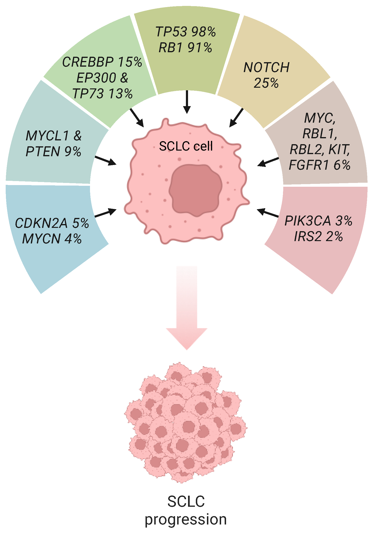
Figure 1. The genomic landscape of SCLC. Percentages refer to the frequency of gene mutations. CDKN2A, cyclin-dependent kinase 2A; CREBBP, CREB binding protein; EP300, E1A binding protein p300; FGFR1, fibroblast growth factor receptor 2; IRS2, insulin receptor substrate 2; MYC, v-myc avian myelocytomatosis viral oncogene homolog; MYCL1, v-myc avian myelocytomatosis viral oncogene homolog 1, lung carcinoma derived; MYCN, v-myc avian myelocytomatosis viral oncogene neuroblastoma derived homolog; PIK3CA, phosphatidylinositol-4,5-bisphosphate 3-kinase catalytic subunit alpha; PTEN, phosphatase and tensin homolog; RB1, retinoblastoma; RBL1, Rb transcriptional corepressor like 1; RBL2, Rb transcriptional corepressor like 2; TP73, tumor protein 73. This figure was created using tools provided by BioRender.com (accessed on 12 February 2024).
Ijms 25 02479 g001
Figure 2. Molecular mechanisms of aberrant p53 and Rb signaling in SCLC. Details are found within the text. The red X sign behind the Rb protein represents Rb inactivation. ATM, ataxia-telangiectasia mutated; ATR, ataxia telangiectasia and Rad3-related protein; BAD, BCL2-associated agonist of cell death; CHK1, checkpoint kinase 1; EZH2, enhancer of zeste homolog 2; HDAC, histone deacetylase; PARP1, poly [ADP-ribose] polymerase 1; PUMA, p53 upregulated modulator of apoptosis; Rb, retinoblastoma; RCOR, REST Corepressor; SIRT3, sirtuin-3; SCLC, small cell lung cancer; Ub, ubiquitin; YAP, yes-associated protein. This figure was created using tools provided by BioRender.com (accessed on 12 February 2024).
Ijms 25 02479 g002
Table 1. Completed clinical trials in patients with deficient p53 and RB SCLC.
AgentNCT ID NumberPhaseTreatment
AURK inhibitorsNCT00858377IAMG 900 monotherapy
n/aIDanusertib monotherapy (24 h infusion)
NCT01045421I-IIAlisertib monotherapy
NCT01923337IAlisertib + Irinotecan
NCT01094288IAlisertib + Docetaxel
NCT01677559IAlisertib + Nab-paclitaxel
2006-003772-35IIDanusertib monotherapy
NCT02038647IIAlisertib + Paclitaxel vs. Placebo + Paclitaxel
CHK1 inhibitorsNCT02735980IIPrexasertib monotherapy
PARP inhibitorsNCT01642251I/IIcisplatin-etoposide (CE) + Veliparib
vs.
Placebo + Veliparib
NCT02446704I/IIOlaparib + TMZ
NCT02734004I/IIOlaparib + Durvalumab
ISRCTN 73164486, EudraCT 2010-021165-76IIOlaparib monotherapy
NCT02289690II1:1:1:
veliparib + carboplatin + etoposide followed by veliparib maintenance,
veliparib + carboplatin + etoposide followed by placebo,
placebo+ carboplatin + etoposide followed by placebo
NCT02484404IIOlaparib + Durvalumab
NCT03958045IIRucaparib + Nivolumab
NCT01638546IITMZ/veliparib
vs.
TMZ/placebo
ZL-2306-005IIINiraparib monotherapy
ATR inhibitorsNCT02487095IIBerzosertib + Topotecan
NCT04768296IIBerzosertib + Topotecan
ATM, ataxia-telangiectasia mutated; ATR, ataxia telangiectasia and Rad3-related protein; AURK, Aurora kinase; CHK1, checkpoint kinase 1; PARP1, poly [ADP-ribose] polymerase 1; TMZ, temozolomide.
Table 2. Ongoing clinical trials in patients with deficient p53 and RB SCLC.
AgentNCT ID NumberPhaseTreatment Regimen
AURK inhibitorsNCT05271292Ib/IIChiauranib
NCT05505825Ib/IIAK104 + Chiauranib
NCT03216343IChiauranib
NCT04830813IIIChiauranib Capsule
NCT06095505IIAlisertib
CDK7 inhibitorsNCT04247126ISY 5609 + gemcitabine
PARP inhibitorsNCT05002868IRP12146
NCT03227016IVeliparib + Topotecan
NCT03532880IOlaparib + Low-dose radiotherapy
NCT03923270IRadiotherapy + durvalumab vs.
durvalumab combinations
(tremelimumab or olaparib)
ES-SCLC-2nd-IIT-SHR3162-APAn/aCamrelizumab + Fluzoparib
NCT04644068I/IIAZD5305 monotherapy or in combination with anti-cancer agents
NCT04826341I/IISacituzumab Govitecan + Berzosertib
NCT04209595I/IIPLX038 (PEGylated SN38) + Rucaparib
NCT04728230I/IICarboplatin + etoposide + durvalumab + olaparib and/or radiation therapy
NCT03830918I/IINiraparib + Temozolomide + Atezolizumab
NCT05975944I/IIOlaparib + Selinexor
NCT02769962I/IIEP0057 + Olaparib
NCT04434482I/IIIMP4297 + Temozolomide
NCT04400188I/IIFluzoparib + Temozolomide ± SHR-1316
NCT04659785I/IIFluzoparib + Apatinib
NCT04538378IIOlaparib + Durvalumab
NCT05411679IIEP0057 + Olaparib
NCT05718323IINiraparib + Anti-PD-L1 therapy
NCT04334941IITalazoparib + Atezolizumab
NCT05162196IIRadiotherapy + Niraparib + Toripalimab
NCT04701307IINiraparib + Dostarlimab
NCT03672773IITalazoparib + low-dose Temozolomide
NCT02498613IICediranib + Olaparib
NCT05623319IIOlaparib + Pembrolizumab
NCT05245994IIOlaparib + Durvalumab
NCT04939662IIOlaparib + Bevacizumab
NCT04624204IIIPembrolizumab + Chemoradiation followed by pembrolizumab ± Olaparib vs. Pembrolizumab + Chemoradiation
NCT04790955ObservationalSBRT and low-dose radiotherapy + PARPi + temozolomide + PD-1/PD-L1 inhibitors
ATR inhibitorsNCT04491942IElimusertib + chemotherapy (cisplatin, or cisplatin and gemcitabine)
NCT02595931IBerzosertib + Irinotecan hydrochloride
NCT04802174I/IIBerzosertib + Lurbinectedin
NCT04826341I/IIBerzosertib + Sacituzumab govitecan
NCT02487095I/IIBerzosertib + Topotecan
NCT03896503IIBerzosertib + Topotecan
ATM inhibitorsNCT04939662IIOlaparib + Bevacizumab
ATM, ataxia-telangiectasia mutated; ATR, ataxia telangiectasia and Rad3-related protein; AURK, Aurora kinase; CDK7, cyclin-dependent kinase 7; ES-SCLC, extensive-stage small cell lung cancer; N/a, not applicable; PARPi, poly [ADP-ribose] polymerase 1 inhibitors; PD1, Programmed cell death protein 1; PDL1, programmed death-ligand 1; SBRT, stereotactic body radiation therapy.
Disclaimer/Publisher’s Note: The statements, opinions and data contained in all publications are solely those of the individual author(s) and contributor(s) and not of MDPI and/or the editor(s). MDPI and/or the editor(s) disclaim responsibility for any injury to people or property resulting from any ideas, methods, instructions or products referred to in the content.