Article

The Impacts of Periconceptional Alcohol on Neonatal Ovaries and Subsequent Adult Fertility in the Rat

1
School of Biomedical Sciences, The University of Queensland, St Lucia, QLD 4072, Australia
2
Mater Research, The University of Queensland, South Brisbane, QLD 4101, Australia
*
Author to whom correspondence should be addressed.
Int. J. Mol. Sci. 2024, 25(5), 2471; https://doi.org/10.3390/ijms25052471
Submission received: 19 January 2024 / Revised: 12 February 2024 / Accepted: 16 February 2024 / Published: 20 February 2024
(This article belongs to the Special Issue Ovarian Dysfunction, Infertility and Its Treatment)

Abstract

:
Maternal exposures during pregnancy can impact the establishment of the ovarian reserve in offspring, the lifetime supply of germ cells that determine a woman’s reproductive lifespan. However, despite alcohol consumption being common in women of reproductive age, the impact of prenatal alcohol on ovarian development is rarely investigated. This study used an established rat model of periconceptional ethanol exposure (PCEtOH; 12.5% v/v ethanol) for 4 days prior to 4 days post-conception. Ovaries were collected from neonates (day 3 and day 10), and genes with protein products involved in regulating the ovarian reserve analyzed by qPCR. Adult offspring had estrous cycles monitored and breeding performance assessed. PCEtOH resulted in subtle changes in expression of genes regulating apoptosis at postnatal day (PN) 3, whilst those involved in regulating growth and recruitment of primordial follicles were dysregulated at PN10 in neonatal ovaries. Despite these gene expression changes, there were no significant impacts on breeding performance in adulthood, nor on F2-generation growth or survival. This contributes additional evidence to suggest that a moderate level of alcohol consumption exclusively around conception, when a woman is often unaware of her pregnancy, does not substantially impact the fertility of her female offspring.

1. Introduction

As women are increasingly delaying childbirth, and fertility is inversely proportional to age [1], it is important to determine environmental factors that may impact the establishment of the ovarian reserve. Ovarian reserve is defined as the number of oocytes stored within primordial follicles in the ovary, and determines a woman’s reproductive lifespan [2,3]. Unlike in males, these germ cells are finite and established prenatally in humans (~weeks 17–19 gestation) and neonatally in rodents (~postnatal day (PN) 3–4) [4,5]. They are gradually depleted throughout life, with a disproportionately high number undergoing apoptosis, while others are recruited into the pool of growing follicles [6]. For this pool of oocytes to be maintained and yet available requires a balance of factors to maintain their quiescence, as well as factors to initiate recruitment to each ‘wave’ of growing follicles (see Ford, et al. [7] for review).
There is evidence that the initial number of non-growing follicles established is proportional to the age at menopause, such that a high ovarian reserve predicts a later age at menopause [8]. There is wide individual variability in the number of primordial follicles established during this early developmental period, and many environmental factors may influence this number via the intrauterine environment [2,9]. However, the impact of prenatal alcohol exposure has been relatively overlooked [2,10]. This is a common maternal exposure in most Western countries, with many women consuming low to moderate amounts of alcohol around the periconceptional period and prior to pregnancy recognition [11,12,13]. Given this is difficult to study in a clinical population, animal models are required.
The current rat study examines the impact of a moderate dose of alcohol exclusively around the time of conception on subsequent ovarian development and fertility. We have previously published multiple negative impacts of this exposure on placental and fetal growth and development [14,15,16]. We have also shown more long-term, often sex-specific, effects in offspring derived from this model, including a diabetic-like phenotype [17], an increased tendency towards obesity [14,18], and impaired renal function [19]. This is despite the fact that only the precursor cells for the placenta and fetus, contained within the early embryo at the blastocyst stage, coincide with the actual timing of exposure. In the present study, we aimed to examine the impact of this periconceptional ethanol (PCEtOH) model on the expression of genes that produce factors regulating ovarian reserve in neonatal offspring, specifically those involved in the apoptotic pathway and primordial follicle activation. We also aimed to examine potential long-term impacts on estrous cyclicity and breeding performance in adulthood.

2. Results

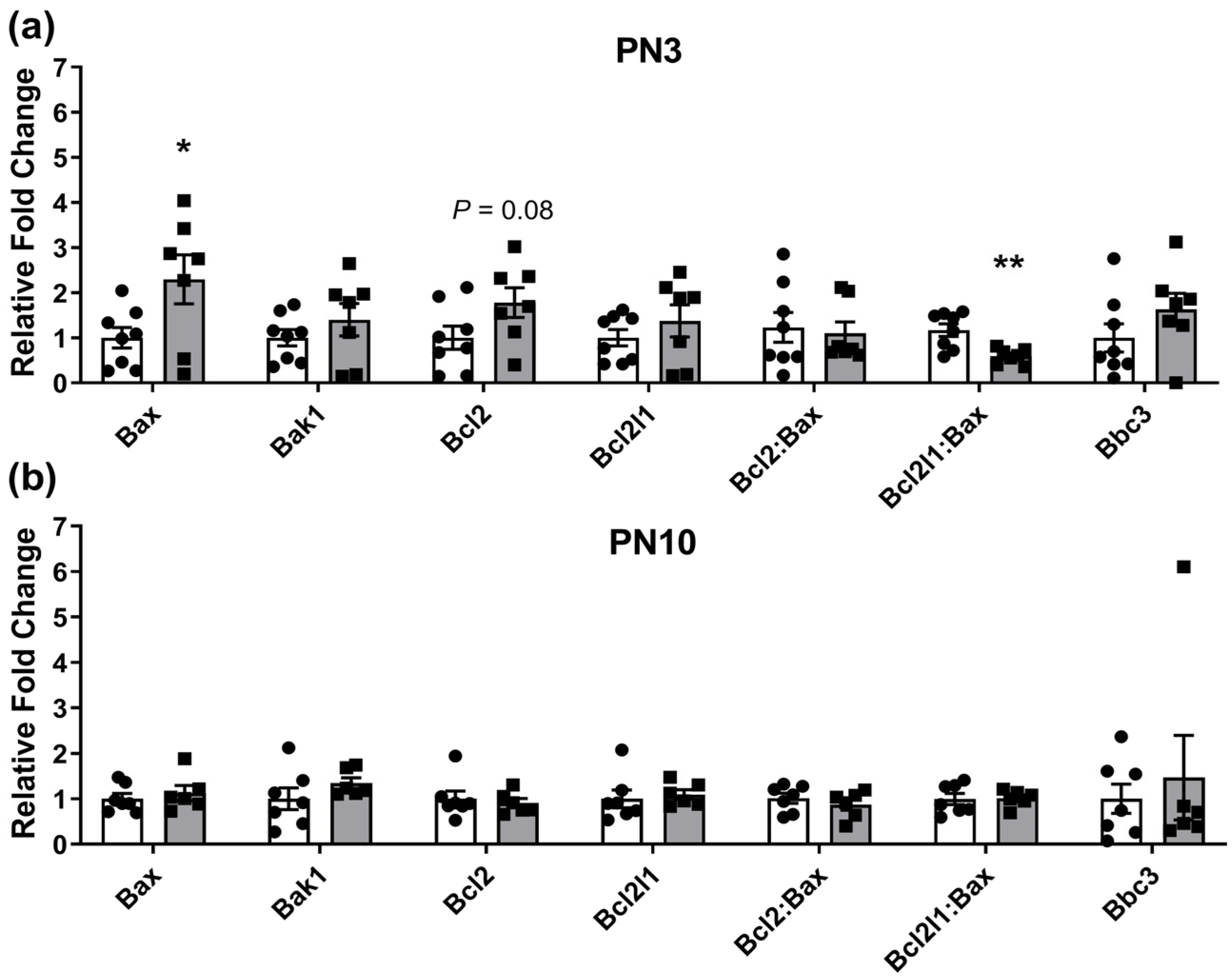
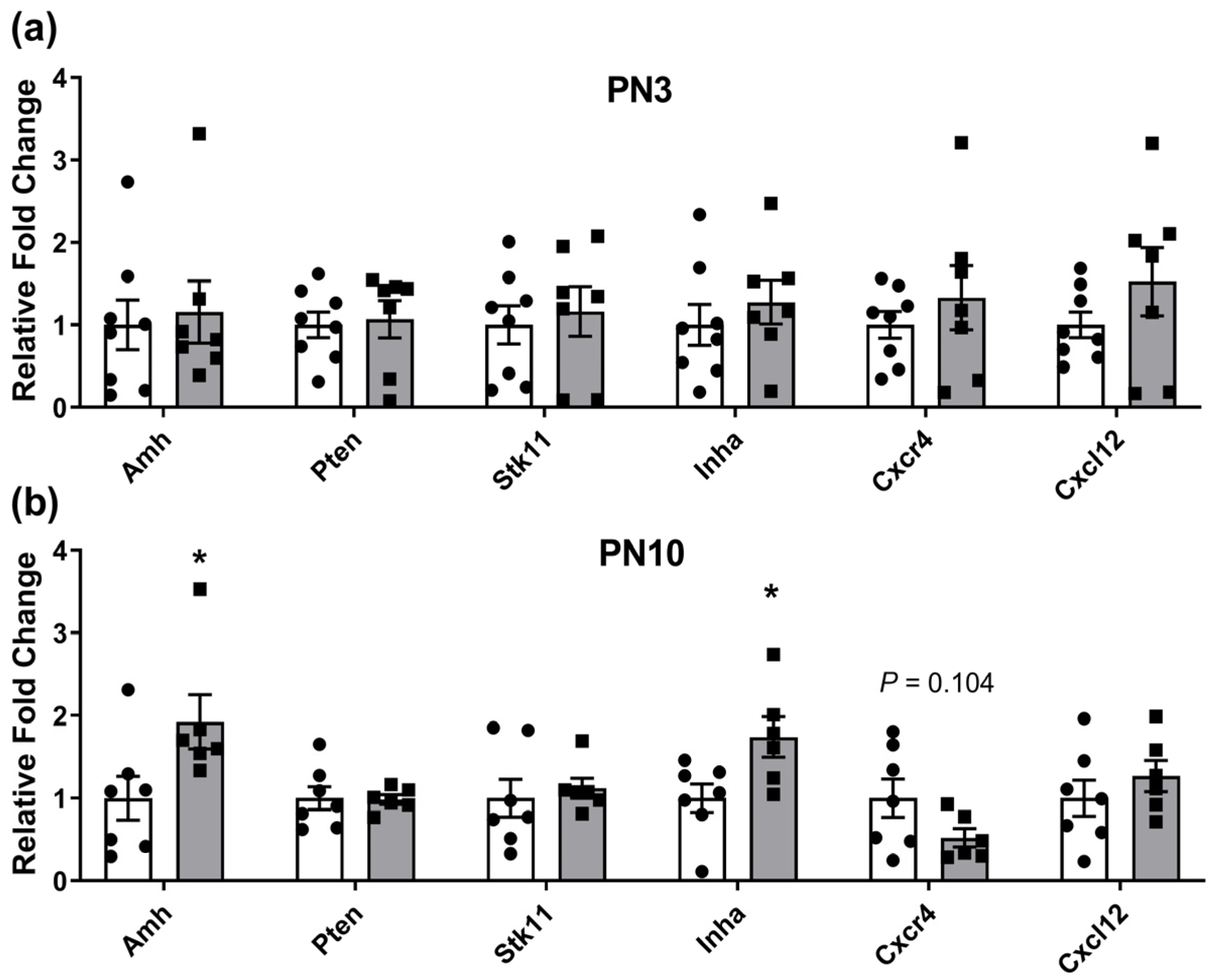
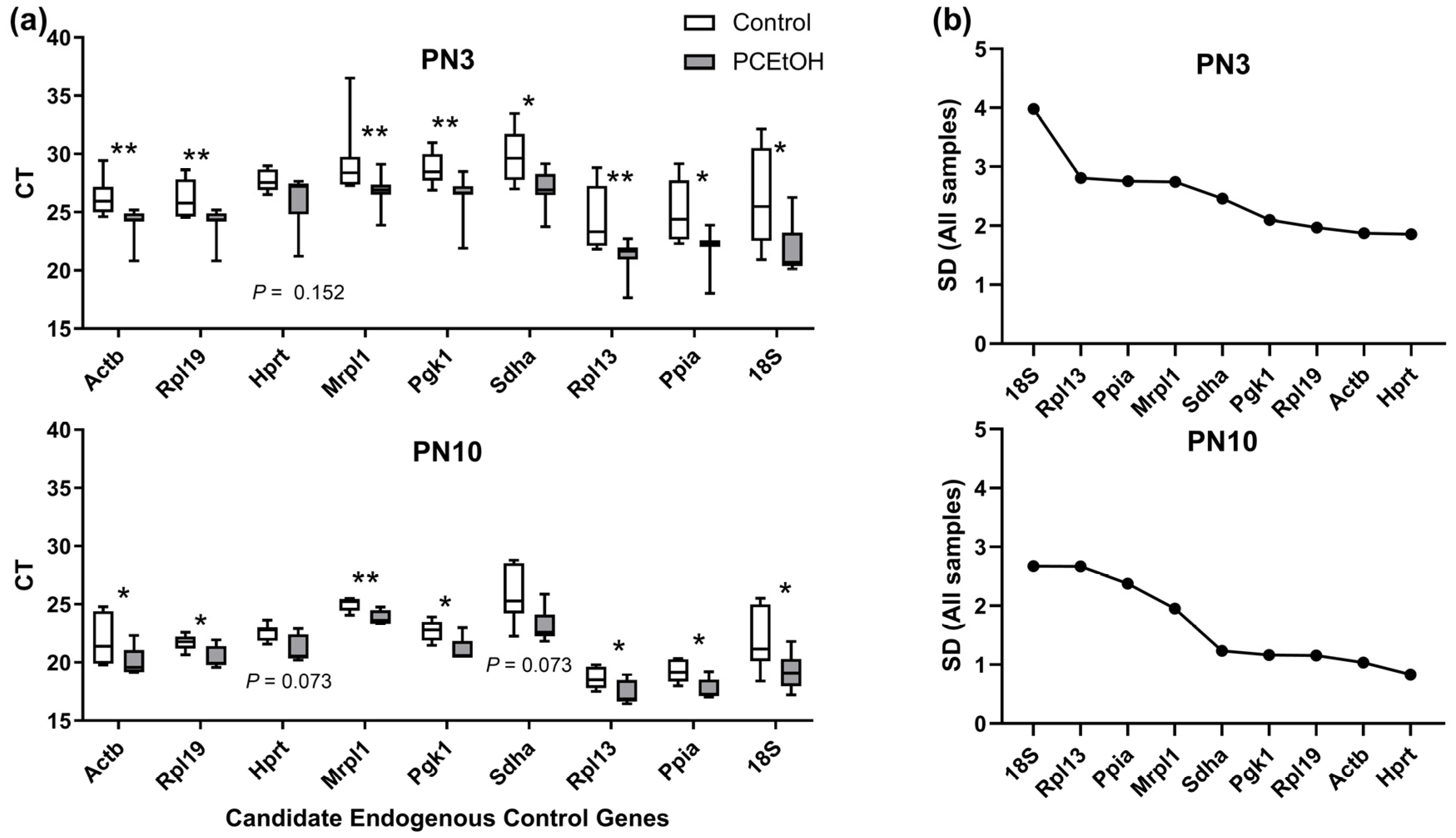
2.1. Ovarian Gene Expression Changes in Neonates Exposed to Periconceptional Alcohol

Aside from differential expression of the tested endogenous control genes in PCEtOH versus control samples (as reported in the Materials and Methods section), genes of interest involved in the apoptotic pathway were dysregulated at PN3, but not at PN10 (Figure 1). Pro-apoptotic BCL2 associated X protein (Bax) had significantly increased expression in ovaries from offspring originating from PCEtOH-exposed pregnancies, while the pro-survival B cell leukemia/lymphoma 2 (Bcl2) gene showed a trend (p < 0.08) for increased expression. The ratio of the pro-survival BCL2 like 1 (Bcl2l1) to Bax was also significantly reduced in PCEtOH ovaries (Figure 1).
In contrast, genes regulating maintenance of the primordial follicle pool were dysregulated at PN10, but not at PN3 (Figure 2). Anti-mullerian hormone (Amh) and Inhibin alpha (Inha) were both increased in ovaries exposed to PCEtOH, suggesting a disproportionate number of larger follicles compared to controls.

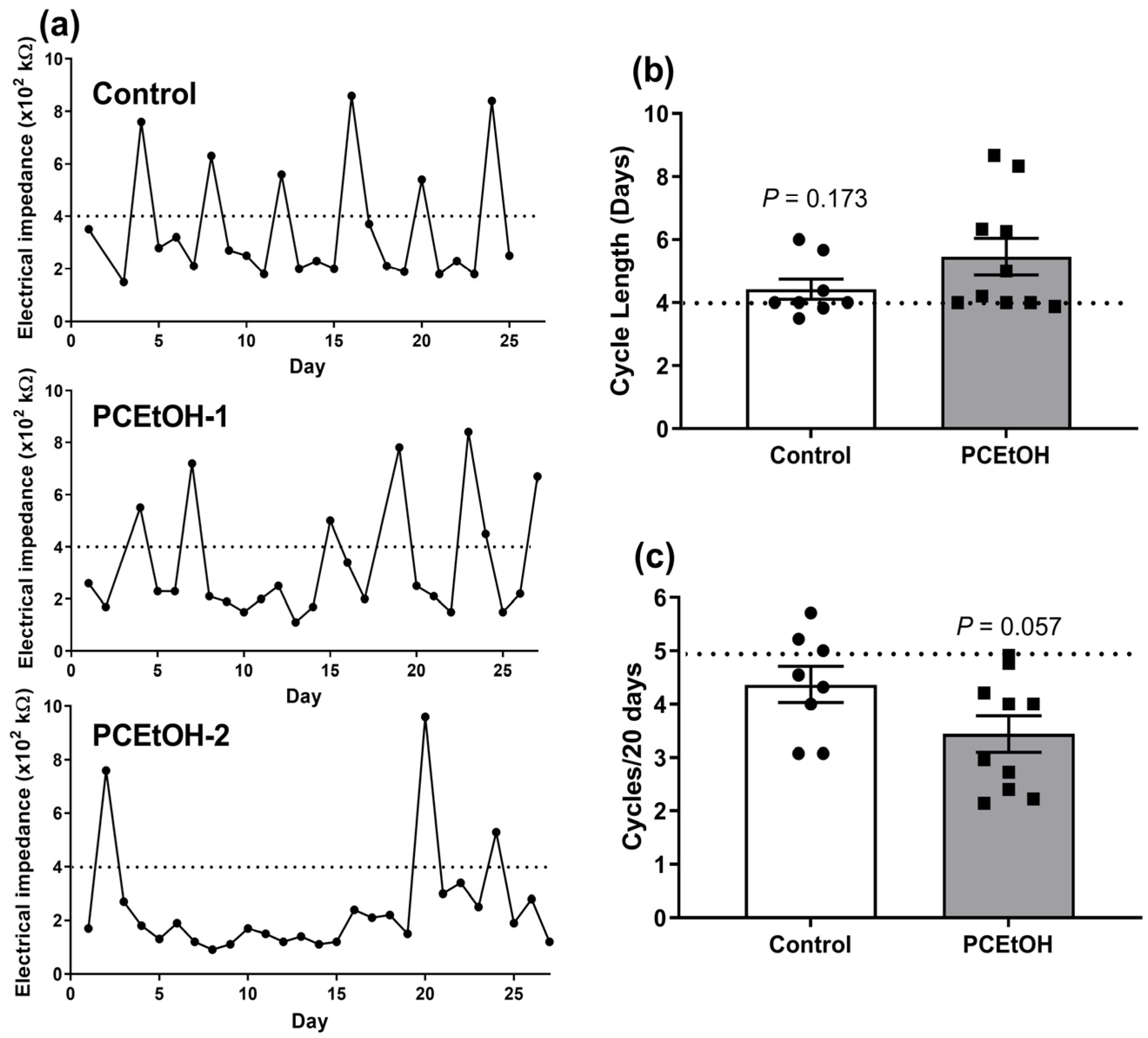
2.2. Estrous Cyclicity and Breeding Performance in Adult Offspring

There was a subtle impact of PCEtOH exposure on estrous cyclicity in 3-month-old F1 offspring, with a subset from pregnancies exposed to alcohol having altered estrus patterns (Figure 3a). This resulted in a trend towards increased average cycle length and an average of one fewer cycles over the monitoring period compared to control animals (Figure 3b,c). There was no significant impact of PCEtOH exposure on F1 female breeding performance, measured by the number of pups born over two consecutive litters, or F2 pup weights and survival (Table 1).

3. Discussion

We measured expression of a subset of potential factors involved in regulating primordial follicle numbers to determine if these were affected by PCEtOH. Numbers of these non-growing follicles are controlled by a balance of factors that (a) regulate activation and recruitment to the pool of growing follicles or maintain quiescence via extrinsic (e.g., Amh, Inha) and intrinsic (e.g., Pten, Cxcr4, Cxcl12, Stk11/Lkb1) factors, or (b) regulate depletion via pro- and anti-apoptotic factors (e.g., Bax, Bak1, Bcl2, Bcl2l1, Bcl2l11, Bbc3/Puma). While many other factors are emerging as being important (see [20,21,22] for review), we chose this subset of relatively established regulators across the spectrum of possible primordial follicle fates (i.e., activation, quiescence or apoptosis).
We found subtle changes in the expression of genes of interest in neonatal ovaries from females originating from PCEtOH-exposed pregnancies, with those involved in the apoptotic pathway, and specifically pro-apoptotic, being upregulated at the earlier PN3 time-point, but not at PN10. Although this was only measured at the transcriptional level, and was not confirmed via measurement of Caspase-3 activity or localization, the lack of subsequent impacts on fertility suggest minimal impacts on folliculogenesis and reproductive capacity. Similarly, although increased levels of Amh and Inha occurred at the later PN10 time-point, suggesting a greater activation of follicles to the growing pool of follicles, and therefore a disproportionate number of preantral follicles compared to controls [23], the similar litter sizes produced by F1 females suggests that these changes were not sufficient to impact on fecundity.
We generally also saw very little evidence of impacts on estrous cyclicity in F1 female offspring, although a subset of females from PCEtOH-exposed pregnancies, but not control pregnancies showed increased cycle length (~2-fold increase) and, therefore, one less cycle on average over a 20-day period. However, there were no apparent impacts on subsequent breeding performance.
These results are similar to our earlier study using an episodic exposure of moderate alcohol at E13.5 and E14.5 of rat pregnancy, which also included assessment of follicle numbers in neonatal ovaries using histology and unbiased stereology [24]. Note that in this previous study, fetal ovaries were present at the time of exposure, whereas in this PCEtOH model, the fetal ovaries have not yet developed. However, we have previously shown that the maternal intra-uterine environment in the PCEtOH model is perturbed immediately following the exposure period, with both progesterone receptor (Pgr) and estrogen receptor alpha (Esr1) down-regulated during the period of receptivity in the rat (E5) and Pgr upregulated during the period of post-implantation decidualization (E7) [15]. Although circulating levels of progesterone and estrogen were unaffected over this peri-implantation period, progesterone-responsive decidualization genes were also dysregulated [15]. Therefore, despite fetal ovarian development occurring after the exposure treatment period, alterations in the maternal environment have the potential to alter developmental trajectories.
We did not directly quantify follicle numbers via histology in neonatal ovaries in the current study, including the non-growing primordial follicle pool. Therefore, the pro-apoptotic state of neonatal ovaries at PN3 and the putative enhanced activation of follicles at PN10 in the PCEtOH-exposed neonates could have reduced the initial ovarian reserve compared to controls. Given that modeling the age-related population of non-growing follicles in humans suggests a reduced initial ovarian reserve can predict an earlier age at menopause [8], this could suggest an earlier age of reproductive senescence in F1 females exposed to PCEtOH during pregnancy in this rodent model. We examined reproductive performance from ~3–6 months of age across two consecutive litters, a period of peak reproductive capacity, and found no differences between PCEtOH-exposed and controls. However, we did not monitor reproductive performance in F1 females as they aged. Sprague Dawley female rats typically start to become sub-fertile at ~8–10 months of age and undergo reproductive senescence at ~12 months of age [25]. Our systematic review [26] highlighted there were no studies exploring long-term fertility in either preclinical models or in women exposed to PAE. Therefore, future preclinical studies examining the impact of prenatal alcohol exposure on female offspring could include an aged cohort (i.e., >8 months of age) to examine the likelihood of premature reproductive senescence. In addition, females diagnosed with FASD in childhood could be monitored as they age to determine if the onset of menopause is affected.
Given that this model of PCEtOH has previously been demonstrated to result in sex-specific programming of long-term health, particularly in males, future studies could examine the impact of this moderate alcohol exposure on male reproductive parameters. This is especially relevant given the reduced nephron endowment reported in offspring from this model [19], and the fact that the kidney and testis both arise from the urogenital ridge during early fetal development [27]. There is some evidence a similar dose of alcohol during pregnancy can affect testis development and long-term reproductive performance in males [28,29,30], although to-date, no studies have examined exposure exclusively around conception.
To our knowledge, only one previous preclinical study in mice has examined the impact of prenatal alcohol exposure exclusively early in gestation (E5-11) on reproductive outcomes in offspring, although this was still after implantation [31]. The dose was comparable to our study (i.e., resulted in a blood alcohol concentration (BAC) of ~0.10–0.15%). However, they only examined the timing of vaginal opening as an indicator of puberty onset, reporting that this was delayed in female offspring from pregnancies exposed to ethanol compared to controls. Future studies could examine impacts of alcohol exposure exclusively during the periconceptional period on puberty onset in offspring, although given our results shown here on fertility, we suspect any impacts would be minor, if any.
An important finding from this study was that there was some evidence of regulation of candidate endogenous control genes by PCEtOH exposure. These should be stably expressed irrespective of treatment, and therefore suggests prenatal alcohol treatment can fundamentally alter the neonatal ovary at the molecular level. We recommend Hprt as a suitable, stably expressed endogenous control for future studies of gene expression in neonatal ovaries in prenatal alcohol studies.
Therefore, although previous studies using this model have reported other sex-specific impacts on F1 offspring metabolic and renal function, these results suggesting minimal impacts on female offspring reproductive function may provide some reassurance for women who have consumed a small amount of alcohol during their early pregnancy.

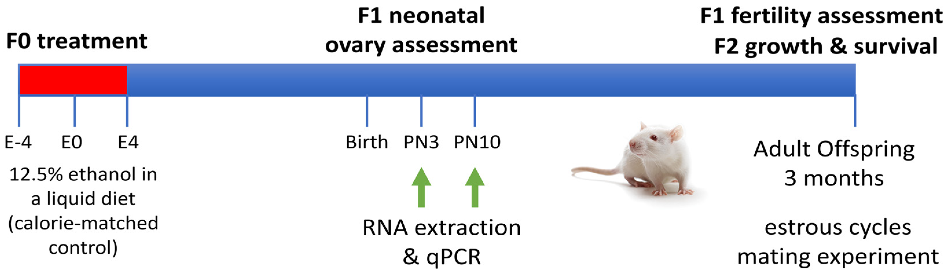
4. Materials and Methods

4.1. Animal Model and Tissue Collection

The housing conditions and treatment protocol for the rat model used in this study of alcohol exposure exclusively around conception have been previously described ([16,32]; Figure 4). Briefly, this involved a 12.5% v/v EtOH liquid diet provided ad libitum from 4 days prior to conception (embryonic day (E)-4)) to 4 days after conception (E4; PCEtOH; n = 11), with calorie-matched controls (Control; n = 11). All animals had a brief (1–2 day) pre-conditioning period to the diet prior to the addition of EtOH, resulting in high acceptance of the diet by all animals (as evidenced by minimal within-group variation and no significant between group difference in calorie intake and weight gain, as reported in [32]). At this dose, our aim was to produce a low-moderate exposure level, clinically relevant for most women. Peak BAC levels occurred 30 min after provision of fresh diet of 0.18 ± 0.04% on day E-2 and 0.25 ± 0.04% on E2 that rapidly declined to 0.07 ± 0.02% at 3h and 0.05 ± 0.02% at 5 h [32]. Although the peak BAC may appear high, this coincided with the initial period of maximal ingestion and quickly reduced to levels only marginally higher than levels found in occasional alcohol users [33]. Additionally, BAC levels were comparable with other preclinical models of EtOH exposure, generally administered via gavage [34,35].
From day E5, pregnant females were fed standard chow (Rat & Mouse Meat-Free Diet, Specialty Feeds). We have previously reported that this model results in equivalent caloric intake and weight gain between treatment and control animals during the treatment period, as well as throughout pregnancy [16]. Ethics approval for all animal experimentation was obtained from the University of Queensland Anatomical Biosciences Animal Ethics Committee (SBMS/467/14/NHMRC) prior to commencement of the study and was conducted in accordance with the Australian Code for the Care and Use of Animals for Scientific Purposes (2013, 8th Edition). Reporting of animal experiments conforms to the ARRIVE guidelines [36,37].
Day of birth was designated as postnatal day (PN) 0 and neonatal ovaries were collected from female F1 offspring during early (PN3) and late (PN10) life from 4 litters per treatment/control, as previously described [24]. These time points correspond with peak numbers of primordial follicles and a period of active recruitment to the growing follicle pool in the rat [5]. Only 1–2 females were used from each litter for analysis of gene expression to remove potential litter effects. The remaining 7 litters per treatment/control remained with the dam and were weaned at PN21, with same sex littermates housed together and females used for fertility assessments as described below.

4.2. Analysis of Gene Expression for Regulators of Ovarian Reserve in F1 Neonatal Ovaries

Expression of genes for factors with putative roles in regulating primordial follicle growth, survival or activation/recruitment (see Table 2 for more details) were analyzed using qPCR as previously described [24]. Briefly, RNA was extracted using either QIAzol Lysis Reagent (Qiagen, Hilden, Germany) and Glycoblue co-precipitant (Thermo Fisher Scientific, Waltham, MA, USA) for PN3 ovaries (with an additional overnight precipitation at −20 °C) or QIAGEN RNeasy Minikit (Qiagen) according to the manufacturer’s instructions. cDNA synthesis was performed using the iScript RT Supermix (Bio-Rad Laboratories, Hercules, CA, USA) using 200 ng of RNA per reaction. RT reactions were performed on a PCR Express Thermal Cycler (Thermo Fisher Scientific). qPCR reactions were performed on an Applied Biosystems Quantstudio 6 Flex Real-Time PCR System (Thermo Fisher Scientific) using 4 ng cDNA, Taq PCR Master Mix (Qiagen; catalogue #201443) and Taqman Assay-on-Demand primer/probe sets (Thermo Fischer Scientific; see Supplementary Table S1 for details) per 10 μL reaction. Nine commonly used endogenous control ‘housekeeper’ genes were tested for stable expression across all samples, with only Hprt identified as not being regulated by ethanol treatment at both time points (Figure 5). Relative gene expression was determined using the comparative threshold method (ΔΔCT) and normalized to Hprt. Fold change was expressed relative to the average of the control group at each age.

4.3. Measurement of Estrous Cyclicity and Fertility in F1 Females

As described above, F1 female offspring from 7 litters/group were grown to 3 months of age and estrous cyclicity assessed over 25–27 days via measurement of vaginal impedance using an EC40 estrous cycle monitor (Fine Science Tools), as previously described [24]. Measurements with an impedance >400 kΩ indicate the female is in proestrus [56].
F1 females were mated and breeding performance was assessed across two consecutive litters. F2 pup weights and survival were also measured at PN3, PN10 and at weaning in a subset of litters. Only 1–2 females were used from each litter to remove potential litter effects for all measures. Remaining offspring were used in previously published experiments examining metabolic and behavioral outcomes (see Introduction).

4.4. Statistical Analysis

All statistical analyses were conducted using GraphPad Prism Version 7.0 Software (GraphPad Software). Ratios of pro-apoptotic and pro-survival factors (i.e., Bcl2:Bax; Bcl2l1:Bax) were calculated, as these are commonly used to examine potential imbalances in cell fate/programmed cell death [57]. Data sets were checked for a normal distribution using the D’Agostino and Pearson test or Shapiro–Wilk test (for small sample size) before conducting parametric or non-parametric analysis as appropriate. Control and PCEtOH groups were compared for each parameter using a Student’s t-test (parametric data) or a Mann–Whitney U-test (non-parametric data). Data are presented as mean ± SEM, with the exception of the analysis of the endogenous control genes which are presented as box plots with median, inter-quartile range (IQR; 25–75%) and 10–90%-ile shown. Data was considered statistically significant at p < 0.05 and considered a trend at 0.05 < p < 0.01.

Supplementary Materials

The following supporting information can be downloaded at: https://www.mdpi.com/article/10.3390/ijms25052471/s1.

Author Contributions

L.K.A. conceived the study, collected samples, analyzed the data and wrote the manuscript. S.E.S. conducted the gene expression experiments/analysis and contributed to earlier drafts of the manuscript. D.J.B. collected the estrous cycle data and reviewed earlier drafts of the manuscript; and K.M.M. conceived the model used in this study, contributed intellectual input and reviewed earlier drafts of the manuscript. All authors have read and agreed to the published version of the manuscript.

Funding

This work was funded by a research grant from the National Health and Medical Research Council of Australia (NHMRC) (APP1046137). KMM was funded by an NHMRC Senior Research Fellowship (APP1078164).

Institutional Review Board Statement

The animal study protocol was approved by the University of Queensland Anatomical Biosciences Animal Ethics Committee (SBMS/467/14/NHMRC).

Informed Consent Statement

Not applicable.

Data Availability Statement

All underlying data used for figures and tables reported in this study are available from the authors by request.

Acknowledgments

The authors acknowledge the assistance of Jacinta Kalisch-Smith, Emily Dorey, Diana Zanfirache, Daniel Browne, Michael Wing, Filip Veljkovic, and Han (Jessica) Dang for assistance with treating and weighing animals; and Barb Arnts, Dave Herne and staff at UQ Biological Resources for maintenance of animal colonies.

Conflicts of Interest

The authors declare no conflicts of interest.

References

  1. Alviggi, C.; Humaidan, P.; Howles, C.M.; Tredway, D.; Hillier, S.G. Biological versus chronological ovarian age: Implications for assisted reproductive technology. Reprod. Biol. Endocrinol. 2009, 7, 101. [Google Scholar] [CrossRef] [PubMed]
  2. Richardson, M.C.; Guo, M.; Fauser, B.C.; Macklon, N.S. Environmental and developmental origins of ovarian reserve. Hum. Reprod. Update 2014, 20, 353–369. [Google Scholar] [CrossRef]
  3. Findlay, J.K.; Hutt, K.J.; Hickey, M.; Anderson, R.A. What is the “ovarian reserve”? Fertil. Steril. 2015, 103, 628–630. [Google Scholar] [CrossRef]
  4. Childs, A.J.; Kinnell, H.L.; Collins, C.S.; Hogg, K.; Bayne, R.A.; Green, S.J.; McNeilly, A.S.; Anderson, R.A. BMP signaling in the human fetal ovary is developmentally regulated and promotes primordial germ cell apoptosis. Stem Cells 2010, 28, 1368–1378. [Google Scholar] [CrossRef]
  5. Picut, C.A.; Dixon, D.; Simons, M.L.; Stump, D.G.; Parker, G.A.; Remick, A.K. Postnatal ovary development in the rat: Morphologic study and correlation of morphology to neuroendocrine parameters. Toxicol. Pathol. 2015, 43, 343–353. [Google Scholar] [CrossRef] [PubMed]
  6. Hansen, K.R.; Knowlton, N.S.; Thyer, A.C.; Charleston, J.S.; Soules, M.R.; Klein, N.A. A new model of reproductive aging: The decline in ovarian non-growing follicle number from birth to menopause. Hum. Reprod. 2008, 23, 699–708. [Google Scholar] [CrossRef] [PubMed]
  7. Ford, E.A.; Beckett, E.L.; Roman, S.D.; McLaughlin, E.A.; Sutherland, J.M. Advances in human primordial follicle activation and premature ovarian insufficiency. Reproduction 2020, 159, R15–R29. [Google Scholar] [CrossRef] [PubMed]
  8. Wallace, W.H.; Kelsey, T.W. Human ovarian reserve from conception to the menopause. PLoS ONE 2010, 5, e8772. [Google Scholar] [CrossRef]
  9. Zhu, Q.; Li, Y.; Ma, J.; Ma, H.; Liang, X. Potential factors result in diminished ovarian reserve: A comprehensive review. J. Ovarian Res. 2023, 16, 208. [Google Scholar] [CrossRef]
  10. Monniaux, D. Factors influencing establishment of the ovarian reserve and their effects on fertility. Anim. Reprod. 2018, 15, 635–647. [Google Scholar] [CrossRef]
  11. McCormack, C.; Hutchinson, D.; Burns, L.; Wilson, J.; Elliott, E.; Allsop, S.; Najman, J.; Jacobs, S.; Rossen, L.; Olsson, C.; et al. Prenatal Alcohol Consumption Between Conception and Recognition of Pregnancy. Alcohol. Clin. Exp. Res. 2017, 41, 369–378. [Google Scholar] [CrossRef]
  12. Muggli, E.; O’Leary, C.; Donath, S.; Orsini, F.; Forster, D.; Anderson, P.J.; Lewis, S.; Nagle, C.; Craig, J.M.; Elliott, E.; et al. “Did you ever drink more?” A detailed description of pregnant women’s drinking patterns. BMC Public Health 2016, 16, 683. [Google Scholar] [CrossRef]
  13. Ishitsuka, K.; Hanada-Yamamoto, K.; Mezawa, H.; Saito-Abe, M.; Konishi, M.; Ohya, Y.; Japan, E.; Children’s Study, G. Determinants of Alcohol Consumption in Women Before and After Awareness of Conception. Matern. Child Health J. 2020, 24, 165–176. [Google Scholar] [CrossRef]
  14. Gardebjer, E.M.; Cuffe, J.S.M.; Ward, L.C.; Steane, S.; Anderson, S.T.; Dorey, E.S.; Kalisch-Smith, J.I.; Pantaleon, M.; Chong, S.; Yamada, L.; et al. Effects of periconceptional maternal alcohol intake and a postnatal high-fat diet on obesity and liver disease in male and female rat offspring. Am. J. Physiol. Endocrinol. Metab. 2018, 315, E694–E704. [Google Scholar] [CrossRef] [PubMed]
  15. Kalisch-Smith, J.I.; Steane, S.E.; Simmons, D.G.; Pantaleon, M.; Anderson, S.T.; Akison, L.K.; Wlodek, M.E.; Moritz, K.M. Periconceptional alcohol exposure causes female-specific perturbations to trophoblast differentiation and placental formation in the rat. Development 2019, 146, dev.172205. [Google Scholar] [CrossRef] [PubMed]
  16. Steane, S.E.; Fielding, A.M.; Kent, N.L.; Andersen, I.; Browne, D.J.; Tejo, E.N.; Gardebjer, E.M.; Kalisch-Smith, J.I.; Sullivan, M.A.; Moritz, K.M.; et al. Maternal choline supplementation in a rat model of periconceptional alcohol exposure: Impacts on the fetus and placenta. Alcohol. Clin. Exp. Res. 2021, 45, 2130–2146. [Google Scholar] [CrossRef] [PubMed]
  17. Gardebjer, E.M.; Anderson, S.T.; Pantaleon, M.; Wlodek, M.E.; Moritz, K.M. Maternal alcohol intake around the time of conception causes glucose intolerance and insulin insensitivity in rat offspring, which is exacerbated by a postnatal high-fat diet. FASEB J. 2015, 29, 2690–2701. [Google Scholar] [CrossRef]
  18. Dorey, E.S.; Cullen, C.L.; Lucia, D.; Mah, K.M.; Manchadi, M.R.; Muhlhausler, B.S.; Moritz, K.M. The impact of periconceptional alcohol exposure on fat preference and gene expression in the mesolimbic reward pathway in adult rat offspring. J. Dev. Orig. Health Dis. 2018, 9, 223–231. [Google Scholar] [CrossRef]
  19. Dorey, E.S.; Walton, S.L.; Kalisch-Smith, J.I.; Paravicini, T.M.; Gardebjer, E.M.; Weir, K.A.; Singh, R.R.; Bielefeldt-Ohmann, H.; Anderson, S.T.; Wlodek, M.E.; et al. Periconceptional ethanol exposure induces a sex specific diuresis and increase in AQP2 and AVPR2 in the kidneys of aged rat offspring. Physiol. Rep. 2019, 7, e14273. [Google Scholar] [CrossRef]
  20. Chang, C.W.; Sung, Y.W.; Hsueh, Y.W.; Chen, Y.Y.; Ho, M.; Hsu, H.C.; Yang, T.C.; Lin, W.C.; Chang, H.M. Growth hormone in fertility and infertility: Mechanisms of action and clinical applications. Front. Endocrinol. 2022, 13, 1040503. [Google Scholar] [CrossRef]
  21. Kaur, S.; Kurokawa, M. Regulation of Oocyte Apoptosis: A View from Gene Knockout Mice. Int. J. Mol. Sci. 2023, 24, 1345. [Google Scholar] [CrossRef]
  22. Frost, E.R.; Ford, E.A.; Peters, A.E.; Lovell-Badge, R.; Taylor, G.; McLaughlin, E.A.; Sutherland, J.M. A New Understanding, Guided by Single-Cell Sequencing, of the Establishment and Maintenance of the Ovarian Reserve in Mammals. Sex. Dev. 2023, 17, 145–155. [Google Scholar] [CrossRef]
  23. Kristensen, S.G.; Andersen, K.; Clement, C.A.; Franks, S.; Hardy, K.; Andersen, C.Y. Expression of TGF-beta superfamily growth factors, their receptors, the associated SMADs and antagonists in five isolated size-matched populations of pre-antral follicles from normal human ovaries. Mol. Hum. Reprod. 2014, 20, 293–308. [Google Scholar] [CrossRef] [PubMed]
  24. McReight, E.K.; Liew, S.H.; Steane, S.E.; Hutt, K.J.; Moritz, K.M.; Akison, L.K. Moderate episodic prenatal alcohol does not impact female offspring fertility in rats. Reproduction 2020, 159, 615–626. [Google Scholar] [CrossRef] [PubMed]
  25. Acuna, E.; Fornes, R.; Fernandois, D.; Garrido, M.P.; Greiner, M.; Lara, H.E.; Paredes, A.H. Increases in norepinephrine release and ovarian cyst formation during ageing in the rat. Reprod. Biol. Endocrinol. 2009, 7, 64. [Google Scholar] [CrossRef] [PubMed]
  26. Akison, L.K.; Moritz, K.M.; Reid, N. Adverse reproductive outcomes associated with fetal alcohol exposure: A systematic review. Reproduction 2019, 157, 329–343. [Google Scholar] [CrossRef] [PubMed]
  27. Himelreich Peric, M.; Takahashi, M.; Jezek, D.; Cunha, G.R. Early development of the human embryonic testis. Differentiation 2023, 129, 4–16. [Google Scholar] [CrossRef] [PubMed]
  28. McGivern, R.F.; Raum, W.J.; Handa, R.J.; Sokol, R.Z. Comparison of two weeks versus one week of prenatal ethanol exposure in the rat on gonadal organ weights, sperm count, and onset of puberty. Neurotoxicol. Teratol. 1992, 14, 351–358. [Google Scholar] [CrossRef] [PubMed]
  29. Udani, M.; Parker, S.; Gavaler, J.; Van Thiel, D.H. Effects of in utero exposure to alcohol upon male rats. Alcohol. Clin. Exp. Res. 1985, 9, 355–359. [Google Scholar] [CrossRef] [PubMed]
  30. Lan, N.; Vogl, A.W.; Weinberg, J. Prenatal ethanol exposure delays the onset of spermatogenesis in the rat. Alcohol. Clin. Exp. Res. 2013, 37, 1074–1081. [Google Scholar] [CrossRef]
  31. Boggan, W.O.; Randall, C.L.; Dodds, H.M. Delayed sexual maturation in female C57BL/6J mice prenatally exposed to alcohol. Res. Commun. Chem. Pathol. Pharmacol. 1979, 23, 117–125. [Google Scholar]
  32. Gardebjer, E.M.; Cuffe, J.S.; Pantaleon, M.; Wlodek, M.E.; Moritz, K.M. Periconceptional alcohol consumption causes fetal growth restriction and increases glycogen accumulation in the late gestation rat placenta. Placenta 2014, 35, 50–57. [Google Scholar] [CrossRef] [PubMed]
  33. Touquet, R.; Csipke, E.; Holloway, P.; Brown, A.; Patel, T.; Seddon, A.J.; Gulati, P.; Moore, H.; Batrick, N.; Crawford, M.J. Resuscitation room blood alcohol concentrations: One-year cohort study. Emerg. Med. J. 2008, 25, 752–756. [Google Scholar] [CrossRef]
  34. Gray, S.P.; Denton, K.M.; Cullen-McEwen, L.; Bertram, J.F.; Moritz, K.M. Prenatal exposure to alcohol reduces nephron number and raises blood pressure in progeny. J. Am. Soc. Nephrol. 2010, 21, 1891–1902. [Google Scholar] [CrossRef] [PubMed]
  35. Chen, L.; Nyomba, B.L. Effects of prenatal alcohol exposure on glucose tolerance in the rat offspring. Metabolism 2003, 52, 454–462. [Google Scholar] [CrossRef]
  36. Kilkenny, C.; Browne, W.; Cuthill, I.C.; Emerson, M.; Altman, D.G.; NC3Rs Reporting Guidelines Working Group. Animal research: Reporting in vivo experiments: The ARRIVE guidelines. Br. J. Pharmacol. 2010, 160, 1577–1579. [Google Scholar] [CrossRef]
  37. Kilkenny, C.; Browne, W.J.; Cuthill, I.C.; Emerson, M.; Altman, D.G. Improving bioscience research reporting: The ARRIVE guidelines for reporting animal research. PLoS Biol. 2010, 8, e1000412. [Google Scholar] [CrossRef]
  38. Greenfeld, C.R.; Pepling, M.E.; Babus, J.K.; Furth, P.A.; Flaws, J.A. BAX regulates follicular endowment in mice. Reproduction 2007, 133, 865–876. [Google Scholar] [CrossRef] [PubMed]
  39. Chen, Q.; Chen, Q.; Song, Y.; Xiang, Y.; Li, Q.; Sang, Y.; Zhang, L.; Bai, L.; Zhu, Y. Downregulation of homeobox A1 in human granulosa cells is involved in diminished ovarian reserve through promoting cell apoptosis and mitochondrial dysfunction. Mol. Cell. Endocrinol. 2024, 580, 112084. [Google Scholar] [CrossRef]
  40. Hutt, K.J. The role of BH3-only proteins in apoptosis within the ovary. Reproduction 2015, 149, R81–R89. [Google Scholar] [CrossRef]
  41. Liew, S.H.; Vaithiyanathan, K.; Hutt, K.J. Taking control of the female fertile lifespan: A key role for Bcl-2 family proteins. Reprod. Fertil. Dev. 2016, 28, 864–871. [Google Scholar] [CrossRef] [PubMed]
  42. Flaws, J.A.; Hirshfield, A.N.; Hewitt, J.A.; Babus, J.K.; Furth, P.A. Effect of bcl-2 on the primordial follicle endowment in the mouse ovary. Biol. Reprod. 2001, 64, 1153–1159. [Google Scholar] [CrossRef] [PubMed]
  43. Ratts, V.S.; Flaws, J.A.; Kolp, R.; Sorenson, C.M.; Tilly, J.L. Ablation of bcl-2 gene expression decreases the numbers of oocytes and primordial follicles established in the post-natal female mouse gonad. Endocrinology 1995, 136, 3665–3668. [Google Scholar] [CrossRef] [PubMed]
  44. Rucker, E.B., 3rd; Dierisseau, P.; Wagner, K.U.; Garrett, L.; Wynshaw-Boris, A.; Flaws, J.A.; Hennighausen, L. Bcl-x and Bax regulate mouse primordial germ cell survival and apoptosis during embryogenesis. Mol. Endocrinol. 2000, 14, 1038–1052. [Google Scholar] [CrossRef] [PubMed]
  45. Nilsson, E.E.; Stanfield, J.; Skinner, M.K. Interactions between progesterone and tumor necrosis factor-alpha in the regulation of primordial follicle assembly. Reproduction 2006, 132, 877–886. [Google Scholar] [CrossRef] [PubMed]
  46. Myers, M.; Morgan, F.H.; Liew, S.H.; Zerafa, N.; Gamage, T.U.; Sarraj, M.; Cook, M.; Kapic, I.; Sutherland, A.; Scott, C.L.; et al. PUMA regulates germ cell loss and primordial follicle endowment in mice. Reproduction 2014, 148, 211–219. [Google Scholar] [CrossRef] [PubMed]
  47. Nguyen, Q.N.; Zerafa, N.; Liew, S.H.; Morgan, F.H.; Strasser, A.; Scott, C.L.; Findlay, J.K.; Hickey, M.; Hutt, K.J. Loss of PUMA protects the ovarian reserve during DNA-damaging chemotherapy and preserves fertility. Cell Death Dis. 2018, 9, 618. [Google Scholar] [CrossRef] [PubMed]
  48. Pangas, S.A. Regulation of the ovarian reserve by members of the transforming growth factor beta family. Mol. Reprod. Dev. 2012, 79, 666–679. [Google Scholar] [CrossRef]
  49. Durlinger, A.L.; Visser, J.A.; Themmen, A.P. Regulation of ovarian function: The role of anti-Mullerian hormone. Reproduction 2002, 124, 601–609. [Google Scholar] [CrossRef]
  50. Durlinger, A.L.; Kramer, P.; Karels, B.; de Jong, F.H.; Uilenbroek, J.T.; Grootegoed, J.A.; Themmen, A.P. Control of primordial follicle recruitment by anti-Mullerian hormone in the mouse ovary. Endocrinology 1999, 140, 5789–5796. [Google Scholar] [CrossRef]
  51. Reddy, P.; Liu, L.; Adhikari, D.; Jagarlamudi, K.; Rajareddy, S.; Shen, Y.; Du, C.; Tang, W.; Hamalainen, T.; Peng, S.L.; et al. Oocyte-specific deletion of Pten causes premature activation of the primordial follicle pool. Science 2008, 319, 611–613. [Google Scholar] [CrossRef]
  52. Jagarlamudi, K.; Liu, L.; Adhikari, D.; Reddy, P.; Idahl, A.; Ottander, U.; Lundin, E.; Liu, K. Oocyte-specific deletion of Pten in mice reveals a stage-specific function of PTEN/PI3K signaling in oocytes in controlling follicular activation. PLoS ONE 2009, 4, e6186. [Google Scholar] [CrossRef]
  53. Jiang, Z.Z.; Hu, M.W.; Ma, X.S.; Schatten, H.; Fan, H.Y.; Wang, Z.B.; Sun, Q.Y. LKB1 acts as a critical gatekeeper of ovarian primordial follicle pool. Oncotarget 2016, 7, 5738–5753. [Google Scholar] [CrossRef] [PubMed]
  54. Myers, M.; Middlebrook, B.S.; Matzuk, M.M.; Pangas, S.A. Loss of inhibin alpha uncouples oocyte-granulosa cell dynamics and disrupts postnatal folliculogenesis. Dev. Biol. 2009, 334, 458–467. [Google Scholar] [CrossRef] [PubMed]
  55. Chand, A.L.; Harrison, C.A.; Shelling, A.N. Inhibin and premature ovarian failure. Hum. Reprod. Update 2010, 16, 39–50. [Google Scholar] [CrossRef] [PubMed]
  56. Jaramillo, L.M.; Balcazar, I.B.; Duran, C. Using vaginal wall impedance to determine estrous cycle phase in Lewis rats. Lab Anim. 2012, 41, 122–128. [Google Scholar] [CrossRef] [PubMed]
  57. Korsmeyer, S.J.; Shutter, J.R.; Veis, D.J.; Merry, D.E.; Oltvai, Z.N. Bcl-2/Bax: A rheostat that regulates an anti-oxidant pathway and cell death. Semin. Cancer Biol. 1993, 4, 327–332. [Google Scholar]
Figure 1. Genes involved in primordial follicle survival in F1 neonatal ovaries. (a) PN3 ovaries; (b) PN10 ovaries. Closed circles (white bars) are controls; closed squares (grey bars) are periconceptional ethanol exposed (PCEtOH) offspring. Data expressed as mean ± SEM. * p < 0.05; ** p < 0.01; unpaired t-test or Mann–Whitney rank sum test used for each comparison. n = 6–8 across 4 litters/group. PN = post-natal day.
Ijms 25 02471 g001
Figure 2. Genes involved in primordial follicle growth and activation/recruitment in F1 neonatal ovaries. (a) PN3 ovaries; (b) PN10 ovaries. Closed circles (white bars) are controls; closed squares (grey bars) are periconceptional ethanol exposed (PCEtOH) offspring. Data expressed as mean ± SEM. * p < 0.05; unpaired t-test or Mann–Whitney rank sum test used for each comparison. n = 6–8 across 4 litters/group. PN = post-natal day.
Ijms 25 02471 g002
Figure 3. Estrous cyclicity in F1 offspring. (a) Estrous cycles were examined in 3-month-old offspring over 25–27 days via measurement of vaginal impedance (>400 kΩ = proestrus, as indicated by the dotted line). The top figure is a representative profile for a control group female, while the bottom two figures are representative of periconceptional ethanol (PCEtOH) group females. (b) Mean ± SEM estrous cycle length in days. Dotted line indicates typical 4-day cycle for Sprague Dawley rats. (c) Number of cycles over a 20-day period. Dotted line indicates typical 5 cycles per 20-day period in this species. p values obtained using a Mann–Whitney rank sum test. n = 8–10 across 7 litters/group.
Ijms 25 02471 g003
Figure 4. Rat model of periconceptional alcohol exposure. The period of exposure is shown in red. E = embryonic day; PN = post-natal day.
Ijms 25 02471 g004
Figure 5. Candidate endogenous control gene expression varied with ethanol exposure. n = 6–8/treatment/age across 4 litters/group. (a) Cycle threshold (CT) displayed as median, IQR (25–75% = box) and 10–90% (whiskers). All genes analyzed using Mann–Whitney rank sum tests due to non-normal distribution of most data sets. * p < 0.05, ** p < 0.01. (b) Standard deviation (SD) graphed across all samples for each gene to further illustrate heterogeneity in expression.
Ijms 25 02471 g005
Table 1. F1 female breeding data and F2 pup weights and survival.
Parameter—Mean (SD)Control (n = 7) 1PCEtOH (n = 7) 1p Value 2
F1 age at 1st mating (weeks)14.71 (1.9)14.63 (2.1)0.937
Pups per litter (Litter 1) 316 (2.4)16 (2.2)0.648
Pups per litter (Litter 2) 3,416 (3.1)14 (2.4)0.364
Survival of F2 pups to PN10 (%) 596.79 (3.2)89.86 (27.3)0.837
F2 Pup weight at PN3 (g)—females 68.19 (1.1)8.34 (0.9)0.794
F2 Pup weight at PN3 (g)—males 68.83 (1.4)8.85 (1.1)0.973
F2 Pup weight at weaning (g)—females 744.36 (3.0)40.06 (2.9)0.085
F2 Pup weight at weaning (g)—males 747.22 (2.3)44.27 (2.6)0.140
1 One F1 female was randomly chosen from each treated F0 female. 2 p values were obtained from either an unpaired t-test (parametric data) or a Mann–Whitney rank sum test (non-parametric data). 3 Pups per litter expressed as whole pups. 4 n = 6 per group. 5 Across 13 litters/group. 6 Across 9 litters/group. 7 Across 4 litters/group. F1/F2 = 1st/2nd generation offspring. PCEtOH = periconceptional ethanol. PN = postnatal day.
Table 2. Details of genes examined in neonatal ovaries from pregnancies exposed to periconceptional ethanol. Many of these functions have been discovered using knockout mouse models but their precise molecular action and regulation is still under investigation.
Gene NameGene SymbolFunction Related to Ovarian ReserveReference(s)
Apoptotic pathway
BCL2 associated X, apoptosis regulatorBaxPro-apoptotic factor[38,39]
BCL2 antagonist/killer 1Bak1Pro-apoptotic factor[40,41]
BCL2 apoptosis regulatorBcl2Pro-survival factor[39,42,43]
BCL2 like 1Bcl2l1 (Bclx)Pro-survival factor[44,45]
BCL2 binding component3Bbc3 (Puma)Pro-apoptotic via activation of Bax/Bak1 or inhibition of pro-survival BCL2 family members[46,47]
Follicle growth and recruitment
Anti-mullerian hormoneAmhMaintenance of ovarian reserve by inhibiting recruitment to the growing follicle pool[48,49,50]
Phosphatase and tensin homologPtenMaintenance of quiescent state of primordial follicles via the PI3K/AKT pathway[51,52]
Serine/threonine kinase 11Stk11 (Lkb1)Maintenance of quiescent state of
primordial follicles via mTOR
[53]
Inhibin subunit alphaInhaIndirectly impacts on follicle growth and recruitment via negative feedback of FSH[54,55]
AKT = protein kinase a; FSH = follicle stimulating hormone; mTOR = mammalian target of rapamycin; PI3K = phosphatidylinositol-3 kinase.
Disclaimer/Publisher’s Note: The statements, opinions and data contained in all publications are solely those of the individual author(s) and contributor(s) and not of MDPI and/or the editor(s). MDPI and/or the editor(s) disclaim responsibility for any injury to people or property resulting from any ideas, methods, instructions or products referred to in the content.

