Article

The Role of Glucose-6-phosphate Dehydrogenase in the Wine Yeast Hanseniaspora uvarum

by
Jürgen J. Heinisch
*,
Andrea Murra
,
Lucía Fernández Murillo
and
Hans-Peter Schmitz
AG Genetik, Fachbereich Biologie/Chemie, Universität Osnabrück, Barbarastr. 11, D-49076 Osnabrück, Germany
*
Author to whom correspondence should be addressed.
Int. J. Mol. Sci. 2024, 25(4), 2395; https://doi.org/10.3390/ijms25042395
Submission received: 29 January 2024 / Revised: 13 February 2024 / Accepted: 16 February 2024 / Published: 18 February 2024
(This article belongs to the Special Issue Microbial Biotechnology in Food)

Abstract

:
Hanseniaspora uvarum is the predominant yeast species in the majority of wine fermentations, which has only recently become amenable to directed genetic manipulation. The genetics and metabolism of H. uvarum have been poorly studied as compared to other yeasts of biotechnological importance. This work describes the construction and characterization of homozygous deletion mutants in the HuZWF1 gene, encoding glucose-6-phosphate dehydrogenase (G6PDH), which provides the entrance into the oxidative part of the pentose phosphate pathway (PPP) and serves as a major source of NADPH for anabolic reactions and oxidative stress response. Huzwf1 deletion mutants grow more slowly on glucose medium than wild-type and are hypersensitive both to hydrogen peroxide and potassium bisulfite, indicating that G6PDH activity is required to cope with these stresses. The mutant also requires methionine for growth. Enzyme activity can be restored by the expression of heterologous G6PDH genes from other yeasts and humans under the control of a strong endogenous promoter. These findings provide the basis for a better adaptation of H. uvarum to conditions used in wine fermentations, as well as its use for other biotechnological purposes and as an expression organism for studying G6PDH functions in patients with hemolytic anemia.

1. Introduction

Glucose-6-phosphate dehydrogenase (G6PDH; EC 1.1.1.49) catalyzes the first step in the oxidative part of the pentose phosphate pathway (PPP, in which glucose-6-phosphate is converted into 6-phosphoglucono lactone. This enables the production of pentose phosphates required as precursors for the synthesis of nucleic acid, cofactors, and histidine, as well as erythrose-4-phosphate in the non-oxidative part of the pathway for aromatic amino acid biosynthesis [1,2]. In addition, the G6PDH reaction is the primary source of NADPH in many cells and tissues, providing the reducing power to drive lipid and fatty acid synthesis and for the detoxification of reactive oxygen species (ROS). This antioxidant activity explains the importance of the G6PDH reaction in cancerogenesis [3,4] and neurodegenerative diseases [5]. NADPH is also required for contractility in cardiomyocytes and is thus important for proper heart function [6]. With respect to human health, G6PDH is of utmost importance in erythrocytes, being the exclusive source of NADPH [7]. In fact, malfunctions of the enzyme are the leading cause of hemolytic anemia, with some 217 mutations described for the encoding gene [8]. Predisposition for this enzymopathic condition may affect as many as 500 million people worldwide, most being asymptomatic unless exposed to certain diets or drugs [9]. In addition, increased enzyme activities have been detected in a variety of human tumors [10].
Mutants in the ZWF1 gene (for the German “Zwischenferment” derived from biochemical studies early in the last century [11]) were first obtained by suppression of the glucose-negative phenotype of a 6-phosphogluconate dehydrogenase mutant [12]. Later on, deletion mutants constructed after gene cloning were found to be hypersensitive towards oxidative stress agents and to require methionine for growth, all attributed to the reduced provision of NADPH for detoxification or biosynthesis, respectively [13,14,15]. Vice versa, oxidative stress inactivates the glycolytic enzymes glyceraldehyde-3-phosphate dehydrogenase and triosephosphate isomerase, which results in enhancing the flux through G6PDH and NADPH production [16]. The latter has been attributed to the recycling of glucose-6-phosphate by means of the non-oxidative part of the PPP rather than metabolite accumulation caused by the block in glycolysis [17]. While a zwf1 deletion has little effect on the growth of glucose in the genetic background of some common laboratory strains of S. cerevisiae [15,18], it grows poorly under standard conditions in a strain more closely related to industrial baker’s yeast compared to its isogenic wild-type [19]. These defects can be complemented by introducing genes encoding heterologous G6PDH enzymes from humans and plants so that the deletion strain has been proposed as an expression organism for production and biochemical studies of the heterologous enzymes [19].
While S. cerevisiae has been employed as a general model system in biochemistry and molecular genetics, it is specialized in alcoholic fermentation and has been selected for this purpose by mankind for thousands of years [20]. In fact, it reduces its respiratory capacity in the presence of high medium sugar concentrations, independent of the availability of oxygen (the so-called Crabtree effect), and thus does not display the Pasteur effect (i.e., switching to respiratory metabolism when oxygen is available, [21]). The PPP is, therefore, of greater importance in Crabtree-negative yeasts, which require respiration for efficient energy production instead of relying on glycolytic capacity [1]. Amongst these yeasts, Kluyveromyces lactis acquired a model status, as it has not undergone a whole genome duplication and can be manipulated by similar techniques of classical and molecular genetics as used for S. cerevisiae [22,23]. Evidence for a more prominent role of the PPP in sugar metabolism by this yeast was provided by the fact that glycolytic mutants, such as Klpgi1 or Klpfk1 Klpfk2 deletions, still grew on glucose as a sole carbon source, in contrast to its S. cerevisiae counterparts [24,25]. The notion of increased flux through the PPP was strongly supported by an additional lack of transaldolase in the respective double (Klpgi1 Kltal1) and triple (Klpfk1 Klpfk2 Kltal1) deletions, which failed to grow on glucose [26]. A lack of G6PDH activity in Klzwf1 deletions leads to reduced biomass production on different carbon sources as compared to the wild-type [27]. The active enzyme in K. lactis is a homotetramer, which is allosterically regulated by NADPH, upon whose binding it dissociates into dimers [28].
Hanseniaspora uvarum is another Crabtree-negative yeast which draws growing attention for its biotechnological potential [29]. Although it is found on grapes in vineyards worldwide and frequently dominates the yeast population in the first hours of wine fermentation [30,31,32], its genetics and physiology have been poorly studied at the molecular level. A first basis for such studies was provided by sequencing the genome and its annotation, accompanied by determinations of glycolytic enzyme activities [33]. The latter work attributed the lower fermentative capacity of H. uvarum as compared to S. cerevisiae to a reduced pyruvate kinase activity as the final step of glycolysis. Based on the genome sequence methods for the introduction of DNA and the construction of deletion mutants have been successfully implemented only recently [34,35]. The construction of autonomously replicating plasmids and homozygous auxotrophic deletion mutants in the diploid-type strain (i.e., Huade2, Huhis3, Huleu2, and Huura3) provided a set of essential tools for future genetic manipulations of this yeast [36].
Here, we applied this toolset for the construction and physiological characterization of deletion mutants in the HuZWF1 gene. These were then employed as a host for the expression of heterologous genes encoding G6PDH from other yeasts, as well as a human isoform.

2. Results

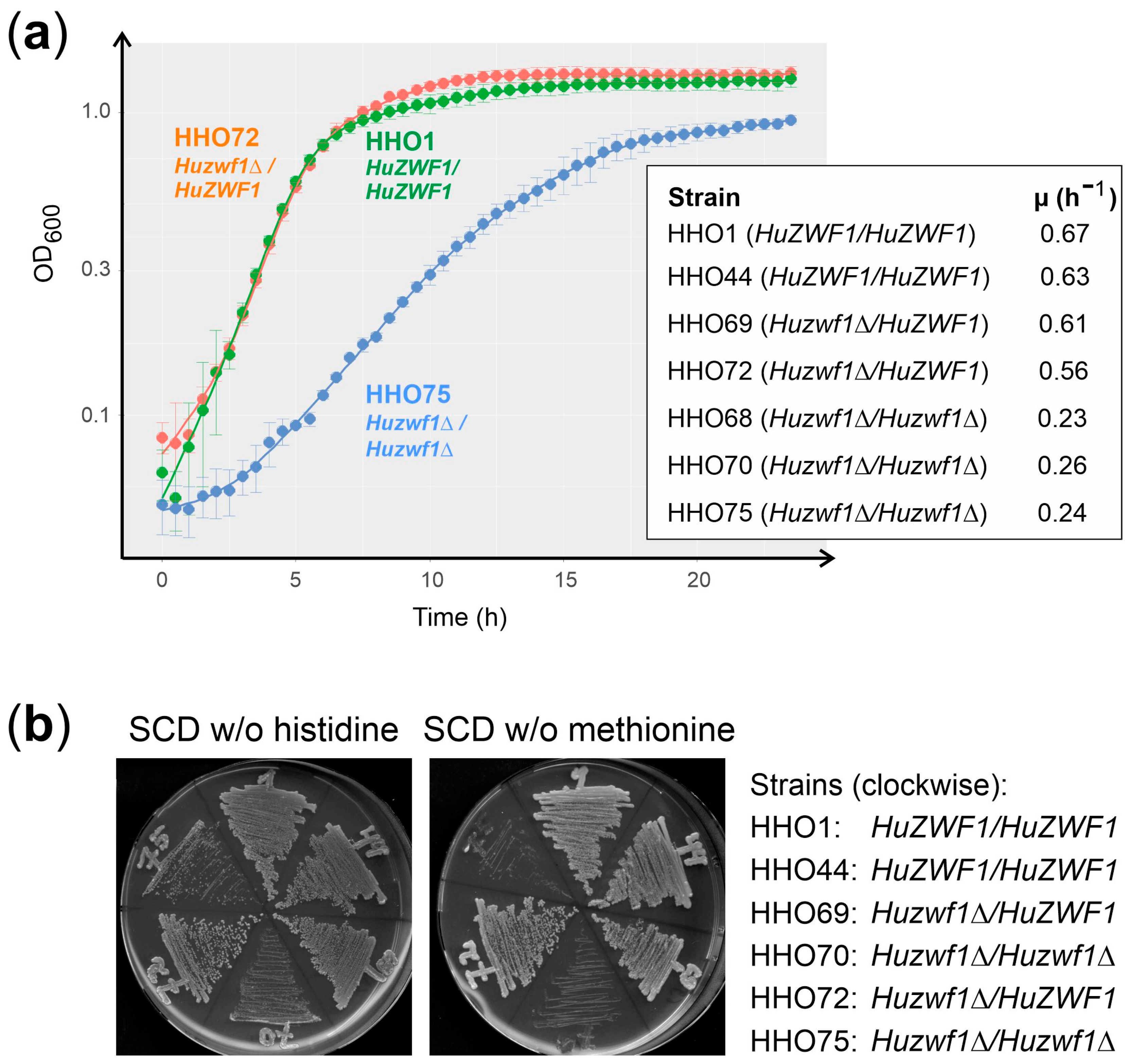
2.1. Homozygous Huzwf1 Deletion Mutants Lack Glucose-6-phosphate Dehydrogenase Activity
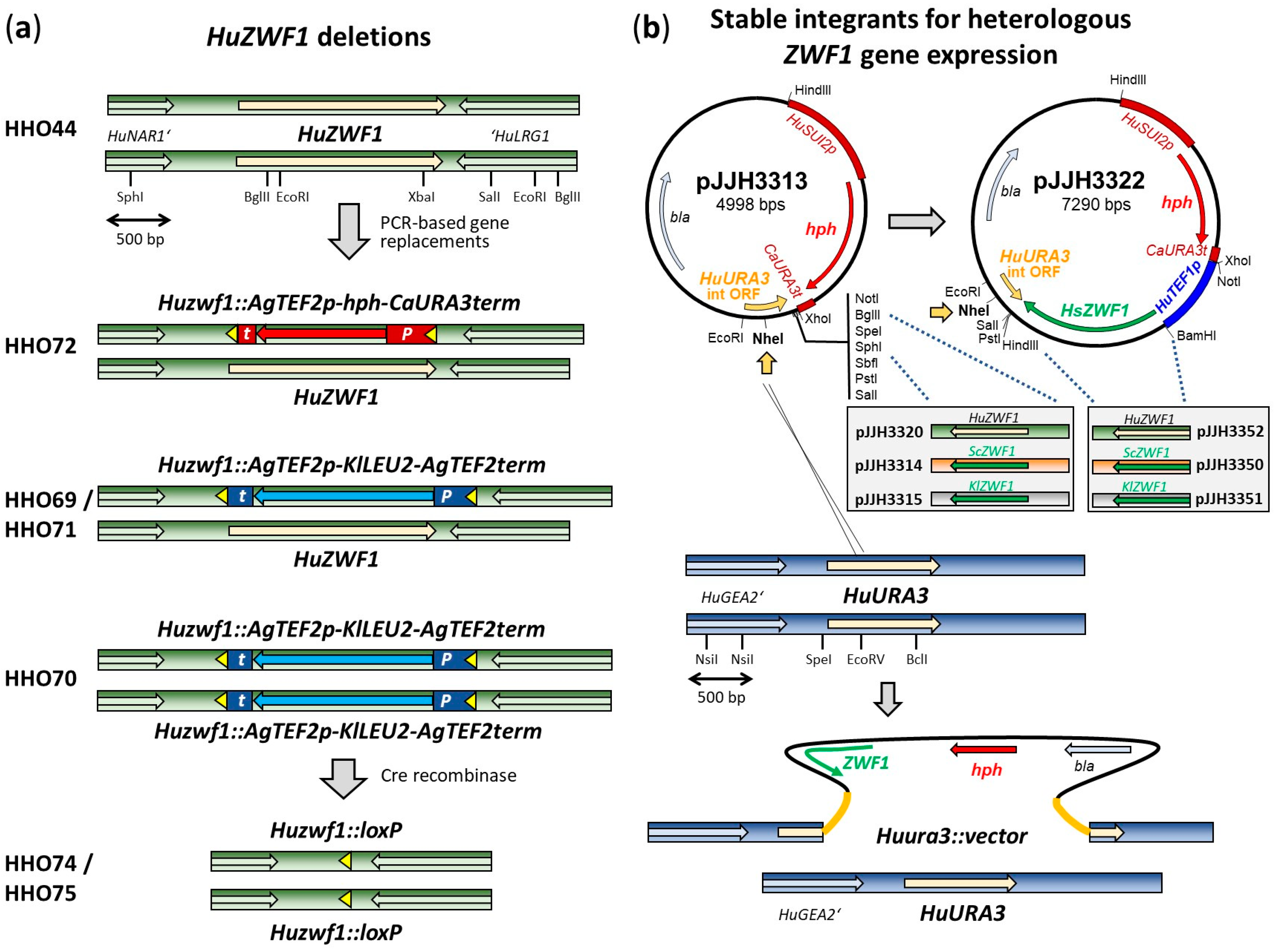
Searching the annotated genome sequence of Hanseniaspora uvarum for homologies to glucose-6-phosphate dehydrogenases (G6PDH), we identified one candidate gene to encode the enzyme, further designated as HuZWF1. The deduced amino acid sequence is comprised of 533 residues and was aligned with the homologues from Saccharomyces cerevisiae (ScZwf1p), Kluyveromyces lactis (KlZwf1p), and a human isoform (HsZwf1p; Figure 1a, see legend for accession numbers), sharing 63%, 67%, and 45% overall identity with HuZwf1, respectively. Domains known to bind glucose, or the nucleotide cofactor, were more strongly conserved, indicating that HuZWF1 indeed encodes a functional homologue. This notion was further supported by the modelling of the three-dimensional structure, which was highly similar to human G6PDH and the enzymes from the two other yeast species for the core structure (Figure 1b). Interestingly, HuZwf1p shares an additional N-terminal helix with the human isoform, which is not present in the other yeast enzymes.
In order to provide experimental proof for this enzyme function, we decided to delete the encoding gene from strain HHO44 (Huleu2/Huleu2). Since this strain is diploid, a strategy to consecutively substitute the two alleles with different marker cassettes was originally employed, as exercised previously for the auxotrophic markers (Figure 2a; [36]). However, in the course of verification by PCR, two clones turned out to carry the homozygous deletion of both alleles with the KlLEU2 cassette in a single transformation of HHO44, which were further employed.
The resulting strains were then grown in rich medium into a logarithmic phase and harvested for the preparation of crude extracts. Specific G6PDH activities were determined and are listed in Table 1 (upper part).
Table 1. Specific G6PDH activities.
StrainRelevant ZWF1 AllelesmU/mg Protein
HHO1Wild-type252 ± 11
HHO44Wild-type257 ± 4
HHO69Huzwf1Δ/HuZWF1132 ± 16
HHO72Huzwf1Δ/HuZWF1123 ± 13
HHO70Huzwf1Δ/Huzwf1Δb.d.
HHO75Huzwf1Δ/Huzwf1Δb.d.
HHO75/pJJH3313Huzwf1Δ/Huzwf1Δ Huura3::vector/HuURA3b.d.
HHO75/pJJH3320Huzwf1Δ/Huzwf1Δ Huura3::HuZWF1/HuURA3168 ± 2
HHO75/pJJH3314Huzwf1Δ/Huzwf1Δ Huura3::ScZWF1/HuURA311 ± 1
HHO75/pJJH3315Huzwf1Δ/Huzwf1Δ Huura3::KlZWF1/HuURA38 ± 1
HHO75/pJJH3352Huzwf1Δ/Huzwf1Δ Huura3::TEF1p-HuZWF1/HuURA3651 ± 27
HHO75/pJJH3350Huzwf1Δ/Huzwf1Δ Huura3::TEF1p-ScZWF1/HuURA3349 ± 27
HHO75/pJJH3351Huzwf1Δ/Huzwf1Δ Huura3::TEF1p-KlZWF1/HuURA3175 ± 5
HHO75/pJJH3322Huzwf1Δ/Huzwf1Δ Huura3::TEF1p-HsZWF1/HuURA398 ± 12
Given are the mean specific activities of at least three biological replicates, each with three technical replicates (with the exception of the second wild-type, HHO44, and the homozygous deletions HHO70 and HHO75, for which biological duplicates were examined). Species designations for ZWF1 genes are Hu = Hanseniaspora uvarum, Sc = Saccharomyces cerevisiae, Kl = Kluyveromyces lactis, and Hs = Homo sapiens. For complete genotypes and constitution of the deletion alleles, consult Table 2. b.d. = below detection (<2 mU/mg protein).
Table 2. Hanseniaspora uvarum strains used in this work.
StrainGenotypeReference
HHO1Wild-type[36]
HHO44Huleu2::loxP/Huleu2::loxP[36]
HHO68Huleu2::loxP/Huleu2::loxP Huzwf1::KlLEU2/Huzwf1::KlLEU2this work
HHO69Huleu2::loxP/Huleu2::loxP Huzwf1::KlLEU2/HuZWF1this work
HHO70Huleu2::loxP/Huleu2::loxP Huzwf1:: KlLEU2/Huzwf1:: KlLEU2this work
HHO71Huleu2::loxP/Huleu2::loxP Huzwf1:: KlLEU2/HuZWF1this work
HHO72Huleu2::loxP/Huleu2::loxP Huzwf1::hph/HuZWF1this work
HHO74Huleu2::loxP/Huleu2::loxP Huzwf1::loxP/Huzwf1::loxPthis work
HHO75Huleu2::loxP/Huleu2::loxP Huzwf1::loxP/Huzwf1::loxPthis work
HHO75/pJJH3313Huleu2::loxP/Huleu2::loxP Huzwf1::loxP/Huzwf1::loxP Huura3::hph/HuURA3this work
HHO75/pJJH3320Huleu2::loxP/Huleu2::loxP Huzwf1::loxP/Huzwf1::loxP Huura3::HuZWF1-hph/HuURA3this work
HHO75/pJJH3314Huleu2::loxP/Huleu2::loxP Huzwf1::loxP/Huzwf1::loxP Huura3::ScZWF1-hph/HuURA3this work
HHO75/pJJH3315Huleu2::loxP/Huleu2::loxP Huzwf1::loxP/Huzwf1::loxP Huura3::KlZWF1-hph/HuURA3this work
HHO75/pJJH3352Huleu2::loxP/Huleu2::loxP Huzwf1::loxP/Huzwf1::loxP Huura3::TEF1p-HuZWF1-hph/HuURA3this work
HHO75/pJJH3350Huleu2::loxP/Huleu2::loxP Huzwf1::loxP/Huzwf1::loxP Huura3::TEF1p-ScZWF1-hph/HuURA3this work
HHO75/pJJH3351Huleu2::loxP/Huleu2::loxP Huzwf1::loxP/Huzwf1::loxP Huura3::TEF1p-KlZWF1-hph/HuURA3this work
HHO75/pJJH3322Huleu2::loxP/Huleu2::loxP Huzwf1::loxP/Huzwf1::loxP Huura3::TEF1p-HsZWF1-hph/HuURA3this work
hph designates the gene conferring resistance to hygromycin B; HHO74 and HHO75 are two clones derived from HHO70 after elimination of the KlLEU2 marker by Cre-induced recombination.
As expected from retaining one intact gene copy, the heterozygous deletion mutants (HHO69 and HHO72) displayed approximately half of the specific G6PDH activity as the wild-type controls (HHO1 and HHO44), whereas the homozygous deletions (HHO74 and HHO75) showed no detectable activity. This clearly demonstrates that HuZWF1 indeed encodes G6PDH, both alleles encode functional subunits, and that the H. uvarum genome carries no further genes encoding a functional enzyme. This notion was further supported by re-introducing a copy of the wild-type HuZWF1 gene into the homozygous deletion strain (HHO75/pJJH3320), which was integrated at the HuURA3 locus by disrupting one of the alleles, which restored enzyme activity to half the level of the wild-type controls (Table 1, lower part).

2.2. Heterologous Genes Restore G6PDH Activity in Huzwf1 Deletion

Given the striking similarities in G6PDH enzyme structures depicted in Figure 1b and the importance of the enzyme in human health, we wondered whether H. uvarum could serve as an expression organism to study enzymatic parameters of genes expressed from patients or simply for comparative studies of G6PDHs from different sources. Therefore, heterologous genes from humans and the two model yeasts S. cerevisiae and K. lactis were cloned into an integrative vector and stably integrated into the genome of HHO75 (Huzwf1Δ/Huzwf1Δ), disrupting one of the two HuURA3 alleles in the process (Figure 2b). Cells were logarithmically grown in rich medium and harvested for the preparation of crude extracts and the determination of specific G6PDH activities (Table 1, lower part). As expected, integration of the empty vector (pJJH3313) did not restore enzymatic activity, but the integration of a single copy of the HuZWF1 gene under the control of its native promoter yielded half the specific activity of the wild-type control. Strains carrying either the gene from S. cerevisiae (ScZWF1) or that from K. lactis (KlZWF1) under their native promoters yielded only very low specific G6PDH activities, indicating that these promoters are poorly recognized for efficient gene expression in H. uvarum. Consequently, both genes were placed under the control of the strong native HuTEF1 promoter. Specific G6PDH activities of approximately 350 mU/mg protein (HuTEF1p-ScZWF1) and 175 mU/mg protein (HuTEF1p-KlZWF1) in crude extracts from these strains demonstrated functional expression in the broad range of the native HuZwf1p enzyme in wild-type strains. Placing the native gene under the strong promoter (HuTEF1p-HuZWF1) yielded the highest specific activities (approximately 650 mU/mg protein), i.e., triplicating the value of the wild-type control. Finally, the strain expressing the human gene (HuTEF1p-HsZWF1) still displayed activities of approximately 100 mU/mg protein. These findings provide proof-of-principle that H. uvarum is a suitable host for heterologous gene expression.

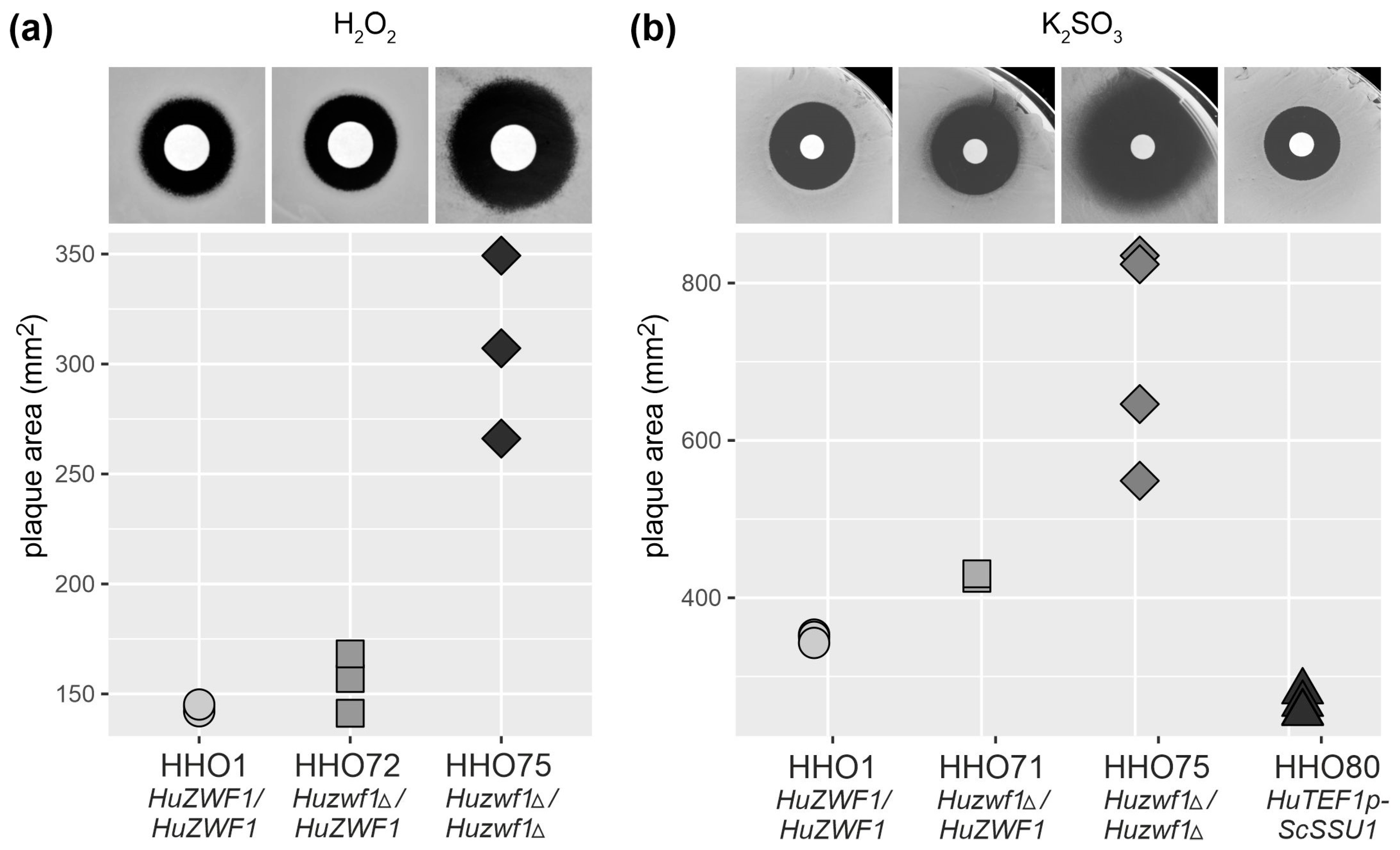
2.3. Lack of HuZwf1 Confers Methionine Auxotrophy and Hypersensitivity towards Hydrogen Peroxide and Sulfite, Whereas Heterologous Expression of a Sulfite Transporter Increases Resistance

As we noticed that the homozygous Huzwf1 deletion mutants produced smaller colonies when streaked out on plates with glucose as a carbon source, we proceeded by recording their growth curves (Figure 3a).
The reducing power in the form of NADPH generated by the G6PDH reaction is required for methionine biosynthesis and to combat oxidative stress both in S. cerevisiae and K. lactis [15,27]. The Huzwf1 deletion strains were therefore tested for their ability to grow on synthetic medium lacking methionine. As evident from Figure 3b, the homozygous deletion strains display severe growth retardation on synthetic medium supplemented with methionine as compared to the wild-type controls and the heterozygous deletion strains still carrying one functional gene copy (note that histidine was omitted from the control plates to demonstrate that the strains did not display a general growth defect in the absence of amino acids). However, the homozygous deletions do not grow when methionine is omitted, in contrast to the other strains. We concluded that while sufficient NADPH for methionine synthesis can be produced by strains carrying one gene copy of HuZWF1, a complete lack of enzyme activity (in strains HHO70 and HHO75) renders them auxotrophic for this amino acid.
In order to test the sensitivity of the strains towards oxidative stress and sulfite, we employed growth inhibition halo assays (Figure 4). To assess the reaction towards oxidative stress, sterile filter discs soaked with 10 µL of different hydrogen peroxide concentrations were placed on a lawn of cells to determine the zones of inhibition after overnight incubation at 28 °C (with a concentration of 2.8 mM shown in the upper part of Figure 4a).
Strains with a heterozygous deletion (Huzwf1Δ/HuZWF1) displayed a similar sensitivity to the wild-type, as indicated by the respective halo areas, whereas homozygous deletions (Huzwf1Δ/Huzwf1Δ) were much more sensitive. We concluded that H. uvarum, like other yeasts, requires G6PDH activity to efficiently cope with oxidative stress.
Wine production commonly involves treatment with sulfite as an antioxidant and for microbial stabilization. Although H. uvarum frequently predominates the yeast population at the beginning of wine fermentations, it is much more sensitive to this treatment than the starter strains of S. cerevisiae frequently employed. In fact, growing the yeasts in rich medium, we found that a diploid laboratory strain of S. cerevisiae (HHD1; [39]) was inhibited by potassium bisulfite concentrations of 3 g/L; a commercial starter strain for vinification (Lalvin EC1118) still grew at more than 4 g/L, whereas the H. uvarum strain HHO1, from which all strains used in this work are derived, already ceased growth at less than 1.5 g/L.
To determine whether G6PDH contributes to sulfite resistance, similar halo assays to those described above for hydrogen peroxide were performed, with potassium bisulfite as an inhibitory agent at different concentrations (Figure 4b shows the results at 3.6 M). The sensitivity of the homozygous deletion strain (Huzwf1Δ/Huzwf1Δ) was clearly increased when compared to the wild-type control, and the heterozygous deletion still carrying one functional allele (Huzwf1Δ/HuZWF1) displayed an intermediary phenotype.
Since sulfite resistance may be of significant biotechnological importance for the future application of H. uvarum, this was further investigated. In wine strains of S. cerevisiae, sulfite resistance can be largely attributed to the overexpression of the SSU1 gene, which encodes a sulfite pump [40]. Consequently, we placed this heterologous gene under the control of the strong HuTEF1 promoter and stably integrated the construct at the HuURA3 locus in strain HHO1, similar to the strategy explained in Figure 2b. As shown in Figure 4b (upper right corner and quantification), this strain (Huura3::TEF1p-SSU1) exhibited smaller halos of inhibition as compared to the wild-type control (HuZWF1/HuZWF1). This indicates that the heterologous SSU1 gene is expressed and produces a functional sulfite exporter in H. uvarum.

3. Discussion

Glucose-6-phosphate dehydrogenase is an important enzyme not only for human health but also to ensure autonomous metabolism in microbial cells, including yeasts [1,2]. Here, we investigated the genetics and physiology of this enzyme in the abundant wine yeast Hanseniaspora uvarum, taking advantage of the recently developed methods for its genomic manipulation.
A homology search against the annotated H. uvarum genome sequence revealed the presence of only one G6PDH-encoding gene, consequently named HuZWF1. Besides the alignment of the deduced amino acid sequence with those of G6PDHs from humans and two other yeast species, revealing high sequence identities, structure predictions provided strong evidence for HuZwf1p being a functional homolog. Interestingly, HuZwf1p and its human homologue both carry an N-terminal extension, which is not present in the enzymes of the yeasts Saccharomyces cerevisiae or Kluyveromyces lactis. Although the extensions are different in their primary sequences, they both form an alpha-helical region according to the structure predictions. Apparently, this is not required for enzymatic catalysis, as the two other yeast enzymes, which do not carry the extension, confer G6PDH activity when expressed in a Huzwf1 deletion strain. It is still possible that the extension could be important for fine-tuning enzyme activity and/or regulatory interaction with other proteins in H. uvarum and humans. While interesting, addressing this question would require elaborate and time-consuming experimental setups beyond the scope of this manuscript.
Given the strong similarities in the core enzyme structure and the fact that both human and yeast G6PDHs form homotetramers, we assume that HuZwf1p also adopts a tetrameric structure [28,41,42]. It yielded specific G6PDH activities in wild-type strains of H. uvarum of approximately 250 mU/mg protein. This is about 2.5-fold higher than the activity in a wild-type S. cerevisiae strain [19,43], indicating that H. uvarum has a higher capacity to channel glucose-6-phosphate into the pentose phosphate pathway. This relates to H. uvarum being a Crabtree-negative yeast, which relies on a more respiratory metabolism, as opposed to the mainly fermentative S. cerevisiae. Yet, the Crabtree-negative milk yeast K. lactis displays G6PDH activities of approximately 430 mU/mg protein, suggesting a still higher capacity [43]. In K. lactis, strong evidence for the PPP contributing substantially to glucose metabolism was provided by mutant analyses. Thus, the lack of activity of either a glycolytic enzyme or of transaldolase in the PPP did not preclude growth on glucose as a sole carbon source, whereas double mutants with blocks in both pathways were glucose-negative [26]. Similar experiments would be required in H. uvarum to demonstrate a significant contribution of the PPP to sugar metabolism. In fact, we are in the process of also constructing deletion mutants in the genes encoding both phosphofructokinase subunits, HuPFK1 and HuPFK2 ([33]; and unpublished results). However, since H. uvarum is diploid, with the necessity to delete two alleles of each gene, and lacks a sexual life cycle, impeding conventional crossing and tetrad analysis to combine genetic markers, construction of homozygous triple mutants may be hard to realize. This is further complicated by the expected glucose-negative phenotype of such triple mutants, which would require physiological studies to find a non-fermentable carbon source that supports the growth of H. uvarum.
Apart from these obstacles, the present work confirms previous findings that homologous recombination can be employed for efficient manipulation of H. uvarum [34,36]. Thus, all constructs for heterologous ZWF1 gene expression integrated readily at the HuURA3 locus, targeted by linearization of the vectors prior to transformation, as is common practice in S. cerevisiae [44]. Moreover, both HuZWF1 alleles were deleted in one step from the diploid HHO44 genome by the use of a deletion cassette with long flanking homologies to the target sequence. This was not an isolated instance, as it also occurred in the deletion of one of the HuPFK genes (unpublished results from our laboratory). For future works, the investigation of a larger number of colonies arising on selective media in the first transformation may thus be worth the effort before applying the two-step deletion strategy to eliminate both target alleles, as suggested previously [36].
In the course of the heterologous ZWF1 gene expression studies, we noticed that the genes from S. cerevisiae and K. lactis were very poorly expressed in H. uvarum under the control of their native promoters, reflected by the low specific G6PDH activities. This stands in contrast to the exchange of these and other genes between S. cerevisiae and K. lactis, which did not pose problems or require promoter modification (see, for example, [25,45] and references therein). However, it confirms previous suggestions that promoters from S. cerevisiae genes may not confer sufficient expression in H. uvarum [34]. Yet, when placed under the control of the strong native HuTEF1 promoter, the heterologous genes were successfully expressed in H. uvarum. As a control, HuZWF1 expressed from the strong promoter displayed almost three-fold higher activities than a wild-type strain. Although the two yeast enzymes and the HuTEF1p-HsZWF1 construct conferred less activity (the latter at the lower end with approximately 100 mU/mg protein as compared to 250 mU/mg protein in wild-type H. uvarum), it would certainly allow further biochemical and biophysical protein characterizations. Yeasts clearly possess several advantages as heterologous expression hosts for human disease alleles: (i) They are eukaryotes and thus more suitable to produce human proteins than E. coli. (ii) With their short generation time, sufficient cell mass can be produced overnight for protein purifications. (iii) They can be easily genetically manipulated, as demonstrated herein. (iv) Most yeast species, including H. uvarum, are generally regarded as safe (GRAS). Regarding H. uvarum as an expression host, it should be noted that features like metabolic bottlenecks, the influence of protein folding, posttranslational modifications, or the burden generated by the overproduction of foreign proteins, as studied in the model yeast S. cerevisiae, still need to be thoroughly investigated. The current study only provides a first step in this direction.
The homozygous Huzwf1 deletion mutant obtained was also subjected to phenotypic characterizations. As expected from data on Sczwf1 mutants, the H. uvarum strain lacking G6PDH activity also required methionine for growth [15]. This has been attributed to a lack of NADPH, three molecules of which are required for reductive reactions in the biosynthesis of one molecule of methionine [13]. This feature of the zwf1 deletion has been exploited for biotechnological purposes in S. cerevisiae, for instance, to improve xylose fermentation [46] or to construct a platform strain which overproduces tyrosine [47]. As methods for genetic manipulation of H. uvarum are only just emerging, and its ability to utilize pentoses as a sole carbon source has not been thoroughly investigated, our results on the role of G6PDH should be kept in mind until similar platform strains are available for this yeast. The Huzwf1 mutant also showed a reduced growth on standard rich medium with glucose, a phenotype only observed in our laboratory strain of S. cerevisiae but not in others [15,18,36]. This indicates that the PPP indeed may contribute, to a larger extent, to sugar catabolism in H. uvarum as compared to S. cerevisiae.
A lack of G6PDH activity in S. cerevisiae has also been related to its oxidative stress response [13]. Accordingly, the first deletion mutant constructed proved to be hypersensitive towards hydrogen peroxide [15], a feature shared by strains of all genetic backgrounds tested so far [13,18,36]. It was thus not surprising to find an increased sensitivity towards this stress agent also for the homozygous Huzwf1 deletion strain. This also makes sense in the natural environment of H. uvarum, as it frequently dominates the yeast population on grape skins [30]. The constant exposure to oxygen, in combination with its respiratory metabolism, is expected to produce reactive oxygen species as a side effect. Efficient detoxification mechanisms are thus a prerequisite. For instance, glutathione reductase is an enzyme for which the G6PDH reaction provides the reducing power as the primary source for NADPH in yeasts [48]. Moreover, glutathione synthesis itself is dependent on methionine metabolism, which, as outlined above, also requires G6PDH activity [49].
Finally, we observed that the homozygous Huzwf1 deletion also results in an increased sensitivity towards sulfite as compared to wild-type H. uvarum strains. Although the zwf1 deletions in other yeasts have not yet been tested for their effect on sulfite tolerance, a connection to oxidative stress response has been insinuated for S. cerevisiae [50]. It is likely that the molecular basis for this feature is the NADPH-dependent sulfite reductase encoded by MET10 in S. cerevisiae, which is required for the reduction of sulfite as a precursor in methionine biosynthesis [51]. Clearly, sulfite resistance is of special interest for the biotechnological application of H. uvarum as the predominant yeast in the early stages of wine fermentations and would be a desirable trait. Thus, expanding the S. cerevisiae starter cultures in large-scale industrial winemaking by using mixed populations to improve wine quality, such as starter cultures of S. cerevisiae plus H. uvarum or other non-conventional yeasts, are under investigation [52,53,54,55]. As sulfite is commonly added for microbial stabilization and an antioxidant in winemaking, strains of H. uvarum with an increased tolerance would be advantageous. One would think that adaptive laboratory evolution (ALE) with stepwise exposure of a wine strain to increasing sulfite concentrations would be a reasonable approach [56]. However, sulfite resistance in wine strains of S. cerevisiae is mainly due to overexpression of the sulfite-pump encoding SSU1 gene caused by chromosomal translocations [40]. In silico analyses revealed that the H. uvarum sequence probably lacks a homologue of this gene, diminishing the likelihood of an effective natural selection approach. Instead, our results suggest that heterologous expression of the ScSSU1 gene in H. uvarum could be an alternative, as strains proved to be more resistant to sulfite than the recipient wild-type strain. Of course, this does not preclude the possibility of using in vitro mutagenesis on the ScSSU1 expression vector, SSU1 genes from other yeast species, and/or ALE on the manipulated H. uvarum strains to further enhance the resistance level. Also, it remains to be determined whether overexpression of HuZWF1 in this context could also contribute to improving sulfite resistance.
In summary, we showed that the homozygous Huzwf1 deletion strain can serve as a host for expression of heterologous ZWF1 genes, including alleles with mutations from human patients. Although this has been previously exercised in a S. cerevisiae host for human G6PDH variants [57], H. uvarum may provide some advantages. For example, it grows faster and reaches higher cell densities than S. cerevisiae, providing more biomass for protein purification [33]. In addition, its respiratory metabolism provides a physiological environment resembling human cells more closely than the strongly fermentative baker’s yeast. The expression of the ScSSU1 gene further demonstrated that heterologous expression is not restricted to G6PDH but can generally be applied to other proteins of interest. The genetic manipulations exercised herein will also increase the potential of H. uvarum for its different biotechnological applications [29].

4. Materials and Methods

4.1. Media, Strains, and Culture Conditions

Rich media were based on yeast extract (1% w/v) and peptone (2% w/v) from Difco Laboratories Inc., Detroit, MI, USA, with glucose (2% w/v) as a carbon source (YEPD). Synthetic media were based on Difco yeast nitrogen base with ammonium sulfate as described in [58], with the addition of amino acids and bases using a mixture provided by MP Biomedicals (Eschwege, Germany; CSM-His-Leu-Trp-Ura) supplemented as required for the selection of plasmids or deletion markers, and 2% glucose (w/v) as carbon source (SCD). In total, 400 mg/L of hygromycin B or 50 mg/L G418 were used for marker selection. For the preparation of solid media, 1.5% agar was added prior to sterilization. Yeasts were incubated at 28 °C, with constant agitation at 180 rpm for liquid cultures.
H. uvarum strains employed in this work are listed in Table 2. For the purpose of plasmid constructions by in vivo recombinations, the diploid S. cerevisiae strain DHD5 was used [58]. Comparative assays for sulfite resistance also included the S. cerevisiae strains HHD1 [39] and the commercial wine yeast strain Lalvin EC1118, kindly provided by Christian von Wallbrunn (Hochschule Geisenheim, Germany).
For work with E. coli, strain DH5α (Invitrogen, Karlsruhe, Germany) was employed, grown at 37 °C in LB medium (0.5% yeast extract, 1% Bacto peptone, 0.5% sodium chloride) and supplemented with ampicillin (50 mg/L) for plasmid selection.

4.2. Construction of Deletion Mutants and Cloning Procedures

For the transformation of H. uvarum, the freeze method was used as previously described [36], with the exception that cells were exposed to a heat shock for 5 min at 28 °C instead of 30 °C after the addition of DNA.
Deletions of the HuZWF1 alleles were obtained by PCR-based one-step gene replacements. Oligonucleotides employed for this and other purposes are listed in Table 3.
To obtain longer flanking regions for homologous recombination in H. uvarum, the HuZWF1 gene was first amplified by PCR with the primer pair 20.277/20.278, using DNA from strain HHO1 as a template and originally cloned as a BamHI/SalI fragment. Several steps of subcloning finally led to the insertion of the fragment into a derivative of the S. cerevisiae multicopy vector YEplac195 [59] to yield pJJH3286. The latter was linearized at the single BglII site, dephosphorylated, and cotransformed with PCR-generated deletion fragments into S. cerevisiae strain DHD5. Deletion cassettes were amplified with the primer pair 22.193/22.194 from pJJH3204 [36] to generate pJJH3287 (Huzwf1::AgTEF2p-hph-CaURA3term) and from pJJH1287 to generate pJJH3388 (Huzwf1::AgTEF2p-KlLEU2-AgTEF2term). Both PCR products included the flanking loxP sites. The insertions with the markers flanked by HuZWF1 up- and downstream sequences were subcloned into pUK1921 [60], yielding pJJH3293 and pJJH3294, respectively, from which they were excised by digestion with BamHI/SphI prior to transformation of HHO44. As exercised previously for the construction of strains with auxotrophic markers [36], a consecutive strategy with the two selection markers was initially followed, resulting in strains with the genomic constitution depicted in Figure 2a and listed in Table 2. However, the approach of substituting HuZWF1 by the KlLEU2 marker from pJJH3394 already yielded two homozygous deletion clones (Huzwf1::KlLEU2/Huzwf1::KlLEU2) in the first attempt amongst several expected heterozygous ones (Huzwf1::KlLEU2/HuZWF1). After verification of the correct deletion by PCR with flanking and internal oligonucleotides, the episomal vector pJJH3203 (kanMX HuTEF1p-Cre) was introduced into one of the homozygous deletion strains to produce the Cre recombinase and remove both KllEU2 marker alleles, retaining single loxP sites substituting the original HuZWF1 open reading frames (Figure 2a; [36]). To induce plasmid loss, cells were grown overnight in non-selective rich medium, streaked out for single colonies on YEPD plates, and incubated for 2 days at 28 °C. They were then replica-plated onto YEPD with or without 50 mg/L of G418, and clones sensitive to the latter were picked for further investigations.
Integrative plasmids for expression of heterologous ZWF1 genes were obtained by subcloning genes from various existing constructs [19], introducing new restriction sites by PCR if required. Clones containing PCR fragments were verified by Sanger sequencing (Seqlab, Göttingen, Germany) as not containing errors in the open reading frames before they were introduced into H. uvarum HHO44. Complete sequences of all plasmids described in the following are available upon request. As a vector for integration/disruption at the HuURA3 locus, pJJH3313 was constructed (Figure 2b). It is based on the pUC19 backbone [61], into which the HuSUI2p-hph-CaURA3term region from pJJH3152 [36] was inserted, together with a polylinker to facilitate the cloning of heterologous genes. It also contains an internal fragment of the HuURA3 gene (from position 112 to 470 in the open reading frame, amplified with the primer pair 21.304/21.305), which was cloned as an EcoRI/SalI fragment with a unique internal NheI site introduced for linearization to promote homologous recombination upon introduction into H. uvarum. This vector was used to insert different ZWF1 genes as follows: The ScZWF1 gene and the KlZWF1 gene with their flanking regions were cloned as SphI/SalI fragments into pJJH3313 to yield pJJH3314 and pJJH3315, respectively. The SphI sites were introduced with the primers 23.089 and 23.090, respectively, in combination with 99.113 or 14.111 as required, amplifying the genes from clones previously obtained [19]. HuZWF1 was cloned as a HindIII/SalI fragment, which also included the hph resistance cassette, to yield pJJH3320. The human G6PDH gene was first placed under the control of the strong HuTEF1 promoter, which was introduced as an EcoRI/BamHI fragment into pJJH2223 [19] to yield pJJH3318. After further cloning steps, it was inserted as a NotI/SalI fragment into pJJH3313, resulting in pJJH3322 (Figure 2b). The latter was used as a basis to replace the HsZWF1 coding sequence with those of the different yeast ZWF1 genes. Thus, ScZWF1 and KlZWF1 were cloned as BamHI/XhoI fragments into pJJH3322 digested with BamHI/SalI to yield pJJH3350 and pJJH3351, respectively. Finally, HuZWF1 was introduced into the pJJH3322 backbone as a BamHI/SalI fragment to give pJJH3352. All integrative plasmids were linearized with NheI and introduced into strain HHO1, selecting for hygromycin resistance. Correct insertions disrupting one of the HuURA3 alleles were verified by PCR.
For heterologous expression of the ScSSU1 gene, which encodes a sulfite pump, a similar strategy was followed. Thus, the ScSSU1 sequence starting with the ATG translation start codon and carrying 345 base pairs of the terminator region was amplified by PCR from DNA of strain DHD5 with the primer pair 20.349/20.350. It was cloned into a HuURA3 integration vector similar to pJJH3322 under the control of the HuTEF1 promoter to yield pJJH3167. Integration was targeted by linearization with Bsp119I prior to the transformation of strain HHO1. Transformants were selected for hygromycin B resistance and verified for correct integration/disruption at the HuURA3 locus.

4.3. Growth Curves

Cells were pregrown overnight in 3 mL of YEPD at 28 °C with shaking at 180 rpm, diluted to an OD600 of 0.3 in fresh SCD medium, and allowed to grow for another 5–6 h. They were adjusted to OD600 of 0.1 in SCD, and three technical replicates of each strain were applied to a microtiter plate. Growth curves were obtained in 100 µL cultures in 96-well plates in SCD medium and recorded in a Varioscan Lux plate reader (Thermo Scientific, Bremen, Germany). Except for the growth temperature of 28 °C, all settings and calculations were as previously described [62]. For statistical analysis and preparation of graphics, R v. 4.1.0 and R Studio v. 1.41717 were used [63]. The maximal growth rates were determined by moving a sliding window of width between 5 and 15 measurements along the dataset and sequentially performing linear regression.

4.4. Enzyme Assays

Cultures for enzymatic determinations were grown overnight in rich medium (YEPD), inoculated in fresh medium to an OD600 of 2.0, and grown for another 4–5 h with shaking at 180 rpm at 28 °C. Cells were then harvested by centrifugation, washed once with water and once with Tris-HCl buffer, pH7.5, and stored at −20 °C until use. Glucose-6-phosphate dehydrogenase activities were determined routinely from yeast crude extracts prepared by breaking of cells with glass beads and centrifugation, as described previously [19]. G6PD activities were determined in Tris-HCl buffer, pH7.5, by following the kinetics of NADP reduction at a final concentration of 0.4 mM, added from a 40 mM stock solution in extraction buffer, in a DU800 spectrophotometer (Beckman-Coulter, Krefeld, Germany) at 340 nm and 30 °C. Glucose-6-phosphate was added at a final concentration of 2 mM. All chemicals used for enzyme tests were obtained from Sigma-Aldrich Chemie GmbH, Munich, Germany.

4.5. Inhibition Halo Assays

To examine growth inhibition, strains were grown overnight in liquid-rich medium (YEPD), diluted to an OD600 of 1.0 (wild-type and heterozygous Huzwf1Δ/HuZWF1 mutants) or 2.0–3.0 (homozygous Huzwf1Δ/Huzwf1Δ mutants), and 300 µL were used to cover SCD plates with 30 mL of medium lacking uracil with a homogenous lawn. After drying the plates in a sterile hood, circular filter papers soaked with the reagents (between 0–13.4 mM for hydrogen peroxide or 0–3.6 M for potassium bisulfite) were deposited.
Plates were incubated for 1–2 days at 28 °C, scanned, and the area of inhibition was determined using the Analyze Particles function of Fiji (based on ImageJ 153c (https://imagej.net/, accessed on 2 February 2024). For statistical analysis and preparation of graphics, R v. 4.1.0 and R Studio v. 1.41717 were used [63].

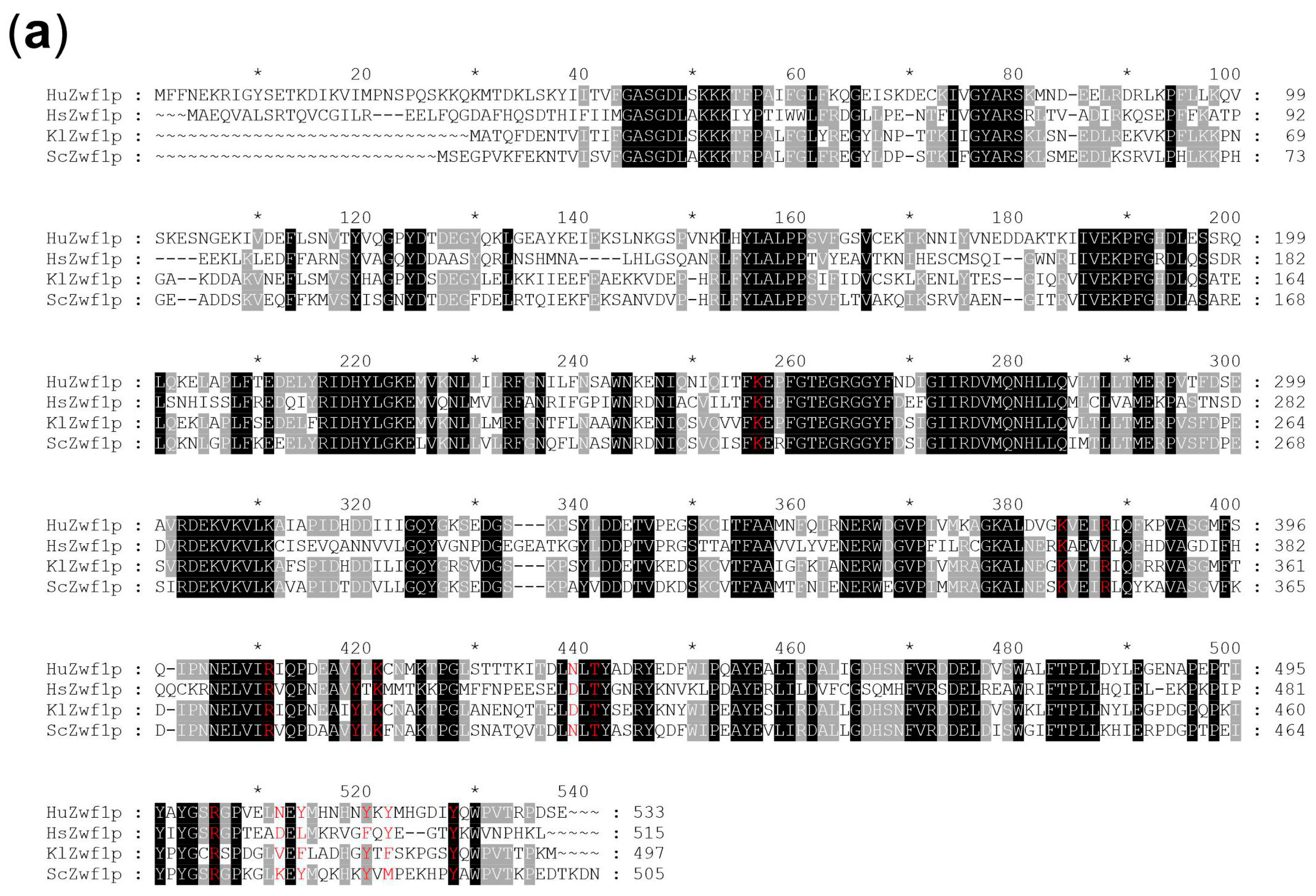
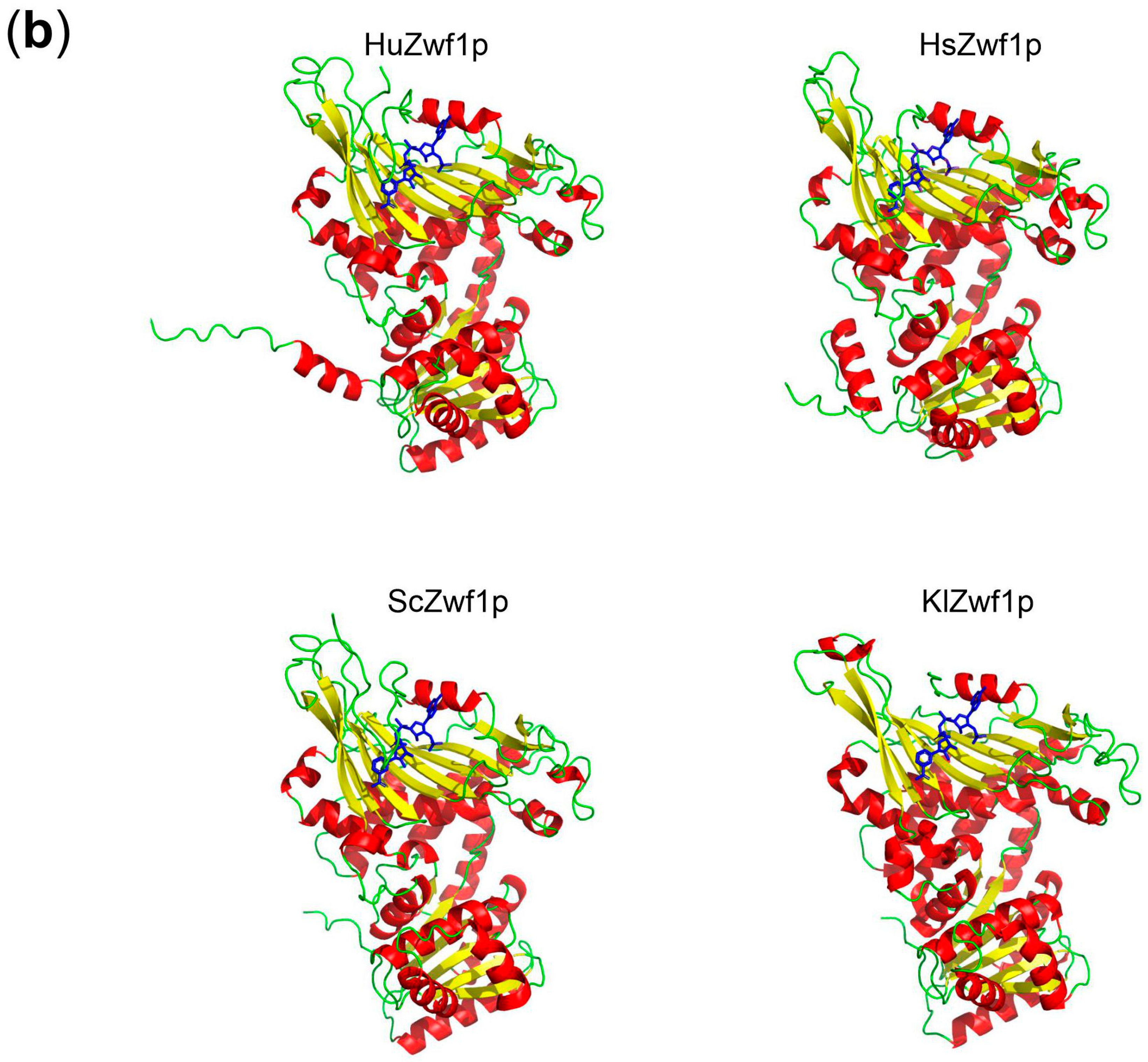
4.6. In Silico Analyses

The annotated H. uvarum genome sequence (GenBank accession number APLS01000000; [33]) was employed for homology searches and to extract the HuZWF1 sequence. Alignments were performed with Clustal Omega [64]. For structure prediction of the HuZwf1p protein, the AlphaFold 2 program was used [37,38]. Calculated structures for G6PDH from the AlphaFold Database were used for S. cerevisiae and H. sapiens. The structure of the Kluyveromyces lactis G6PDH is an experimentally determined structure [42]. The Open-Source version of PyMOL v. 2.6.0a0 [65] was used for visualization and alignment of the protein structures.

Author Contributions

Conceptualization, J.J.H.; methodology, J.J.H., L.F.M., A.M. and H.-P.S.; software, H.-P.S.; validation, J.J.H., L.F.M., A.M. and H.-P.S.; formal analysis, J.J.H. and H.-P.S.; investigation, J.J.H., L.F.M. and A.M.; resources, J.J.H.; data curation, J.J.H., A.M. and H.-P.S.; writing—original draft preparation, J.J.H.; writing—review and editing, J.J.H. and H.-P.S.; supervision, J.J.H.; project administration, J.J.H.; funding acquisition, J.J.H. All authors have read and agreed to the published version of the manuscript.

Funding

This research was funded by a research allowance from the University of Osnabrück and a grant from the Forschungsring des Deutschen Weinbaus.

Institutional Review Board Statement

Not applicable.

Informed Consent Statement

Not applicable.

Data Availability Statement

Protein and nucleic acid sequences are available from the public databases as cited. Sequences of plasmids constructed and manipulated chromosomal loci are available upon request.

Acknowledgments

We appreciate the contribution of Philip Harpering in optimizing the transformation procedure during an undergraduate course.

Conflicts of Interest

The authors declare no conflicts of interest.

Abbreviations

CFU = colony forming units; G6PDH = glucose-6-phosphate dehydrogenase; PPP = pentose phosphate pathway; ROS = reactive oxygen species; ZWF = gene encoding G6PDH (“Zwischenferment”).

References

  1. Bertels, L.K.; Fernandez Murillo, L.; Heinisch, J.J. The pentose phosphate pathway in yeasts—More than a poor cousin of glycolysis. Biomolecules 2021, 11, 725. [Google Scholar] [CrossRef] [PubMed]
  2. Stincone, A.; Prigione, A.; Cramer, T.; Wamelink, M.M.; Campbell, K.; Cheung, E.; Olin-Sandoval, V.; Gruning, N.M.; Kruger, A.; Tauqeer Alam, M.; et al. The return of metabolism: Biochemistry and physiology of the pentose phosphate pathway. Biol. Rev. Camb. Philos. Soc. 2015, 90, 927–963. [Google Scholar] [CrossRef] [PubMed]
  3. Patra, K.C.; Hay, N. The pentose phosphate pathway and cancer. Trends Biochem. Sci. 2014, 39, 347–354. [Google Scholar] [CrossRef] [PubMed]
  4. Riganti, C.; Gazzano, E.; Polimeni, M.; Aldieri, E.; Ghigo, D. The pentose phosphate pathway: An antioxidant defense and a crossroad in tumor cell fate. Free Radic. Biol. Med. 2012, 53, 421–436. [Google Scholar] [CrossRef] [PubMed]
  5. Ho, H.Y.; Cheng, M.L.; Chiu, D.T. Glucose-6-phosphate dehydrogenase--beyond the realm of red cell biology. Free Radic. Res. 2014, 48, 1028–1048. [Google Scholar] [CrossRef] [PubMed]
  6. Jain, M.; Brenner, D.A.; Cui, L.; Lim, C.C.; Wang, B.; Pimentel, D.R.; Koh, S.; Sawyer, D.B.; Leopold, J.A.; Handy, D.E.; et al. Glucose-6-phosphate dehydrogenase modulates cytosolic redox status and contractile phenotype in adult cardiomyocytes. Circ. Res. 2003, 93, e9–e16. [Google Scholar] [CrossRef] [PubMed]
  7. Mehta, A.; Mason, P.J.; Vulliamy, T.J. Glucose-6-phosphate dehydrogenase deficiency. Baillieres Best Pract. Res. Clin. Haematol. 2000, 13, 21–38. [Google Scholar] [CrossRef] [PubMed]
  8. Gomez-Manzo, S.; Marcial-Quino, J.; Vanoye-Carlo, A.; Serrano-Posada, H.; Ortega-Cuellar, D.; Gonzalez-Valdez, A.; Castillo-Rodriguez, R.A.; Hernandez-Ochoa, B.; Sierra-Palacios, E.; Rodriguez-Bustamante, E.; et al. Glucose-6-phosphate dehydrogenase: Update and analysis of new mutations around the world. Int. J. Mol. Sci. 2016, 17, 2069. [Google Scholar] [CrossRef]
  9. Luzzatto, L.; Ally, M.; Notaro, R. Glucose-6-phosphate dehydrogenase deficiency. Blood 2020, 136, 1225–1240. [Google Scholar] [CrossRef]
  10. Ahamed, A.; Hosea, R.; Wu, S.; Kasim, V. The emerging roles of the metabolic regulator G6PD in human cancers. Int. J. Mol. Sci. 2023, 24, 17238. [Google Scholar] [CrossRef]
  11. Horecker, B.L. The pentose phosphate pathway. J. Biol. Chem. 2002, 277, 47965–47971. [Google Scholar] [CrossRef]
  12. Lobo, Z.; Maitra, P.K. Pentose phosphate pathway mutants of yeast. Mol. Gen. Genet. 1982, 185, 367–368. [Google Scholar] [CrossRef]
  13. Campbell, K.; Vowinckel, J.; Keller, M.A.; Ralser, M. Methionine metabolism alters oxidative stress resistance via the pentose phosphate pathway. Antioxid. Redox Signal. 2016, 24, 543–547. [Google Scholar] [CrossRef]
  14. Nogae, I.; Johnston, M. Isolation and characterization of the ZWF1 gene of Saccharomyces cerevisiae, encoding glucose-6-phosphate dehydrogenase. Gene 1990, 96, 161–169. [Google Scholar] [CrossRef]
  15. Thomas, D.; Cherest, H.; Surdin-Kerjan, Y. Identification of the structural gene for glucose-6-phosphate dehydrogenase in yeast. Inactivation leads to a nutritional requirement for organic sulfur. EMBO J. 1991, 10, 547–553. [Google Scholar] [CrossRef]
  16. Ralser, M.; Wamelink, M.M.; Kowald, A.; Gerisch, B.; Heeren, G.; Struys, E.A.; Klipp, E.; Jakobs, C.; Breitenbach, M.; Lehrach, H.; et al. Dynamic rerouting of the carbohydrate flux is key to counteracting oxidative stress. J. Biol. 2007, 6, 10. [Google Scholar] [CrossRef]
  17. Dick, T.P.; Ralser, M. Metabolic remodeling in times of stress: Who shoots faster than his shadow? Mol. Cell 2015, 59, 519–521. [Google Scholar] [CrossRef] [PubMed]
  18. Miosga, T.; Zimmermann, F.K. Cloning and characterization of the first two genes of the non-oxidative part of the Saccharomyces cerevisiae pentose-phosphate pathway. Curr. Genet. 1996, 30, 404–409. [Google Scholar] [CrossRef] [PubMed]
  19. Heinisch, J.J.; Knuesting, J.; Scheibe, R. Investigation of heterologously expressed glucose-6-phosphate dehydrogenase genes in a yeast zwf1 deletion. Microorganisms 2020, 8, 546. [Google Scholar] [CrossRef]
  20. Barnett, J.A.; Payne, R.W.; Yarrow, D. Yeasts: Characteristics and Identification; Cambridge University Press: Cambridge, UK, 2000. [Google Scholar]
  21. Lagunas, R.; Dominguez, C.; Busturia, A.; Saez, M.J. Mechanisms of appearance of the Pasteur effect in Saccharomyces cerevisiae: Inactivation of sugar transport systems. J. Bacteriol. 1982, 152, 19–25. [Google Scholar] [CrossRef] [PubMed]
  22. Heinisch, J.J.; Buchwald, U.; Gottschlich, A.; Heppeler, N.; Rodicio, R. A tool kit for molecular genetics of Kluyveromyces lactis comprising a congenic strain series and a set of versatile vectors. FEMS Yeast Res. 2010, 10, 333–342. [Google Scholar] [CrossRef]
  23. Rodicio, R.; Heinisch, J.J. Yeast on the milky way: Genetics, physiology and biotechnology of Kluyveromyces lactis. Yeast 2013, 30, 165–177. [Google Scholar] [CrossRef]
  24. Goffrini, P.; Wesolowski-Louvel, M.; Ferrero, I. A phosphoglucose isomerase gene is involved in the Rag phenotype of the yeast Kluyveromyces lactis. Mol. Gen. Genet. 1991, 228, 401–409. [Google Scholar] [CrossRef]
  25. Heinisch, J.; Kirchrath, L.; Liesen, T.; Vogelsang, K.; Hollenberg, C.P. Molecular genetics of phosphofructokinase in the yeast Kluyveromyces lactis. Mol. Microbiol. 1993, 8, 559–570. [Google Scholar] [CrossRef]
  26. Jacoby, J.; Hollenberg, C.P.; Heinisch, J.J. Transaldolase mutants in the yeast Kluyveromyces lactis provide evidence that glucose can be metabolized through the pentose phosphate pathway. Mol. Microbiol. 1993, 10, 867–876. [Google Scholar] [CrossRef]
  27. Saliola, M.; Scappucci, G.; De Maria, I.; Lodi, T.; Mancini, P.; Falcone, C. Deletion of the glucose-6-phosphate dehydrogenase gene KlZWF1 affects both fermentative and respiratory metabolism in Kluyveromyces lactis. Eukaryot. Cell 2007, 6, 19–27. [Google Scholar] [CrossRef]
  28. Saliola, M.; Tramonti, A.; Lanini, C.; Cialfi, S.; De Biase, D.; Falcone, C. Intracellular NADPH levels affect the oligomeric state of the glucose 6-phosphate dehydrogenase. Eukaryot. Cell 2012, 11, 1503–1511. [Google Scholar] [CrossRef] [PubMed]
  29. van Wyk, N.; Badura, J.; von Wallbrunn, C.; Pretorius, I.S. Exploring future applications of the apiculate yeast Hanseniaspora. Crit. Rev. Biotechnol. 2023, 44, 100–119. [Google Scholar] [CrossRef]
  30. Fleet, G.H. Yeast interactions and wine flavour. Int. J. Food Microbiol. 2003, 86, 11–22. [Google Scholar] [CrossRef] [PubMed]
  31. Wang, C.; Garcia-Fernandez, D.; Mas, A.; Esteve-Zarzoso, B. Fungal diversity in grape must and wine fermentation assessed by massive sequencing, quantitative PCR and DGGE. Front. Microbiol. 2015, 6, 1156. [Google Scholar] [CrossRef] [PubMed]
  32. Zott, K.; Miot-Sertier, C.; Claisse, O.; Lonvaud-Funel, A.; Masneuf-Pomarede, I. Dynamics and diversity of non-Saccharomyces yeasts during the early stages in winemaking. Int. J. Food Microbiol. 2008, 125, 197–203. [Google Scholar] [CrossRef] [PubMed]
  33. Langenberg, A.K.; Bink, F.J.; Wolff, L.; Walter, S.; von Wallbrunn, C.; Grossmann, M.; Heinisch, J.J.; Schmitz, H.P. Glycolytic functions are conserved in the genome of the wine yeast Hanseniaspora uvarum, and pyruvate kinase limits its capacity for alcoholic fermentation. Appl. Environ. Microbiol. 2017, 83, e01580-17. [Google Scholar] [CrossRef]
  34. Badura, J.; van Wyk, N.; Brezina, S.; Pretorius, I.S.; Rauhut, D.; Wendland, J.; von Wallbrunn, C. Development of genetic modification tools for Hanseniaspora uvarum. Int. J. Mol. Sci. 2021, 22, 1943. [Google Scholar] [CrossRef] [PubMed]
  35. Badura, J.; van Wyk, N.; Zimmer, K.; Pretorius, I.S.; von Wallbrunn, C.; Wendland, J. PCR-based gene targeting in Hanseniaspora uvarum. FEMS Yeast Res. 2023, 23, foad034. [Google Scholar] [CrossRef] [PubMed]
  36. Heinisch, J.J.; Murra, A.; Jurgens, K.; Schmitz, H.P. A versatile toolset for genetic manipulation of the wine yeast Hanseniaspora uvarum. Int. J. Mol. Sci. 2023, 24, 1859. [Google Scholar] [CrossRef] [PubMed]
  37. Jumper, J.; Evans, R.; Pritzel, A.; Green, T.; Figurnov, M.; Ronneberger, O.; Tunyasuvunakool, K.; Bates, R.; Žídek, A.; Potapenko, A.; et al. Highly accurate protein structure prediction with AlphaFold. Nature 2021, 596, 583–592. [Google Scholar] [CrossRef]
  38. Mirdita, M.; Schutze, K.; Moriwaki, Y.; Heo, L.; Ovchinnikov, S.; Steinegger, M. ColabFold: Making protein folding accessible to all. Nat. Methods 2022, 19, 679–682. [Google Scholar] [CrossRef]
  39. Schehl, B.; Muller, C.; Senn, T.; Heinisch, J.J. A laboratory yeast strain suitable for spirit production. Yeast 2004, 21, 1375–1389. [Google Scholar] [CrossRef]
  40. Garcia-Rios, E.; Nuevalos, M.; Barrio, E.; Puig, S.; Guillamon, J.M. A new chromosomal rearrangement improves the adaptation of wine yeasts to sulfite. Environ. Microbiol. 2019, 21, 1771–1781. [Google Scholar] [CrossRef] [PubMed]
  41. Au, S.W.; Gover, S.; Lam, V.M.; Adams, M.J. Human glucose-6-phosphate dehydrogenase: The crystal structure reveals a structural NADP(+) molecule and provides insights into enzyme deficiency. Structure 2000, 8, 293–303. [Google Scholar] [CrossRef] [PubMed]
  42. Vu, H.H.; Jin, C.; Chang, J.H. Structural basis for substrate recognition of glucose-6-phosphate dehydrogenase from Kluyveromyces lactis. Biochem. Biophys. Res. Commun. 2021, 553, 85–91. [Google Scholar] [CrossRef]
  43. Jacoby, J.J.; Heinisch, J.J. Analysis of a transketolase gene from Kluyveromyces lactis reveals that the yeast enzymes are more related to transketolases of prokaryotic origins than to those of higher eukaryotes. Curr. Genet. 1997, 31, 15–21. [Google Scholar] [CrossRef]
  44. Orr-Weaver, T.L.; Szostak, J.W.; Rothstein, R.J. Yeast transformation: A model system for the study of recombination. Proc. Natl. Acad. Sci. USA 1981, 78, 6354–6358. [Google Scholar] [CrossRef]
  45. Rodicio, R.; Koch, S.; Schmitz, H.P.; Heinisch, J.J. KlRHO1 and KlPKC1 are essential for cell integrity signalling in Kluyveromyces lactis. Microbiology 2006, 152, 2635–2649. [Google Scholar] [CrossRef]
  46. Jeppsson, M.; Traff, K.; Johansson, B.; Hahn-Hagerdal, B.; Gorwa-Grauslund, M.F. Effect of enhanced xylose reductase activity on xylose consumption and product distribution in xylose-fermenting recombinant Saccharomyces cerevisiae. FEMS Yeast Res. 2003, 3, 167–175. [Google Scholar] [CrossRef]
  47. Gold, N.D.; Gowen, C.M.; Lussier, F.X.; Cautha, S.C.; Mahadevan, R.; Martin, V.J. Metabolic engineering of a tyrosine-overproducing yeast platform using targeted metabolomics. Microb. Cell Fact. 2015, 14, 73. [Google Scholar] [CrossRef] [PubMed]
  48. Izawa, S.; Maeda, K.; Miki, T.; Mano, J.; Inoue, Y.; Kimura, A. Importance of glucose-6-phosphate dehydrogenase in the adaptive response to hydrogen peroxide in Saccharomyces cerevisiae. Biochem. J. 1998, 330 Pt 2, 811–817. [Google Scholar] [CrossRef] [PubMed]
  49. Kitada, M.; Ogura, Y.; Monno, I.; Xu, J.; Koya, D. Effect of methionine restriction on aging: Its relationship to oxidative stress. Biomedicines 2021, 9, 130. [Google Scholar] [CrossRef] [PubMed]
  50. Garcia-Rios, E.; Guillamon, J.M. Sulfur dioxide resistance in Saccharomyces cerevisiae: Beyond SSU1. Microb. Cell 2019, 6, 527–530. [Google Scholar] [CrossRef] [PubMed]
  51. Thomas, D.; Surdin-Kerjan, Y. Metabolism of sulfur amino acids in Saccharomyces cerevisiae. Microbiol. Mol. Biol. Rev. 1997, 61, 503–532. [Google Scholar] [PubMed]
  52. Domizio, P.; Romani, C.; Lencioni, L.; Comitini, F.; Gobbi, M.; Mannazzu, I.; Ciani, M. Outlining a future for non-Saccharomyces yeasts: Selection of putative spoilage wine strains to be used in association with Saccharomyces cerevisiae for grape juice fermentation. Int. J. Food Microbiol. 2011, 147, 170–180. [Google Scholar] [CrossRef]
  53. Jolly, N.P.; Varela, C.; Pretorius, I.S. Not your ordinary yeast: Non-Saccharomyces yeasts in wine production uncovered. FEMS Yeast Res. 2014, 14, 215–237. [Google Scholar] [CrossRef] [PubMed]
  54. Masneuf-Pomarede, I.; Bely, M.; Marullo, P.; Albertin, W. The genetics of non-conventional wine yeasts: Current knowledge and future challenges. Front. Microbiol. 2015, 6, 1563. [Google Scholar] [CrossRef] [PubMed]
  55. Zhang, Z.; Wang, H.; Xia, H.; Sun, L.; Zhang, Q.; Yang, H.; Zhang, J. Wine aroma modification by Hanseniaspora uvarum: A multiple-step strategy for screening potential mixed starters. Food Chem. X 2023, 20, 100930. [Google Scholar] [CrossRef]
  56. Fernandes, T.; Osorio, C.; Sousa, M.J.; Franco-Duarte, R. Contributions of adaptive laboratory evolution towards the enhancement of the biotechnological potential of non-conventional yeast species. J. Fungi 2023, 9, 186. [Google Scholar] [CrossRef]
  57. Merritt, J.; Butz, J.A.; Ogunnaike, B.A.; Edwards, J.S. Parallel analysis of mutant human glucose 6-phosphate dehydrogenase in yeast using PCR colonies. Biotechnol. Bioeng. 2005, 92, 519–531. [Google Scholar] [CrossRef] [PubMed]
  58. Kirchrath, L.; Lorberg, A.; Schmitz, H.P.; Gengenbacher, U.; Heinisch, J.J. Comparative genetic and physiological studies of the MAP kinase Mpk1p from Kluyveromyces lactis and Saccharomyces cerevisiae. J. Mol. Biol. 2000, 300, 743–758. [Google Scholar] [CrossRef] [PubMed]
  59. Gietz, R.D.; Sugino, A. New yeast-Escherichia coli shuttle vectors constructed with in vitro mutagenized yeast genes lacking six-base pair restriction sites. Gene 1988, 74, 527–534. [Google Scholar] [CrossRef]
  60. Heinisch, J.J. PFK2, ISP42, ERG2 and RAD14 are located on the right arm of chromosome XIII. Yeast 1993, 9, 1103–1105. [Google Scholar] [CrossRef]
  61. Yanisch-Perron, C.; Vieira, J.; Messing, J. Improved M13 phage cloning vectors and host strains: Nucleotide sequences of the M13mp18 and pUC19 vectors. Gene 1985, 33, 103–119. [Google Scholar] [CrossRef]
  62. Sterk, C.; Graber, L.; Schmitz, H.P.; Heinisch, J.J. Analysis of functional domains in Rho5, the yeast homolog of human Rac1 GTPase, in oxidative stress response. Int. J. Mol. Sci. 2019, 20, 5550. [Google Scholar] [CrossRef] [PubMed]
  63. R Core Team R: A Language and Environment for Statistical Computing. R Foundation for Statistical Computing. Available online: https://www.R-project.org/ (accessed on 2 February 2024).
  64. Sievers, F.; Wilm, A.; Dineen, D.; Gibson, T.J.; Karplus, K.; Li, W.; Lopez, R.; McWilliam, H.; Remmert, M.; Soding, J.; et al. Fast, scalable generation of high-quality protein multiple sequence alignments using Clustal Omega. Mol. Syst. Biol. 2011, 7, 539. [Google Scholar] [CrossRef] [PubMed]
  65. Schrödinger, LLC. The PyMOL Molecular Graphics System, Version 1.8. 2015. Available online: https://pymol.org/2/ (accessed on 2 February 2024).
Figure 1. In silico analyses of HuZwf1. (a) Alignment of the deduced amino acid sequences of HuZwf1p (Uniprot accession number A0A1E5S1A9) with the G6PDHs from a human isoform (designated here as HsZwf1p; Uniprot accession number P11413), baker’s yeast (ScZwf1p; Uniprot accession number P11412), and (KlZwf1p; Uniprot accession number P48828). Identical amino acid residues are shown with a black background and conserved residues with a light gray background. Amino acid residues known to participate in glucose-6-phosphate binding are highlighted in red letters. Numbering of amino acids is in intervals of twenty, with an “*” after ten residues in between. (b) Proposed structures of HuZwf1p compared to the human G6PDH obtained with the AlphaFold 2 program ([37,38]). Alpha-helical domains are shown in red and ß-sheets as yellow arrows. The cofactor (NADP+) in its binding pocket is indicated in blue. The latter is located close to the site of glucose-6-phosphate binding at the interfaces of monomers in the tetrameric enzyme.
Ijms 25 02395 g001aIjms 25 02395 g001b
Figure 2. Genomic constitution of manipulated H. uvarum strains. Bars of different colors represent the genomic loci. Open reading frames are depicted as colored arrows. Promoter (P) and terminator (t) sequences are shown as boxes. (a) HuZWF1 locus of strains carrying deletions of one or two HuZWF1 alleles. The diploid recipient strain HHO44 also carries a homozygous deletion at the HuLEU2 locus. Names of the resulting strains are shown at the left. Yellow triangles represent loxP sites (not shown to scale) as target sequences for the Cre recombinase. Selected restriction sites are indicated in the upper part for orientation. HuNAR1′ and ‘HuLRG1 designate the ends of the flanking open reading frames from genes homologous to the S. cerevisae genome. (b) Construction of stable integrants for expression of heterologous ZWF1 genes in HHO75. For expression of heterologous genes under their native promoter plasmid, pJJH3313 was constructed as described in materials and methods (Section 4.2). A unique NheI site not present in the genomic sequence was introduced into the internal fragment of the HuURA3 open reading frame (HuURA3 int ORF) and used to direct integration by homologous recombination. The gray box in the middle lists the genes inserted into the polylinker region and the names of the resulting constructs (not drawn to scale). For the expression of the heterologous genes under the control of the strong HuTEF1 promoter, pJJH3322 was used as a recipient vector (upper right corner). The HsZWF1 gene was replaced by the fragments indicated in the gray box below the plasmid map, with names of the resulting constructs indicated (not drawn to scale). Integration into the HuURA3 locus resulted in the disruption of one of the alleles, as shown at the bottom. HuGEA2′ depicts the end of the open reading frame of a gene homologous to S. cerevisiae. Scale bars in figure (a,b) apply to all linear maps, unless indicated otherwise.
Ijms 25 02395 g002
Figure 3. Strains lacking HuZWF1 have a reduced growth rate and require methionine. (a) Growth curves were recorded with a Varioscan Lux plate reader at 28 °C as detailed in materials and methods (Section 4.3). Error bars at each point indicate the standard deviations of three technical replicates for each strain. A representative example of each genetic constitution is depicted in the growth curves. Note that the Y-axis is a logarithmic scale. The table lists the maximal growth rates (µ) determined for the strains and relevant genotypes as indicated. (b) Cells from overnight cultures in rich medium (YEPD) were streaked out onto the media indicated and incubated for 3 days at 25 °C. Numbers on the plates refer to the HHO nomenclature of the strains as indicated at the right, also showing the relevant genotypes (with HHO1 as the wild-type control).
Ijms 25 02395 g003
Figure 4. Strains lacking HuZWF1 are hypersensitive towards oxidative stress and sulfite. In the upper row, representative examples of the halo assays are shown for the strains indicated at the bottom. Quantitative analyses of at least two biological replicates for each strain with the area of inhibition are shown in the lower part. (a) Resistance towards hydrogen peroxide (2.8 mM). (b) Resistance towards potassium bisulfite (3.6 M).
Ijms 25 02395 g004
Table 3. Oligonucleotides used in this work.
NumberNameSequence (5′ → 3′) 1
20.277HuZWF1forBamCTTTGgatCCAGTAGTTGTGAATTATACGG
20.278HuZWF1revSalTATGTGTCGACCTTTTCAAGAGG
20.287HuZWF1startBamgcgtggatccATGTTTTTTAACGAAAAACGAATCGG
21.304HuURA3intforEcoTGGAaTtcGTCAACACAACAGCTGAATTATTAG
21.305HuURA3intforEcoggctgtcGACCCTTTAGAACTTAATTCAG
22.193Huzwf1del5TTATTAGCGTTGTATATAACGACTTTGCTTGATTTTGTATGATGTTTGAATTTAT-
GCTTCGTACGCTGCAGGTCGAC
22.194Huzwf1del3TTTAAAACCTTTTTTTTGATATAAAAACAATTGATATTTTATATATAATATGAAGCATAGGCCACTAGTGGATCTG
23.078HuZWF1wforGGAACAAGAAAACAGTTCTTTAGAACTTG
23.079HuZWF1wrevGGCAGGTTCTATGCATGATTTGGATAG
23.080HuZWF1forGTGCCTGATGAGGAATCATCTTC
23.081HuZWF1revCAAGAGGTTCCAAAATGGATATAC
23.089ScZWF1forSphgcgtgcatgCTGGTAAGTAAGGTGTAGTTTTG
23.090KlZWF1forSphgcgtgcatGCGATGAAAGCCAACAGTGGTTAG
23.143HuZWF1forBamgtgaggatccATGTTTTTTAACGAAAAACGAATCGG
20.349ScSSU1BamATGgcgtggatccATGGTTGCCAATTGGGTACTTG
20.350ScSSU1revSalggctgtcgacGGTTGCAGAAAGCATAAAGTTCTG
99.113universal-47CGCCAGGGTTTTCCCAGTCACGAC
14.111reverse-60GCTTCCGGCTCGTATGTTGTGTGG
06.252AgTEF2term3outTCGCCTCGACATCATCTGCCCAG
10.225AgTEF2prom5outCCTCAGTGGCAAATCCTAAC
21.216Hygro5outGACAATTGCATCAAATCAGAAACAG
21.217Hygro3outGTTTTGGCTGATTCTGGTAATAGAAG
1 Sequences introduced for cloning purposes, which are not present in the template DNA, are highlighted in lowercase letters in red print. Red uppercase letters designate sequences binding to deletion cassettes. Blue letters indicate bases of restriction sites also present in the template sequence. General primers listed in the lower part were employed for cloning purposes and verification of deletions and integrations.
Disclaimer/Publisher’s Note: The statements, opinions and data contained in all publications are solely those of the individual author(s) and contributor(s) and not of MDPI and/or the editor(s). MDPI and/or the editor(s) disclaim responsibility for any injury to people or property resulting from any ideas, methods, instructions or products referred to in the content.

