Article

Identification of the Reference Genes for Relative qRT-PCR Assay in Two Experimental Models of Rabbit and Horse Subcutaneous ASCs

Department of Pharmacology, Animal Physiology, Biochemistry and Chemistry, Faculty of Veterinary Medicine, Trakia University, 6000 Stara Zagora, Bulgaria
*
Author to whom correspondence should be addressed.
Int. J. Mol. Sci. 2024, 25(4), 2292; https://doi.org/10.3390/ijms25042292
Submission received: 22 January 2024 / Revised: 9 February 2024 / Accepted: 13 February 2024 / Published: 14 February 2024
(This article belongs to the Section Molecular Genetics and Genomics)

Abstract

:
Obtaining accurate and reliable gene expression results in real-time RT-PCR (qRT-PCR) data analysis requires appropriate normalization by carefully selected reference genes, either a single or a combination of multiple housekeeping genes (HKGs). The optimal reference gene/s for normalization should demonstrate stable expression across varying conditions to diminish potential influences on the results. Despite the extensive database available, research data are lacking regarding the most appropriate HKGs for qRT-PCR data analysis in rabbit and horse adipose-derived stem cells (ASCs). Therefore, in our study, we comprehensively assessed and compared the suitability of some widely used HKGs, employing RefFinder and NormFinder, two extensively acknowledged algorithms for robust data interpretation. The rabbit and horse ASCs were obtained from subcutaneous stromal vascular fraction. ASCs were induced into tri-lineage differentiation, followed by the eicosapentaenoic acid (EPA) and docosahexaenoic acid (DHA) treatment of the adipose-differentiated rabbit ASCs, while horse experimental groups were formed based on adipogenic, osteogenic, and chondrogenic differentiation. At the end of the experiment, the total mRNA was obtained and used for the gene expression evaluation of the observed factors. According to our findings, glyceraldehyde 3-phosphate dehydrogenase was identified as the most appropriate endogenous control gene for rabbit ASCs, while hypoxanthine phosphoribosyltransferase was deemed most suitable for horse ASCs. The obtained results underscore that these housekeeping genes exhibit robust stability across diverse experimental conditions, remaining unaltered by the treatments. In conclusion, the current research can serve as a valuable baseline reference for experiments evaluating gene expression in rabbit and horse ASCs. It highlights the critical consideration of housekeeping gene abundance and stability in qPCR experiments, emphasizing the need for an individualized approach tailored to the specific requirements of the study.

1. Introduction

Polymerase chain reaction (PCR) is an exceptionally preferred and dependable technique for scrutinizing gene expression, and real-time PCR stands as a favored approach due to its remarkable sensitivity, susceptibility, and reproducibility [1]. However, some factors can easily affect the method’s accuracy and precision. The reliability of the obtained results largely depends on the quality and quantity of mRNA used as a template for the qRT-PCR reaction, as well as on factors such as the efficiency of the reverse transcription reaction, primer design and concentration, incubation time, running temperature, annealing temperature, template concentration, and reaction conditions [2,3]. Nevertheless, even if all steps are performed correctly, a crucial aspect that can affect the analysis results’ accuracy still remains. To mitigate the effect of changes in experimental conditions, including media composition, cell morphological variations, and other influencing qRT-PCR analysis factors, selecting an appropriate internal standard or reference gene, commonly referred to as a housekeeping gene, is imperative. The optimal reference gene is characterized by stable expression levels without being significantly affected by metabolic changes or exposure to diverse substances. Regardless of the cell differentiation stage, its expression remains constant and comparable to the target gene across different tissue types [4]. Ideally, the internal control gene should be associated with vital cellular processes, such as primary metabolism or cellular structure [5,6].
Several web-based analysis tools, such as NormFinder [7], BestKeeper [8], geNorm [9], and RefFinder [10], have been developed to evaluate the stability and suitability of candidate reference genes for real-time PCR data normalization in a given set of biological samples. However, they significantly differ in their approach, and the results obtained by the distinct mathematical algorithms can be quite contradictory [11].
It should also be emphasized that utilizing universal reference genes across diverse sample types and experimental conditions is not feasible [12]. Traditional housekeeping genes commonly employed for normalization, such as hypoxanthine phosphoribosyl transferase 1 (HPRT1), 18S, β-actin (ACTB), and glyceraldehyde-3-phosphate dehydrogenase (GAPDH), often exhibit substantial variability in expression due to their multifaceted and context-dependent functionality [5,13,14].
Previously, GAPDH was commonly considered a stably expressed gene primarily associated with glycolysis, making it a viable candidate for internal control in various experimental studies. However, recent research reveals that GAPDH has a vital role in a variety of other cellular processes, including DNA repair [15] and tRNA export, by regulating the components of the nuclear pore complex, involved in the transport of molecules between the cytoplasm and the nucleus [16]. GAPDH has been implicated in the fusion of vesicles with cell membranes and the regulation of cytoskeletal components, contributing to cell structure, movement, and division [17]. Finally, GAPDH has been linked to cell death as a mediator of programmed cell death (apoptosis) and has been considered a potential factor in cell death associated with neurodegenerative diseases [18]. These findings suggest that GAPDH has a much broader range of cell functions than was initially recognized and may be a key player in various cellular processes beyond glycolysis [19].
Another widespread housekeeping gene is HPRT, encoding an enzyme engaged in the purine salvage pathway, which recycles purine bases such as hypoxanthine and guanine to produce nucleotides for DNA and RNA synthesis [20]. HPRT catalyzes the transfer of a phosphoribose group from phosphoribosyl pyrophosphate to either hypoxanthine or guanine, producing inosine monophosphate or guanosine monophosphate, respectively. These nucleotides can be further converted to other nucleotides, such as adenosine monophosphate and cytidine monophosphate, or incorporated directly into nucleic acids. Since guanosine triphosphate is required for both DNA synthesis and as an energy molecule in the cell, HPRT is considered a housekeeping gene produced at relatively constant levels in all somatic tissues [21]. Its stable expression makes it a commonly used endogenous control or reference gene in gene expression studies [22]. However, its stability during metabolically active states, such as stem cell differentiation, warrants further investigation.
Similarly, β-actin and 18S are commonly used as reference genes because they are permanently expressed in most eukaryotic cells and are considered relatively stable under different experimental conditions [23]. As a cytoskeletal protein, β-actin is involved in cell structure and movement, and its mRNA is transcribed continuously in most cells. As a component of the small ribosomal subunit, 18S rRNA is required for protein translation [24].
However, it is imperative to highlight that the stability and appropriateness of GAPDH, HPRT, 18S, and β-actin as reference genes are subject to the influence of diverse factors, including sample type, experimental treatments, and normalization methods, especially when cells undergo functional morphological alterations [25,26,27]. Therefore, it is crucial to validate the expression stability of candidate reference genes under specific trial conditions before using them in gene expression analyses.
Adipose-derived stem cells (ASCs) have emerged as valuable human and animal research model systems. The utilization of ASCs provides numerous advantages in experimental investigations. Firstly, adipose tissue is an easily accessible and abundant source of stem cells, mitigating ethical concerns and simplifying experimental procedures. Furthermore, ASCs can be readily isolated and expanded in culture, facilitating large-scale experiments and allowing for repeated sampling. The remarkable differentiating capacity of ASCs across multiple cell lineages further enables studying diverse biological processes and disease models [28,29,30].
A notable trend has emerged in recent years wherein traditional cell-based experimental models employing mice and rats are substituted with stem cells isolated from rabbits and horses. This shift is particularly evident in research on regenerative medicine, cellular therapies, obesity, lipolytic nutritional supplements, and diabetes [31,32]. Horses are widely acknowledged and well-established animal models in regenerative medicine, especially for musculoskeletal disorders, due to their clinical relevance and firmly established nature [33,34]. Several diseases affecting animals and humans, including those related to development, infection, autoimmunity, or allergies, share a common underlying physiological basis. Therefore, using animals like horses as test models to evaluate the therapies’ effectiveness and safety in human medicine appears to be a reasonable approach [35]. On the other hand, rabbits seem very promising, as they have lipid metabolism and a lipid profile similar to humans, which makes them suitable for testing the influence of different additives, such as omega-3 polyunsaturated fatty acids (PUFAs), on obesity markers [36]. However, to our knowledge, an evaluation of suitable reference genes for normalizing gene expression in equine and rabbit ASCs has received limited attention.
The aim of this study was to identify the most suitable endogenous control gene for normalizing qRT-PCR data in adipose-derived stem cells from horses and rabbits. We evaluated the stability of expression of widely accepted reference genes (18S, GAPDH, and HPRT), in adipogenically induced rabbit ASCs treated with frequently used omega-3 PUFAs (eicosapentaenoic (EPA) and docosahexaenoic acid (DHA)) and in equine ASCs undergoing adipogenic, chondrogenic, and osteogenic differentiation. A key strength of our research is its contribution to filling a gap in the scientific field by validating the most suitable gene in ASCs within the chosen experimental conditions. Unlike other studies, in which researchers often rely solely on literature data when selecting a control gene, this research provides prevalidation through the experimental testing of these commonly used housekeeping genes.

2. Results

2.1. Identification and Characterization of ASCs

Tri-lineage differentiation was performed to identify and characterize ASCs (adipogenic, chondrogenic, and osteogenic) (Figure 1). The results of histochemical staining demonstrate typical cell inclusions, such as intracellular lipid droplets, extracellular cartilage-specific proteoglycans, and calcium deposits.

2.2. Expression Stability Evaluation of the Reference Genes Using Two Popular Algorithms

We analyzed three distinct experimental groups of rabbit ASCs that previously underwent adipogenic differentiation. Two of the three groups were treated with EPA and DHA, while the third group served as the untreated control. In the equine analyses, we assessed the gene stability of three groups categorized according to differentiation type: adipogenic ASCs (aASCs), osteogenic ASCs (oASCs), and chondrogenic ASCs (chASCs).

2.2.1. NormFinder

NormFinder ranks the set of candidate internal control genes based on their expression stability using inter- and intragroup variations. The most stable reference genes within and between the groups are those with the lowest stability value [7]. According to this approach, in rabbit subcutaneous adipose tissue, the best housekeeping gene was GAPDH, while in horse ASCs, HPRT was the most stable reference gene (Table 1).

2.2.2. RefFinder

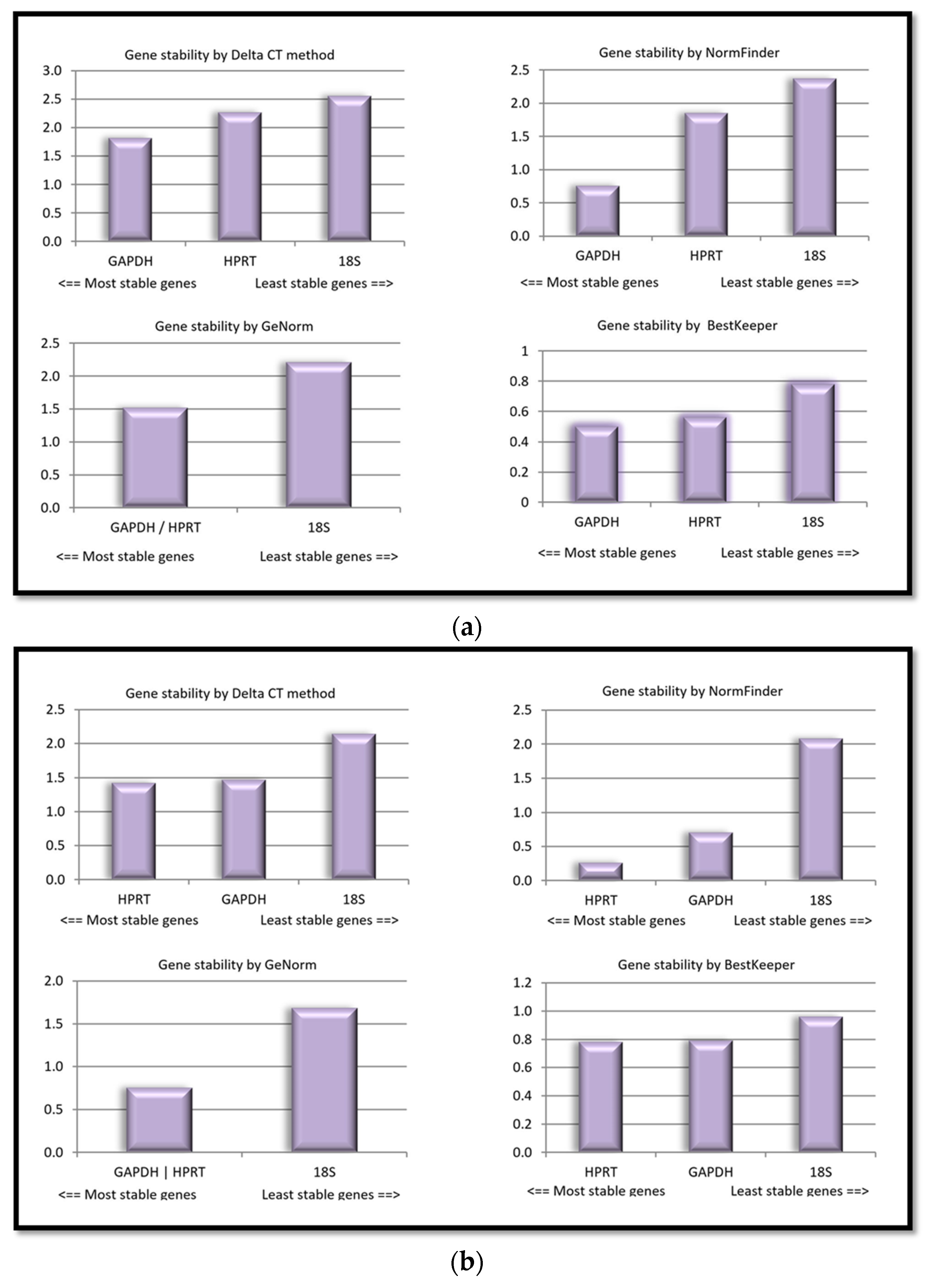
RefFinder, a web-based software program, integrates different computational algorithms, including geNorm, NormFinder, and BestKeeper, to evaluate the stability of candidate normalization genes and rank them accordingly [10]. RefFinder uses the comparative delta Ct method to compare the expression levels of different candidate reference genes. It is important to note that the stability of reference genes may vary depending on the specific experimental conditions, and it is recommended to validate the selected reference genes for each experiment. The use of multiple algorithms and integrated computational programs, as provided by RefFinder, can help enhance the accuracy and reliability of reference gene selection in gene expression studies. Figure 2a,b depict the expression stability ranking of the three candidate reference genes in rabbit and horse ASCs.
The results indicate that significant biological differences between the two species contribute to the observed gene expression variations. Based on the data from the experiment, it was affirmed with a high degree of confidence that GAPDH had the most exceptional stability among the tested genes in rabbit ASCs. However, in horse ASCs, HPRT was confirmed to exhibit the highest level of stability.

2.3. Expression Levels and Variation in Candidate Housekeeping Genes

An inverse correlation exists between the Ct value and gene expression levels. A lower Ct value signifies higher gene expression, while a higher Ct value indicates lower gene expression [6]. Nevertheless, genes with a standard deviation (SD) value exceeding one are considered unstable, aligning with our findings (Table 2).

2.4. Efficiency of the Reactions

PCR efficiency refers to how effectively the target DNA is amplified with each reaction cycle. According to the obtained results from PCR reactions, the efficiency values of examined housekeeping genes varied between 95% and 100%, underscoring their accuracy and reliability in the amplification process (Table 3).
The slope of the standard curve is indicative of the amplification efficiency of the PCR reaction. An efficiency of 100% corresponds to a slope of −3.32 in the standard curve. Deviations from this slope indicate variations in the amplification efficiency of the reaction.

3. Discussion

Stem cells can regenerate the functionally and anatomically impaired integrity of an organ due to their ability to differentiate into various classes of specialized cell types [37]. Among these, mesenchymal stem cells (MSCs) are widely regarded as promising in therapeutic interventions owing to their ease of isolation, storage, cultivation, differentiation, and secretion of regenerative bioactive factors. Historically, the prevailing belief was that MSCs predominantly originated from bone marrow. However, contemporary research has outlined adipose tissue as a preferred and even more abundant source of these cells, offering facilitated accessibility and less invasive harvesting methods [38]. Consequently, adipose-derived stem cells (ASCs) have become increasingly prominent in both human and animal therapies and serve as suitable experimental models in research areas like obesity, cancer, tissue engineering, and drug discovery [39,40,41]. In molecular research on these stem cells, a critical aspect is the selection of appropriate reference genes for gene expression analysis. During differentiation, ASCs undergo significant morphological and physiological changes, complicating the identification of a stable and suitable internal control gene, a challenge further deepened by variability across various biological species and experimental conditions.
Our study was focused on identifying a suitable internal control gene in ASCs from subcutaneous adipose tissue from two poorly investigated animal species—horses and rabbits—during tri-lineage differentiation—adipogenesis, osteogenesis, and chondrogenesis. According to the obtained results, GAPDH exhibited the most stable expression in adipogenically differentiated ASCs isolated from rabbits and treated with omega-3 PUFAs, while 18S and HPRT showed significant variability. GAPDH is extensively employed as a housekeeping gene and is widely preferred in genomic studies, primarily due to its pivotal role in glycolysis—a central metabolic pathway crucial for cellular energy production. Its expression remains consistent across various experimental conditions, cell types, and developmental stages [42,43,44]. However, its suitability in the context of adipogenesis is still questionable due to GAPDH’s involvement in triglyceride synthesis. Some researchers consider GAPDH as a later marker of adipogenesis and believe that its expression reflects its dynamic role in the lipid accumulation phase of adipocyte development [45,46,47]. By contrast, Gorzelniak et al. [48] identified GAPDH as a housekeeping gene with the highest stability both in preadipocytes and mature adipocytes, regardless of hormone exposure. In the context of adipogenesis, other researchers have also reported that GAPDH exhibits the most stable expression and, similar to our results, outlined 18S as an unreliable internal control [49,50].
Over the past few years, numerous scientific studies have focused on different research fields related to equine ASCs. However, a notable gap exists in the literature concerning the validation of internal controls for ASCs. Investigations on the stability of housekeeping genes in adipose tissue across different species, including horses, humans, rats, and rabbits, have highlighted genes such as 18S, ACTB, GAPDH, and HPRT for their consistent expression [51,52,53]. In equine experiments, many researchers utilize housekeeping genes like HPRT as internal control without directly verifying its stability [54,55,56]. Bruynsteen et al. [57] identified both HPRT and GAPDH as the most stable housekeeping genes in equine adipose tissue. Contrarily, some authors have shown significant variability in GAPDH gene expression under certain treatment conditions [27,58,59].
Our research identified HPRT as a stable housekeeping gene in horse ASCs undergoing tri-linear differentiation, namely adipogenesis, osteogenesis, and chondrogenesis, suggesting its potential utility under these specific conditions. Almeida-Oliveira et al. [27] highlighted that selecting suitable housekeeping genes for research is notably influenced by the validation methodology employed. Their study revealed that using multiple software tools for validation often leads to conflicting results, further complicating the identification of a reliable internal control for experimental studies. The research methodology in our experiment includes two software programs—NormFinder and RefFinder. NormFinder, the most commonly used tool, is based on a mathematical model ranking candidate normalization genes on their expression stability [7]. It assesses the gene variation within and between sample groups for proper estimation across the experimental conditions by calculating the coefficient of variation (CV) and pairwise variation (V). Genes with lower CV or V values are considered more stable and are prioritized for selection as reference genes. According to the software analysis, HPRT showed less intra- and intergroup variations in horse ASCs, while in rabbit ASCs, GAPDH was the most unvarying. To confirm the results obtained from NormFinder, we further evaluated the raw Cq values using RefFinder, which has recently emerged as a comprehensive alternative tool that amalgamates various commonly used algorithms—NormFinder, BestKeeper, delta Ct, and geNorm. BestKeeper, an Excel-based tool, interprets the data based on the variations in Ct values and intergene relations in all reference pairs (Pearson correlation). Genes exhibiting a standard deviation (SD) value exceeding one are regarded as unstable (A). The delta Ct method compares the relative expression of pairs of housekeeping genes within each sample and progressively eliminates these with larger delta Ct value deviations (B). The geNorm algorithm calculates the average pairwise variation between a reference gene and all other genes, resulting in an M value. The lowest M value signifies the gene with the highest degree of stability (C). Finally, RefFinder generates a summarized report based on the coefficient of variation (CV) and pairwise variation (V) and provides a more robust evaluation of gene stability compared to single-measure tools. Its iterative refinement process enhances the accuracy of results, making it suitable for complex datasets [10].
De Spiegelaere [60] claimed that the implementation of the original software packages (BestKeeper, NormFinder, and geNorm) generated quite different outcomes compared to the results obtained by RefFinder. The most probable reason is the lack of consideration for PCR efficiencies. When the raw data were reanalyzed, assuming 100% efficiency for all genes, the outputs of the original software packages closely resembled those generated by the RefFinder software.

4. Materials and Methods

Before performing the collection, horses were sedated intravenously with a mixture of 0.01 mg/kg of detomidine hydrochloride (Domosedan, Orion Pharma, Finland) and 0.025 mg/kg of butorphanol tartrate (Butomidor, Richter Pharma, Austria), followed by local infiltration with 5 mL of lidocaine 2% solution (Sopharma, Bulgaria) for regional desensitization. Additionally, rabbits were sacrificed based on the recommendations and approval of the Local Committee for Human Treatment of Animals. Adipose tissue samples weighing 1–1.5 g were then obtained from the gluteal region subcutaneous layer of 18-year-old mix breed horses through a longitudinal skin incision in the Regio radical caudal [61], while in rabbits, the samples were obtained from the subcutaneous (interscapular region) depots of 28-day-old New Zealand rabbits. The isolation procedure was performed according to Vachkova et al. [62]. Initially, the samples were minced in a laminar flow hood, collagenase-digested for 2 h, and centrifuged at 300× g for 10 min. The resulting pellet representing a heterogeneous population of cells known as a stromal vascular fraction (SVF) was resuspended and seeded into T25 tissue flasks in cell culture media containing Dulbecco’s modified Eagle’s medium (DMEM), 10% fetal bovine serum (FBS), and 10 mL/L antibiotic–antimycotic solution, all from Sigma (St. Louis, MO, USA). The cells were cultured and incubated under humidified, 5% CO2 conditions at 37 °C until reaching confluence.

4.1. Experimental Design Rabbits

The cells were cultured up to passage four and further induced in an adipogenic differentiation for 21 days. At this stage, the following groups were formed: control group, treated with DMEM; FBS, L-glutamine, antibiotic mixed solution (control medium); EPA group, treated with the control medium supplemented with 100 µg EPA; and DHA group, treated with the control medium + 100 µg DHA. The cells were cultured in these conditions for 48 h. The cells were additionally induced in osteogenic and chondrogenic differentiation media to demonstrate their functional multipotency. The protocols of tri-lineage differentiation were performed as described by Vachkova et al. [62].

4.2. Experimental Design Horses

The functional multipotency of the isolated cells was assessed through adipogenic (aASC group), osteogenic (oASC group), and chondrogenic (chASC group) differentiation. The fifth passage cells were seeded in 12-well plates at a density of 5 × 10⁵/mL in a basal medium with FBS until reaching confluence.

4.2.1. Adipogenic Differentiation

The cells were induced in an adipogenic medium (DMEM-HG, 10% HS (heat-inactivated horse serum), 1 μM dexamethasone, 10 μg/mL insulin, 0.05 mM indomethacin, 0.5 mM IBMX, and 10 mL/L antibiotic–antimycotic solution) for 18 days. To visualize the intracellular lipid droplets (LDs), the cells were stained with Oil Red O.

4.2.2. Osteogenic Differentiation

The cells were treated with an osteogenic medium (4% HS, 1% ITS, 0.1 μM dexamethasone, 10 mM β-glycerolphosphate, 50 µM L-ascorbic acid, 20 mL/L L-glutamine, 10 mL/L antibiotic–antimycotic solution, and DMEM, all from Sigma). After 21 days, Alizarin Red S was applied to the cells to observe the calcium deposits outside the cells.

4.2.3. Chondrogenic Differentiation

The cells were cultured with chondrogenesis-inducing media (1% ITS, 4% HS, 0.1 μM dexamethasone, 10 ng/mL TGFβ1, 50 µM L-ascorbic acid, 20 mL/L L-glutamine, 10 mL/L antibiotic–antimycotic solution, and DMEM, all from Sigma) for 21 days. The visualization of extracellular cartilage-specific proteoglycans was carried out by utilizing Alcian Blue staining.

4.2.4. Negative Controls

Non-induced ASCs were cultured in parallel only with the basic growth medium and used as negative controls for each type of differentiation, stained with Oil Red O, Alizarin Red S, and Alcian Blue, respectively.
Stained cells were subsequently observed under a Leica DM1000 LED inverted microscope (Heerbrugg, Switzerland), equipped with a 5.0 MP resolution DMi1 camera version and the software platform Leica Application Suite Core, used for all micrographs.

4.3. Gene Expression Analysis (RT-qPCR)

4.3.1. Rabbit Protocol

For the isolation and purification of the total RNA from subcutaneous tissue samples, an RNeasy Lipid Tissue Kit from Qiagen (Hilden, Germany) was used. Then, to determine the RNA quantity, the absorbance was measured at 260 and 280 nm with a GeneQuant 1300 spectrophotometer (GE Healthcare Bio-Sciences AB, Uppsala, Sweden). Reverse transcription reaction was implemented with a RevertAid First Strand cDNA Synthesis Kit (Fermentas Life Science, Thermo Fisher Scientific™, Waltham, MA, USA) using a Quanta Biotech QB-96 cycler (Quanta Biotech Ltd., Surrey, UK). cDNA was stored at −20 until use. The real-time PCR reaction was carried out using the ABI Prism 7900H sequence detection system (AppliedBiosystems, Foster City, CA, USA).

4.3.2. Horse Protocol

The total cellular RNA was extracted using a Universal RNA Purification Kit (EURx, Gdańsk, Poland) per the manufacturer’s protocol. BioTek’s Take3 plate measured RNA purity and quantity, Synergy™ LX Multi-Mode Microplate Reader (BioTek, Santa Clara, CA, USA) at 260 and 280 nm absorbance. All RNA samples were purified from genomic DNA using RNAse-Free DNase Set (Qiagen, Germany) and were reverse-transcribed using a RevertAid First Strand cDNA Synthesis Kit (Thermo Scientific, Waltham, MA, USA), following the manufacturer’s instructions. The analysis of the expression levels of the housekeeping genes, 18S, HPRT, and GAPDH, was performed with real-time qPCR thermocycler Gentier 96E (Xi’an Tialong Science and Technology, Xi’an, China) using KAPA SYBR FAST Master Mix (2X) (Roche) in duplicates with two-step qPCR. The amplification of cDNA was performed following the manufacturer’s conditions.

4.3.3. Standard Curve Construction

The efficiency of the PCR reaction was assessed by generating a standard curve. This curve was constructed using a five-fold serial dilution of a pool of all the DNA samples used in the PCR reaction following the above-mentioned algorithm. Finally, PCR software (Xi’an Tianlong Science and Technology Co., Ltd., https://www.medtl.net/ accessed on 19 February 2023) automatically calculated the efficiency, based on the slope of the standard curve.
The primers for all housekeeping genes were designed using open access data in NCBI (www.ncbi.nlm.nih.gov, accessed on 19 February 2023) and Ensembl (www.ensambl.org, accessed on 22 February 2023). The product length and annealing temperatures were determined via the web-based software Primer 3 (http://bioinfo.ut.ee/primer3/, accessed on 25 February 2023). The primer sequences for the housekeeping genes are presented in Table 4. The primer sequence of 18S for horse samples was designed by Arnhold et al. [63].

4.4. Evaluation of Expression Stability of Housekeeping Genes

We employed two commonly used algorithms to identify the most stable and suitable endogenous control: NormFinder (an Excel add-in, MS Office version 2016) [7] and RefFinder (http://blooge.cn//RefFinder/, accessed on 25 February 2023) [10].

4.5. Statistical Analysis

To evaluate the standard error of the mean (SEM) and the mean values for each gene, Statistica v.10 (StatSoft, Inc., Tulsa, OK, USA (2011) was used. Furthermore, the least significant digit (LSD) test was performed to estimate intergroup variations. A statistically significant difference was considered as a p-value of less than 0.05.

5. Conclusions

In conclusion, our investigation demonstrated that GAPDH exhibited stable expression in rabbit ASCs after adipogenesis and treated with omega-3 PUFAs. At the same time, HPRT was identified as a stable internal control in horse ASCs during tri-lineage differentiation. This research emphasizes the importance of a precise validation methodology, employing multiple software tools such as NormFinder and RefFinder, to comprehensively assess gene stability. The choice of appropriate housekeeping genes should be tailored to the specific requirements of each trial, highlighting the need for an individual approach.
We believe that our findings can contribute to selecting suitable internal controls in ASCs, particularly in less-explored species, and highlight the significance of methodological integrity in gene expression studies involving stem cells.

Author Contributions

Conceptualization, E.V., Z.I., N.G., and V.P.; methodology, E.V., Z.I. and V.P.; software, Z.I., N.G. and V.P.; validation, Z.I. and V.P.; formal analysis, Z.I. and V.P.; investigation, E.V., Z.I., N.G. and V.P.; resources, Z.I. and V.P.; data curation, E.V., Z.I. and V.P.; writing—original draft preparation, Z.I. and V.P.; writing—review and editing, N.G. and E.V.; visualization, Z.I., N.G. and V.P.; supervision, N.G. and E.V.; project administration, E.V., Z.I. and V.P.; funding acquisition, V.P. and E.V. All authors have read and agreed to the published version of the manuscript.

Funding

This work was supported by the Faculty of Veterinary Medicine, Trakia University, Stara Zagora, Bulgaria (Project N 14/20 and by the Ph.D. Fellowship of Valeria Petrova).

Institutional Review Board Statement

This study was conducted in accordance with the Declaration of Helsinki and national animal welfare regulations and approved by the National Ethics Commission (Permission № 322/9 December 2021 issued by the Bulgarian Food Safety Agency within the Bulgarian Ministry of Agriculture, for the horse experimental design) and by the Commission of Ethics at the Faculty of Veterinary Medicine of Trakia University, Stara Zagora (Permission № 53/29 September 2012, for the rabbit experimental procedure).

Informed Consent Statement

Not applicable.

Data Availability Statement

The datasets generated for this study are available from the corresponding author upon request.

Acknowledgments

The experiment and all listed analyses were performed in the laboratory of Experimental Cellular Physiology and Therapeutic Drug Monitoring of the Faculty of Veterinary Medicine, Trakia University, Stara Zagora, Bulgaria.

Conflicts of Interest

The authors declare no conflicts of interest.

References

  1. Ginzinger, D.G. Gene quantification using real-time quantitative PCR: An emerging technology hits the mainstream. Exp. Hematol. 2002, 30, 503–512. [Google Scholar] [CrossRef] [PubMed]
  2. Dheda, K.; Huggett, J.F.; Bustin, S.A.; Johnson, M.A.; Rook, G.; Zumla, A. Validation of housekeeping genes for normalizing RNA expression in real-time PCR. Biotechniques 2004, 37, 112–119. [Google Scholar] [CrossRef] [PubMed]
  3. Nestorov, J.; Matić, G.; Elaković, I.; Tanić, N. Gene expression studies: How to obtain accurate and reliable data by quantitative real-time RT PCR. J. Med. Biochem. 2013, 32, 325–338. [Google Scholar] [CrossRef]
  4. Eisenberg, E.; Levanon, E.Y. Human housekeeping genes, revisited. Trends Genet. 2013, 29, 569–574. [Google Scholar] [CrossRef] [PubMed]
  5. Suzuki, T.; Higgins, P.J.; Crawford, D.R. Control selection for RNA quantitation. Biotechniques 2000, 29, 332–337. [Google Scholar] [CrossRef]
  6. You, S.; Cao, K.; Chen, C.; Li, Y.; Wu, J.; Zhu, G.; Fang, W.; Wang, X.; Wang, L. Selection and validation reference genes for qRT-PCR normalization in different cultivars during fruit ripening and softening of peach (Prunus persica). Sci. Rep. 2021, 11, 7302. [Google Scholar] [CrossRef] [PubMed]
  7. Andersen, C.L.; Jensen, J.L.; Ørntoft, T.F. Normalization of real-time quantitative reverse transcription-PCR data: A model-based variance estimation approach to identify genes suited for normalization, applied to bladder and colon cancer data sets. Cancer Res. 2004, 64, 5245–5250. [Google Scholar] [CrossRef]
  8. Pfaffl, M.W.; Tichopad, A.; Prgomet, C.; Neuvians, T.P. Determination of stable housekeeping genes, differentially regulated target genes and sample integrity: BestKeeper--Excel-based tool using pair-wise correlations. Biotechnol. Lett. 2004, 26, 509–515. [Google Scholar] [CrossRef]
  9. Vandesompele, J.; De Preter, K.; Pattyn, F.; Poppe, B.; Van Roy, N.; De Paepe, A.; Speleman, F. Accurate normalization of real-time quantitative RT-PCR data by geometric averaging of multiple internal control genes. Genome Biol. 2002, 3, research0034.1. [Google Scholar] [CrossRef]
  10. Xie, F.; Xiao, P.; Chen, D.; Xu, L.; Zhang, B. miRDeepFinder: A miRNA analysis tool for deep sequencing of plant small RNAs. Plant Mol. Biol. 2012, 80, 75–84. [Google Scholar] [CrossRef]
  11. Song, J.; Cho, J.; Park, J.; Hwang, J.H. Identification and validation of stable reference genes for quantitative real time PCR in different minipig tissues at developmental stages. BMC Genom. 2022, 23, 585. [Google Scholar] [CrossRef]
  12. Tsotetsi, T.N.; Collins, N.E.; Oosthuizen, M.C.; Sibeko-Matjila, K.P. Selection and evaluation of housekeeping genes as endogenous controls for quantification of mRNA transcripts in Theileria parva using quantitative real-time polymerase chain reaction (qPCR). PLoS ONE 2018, 13, 0196715. [Google Scholar] [CrossRef]
  13. De Jonge, H.J.M.; Fehrmann, R.S.N.; de Bont, E.S.J.M.; Hofstra, R.M.W.; Gerbens, F.; Kamps, W.A.; de Vries, E.G.E.; van der Zee, A.G.J.; te Merrman, G.J.; ter Elst, A. Evidence based selection of housekeeping genes. PLoS ONE 2007, 2, e898. [Google Scholar] [CrossRef]
  14. Guaita-Cespedes, M.; Grillo-Risco, R.; Hidalgo, M.R.; Fernández-Veledo, S.; Burks, D.J.; de la Iglesia-Vayá, M.; Galan, A.; Garcia-Garcia, F. Deciphering the sex bias in housekeeping gene expression in adipose tissue: A comprehensive meta-analysis of transcriptomic studies. Biol. Sex Differ. 2023, 14, 20. [Google Scholar] [CrossRef] [PubMed]
  15. Meyer-Siegler, K.; Mauro, D.J.; Seal, G.; Wurzer, J.; Deriel, J.K.; Sirover, M.A. A human nuclear uracil DNA glycosylase is the 37-kDa subunit of glyceraldehyde-3-phosphate dehydrogenase. Proc. Natl. Acad. Sci. USA 1991, 88, 8460–8464. [Google Scholar] [CrossRef] [PubMed]
  16. Singh, R.; Green, M.R. Sequence-specific binding of transfer RNA by glyceraldehyde-3-phosphate dehydrogenase. Science 1993, 259, 365–368. [Google Scholar] [CrossRef] [PubMed]
  17. Tisdale, E.J. Glyceraldehyde-3-phosphate dehydrogenase is required for vesicular transport in the early secretory pathway. J. Biol. Chem. 2001, 276, 2480–2486. [Google Scholar] [CrossRef] [PubMed]
  18. Tarze, A.; Deniaud, A.; Le Bras, M.; Maillier, E.; Mollé, D.; Larochette, N.; Zamzami, N.; Jan, G.; Kroemer, G.; Brenner, C. GAPDH, a novel regulator of the pro-apoptotic mitochondrial membrane permeabilization. Oncogene 2007, 26, 2606–2620. [Google Scholar] [CrossRef]
  19. Hara, M.R.; Agrawal, N.; Kim, S.F.; Cascio, M.B.; Fujimuro, M.; Ozeki, Y.; Takahashi, M.; Cheah, J.H.; Tankou, S.K.; Hester, L.D.; et al. S-nitrosylated GAPDH initiates apoptotic cell death by nuclear translocation following Siah1 binding. Nat. Cell Biol. 2005, 7, 665–674. [Google Scholar] [CrossRef]
  20. Wilson, J.M.; Tarr, G.E.; Kelley, W.N. Human hypoxanthine (guanine) phosphoribosyltransferase: An amino acid substitution in a mutant form of the enzyme isolated from a patient with gout. Proc. Nat. Acad. Sci. USA 1983, 80, 870–873. [Google Scholar] [CrossRef]
  21. Stout, J.T.; Caskey, C.T. HPRT: Gene structure, expression, and mutation. Annu. Rev. Genet. 1985, 19, 127–148. [Google Scholar] [CrossRef]
  22. Becerra, A.; Lazcano, A. The role of gene duplication in the evolution of purine nucleotide salvage pathways. Orig. Life Evol. Biosphere. 1998, 28, 539–553. [Google Scholar] [CrossRef] [PubMed]
  23. Yamada, H.; Chen, D.; Monstein, H.J.; Hakanson, R. Effects of fasting on the expression of gastrin, cholecystokinin, and somatostatin genes and of various housekeeping genes in the pancreas and upper digestive tract of rats. Biochem. Biophys. Res. Commun. 1997, 231, 835–838. [Google Scholar] [CrossRef] [PubMed]
  24. Ferreira-Cerca, S.; Pöll, G.; Gleizes, P.E.; Tschochner, H.; Milkereit, P. Roles of eukaryotic ribosomal proteins in maturation and transport of pre-18S rRNA and ribosome function. Mol. Cell 2005, 20, 263–275. [Google Scholar] [CrossRef] [PubMed]
  25. Ferguson, B.S.; Nam, H.; Hopkins, R.G.; Morrison, R.F. Impact of reference gene selection for target gene normalization on experimental outcome using real-time qRT-PCR in adipocytes. PLoS ONE 2010, 5, e15208. [Google Scholar] [CrossRef]
  26. Gong, H.; Sun, L.; Chen, B.; Han, Y.; Pang, J.; Wu, W.; Qi, R.; Zhang, T.M. Evaluation of candidate reference genes for RT-qPCR studies in three metabolism related tissues of mice after caloric restriction. Sci. Rep. 2016, 6, 38513. [Google Scholar] [CrossRef]
  27. Almeida-Oliveira, F.; Leandro, J.G.; Ausina, P.; Sola-Penna, M.; Majerowicz, D. Reference genes for quantitative PCR in the adipose tissue of mice with metabolic disease. Biomed. Pharmacother. 2017, 88, 948–955. [Google Scholar] [CrossRef]
  28. Baer, P.C.; Geiger, H. Adipose-derived mesenchymal stromal/stem cells: Tissue localization, characterization, and heterogeneity. Stem Cells Int. 2012, 2012, 812693. [Google Scholar] [CrossRef]
  29. Vidal, M.A.; Kilroy, G.E.; Lopez, M.J.; Johnson, J.R.; Moore, R.M.; Gimble, J.M. Characterization of equine adipose tissue-derived stromal cells: Adipogenic and osteogenic capacity and comparison with bone marrow-derived mesenchymal stromal cells. Vet. Surg. 2007, 36, 613–622. [Google Scholar] [CrossRef]
  30. Corsini, N.S.; Knoblich, J.A. Human organoids: New strategies and methods for analyzing human development and disease. Cell 2022, 185, 2756–2769. [Google Scholar] [CrossRef]
  31. Tirpáková, M.; Vašíček, J.; Svoradová, A.; Baláži, A.; Tomka, M.; Bauer, M.; Makarevich, A.; Chrenek, P. Phenotypical characterization and neurogenic differentiation of rabbit adipose tissue-derived mesenchymal stem cells. Genes 2021, 12, 431. [Google Scholar] [CrossRef]
  32. Bukowska, J.; Szóstek-Mioduchowska, A.Z.; Kopcewicz, M.; Walendzik, K.; Machcińska, S.; Gawrońska-Kozak, B. Adipose-derived stromal/stem cells from large animal models: From basic to applied science. Stem Cell Rev. Rep. 2021, 17, 719–738. [Google Scholar] [CrossRef]
  33. Al Naem, M.; Bourebaba, L.; Kucharczyk, K.; Röcken, M.; Marycz, K. Therapeutic mesenchymal stromal stem cells: Isolation, characterization and role in equine regenerative medicine and metabolic disorders. Stem Cell Rev. Rep. 2020, 16, 301–322. [Google Scholar] [CrossRef]
  34. Ribitsch, I.; Baptista, P.M.; Lange-Consiglio, A.; Melotti, L.; Patruno, M.; Jenner, F.; Schnabl-Feichter, E.; Dutton, L.; Connolly, D.J.; van Steenbeek, F.; et al. Large animal models in regenerative medicine and tissue engineering: To do or not to do. Front. Bioeng. Biotechnol. 2020, 8, 972. [Google Scholar] [CrossRef] [PubMed]
  35. Gugjoo, M.B.; Sharma, G.T. Equine mesenchymal stem cells: Properties, sources, characterization, and potential therapeutic applications. J. Equine Vet. Sci. 2019, 72, 16–27. [Google Scholar] [CrossRef] [PubMed]
  36. Matsuhisa, F.; Kitajima, S.; Nishijima, K.; Akiyoshi, T.; Morimoto, M.; Fan, J. Transgenic rabbit models: Now and the future. Appl. Sci. 2020, 10, 7416. [Google Scholar] [CrossRef]
  37. Bacakova, L.; Zarubova, J.; Travnickova, M.; Musilkova, J.; Pajorova, J.; Slepicka, P.; Kasalkova, N.S.; Svorcik, V.; Kolska, Z.; Motarjemi, H.; et al. Stem cells: Their source, potency and use in regenerative therapies with focus on adipose-derived stem cells—A review. Biotechnol. Adv. 2018, 36, 1111–1126. [Google Scholar] [CrossRef]
  38. Fraser, J.K.; Wulur, I.; Alfonso, Z.; Hedrick, M.H. Fat tissue: An underappreciated source of stem cells for biotechnology. Trends Biotechnol. 2006, 24, 150–154. [Google Scholar] [CrossRef]
  39. Farré-Guasch, E.; Bravenboer, N.; Helder, M.N.; Schulten, E.A.; Ten Bruggenkate, C.M.; Klein-Nulend, J. Blood vessel formation and bone regeneration potential of the stromal vascular fraction seeded on a calcium phosphate scaffold in the human maxillary sinus floor elevation model. Materials 2018, 11, 161. [Google Scholar] [CrossRef] [PubMed]
  40. Scioli, M.G.; Storti, G.; D’Amico, F.; Gentile, P.; Kim, B.S.; Cervelli, V.; Orlandi, A. Adipose-derived stem cells in cancer progression: New perspectives and opportunities. Int. J. Mol. Sci. 2019, 20, 3296. [Google Scholar] [CrossRef]
  41. Zuk, P.A.; Zhu, M.; Mizuno, H.; Huang, J.J.; Futrell, W.; Katz, A.J.; Benhaim, P.; Lorenz, H.P.; Hedrick, M.H. Multilineage cells from human adipose tissue: Implications for cell-based therapies. Tissue Eng. 2001, 7, 211–228. [Google Scholar] [CrossRef]
  42. Quiroz, F.G.; Posada, O.M.; Gallego-Perez, D.; Higuita-Castro, N.; Sarassa, C.; Hansford, D.J.; Agudelo-Florez, P.; López, L.E. Housekeeping gene stability influences the quantification of osteogenic markers during stem cell differentiation to the osteogenic lineage. Cytotechnology 2010, 62, 109–120. [Google Scholar] [CrossRef]
  43. Wiraja, C.; Yeo, D.C.; Chong, M.S.; Xu, C. Nanosensors for continuous and noninvasive monitoring of mesenchymal stem cell osteogenic differentiation. Small 2016, 12, 1342–1350. [Google Scholar] [CrossRef]
  44. Zainuddin, A.; Chua, K.H.; Abdul Rahim, N.; Makpol, S. Effect of experimental treatment on GAPDH mRNA expression as a housekeeping gene in human diploid fibroblasts. BMC Mol. Biol. 2008, 9, 59. [Google Scholar] [CrossRef] [PubMed]
  45. Zhang, J.; Tang, H.; Zhang, Y.; Deng, R.; Shao, L.; Liu, Y.; Li, F.; Wang, X.; Zhou, L. Identification of suitable reference genes for quantitative RT-PCR during 3T3-L1 adipocyte differentiation. Int. J. Mol. Med. 2014, 33, 1209–1218. [Google Scholar] [CrossRef] [PubMed]
  46. Salmerón, C.; Riera-Heredia, N.; Gutiérrez, J.; Navarro, I.; Capilla, E. Adipogenic gene expression in gilthead sea bream mesenchymal stem cells from different origin. Front. Endocrinol. 2016, 7, 113. [Google Scholar] [CrossRef] [PubMed]
  47. Krautgasser, C.; Mandl, M.; Hatzmann, F.M.; Waldegger, P.; Mattesich, M.; Zwerschke, W. Reliable reference genes for expression analysis of proliferating and adipogenically differentiating human adipose stromal cells. Cell. Mol. Biol. Lett. 2019, 24, 14. [Google Scholar] [CrossRef] [PubMed]
  48. Gorzelniak, K.; Janke, J.; Engeli, S.; Sharma, A.M. Validation of endogenous controls for gene expression studies in human adipocytes and preadipocytes. Horm. Metab. Res. 2001, 33, 625–627. [Google Scholar] [CrossRef]
  49. Fink, T.; Lund, P.; Pilgaard, L.; Rasmussen, J.G.; Duroux, M.; Zachar, V. Instability of standard PCR reference genes in adipose-derived stem cells during propagation, differentiation and hypoxic exposure. BMC Mol. Biol. 2008, 9, 98. [Google Scholar] [CrossRef]
  50. Ebrahimi, R.; Toolabi, K.; Jannat Ali Pour, N.; Mohassel Azadi, S.; Bahiraee, A.; Zamani-Garmsiri, F.; Emamgholipour, S. Adipose tissue gene expression of long non-coding RNAs; MALAT1, TUG1 in obesity: Is it associated with metabolic profile and lipid homeostasis-related genes expression? Diabetol. Metab. Syndr. 2020, 12, 36. [Google Scholar] [CrossRef]
  51. Bourebaba, L.; Kornicka-Garbowska, K.; Al Naem, M.; Röcken, M.; Łyczko, J.; Marycz, K. MSI-1436 improves EMS adipose derived progenitor stem cells in the course of adipogenic differentiation through modulation of ER stress, apoptosis, and oxidative stress. Stem Cell Res. Ther. 2021, 12, 97. [Google Scholar] [CrossRef]
  52. El-Gindy, Y.M.; Hafsa, S.H.A.; El-Deeb, N.M. The expression of liver TNF-α gene, liver and small intestine histology of thermal stressed growing rabbits affected by allicin and lycopene. J. Therm. Biol. 2023, 113, 103521. [Google Scholar] [CrossRef]
  53. Radtke, C.L.; Nino-Fong, R.; Gonzalez, B.P.E.; Stryhn, H.; McDuffee, L.A. Characterization and osteogenic potential of equine muscle tissue–and periosteal tissue–derived mesenchymal stem cells in comparison with bone marrow–and adipose tissue–derived mesenchymal stem cells. Am. J. Vet. Res. 2013, 74, 790–800. [Google Scholar] [CrossRef] [PubMed]
  54. Azarpeykan, S.; Dittmer, K.E. Evaluation of housekeeping genes for quantitative gene expression analysis in the equine kidney. J. Equine Sci. 2016, 27, 165–168. [Google Scholar] [CrossRef] [PubMed]
  55. Hotham, W.E.; Thompson, C.; Szu-Ting, L.; Henson, F.M.D. The anti-inflammatory effects of equine bone marrow stem cell-derived extracellular vesicles on autologous chondrocytes. Vet. Rec. Open 2021, 8, 22. [Google Scholar] [CrossRef]
  56. Jarazo, J.M. Effect of Tissue Source on Adult Equine Multipotent Stromal Cell Pluripotency Induction Treatment with Synthetic mRNA. Master’s Thesis, Louisiana State University and Agricultural & Mechanical College, Baton Rouge, LA, USA, 2014. [Google Scholar]
  57. Bruynsteen, L.; Erkens, T.; Peelman, L.J.; Ducatelle, R.; Janssens, G.P.J.; Harris, P.A.; Hesta, M. Expression of inflammation-related genes is associated with adipose tissue location in horses. BMC Vet. Res. 2013, 9, 240. [Google Scholar] [CrossRef]
  58. Dheda, K.; Huggett, J.F.; Chang, J.S.; Kim, L.U.; Bustin, S.A.; Johnson, M.A.; Rook, G.A.W.; Zumla, A. The implications of using an inappropriate reference gene for real-time reverse transcription PCR data normalization. Anal. Biochem. 2005, 344, 141–143. [Google Scholar] [CrossRef]
  59. Nazari, F.; Parham, A.; Maleki, A.F. GAPDH, β-actin and β2-microglobulin, as three common reference genes, are not reliable for gene expression studies in equine adipose-and marrow-derived mesenchymal stem cells. J. Anim. Sci. Technol. 2015, 57, 18. [Google Scholar] [CrossRef] [PubMed]
  60. De Spiegelaere, W.; Dern-Wieloch, J.; Weigel, R.; Schumacher, V.; Schorle, H.; Nettersheim, D.; Bergmann, M.; Brehm, R.; Kliesch, S.; Vandekerckhove, L.; et al. Reference gene validation for RT-qPCR, a note on different available software packages. PLoS ONE 2015, 10, e0122515. [Google Scholar] [CrossRef]
  61. Raabe, O.; Shell, K.; Würtz, A.; Reich, C.M.; Wenisch, S.; Arnhold, S. Further insights into the characterization of equine adipose tissue-derived mesenchymal stem cells. Vet. Res. Commun. 2011, 35, 355–365. [Google Scholar] [CrossRef]
  62. Vachkova, E.; Bosnakovski, D.; Yonkova, P.; Grigorova, N.; Ivanova, Z.; Todorov, P.; Penchev, G.; Milanova, A.; Simeonova, G.; Stanilova, S.; et al. Adipogenic potential of stem cells derived from rabbit subcutaneous and visceral adipose tissue in vitro. Vitr. Cell. Dev. Biol.-Anim. 2016, 52, 829–837. [Google Scholar] [CrossRef]
  63. Arnhold, S.; Elashry, M.I.; Klymiuk, M.C.; Geburek, F. Investigation of Stemness and Multipotency of Equine Adipose-Derived Mesenchymal Stem Cells (ASCs) from Different Fat Sources in Comparison with Lipoma. Stem Cell Res. Ther. 2019, 10, 309. [Google Scholar] [CrossRef]
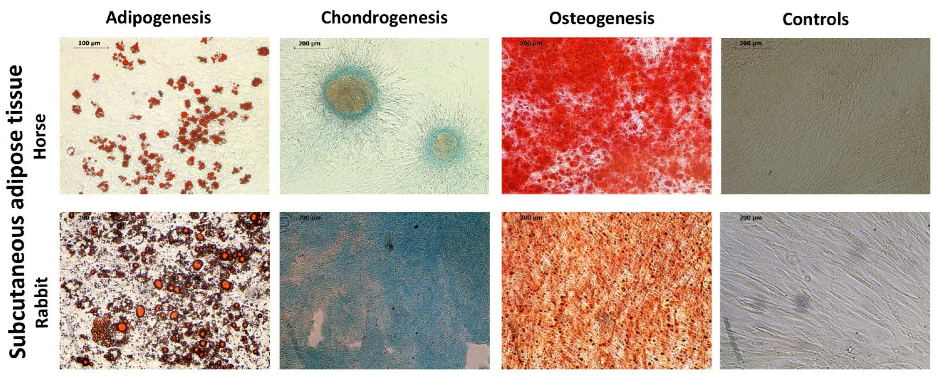
Figure 1. Tri-lineage differentiation of horse and rabbit ASCs: for adipogenesis, the samples were stained with Oil Red O, scale bars 100 μm (horses) and 200 μm (rabbits); for chondrogenesis, the samples stained with Alcian Blue, scale bars 200 μm; for osteogenesis, the samples were stained with Alizarin Red, scale bars 200 μm; controls, scale bars 200 μm.
Ijms 25 02292 g001
Figure 2. The stability ranking of candidate housekeeping gene identified by RefFinder in rabbit and horse ASCs (a,b), respectively, based on their Ct values. The following groups were analyzed: for rabbit ASCs, control as well as EPA- and DHA-treated groups; for horse ASCs, the groups undergoing adipogenic, osteogenic, and chondrogenic differentiation.
Ijms 25 02292 g002
Table 1. Expression stability evaluation of the reference genes using NormFinder, based on their quantity.
Gene NameStability Value
RabbitHorse
18S0.4730.589
GAPDH0.1360.235
HPRT0.2630.071
Table 2. Mean threshold cycles (Ct) and standard deviation (SD) of 18S, GAPDH, and HPRT in rabbit and horse ASCs.
Gene NameMean Ct Values with Their SD
RabbitHorse
18S17.01 ± 1.0519.03 ± 1.15
GAPDH23.28 ± 0.4023.48 ± 1.26
HPRT27.74 ± 0.8826.91 ± 0.94
Table 3. Slope and efficiency determination by standard curve analysis in qPCR of 18S, GAPDH, and HPRT genes in rabbit and horse ASCs.
Gene NameSlopes/Efficiency (%) of PCR Reaction
RabbitHorse
18S−3.449/94.96−3.389/97.28
GAPDH−3.333/99.54−3.337/99.38
HPRT−3.331/99.62−3.345/99.04
Table 4. Primer sequences, product length, and accession number of candidate reference genes for qPCR normalization in adipose-derived stem cells.
AbbreviationFull NameForwardReverseProduct LengthNCBI
Accession №
HORSE
HPRTHypoxanthine phosphoribosyltransferaseCCAGTCAACAGGGGACATAAGCTTGCGACCTTGACCATCT163AY372182.1
GAPDHGlyceraldehyde 3-phosphate dehydrogenaseTCCCTGCTTCTACTGGTGCTCGTATTTGGCAGCTTTCTCC147NM_001163856.1
18S18S ribosomal RNAATGCGGCGGCGTTATTCCGCTATCAATCTGTCAATCCTGTCC204NR_046271.1
RABBITS
HPRTHypoxanthine phosphoribosyltransferaseCCCCAGCGTTGTGATTAGTGGCCTCCCATCTCCTTCATCA163ENSOCUT00000030995.2
GAPDHGlyceraldehyde 3-phosphate dehydrogenaseGAACGGGAAGCTGGTCATCAGAAGACGCCAGTGGATTCCA118GCA_000003625.1
18S18S ribosomal RNAATCAGATACCGTCGTAGTTCTTCCGTCAATTCCTTTAAG167NR_033238.1
Disclaimer/Publisher’s Note: The statements, opinions and data contained in all publications are solely those of the individual author(s) and contributor(s) and not of MDPI and/or the editor(s). MDPI and/or the editor(s) disclaim responsibility for any injury to people or property resulting from any ideas, methods, instructions or products referred to in the content.