Article

Protective Effect of Probiotics against Pseudomonas aeruginosa Infection of Human Corneal Epithelial Cells

Department of Chemical, Biological, Pharmaceutical and Environmental Sciences, University of Messina, 98166 Messina, Italy
*
Author to whom correspondence should be addressed.
These authors contributed equally to this work.
Int. J. Mol. Sci. 2024, 25(3), 1770; https://doi.org/10.3390/ijms25031770
Submission received: 31 December 2023 / Revised: 27 January 2024 / Accepted: 30 January 2024 / Published: 1 February 2024
(This article belongs to the Special Issue Molecular Research in Prebiotics, Probiotics and Postbiotics)

Abstract

:
Probiotic therapy needs consideration as an alternative strategy to prevent and possibly treat corneal infection. This study aimed to assess the preventive effect of Lactobacillus reuteri and Bifidobacterium longum subsp. infantis on reducing the infection of human corneal epithelial (HCE) cells caused by Pseudomonas aeruginosa. The probiotics’ preventive effect against infection was evaluated in cell monolayers pretreated with each probiotic 1 h and 24 h prior to P. aeruginosa challenge followed by 1 h and 24 h of growth in combination. Cell adhesion, cytotoxicity, anti-inflammatory, and antinitrosative activities were evaluated. L. reuteri and B. longum adhered to HCE cells, preserved occludin tight junctions’ integrity, and increased mucin production on a SkinEthicTM HCE model. Pretreatment with L. reuteri or B. longum significantly protected HCE cells from infection at 24 h, increasing cell viability at 110% (110.51 ± 5.15; p ≤ 0.05) and 137% (137.55 ± 11.97; p ≤ 0.05), respectively. Each probiotic showed anti-inflammatory and antinitrosative activities, reducing TNF-α level (p ≤ 0.001) and NOx amount (p ≤ 0.001) and reestablishing IL-10 level (p ≤ 0.001). In conclusion, this study demonstrated that L. reuteri and B. longum exert protective effects in the context of corneal infection caused by P. aeruginosa by restoring cell viability and modulating inflammatory cytokine release.

1. Introduction

Infectious keratitis is considered a medical emergency as it can cause vision loss and blindness. Among the most common pathogens implicated in bacterial keratitis is Pseudomonas aeruginosa, which is of particular concern for several reasons [1,2]. It is highly virulent, difficult to treat, and results in worse corneal ulcers compared to those caused by other bacteria [3,4]. Predisposing factors for infectious keratitis include ocular disease, ocular injury, surgical procedures, and antibiotic therapy [5]. Moreover, P. aeruginosa infections can develop biofilms which facilitate a higher incidence of disease in extended-wear contact lens users [6,7]. Currently, treatment for several ocular surface infections is based on eye drops containing antibiotics that can stimulate changes in the healthy eye microbiota, contribute to the onset of eye diseases, and increase the resistance of pathogenic strains [8].
In healthy conditions, the ocular microbiota play an important role in maintaining local homeostasis: they modulate the immune response and, through a barrier effect, prevent the invasion and proliferation of pathogenic or opportunistic microorganisms [9,10]. The microbiome consists of bacteria, fungi, viruses, and protozoa. Recently, next-generation sequencing 16S rRNA technology has demonstrated the most abundant bacterial phyla are represented by Proteobacteria (64%), Actinobacteria (19.6%), and Firmicutes (3.9%) [10]. Most metagenomic sequencing results support Corynebacterium, Propionibacterium, and Staphylococcus as the dominant taxons of the healthy ocular surface. However, the microbiome and microbiota can undergo dynamic changes during the lifespan of a human being. The microbiota of the ocular surface can be affected by environmental conditions, age, sex, seasonality, personal habits, use of contact lenses, pathological states, infections, antibiotics, etc. [9].
Ocular surface dysbiosis, a substantial change in microbiota composition, can be associated to several diseases and conditions, including infections [2,11]. Keratitis caused by P. aeruginosa is refractory and difficult to treat because of its extensive and emerging antibiotic resistance. During the infection process, P. aeruginosa produces several virulence factors, forms a biofilm, and displays resistance to many antibiotics [12,13]. Biofilms are communities of microorganisms in nature that are attached to a biological or abiotic surface and are surrounded by a self-generated extracellular matrix that is mainly composed of polysaccharides, secreted proteins, and extracellular DNAs. Biofilm bacteria exhibit different phenotypic characteristics from their planktonic counterparts, including an increased resistance to antibiotics, and steep rises in the incidence of microbial keratitis have been linked to the increased popularity of contact lenses [13,14].
Treatment failure in clinical practice leads to the development of new therapeutic strategies. Among these strategies, probiotic therapy may have great potential in preventing and treating several ocular diseases [10,15]. According to a definition by the Food and Drug Administration (FDA), probiotics are classified as “live biotherapeutics: live microorganisms that have a positive impact on the health and physiology of the host” [16]. Numerous studies have proven that Lactobacilli, Bifidobacteria, Escherichia coli, Saccharomyces cerevisiae, S. boulardii, S. lactis, etc., could be used as probiotics [17,18]. Probiotics may modify immunological activity by increasing innate and adaptive immune responses, altering microbial habitats, producing antimicrobial compounds, and improving barrier function and competitive adhesion to the mucosa and epithelium [19]. Some strains can inhibit the adhesion of pathogens to the mucosa by forming a barrier via auto-aggregation or by direct coaggregation with the pathogens. Auto-aggregation facilitates the communication between cells as well as antagonism and estrangement of pathogenic bacteria [20]. Coaggregation allows for close interaction between the probiotic strain and the pathogen, and through the production of antimicrobial substances, the probiotic can inhibit the growth of pathogens [21,22].
Moreover, several probiotic strains form biofilms that can counteract the establishment of pathogenic biofilms [23].The main antimicrobial compounds produced by probiotic strains are organic acids, such as lactic, propionic, acetic, and succinic acid, and ethanol, hydrogen sulfide, biosurfactants, carbon dioxide, exopolysaccharides, and bacteriocins [24]. Probiotics are used to prevent and treat conditions such as diarrhea, acute pancreatitis, colon cancer, hypertension, diabetes, Helicobacter pylori infection, ventilator-associated pneumonia, migraine, and autism in clinical settings [19]. Nevertheless, in most cases, probiotic activity is strain-specific as demonstrated by Bubnov and colleagues [25] who investigated specific properties, such as adhesive ability and resistance to antibiotics, of lactic acid bacteria (LAB) and Bifidobacteria.
Nowadays, research on microbial biotherapy for ocular diseases is still in its infancy. However, some studies have focused their attention on how probiotics can restore the ocular microbiota and improve the course of some ocular surface diseases. Among these, some clinical trials reported the efficacy of topically applied probiotics such as L. acidophilus or B. lactis and B. bifido formulations to correct vernal keratoconjunctivitis or dry eye, respectively [26,27].
Currently, there are still few studies that investigate the effect of probiotics on ocular surface infections.
Probiotic therapy could promote a rapid and stable restoration of the healthy ocular microbiota, faster anti-inflammatory effect, and antagonistic action against pathogens [10].
The aim of this study was to evaluate the influence of L. reuteri and B. longum subsp. infantis on preventing P. aeruginosa infection of human corneal epithelial (HCE) cells. The experimental design involved two distinct phases in order to (i) determine the strain-specific characteristics of these probiotics, (ii) evaluate whether pretreatment of HCE cells with these probiotic strains can protect them from P. aeruginosa infection-induced damage.

2. Results

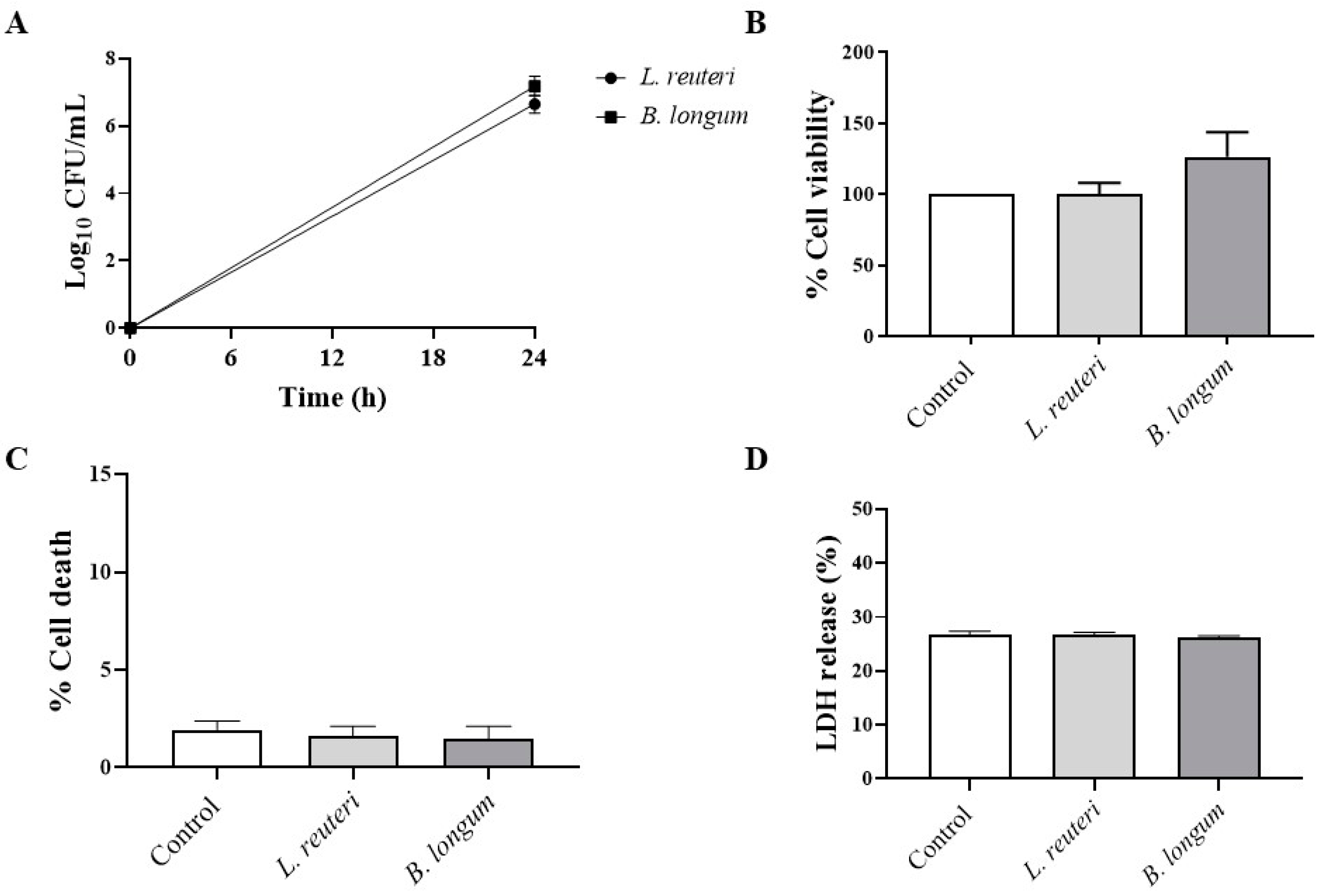
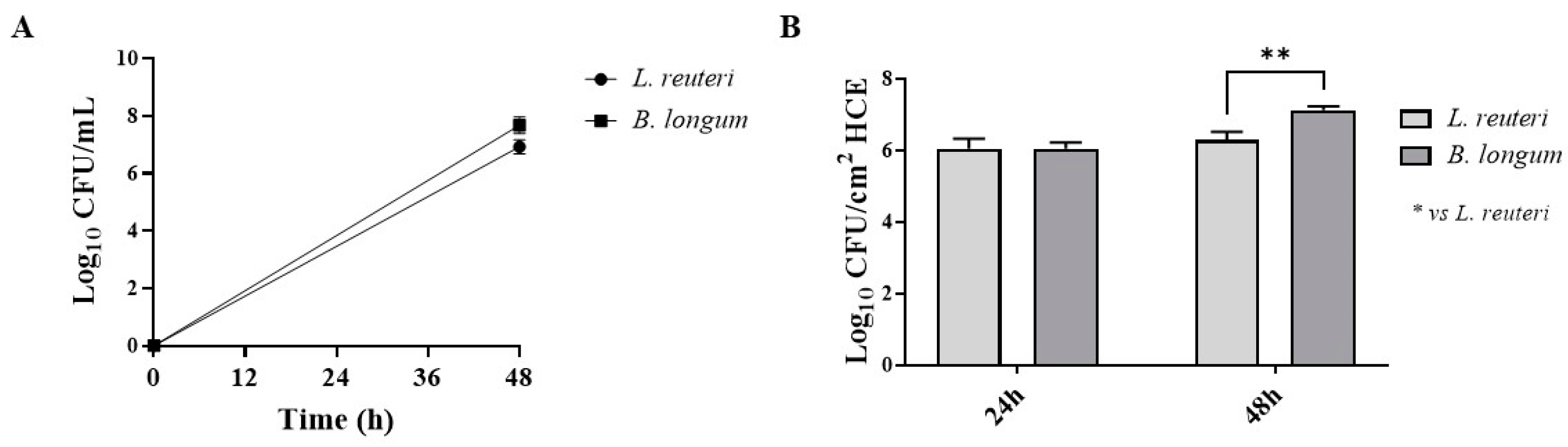
2.1. Strains and Growth Conditions

Overnight cultures of L. reuteri and B. longum strains in MRSB, with or without cysteine (0.05 g/L) and incubated at 37 °C under 5% CO2 conditions were diluted to obtain an optical density of 0.5 at 570 nm (equivalent to approximately 5 × 10⁸ CFU/mL). Both strains, with and without 0.05 g/L cysteine, showed the same concentrations when plated on MRSA (about 5 × 10⁸ CFU/mL). In the following assays, these strains were cultivated in MRSB or MRSA without cysteine at 37 °C under 5% CO2 conditions, in anticipation of subsequent tests where the strains would be put in contact with human corneal epithelial cells (HCE).

2.1.1. Aggregation and Coaggregation with P. aeruginosa

The auto- and coaggregation abilities of the probiotics L. reuteri and B. longum elucidated in our study are summarized in Figure 1A,B. After 5 h of incubation, a significantly higher percentage of aggregation was observed for B. longum (22.35%) than for L. reuteri (7.70%). The best auto-aggregation ability was shown by P. aeruginosa (37.61%). B. longum also demonstrated a higher coaggregation ability with P. aeruginosa (25.59%) than L. reuteri (11.60%).

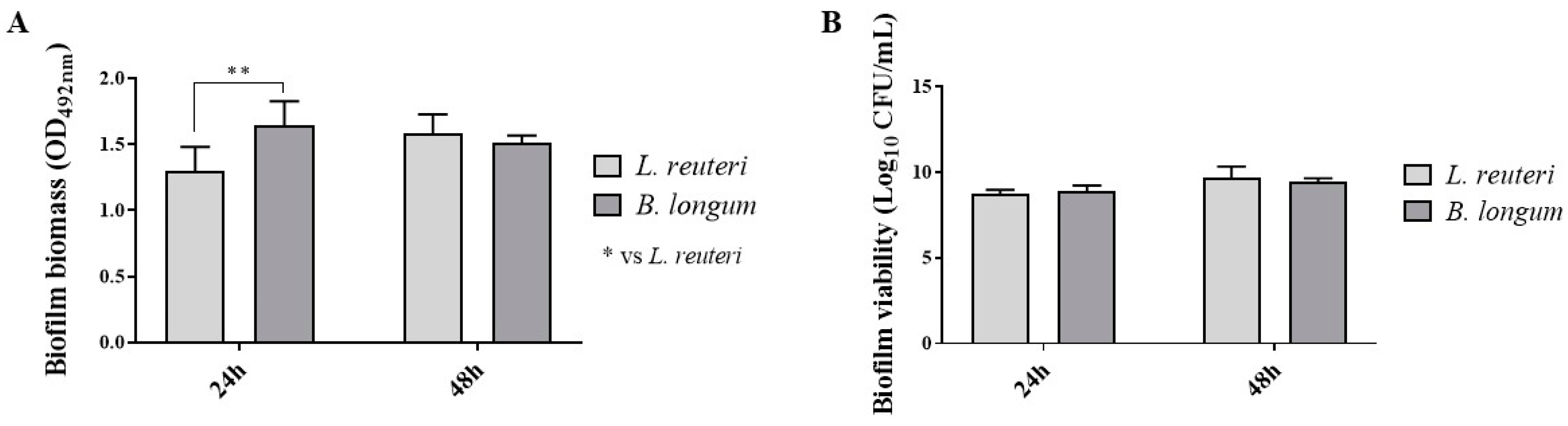
2.1.2. Biofilm Production

In this study, the probiotic strains’ ability to develop a biofilm was determined by evaluating biomass and viability in the sessile phase. As shown in Figure 2A, after 24 h of growth, B. longum showed a significantly greater ability to produce biomass with respect to L. reuteri. After 48 h of growth, B. longum and L. reuteri showed the same ability. In Figure 2B, the results of biofilm cell viability show that both strains reached the same colony-forming unit count after 24 h and 48 h of incubation.

2.1.3. Effect on HCE Cell Viability

Before exploring the effects of each probiotic strain against P. aeruginosa infection, we assessed their effect on HCE cell viability via MTT assay. Figure 3A shows the growth curves of probiotics coincubated with HCE cells at 24 h. The coincubation of bacteria with HCE cells did not exert any cytotoxic effect (Figure 3B). The viability of HCE cells was 100% and 126% when L. reuteri and B. longum were added, respectively, compared to untreated HCE cells (Figure 3B). MTT results were confirmed by trypan blue staining (Figure 3C) and LDH assay (Figure 3D), demonstrating that L. reuteri and B. longum did not increase cell death, exerting a protective effect on HCE cells.

2.1.4. Adhesion to HCE Cells

Probiotic strains with adhesion ability can prevent the adhesion of pathogens by competing for host cell binding sites [28]. The growth curves of probiotics incubated with HCE at 48 h of contact are shown in Figure 4A. As shown in Figure 4B, L. reuteri and B. longum demonstrated great ability to adhere to HCE cells after 24 h of contact, increasing the concentration of sessile cells after 48 h.

2.1.5. Effect on Occludin and Mucin-1 in SkinEthicTM HCE Model

The cornea is characterized by the presence of intercellular junctions which contribute to the first line of protection against pathogens and allergens [29,30]. Thus, we decided to evaluate the effects of L. reuteri and B. longum on occludin level, a tight-junction-associated protein that is abundantly expressed in the corneal epithelium. As demonstrated in our study, the corneal epithelium of the control group showed basal levels of occludin staining, forming a continuous ring around the cells (Figure 5A,A1); the same condition was observed also in the L. reuteri and B. longum groups (Figure 5B,B1 and C,C1, respectively). Moreover, we investigated the effects of the probiotic strains on mucin-1 (MUC-1), a transmembrane glycoprotein expressed in the apical surface of corneal cells which plays multiple roles in the protection of mucosal surfaces [29]. The data showed that MUC-1 was abundantly expressed in the superficial layer of cornea in the control group (Figure 6A,A1) as well as in the L. reuteri and B. longum groups (Figure 6B,B1,C,C1), suggesting that these strains exert beneficial effects on corneal structure, promoting MUC-1 and occludin levels. The results of immunofluorescence (IF) for occludin and MUC-1 were confirmed by Western blot analysis, highlighting the beneficial effects of probiotics on the corneal epithelium as shown in Figure 5D and Figure 6D (see densitometric analysis Figure 5D1 and Figure 6D1, respectively).

2.2. Prevention of P. aeruginosa Infection Damage via Pretreatment of HCE Cells with Probiotic Strains

2.2.1. Effect on HCE Cell Viability

HCE cells were treated with each probiotic strain for 1 h (short contact time) prior to P. aeruginosa inoculation. As shown in Figure 7A, P. aeruginosa infection (group 3) did not significantly reduce HCE cell viability compared to untreated HCE cells and HCE cells treated with each probiotic alone. MTT assay data were confirmed by trypan blue staining (Figure 7B) and LDH assay (Figure 7C), demonstrating that P. aeruginosa infection did not induce significant cell death or a marked LDH release after a short contact time.
The effect of probiotic strains on HCE cell viability was assessed at 24 h (long contact time) (Figure 8). The results showed that pretreatment with L. reuteri and B. longum (groups 3 and 4) significantly increased the viability of HCE cells compared to P. aeruginosa infection (group 3) (Figure 8A). MTT assay data were confirmed by trypan blue staining (Figure 8B) and LDH assay (Figure 8C), demonstrating that probiotic strains may reduce the % of cell death and LDH release compared to P. aeruginosa infection (group 3), exerting a protective effect.

2.2.2. Antagonistic Activity against P. aeruginosa Adhesion

The results demonstrated that the probiotic strains did not prevent the adhesion of P. aeruginosa to HCE cells in the experimental conditions used in this study (Table 1).

2.2.3. Anti-Inflammatory Activity against P. aeruginosa

Considering the key role of the inflammatory process in corneal epithelial infections induced by P. aeruginosa [31], it was decided to investigate the effect of L. reuteri and B. longum at 24 h of coincubation on the levels of pro-inflammatory cytokines, such as tumor necrosis factor-α (TNF-α), and on levels of anti-inflammatory cytokines, such as interleukin (IL-10), in HCE cell supernatants. Our results demonstrated that P. aeruginosa infection (group 3) is characterized by an increase in the levels of the pro-inflammatory cytokine TNF-α and a decrease in the levels of the anti-inflammatory cytokine IL-10 compared to untreated HCE cells (group 1) and to HCE cells treated with each probiotic alone (group 2) (long contact time; see Figure 9A,B). However, L. reuteri and B. longum significantly reduced TNF-α and increased IL-10 levels compared to P. aeruginosa infection (group 3), as shown in Figure 9A,B, counteracting the inflammatory process.

2.2.4. Antinitrosative Activity against P. aeruginosa

Nitrosative stress is a process characterized by the overproduction of nitric oxide (NO); it is characterized by the simultaneous production of superoxide anions, which results in the formation of peroxynitrite, and of other reactive nitrogen species (RNS), which contribute to mitochondrial dysfunction and inflammatory process [32,33,34]. Based on these findings, the effect of probiotics on NOx levels in HCE cell supernatants following P. aeruginosa infection was investigated. HCE cells were treated with each probiotic strain for 24 h (long contact time) prior to P. aeruginosa inoculation. At 24 h of coincubation, the supernatants were collected and analyzed. As shown in Figure 10, the results revealed that P. aeruginosa infection (group 3) was characterized by a significant increase in NOx levels compared to untreated HCE cells (group 1) and HCE cells treated with each probiotic alone (group 2); however, L. reuteri and B. longum significantly reduced NOx levels (group 4) at 24 h (long contact time; see Figure 10), counteracting nitrosative stress.

3. Discussion

Keratitis caused by P. aeruginosa is of particular concern because it develops rapidly, triggering an inflammatory/immune reaction that may lead to vision loss after ocular trauma or in contact lens wearers [35]. Traditional strategies for infection include antibiotics and steroids in the form of eye drops. However, eye drops containing antibiotics may lead to dysbiosis, causing an imbalance in favor of pathogenic strains and increasing the risk of resistance [10,36,37]. Those containing steroids may bring side effects, such as increased intraocular pressure or corneal complications. Probiotics could be one of the most promising biotherapies in the prophylaxis of and therapeutic usage for a variety of diseases, including infections [10]. The goal of prophylactic probiotics is to keep “good” bacteria colonyzed to combat pathogen invasion [19]. Several experimental and clinical results have shown the benefits of probiotics in the prevention of P. aeruginosa infection [38,39,40]. Preventing overt microbiological damage or imbalance may determine improvement in clinical illness as well as reduce the frequency and prevalence of the chronic infections [41].
In line with this, our results demonstrated that pretreatment with probiotic strains significantly protected HCE cells by reducing damage caused by P. aeruginosa infection. We demonstrated that probiotic strains preserved HCE cell viability, promoting occludin distribution and mucin production, suggesting their beneficial effects on HCE tissue structure. Regarding the efficacy of pretreating HCE cells with probiotic strains before P. aeruginosa infection, a significant restoration of cell viability was demonstrated compared to untreated and infected HCE cells. Moreover, L. reuteri and B. longum significantly reduced the levels of pro-inflammatory cytokines such as TNF-α and reestablished IL-10 levels, reducing the inflammatory response following a long contact time. In addition, with reference to antinitrosative activity, pretreatment with the probiotic strains showed a significant reduction in NOx level, counteracting the overproduction of nitrogen-based free radicals at 24 h of pretreatment. However, the adhesion of P. aeruginosa to HCE was not countered by probiotic strains, probably because of their modest capacity for auto-aggregation, coaggregation and adhesion with P. aeruginosa.
Many studies have proven the beneficial effects of probiotics on many diseases, achieved by promoting cell survival, cell proliferation, barrier function, and by stimulating the immune response [42,43,44,45,46]. Pretreatment with probiotic bacteria such as Lactobacilli exerts antioxidant and antiapoptotic activity, both in vitro and in vivo, improving cell viability [47,48]. Recent reports demonstrated that the application of probiotics might be effective in the management of eye diseases, suggesting probiotics as a potential alternative therapeutic treatment thanks to their ability to regulate epithelial barriers, secrete mucus and antimicrobial peptides, as well as to stimulate cell proliferation and the immune system [49].
Among the strains commonly found in the human microbiota, lactobacilli are mainly predominant in the gastric region and upper gastro-intestinal tract, while bifidobacteria in the lower intestinal tract. L. reuteri, used in this study, belongs to clade II. It has been demonstrated that clade II strains can be considered immunosuppressive and anti-inflammatory [50,51,52]. Moreover, L. reuteri can attach to mucin and suppress potentially deleterious effects of mucosal inflammation but it produces low levels of antimicrobial compounds such as reuterin [53,54,55,56]. B. longum subsp. infantis, within the genus Bifidobacterium, has shown several beneficial effects attributed to strains belonging to the subspecies [57]. This strain has proven to be particularly effective in protecting against infectious diseases and in increasing the maturation of the immune response to suppress inflammation [58,59,60]. In light of this, a recent clinical study demonstrated that the application of Lactobacillus and Bifidobacterium strains could be a potential alternative therapeutic treatment for eye diseases by modulating the immune system, cell proliferation, and the secretion of mucus [49].
In this study, it was demonstrated that topical pretreatment of HCE cells infected with P. aeruginosa with L. reuteri or B. longum subsp. infantis can restore cell viability and reduce the inflammatory reaction caused by the pathogen, while also showing an antinitrosative effect. The prophylactic topical administration of a probiotic or a mixture of probiotic strains can reduce the number, duration, and severity of recurrent keratitis caused by P. aeruginosa and prevent ulcer formation. However, further studies are necessary to clarify the mechanisms by which probiotic strains act in preventing P. aeruginosa infection.
Considering the obtained in vitro data, in the next study, we will evaluate these benefit activities in an in vivo model. At this moment, in the field of utilizing probiotics as a new therapy for eye infection, much remains to be done. More experimental studies are needed to investigate the mode of action of homeostatic microorganisms in the prevention of several ophthalmic diseases. Knowing the efficacy and safety of single probiotic strains in contact with HCE cells could lead to the formulation of a mixture of probiotic strains with different properties that could synergistically assist antibiotic and/or anti-inflammatory drugs in preventing or treating corneal infections.

4. Materials and Methods

4.1. Strains and Growth Conditions

The probiotic strains tested in this study were L. reuteri DSM20016 and B. longum subsp. infantis DSM20088 (Leibniz Institute DSMZ-German Collection of Microorganisms and Cell Cultures, Braunschweig, Germany) [53,61]. The pathogen strain used was P. aeruginosa American Type Culture Collections (ATCC 9027). The L. reuteri and B. longum strains were grown in De Man, Rogosa, and Sharpe broth (MRSB, Oxoid, Milan, Italy) with and without the addition of 0.05% cysteine for 24–48 h at 37 °C under 5% CO2 conditions. P. aeruginosa was grown in tryptic soy broth (TSB, Oxoid) at 37 °C for 24 h in aerobic conditions. The strains were stored at −70 °C in Microbanks™ (Pro-lab Diagnostics, Neston, UK). All reagents were purchased from Sigma-Aldrich (Milan, Italy) unless otherwise specified in the text.

4.2. Aggregation and Coaggregation with P. aeruginosa

Auto-aggregation assay. The cultures of probiotic strains in steady state were centrifugated at 10,000× g for 15 min and the bacteria were resuspended in 10 mL of phosphate-buffered saline solution (PBS) (pH 7.4) to approximately 2 × 10⁸ CFU/mL (OD at 600 nm 0.5 for L. reuteri and B. longum strains). Each suspension was vortexed for 10 s and incubated for 5 h at room temperature. At each hour, 1 mL of the upper part of each suspension was withdrawn to measure absorbance. The percentage of auto-aggregation was calculated according to the following formula:
Auto-aggregation% = [1 − ODt/ODi] × 100
where ODi is the initial OD of the microbial suspension and ODt is optical density at different time points.
Coaggregation assay. The suspensions of probiotic strains were treated as mentioned above. P. aeruginosa was harvested in stationary phase by centrifugation during 10 min at 5000× g and resuspended in PBS (pH 7.2) to obtain a P. aeruginosa suspension of about 2 × 10⁸ CFU/mL (OD600 0.2). Aliquots of pairs of bacterial suspensions (each probiotic and pathogen strain) totaling 10 mL each were vortexed for 10 s. Samples containing a single bacterial suspension were used as control. Each suspension was vortexed for 10 s and maintained for 5 h at room temperature. At each hour, 1 mL of the upper part of each suspension was withdrawn to measure absorbance as described above. The percentage of coaggregation was then calculated according to the following formula:
Co-aggregation % = [ ( Ax + Ay ) / 2 ] At ( x + y ) × 100 ( Ax + Ay ) / 2
where Ax (pathogen) and Ay (probiotic) are the OD values of each probiotic or pathogen strain suspension, and At (x + y) is the OD of the combined aggregation of each probiotic strain with the pathogen at different time points [62].

4.3. Biofilm Production

The evaluation of biofilm formation was carried out by determining the biomass and viability of each probiotic strain in the sessile phase. Overnight cultures of L. reuteri and B. longum strains in both MRSB and MRSB with 0.05% cysteine were standardized to a concentration of 5 × 10⁵ CFU/mL, placed in a flat-bottomed polystyrene microplate (Corning Inc., Corning, NY, USA), and incubated for 24–48 h at 37 °C under 5% CO2 conditions. Then, the planktonic phase was gently removed, and the wells were washed with PBS (pH 7.4) 3 times. To assess the biomass of the strains, the microplate wells were dried, colored with 0.1% safranin, and washed again with PBS. The colored biofilm was resuspended in 30% acetic acid (v/v) and then OD492 was measured by spectrophotometer. Control wells containing only the medium were included. The production of biofilm was obtained by comparing the OD value obtained from the arithmetic mean of the values of each strain with the cut-off value (ODc), defined as the average of the values that was obtained from the control. To assess the cell viability of the samples, microplate wells with adherent biofilm were scarified in PBS, serially diluted, and seeded on MRSA (Oxoid) plates. After an incubation period of 24 h and 48 h at 37 °C, CFU/mL were counted [63,64].

4.4. Cell Culture

Human corneal epithelial (HCE) cells were kindly provided by the Sooft Research Center, Catania, Italy, SpA. HCE cells were isolated from the human cornea of a donor patient as described by Cristaldi and colleagues [65]. HCE cells were cultured in 75 cm2 flasks in DMEM-F12 Advanced (ATCC, cat. no. 12634010) and supplemented with 1% penicillin/streptomycin (penicillin 1000 units–streptomycin 0.1 mg/L, Sigma-Aldrich® Catalog No. P4333; St. Louis, MO, USA), 2% fetal bovine serum (FBS) (Sigma-Aldrich, St. Louis, MO, USA, cat. no. F7524), and specific corneal epithelial growth factors (ATCC, cat. no. PCS-700-040) at 37 °C in a humidified atmosphere containing 5% CO2.

4.5. Viability Assays

Cell viability was measured using a mitochondria-dependent dye for live cells ((3-(4,5 Dimethylthiazol-2-yl)-2,5-diphenyltetrazolium bromide); MTT), lactate dehydrogenase (LDH) assay, and trypan blue staining. For MTT assay, HCE cells were seeded at a density of 4 × 10⁴ cells/well in 96-well plates for each probiotic or pathogen in DMEM without antibiotic supplements and incubated at 37 °C and 5% CO2 in the incubator. After 24 h, the cells were incubated with each probiotic strain suspension (1 × 10⁷ CFU/mL) [66,67]. After 24 h, HCE cells were incubated at 37 °C with MTT (0.2 mg/mL) for 1 h. The medium was removed by aspiration and the cells were lysed with DMSO (100 μL). Absorbance (OD570) was measured using a microplate reader [68].
To confirm MTT results, cell membrane integrity was analyzed using the cytotoxicity assay (G1780, Promega, Madison, WI, USA) according to the manufacturer’s instructions. The percentage of LDH released was calculated using the following formula:
% LDH   release = 100 × Experimental   LDH   release   ( O D 490 ) ( Maximum   LDH   release   ( O D 490 )
For trypan blue staining, after incubation with probiotic strains, trypan blue solution (0.4%) was mixed with trypsinized cell suspensions at a ratio of 1:1 to evaluate the % of cell death. After 5 min incubation, cells were loaded onto a hemocytometer, and both live (unstained) and dead (blue-stained) cells were counted under a light microscope [69]. The number of cells died was expressed as %.

4.6. Adhesion to HCE Cells

Bacterial adhesion tests on HCE cell monolayers were carried out in 96-well tissue culture plates. Cells were grown routinely in DMEM, supplemented as mentioned above without antibiotic to reach a density of 4 × 10⁴ cells/well in 96-well plates, and incubated at 37 °C under 5% CO2 conditions for 24 h until the formation of a dense cell layer. Probiotic strains were cultivated for 24 h in a standard culture broth before the adhesion experiment. The bacterial suspension was then washed twice in PBS and finally resuspended in DMEM to a 1 × 10⁷ CFU/mL final concentration. Subsequently, each bacterial suspension was added to the wells of the microtiter in monolayers and incubated at 37 °C under 5% CO2 conditions for 24 and 48 h. The supernatant of each probiotic strain was eliminated and unattached cells were gently removed by PBS buffer 3 times. The cells were then lysed by 0.1% Triton X-100 lysis buffer (10 mM Tris-HCl, 2 mM MgCl2, 0.25% Triton X-100, pH 8.0) and the number of viable adherent bacteria was determined by plating serial dilutions on MRSA to quantify the total number of cell-associated bacteria (CFU/cm2) [67,70].

4.7. SkinEthicTM HCE Model

The SkinEthicTM HCE model was purchased from EPISKIN Laboratories (Lyon, France, UE). It is an in vitro model of transformed human corneal keratinocytes cultivated on an inert polycarbonate filter which is structurally, morphologically, and functionally like the human cornea, with the presence of basal and wing cells and mucus production [71]. The SkinEthicTM HCE model was cultured in a chemically defined medium as previously described [71].
Experimental groups:
  • SkinEthicTM HCE cells in DMEM (control group);
  • SkinEthicTM HCE cells treated with each probiotic strain at 1 × 10⁷ CFU/mL for 24 h at 37 °C, under 5% CO2 conditions.

4.8. Immunofluorescence (IF) Staining for Intercellular Junctions and Mucin Localization

SkinEthicTM HCE sections of 7 μm were incubated with the primary antibodies antioccludin (1:100; 71-1500 Invitrogen, Carlsbad, CA, USA) or antimucin 1 (1:200; sc-7313, Santa Cruz Biotechnology, Santa Cruz, CA, USA) in a humidified chamber at 37 °C overnight. Sections were washed three times with PBS and were incubated with secondary antibody Alexa Fluor-488 (1:1000 in PBS, v/v, Molecular Probes, Altrincham, UK) for 1 h at 37 °C. Sections were washed in PBS and, for nuclear staining, 4′,6′-diamidino-2-phenylindole (DAPI; Hoechst, Frankfurt, Germany) (2 μg/mL) was added to PBS [72]. Sections were observed and photographed at 100× magnification using a Leica DM2000 microscope (Wetzlar, Germany).

4.9. Western Blot Analysis

Western blot analysis was performed on SkinEthicTM HCE cells as previously described [73]. Protein concentration was estimated by the Bio-Rad protein assay using bovine serum albumin (BSA) as standard. Samples were heated at 95 °C for 5 min, and the same amounts of protein were separated on 12% SDS-PAGE gel and blotted to PVDF membrane (Immobilon-P). Membranes were incubated overnight (O/N) at 4 °C with the following primary antibodies: anti-occludin (1:500, Cat#91131, Cell signaling, Danvers, MA, USA) or anti-mucin 1 (1:500; sc-7313, Santa Cruz Biotechnology, Santa Cruz, CA, USA). To ascertain that the blots were loaded with equal amounts of proteins, the membranes were also incubated in the presence of the antibody against β-actin protein (1:1000; sc-47778, Santa Cruz Biotechnology, Santa Cruz, CA, USA). Results are expressed as % of the control.

4.10. Pretreatment of HCE Cells with Probiotic Strains Prevents P. aeruginosa Infection Damage

After washing the cell monolayers 3 times with PBS, each probiotic strain (approximately 1 × 10⁷ CFU/mL) was added 1 h (short contact time) and 24 h (long contact time) prior to P. aeruginosa inoculation (1 × 10⁶ CFU/mL). Then, the short-contact-time group was incubated for 1 h and the long-contact-time group for 24 h at 37 °C under 5% CO2 conditions. Control wells (HCE cells in medium) and HCE cells with P. aeruginosa challenge were included.
Experimental groups:
  • HCE cells in DMEM (control group);
  • HCE cells pretreated with each probiotic strain (pretreated cells);
  • HCE cells infected with P. aeruginosa (infected cells);
  • HCE cells pretreated with each probiotic strain and then infected with P. aeruginosa (pretreated and infected cells).

4.11. Viability Assays

After 1 h (short contact time) and 24 h (long contact time) of growth, the HCE control monolayer (group 1), pretreated monolayers (group 2), monolayers infected with P. aeruginosa (group 3), and pretreated monolayers infected with P. aeruginosa (group 4) were incubated at 37 °C with MTT. To confirm MTT results, LDH assay and trypan blue staining were performed following the above-described protocol.

4.12. Antagonistic Activity against P. aeruginosa Adhesion to HCE Cells

After 1 h (short contact time) and 24 h (long contact time) of growth, the HCE control monolayer (group 1), pretreated monolayers (group 2), monolayers infected with P. aeruginosa (group 3), and pretreated monolayers infected with P. aeruginosa (group 4) were washed twice with PBS and lysed with 1 mL/L (v/v) Triton X-100 for 5 min and were serially diluted. Bacteria were determined by plate counting on MRSA for L. reuteri and B. longum strains and on Pseudomonas agar base (Oxoid) for P. aeruginosa. The total number of cell-associated bacteria was expressed as CFU/cm2 [74,75].

4.13. Enzyme-Linked Immunosorbent Assay for TNFα and IL-10 in HCE Cell Supernatant

At 24 h (long contact time), the anti-inflammatory effect on pretreated HCE monolayers infected with P. aeruginosa (group 4), monolayers infected with P. aeruginosa (group 3), pretreated monolayers (group 2), and the control monolayer (group 1) were evaluated by enzyme-linked immunosorbent assay (ELISA). ELISA kits for tumor necrosis factor-α (TNF-α) and interleukin-10 (IL-10) were used according to the manufacturer’s instructions (TNF-α ELISA Kit My BioSource; cat No. MBS267654; IL-10 ELISA Kit eBioscience; Catalog Number: 88-7106). Cell supernatants were centrifuged for 10 min at 3000 rpm; then, supernatants were collected for immediate testing and/or stored at −20 °C. Absorbance was measured at 450 nm. The authors decided to evaluate cytokines release only at 24 h (long contact time) due to cell viability results.

4.14. Measurement of NOx Levels in HCE Cell Supernatant

At 24 h (long contact time), total nitrite levels, as an indicator of nitric oxide (NO) production, were measured in pretreated HCE monolayers infected with P. aeruginosa (group 4), monolayers infected with P. aeruginosa (group 3), pretreated monolayers (group 2), and the control monolayer (group 1) in the supernatant as previously described [76]. Briefly, nitrate in the medium was reduced to nitrite by incubation with nitrate reductase (670 mU/mL) and β-nicotinamide adenine dinucleotide 3-phosphate (160 mM) at room temperature for 3 h. Entire nitrite concentration was later calculated via the Griess reaction by adding 100 μL Griess reagent [0.1% (w/v) N-(1-naphthyl) ethylenediamine dihydrochloride in H2O and 1% (w/v) sulfanilamide in 5% (v/v) concentrated H3PO4; volume 1:1] to the 100-μL sample. OD570 was determined using a microplate reader (Tecan, Männedorf, Switzerland). The authors decided to evaluate NOx levels only at 24 h (long contact time) due to cell viability results.

4.15. Statistical Analysis

The results from the 3 experiments, expressed as means ± standard deviation, were statistically analyzed using the software GraphPad Prism 9.5.1. The results were analyzed using a one-way analysis of variance (ANOVA) and t-test procedure. Multiple comparisons were performed among groups by the Bonferroni correction test. Differences were considered statistically significant at p ≤ 0.05.

5. Conclusions

In conclusion, based on the obtained results, we demonstrated that L. reuteri and B. longum exert several beneficial effects in the context of corneal infection caused by P. aeruginosa thanks to their ability to restore cell viability and modulate inflammatory and nitrosative processes. However, despite these interesting data, further studies are needed to better understand their efficacy and mechanisms in the process of corneal infection.

Author Contributions

Conceptualization, A.M. and I.P.; methodology, S.A.S. and L.C.; data curation, A.M., E.E., A.N. and I.P.; writing—original draft preparation, I.P., S.A.S. and A.M.; supervision, A.M. and I.P. All authors have read and agreed to the published version of the manuscript.

Funding

This research received no external funding.

Institutional Review Board Statement

Not applicable.

Informed Consent Statement

Not applicable.

Data Availability Statement

All data generated or analyzed during this study are included in this article.

Acknowledgments

The authors thank Vincenza Isabella for her assistance with technical English in this study.

Conflicts of Interest

The authors declare no conflicts of interest.

References

  1. Al-Mujaini, A.; Al-Kharusi, N.; Thakral, A.; Wali, U.K. Bacterial keratitis: Perspective on epidemiology, clinico-pathogenesis, diagnosis and treatment. Sultan Qaboos Univ. Med. J. 2009, 9, 184–195. [Google Scholar]
  2. Xue, W.; Li, J.J.; Zou, Y.; Zou, B.; Wei, L. Microbiota and Ocular Diseases. Front. Cell Infect. Microbiol. 2021, 11, 759333. [Google Scholar] [CrossRef]
  3. Sy, A.; Srinivasan, M.; Mascarenhas, J.; Lalitha, P.; Rajaraman, R.; Ravindran, M.; Oldenburg, C.E.; Ray, K.J.; Glidden, D.; Zegans, M.E.; et al. Pseudomonas aeruginosa keratitis: Outcomes and response to corticosteroid treatment. Investig. Ophthalmol. Vis. Sci. 2012, 53, 267–272. [Google Scholar] [CrossRef]
  4. Rossi, E.; Ghoul, M.; La Rosa, R. Editorial: Pseudomonas aeruginosa Pathogenesis: Virulence, Antibiotic Tolerance and Resistance, Stress Responses and Host-Pathogen Interactions. Front. Cell Infect. Microbiol. 2022, 12, 860314. [Google Scholar] [CrossRef]
  5. Fleiszig, S.M.; Evans, D.J. The pathogenesis of bacterial keratitis: Studies with Pseudomonas aeruginosa. Clin. Exp. Optom. 2002, 85, 271–278. [Google Scholar] [CrossRef]
  6. Hazlett, L.D.; Huang, X.; McClellan, S.A.; Barrett, R.P. Further studies on the role of IL-12 in Pseudomonas aeruginosa corneal infection. Eye 2003, 17, 863–871. [Google Scholar] [CrossRef] [PubMed]
  7. Stapleton, F.; Bakkar, M.; Carnt, N.; Chalmers, R.; Vijay, A.K.; Marasini, S.; Ng, A.; Tan, J.; Wagner, H.; Woods, C.; et al. CLEAR—Contact lens complications. Cont. Lens Anterior Eye 2021, 44, 330–367. [Google Scholar] [CrossRef] [PubMed]
  8. Chiang, M.C.; Chern, E. More than Antibiotics: Latest Therapeutics in the Treatment and Prevention of Ocular Surface Infections. J. Clin. Med. 2022, 11, 4195. [Google Scholar] [CrossRef] [PubMed]
  9. Aragona, P.; Baudouin, C.; Benitez Del Castillo, J.M.; Messmer, E.; Barabino, S.; Merayo-Lloves, J.; Brignole-Baudouin, F.; Inferrera, L.; Rolando, M.; Mencucci, R.; et al. The ocular microbiome and microbiota and their effects on ocular surface pathophysiology and disorders. Surv. Ophthalmol. 2021, 66, 907–925. [Google Scholar] [CrossRef] [PubMed]
  10. Petrillo, F.; Pignataro, D.; Lavano, M.A.; Santella, B.; Folliero, V.; Zannella, C.; Astarita, C.; Gagliano, C.; Franci, G.; Avitabile, T.; et al. Current Evidence on the Ocular Surface Microbiota and Related Diseases. Microorganisms 2020, 8, 1033. [Google Scholar] [CrossRef] [PubMed]
  11. Lu, L.J.; Liu, J. Human Microbiota and Ophthalmic Disease. Yale J. Biol. Med. 2016, 89, 325–330. [Google Scholar]
  12. Hilliam, Y.; Kaye, S.; Winstanley, C. Pseudomonas aeruginosa and microbial keratitis. J. Med. Microbiol. 2020, 69, 3–13. [Google Scholar] [CrossRef]
  13. Urwin, L.; Okurowska, K.; Crowther, G.; Roy, S.; Garg, P.; Karunakaran, E.; MacNeil, S.; Partridge, L.J.; Green, L.R.; Monk, P.N. Corneal Infection Models: Tools to Investigate the Role of Biofilms in Bacterial Keratitis. Cells 2020, 9, 2450. [Google Scholar] [CrossRef]
  14. Mohamed, S.; Elmohamady, M.N.; Abdelrahman, S.; Amer, M.M.; Abdelhamid, A.G. Antibacterial effects of antibiotics and cell-free preparations of probiotics against Staphylococcus aureus and Staphylococcus epidermidis associated with conjunctivitis. Saudi Pharm. J. 2020, 28, 1558–1565. [Google Scholar] [CrossRef] [PubMed]
  15. Chiang, M.C.; Chern, E. Ocular surface microbiota: Ophthalmic infectious disease and probiotics. Front. Microbiol. 2022, 13, 952473. [Google Scholar] [CrossRef] [PubMed]
  16. Martin, R.; Langella, P. Emerging Health Concepts in the Probiotics Field: Streamlining the Definitions. Front. Microbiol. 2019, 10, 1047. [Google Scholar] [CrossRef] [PubMed]
  17. Zheng, J.; Wittouck, S.; Salvetti, E.; Franz, C.; Harris, H.M.B.; Mattarelli, P.; O’Toole, P.W.; Pot, B.; Vandamme, P.; Walter, J.; et al. A taxonomic note on the genus Lactobacillus: Description of 23 novel genera, emended description of the genus Lactobacillus Beijerinck 1901, and union of Lactobacillaceae and Leuconostocaceae. Int. J. Syst. Evol. Microbiol. 2020, 70, 2782–2858. [Google Scholar] [CrossRef] [PubMed]
  18. Das, S.; Vishakha, K.; Banerjee, S.; Bera, T.; Mondal, S.; Ganguli, A. A novel probiotic strain of Lactobacillus fermentum TIU19 isolated from Haria beer showing both in vitro antibacterial and antibiofilm properties upon two multi resistant uro-pathogen strains. Curr. Res. Microb. Sci. 2022, 3, 100150. [Google Scholar] [CrossRef] [PubMed]
  19. Tegegne, B.A.; Kebede, B. Probiotics, their prophylactic and therapeutic applications in human health development: A review of the literature. Heliyon 2022, 8, e09725. [Google Scholar] [CrossRef] [PubMed]
  20. Isenring, J.; Geirnaert, A.; Lacroix, C.; Stevens, M.J.A. Bistable auto-aggregation phenotype in Lactiplantibacillus plantarum emerges after cultivation in in vitro colonic microbiota. BMC Microbiol. 2021, 21, 268. [Google Scholar] [CrossRef]
  21. Nwoko, E.Q.A.; Okeke, I.N. Bacteria autoaggregation: How and why bacteria stick together. Biochem. Soc. Trans. 2021, 49, 1147–1157. [Google Scholar] [CrossRef]
  22. Tuo, Y.; Yu, H.; Ai, L.; Wu, Z.; Guo, B.; Chen, W. Aggregation and adhesion properties of 22 Lactobacillus strains. J. Dairy Sci. 2013, 96, 4252–4257. [Google Scholar] [CrossRef]
  23. Salas-Jara, M.J.; Ilabaca, A.; Vega, M.; Garcia, A. Biofilm Forming Lactobacillus: New Challenges for the Development of Probiotics. Microorganisms 2016, 4, 35. [Google Scholar] [CrossRef]
  24. Carvalho, F.M.; Teixeira-Santos, R.; Mergulhao, F.J.M.; Gomes, L.C. The Use of Probiotics to Fight Biofilms in Medical Devices: A Systematic Review and Meta-Analysis. Microorganisms 2020, 9, 27. [Google Scholar] [CrossRef]
  25. Bubnov, R.V.; Babenko, L.P.; Lazarenko, L.M.; Mokrozub, V.V.; Spivak, M.Y. Specific properties of probiotic strains: Relevance and benefits for the host. EPMA J. 2018, 9, 205–223. [Google Scholar] [CrossRef] [PubMed]
  26. Chisari, G.; Chisari, E.M.; Francaviglia, A.; Chisari, C.G. The mixture of bifidobacterium associated with fructo-oligosaccharides reduces the damage of the ocular surface. Clin. Ter. 2017, 168, e181–e185. [Google Scholar] [CrossRef] [PubMed]
  27. Iovieno, A.; Lambiase, A.; Sacchetti, M.; Stampachiacchiere, B.; Micera, A.; Bonini, S. Preliminary evidence of the efficacy of probiotic eye-drop treatment in patients with vernal keratoconjunctivitis. Graefes Arch. Clin. Exp. Ophthalmol. 2008, 246, 435–441. [Google Scholar] [CrossRef]
  28. Monteagudo-Mera, A.; Rastall, R.A.; Gibson, G.R.; Charalampopoulos, D.; Chatzifragkou, A. Adhesion mechanisms mediated by probiotics and prebiotics and their potential impact on human health. Appl. Microbiol. Biotechnol. 2019, 103, 6463–6472. [Google Scholar] [CrossRef] [PubMed]
  29. Mantelli, F.; Argueso, P. Functions of ocular surface mucins in health and disease. Curr. Opin. Allergy Clin. Immunol. 2008, 8, 477–483. [Google Scholar] [CrossRef]
  30. Mantelli, F.; Mauris, J.; Argueso, P. The ocular surface epithelial barrier and other mechanisms of mucosal protection: From allergy to infectious diseases. Curr. Opin. Allergy Clin. Immunol. 2013, 13, 563–568. [Google Scholar] [CrossRef]
  31. Hazlett, L.D. Corneal response to Pseudomonas aeruginosa infection. Prog. Retin. Eye Res. 2004, 23, 1–30. [Google Scholar] [CrossRef] [PubMed]
  32. Bourges, J.L.; Torriglia, A.; Valamanesh, F.; Benezra, D.; Renard, G.; Behar-Cohen, F.F. Nitrosative stress and corneal transplant endothelial cell death during acute graft rejection. Transplantation 2007, 84, 415–423. [Google Scholar] [CrossRef] [PubMed]
  33. Canto, A.; Olivar, T.; Romero, F.J.; Miranda, M. Nitrosative Stress in Retinal Pathologies: Review. Antioxidants 2019, 8, 543. [Google Scholar] [CrossRef] [PubMed]
  34. Wang, F.; Yuan, Q.; Chen, F.; Pang, J.; Pan, C.; Xu, F.; Chen, Y. Fundamental Mechanisms of the Cell Death Caused by Nitrosative Stress. Front. Cell Dev. Biol. 2021, 9, 742483. [Google Scholar] [CrossRef] [PubMed]
  35. Shan, Q.; Dwyer, M.; Rahman, S.; Gadjeva, M. Distinct susceptibilities of corneal Pseudomonas aeruginosa clinical isolates to neutrophil extracellular trap-mediated immunity. Infect. Immun. 2014, 82, 4135–4143. [Google Scholar] [CrossRef] [PubMed]
  36. Miller, D.; Iovieno, A. The role of microbial flora on the ocular surface. Curr. Opin. Allergy Clin. Immunol. 2009, 9, 466–470. [Google Scholar] [CrossRef] [PubMed]
  37. Mshangila, B.; Paddy, M.; Kajumbula, H.; Ateenyi-Agaba, C.; Kahwa, B.; Seni, J. External ocular surface bacterial isolates and their antimicrobial susceptibility patterns among pre-operative cataract patients at Mulago National Hospital in Kampala, Uganda. BMC Ophthalmol. 2013, 13, 71. [Google Scholar] [CrossRef]
  38. Huang, F.C.; Lu, Y.T.; Liao, Y.H. Beneficial effect of probiotics on Pseudomonas aeruginosa-infected intestinal epithelial cells through inflammatory IL-8 and antimicrobial peptide human beta-defensin-2 modulation. Innate Immun. 2020, 26, 592–600. [Google Scholar] [CrossRef]
  39. Forestier, C.; Guelon, D.; Cluytens, V.; Gillart, T.; Sirot, J.; De Champs, C. Oral probiotic and prevention of Pseudomonas aeruginosa infections: A randomized, double-blind, placebo-controlled pilot study in intensive care unit patients. Crit. Care 2008, 12, R69. [Google Scholar] [CrossRef]
  40. Valdez, J.C.; Peral, M.C.; Rachid, M.; Santana, M.; Perdigon, G. Interference of Lactobacillus plantarum with Pseudomonas aeruginosa in vitro and in infected burns: The potential use of probiotics in wound treatment. Clin. Microbiol. Infect. 2005, 11, 472–479. [Google Scholar] [CrossRef]
  41. Milner, E.; Stevens, B.; An, M.; Lam, V.; Ainsworth, M.; Dihle, P.; Stearns, J.; Dombrowski, A.; Rego, D.; Segars, K. Utilizing Probiotics for the Prevention and Treatment of Gastrointestinal Diseases. Front. Microbiol. 2021, 12, 689958. [Google Scholar] [CrossRef] [PubMed]
  42. Castiblanco, G.A.; Yucel-Lindberg, T.; Roos, S.; Twetman, S. Effect of Lactobacillus reuteri on Cell Viability and PGE(2) Production in Human Gingival Fibroblasts. Probiotics Antimicrob. Proteins 2017, 9, 278–283. [Google Scholar] [CrossRef]
  43. Maldonado Galdeano, C.; Cazorla, S.I.; Lemme Dumit, J.M.; Velez, E.; Perdigon, G. Beneficial Effects of Probiotic Consumption on the Immune System. Ann. Nutr. Metab. 2019, 74, 115–124. [Google Scholar] [CrossRef]
  44. Maudsdotter, L.; Jonsson, H.; Roos, S.; Jonsson, A.B. Lactobacilli reduce cell cytotoxicity caused by Streptococcus pyogenes by producing lactic acid that degrades the toxic component lipoteichoic acid. Antimicrob. Agents Chemother. 2011, 55, 1622–1628. [Google Scholar] [CrossRef]
  45. Zhao, X.; Zhou, J.; Liang, W.; Sheng, Q.; Lu, L.; Chen, T.; Chen, J.; Tan, K.; Lv, Z. Probiotics mixture reinforces barrier function to ameliorate necrotizing enterocolitis by regulating PXR-JNK pathway. Cell Biosci. 2021, 11, 20. [Google Scholar] [CrossRef] [PubMed]
  46. Sultana, R.; McBain, A.J.; O’Neill, C.A. Strain-dependent augmentation of tight-junction barrier function in human primary epidermal keratinocytes by Lactobacillus and Bifidobacterium lysates. Appl. Environ. Microbiol. 2013, 79, 4887–4894. [Google Scholar] [CrossRef]
  47. Wang, J.; Zhang, W.; Wang, S.; Wang, Y.; Chu, X.; Ji, H. Lactobacillus plantarum Exhibits Antioxidant and Cytoprotective Activities in Porcine Intestinal Epithelial Cells Exposed to Hydrogen Peroxide. Oxid. Med. Cell Longev. 2021, 2021, 8936907. [Google Scholar] [CrossRef] [PubMed]
  48. Iorio, R.; Petricca, S.; Luzi, C.; Bellio, P.; Cristiano, L.; Festuccia, C.; Amicosante, G.; Celenza, G. Lactobacillus sakei Pro-Bio65 Reduces TNF-alpha Expression and Upregulates GSH Content and Antioxidant Enzymatic Activities in Human Conjunctival Cells. Transl. Vis. Sci. Technol. 2021, 10, 8. [Google Scholar] [CrossRef]
  49. Tavakoli, A.; Markoulli, M.; Papas, E.; Flanagan, J. The Impact of Probiotics and Prebiotics on Dry Eye Disease Signs and Symptoms. J. Clin. Med. 2022, 11, 4889. [Google Scholar] [CrossRef]
  50. Pena, J.A.; Rogers, A.B.; Ge, Z.; Ng, V.; Li, S.Y.; Fox, J.G.; Versalovic, J. Probiotic Lactobacillus spp. diminish Helicobacter hepaticus-induced inflammatory bowel disease in interleukin-10-deficient mice. Infect. Immun. 2005, 73, 912–920. [Google Scholar] [CrossRef]
  51. Oh, P.L.; Benson, A.K.; Peterson, D.A.; Patil, P.B.; Moriyama, E.N.; Roos, S.; Walter, J. Diversification of the gut symbiont Lactobacillus reuteri as a result of host-driven evolution. ISME J. 2010, 4, 377–387. [Google Scholar] [CrossRef]
  52. Hemarajata, P.; Gao, C.; Pflughoeft, K.J.; Thomas, C.M.; Saulnier, D.M.; Spinler, J.K.; Versalovic, J. Lactobacillus reuteri-specific immunoregulatory gene rsiR modulates histamine production and immunomodulation by Lactobacillus reuteri. J. Bacteriol. 2013, 195, 5567–5576. [Google Scholar] [CrossRef]
  53. Spinler, J.K.; Sontakke, A.; Hollister, E.B.; Venable, S.F.; Oh, P.L.; Balderas, M.A.; Saulnier, D.M.; Mistretta, T.A.; Devaraj, S.; Walter, J.; et al. From prediction to function using evolutionary genomics: Human-specific ecotypes of Lactobacillus reuteri have diverse probiotic functions. Genome Biol. Evol. 2014, 6, 1772–1789. [Google Scholar] [CrossRef]
  54. Mu, Q.; Tavella, V.J.; Luo, X.M. Role of Lactobacillus reuteri in Human Health and Diseases. Front. Microbiol. 2018, 9, 757. [Google Scholar] [CrossRef]
  55. Hou, C.; Zeng, X.; Yang, F.; Liu, H.; Qiao, S. Study and use of the probiotic Lactobacillus reuteri in pigs: A review. J. Anim. Sci. Biotechnol. 2015, 6, 14. [Google Scholar] [CrossRef] [PubMed]
  56. Li, X.J.; Yue, L.Y.; Guan, X.F.; Qiao, S.Y. The adhesion of putative probiotic lactobacilli to cultured epithelial cells and porcine intestinal mucus. J. Appl. Microbiol. 2008, 104, 1082–1091. [Google Scholar] [CrossRef] [PubMed]
  57. Blanco, G.; Ruiz, L.; Tames, H.; Ruas-Madiedo, P.; Fdez-Riverola, F.; Sanchez, B.; Lourenco, A.; Margolles, A. Revisiting the Metabolic Capabilities of Bifidobacterium longum susbp. longum and Bifidobacterium longum subsp. infantis from a Glycoside Hydrolase Perspective. Microorganisms 2020, 8, 723. [Google Scholar] [CrossRef] [PubMed]
  58. Chichlowski, M.; Shah, N.; Wampler, J.L.; Wu, S.S.; Vanderhoof, J.A. Bifidobacterium longum Subspecies infantis (B. infantis) in Pediatric Nutrition: Current State of Knowledge. Nutrients 2020, 12, 1581. [Google Scholar] [CrossRef] [PubMed]
  59. Kim, S.; Han, S.Y.; Lee, J.; Kim, N.R.; Lee, B.R.; Kim, H.; Kwon, M.; Ahn, K.; Noh, Y.; Kim, S.J.; et al. Bifidobacterium longum and Galactooligosaccharide Improve Skin Barrier Dysfunction and Atopic Dermatitis-like Skin. Allergy Asthma Immunol. Res. 2022, 14, 549–564. [Google Scholar] [CrossRef] [PubMed]
  60. Quinn, E.M.; Slattery, H.; Walsh, D.; Joshi, L.; Hickey, R.M. Bifidobacterium longum subsp. infantis ATCC 15697 and Goat Milk Oligosaccharides Show Synergism In Vitro as Anti-Infectives against Campylobacter jejuni. Foods 2020, 9, 348. [Google Scholar] [CrossRef] [PubMed]
  61. Greppi, A.; Asare, P.T.; Schwab, C.; Zemp, N.; Stephan, R.; Lacroix, C. Isolation and Comparative Genomic Analysis of Reuterin-Producing Lactobacillus reuteri From the Chicken Gastrointestinal Tract. Front. Microbiol. 2020, 11, 1166. [Google Scholar] [CrossRef]
  62. Golowczyc, M.A.; Mobili, P.; Garrote, G.L.; Abraham, A.G.; De Antoni, G.L. Protective action of Lactobacillus kefir carrying S-layer protein against Salmonella enterica serovar Enteritidis. Int. J. Food Microbiol. 2007, 118, 264–273. [Google Scholar] [CrossRef]
  63. Speranza, B.; Liso, A.; Russo, V.; Corbo, M.R. Evaluation of the Potential of Biofilm Formation of Bifidobacterium longum subsp. infantis and Lactobacillus reuteri as Competitive Biocontrol Agents Against Pathogenic and Food Spoilage Bacteria. Microorganisms 2020, 8, 177. [Google Scholar] [CrossRef]
  64. Szewczyk, A.; Marino, A.; Taviano, M.F.; Cambria, L.; Davi, F.; Trepa, M.; Grabowski, M.; Miceli, N. Studies on the Accumulation of Secondary Metabolites and Evaluation of Biological Activity of In Vitro Cultures of Ruta montana L. in Temporary Immersion Bioreactors. Int. J. Mol. Sci. 2023, 24, 7045. [Google Scholar] [CrossRef]
  65. Cristaldi, M.; Olivieri, M.; Spampinato, G.; Anfuso, C.D.; Scalia, M.; Lupo, G.; Rusciano, D. Isolation and Characterization of a New Human Corneal Epithelial Cell Line: HCE-F. Cornea 2020, 39, 1419–1425. [Google Scholar] [CrossRef]
  66. Tuo, Y.F.; Zhang, L.W.; Yi, H.X.; Zhang, Y.C.; Zhang, W.Q.; Han, X.; Du, M.; Jiao, Y.H.; Wang, S.M. Short communication: Antiproliferative effect of wild Lactobacillus strains isolated from fermented foods on HT-29 cells. J. Dairy Sci. 2010, 93, 2362–2366. [Google Scholar] [CrossRef]
  67. Polak-Berecka, M.; Wasko, A.; Paduch, R.; Skrzypek, T.; Sroka-Bartnicka, A. The effect of cell surface components on adhesion ability of Lactobacillus rhamnosus. Antonie Van Leeuwenhoek 2014, 106, 751–762. [Google Scholar] [CrossRef] [PubMed]
  68. Paterniti, I.; Cordaro, M.; Campolo, M.; Siracusa, R.; Cornelius, C.; Navarra, M.; Cuzzocrea, S.; Esposito, E. Neuroprotection by association of palmitoylethanolamide with luteolin in experimental Alzheimer’s disease models: The control of neuroinflammation. CNS Neurol. Disord. Drug Targets 2014, 13, 1530–1541. [Google Scholar] [CrossRef] [PubMed]
  69. Jung, I.G.; Jeong, J.Y.; Yum, S.H.; Hwang, Y.J. Inhibitory Effects of Selected Medicinal Plants on Bacterial Growth of Methicillin-Resistant Staphylococcus aureus. Molecules 2022, 27, 7780. [Google Scholar] [CrossRef] [PubMed]
  70. Gopal, P.K.; Prasad, J.; Smart, J.; Gill, H.S. In vitro adherence properties of Lactobacillus rhamnosus DR20 and Bifidobacterium lactis DR10 strains and their antagonistic activity against an enterotoxigenic Escherichia coli. Int. J. Food Microbiol. 2001, 67, 207–216. [Google Scholar] [CrossRef] [PubMed]
  71. Alepee, N.; Grandidier, M.H.; Teluob, S.; Amaral, F.; Caviola, E.; De Servi, B.; Martin, S.; Meloni, M.; Nardelli, L.; Pasdelou, C.; et al. Validation of the SkinEthic HCE Time-to-Toxicity test method for eye hazard classification of chemicals according to UN GHS. Toxicol. Vitr. 2022, 80, 105319. [Google Scholar] [CrossRef]
  72. Mannino, D.; Scuderi, S.A.; Casili, G.; Bova, V.; Cucinotta, L.; Lanza, M.; Filippone, A.; Esposito, E.; Paterniti, I. Neuroprotective effects of GSK-343 in an in vivo model of MPTP-induced nigrostriatal degeneration. J. Neuroinflamm. 2023, 20, 155. [Google Scholar] [CrossRef] [PubMed]
  73. Khoh-Reiter, S.; Jessen, B.A. Evaluation of the cytotoxic effects of ophthalmic solutions containing benzalkonium chloride on corneal epithelium using an organotypic 3-D model. BMC Ophthalmol. 2009, 9, 5. [Google Scholar] [CrossRef] [PubMed]
  74. Wang, C.; Schaefer, L.; Bian, F.; Yu, Z.; Pflugfelder, S.C.; Britton, R.A.; de Paiva, C.S. Dysbiosis Modulates Ocular Surface Inflammatory Response to Liposaccharide. Investig. Ophthalmol. Vis. Sci. 2019, 60, 4224–4233. [Google Scholar] [CrossRef] [PubMed]
  75. Mohammedsaeed, W.; McBain, A.J.; Cruickshank, S.M.; O’Neill, C.A. Lactobacillus rhamnosus GG inhibits the toxic effects of Staphylococcus aureus on epidermal keratinocytes. Appl. Environ. Microbiol. 2014, 80, 5773–5781. [Google Scholar] [CrossRef]
  76. Paterniti, I.; Impellizzeri, D.; Cordaro, M.; Siracusa, R.; Bisignano, C.; Gugliandolo, E.; Carughi, A.; Esposito, E.; Mandalari, G.; Cuzzocrea, S. The Anti-Inflammatory and Antioxidant Potential of Pistachios (Pistacia vera L.) In Vitro and In Vivo. Nutrients 2017, 9, 915. [Google Scholar] [CrossRef]
Figure 1. Percentage of auto-aggregation (A) and coaggregation with P. aeruginosa (B) of the probiotics after 5 h of contact. Results are expressed as mean ± SD (A) (*** p ≤ 0.001, +++ p ≤ 0.001); (B) (*** p ≤ 0.001).
Ijms 25 01770 g001
Figure 2. Biofilm formation ability of probiotics on polystyrene microplates. Percentage of biofilm biomass (A). Quantification of cell viability (B). Results are expressed as mean ± SD (A) (** p ≤ 0.01).
Ijms 25 01770 g002
Figure 3. Growth curves of probiotics incubated with HCE cells at 24 h (A). Evaluation of effect of probiotics on HCE cell viability by MTT (B), trypan blue (C), and LDH (D) assays, respectively. Results are expressed as mean ± SD.
Ijms 25 01770 g003
Figure 4. Growth curves of probiotics incubated with HCE at 48 h of contact (A). Quantification of probiotic bacteria adherent to HCE cells (B). Results are expressed as mean ± SD (B) (** p ≤ 0.01).
Ijms 25 01770 g004
Figure 5. Evaluation of occludin staining on SkinEthicTM HCE model. IF staining demonstrated that L. reuteri (B,B1) and B. longum (C,C1) guarantee the health of corneal structure compared to control group (A,A1). The results of IF were confirmed by Western blot analysis (D,D1). The yellow arrows indicate the positive staining. Results are expressed as mean ± SD (D,D1) (* p ≤ 0.05).
Ijms 25 01770 g005
Figure 6. Evaluation of MUC-1 staining on SkinEthicTM HCE model. IF staining demonstrated that L. reuteri (B,B1) and B. longum (C,C1) promote the health of corneal tissue compared to control group (A,A1). The results of IF were confirmed by Western blot analysis (D,D1). The yellow arrows indicate the positive staining. Results are expressed as mean ± SD (D,D1) (* p ≤ 0.05).
Ijms 25 01770 g006
Figure 7. Effects of probiotics on HCE cell viability after P. aeruginosa infection at 1 h. P. aeruginosa infection did not exert any cytotoxicity on HCE cells compared to control and HCE cells treated with each probiotic alone at 1 h (A). Trypan blue staining (B) and LDH assay (C) were used to confirm MTT data. Results are expressed as mean ± SD (C) (* p ≤ 0.05).
Ijms 25 01770 g007
Figure 8. Effects of probiotics on HCE cell viability after P. aeruginosa infection at 24 h. Probiotics significantly restored HCE viability after P. aeruginosa infection at 24 h (A). Trypan blue staining (B) and LDH assay (C) were used to confirm MTT data. Results are expressed as mean ± SD (A) (# p ≤ 0.05); (B) (* p ≤ 0.05; # p ≤ 0.05; ## p ≤ 0.01); (C) (*** p ≤ 0.001; ### p ≤ 0.001).
Ijms 25 01770 g008
Figure 9. Evaluation of effect of probiotics on TNF-α and IL-10 levels. P. aeruginosa infection induced an increase in TNF-α level and a decrease in IL-10 level compared to control and to HCE cells treated with each probiotic alone. The probiotics significantly reduced TNF-α and restored IL-10 levels after P. aeruginosa infection at 24 h (A,B). Results are expressed as mean ± SD (A) (### p ≤ 0.001, *** p ≤ 0.001); (B) (*** p ≤ 0.001, ### p ≤ 0.001).
Ijms 25 01770 g009
Figure 10. Effects of probiotics on NOx levels. P. aeruginosa infection induced a significant increase in NOx levels compared to control and HCE cells treated with each probiotic alone at 24 h. The probiotics significantly reduced NOx levels at 24 h of coincubation. Results are expressed as mean ± SD (*** p ≤ 0.001, ### p ≤ 0.001).
Ijms 25 01770 g010
Table 1. Adhesion of P. aeruginosa to HCE cells in association with each probiotic strain with respect to controls.
Contact TimeP.a.P.a. + L.r.P.a. + B.l
Short7.1 ± 0.26.4 ± 0.66.5 ± 0.1
Long8.8 ± 0.48.7 ± 0.38.4 ± 0.5
P.a. = Pseudomonas aeruginosa; L.r. = Lactobacillus reuteri; B.l. = Bifidobacterium longum. Results (expressed in units of Log10 CFU/cm2) are expressed as mean ± SD.
Disclaimer/Publisher’s Note: The statements, opinions and data contained in all publications are solely those of the individual author(s) and contributor(s) and not of MDPI and/or the editor(s). MDPI and/or the editor(s) disclaim responsibility for any injury to people or property resulting from any ideas, methods, instructions or products referred to in the content.

