Article

TonEBP: A Key Transcription Factor in Microglia Following Intracerebral Hemorrhage Induced-Neuroinflammation

1
Department of Neurosurgery Children’s Hospital of Chongqing Medical University, National Clinical Research Center for Child Health and Disorders, Ministry of Education Key Laboratory of Child Development and Disorders, Chongqing Key Laboratory of Child Neurodevelopment and Cognitive Disorders, Chongqing 400010, China
2
Center for Neuroscience Research, Chongqing Medical University, Chongqing 400016, China
*
Authors to whom correspondence should be addressed.
Int. J. Mol. Sci. 2024, 25(3), 1438; https://doi.org/10.3390/ijms25031438
Submission received: 5 December 2023 / Revised: 15 January 2024 / Accepted: 19 January 2024 / Published: 24 January 2024
(This article belongs to the Special Issue Recent Advances in Microglia Research)

Abstract

:
Transcription factors within microglia contribute to the inflammatory response following intracerebral hemorrhage (ICH). Therefore, we employed bioinformatics screening to identify the potential transcription factor tonicity-responsive enhancer-binding protein (TonEBP) within microglia. Inflammatory stimuli can provoke an elevated expression of TonEBP in microglia. Nevertheless, the expression and function of microglial TonEBP in ICH-induced neuroinflammation remain ambiguous. In our recent research, we discovered that ICH instigated an increased TonEBP in microglia in both human and mouse peri-hematoma brain tissues. Furthermore, our results indicated that TonEBP knockdown mitigates lipopolysaccharide (LPS)-induced inflammation and the activation of NF-κB signaling in microglia. In order to more deeply comprehend the underlying molecular mechanisms of how TonEBP modulates the inflammatory response, we sequenced the transcriptomes of TonEBP-deficient cells and sought potential downstream target genes of TonEBP, such as Pellino-1 (PELI1). PELI has been previously reported to mediate nuclear factor-κB (NF-κB) signaling. Through the utilization of CUT & RUN, a dual-luciferase reporter, and qPCR, we confirmed that TonEBP is the transcription factor of Peli1, binding to the Peli1 promoter. In summary, TonEBP may enhance the LPS-induced inflammation and activation of NF-κB signaling via PELI1.

Graphical Abstract

1. Introduction

Intracerebral hemorrhage (ICH), a severe form of stroke, causes roughly 2.8 million deaths annually across the world [1]. Survivors of ICH frequently experience impaired mobility and cognitive disorders due to the lack of specific treatments [2]. Primary brain injury, characterized by intracranial hypertension and neurological impairment, arises from extensive hematoma and mechanical compression [3]. In the hours following the ICH, secondary brain injury develops, causing tissue injury over the following days or weeks [4]. Multiple pathophysiological processes initiate and propagate secondary injury, such as the leakage of damage-associated molecular patterns (DAMPs), erythrolysis, microglial activation, and peripheral macrophage and neutrophil infiltration [5,6]. Activated microglia in the peri-hematoma region, which surrounds the hematoma in the brain in hemorrhagic stroke, secrete numerous inflammation-related molecules, including cytokines and chemokines, exacerbating edema and the infiltration of peripheral neutrophils and macrophages, thereby exacerbating neurological deficits [7,8]. There have been numerous studies demonstrating that transcription factors in microglia regulate the expression of pro-inflammatory cytokines [8]. To identify the key transcription factors in microglia that regulate inflammatory responses, we utilized bioinformatics to screen for tonicity-responsive enhancer-binding protein (TonEBP), a transcription factor associated with microglia-induced inflammation.
TonEBP was previously reported as an important hypertonic stress modulator in the renal medulla [9]. However, the critical role of TonEBP in many physiological or pathological processes, in an osmostress-independent manner, has been reported [10,11]. High-fat-diet-induced diabetes in mice results in increased TonEBP expression in the brain [12]. Furthermore, inflammatory triggers such as interferon-γ and interleukin-4 have been documented to augment TonEBP in primary microglia [13]. Within macrophages, TonEBP stimulates the induction of pro-inflammatory cytokines like interleukin (IL)-6 and IL-1β, polarizes M1 macrophages, and inhibits M2 alternation [14]. Additionally, the absence of myeloid TonEBP diminishes macrophage activation, enhances arginase-1 and IL-10 (M2 phonotype markers), and suppresses inflammation responses [10]. Consequently, TonEBP may play a role in neuroinflammation following ICH.
The nuclear factor-κB (NF-κB) complex is involved in a variety of cellular processes, including immunity, inflammation, tissue growth, survival, and apoptosis [15]. Under stimulation with damage-associated molecular patterns (DAMPs), the NF-κB signaling pathway is activated, subsequently activating pro-inflammatory microglia [16]. The NF-κB signaling boosts pro-inflammatory cytokines’ secretion in microglia, such as tumor necrosis factor (TNF)-α, IL-1β, and IL-6, thereby facilitating neuroinflammation [16,17]. In our current research, we discovered that TonEBP modulates lipopolysaccharide (LPS)-evoked inflammation and NF-κB signaling. However, the mechanisms through which TonEBP regulates inflammatory responses warrant further investigation. In this work, our team conducted RNA-seq to identify a novel downstream target of TonEBP, PELI1.
Pellino-1 (PELI1) is a E3 ubiquitin ligase, which is part of the Pellino family [18]. According to recent studies, PELI1 mediates NF-κB and MAPK signaling and subsequently modulates the M1-type activation of macrophages [19,20]. In the central nervous system, PELI1 exhibits notable prominence because of its enhanced expression in microglial cells post LPS treatment [21,22]. Additionally, PELI1 is progressively becoming acknowledged as a regulator specific to microglia, contributing to the pathological progression of experimental autoimmune encephalomyelitis (EAE), viral encephalitis, West Nile virus (WNV) encephalitis, and subarachnoid hemorrhage (SHA) [22,23]. Nevertheless, PELI1’s function in ICH is still unresearched, as is its potential association with TonEBP.
Here, we discovered a key transcription factor in microglia and dissected the molecular mechanisms involved in the inflammatory response after ICH. In our study, we carried out a detailed analysis of TonEBP expression in human and mouse brain tissue post-ICH. We further explored the role and mechanism of TonEBP in neuroinflammation. Based on our study, TonEBP enhances LPS-evoked inflammation and NF-κB pathway activation in microglia. Moreover, our findings suggest that TonEBP potentially operates as the transcription factor of PELI1, thereby enhancing the transcriptional activity of the Peli1 promoter. In conclusion, our study uncovers a previously unidentified downstream target gene of TonEBP, which also represents a promising therapeutic candidate for the improvement of ICH outcomes.

2. Results

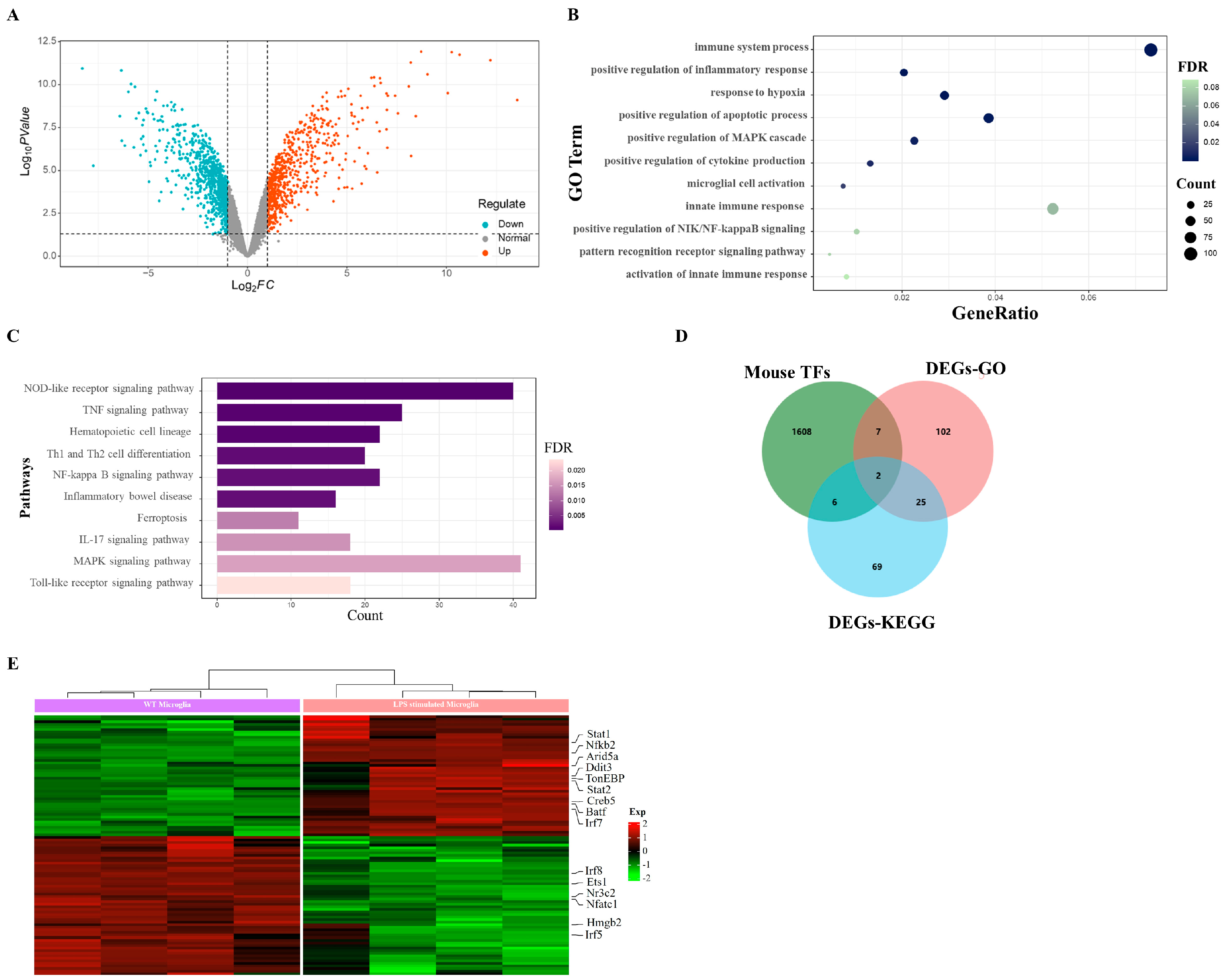
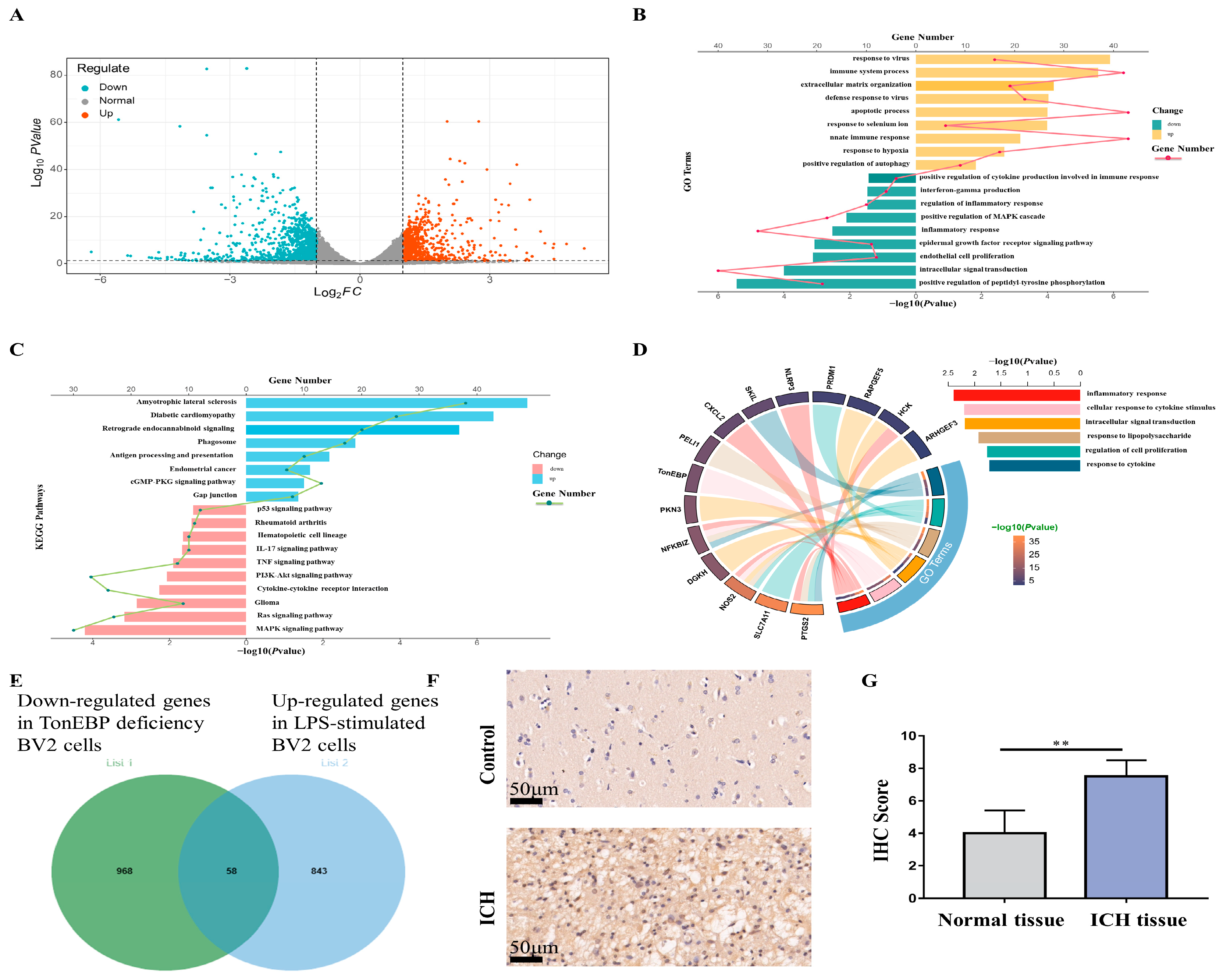
2.1. LPS Evokes Upregulated Transcription Factors in Microglia

To elucidate the key genes implicated in neuroinflammation, we analyzed the RNA-seq data derived from GEO (GSE102482). The bioinformatic analysis led to the identification of 683 upregulated and 816 downregulated differentially expressed genes (DEGs) (Figure 1A). For the purpose of understanding the functional pathways linked to these DEGs, we executed a gene ontology (GO) functional analysis and Kyoto Encyclopedia of Genes and Genomes (KEGG) pathway enrichment analysis. The GO functional analysis unveiled significantly enriched biological processes, including the immune system response, positive regulation of inflammation response, cytokine production augmentation, microglial cell activation, positive regulation of NIK/NF-κB signaling, and the pattern recognition receptor signaling pathway (Figure 1B). Furthermore, the KEGG pathway enrichment analysis revealed a robust correlation between these DEGs and inflammation-associated pathways, including the NOD-like receptor signaling pathway, TNF signaling pathway, NF-kappa B signaling pathway, IL-17 signaling pathway, MAPK signaling pathway, and Toll-like receptor signaling pathway (Figure 1C).
In an effort to pinpoint the crucial transcription factors governing the inflammatory response in activated microglia, we intersected these differentially expressed genes (DEGs) with a dataset comprising mouse transcription factors from Animal TFDB4 (hust.edu.cn). This intersection led to the identification of 101 differentially expressed transcription factors (Figure 1D). Out of these, 15 were implicated in inflammation-related biological functions and pathways (Figure 1E). Among the 15 transcription factors, the upregulated transcription factors included signal transducer and activator of transcription 1 (STAT1), nuclear factor kappa B subunit 2 (NFKB2), DNA damage-inducible transcript 3 (DDIT3), tonicity-responsive enhancer-binding protein (TonEBP), signal transducer and activator of transcription 2 (STAT2), CAMP-responsive element-binding protein 5 (CREB5), basic leucine zipper ATF-like transcription factor (BATF), and interferon regulatory factor 7 (IRF7). Conversely, the downregulated transcription factors comprised interferon regulatory factor 8 (IRF8), ETS proto-oncogene 1, ETS proto-oncogene 1, transcription factor (ETS1), nuclear receptor subfamily 3 group C member 2 (NR3C2), nuclear factor of activated T cells 1 (NFATC1), high-mobility group box 2 (HMGB2), and interferon regulatory factor 5 (IRF5) (Figure 1E).

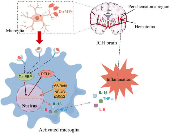
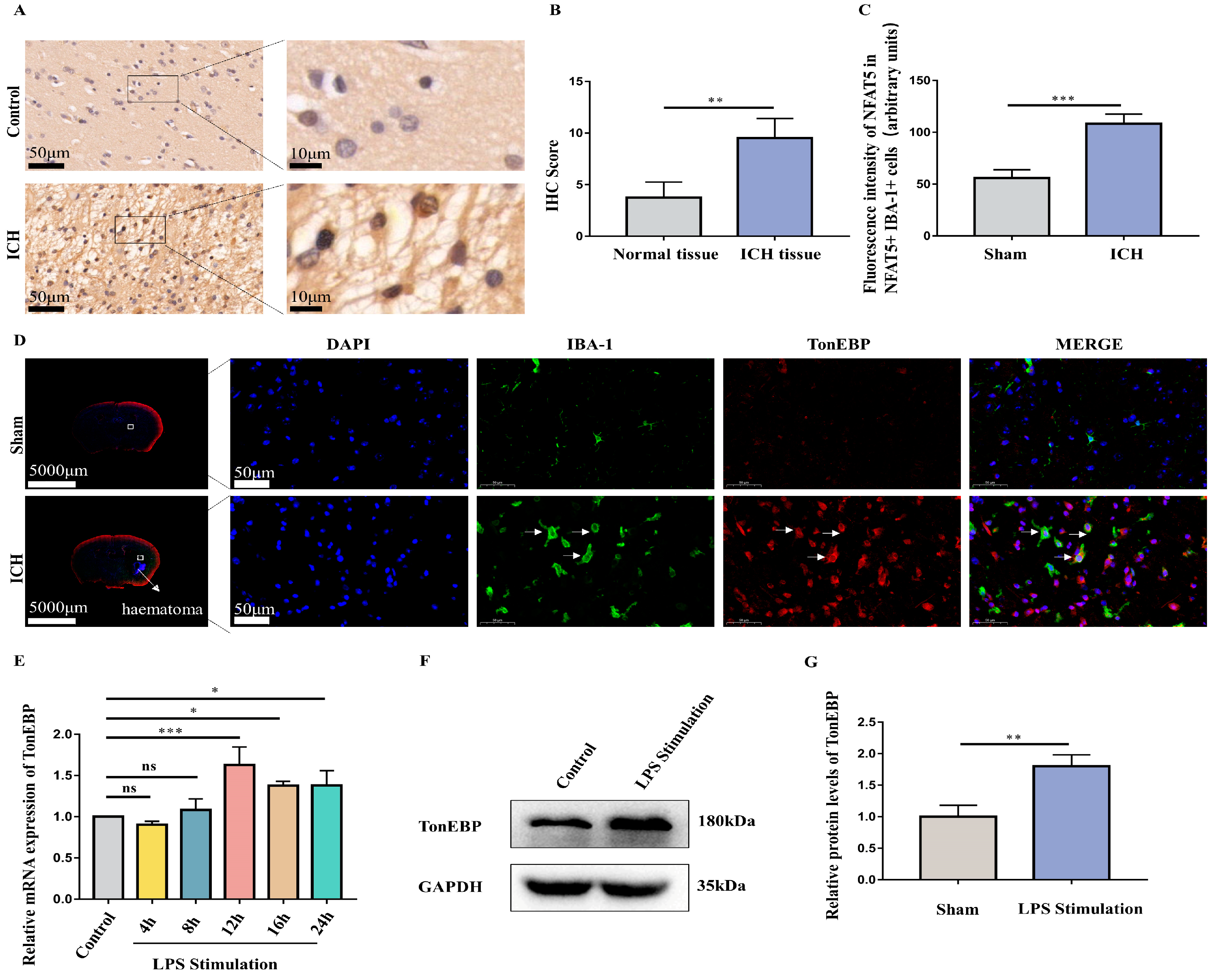
2.2. ICH and LPS Cause the Upregulation of Microglial TonEBP

Considering that the ICH triggered neuroinflammation, particularly in near-hematoma regions, we conducted immunohistochemistry on control tissue and post-ICH tissue in the human brain. Among the various transcription factors examined, our findings revealed an enhanced TonEBP in the peri-hematoma region of the human brain post-ICH (Figure 2A,B). Furthermore, we established ICH models in mice and collected their brain tissue 3 days after the establishment of the models. Microglia, labeled in green with Iba-1, and TonEBP, labeled in red, were examined using immunofluorescence. It was found that TonEBP was co-localized with Iba-1, and that the TonEBP fluorescence intensity in Iba-1-positive cells was increased after ICH (Figure 2C,D). In order to simulate the inflammation in microglia, we stimulated the BV2 with 1 μg/mL concentrations of LPS. Furthermore, BV2 were treated with LPS for different durations (4, 8, 12, 16, and 24 h). It was found that LPS needed to be sustained for at least 12 h to cause a sustained and stable elevation in TonEBP (Figure 2E). Additionally, our Western blot results demonstrated that the protein levels of TonEBP in BV2 were elevated under the 1 μg/mL LPS treatment for 12 h (Figure 2F,G).

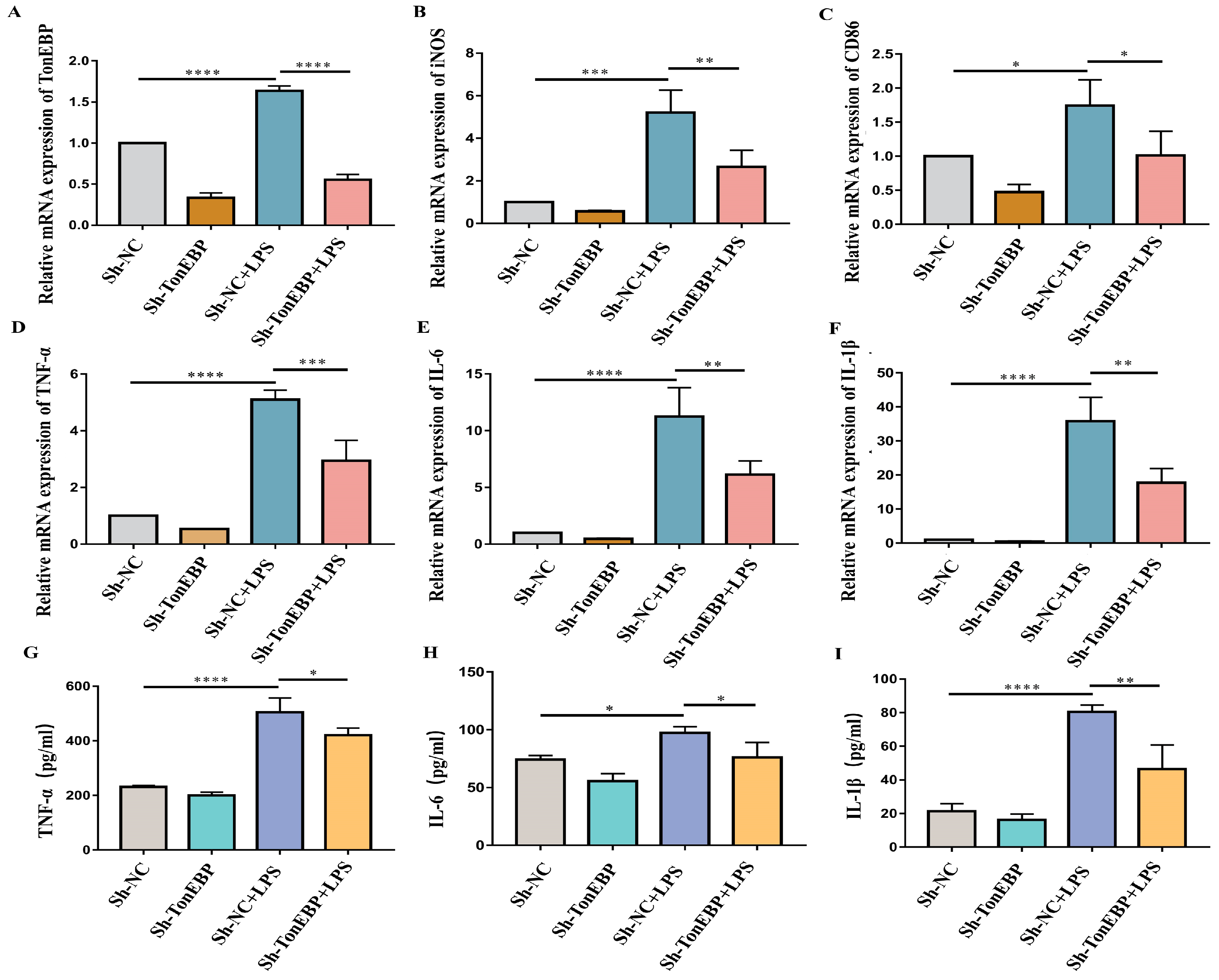
2.3. TonEBP Exacerbates Neuroinflammation and Microglial Activation

To demonstrate the influence of TonEBP on neuroinflammation and microglia activation upon LPS stimulation, we generated TonEBP-knockdown cells using sh-RNA and evaluated the efficiency of TonEBP knockdown with RT-PCR assays (Figure 3A). Our RT-PCR results demonstrated a successful knockdown of TonEBP expression in BV2 cells. Additionally, RT-qPCR assays were conducted to measure the mRNA levels of the pro-inflammatory cytokines TNF-α, IL-6, and IL-1β. The findings indicated that TonEBP deficiency in BV2 cells remarkably attenuated the mRNA of TNF-α, IL-6, and IL-1β after LPS treatment (Figure 3D–F). Moreover, a medium from BV2 was also obtained to evaluate the protein levels of TNF-α, IL-6, and IL-1β using ELISA assays. LPS treatment increased the protein levels of TNF-α, IL-6, and IL-1β in the medium from BV2, while TonEBP deficiency reduced the protein levels of these cytokines elicited by LPS stimulation (Figure 3G–I). Furthermore, the pro-inflammatory microglia markers iNOS (nitric oxide synthase 2) and CD86 (CD86 molecule) were elevated upon LPS stimulation, while TonEBP deficiency ameliorated the elevation of iNOS and CD86 caused by LPS stimulation (Figure 3B,C). These results indicate that TonEBP deficiency reduces the mRNA and protein levels of pro-inflammatory cytokines and ameliorates microglia activation in response to LPS stimulation.

2.4. TonEBP Contributes to the Activation of the NF-κB Signaling Pathway

Researchers have reported that the NF-κB pathway is essential for pro-inflammatory cytokine secretion. NF-κB signaling is triggered by DAMPs (damage-associated molecular patterns), promoting the shifting of the RELA/p65-NFKB1/p50 heterodimeric complex from the cytoplasm to the nucleus. After translocation to the nucleus, this complex binds to chromatin and subsequently activates the production of various pro-inflammatory cytokines, including TNF-α, IL-6, and IL-1β. To more deeply investigate the function of TonEBP in the modulation of NF-κB signaling, we assessed the phosphorylation of the RELA/p65 subunits of NF-κB. Western blot results demonstrated upregulation in the phosphorylation of RELA/p65, whereas the knockdown of TonEBP by sh-RNA decreased the phosphorylation of RELA/p65 under LPS stimulation in BV2 (Figure 4A,B). Additionally, LPS treatment decreased cytoplasmic NF-κB, while TonEBP deficiency mitigated this decrease (Figure 4C,D). These results demonstrate that TonEBP facilitates the activation of NF-κB signaling.

2.5. RNA-seq for Potential Targets of TonEBP

Next, for the purpose of exploring downstream of TonEBP, we conducted RNA-seq analysis on control and TonEBP-deficient BV2. The volcano plot of the RNA-seq analysis revealed 779 upregulated genes and 1026 downregulated genes in the sh-TonEBP BV2 cells compared to the sh-NC cells (Figure 5A). Subsequently, we performed GO function analysis and KEGG pathway enrichment analysis on the differentially expressed genes (DEGs). The bar plot displayed the significantly enriched GO terms in the upregulated and downregulated DEGs. The increased genes were notably clustered in biological processes such as the response to virus, immune system processes, extracellular matrix organization, the defense response to virus, and apoptotic processes. Nevertheless, the downregulated genes were prominently enriched in processes such as the positive regulation of peptidyl-tyrosine phosphorylation, intracellular signal transduction, the inflammatory response, positive regulation of MAPK cascade, and the positive regulation of cytokine production involved in the immune response (Figure 5B). The subsequent bar plot illustrated the prominently enriched KEGG pathways. The upregulated DEGs were involved in pathways such as amyotrophic lateral sclerosis, diabetic cardiomyopathy, retrograde endocannabinoid signaling, phagosome, antigen processing and presentation, and the cGMP-PKG signaling pathway. Conversely, the downregulated DEGs were mainly associated with pathways such as the MAPK signaling pathway, Ras signaling pathway, cytokine–cytokine receptor interaction, TNF signaling pathway, and IL-17 signaling pathway (Figure 5C). These results indicate significant changes in the inflammation-related biological functions and pathways in the downregulated DEGs.
To further investigate the potential target of TonEBP, we conducted an analysis by comparing the downregulated genes in TonEBP-deficient BV2 cells with the upregulated genes in LPS-stimulated BV2 cells. This analysis revealed 58 genes that were found to be common to both conditions (Figure 5E). Subsequently, we performed a functional analysis of these 58 genes using GO analysis to identify the genes associated with inflammatory immunity. As a result, we identified 15 genes that are involved in various inflammatory processes, including the inflammatory response (PTGS, NOS2, NFKBIZ, CXCL2, NLRP3, and HCK), the cellular response to cytokine stimulus (TonEBP), intracellular signal transduction (DGKH, PKN3, RAPGEF5, and ARHGEF3), the response to lipopolysaccharide (PELI1), the regulation of cell proliferation (SLC7A11 and PRDM1), and the response to cytokine (SKIL) (Figure 5D). Notably, our immunohistochemical staining results demonstrated an enhanced expression of PELI1 in human brain tissue with ICH (Figure 5F,G).

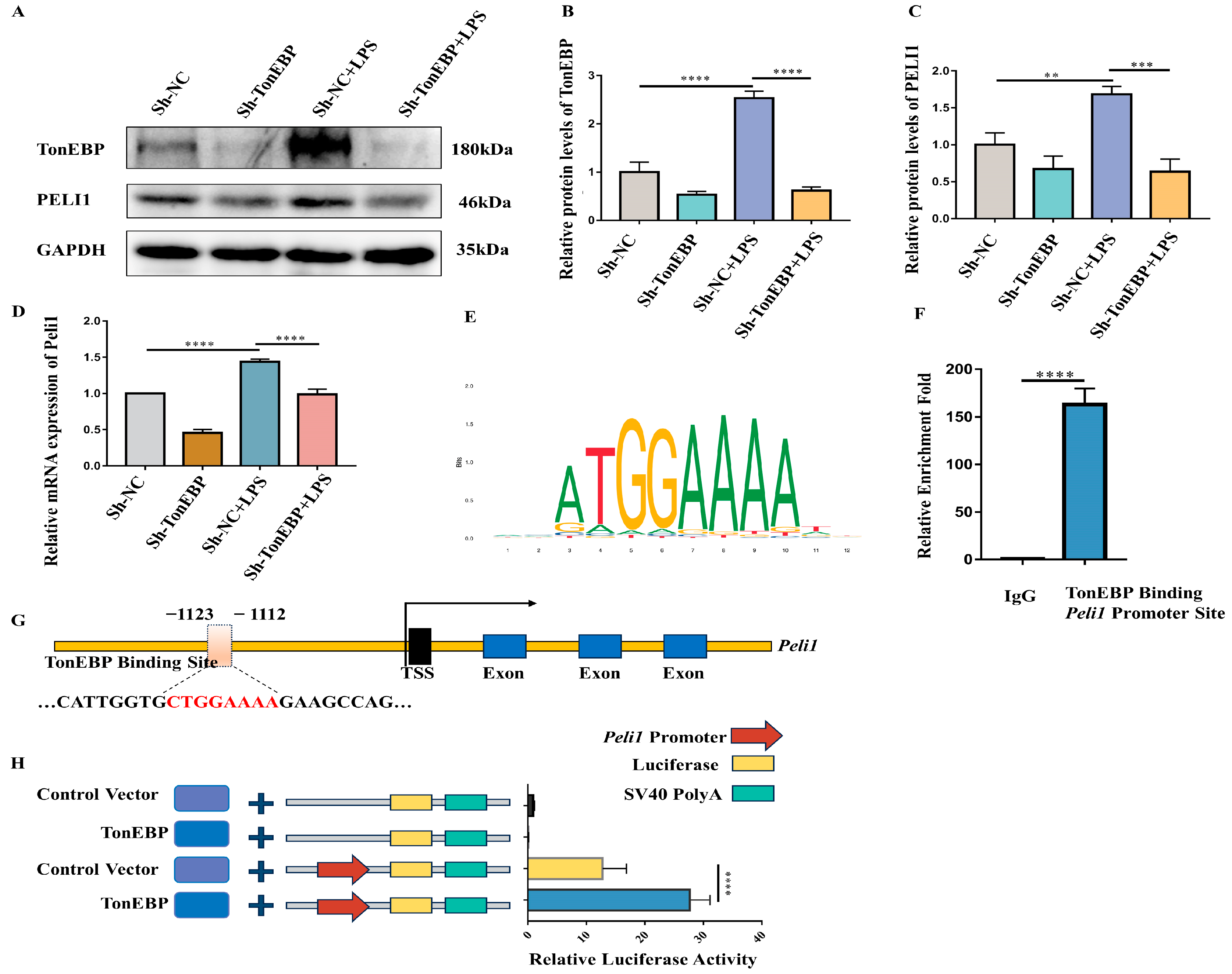
2.6. TonEBP Regulates PELI1 Expression at the Transcriptional Level

Next, we began to explore the possibility that PELI1 is downstream of TonEBP. Firstly, we performed a Western blot analysis to examine PELI1 in TonEBP-deficient BV2 under LPS stimulation. The results revealed that TonEBP and PELI1 were enhanced after LPS stimulation in sh-NC BV2 cells, while TonEBP and PELI1 expression decreased in TonEBP-deficient cells under LPS stimulation. (Figure 6A–C). To assess whether TonEBP regulates the mRNA level of PELI1 under LPS stimulation, we conducted RT-PCR to measure the mRNA levels of PELI1. The RT-PCR showed that TonEBP deficiency reduced the mRNA of PELI under LPS stimulation (Figure 6D). Next, we asked whether TonEBP regulates the transcription of Peli1. To address this question, we predicted the potential binding site of TonEBP to the Peli1 promoter region using the JASPAR website. The analysis showed the presence of a potential binding site upstream of the Peli1 transcription start site (Figure 6E,G). Subsequently, we performed a CUT & RUN experiment to verify the interaction between TonEBP and the Peli1 promoter region. The results of the CUT & RUN experiment indicated a strong interaction between TonEBP and the Peli1 promoter region 1123 to 1112, upstream of the transcription start site of the Peli1 gene. (Figure 6F). Furthermore, to substantiate whether NAFT5 would promote the transcription activity of Peli1, we conducted a dual-luciferase reporter experiment. The dual-luciferase reporter assay results indicated that TonEBP strongly promoted the transcription activity of the Peli1 gene. (Figure 6H). These findings suggest that TonEBP interacts with the Peli1 promoter region and promotes Peli1 transcriptional activity, leading to elevated PELI1 expression, which ultimately regulates inflammation in BV2 cells.

3. Discussion

Numerous studies have provided compelling evidence that transcription factors (TFs) are instrumental in modulating microglial functions across a variety of biological processes, including microglial activation [8,24], such as the NFE2-like BZIP transcription factor 2 (Nrf2), activating transcription factor 4 (ATF4), and signal transducer and activator of transcription family (STATs) [25,26,27]. Here, we report a potential transcription factor, TonEBP, which may regulate microglial activation. Some studies have reported abnormal TonEBP expression in the lesion regions of various diseases. Laban et al. published a study stating that hypoxia increased TonEBP levels in the lung and cultured smooth muscle cells within 24 h [28]. In addition, Shin et al. reported enhanced TonEBP expression in the brains of a kainic-acid-induced mouse seizure model [29]. Our current work suggests increased TonEBP expression within the peri-hematoma region of the human brain following ICH. Furthermore, our immunofluorescence experiments also demonstrated the specific co-localization of TonEBP with the microglial marker Iba-1 and elevated TonEBP fluorescence intensity in Iba-1-positive microglial cells in post-ICH mice compared to the sham group. These findings indicated elevated TonEBP in the human hemorrhagic brain and in the microglia of post-ICH mouse brains.
In further in vitro experiments, we observed that microglial TonEBP is triggered by LPS cellular stimulation. Moreover, a deficiency in microglial TonEBP mitigated the induction of pro-inflammatory cytokines like TNF-α, IL-6, and IL-1β. This deficiency also inhibited the expression of the activated microglial markers iNOS and CD86. Our findings align with previous research on macrophages, in that elevated TonEBP propels the differentiation of macrophages into the M1 phenotype when responding to inflammatory signals, including LPS or hyperglycemia [30,31]. Taken together, our results suggest that TonEBP may function as a key transcription factor in microglia activation and the microglia-mediated inflammation response. Nevertheless, due to the limitations of in vivo experiments, it is difficult to evaluate whether TonEBP participates in neurological deficits in an in vivo ICH mouse model. Therefore, additional research and experimental studies are imperative to clarify the function of TonEBP in ICH.
The substantial reduction in pro-inflammatory cytokines and the microglia activation in LPS-stimulated TonEBP-deficient BV2 led us to hypothesize that the inflammation regulatory role of TonEBP might be linked to NF-κB signaling pathways. It is widely recognized that nuclear factor kappa B (NF-κB) controls the majority of activated microglia cell signature genes in central nervous system diseases [32]. However, the correlation between TonEBP and NF-κB has been a subject of debate. Some studies have reported that TonEBP regulates the phosphorylation of NF-κB and subsequent NF-κB signaling pathway activation [33,34,35]. However, other findings have revealed a connection between TonEBP and the NF-κB complex via a different mechanism, suggesting that TonEBP enhances the transcriptional activity of NF-κB in response to LPS by acting as a cofactor for NF-κB [11,30,36]. Our results align with former studies stating that TonEBP deficiency significantly diminishes the phosphorylation of p65, a crucial NF-κB complex, in LPS-stimulated BV2 cells. Moreover, our results also indicate that TonEBP deficiency in LPS-stimulated BV2 cells increases the levels of the cytoplasmic p65 protein, suggesting that TonEBP regulates NF-κB phosphorylation and its subsequent translocation.
The RNA-seq analysis we conducted provided us with a more profound understanding through which to explore the regulation of NF-κB activation in microglia cells by TonEBP. The gene ontology (GO) biological enrichment process identified 15 potential inflammation-related genes targeted by TonEBP. Among these genes, NLRP3 (NLR family pyrin domain-containing 3), CXCL2 (C-X-C motif chemokine ligand 2), and NOS2 (nitric oxide synthase, inducible) have been reported to recruit TonEBP to their promoter regions, thereby enhancing transcriptional activity [37,38,39]. These findings led us to hypothesize that the new downstream gene of TonEBP could be among these 15 potential target genes. Intriguingly, the detection of upregulated PELI1 expression in the peri-hematoma region of ICH human brain provided us with a direction for further investigation into the relationship between TonEBP and PELI1. Emerging evidence suggests that PELI1 plays a crucial role in the inflammation regulation in immune cells, including T cells, macrophages, and microglia, across multiple diseases [40,41]. In the central nervous system, due to the cell specificity of PLEI1 to microglia, the critical inflammation regulatory role of PELI1 is primarily focused on microglia [21,22,42]. Mechanistically, PELI1 regulates the K63-ubiquitination of IRAK1 (interleukin-1 receptor-associated kinase 1) and Traf3/Traf6 (TNF receptor-associated factor 3,6), facilitating subsequent NF-κB activation [21,22,43,44]. As anticipated, our research revealed that PELI1 was upregulated in LPS-stimulated BV2 cells. Moreover, TonEBP knockdown significantly reduced PELI1 expression at both the protein and mRNA levels. Furthermore, the interaction between TonEBP and the Peli1 promoter and the enhanced Peli1 transcriptional activity by TonEBP suggest that PELI1 is a direct target of TonEBP. In summary, our findings suggest that TonEBP may regulate NF-κB activation by transcriptionally modulating PELI1.

4. Materials and Methods

4.1. Human Tissues

This study was approved by the Ethics Committee of the Children’s Hospital of Chongqing Medical University (File No. 2021295) and was conducted in compliance with ethical standards and the Declaration of Helsinki. Brain tissues were obtained from patients with spontaneous intracerebral hemorrhage (n = 3 per group). Control tissues were provided by the Department of Forensic Medicine, Chongqing Medical University (n = 3, per group). The details are shown in Supplementary Table S1.

4.2. Animals

The Institutional Animal Ethics Committee of Chongqing Medical University sanctioned all animal care and procedures. Male C57BL/6 mice, aged between 8 and 10 weeks (n = 3 per group), were procured from the Laboratory Animal Center of the same university. The mice were housed under a standard light/dark cycle and were also provided with unrestricted access to nourishment and hydration.

4.3. ICH Model

The induction of the murine intracerebral hemorrhage (ICH) model was achieved through the stereotactic-guided delivery of bacterial collagenase VII (C0773, Sigma-Aldrich, St. Louis, MO, USA) into the right basal ganglia [45]. Initially, mice were anesthetized with an intraperitoneal administration of pentobarbital (40 mg/kg) and subsequently positioned within a stereotaxic frame (RWD Life Science, Shenzhen, China). Following this, the bacterial collagenase VII (0.1 U in 1 μL) was administered at 0.2 mm posterior and 2.2 mm lateral of the bregma, 3.5 mm in depth, at a speed of 0.1 μL/min. The burr hole was subsequently closed with sterilized medical bone wax after the syringe remained in place for 10 min post administration. For the sham mice, an identical procedure was conducted with the replacement of bacterial collagens with saline. Throughout both the experimental and recovery periods, the animals’ body temperatures were maintained at 37.0 ± 0.5 °C.

4.4. Cell Culture and LPS Treatment

The BV-2 cell, a widely utilized murine microglia cell line (National Infrastructure of Cell Line Resource, Beijing, China), was cultivated in Dulbecco’s Modified Eagle Medium (Gibco, Thermo Fisher Scientific, New York, NY, USA) supplimented with 10% FBS (VivaCell, Shanghai, China), 100 U/mL penicillin, and 100 g/mL streptomycin (15140122, Gibco, Thermo Fisher Scientific, New York, USA). The cells were maintained in a humidified atmosphere with 5% CO2 at a temperature of 37 °C. For LPS stimulation, BV2 cells were treated with 1 μg/mL LPS [46] for the indicated time.

4.5. RNA Extraction and Real-Time Quantitative PCR

RNA was isolated utilizing the TRIZOL reagent, adhering to the protocol provided by the manufacturer (Invitrogen, San Diego, CA, USA). Subsequently, a volume of 800 nanograms of RNA was reversed into cDNA via the ABScript III RT Master Mix (RK20429, ABclonal, Wuhan, China). The quantitative PCR process was executed utilizing the SYBR Green Fast qPCR Mix (RK21203, ABclonal, Wuhan, China). The relative expression was quantified via the 2-ΔΔCT method, which underwent normalization to GAPDH. The primers used in the article are shown in Supplementary Table S2.

4.6. ELISA Assay for Cytokines

Enzyme-linked immunosorbent assay (ELISA) kits (Jiubang, Quanzhou, China) were used to determine the levels of TNF-α (QZ-10225), IL-6 (QZ-10260), and IL-1β (QZ-10247) in the treated BV2 cell culture medium as per the manufacturer’s instructions.

4.7. Immunofluorescence

Mice were euthanized and sacrificed 72 h post-ICH model induction. The mouse brains were fixed with a 4% paraformaldehyde solution prior to being sectioned into 8 μm slices. These sections were permeabilized and blocked with a 5% goat serum solution containing 0.5% Triton X-100. Next, the section was incubated with the indicated primary antibody at 4 °C overnight, followed by treatment with the appropriate secondary antibody for 1 h at 37 °C. Finally, the sections were subjected to incubation with DAPI and observation with a fluorescence microscope. The antibodies used for immunofluorescence were rabbit anti-TonEBP (ab3446, Abcam, Waltham, MA, USA) and anti-Iba-1 (GB15105-100, Servicebio, Wuhan, China).

4.8. Immunohistochemical Staining

The paraffin-embedded human brain tissue sections underwent deparaffinization and rehydration prior to antigen retrieval. Subsequently, the brain sections were incubated with the indicated primary and secondary antibodies after the inhibition of internal peroxidase activity. A DAB substrate kit (ZLI-9018; ZSGB, Beijing, China) was employed for visualization. Lastly, the brain tissue was counterstained with hematoxylin and sealed. The immunostaining intensity was scored as follows: 0 (no immunostaining), 1 (weak immunostaining), 2 (moderate immunostaining), or 3 (strong immunostaining). The percentage of positive cells was scored as 0 (<5%), 1 (5–25%), 2 (26–50%), 3 (51–75%), or 4 (76–100%). Finally, the immunostaining intensity score was multiplied by the percentage-of-positive-cells score to calculate each sample’s score. The antibodies used for immunohistochemical staining were rabbit anti-TonEBP (ab3446, Abcam, Waltham, USA) and rabbit anti-PELI1 (ab199336, Abcam, Waltham, USA).

4.9. Construction and Transfection of Plasmids

Full-length sequences of TonEBP were procured from the cDNA of the BV2 cell and subsequently cloned into the Plenti6/TR vector. The Peli1 promoter region was synthesized by BGI (Beijing Genomics Institute, Beijing, China). Plasmids were transiently transfected into HEK293T cells with Linea Polyethylenimine (PEI) in accordance with the manufacturer’s protocol (polyscience, Niles, IL, USA). For BV2 cells, the scrambled sh-RNA and TonEBP lentivirus vector were constructed by OBiO (Shanghai, China) and transfected to the BV2 cells. The primers used for the plasmid construction and sh-RNA sequence are shown in Supplementary Table S3.

4.10. Promoter Luciferase Reporter Assay

The promoter region of the Peli1 gene, extending from the −2000 base site to the transcription start site, was integrated into the modified pGL4.10 vector (Promega, Madison, WI, USA). Full-length sequences of TonEBP were extracted from the cDNA of the BV2 cell and cloned into the Plenti6/TR vector. For plasmid transfection, HEK293T cells were cultured on 24-well plates and transfected with indicated plasmids 48 h before harvesting. The activity of the Firefly luciferase and the signal of the Renilla luciferase were quantified using the dual-luciferase reporter assay kit (Promega, Madison, USA) and detected using a GloMax 20/20 Luminometer (Promega).

4.11. Cleavage under Targets and Release Using Nuclease (CUT&RUN) Assay

The interaction between protein and DNA was ascertained using the hyperactive pG-MNase CUT & RUN assay kit for PCR/qPCR (Vazyme, HD101, Nanjing, China). Initially, the potential binding site of TonEBP on the Peli1 promoter was predicted using the JASPAR website [47]; the primers were designed based on this predicted binding site. For the CUT & RUN assay, 3 × 10⁵ BV2 cells were harvested and incubated with pre-treated ConA Beads Pro for 10 min at room temperature. Following this, 6 ug of antibodies were added to the cell–bead crosslink and incubated overnight at 4 °C. Subsequently, CaCl2 was added to initiate cleavage after the pG-MNase enzyme was linked to the antibody–cell–bead crosslink. Finally, the released DNA fragments were collected and quantified using RT-qRCR with ChamQ Universal SYBR qPCR Master Mix. The primers were forward primer TGGGCATCCAGGGTTTTTGA and reverse primer GGTAATTTGCCTCAGTTTCCCT.

4.12. RNA Sequencing and Analysis

For RNA-seq analysis, total RNA was extracted from BV2 cells with a TRIzolKit (Invitrogen, California, USA). RNA-seq library preparation and subsequent RNA-seq sequencing were conducted using SHANGHAI BIOPROFILE (Shanghai, China), in which the prepared library was sequenced on the Illumina (San Diego, CA, USA) Novaseq6000 platform. The RNA-seq data were uploaded to the GEO database under accession number GSE249968. The RNA-seq data on LPS-stimulated primary microglia and the microarray data on LPS-stimulated BV2 cells were obtained from the GEO database under accession numbers GSE102482 and GSE103156, respectively.
For data processing and analysis, raw counts were used to analyze differentially expressed genes (DEGs) using the DESeq2 R package, with cutoffs at log2 fold change > 1 or < −1 and p-value < 0.05. GO (gene ontology) and KEGG (Kyoto Encyclopedia of Genes and Genomes) analyses were performed on the DAVID website [48,49], and the results were visualized in R (4.2.2). The DEGs were compared using the website tool Jvenn [50].

4.13. Lentivirus Transfection

OBiO Technology (Shanghai, China) engineered the lentivirus. The pSLenti-puro vector was utilized to clone both the silencing and non-silencing shRNA sequences of murine TonEBP, which were subsequently packaged into the lentivirus. Following this, BV2 cells underwent a 14 h transfection process with the lentivirus. A selection process was implemented 72 h post-infection, utilizing 2 μg/mL of puromycin (T19978, TargetMol, Boston, MA, USA).

4.14. Western Blot

Protein extraction was performed on BV2 and mouse brain samples using RIPA lysis buffer (P0013B, Beyotime, Shanghai, China) supplemented with a 1 mM protease inhibitor cocktail (B14001, Selleck, Houston, TX, USA). The lysates were then centrifuged to gather supernatants. Nuclear and cytoplasmic proteins were extracted using a cytoplasmic and nuclear protein extraction kit (AR0106, Boster, Wuhan, China). The proteins were transferred onto a PVDF membrane (Millipore, Shanghai, China) after being resolved using suitable SDS-PAGE. The membrane was blocked in 5% non-fat milk solution for one hour at room temperature, then incubated overnight at 4 °C with primary antibodies. Immunoblotting was identified using horseradish peroxidase-linked anti-rabbit or anti-mouse secondary antibodies, in conjunction with the BeyoECL Moon super sensitivity detection kit (Beyotime, Shanghai, China). The antibodies and concentrations used for Western blotting were as follows: rabbit anti-TonEBP (ab3446, Abcam, Waltham, USA) 1:1000, rabbit anti-NF-κB p65 (8242, Cell Signaling Technology, Danvers, MA, USA) 1:2000, rabbit anti-phospho-NF-κB p65 (Ser536) (3033, Cell Signaling Technology, Danvers, USA) 1:500, mouse anti-PELI1 (TA807629S, ORIGENE, Rockville, MD, USA) 1:2000, and mouse anti-GAPDH (60004-1-Ig, Proteintech, Wuhan, China) 1:50,000.

4.15. Statistical Analysis

Data are presented as mean ± SD. GraphPad Prism software (version 8.0) was used for statistical analyses. Two groups were analyzed using Student’s t-test. One-way ANOVA and Tukey’s multiple comparison test were used to analyze data between more than two groups. Differences between means were considered statistically significant when p < 0.05.

5. Conclusions

In summary, our current research investigates an increased transcription factor, TonEBP, after hemorrhagic stroke in the peri-hematoma region of the brain in humans and mice. Furthermore, our in vitro experiment shows that TonEBP may act as a transcription factor of PELI1, which has been reported to be a regulator of the NF-κB pathway. Moreover, we demonstrate that the knockdown of microglial TonEBP could regulate the NF-κB pathway and attenuate the inflammation response. Taken together, TonEBP may mediate the regulation of the NF-κB pathway and the inflammatory response via promoting the expression of PELI1 transcriptionally. Our research may provide a potential therapeutic target for intracerebral hemorrhage and elucidate part of the underlying mechanism of TonEBP in the inflammatory response. However, the role of microglial TonEBP in neuroinflammation after a hemorrhagic stroke in mice deserves our further attention.

Supplementary Materials

The following supporting information can be downloaded at: https://www.mdpi.com/article/10.3390/ijms25031438/s1. Supplementary Table S1: Human tissue information. Supplementary Table S2: Primer sequences for RT-qPCR, Supplementary Table S3: Sh-RNA sequences and primer sequences for plasmids construction.

Author Contributions

Conceptualization: H.G. and A.P.; methodology, A.P. and L.Q.; software, A.P. and H.G.; validation, Y.L., M.Z. and Y.D.; investigation, M.Z. and Y.D.; data curation, Y.L.; writing—original draft preparation, H.G. and A.P.; writing—review and editing, H.G. and X.Z.; supervision, H.G. and X.Z.; funding acquisition, H.G. and X.Z. All authors have read and agreed to the published version of the manuscript.

Funding

This research was supported by the National Natural Science Foundation of China (grant number: 81971217 to Xuan Zhai), the National Natural Science Foundation of China (youth fund) (grant number: 82301512 to Hui Gan), the China Postdoctoral Science Foundation (2022MD723734 to Hui Gan), the Chongqing Postdoctoral Science Foundation (CST2023NSCQ-BHX0121 to Hui Gan), and the Program for Youth Innovation in Future Medicine, Chongqing Medical University (to Xuan Zhai).

Institutional Review Board Statement

This study was conducted in accordance with animal protocols and approved by the Ethics Committee of Chongqing Medical University (Chongqing, China). All animal experiments were in strict accordance with the UK’s Animals (Scientific Procedures) Act 1986 and associated guidelines (NIH Publication No. 85-23, revised 1996). All patients who participated in this study were approved by the Ethics Committee of Children’s Hospital of Chongqing Medical University (File No. 2021295).

Informed Consent Statement

Informed consent was obtained from all subjects involved in the study.

Data Availability Statement

Data are contained within the article and Supplementary Materials.

Conflicts of Interest

The authors have declared that no conflicts of interest exist.

References

  1. Puy, L.; Parry-Jones, A.R.; Sandset, E.C.; Dowlatshahi, D.; Ziai, W.; Cordonnier, C. Intracerebral haemorrhage. Nat. Rev. Dis. Primers 2023, 9, 14. [Google Scholar] [CrossRef] [PubMed]
  2. Hemphill, J.C., III; Greenberg, S.M.; Anderson, C.S.; Becker, K.; Bendok, B.R.; Cushman, M.; Fung, G.L.; Goldstein, J.N.; Macdonald, R.L.; Mitchell, P.H.; et al. Guidelines for the Management of Spontaneous Intracerebral Hemorrhage: A Guideline for Healthcare Professionals From the American Heart Association/American Stroke Association. Stroke 2015, 46, 2032–2060. [Google Scholar] [CrossRef] [PubMed]
  3. Wilkinson, D.A.; Pandey, A.S.; Thompson, B.G.; Keep, R.F.; Hua, Y.; Xi, G. Injury mechanisms in acute intracerebral hemorrhage. Neuropharmacology 2018, 134 Pt B, 240–248. [Google Scholar] [CrossRef]
  4. Zheng, H.; Chen, C.; Zhang, J.; Hu, Z. Mechanism and Therapy of Brain Edema after Intracerebral Hemorrhage. Cerebrovasc. Dis. 2016, 42, 155–169. [Google Scholar] [CrossRef]
  5. Keep, R.F.; Hua, Y.; Xi, G. Intracerebral haemorrhage: Mechanisms of injury and therapeutic targets. Lancet. Neurol. 2012, 11, 720–731. [Google Scholar] [CrossRef] [PubMed]
  6. Aronowski, J.; Zhao, X. Molecular pathophysiology of cerebral hemorrhage: Secondary brain injury. Stroke 2011, 42, 1781–1786. [Google Scholar] [CrossRef] [PubMed]
  7. Zhang, Y.; Khan, S.; Liu, Y.; Zhang, R.; Li, H.; Wu, G.; Tang, Z.; Xue, M.; Yong, V.W. Modes of Brain Cell Death Following Intracerebral Hemorrhage. Front. Cell. Neurosci. 2022, 16, 799753. [Google Scholar] [CrossRef] [PubMed]
  8. Zhang, Z.; Zhang, Z.; Lu, H.; Yang, Q.; Wu, H.; Wang, J. Microglial Polarization and Inflammatory Mediators after Intracerebral Hemorrhage. Mol. Neurobiol. 2017, 54, 1874–1886. [Google Scholar] [CrossRef]
  9. Miyakawa, H.; Woo, S.K.; Dahl, S.C.; Handler, J.S.; Kwon, H.M. Tonicity-responsive enhancer binding protein, a rel-like protein that stimulates transcription in response to hypertonicity. Proc. Natl. Acad. Sci. USA 1999, 96, 2538–2542. [Google Scholar] [CrossRef]
  10. Yoo, E.J.; Oh, K.H.; Piao, H.; Kang, H.J.; Jeong, G.W.; Park, H.; Lee, C.J.; Ryu, H.; Yang, S.H.; Kim, M.G.; et al. Macrophage transcription factor TonEBP promotes systemic lupus erythematosus and kidney injury via damage-induced signaling pathways. Kidney Int. 2023, 104, 163–180. [Google Scholar] [CrossRef]
  11. Xu, J.; Gao, C.; He, Y.; Fang, X.; Sun, D.; Peng, Z.; Xiao, H.; Sun, M.; Zhang, P.; Zhou, T.; et al. NLRC3 expression in macrophage impairs glycolysis and host immune defense by modulating the NF-κB-NFAT5 complex during septic immunosuppression. Mol. Ther. J. Am. Soc. Gene Ther. 2023, 31, 154–173. [Google Scholar] [CrossRef] [PubMed]
  12. Lee, J.Y.; Jeong, E.A.; Kim, K.E.; Yi, C.O.; Jin, Z.; Lee, J.E.; Lee, D.H.; Kim, H.J.; Kang, S.S.; Cho, G.J.; et al. TonEBP/NFAT5 haploinsufficiency attenuates hippocampal inflammation in high-fat diet/streptozotocin-induced diabetic mice. Sci. Rep. 2017, 7, 7837. [Google Scholar] [CrossRef] [PubMed]
  13. Jeong, G.R.; Im, S.K.; Bae, Y.H.; Park, E.S.; Jin, B.K.; Kwon, H.M.; Lee, B.J.; Bu, Y.; Hur, E.M.; Lee, B.D. Inflammatory signals induce the expression of tonicity-responsive enhancer binding protein (TonEBP) in microglia. J. Neuroimmunol. 2016, 295, 21–29. [Google Scholar] [CrossRef] [PubMed]
  14. Tellechea, M.; Buxadé, M.; Tejedor, S.; Aramburu, J.; López-Rodríguez, C. NFAT5-Regulated Macrophage Polarization Supports the Proinflammatory Function of Macrophages and T Lymphocytes. J. Immunol. 2018, 200, 305–315. [Google Scholar] [CrossRef] [PubMed]
  15. Zhang, Z.L.; Liu, Y.G.; Huang, Q.B.; Wang, H.W.; Song, Y.; Xu, Z.K.; Li, F. Nuclear factor-κB activation in perihematomal brain tissue correlates with outcome in patients with intracerebral hemorrhage. J. Neuroinflamm. 2015, 12, 53. [Google Scholar] [CrossRef] [PubMed]
  16. Kopitar-Jerala, N. Innate Immune Response in Brain, NF-Kappa B Signaling and Cystatins. Front. Mol. Neurosci. 2015, 8, 73. [Google Scholar] [CrossRef] [PubMed]
  17. Yu, H.; Lin, L.; Zhang, Z.; Zhang, H.; Hu, H. Targeting NF-κB pathway for the therapy of diseases: Mechanism and clinical study. Signal Transduct. Target. Ther. 2020, 5, 209. [Google Scholar] [CrossRef]
  18. Jin, W.; Chang, M.; Sun, S.C. Peli: A family of signal-responsive E3 ubiquitin ligases mediating TLR signaling and T-cell tolerance. Cell Mol. Immunol. 2012, 9, 113–122. [Google Scholar] [CrossRef]
  19. Mo, H.; Wang, Z.; He, Z.; Wan, J.; Lu, R.; Wang, C.; Chen, A.; Cheng, P. Decreased Peli1 expression attenuates osteoarthritis by protecting chondrocytes and inhibiting M1-polarization of macrophages. Bone Jt. Res. 2023, 12, 121–132. [Google Scholar] [CrossRef]
  20. Chen, H.; Hou, Y.; Zhai, Y.; Yang, J.; Que, L.; Liu, J.; Lu, L.; Ha, T.; Li, C.; Xu, Y.; et al. Peli1 deletion in macrophages attenuates myocardial ischemia/reperfusion injury by suppressing M1 polarization. J. Leukoc. Biol. 2023, 113, 95–108. [Google Scholar] [CrossRef]
  21. Xiao, Y.; Jin, J.; Chang, M.; Chang, J.H.; Hu, H.; Zhou, X.; Brittain, G.C.; Stansberg, C.; Torkildsen, Ø.; Wang, X.; et al. Peli1 promotes microglia-mediated CNS inflammation by regulating Traf3 degradation. Nat. Med. 2013, 19, 595–602. [Google Scholar] [CrossRef] [PubMed]
  22. Wang, L.; Yin, C.; Liu, T.; Abdul, M.; Zhou, Y.; Cao, J.L.; Lu, C. Pellino1 regulates neuropathic pain as well as microglial activation through the regulation of MAPK/NF-κB signaling in the spinal cord. J. Neuroinflamm. 2020, 17, 83. [Google Scholar] [CrossRef] [PubMed]
  23. Xiao, Y.; Jin, J.; Zou, Q.; Hu, H.; Cheng, X.; Sun, S.C. Peli1 negatively regulates type I interferon induction and antiviral immunity in the CNS. Cell Biosci. 2015, 5, 34. [Google Scholar] [CrossRef]
  24. Yeh, H.; Ikezu, T. Transcriptional and Epigenetic Regulation of Microglia in Health and Disease. Trends Mol. Med. 2019, 25, 96–111. [Google Scholar] [CrossRef] [PubMed]
  25. Zhao, X.; Aronowski, J. Nrf2 to pre-condition the brain against injury caused by products of hemolysis after ICH. Transl. Stroke Res. 2013, 4, 71–75. [Google Scholar] [CrossRef]
  26. Karuppagounder, S.S.; Alim, I.; Khim, S.J.; Bourassa, M.W.; Sleiman, S.F.; John, R.; Thinnes, C.C.; Yeh, T.L.; Demetriades, M.; Neitemeier, S.; et al. Therapeutic targeting of oxygen-sensing prolyl hydroxylases abrogates ATF4-dependent neuronal death and improves outcomes after brain hemorrhage in several rodent models. Sci. Transl. Med. 2016, 8, 328ra29. [Google Scholar] [CrossRef]
  27. Gong, L.; Manaenko, A.; Fan, R.; Huang, L.; Enkhjargal, B.; McBride, D.; Ding, Y.; Tang, J.; Xiao, X.; Zhang, J.H. Osteopontin attenuates inflammation via JAK2/STAT1 pathway in hyperglycemic rats after intracerebral hemorrhage. Neuropharmacology 2018, 138, 160–169. [Google Scholar] [CrossRef]
  28. Laban, H.; Siegmund, S.; Zappe, M.; Trogisch, F.A.; Heineke, J.; Torre, C.; Fisslthaler, B.; Arnold, C.; Lauryn, J.; Büttner, M.; et al. NFAT5/TonEBP Limits Pulmonary Vascular Resistance in the Hypoxic Lung by Controlling Mitochondrial Reactive Oxygen Species Generation in Arterial Smooth Muscle Cells. Cells 2021, 10, 3293. [Google Scholar] [CrossRef]
  29. Shin, H.J.; Kim, H.; Heo, R.W.; Kim, H.J.; Choi, W.S.; Kwon, H.M.; Roh, G.S. Tonicity-responsive enhancer binding protein haplodeficiency attenuates seizure severity and NF-κB-mediated neuroinflammation in kainic acid-induced seizures. Cell Death Differ. 2014, 21, 1095–1106. [Google Scholar] [CrossRef] [PubMed]
  30. Lee, H.H.; Sanada, S.; An, S.M.; Ye, B.J.; Lee, J.H.; Seo, Y.K.; Lee, C.; Lee-Kwon, W.; Küper, C.; Neuhofer, W.; et al. LPS-induced NFκB enhanceosome requires TonEBP/NFAT5 without DNA binding. Sci. Rep. 2016, 6, 24921. [Google Scholar] [CrossRef]
  31. Choi, S.Y.; Lim, S.W.; Salimi, S.; Yoo, E.J.; Lee-Kwon, W.; Lee, H.H.; Lee, J.H.; Mitchell, B.D.; Sanada, S.; Parsa, A.; et al. Tonicity-Responsive Enhancer-Binding Protein Mediates Hyperglycemia-Induced Inflammation and Vascular and Renal Injury. J. Am. Soc. Nephrol. JASN 2018, 29, 492–504. [Google Scholar] [CrossRef] [PubMed]
  32. Taetzsch, T.; Levesque, S.; McGraw, C.; Brookins, S.; Luqa, R.; Bonini, M.G.; Mason, R.P.; Oh, U.; Block, M.L. Redox regulation of NF-κB p50 and M1 polarization in microglia. Glia 2015, 63, 423–440. [Google Scholar] [CrossRef] [PubMed]
  33. Li, C.; Liu, M.; Bo, L.; Liu, W.; Liu, Q.; Chen, X.; Xu, D.; Li, Z.; Jin, F. NFAT5 participates in seawater inhalation-induced acute lung injury via modulation of NF-κB activity. Mol. Med. Rep. 2016, 14, 5033–5040. [Google Scholar] [CrossRef] [PubMed]
  34. Roth, I.; Leroy, V.; Kwon, H.M.; Martin, P.Y.; Féraille, E.; Hasler, U. Osmoprotective transcription factor NFAT5/TonEBP modulates nuclear factor-kappaB activity. Mol. Biol. Cell 2010, 21, 3459–3474. [Google Scholar] [CrossRef]
  35. Xie, X.; Huang, C.; Xu, D.; Liu, Y.; Hu, M.; Long, J.; Fang, X. Elevation of hypertonicity-induced protein NFAT5 promotes apoptosis of human umbilical vein endothelial cells through the NF-κB pathway. Mol. Med. Rep. 2021, 23, 184. [Google Scholar] [CrossRef] [PubMed]
  36. Jeong, G.W.; Lee, H.H.; Lee-Kwon, W.; Kwon, H.M. Microglial TonEBP mediates LPS-induced inflammation and memory loss as transcriptional cofactor for NF-κB and AP-1. J. Neuroinflamm. 2020, 17, 372. [Google Scholar] [CrossRef] [PubMed]
  37. Buxadé, M.; Lunazzi, G.; Minguillón, J.; Iborra, S.; Berga-Bolaños, R.; Del Val, M.; Aramburu, J.; López-Rodríguez, C. Gene expression induced by Toll-like receptors in macrophages requires the transcription factor NFAT5. J. Exp. Med. 2012, 209, 379–393. [Google Scholar] [CrossRef] [PubMed]
  38. Ma, P.; Zha, S.; Shen, X.; Zhao, Y.; Li, L.; Yang, L.; Lei, M.; Liu, W. NFAT5 mediates hypertonic stress-induced atherosclerosis via activating NLRP3 inflammasome in endothelium. Cell Commun. Signal. 2019, 17, 102. [Google Scholar] [CrossRef] [PubMed]
  39. Johnson, Z.I.; Doolittle, A.C.; Snuggs, J.W.; Shapiro, I.M.; Le Maitre, C.L.; Risbud, M.V. TNF-α promotes nuclear enrichment of the transcription factor TonEBP/NFAT5 to selectively control inflammatory but not osmoregulatory responses in nucleus pulposus cells. J. Biol. Chem. 2017, 292, 17561–17575. [Google Scholar] [CrossRef]
  40. Humphries, F.; Moynagh, P.N. Molecular and physiological roles of Pellino E3 ubiquitin ligases in immunity. Immunol. Rev. 2015, 266, 93–108. [Google Scholar] [CrossRef]
  41. Moynagh, P.N. The roles of Pellino E3 ubiquitin ligases in immunity. Nat. Rev. Immunol. 2014, 14, 122–131. [Google Scholar] [CrossRef] [PubMed]
  42. Dai, D.; Yuan, J.; Wang, Y.; Xu, J.; Mao, C.; Xiao, Y. Peli1 controls the survival of dopaminergic neurons through modulating microglia-mediated neuroinflammation. Sci. Rep. 2019, 9, 8034. [Google Scholar] [CrossRef] [PubMed]
  43. Chang, M.; Jin, W.; Sun, S.C. Peli1 facilitates TRIF-dependent Toll-like receptor signaling and proinflammatory cytokine production. Nat. Immunol. 2009, 10, 1089–1095. [Google Scholar] [CrossRef]
  44. Xu, W.; Yang, T.; Lou, X.; Chen, J.; Wang, X.; Hu, M.; An, D.; Gao, R.; Wang, J.; Chen, X. Role of the Peli1-RIPK1 Signaling Axis in Methamphetamine-Induced Neuroinflammation. ACS Chem. Neurosci. 2023, 14, 864–874. [Google Scholar] [CrossRef] [PubMed]
  45. Zheng, J.; Lu, J.; Mei, S.; Wu, H.; Sun, Z.; Fang, Y.; Xu, S.; Wang, X.; Shi, L.; Xu, W.; et al. Ceria nanoparticles ameliorate white matter injury after intracerebral hemorrhage: Microglia-astrocyte involvement in remyelination. J. Neuroinflamm. 2021, 18, 43. [Google Scholar] [CrossRef] [PubMed]
  46. Bi, W.; Bao, K.; Zhou, X.; Deng, Y.; Li, X.; Zhang, J.; Lan, X.; Zhao, J.; Lu, D.; Xu, Y.; et al. PSMC5 regulates microglial polarization and activation in LPS-induced cognitive deficits and motor impairments by interacting with TLR4. J. Neuroinflamm. 2023, 20, 277. [Google Scholar] [CrossRef] [PubMed]
  47. Rauluseviciute, I.; Riudavets-Puig, R.; Blanc-Mathieu, R.; Castro-Mondragon, J.A.; Ferenc, K.; Kumar, V.; Lemma, R.B.; Lucas, J.; Chèneby, J.; Baranasic, D.; et al. JASPAR 2024: 20th anniversary of the open-access database of transcription factor binding profiles. Nucleic Acids Res. 2023, 52, D174–D182. [Google Scholar] [CrossRef] [PubMed]
  48. Huang, D.W.; Sherman, B.T.; Lempicki, R.A. Systematic and integrative analysis of large gene lists using DAVID bioinformatics resources. Nat. Protoc. 2009, 4, 44–57. [Google Scholar] [CrossRef]
  49. Sherman, B.T.; Hao, M.; Qiu, J.; Jiao, X.; Baseler, M.W.; Lane, H.C.; Imamichi, T.; Chang, W. DAVID: A web server for functional enrichment analysis and functional annotation of gene lists (2021 update). Nucleic Acids Res. 2022, 50, W216–W221. [Google Scholar] [CrossRef]
  50. Bardou, P.; Mariette, J.; Escudié, F.; Djemiel, C.; Klopp, C. Jvenn: An interactive Venn diagram viewer. BMC Bioinform. 2014, 15, 293. [Google Scholar] [CrossRef]
Figure 1. Upregulated transcription factors in the lipopolysaccharide (LPS)-induced inflammatory response in microglia. (A) Volcano plot of DEGs at 12 h after lipopolysaccharide (LPS). P-adj < 0.05 and |(log2FoldChange)| > 1. (B) GO enrichment scatter plot. (C) KEGG pathway enrichment analysis plot. (D) Wayne diagram showing DEGs-GO and DEGs-KEGG of activated microglia and mouse transcription factors. (E) Heat map of different transcription factors in WT microglia and LPS-stimulated microglia.
Ijms 25 01438 g001
Figure 2. The expression of TonEBP in brain tissues of humans and mice after ICH and in microglia stimulated by LPS. (A,B) Immunohistochemistry (scale bar = 50 μm or 10 μm) (A) and IHC score (B) for TonEBP in brain tissues of humans in control group and ICH group. N = 3. (C,D) Immunofluorescence staining (scale bar = 5000 μm or 50 μm) (D) and fluorescence intensity (C) of TonEBP and Iba-1in in brain tissues of mice in sham group and ICH group. N = 3. The Iba-1 and TonEBP positive cells were indicated by white arrows. (E) RT-PCR of TonEBP in control and 1 μg/mL LPS-treated BV2 cells for different durations (4, 8, 12, 16, and 24 h). N = 3. (F,G) Western blot of TonEBP in control group and LPS stimulation group. N = 3. The data are presented as mean ± SD. * p < 0.05, ** p < 0.01, *** p < 0.001, ns: non significant.
Ijms 25 01438 g002
Figure 3. Inflammation and microglial activation were ameliorated by TonEBP deficiency. (AC) RT-PCR of TonEBP (A), iNOS (B), and CD86 (C) in sh-NC, sh-TonEBP, sh-NC+LPS, and sh-TonEBP+LPS groups. N = 3. (DF) RT-PCR of TNF-α (D), IL-6 (E), and IL-1β (F) in sh-NC, sh-TonEBP, sh-NC+LPS, and sh-TonEBP+LPS groups. N = 3. (GI) ELISA of TNF-α (D), IL-6 (E), and IL-1β (F) in sh-NC, sh-TonEBP, sh-NC+LPS, and sh-TonEBP+LPS groups. N = 3. The data are presented as mean ± SD. * p < 0.05, ** p < 0.01, *** p < 0.001, **** p < 0.0001.
Ijms 25 01438 g003
Figure 4. TonEBP deficiency reduced the activation of the NF-κB signaling pathway. (A,B) Western blot of TonEBP, p-NF-κB, and NF-κB of total cell lysis in sh-NC, sh-TonEBP, sh-NC+LPS, and sh-TonEBP+LPS groups. N = 3. (C,D) Western blot of NF-κB of cytoplasm in sh-NC, sh-TonEBP, sh-NC+LPS, and sh-TonEBP+LPS groups. N = 3. The data are presented as mean ± SD. * p < 0.05, *** p < 0.001, **** p < 0.0001.
Ijms 25 01438 g004
Figure 5. RNA-seq for potential target of TonEBP. (A) Volcano plot of DEGs between the sh-NC group and sh-TonEBP group. p-value < 0.05 and |(log2FoldChange)| > 1. (B) GO enrichment scatter plot. (C) KEGG pathway enrichment analysis plot. (D) GO enrichment chord diagram and bar plot legend for GO terms. (E) Wayne diagram showing upregulated DEGs in LPS-stimulated BV2 cells and downregulated DEGs in TonEBP-deficient BV2 cells. (F,G) Immunohistochemistry (scale bar = 50 μm) (F) and IHC score (G) for PELI1 in brain tissues of humans in control group and ICH group. N = 3. The data are presented as mean ± SD. ** p < 0.01.
Ijms 25 01438 g005
Figure 6. TonEBP acted as a transcription factor of PELI1. (AC) Western blot of TonEBP (A,B) and PELI1 (A,C) in sh-NC, sh-TonEBP, sh-NC+LPS, and sh-TonEBP+LPS groups. N = 3. (D) RT-PCR of PELI1 (D) in sh-NC, sh-TonEBP, sh-NC+LPS, and sh-TonEBP+LPS groups. N = 3. (E) JASPAR-derived TonEBP chromatin binding motif. (F) CUT & RUN experiment for TonEBP and Peli1 promoter. N = 3. (G) Scheme of TonEBP binding sequence in Peli1 promoter. (H) The dual-luciferase reporter assay of TonEBP and Peli1 promoter. N = 7. ** p < 0.01, *** p < 0.001, **** p < 0.0001.
Ijms 25 01438 g006
Disclaimer/Publisher’s Note: The statements, opinions and data contained in all publications are solely those of the individual author(s) and contributor(s) and not of MDPI and/or the editor(s). MDPI and/or the editor(s) disclaim responsibility for any injury to people or property resulting from any ideas, methods, instructions or products referred to in the content.

