Article

Biological Function of Prophage-Related Gene Cluster ΔVpaChn25_RS25055VpaChn25_0714 of Vibrio parahaemolyticus CHN25

1
Key Laboratory of Quality and Safety Risk Assessment for Aquatic Products on Storage and Preservation (Shanghai), Ministry of Agriculture and Rural Affairs of China, College of Food Science and Technology, Shanghai Ocean University, Shanghai 201306, China
2
Department of Internal Medicine, Virginia Commonwealth University/McGuire VA Medical Centre, Richmond, VA 23284, USA
3
Key Laboratory of Exploration and Utilization of Aquatic Genetic Resources, Ministry of Education, Shanghai Ocean University, Shanghai 201306, China
*
Author to whom correspondence should be addressed.
Int. J. Mol. Sci. 2024, 25(3), 1393; https://doi.org/10.3390/ijms25031393
Submission received: 14 December 2023 / Revised: 15 January 2024 / Accepted: 17 January 2024 / Published: 23 January 2024
(This article belongs to the Special Issue Bacteriophage—Molecular Studies 5.0)

Abstract

:
Vibrio parahaemolyticus is the primary foodborne pathogen known to cause gastrointestinal infections in humans. Nevertheless, the molecular mechanisms of V. parahaemolyticus pathogenicity are not fully understood. Prophages carry virulence and antibiotic resistance genes commonly found in Vibrio populations, and they facilitate the spread of virulence and the emergence of pathogenic Vibrio strains. In this study, we characterized three such genes, VpaChn25_0713, VpaChn25_0714, and VpaChn25_RS25055, within the largest prophage gene cluster in V. parahaemolyticus CHN25. The deletion mutants ΔVpaChn25_RS25055, ΔVpaChn25_0713, ΔVpaChn25_0714, and ΔVpaChn25_RS25055-0713-0714 were derived with homologous recombination, and the complementary mutants ΔVpaChn25_0713-com, ΔVpaChn25_0714-com, ΔVpaChn25_RS25055-com, ΔVpaChn25_RS25055-0713-0714-com were also constructed. In the absence of the VpaChn25_RS25055, VpaChn25_0713, VpaChn25_0714, and VpaChn25_RS25055-0713-0714 genes, the mutants showed significant reductions in low-temperature survivability and biofilm formation (p < 0.001). The ΔVpaChn25_0713, ΔVpaChn25_RS25055, and ΔVpaChn25_RS25055-0713-0714 mutants were also significantly defective in swimming motility (p < 0.001). In the Caco-2 model, the above four mutants attenuated the cytotoxic effects of V. parahaemolyticus CHN25 on human intestinal epithelial cells (p < 0.01), especially the ΔVpaChn25_RS25055 and ΔVpaChn25_RS25055-0713-0714 mutants. Transcriptomic analysis showed that 15, 14, 8, and 11 metabolic pathways were changed in the ΔVpaChn25_RS25055, ΔVpaChn25_0713, ΔVpaChn25_0714, and ΔVpaChn25_RS25055-0713-0714 mutants, respectively. We labeled the VpaChn25_RS25055 gene with superfolder green fluorescent protein (sfGFP) and found it localized at both poles of the bacteria cell. In addition, we analyzed the evolutionary origins of the above genes. In summary, the prophage genes VpaChn25_0713, VpaChn25_0714, and VpaChn25_RS25055 enhance V. parahaemolyticus CHN25’s survival in the environment and host. Our work improves the comprehension of the synergy between prophage-associated genes and the evolutionary process of V. parahaemolyticus.

1. Introduction

Vibrio parahaemolyticus is a Gram-negative, halophilic, and rod-shaped bacterium that is found growing in coastal areas and river–sea junctures at a global scale [1,2]. V. parahaemolyticus can cause acute diarrhea, abdominal cramps, vomiting, and fever in humans, and even death [3]. The bacterium was first discovered in Japan in 1950 when the consumption of contaminated semi-dried juvenile sardines caused 272 illnesses and 20 deaths [4]. Since then, outbreaks of the foodborne illness caused by V. parahaemolyticus have occurred in many Asian countries, including Bangladesh, China, India, and Malaysia, and then spread to Asia, America, Africa, and Europe [5,6,7]. According to the Mortality Weekly Report of the CDC (Centers for Disease Control and Prevention) of America, V. parahaemolyticus causes 45,000 illnesses annually in the United States (https://www.cdc.gov/vibrio/faq.html, accessed on 5 March 2019). Approximately 23.12% of foodborne disease outbreaks in coastal cities in China are associated with V. parahaemolyticus [8]. For instance, Luo et al. estimated that the annual incidence rate of V. parahaemolyticus gastroenteritis was 183 cases per 100,000 individuals [9]. The hallmark virulence factors associated with V. parahaemolyticus include thermostable direct hemolysin (TDH) and TDH-related hemolysin (TRH) [10]. However, some clinical isolates were negative for the two toxins and the type III secretion system (T3SS), indicating that other virulence-associated determinants exist.
Phages, the most abundant biological entities in the biosphere, are viruses that infect bacteria [11,12,13,14,15]. Horizontal gene transfer (HGT) facilitated by prophages strongly influences bacterial evolution by granting them access to novel ecological habitats, including pathogenic traits [16]. For example, there are ~300 genes novel to V. parahaemolyticus BB22OP and ~400 genes novel to V. parahaemolyticus RIMD2210633. Many of these novel genes are remnants of transposons or phages [17]. Zabala et al. [18] revealed that in the pandemic V. parahaemolyticus O3:K6 clonal complex, the presence of a 42 kb prophage led to a variant, and V. parahaemolyticus O3:K6 carrying this prophage displayed an ultraviolet radiation sensitivity that was 7–15 times higher. The prophages in V. parahaemolyticus, such as VP06, Vp882, and Vp58.5, contribute to various functions, including increased sensitivity to ultraviolet radiation, DNA methylase activity, quorum sensing, and improved resistance to environmental stress [19]. Yang and co-workers [20] found that V. parahaemolyticus carrying prophages 12B12, VEJphi, VCY_phi, and VFJ caused acute hepatopancreatic necrosis disease (AHPND) in shrimps. Prophages are essential for the biological properties of bacterial hosts; thus, it is necessary to recognize them accurately and understand their function via nucleotide sequence analysis [21].
Our previous studies isolated a V. parahaemolyticus CHN25 strain (serotype: O5: KUT) of aquatic animal origin, followed by identification and characterization [22,23,24,25,26]. It was found that prophage gene clusters were present in chromosome 1 (3,416,467 bp) of the V. parahaemolyticus CHN25 genome [24], within which the biological functions of two genes VpaChn25_0734 (543 bp) and VpaChn25_0724 (294 bp) have been characterized recently. The VpaChn25_0734 gene encodes a predicted phage virion morphogenetic protein with conserved structural domains belonging to the Phage_tail_S superfamily, and the VpaChn25_0724 gene encodes an unknown hypothetical protein without conserved structural domains [15,25]. Sequence analysis revealed that the VpaChn25_RS25055, VpaChn25_0713, and VpaChn25_0714 genes belong to the same prophage gene cluster, but their biological functions are unknown. Therefore, we focused on these unknown genes in this study. To facilitate an improved understanding of the biological functions of unknown protein-encoding genes in the prophage clusters retained in the V. parahaemolyticus genome, herein, we investigated the impact of the VpnChn25_RS25055, VpnChn25_0713, and VpnChn25_0714 genes on the survival of the host for the first time. The objectives of this study were (1) to construct three single-gene mutants, ΔVpaChn25_RS25055, ΔVpaChn25_0713, and ΔVpaChn25_0714, as well as a triple-gene mutant, ΔVpaChn25_RS25055-0713-0714, using the homologous recombination technique. Meanwhile, the complementary mutants ΔVpaChn25_RS25055-com, ΔVpaChn25_0713-com, ΔVpaChn25_0714-com, and ΔVpaChn25_RS25055-0713-0714-com were also established; (2) to evaluate the motility, growth, cell toxicity, and biofilm formation of the ΔVpaChn25_RS25055, ΔVpaChn25_0713, ΔVpaChn25_0714, and ΔVpaChn25_RS25055-0713-0714 mutants compared to V. parahaemolyticus CHN25 wild type (WT) and the complementary mutants; (3) to elucidate the molecular mechanisms underlying the changed phenotypes of ΔVpaChn25_0713, ΔVpaChn25_0714, ΔVpaChn25_RS25055, and ΔVpaChn25_RS25055-0713-0714 mutants with comparative transcriptomic analysis; and (4) to label the prophage gene VpaChn25_RS25055 with sfGFP and monitor its position in the cell. Our findings could enhance the comprehension of V. parahaemolyticus genome evolution and pathogenicity.

2. Results

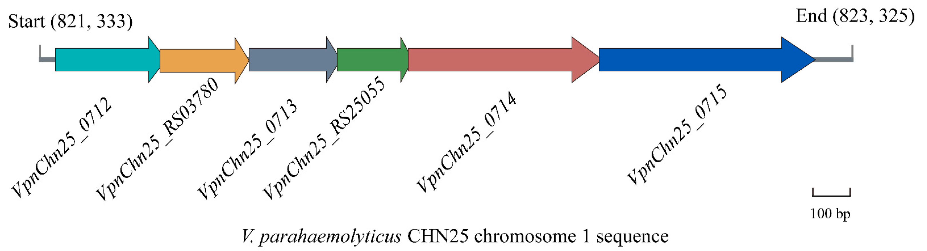
2.1. Prophage-Related Genes VpaChn25_RS25055, VpaChn25_0713, and VpaChn25_0714 in V. parahaemolyticus CHN25

The V. parahaemolyticus CHN25 genome contains a large prophage-like gene cluster, which shows high sequence similarity with the Vibrio phage martha 12B12 (33, 277 bp, GenBank accession no. HQ_316581) containing 50 predicted genes [24]. Of the twenty-four genes present in chromosome 1 (3,416,467 bp) of the V. parahaemolyticus CHN25 genome, seven coded for phage proteins, eight encoded predictive regulators, and nine coded for hypothetical proteins with unknown functions in the current databases [25]. Of these unknown genes, sequence analysis showed that the VpaChn25_0713 gene encodes a hypothetical protein that contains a conserved structural domain of the ku superfamily. The VpaChn25_0714- and VpaChn25_RS25055-encoding proteins had no hits against any conserved structural domains. Meanwhile, VpaChn25_0713 has a 29 bp overlap with VpaChn25_RS25055, and VpaChn25_RS25055 has a 4 bp overlap with VpaChn25_0714 (Figure 1).

2.2. Deletion and Reverse Complementation of the VpaChn25_RS25055, VpaChn25_0713, and VpaChn25_0714 Genes in V. parahaemolyticus CHN25 Genome

Unmarked in-frame single-gene-deletion mutants ΔVpaChn25_RS25055, ΔVpaChn25_0713, and ΔVpaChn25_0714, as well as the triple-deletion mutant ΔVpaChn25_RS25055-0713-0714, were derived with homologous recombination methods. For instance, for the construction of the ΔVpaChn25_0713 mutant, two primer pairs, VpaChn25_0713-up-F/R and VpaChn25_0713-down-F/R (Table 1), were designed to target upstream (454 bp) and downstream (322 bp) sequences of the VpaChn25_0713 gene in the V. parahaemolyticus CHN25 genome, respectively. The upstream and downstream sequences were retrieved by the polymerase chain reaction (PCR) and subsequently cloned into pDS132. Subsequently, the ligated DNA was transfected into E. coli DH5α λpir-competent cells, and subsequent screening identified the positive transformants as obtaining the recombinant vector pDS132+VpaChn25_0713. The recombinant vector was introduced into E. coli β2155-competent cells, followed by conjugation with V. parahaemolyticus CHN25. Positive exconjugants were obtained using the two-step allele exchange approach [25]. The 234 bp VpaChn25_0713 deletion was verified by the DNA sequencing and PCR analyses (Figure S1). Likewise, we used the same method to construct the deletion mutants ΔVpaChn25_0714, ΔVpaChn25_RS25055, and ΔVpaChn25_RS25055-0713-0714 (Figures S2–S4). Sequencing chromatographs of V. parahaemolyticus CHN25 WT and ΔVpaChn25_0713, ΔVpaChn25_RS25055, ΔVpaChn25_0714, and ΔVpaChn25_RS25055-0713-0714 mutants are presented in Figure S5.
Subsequently, four reverse mutants, ΔVpaChn25_RS25055-com, ΔVpaChn25_0713-com, ΔVpaChn25_0714-com, and ΔVpaChn25_RS25055-0713-0714-com, were also successfully constructed, respectively. For example, for the construction of the complementary mutant ΔVpaChn25_0713-com, the 234 bp VpaChn25_0713 was subjected to amplification via the PCR assay, followed by cloning into pMMB207 (9076 bp). The ligated DNA was introduced into E. coli DH5α, and positive transformants were screened to obtain pMMB207+VpaChn25_0713. Electrotransformation introduced the recombinant vector into the ΔVpaChn25_0713 mutant, generating the reverse mutant ΔVpaChn25_0713-com. Confirmation of this mutant was carried out using the methods previously mentioned (Figure S6). Similarly, the same method was used to construct ΔVpaChn25_0714-com, ΔVpaChn25_RS25055-com, and ΔVpaChn25_RS25055-0713-0714-com (Figures S7–S9).

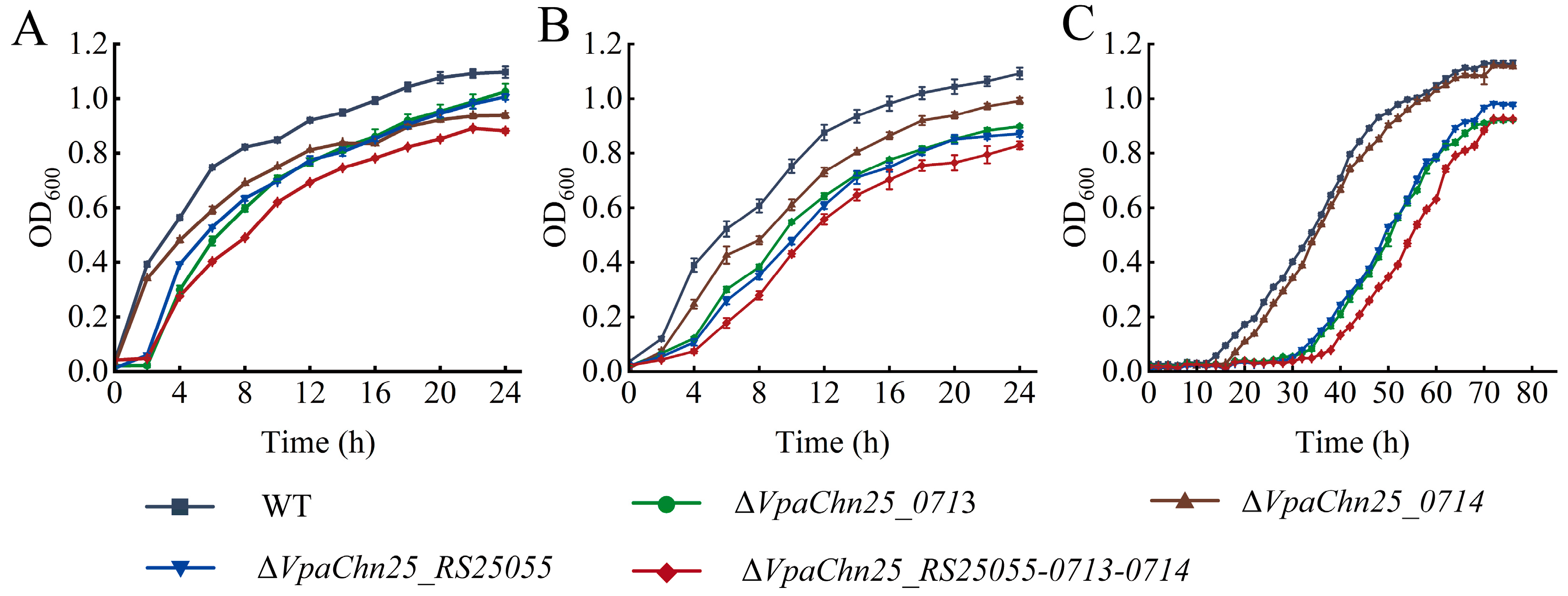
2.3. Survival of ΔVpaChn25_0713, ΔVpaChn25_0714, ΔVpaChn25_RS25055, and ΔVpaChn25_RS25055-0713-0714 Mutants at Different Temperatures and pH Conditions

V. parahaemolyticus can grow in a broad range of environmental conditions such as pH 5.0–9.0 and NaCl 0.5–3% [27]. To investigate the influence of the deletion of the VpaChn25_RS25055, VpaChn25_0713, VpaChn25_0714, and VpaChn25_RS25055-0713-0714 genes on V. parahaemolyticus CHN25 survival, growth curves of the WT strain, four deletion mutants, and four complementary mutants were determined at varying temperatures (15, 25, and 37 °C) and pH conditions (pH 5.5 to 8.0), and the data are presented in Figure 2, Figure 3, Figure S10, and Figure S11, respectively.
As shown in Figure 2A, at 37 °C, the ΔVpaChn25_0713, ΔVpaChn25_RS25055, and ΔVpaChn25_RS25055-0713-0714 mutants grew in TSB medium (pH 8.5, 3% NaCl) with a delay phase (DP) of 2 h when compared with the WT strain. The maximum OD600 value of ΔVpaChn25_RS25055-0713-0714 (0.89 ± 0.01) was significantly lower than that of the WT strain (1.10 ± 0.02) (p < 0.01).
At 25 °C, the maximum OD600 value of ΔVpaChn25_0713, ΔVpaChn25_RS25055, and ΔVpaChn25_RS25055-0713-0714 mutants was significantly lower than that of the WT strain (1.09 ± 0.02) (p < 0.01) (Figure 2B).
At 15 °C, the growth of the WT and four mutant strains were all delayed (Figure 2C). The WT strain entered the logarithmic phase (LP) after 8 h and the stationary phase (SP) after 60 h with a maximum OD600 value of 1.13 ± 0.01. The ΔVpaChn25_0713 mutant grew more slowly during the first 30 h of incubation and entered the LP after 32 h and the SP after 68 h with the maximum OD600 value of 0.91 ± 0.01. Similarly, the lag phases of the ΔVpaChn25_RS25055 and ΔVpaChn25_RS25055-0713-0714 mutants were 3.75-fold and 4.75-fold longer than that of the WT strain, respectively (Figure 2C).
These results indicated that the VpaChn25_RS25055 and VpaChn25_0713 genes could enhance the adaptability of V. parahaemolyticus CHN25 for colder conditions. Growth of ΔVpaChn25_RS25055-0713-0714 was more strongly inhibited than the single-gene-deletion mutants ΔVpaChn25_RS25055, ΔVpaChn25_0713, and ΔVpaChn25_0714 at 37 °C, 25 °C, and 15 °C, indicating a positively superposed regulation of the VpaChn25_RS25055, VpaChn25_0713, and VpaChn25_0714 genes on the growth of V. parahaemolyticus CHN25.
V. parahaemolyticus survives the stomach’s harsh acidic environment and establishes intestinal colonization in the host [28]. When consumed with raw, undercooked, or mishandled seafood, V. parahaemolyticus is challenged by the very low pH environment of the human stomach (which is normally between 1–3 but can rise above 6.0 after consumption of the food) and reaches the human gastrointestinal tract, where it can cause gastroenteritis [22]. Therefore, we studied the growth of the WT and the four mutants in TSB (3% NaCl) at pH values between 5.5 and 8.0, and the results are shown in Figure 3.
Under acidic (pH 5.5–6.5) and neutral (pH 7.0) conditions, the growth of the WT strain and four mutant strains were greatly inhibited, with the maximum OD600 values below 0.6 at the SP (Figure 3A–D). Notably, the maximum OD600 value of the ΔVpaChn25_RS25055 mutant was significantly lower than the WT strain in acidic and neutral conditions (p < 0.01). Interestingly, the ΔVpaChn25_RS25055-0713-0714 mutant grew in the TSB medium with DPs of 2.5 h, 3.0 h, 3.0 h, and 2.5 h when compared with the WT strain at pH 5.5, pH 6, pH 6.5, and pH 7.0, respectively (p < 0.01).
Under alkaline conditions, the maximum biomass of the ΔVpaChn25_RS25055, ΔVpaChn25_0713, ΔVpaChn25_0714, and ΔVpaChn25_RS25055-0713-0714 mutants was significantly lower than WT at pH 7.5 (p < 0.01) (Figure 3E). At pH 8.0, the growth of the ΔVpaChn25_RS25055-0713-0714 mutant was still poor, showing a maximum biomass OD600 value of 0.75 ± 0.01 (Figure 3F).
It was observed that both VpaChn25_0713 and VpaChn25_0714 genes are beneficial to V. parahaemolyticus CHN25 survival at pH 7.0–8.0, while the VpaChn25_RS25055 gene can amplify V. parahaemolyticus CHN25 persistence at pH 5.5–8.0. Interestingly, the LP of ΔVpaChn25_RS25055-0713-0714 was significantly extended compared to the ΔVpaChn25_RS25055, ΔVpaChn25_0713, and ΔVpaChn25_0714 mutants at pH 5.5–8.0, indicating a positively superposed regulation of VpaChn25_RS25055, VpaChn25_0713, and VpaChn25_0714 genes on the acid tolerance of V. parahaemolyticus CHN25.

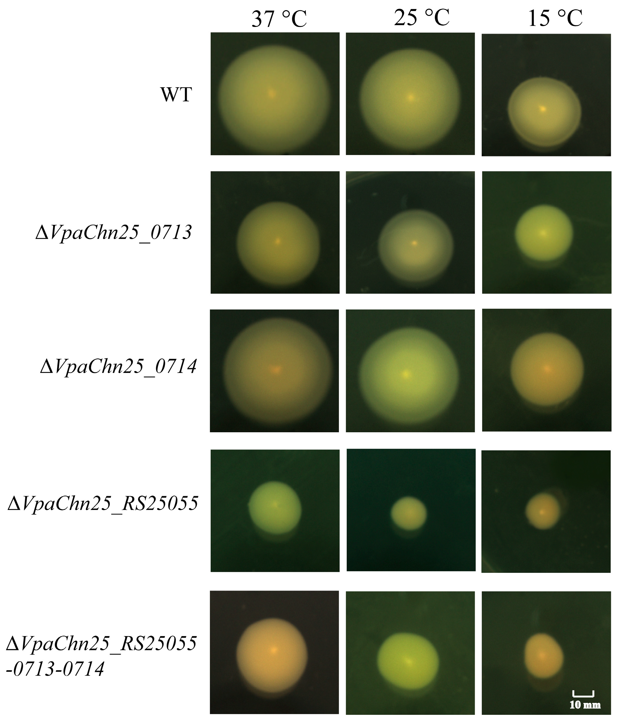
2.4. Swimming Motility of the ΔVpaChn25_RS25055, ΔVpaChn25_0713, ΔVpaChn25_0714, and ΔVpaChn25_RS25055-0713-0714 Mutants

Motility has been identified as an essential virulence factor for the survival and colonization of V. parahaemolyticus [29]. Herein, swimming of the WT strain, four mutants, and four complementary mutants were examined at different temperatures; the results are presented in Figure 4 and Figure S12.
When the strains were separately incubated in semi-solid TSB containing 0.25% agar at 37 °C, the ΔVpaChn25_0713 mutant swam (1.68 ± 0.05 cm) remarkably slower than the WT strain (3.37 ± 0.14 cm) (p < 0.01) (Figure 4). A similar case was observed under lower temperatures (Figure 4). This highlighted that VpaChn25_0713 gene deletion markedly suppressed the motility of V. parahaemolyticus CHN25.
Similarly, the ΔVpaChn25_RS25055 mutant also swam significantly slower than the WT strain at 15, 25, and 37 °C, respectively (p < 0.01) (Figure 4).
The swimming diameters of the ΔVpaChn25_RS25055-0713-0714 mutant were 1.60 ± 0.07 cm, 1.46 ± 0.18 cm, and 1.12 ± 0.09 cm at 37, 25, and 15 °C, respectively, which were 0.47-fold, 0.56-fold, and 0.42-fold smaller than those of the WT strain (p < 0.001) (Figure 4).
In contrast, as depicted in Figure 4, no obvious differences in swimming circles were found between the ΔVpaChn25_0714 mutant and the WT strain at 15, 25, and 37 °C (p > 0.05).
Taken together, these findings demonstrated that a deficiency in motility was induced by VpaChn25_RS25055, VpaChn25_0713, and VpaChn25_RS25055-0713-0714 deletion in V. parahaemolyticus CHN25. Interestingly, when the strains were separately incubated in semi-solid TSB containing 0.25% agar, the swimming diameters of ΔVpaChn25_RS25055 were significantly lower than those of ΔVpaChn25_0713 and ΔVpaChn25_0714 at 15, 25, and 37 °C (p < 0.05).

2.5. Biofilm Formation of the ΔVpaChn25_RS25055, ΔVpaChn25_0713, ΔVpaChn25_0714, and ΔVpaChn25_RS25055-0713-0714 Mutants

V. parahaemolyticus can produce adherence factors that facilitate surface attachment and promote biofilm formation, thereby increasing its environmental survival, infectivity, and transmission [30]. Herein, biofilm formation of the WT, four deletion mutants, and four complementary mutants were analyzed by crystalline violet staining at 37 °C for 60 h. The data are presented in Figure 5A and Figure S13A. All strains showed similar biofilm development, maturation, and diffusion stages, but the maximum biofilm biomass formed by the four mutants was remarkably smaller than the WT strain (p < 0.001).
As shown in Figure 5A, at 0 to 12 h, the biofilm of the WT strain formed slowly; at 12 to 36 h, it increased rapidly and reached the maximum biomass (OD 600 = 1.074 ± 0.05) at 36 h; at 36 to 60 h, the biofilm decreased sharply (OD 600 = 0.614 ± 0.01), which may have resulted from nutrient depletion and accumulation of metabolic waste in the orifice plates.
Compared to the WT strain, the ΔVpaChn25_0713 mutant showed significantly slower biofilm formation at all stages (p < 0.01), reaching maximum biofilm formation at 36 h, which was 0.78-fold less than that of the WT strain (Figure 5A). Similar cases were observed for ΔVpaChn25_0714 and ΔVpaChn25_RS25055-0713-0714 (Figure 5A).
In addition, in the absence of the VpaChn25_RS25055 gene, the biofilm formed by V. parahaemolyticus reached the maximum biomass at 24 h, which was 0.74-fold less than that of WT (Figure 5A).
These findings demonstrated that the absence of the VpaChn25_RS25055, VpaChn25_0713, VpaChn25_0714, and VpaChn25_RS25055-0713-0714 genes led to a decrease in biofilm formation of V. parahaemolyticus CHN25. Notably, the maximum biofilm formation of the ΔVpaChn25_RS25055-0713-0714 mutant was significantly less than those of the ΔVpaChn25_0713 and ΔVpaChn25_0714 mutants (p < 0.01), indicating the positively superposed regulation of VpaChn25_RS25055, VpaChn25_0713, and VpaChn25_0714 genes on the biofilm formation of V. parahaemolyticus CHN25.

2.6. Cell Surface Hydrophobicity, Cell Membrane Permeability, and Fluidity of the ΔVpaChn25_RS25055, ΔVpaChn25_0713, ΔVpaChn25_0714, and ΔVpaChn25_RS25055-0713-0714 Mutants

Cell membranes serve as selective semi-permeable barriers whose integrity, fluidity, and selective permeation control the movement of various substances, playing a pivotal role in microbial growth and pathogenicity [31]. Based on the above results, we further asked whether the deletion of the VpaChn25_RS25055, VpaChn25_0713, and VpaChn25_0714 genes would influence bacterial cell membrane structure (Figure 5 and Figure S13).
o-nitrophenyl-β-D galactopyranoside (ONPG) was used as a probe to examine the cell inner membrane permeability of the strains. As shown in Figure 5B, no apparent differences in cell inner membrane permeability were found between the WT strain and the ΔVpaChn25_RS25055, ΔVpaChn25_0714, and ΔVpaChn25_RS25055-0713-0714 mutants, but the inner membrane permeability of ΔVpaChn25_0713 was significantly reduced (p < 0.001).
As displayed in Figure 5C, the cell membrane fluidity of the ΔVpaChn25_RS25055, ΔVpaChn25_0713, and ΔVpaChn25_0714 mutants were 1.65-fold, 1.32-fold, and 1.13-fold higher than those of the WT strain, respectively. Additionally, the cell surface hydrophobicity of ΔVpaChn25_RS25055 and ΔVpaChn25_RS25055-0713-0714 was 0.56-fold and 0.62-fold lower than that of WT, respectively (Figure 5D).

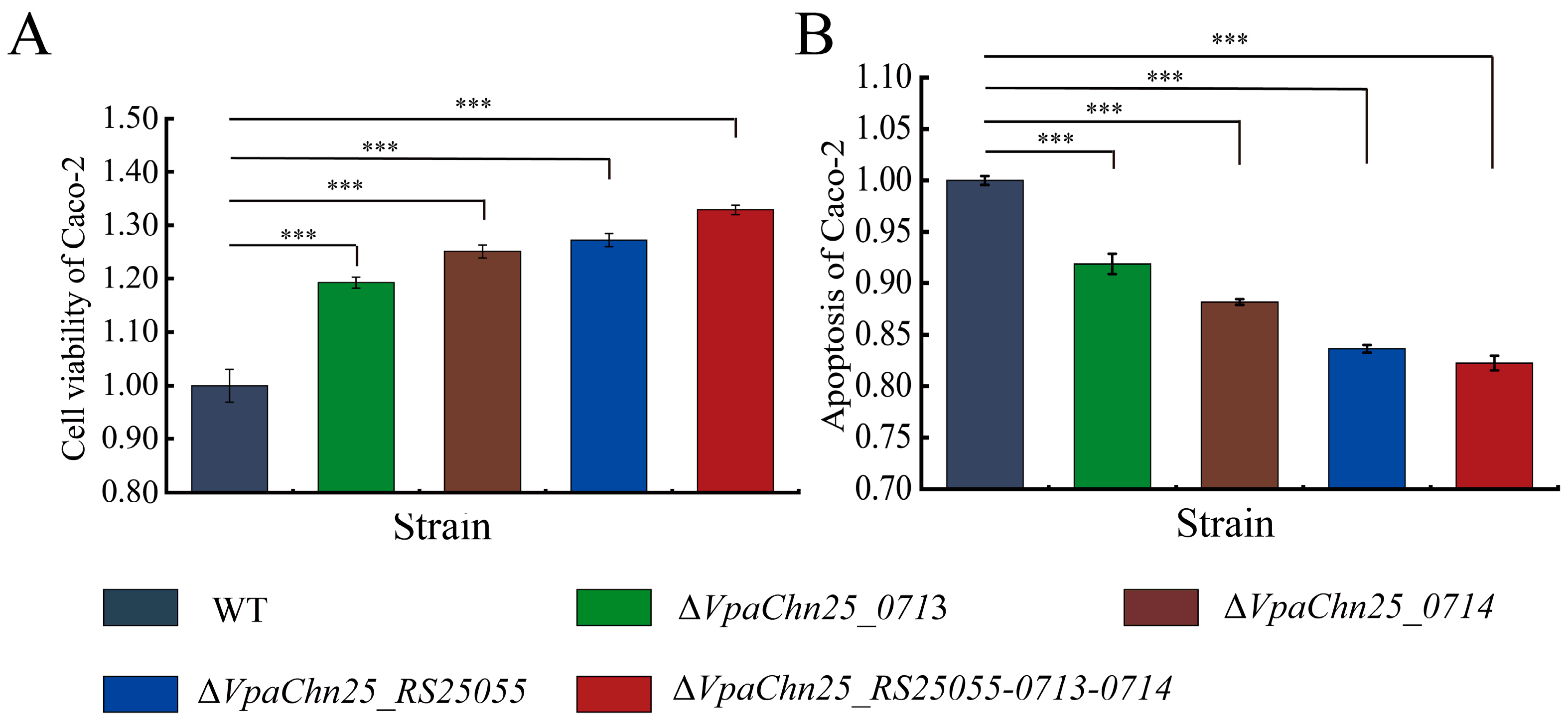
2.7. Interaction between the ΔVpaChn25_RS25055, ΔVpaChn25_0713, ΔVpaChn25_0714, and ΔVpaChn25_RS25055-0713-0714 Mutants and Host Intestinal Epithelial Cells

In this study, Caco-2 was employed as a cell model for in vitro cell interaction assessment, and the data are presented in Figure 6 and Figure S14. Following infection with the ΔVpaChn25_RS25055, ΔVpaChn25_0713, ΔVpaChn25_0714, and ΔVpaChn25_RS25055-0713-0714 mutants at 37 °C for 4 h, the survival of Caco-2 cells was remarkably increased by 1.27-fold, 1.19-fold, 1.23-fold, and 1.33-fold, respectively, as compared to the WT strain (p < 0.01) (Figure 6A). Concurrently, Caco-2 cells were subjected to double staining with the membrane-linked protein V-PI and FITC, followed by a flow cytometry assay analysis. It was found that the ΔVpaChn25_RS25055, ΔVpaChn25_0713, ΔVpaChn25_0714, and ΔVpaChn25_RS25055-0713-0714 mutants induced Caco-2 cell apoptosis at 0.85-fold, 0.92-fold, 0.88-fold, and 0.83-fold lower rates than that of the WT strain following 4 h of infection, respectively (p < 0.01) (Figure 6B). This indicates that the deletion of the VpaChn25_0713, VpaChn25_0714, and VpaChn25_RS25055 genes could reduce the ability of V. parahaemolyticus CHN25 to infect and apoptose the host intestinal epithelial Caco-2 cells.

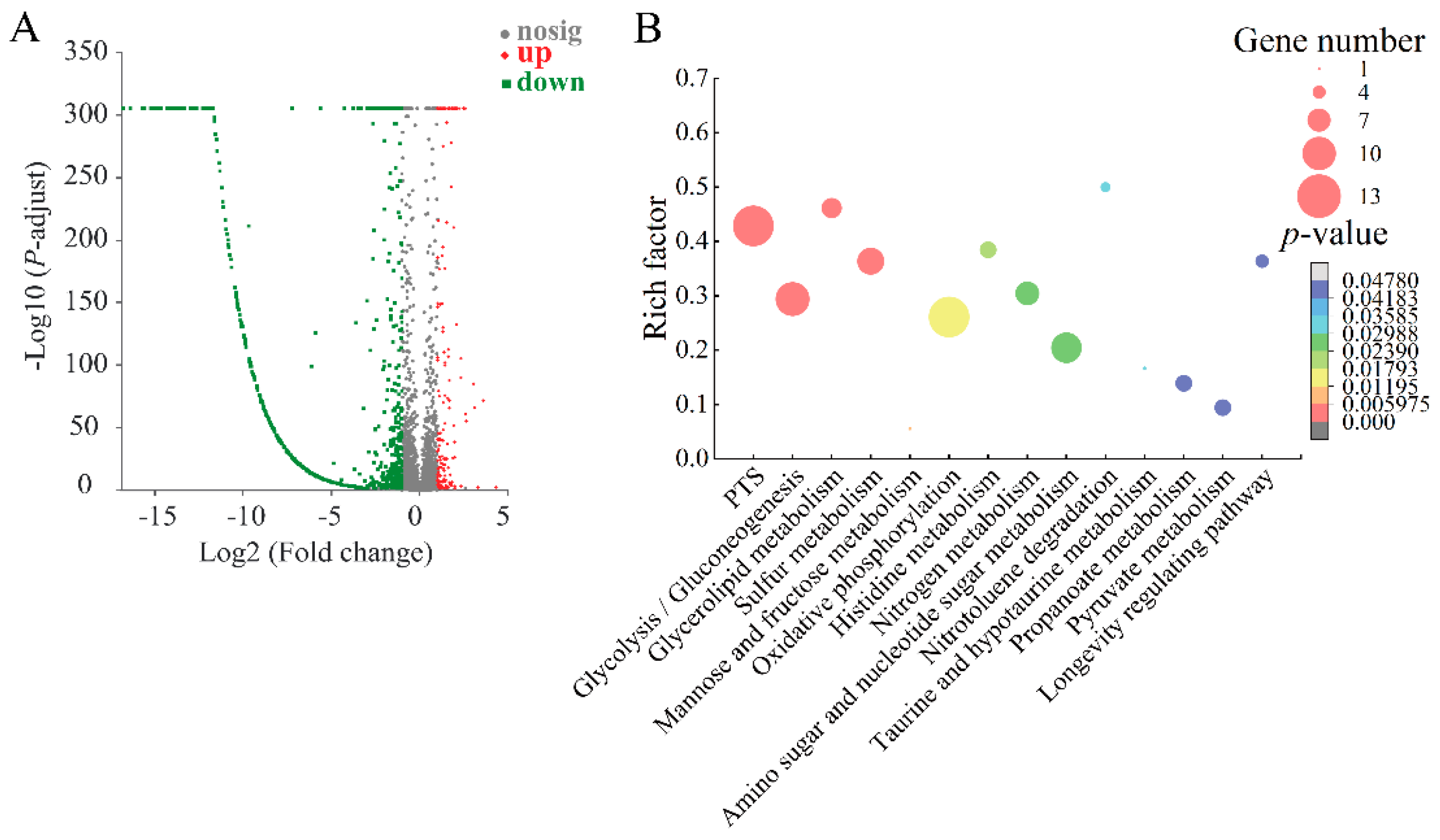
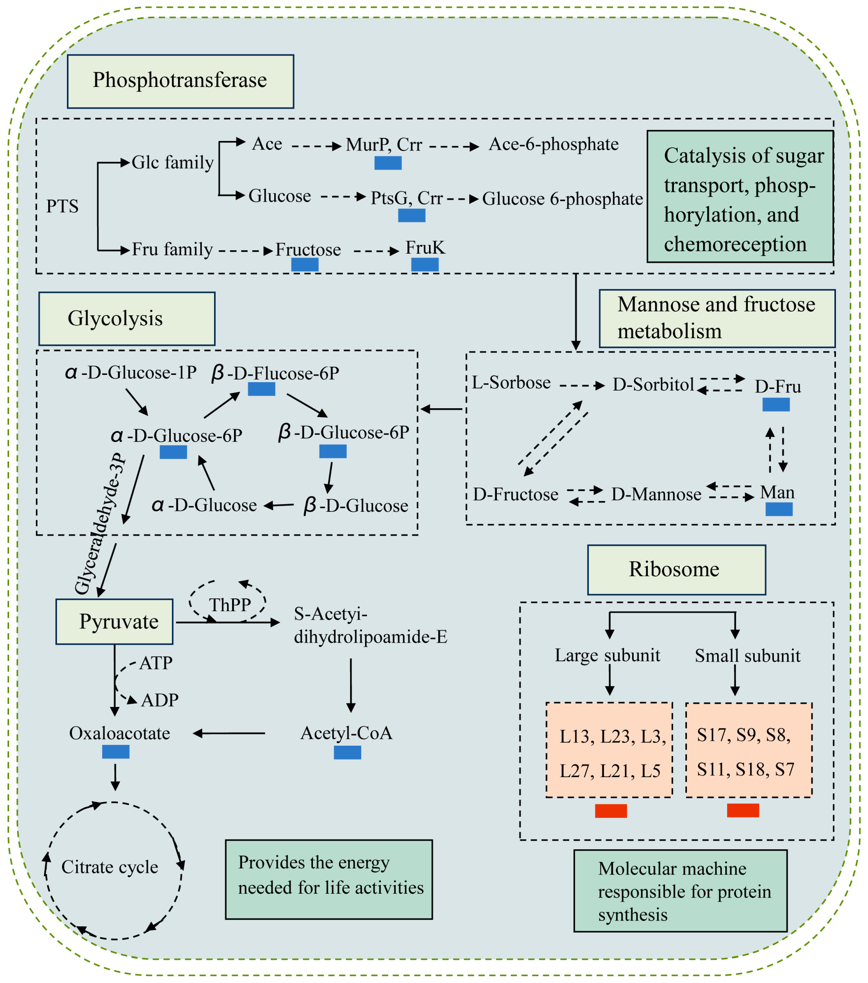
2.8. The Major Changed Metabolic Pathways in the ΔVpaChn25_RS25055, ΔVpaChn25_0713, ΔVpaChn25_0714, and ΔVpaChn25_RS25055-0713-0714 Mutants

To assess the global-level expression alterations regulated by VpaChn25_RS25055, VpaChn25_0713, VpaChn25_0714, and VpaChn25_RS25055-0713-0714 gene deletion, we determined the transcriptomes of the WT, the four mutants (ΔVpaChn25_0713, ΔVpaChn25_RS25055, ΔVpaChn25_0714, ΔVpaChn25_RS25055-0713-0714), and the four complementary mutants (ΔVpaChn25_0713-com, ΔVpaChn25_RS25055-com, ΔVpaChn25_0714-com, ΔVpaChn25_RS25055-0713-0714-com) cultivated in TSB medium to mid-logarithmic growth phase (mid-LGP) at 37 °C. The DEGs across all nine strains were deposited in the NCBI-SRA database (http://www.ncbi.nlm.nih.gov/sra/ (accessed on 27 February 2023); PRJNA938975). To validate the transcriptome data, we detected six representative genes in the ΔVpaChn25_RS25055, ΔVpaChn25_0713, ΔVpaChn25_0714, and ΔVpaChn25_RS25055-0713-0714 mutants by RT-qPCR analysis, respectively (Table S1). The resulting data were correlated with those derived from transcriptome analyses (Table S2).

2.8.1. The Major Changed Metabolic Pathways in the ΔVpaChn25_0713 Mutant

In the ΔVpaChn25_0713 mutant, differential expression changes were observed in approximately 17.32% (812/4810) of the bacterial genes compared to both the WT and ΔVpaChn25_0713-com strains. According to KEGG database analysis of transcriptomic data, 14 significantly altered metabolic pathways were detected: the phosphotransferase system (PTS), glycolysis/gluconeogenesis, glycerolipid metabolism, mannose and fructose metabolism, sulfur metabolism, oxidative phosphorylation, histidine metabolism, nitrogen metabolism, amino sugar and nucleotide sugar metabolism, nitrotoluene degradation, taurine and hypotaurine metabolism, propanoate metabolism, longevity-regulating pathway, and pyruvate metabolism (Figure 7, Table S3).
For example, in the PTS, nine DEGs were markedly down-regulated at the mRNA level (0.057-fold to 0.491-fold) (p < 0.05). The PTS performs dual roles, facilitating the transport and phosphorylation of various sugars and their derivatives while also serving as a regulatory hub governing carbon, nitrogen, and phosphate metabolism, chemotaxis, potassium transport, and influencing the virulence of specific pathogens [32]. The significantly down-regulated DEGs may be consistent with the growth and biofilm-deficient phenotype of the ΔVpaChn25_0713 mutant.
In glycolysis/gluconeogenesis, seven DEGs were remarkably decreased (0.203-fold to 0.494-fold) (p < 0.05): the type I glyceraldehyde-3-phosphate dehydrogenase (Vpachn25_RS10585), D-hexose-6-phosphate mutarotase (VpaChn25_RS10590), bifunctional acetaldehyde-CoA/alcohol dehydrogenase (VpaChn25_RS10475), pyruvate kinase (VpaChn25_RS19575), 6-phospho-β-glucosidase (VpaChn25_RS23575), triose-phosphate isomerase (VpaChn25_RS01325), and phosphoenolpyruvate synthase (VpaChn25_RS17630). The down-regulated DEGs associated with glycolysis/gluconeogenesis could lead to a polysaccharide deficiency. This deficiency might contribute to the biofilm formation observed in the ΔVpaChn25_0713 mutant [33].
All five DEGs were markedly down-regulated in the pyruvate metabolism (0.162-fold to 0.421-fold) (p < 0.05), such as phosphoenolpyruvate carboxylase (VpaChn25_RS14110, 0.342-fold) (p < 0.05), which catalyzes the irreversible reaction between phosphoenolpyruvate (PEP) and bicarbonate to form inorganic phosphate and oxaloacetate, an essential step in bacteria and plants [34]. Four DEGs were decreased in amino sugar and nucleotide sugar metabolism (0.001-fold to 0.164-fold) (p < 0.05). Among these, the DEG encoding the UDP-glucose 4-epimerase GalE (VpaChn25_RS21110) was highly inhibited (0.001-fold). It facilitates the NAD-dependent interconversion of galacto- and gluco-hexoses, which is linked to UDP and holds a crucial position in the galactose metabolism of diverse organisms [35]. These two metabolic pathways are linked to carbohydrate metabolism, which is essential for all life and has implications for organisms’ growth, reproduction, and maintenance [36].
Remarkably, a total of 27 DEGs involved in energy metabolism were significantly changed, including nitrogen metabolism, sulfur metabolism, and oxidative phosphorylation, with 17 DEGs showing down-regulation (0.096-fold to 0.499-fold) (p < 0.05) and 10 genes showing higher transcript levels (2.081-fold to 4.053-fold) (p < 0.05). For example, in nitrogen metabolism, the DEG encoding a glutamate synthase subunit β (GltS β subunit) (VpaChn25_RS02355) was highly inhibited (0.096-fold), which is a flavin adenosine dinucleotide (FAD)-dependent nicotinamide adenine dinucleotide phosphate (NADPH) oxidoreductase, and serves to input electrons into the GltS α subunit for glutamate synthesis [37]. The expression of ATP binding cassette (ABC) transporter ATP-binding protein (VpaChn25_RS20680) was significantly decreased (0.231-fold); ABC proteins transport a huge range of diverse substrates, from simple ions through molecules to peptides, complex lipids, and even small proteins [38]. Moreover, in the sulfur metabolism, the DEG encoding CysK (VpaChn25_RS04200) was significantly inhibited (0.498-fold) (p < 0.05). Singh et al. showed that CysK is a key enzyme in the cysteine biosynthetic pathway involved in promoting biofilm formation [39]. These data suggested inactive transport and utilization of the carbon sources and repressed energy production in the ΔVpaChn25_0713 mutant.
Comparative transcriptome analysis also showed the remarkably up-regulated metabolic routes (p < 0.05) in the ΔVpaChn25_0713 mutant, such as histidine metabolism and propanoate metabolism. For example, four DEGs for histidine metabolism were remarkably increased (3.843-fold to 4.716-fold) (p < 0.05). Of these, imidazolonepropionase (VpaChn25_RS06780), which catalyzes histidine degradation, was substantially up-regulated (4.142-fold) (p < 0.05), while it mediates the third stage in the histidine degradation pathway. This enzyme hydrolyzes the carbon- nitrogen bonds within 4-imidazolone-5-propionic acid, forming N-formimino-l-glutamic acid [40]. The substantial elevation of these enzymes indicates that deletion of the VpaChn25_0713 gene affects histidine metabolism and may promote histidine degradation.
Moreover, all six DEGs involved in glycerolipid metabolism were significantly up-regulated (p < 0.05). Glycerolipids are a class of biological molecules required for membrane formation, caloric storage, and important intracellular signaling processes [41]. The overall up-regulation of the glycerolipid metabolism provides new insight into the mechanism by which VpaChn25_0713 gene deletion in V. parahaemolyticus may affect lipid metabolism. In contrast, nitrotoluene degradation involved three genes that underwent a significant decrease (p < 0.05). Nitroreductases in the intestinal microbiota are involved in the biotransformation of several poisonous, mutagenic, and carcinogenic nitroaromatic chemicals’ reduction products to their hazardous metabolites [42]. Therefore, it is hypothesized that changes in this metabolic pathway correlate with the relevant results of cytotoxicity assays.
Taken together, these data suggested that VpaChn25_0713 gene deletion could inhibit the transportation and phosphorylation of sugar compounds and their derivatives, and suppress the glycolytic/glucose metabolic pathway, leading to polysaccharide deficiency; it inhibited the production of glutamate as well as cysteine, thus affecting energy production. The above changes may contribute to the reduced swimming ability, biofilm formation, and decreased virulence of the ΔVpaChn25_0713 mutant.

2.8.2. The Major Changed Metabolic Pathways in the ΔVpaChn25_0714 Mutant

In the ΔVpaChn25_0714 mutant, differential expression changes were observed in approximately 19.17% (922/4810) of the bacterial genes compared to both the WT and ΔVpaChn25_0714-com strains. According to KEGG database analysis of transcriptomic data, 13 significantly altered metabolic pathways were detected: the propanoate metabolism, mannose and fructose metabolism, nitrotoluene degradation, PTS, lysine degradation, glycolysis/gluconeogenesis, benzoate degradation, pyruvate metabolism, ascorbate and aldarate metabolism, butanoate metabolism, fatty acid degradation, histidine metabolism, and β-lactam resistance (Figure 8, Table S4).
Similar to the ΔVpaChn25_0713 mutant, most DEGs related to mannose and fructose metabolism, nitrotoluene degradation, PTS, glycolysis/gluconeogenesis, and pyruvate metabolism were also markedly reduced in ΔVpaChn25_0714. For example, two DEGs were down-regulated at the mRNA level in mannose and fructose metabolism (0.08-fold to 0.426-fold) (p < 0.05). Apart from being an energy and carbon source, fructose metabolism has been shown to impact various cellular processes, including biofilm formation in streptococci and the pathogenicity of bacteria in plants [43].
In addition, 22 DEGs were significantly down-regulated (0.080-fold to 0.494-fold) (p < 0.05), and 9 DEGs were significantly up-regulated (2.017-fold to 3.217-fold) (p < 0.05) in carbohydrate metabolism. They involved a total of six types of metabolic pathways, which were propanoate metabolism, mannose and fructose metabolism, glycolysis/gluconeogenesis metabolism, pyruvate metabolism, ascorbate and aldarate metabolism, and butanoate metabolism. Overall, these metabolic pathways showed an overall trend of down-regulation. For example, in propanoate metabolism, five DEGs underwent significant down-regulation (0.233-fold to 0.435-fold) (p < 0.05). Of these, acetate kinase is an enzyme widely distributed in the bacteria and archaea domains which catalyzes the phosphorylation of acetate [44]. In glycolysis/gluconeogenesis metabolism, six DEGs underwent significant down-regulation (0.228-fold to 0.483-fold) (p < 0.05). Of these, the DEG encoding type I glyceraldehyde-3-phosphate dehydrogenase (VpaChn25_RS10585, 0.285-fold) was significantly down-regulated, which is essential for glycolysis [45]. The down-regulated DEGs involved in carbohydrate metabolism may be responsible for the biofilm formation defect in the ΔVpaChn25_0714 mutant.
In β-lactam resistance, all eight DEGs were remarkably decreased (0.119-fold to 0.462-fold) in the ΔVpaChn25_0714 mutant (p < 0.05). Resistance is frequently acquired through the action of β-lactamases or the expression of alternative β-lactam-resistant penicillin-binding proteins (PBPs) [46].
Comparative transcriptome analysis indicated that several metabolic routes were markedly elevated in ΔVpaChn25_0714 (p < 0.05). For example, in histidine metabolism, four DEGs were markedly increased (3.400-fold to 4.629-fold) (p < 0.05): histidine ammonia-lyase (VpaChn25_RS06765), imidazolonepropionase (VpaChn25_RS06780), urocanate hydratase (VpaChn25_RS06770), and formimidoylglutamase (VpaChn25_RS06775). Of these, the DEG encoding histidine ammonia-lyase (VpaChn25_RS06765, 3.4-fold) regulates the initial step in histidine catabolism by catalyzing the deamination of histidine into urocanate and ammonia [47]. Meanwhile, in fatty acid degradation, three DEGs were markedly elevated (2.100-fold to 2.425-fold) (p < 0.05), including the fatty acid oxidation complex subunit α FadJ (VpaChn25_RS10820), acetyl-CoA C-acyltransferase FadI (VpaChn25_RS10825), and acetyl-CoA C-acetyltransferase (VpaChn25_RS18175). Fatty acids play an important role in the structural composition of cellular membranes and serve various functions in biological processes [48].
Taken together, similar to the ΔVpaChn25_0713 mutant, the deletion of the VpaChn25_0714 gene also inhibits the PTS pathway, promoting fatty acid degradation and suppressing the glycolytic/glucose metabolic pathway, leading to polysaccharide deficiency. The above changes may be related to the growth and biofilm-deficient phenotype of the ΔVpaChn25_0714 mutant.

2.8.3. The Major Changed Metabolic Pathways in the ΔVpaChn25_RS25055 Mutant

In the ΔVpaChn25_RS25055 mutant, there were differential expression changes observed in approximately 16.59% (798/4810) of the bacterial genes when compared to both the WT and ΔVpaChn25_RS25055-com strains. According to the KEGG database analysis of transcriptomic data, eight significantly altered metabolic pathways were detected: sulfur metabolism, glyoxylate and dicarboxylate metabolism, arginine biosynthesis, longevity-regulating pathway, glycolysis/gluconeogenesis, NOD-like receptor signaling pathway, ribosome, and monobactam biosynthesis (Figure 9, Table S5).
Similar to the ΔVpaChn25_0713 mutant, most DEGs in the sulfur metabolism, glycolysis/gluconeogenesis, and longevity-regulating pathways were also remarkably reduced in the ΔVpaChn25_RS25055 mutant.
In addition, all five DEGs were significantly down-regulated (0.084-fold to 0.305-fold) (p < 0.05) in arginine biosynthesis. Among them, N-acetylglutamate kinase (NAGK) catalyzes the second step of arginine biosynthesis [49]. Similarly, argininosuccinate synthetase 1 (ASS1) is a rate-limiting enzyme in arginine biosynthesis [50]. Overall, E. coli uses arginine as its only nitrogen supply, and many other bacteria use it as a source of nitrogen, carbon, and energy [51]. It has been shown that intracellular arginine deficiency may affect the formation of biofilms [52,53].
In the NOD-like receptor signaling pathway, all four DEGs were significantly down-regulated (0.317-fold to 0.451-fold) (p < 0.05), including flagellin (VpaChn25_RS04175, VpaChn25_RS11070, VpaChn25_RS11075) and the molecular chaperone HtpG (VpaChn25_RS04340). Flagellin, which polymerizes into flagellar filament, is essential for bacterial motility, and flagella-driven motility is an important trait of bacterial colonization and virulence [54,55].
Comparative transcriptome analysis demonstrated that several metabolic routes were remarkably up-regulated (p < 0.05) in ΔVpaChn25_RS25055, including ribosome, glyoxylate, and dicarboxylate metabolism. Briefly, 18 DEGs had significantly up-regulated transcript levels (2.006-fold to 6.026-fold) (p < 0.05). In the ribosome, all 13 DEGs were increased (2.006-fold to 2.705-fold) (p < 0.05). Ribosomes are macromolecular complexes in the cytoplasm, consisting of proteins and RNA, which connect amino acids and synthesize new proteins [56]. For example, the DEG (VpaChn25_RS02135) encoding a 50S ribosomal protein L13 was up-regulated (2.191-fold) (p < 0.05). Aseev et al. revealed that L13 was a major protein in the assembly of the 50S ribosomal subunit and serves as a repressor of rplM-rpsI expression in vivo. [57]. This indicated dramatic cellular reprogramming in the ΔVpaChn25_RS25055 mutant [58].
Taken together, this indicates that the deletion of the VpaChn25_RS25055 gene inhibits the transport and utilization of carbon sources, inhibits the biosynthesis of arginine and the formation of flagella, and changes the biosynthesis of ribosomes. These DEGs induced by VpaChn25_RS25055 deletion may affect swimming and virulence.

2.8.4. The Major Changed Metabolic Pathways in the ΔVpaChn25_RS25055-0713-0714 Mutant

In the ΔVpaChn25_RS25055-0713-0714 mutant, there were differential expression changes observed in approximately 19.90% (957/4810) of the bacterial genes when compared to both the WT and ΔVpaChn25_RS25055-0713-0714-com strains. According to KEGG database analysis of transcriptomic data, 11 significantly altered metabolic pathways were detected, such as the mannose and fructose metabolism, propanoate metabolism, alanine, aspartate and glutamate metabolism, NOD-like receptor signaling pathway, PTS, amino sugar and nucleotide sugar metabolism, arginine and proline metabolism, oxidative phosphorylation, glycolysis/gluconeogenesis, thiamine metabolism, and arginine biosynthesis (Figure 10, Table S6).
Similar to the ΔVpaChn25_0713 mutant, most DEGs related to mannose and fructose metabolism, glycolysis/gluconeogenesis, and PTS were decreased in the ΔVpaChn25_RS25055-0713-0714 mutant. Meanwhile, most DEGs related to NOD-like receptor signal transduction and arginine biosynthesis were also markedly reduced in the ΔVpaChn25_RS25055-0713-0714 mutant, similar to the ΔVpaChn25_RS25055 mutant.
The major metabolic pathway altered by the triple-gene-deletion mutant (ΔVpaChn25_RS25055-0713-0714) was not identical to that altered by the single-gene mutants (ΔVpaChn25_RS25055, ΔVpaChn25_0713, ΔVpaChn25_0714). Comparative transcriptomics revealed that the metabolic pathway of glycolysis/gluconeogenesis exhibited an overall down-regulation in the deletion of the single genes VpaChn25_RS25055, VpaChn25_0713, and VpaChn25_0714, while the deletion of three genes (VpaChn25_RS25055-0713-0714) showed an overall down-regulated superposition effect. In addition, significant changes in mannose and fructose metabolism, propanoate metabolism, and amino sugar and sugar metabolism were simultaneously observed in the four deletion mutants in this study. Carbohydrates can be catabolized for energy (ATP) or employed for anabolic functions [59]. Combining the experimental data on growth, swimming, and biofilm-related phenotypes mentioned in the previous section, it is hypothesized that the genes VpaChn25_RS25055, VpaChn25_0713, and VpaChn25_0714 are synergistically involved in regulating the active transport and utilization of carbon sources.
In the NOD-like receptor signaling pathway, all six DEGs were remarkably decreased (0.331-fold to 0.425-fold) (p < 0.05). Bacterial polar flagella, containing flagellin, play a vital role in bacterial motility. Swimming motility is an essential virulence factor for the pathogenesis of many Vibrio species [60,61]. It has been reported that in gut inflammation, Clostridioides difficile flagellin FliC plays a role in toxin contribution by interacting with the TLR5 of the immune system, triggering the activation of NF-kB and MAPK signal transduction [62]. The data indicate a potential issue with the flagellar basal body in the ΔVpaChn25_RS25055-0713-0714 mutant, which could have affected its impaired swimming ability and reduced virulence.
In the PTS, eight DEGs were transcriptionally significantly repressed (0.056-fold to 0.405-fold) (p < 0.05). Comparative transcriptomics revealed that PTS showed an overall down-regulation in the deletion of single genes VpaChn25_0713 and VpaChn25_0714, while the deletion of three genes (VpaChn25_RS25055-0713-0714) showed a superimposed effect of multiple genes acting together. For example, the ΔVpaChn25_RS25055-0713-0714 mutant encoding fused PTS fructose transporter subunit IIA/HPr (VpaChn25_RS19530, 0.122-fold) showed lower expression than ΔVpaChn25_0713 (0.136-fold) and ΔVpaChn25_0714 (0.134-fold). The PTS mediates both the uptake of carbohydrates across the cytoplasmic membrane and their phosphorylation [63]. Combined with the experimental data of biofilm-related phenotypes, we believe that VpaChn25_0713 and VpaChn25_0714 synergistically regulate membrane transport.
In addition, six DEGs were significantly down-regulated in alanine, aspartate, and glutamate metabolism (0.098-fold to 0.480-fold) (p < 0.05). Meanwhile, in arginine biosynthesis, all six DEGs were significantly repressed (0.101-fold to 0.376-fold) (p < 0.05). This may help V. parahaemolyticus maintain the stability of bacterial cell structure and function.
Comparative transcriptome analysis also revealed the significantly up-regulated metabolic pathways (p < 0.05) in the ΔVpaChn25_RS25055-0713-0714 mutant. For example, all DEGs were significantly up-regulated in oxidative phosphorylation (2.218-fold to 5.684-fold) (p < 0.05), a metabolic pathway related to energy metabolism; all DEGs were significantly up-regulated in thiamine metabolism, a metabolic pathway related to the metabolism of cofactors and vitamins; in arginine and proline metabolism, six DEGs were significantly up-regulated (2.105-fold to 3.110-fold) (p < 0.05).
Taken together, it is indicated that the deletion of the VpaChn25_RS25055-0713-0714 gene inhibits the transport and utilization of carbon sources, the formation of flagella, and the biosynthesis of glutamate and arginine. The above changes may also contribute to the delayed growth, reduced swimming ability, biofilm formation, and decreased virulence of the ΔVpaChn25_RS25055-0713-0714 mutant.

2.8.5. Possible Molecular Mechanisms of the ΔVpaChn25_RS25055, ΔVpaChn25_0713, ΔVpaChn25_0714, and ΔVpaChn25_RS25055-0713-0714 Mutants

In the present study, the transcriptome analyses revealed several DEGs involved in multiple pathways of biosynthesis, degradation, interconversion, and transport of the compounds in the ΔVpaChn25_RS25055, ΔVpaChn25_0713, ΔVpaChn25_0714, and ΔVpaChn25_RS25055-0713-0714 mutants, indicating a complex molecular regulation network in the absence of the above prophage genes (Figure 11).
The same metabolic pathways were elicited in the deletion mutants. For instance, the repressed glycolysis/gluconeogenesis metabolism in the ΔVpaChn25_RS25055, ΔVpaChn25_0713, ΔVpaChn25_0714, and ΔVpaChn25_RS25055-0713-0714 mutants; the repressed PTS, mannose, and fructose metabolism in the ΔVpaChn25_0713, ΔVpaChn25_0714, and ΔVpaChn25_RS25055-0713-0714 mutants; and the repressed pyruvate metabolism in the ΔVpaChn25_0713 and ΔVpaChn25_0714 mutants.
We also observed different metabolic pathways occurring in the above mutants. For instance, in the ΔVpaChn25_RS25055 mutant, 13 DEGs in the ribosome were markedly increased (2.006-fold to 2.705-fold) (p < 0.05).
Overall, multiple metabolic pathways were changed in the above mutants: (1) the PTS was down-regulated, which affects sugar transport, phosphorylation, and chemoreception; (2) the mannose and fructose metabolism, glycolysis, and pyruvate metabolism were down-regulated, thereby affecting energy production; and (3) the amino acid synthesis was decreased to delay cell growth.

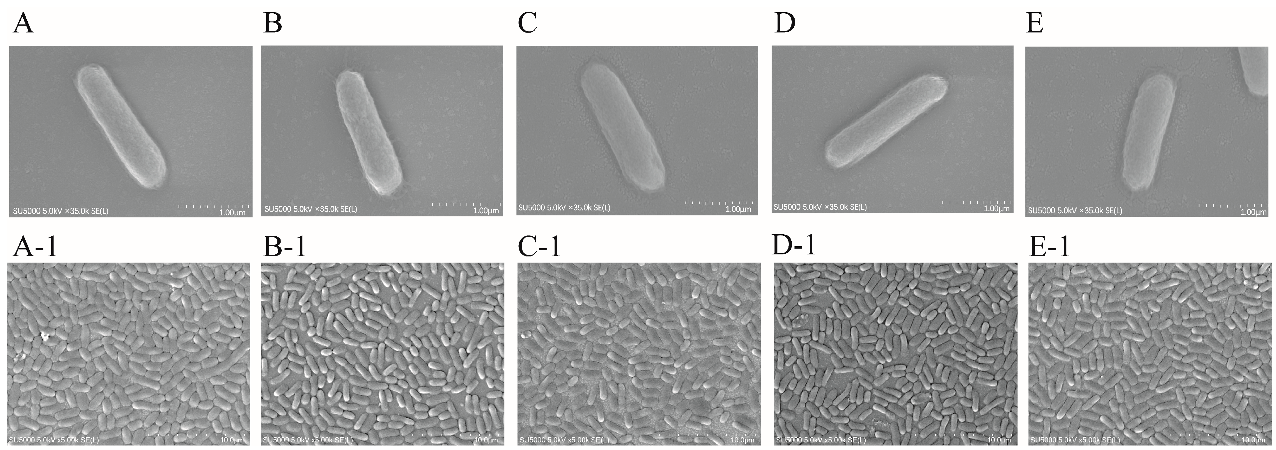
2.9. SEM Observation of Cell Structure of the ΔVpaChn25_RS25055, ΔVpaChn25_0713, ΔVpaChn25_0714, and ΔVpaChn25_RS25055-0713-0714 Mutants

The bacterial cell structures of the V. parahaemolyticus CHN25 WT, four mutants, and four complementary mutants were evaluated by SEM analysis (Figure 12 and Figure S15). As shown in Figure 12, all strains have intact cell surface structures, showing rod-shaped cells with a flat surface in the TSB medium (3% NaCl, pH 8.5) at 37 °C.

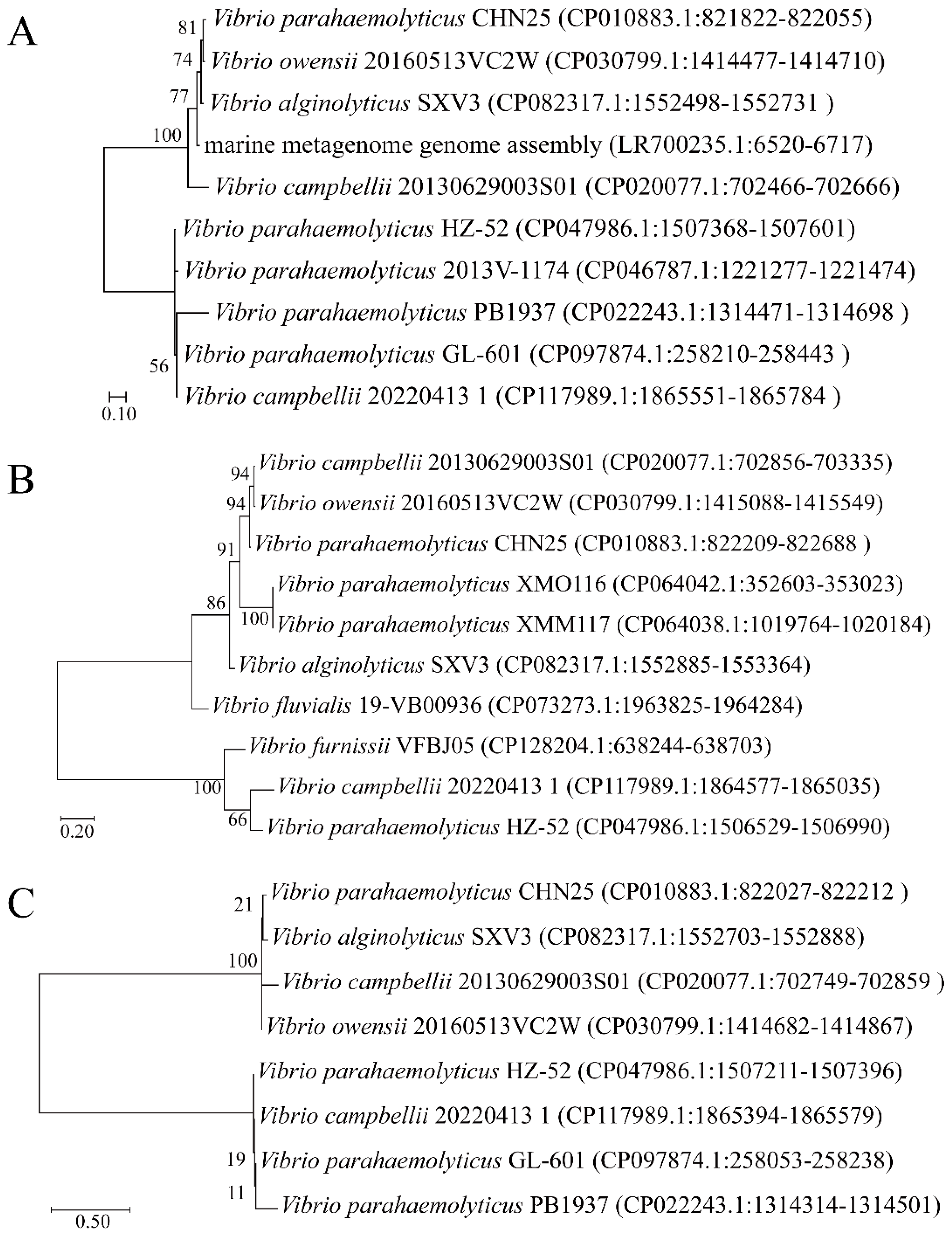
2.10. Distribution of the VpaChn25_RS25055, VpaChn25_0713, and VpaChn25_0714 Genes in Bacteria

A total of 119 V. parahaemolyticus isolates, which were recovered from aquatic products collected in Shanghai, China [64], were tested for the VpaChn25_RS25055, VpaChn25_0713, and VpaChn25_0714 genes via the PCR assays. The findings indicated that 1.68% (n = 1) of the isolates harbored VpaChn25_RS25055, VpaChn25_0713, and VpaChn25_0714 homologs, respectively.
Analysis using BLAST against the GenBank database demonstrated the presence of the homologs of VpaChn25_0713 in a single Vibrio phage, vB_VpaM_VP-3212, which is part of the marine metagenome genome assembly LR700235; and eight Vibrio species, including Vibrio owensii 20160513VC2W (CP030799), Vibrio alginolyticus SXV3 (CP082317), Vibrio campbellii 20130629003S01 (CP020077), and V. parahaemolyticus 2013V-1174 (CP046787), etc. (Figure 13A).
BLAST analysis against the GenBank database showed that VpaChn25_0714 homologs were present in nine Vibrio species, including V. campbellii 20130629003S01 (Genbank accession no.: CP020077), V. owensii 20160513VC2W (Genbank accession no.: CP030799), V. alginolyticus SXV3 (Genbank accession no.: CP082317), V. parahaemolyticus XMO116 (Genbank accession no.: CP064042), and Vibrio fluvialis 19-VB00936 (Genbank accession no.: CP073273), etc. (Figure 13B).
BLAST analysis against the GenBank database also revealed that VpaChn25_RS25055 homologs were present in seven Vibrio species, including V. owensii 20160513VC2W (Genbank accession no.: CP030799), V. alginolyticus SXV3 (Genbank accession no.: CP082317), V. parahaemolyticus PB1937 (Genbank accession no.: CP022243), and V. campbellii 20130629003S01 (Genbank accession no.: CP020077), etc. (Figure 13C).
It was observed that the VpaChn25_RS25055, VpaChn25_0713, and VpaChn25_0714 genes exist in V. parahaemolyticus and the Vibrio genus. Notably, the homologs of the genes VpaChn25_RS25055, VpaChn25_0713, and VpaChn25_0714 are all present in V. campbellii 20130629003S01. V. campbellii is a crucial aquatic pathogen, capable of causing vibriosis in shrimp and fish, leading to substantial economic losses [65].

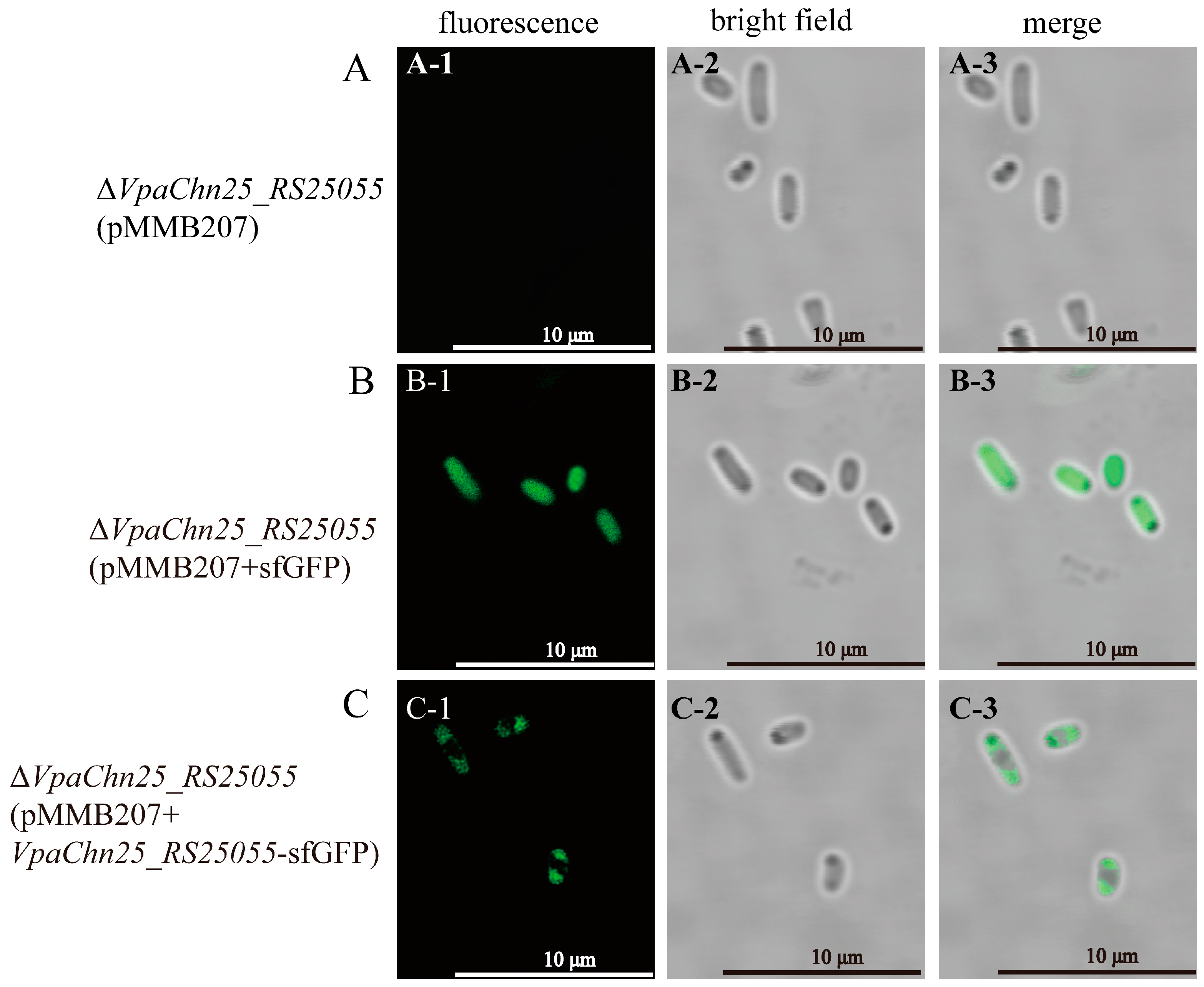
2.11. Cellular Localization of VpaChn25_RS25055 in V. parahaemolyticus CHN25

sfGFP has a stable β-barrel structure and superior features among GFP mutants, such as high solubility, bright fluorescence, fast folding ability, and high denaturant resistance [66]. ΔVpaChn25_RS25055 (pMMB207+VpaChn25_RS25055-sfGFP) was successfully constructed by transferring VpaChn25_RS25055-sfGFP into ΔVpaChn25_RS25055 to study the localization of VpaChn25_RS25055 in cells (Figure S16). Meanwhile, ΔVpaChn25_RS25055 (pMMB207+sfGFP) and ΔVpaChn25_RS25055 (pMMB207) were constructed as controls. Sequencing results of ΔVpaChn25_RS25055 (pMMB207), ΔVpaChn25_RS25055 (pMMB207+sfGFP), and ΔVpaChn25_RS25055 (pMMB207+VpaChn25_RS25055-sfGFP) are presented in Figures S17–S19.
The ΔVpaChn25_RS25055 (pMMB207), ΔVpaChn25_RS25055 (pMMB207+sfGFP), and ΔVpaChn25_RS25055 (pMMB207+VpaChn25_RS25055-sfGFP) strains cultured in the TSB medium (pH 8.5, 3% NaCl) at 37 °C to mid-LGP were observed by a High-Resolution Laser Confocal Microscope. Figure 14 indicates no fluorescence in the negative control of ΔVpaChn25_RS25055 (pMMB207) (A-1, A-2, and A-3 in Figure 14). Fluorescence in ΔVpaChn25_RS25055 (pMMB207+sfGFP) was distributed in the cytoplasm (B-1, B-2, and and B-3 in Figure 14). However, the fluorescence of the fusion protein pMMB207+VpaChn25_RS25055-sfGFP was located at both poles of V. parahaemolyticus (C-1, C-2, and C-3 in Figure 14). This indicated that at this stage, the prophage gene VpaChn25_RS25055 is localized at both cell poles.

3. Discussion

V. parahaemolyticus is a common foodborne pathogen capable of inducing acute gastroenteritis in humans [67]. The complete biological functions of the prophage-associated gene found in V. parahaemolyticus have yet to be comprehensively elucidated. In this study, the VpaChn25_RS25055, VpaChn25_0713, and VpaChn25_0714 genes, which encoded hypothetical proteins in the V. parahaemolyticus CHN25 genome, were systematically studied for the first time. We successfully constructed the deletion mutants and complementary mutants. Our data indicated that the deletion of VpaChn25_RS25055, VpaChn25_0713, VpaChn25_0714, and VpaChn25_RS25055-0713-0714 genes resulted in a defect in the growth of V. parahaemolyticus CHN25 at lower temperatures. In addition, the ΔVpaChn25_RS25055-0713-0714 mutant, deleted with three genes, had a more extended lag phase at pH 5.5–8.0 than the WT strain and other mutants.
Flagella are organelles of locomotion that play an essential role in attachment, biofilm formation, and pathogenesis [68]. In numerous bacterial species, the ability for swimming motility is essential for their interactions with hosts [60]. In this study, our results indicated that the swimming motility of ΔVpaChn25_RS25055, ΔVpaChn25_0713, and ΔVpaChn25_RS25055-0713-0714 mutants were significantly inhibited. Furthermore, the biofilm formation of V. parahaemolyticus CHN25 was reduced when VpaChn25_RS25055, VpaChn25_0713, VpaChn25_0714, and VpaChn25_RS25055-0713-0714 genes were absent.
Traditionally, membrane fluidity has been regarded as a fundamental physical property influencing cell adhesion [69]. A noticeable increase in the membrane fluidity was seen in the ΔVpaChn25_0713, ΔVpaChn25_0714, and ΔVpaChn25_RS25055 mutants compared to the WT (p < 0.01). As membrane fluidity increases, membrane permeability to water and other hydrophilic small molecules increases. Additionally, the hydrophobicity of bacterial cell surfaces is an important characteristic that plays a role in determining a bacterium’s capacity to adhere to non-reactive surfaces [70]. In this study, the ΔVpaChn25_RS25055 and ΔVpaChn25_RS25055-0713-0714 mutants underwent a significant decrease in cell surface hydrophobicity.
In the Caco-2 cell model in vitro, all four mutants significantly reduced the cytotoxicity of V. parahaemolyticus CHN25 to human intestinal epithelial cells (p < 0.01). Notably, the apoptosis rate of Caco-2 cells infected by ΔVpaChn25_RS25055 and ΔVpaChn25_RS25055-0713-0714 were significantly smaller than that of ΔVpaChn25_0713 and ΔVpaChn25_0714 mutants. This showed that expression of the VpaChn25_RS25055, VpaChn25_0713, and VpaChn25_0714 genes benefited V. parahaemolyticus CHN25 for its infection of host cells.
Transcriptomic analysis showed that 15, 14, 8, and 11 metabolic pathways were significantly changed in the ΔVpaChn25_RS25055, ΔVpaChn25_0713, ΔVpaChn25_0714, and ΔVpaChn25_RS25055-0713-0714 mutants, respectively. Moreover, they were mainly involved in membrane transport, carbohydrate metabolism, energy metabolism, translation, and other metabolic pathways. In summary, the prophage-encoding genes VpaChn25_RS25055, VpaChn25_0713, VpaChn25_0714, and VpaChn25_RS25055-0713-0714 enhance the adaptability of V. parahaemolyticus CHN25 to survive in the environment and the host. The results of this study contribute to a better understanding of the pathogenicity and evolution of V. parahaemolyticus.
Remarkably, an overall down-regulation of glycolysis/gluconeogenesis metabolism occurred in the ΔVpaChn25_RS25055, ΔVpaChn25_0713, ΔVpaChn25_0714, and ΔVpaChn25_RS25055-0713-0714 mutants. In particular, VpaChn25_RS10585 (type I GAPDH), VpaChn25_RS10590 (D-hexose-6-phosphate mutarotase), VpaChn25_RS10475 (bifunctional acetaldehyde-CoA/alcohol dehydrogenase), and VpaChn25_RS23575 (6-phospho-β-glucosidase) were significantly down-regulated in all deletion strains. Among these, GAPDH contributes to biological processes such as membrane fusion, DNA replication and repair, and apoptosis [45]. Recent studies have shown that type 1 GAPDH (Ec GAPDH1) from E. coli is responsible for protein synthesis, protein folding, and DNA repair [45]. The down-regulated DEGs associated with glycolysis/gluconeogenesis led to a deficiency in polysaccharide production, which, in turn, contributed to the biofilm formation defect observed in the deletion mutants [33].
Meanwhile, in the ΔVpaChn25_0713 mutant, six DEGs were remarkably decreased in the pyruvate metabolism (0.162-fold to 0.421-fold) (p < 0.05). Four DEGs were markedly reduced in amino sugar and nucleotide sugar metabolism (0.001-fold to 0.164-fold) (p < 0.05). In the ΔVpaChn25_0714 mutant, propanoate, fructose, mannose, and pyruvate metabolism underwent an overall down-regulation. Mannose and fructose metabolism were also obviously decreased in the ΔVpaChn25_RS25055-0713-0714 mutant, which is all related to carbohydrate metabolism. These findings indicate a lack of active transportation and usage of carbon sources, along with suppressed energy production in ΔVpaChn25_RS25055, ΔVpaChn25_0713, ΔVpaChn25_0714, and ΔVpaChn25_RS25055-0713-0714 mutants.
PTS also serves as a sophisticated protein kinase system that governs a diverse range of metabolic processes, transport mechanisms, and the expression of numerous genes. It establishes a connection between the PTS and the virulence of specific pathogens [71,72]. In this study, the overall down-regulation of PTS metabolism occurred in the ΔVpaChn25_0713, ΔVpaChn25_0714, and ΔVpaChn25_RS25055-0713-0714 mutants.
In addition, significant down-regulation of all DEGs in the NOD-like receptor signaling pathway occurred in the ΔVpaChn25_RS25055 and ΔVpaChn25_RS25055-0713-0714 mutants. Flagella are essential in attachment, biofilm formation, and pathogenesis [68]. Down-regulation of these genes in the ΔVpaChn25_RS25055 and ΔVpaChn25_RS25055-0713-0714 mutants may have contributed to their observed defects in swimming motility, biofilm formation, and cytotoxicity to the host cells.
Comparative transcriptome analysis also revealed that a few metabolic pathways were significantly up-regulated (p < 0.05) in the mutants. For example, in the ΔVpaChn25_0713 mutant, the oxidative phosphorylation, histidine metabolism, and propanoate metabolism were up-regulated (p < 0.05). Four DEGs in histidine metabolism were significantly up-regulated (3.843-fold to 4.716-fold) (p < 0.05), such as the highly up-regulated gene, VpaChn25_RS06770 (4.374-fold) (p < 0.05) encoding a urocanate hydratase, which is involved in the L-histidine catabolic pathway and plays a significant role in providing intermediates for the TCA cycle [73]. In the ΔVpaChn25_0714 mutant, four DEGs in histidine metabolism (3.400-fold to 4.629-fold) (p < 0.05) and three DEGs in fatty acid decomposition (2.100-fold to 2.425-fold) (p < 0.05) were significantly up-regulated. Fatty acids are essential components of cell membranes and an important source of metabolic energy in all organisms [74,75]. Remarkably, in the ΔVpaChn25_RS25055 mutant, 13 DEGs in the ribosome were markedly increased (2.006-fold to 2.705-fold) (p < 0.05). Ribosomes are large molecular complexes that translate the genetic code into functional proteins [76]. The biogenesis of ribosomes includes rDNA transcription, rRNA processing, and the assembly of ribosomal proteins with rRNA; ribosomal protein has been shown to affect the RNA-to-protein ratio, and is necessary for cell growth [56]. In the ΔVpaChn25_RS25055-0713-0714 mutant, all DEGs in oxidative phosphorylation (2.218-fold to 5.684-fold) (p < 0.05) and thiamine metabolism (2.127-fold to 3.115-fold) (p < 0.05) were significantly up-regulated, which were related to energy metabolism and metabolism of cofactors and vitamins, respectively.
As was observed by fluorescence measurements, the fusion protein VpaChn25_RS25055-sfGFP is located at both poles of V. parahaemolyticus CHN25 during the mid-LGP stage. It has been reported that cell poles are specific assemblies of surface organelles such as flagella, pili, and virulence factor secretion systems, allowing the cell to orient itself for directional motility and interaction with surfaces [77]. It is also consistent with this study’s V. parahaemolyticus CHN25-deficient phenotype resulting from the VpaChn25_RS25055 gene deletion.

4. Materials and Methods

4.1. Bacterial Strains, Plasmids, and Culture Conditions

Herein, the V. parahaemolyticus CHN25 strain was employed. Escherichia coli DH5α λpir [BEINUO Biotech, Shanghai, China] was used as a host strain for DNA cloning. Conjugation experiments involved the E. coli β2155 λpir and pDS132 plasmid, which served as a donor strain and a suicide vector, respectively [24]. For constructing the reverse mutant, the pMMB207 plasmid (Biovector Science Lab, Beijing, China) was utilized as an expression vector [24].

4.2. Construction of the Gene Deletion Mutants and Reverse Complementation

Genomic DNA extraction was carried out using the TaKaRa-MiniBEST Bacterial Genomic DNA Extraction Kit (Japan TaKaRa BIO, Dalian Company, Dalian, China). Plasmid DNA was extracted utilizing the TIANpure Midi Plasmid Kit (Tiangen Biotech Beijing Co. Ltd., Beijing, China). Construction of prophage gene deletion and complementary mutants for V. parahaemolyticus CHN25 followed a previous method outlined in our earlier studies [24,25]. DNA sequencing was conducted by Sangon in China.

4.3. Growth Curve Assay

V. parahaemolyticus strains were cultivated in TSB at varying temperatures (15, 25, 37 °C) for 24 h to 60 h intervals. These growth experiments were conducted using the Bioscreen C Automated Growth Curve Analyzer (Lab Systems, Helsinki, Finland). Additionally, growth curves of V. parahaemolyticus strains were analyzed in TSB across a spectrum of pH conditions, from pH 5.5 to 8.0 [22,24,25].

4.4. Swimming Motility Assays

As previously mentioned, the swimming motility of V. parahaemolyticus strains was determined [78,79]. In brief, V. parahaemolyticus strains were cultured in TSB at 37 °C to the mid-LGP. A 0.5 μL bacteria solution was pipetted into 0.25% semi-solid TSB. The diameter size was measured and recorded by incubating at 15, 25, and 37 °C for 48, 24, and 12 h, respectively.

4.5. Biofilm Formation Assay

As described previously, biofilm formation was determined by crystalline violet staining [80]. Briefly, V. parahaemolyticus cultivated in TSB medium at 37 °C was diluted to an OD600 of 0.4, and 1 mL of dilution was then inoculated individually into sterile 24-well plates. Planktonic bacteria were removed after incubation at 37 °C for 12, 24, 36, 48, and 60 h. Plates were rinsed 3 times with 1 mL 0.1 M PBS (phosphate-buffered saline, pH 7.2–7.4, Sangon, Shanghai, China). The biofilm was subsequently fixed with 0.1% (w/v) crystalline violet (Sangon, Shanghai, China). The staining solution was removed and rinsed 3 times with 1 mL PBS each time, dried for 30 min, and then eluted with 1 mL of 95% ethanol for 15 min. A total of 200 μL of the eluate was aspirated in a 96-well plate. The absorbance values were measured at 600 nm using a BioTek Synergy 2 (BioTek, Winooski, VT, USA).

4.6. Bacterial Cell Membrane Damage, Hydrophobicity, and Fluidity Assays

Intracellular membrane permeability was determined following a previous method [25]. In brief, 200 μL of bacterial suspension and 2.5 μL 10 mM ONPG solution were added to a 96-well cell culture plate and incubated at 37 °C. The OD415 absorbance was measured every 30 min using a BioTek Synergy 2 and labeled as OD1; the non-treated suspension was employed as a negative control labeled OD2. Determination of cell membrane hydrophobicity and fluidity was carried out according to a previously described method [81].

4.7. Human Intestinal Epithelial Cell Viability and Apoptosis Assay

Cell viability of Caco-2 cells infected with V. parahaemolyticus was detected following a previous method [15]. Briefly, Caco-2 cells cultured in DMEM (Dulbecco’s modified eagle medium, Gibco, CA, USA) were inoculated into cell culture plates at 5 × 10⁴ cells/mL/well and incubated at 37 °C and 5% CO2 for 24 h. Subsequently, Caco-2 cells were rinsed with 0.1 M PBS (pH 7.2–7.4). At the same time, V. parahaemolyticus cultivated to mid-LGP at 37 °C was collected, washed, and then adjusted to an OD490 of 0.2 ± 0.02 with DMEM medium without phenol red. Cell culture plates containing Caco-2 cells were added with 100 µL of bacterial suspension and 10 µL CCK-8 and then incubated with 5% CO2 for 4 h at 37 °C. Caco-2 cell viability was determined following a previous method [25]. V. parahaemolyticus-infected Caco-2 cell apoptosis was detected following the method described by Yang and co-workers [25].

4.8. Illumina RNA Sequencing

V. parahaemolyticus strains were separately cultivated in TSB medium at (3% NaCl, pH 8.5) to mid-LGP. For each Illumina RNA-sequencing experiment, 3 independent RNA specimens were employed. The sequencing was performed using the Illumina HiSeq 2500 platform (Illumina, CA, USA) [15].

4.9. Scanning Electron Microscopy (SEM) Analysis

The thermal field emission SEM (Hitachi, 5.0 kV, ×5000; SU5000, Tokyo, Japan) was used to observe and record the cell structure of V. parahaemolyticus strains cultivated in TSB to mid-LGP at 37 °C.

4.10. Real-Time Reverse Transcription-PCR Assay

RT-qPCR assays were conducted following the methods outlined in a prior work [15]. The 16S rRNA gene was utilized as a housekeeping gene in the RT-qPCR analysis. The mRNA levels of target genes were detected using the 2−ΔΔCt approach. This approach provides a reliable means of quantifying and comparing gene expression levels in the experimental samples (Table S1).

4.11. Construction of Recombinant Vectors for Cell Localization Experiments

The VpaChn25_RS25055 gene was fused to sfGFP to study the localization of VpaChn25_RS25055 in V. parahaemolyticus CHN25 cells [82,83]. sfGFP was synthesized by Sangon and then constructed into the PUC57 vector. The VpaChn25_RS25055 gene was amplified from the genomic DNA of V. parahaemolyticus CHN25 using PCR, employing the RS-sfGFP-F and RS-R primers (Table 1). Simultaneously, the sfGFP gene was amplified from the PUC57 vector, utilizing the sfGFP-F and RS-sfGFP-R primers. These gene fragments, VpaChn25_RS25055 and sfGFP, were then fused via fusion PCR, creating the VpaChn25_RS25055-sfGFP composite fragment. Subsequently, the VpaChn25_RS25055-sfGFP was integrated into the EcoRI and XbaI sites in the expression vector pMMB207 using the infusion technique [84]. This ligated DNA construct was then introduced into E. coli DH5α and positive transformants were identified. The recombinant plasmid pMMB207+VpaChn25_RS25055-sfGFP was prepared and introduced into the ΔVpaChn25_RS25055 mutant through electrotransformation. Positive electrotransformants, designated as ΔVpaChn25_RS25055 (pMMB207+VpaChn25_RS25055-sfGFP), were identified via colony PCR utilizing the pMMB207-F/R and tlh-F/R primer pairs provided in Table 1. The confirmation process involved the methodologies discussed earlier.
We constructed ΔVpaChn25_RS25055 (pMMB207+sfGFP) and ΔVpaChn25_RS25055 (pMMB207) as controls. For ΔVpaChn25_RS25055 (pMMB207+sfGFP), the sfGFP gene was amplified from the PUC57 vector, utilizing the sfGFP-F2 and sfGFP-R2 primers. Subsequently, the sfGFP gene was integrated into the EcoRI/XbaI sites in the expression vector pMMB207 using the infusion technique. The ligated DNA was introduced into E. coli DH5α, and subsequently, positive transformants were identified through screening. Following this, the modified plasmid pMMB207+sfGFP was prepared and introduced into the ΔVpaChn25_RS25055 mutant using the electrotransformation procedure detailed earlier. The positively transformed cells, designated as ΔVpaChn25_RS25055 (pMMB207+sfGFP), were then subjected to screening via colony PCR, employing the pMMB207-F/R and tlh-F/R primer pairs outlined in Table 1. Meanwhile, the plasmid PMMB207 was electrotransformed into the ΔVpaChn25_RS25055 mutant, and the positively transformed cells were designated as ΔVpaChn25_RS25055 (pMMB207). The confirmation process involved the same methods discussed previously.

4.12. Preparation of Cells for Microscopy

A High-Resolution Laser Confocal Microscope (Leica STELLARIS, Wetzlar, Germany) was used to observe and record the cell structure of V. parahaemolyticus strains. Briefly, the strains were cultured in TSB at 37 °C to mid-LGP. Then, the cellular morphology was observed using a confocal microscope.

4.13. Data Analysis

Sequence analysis was conducted using BLAST (http://www.ncbi.nlm.nih.gov/BLAST (accessed on 5 October 2023)) [24]. Expression of each gene was calculated using RNA-Seq by Expectation-Maximization (RSEM, http://deweylab.github.io/RSEM/ (accessed on 16 May 2023)). Genes with the criteria of fold changes ≥ 2.0 or ≤ 0.5, and p-values by BH (fdr correction with Benjamini/Hochberg) < 0.05 relative to the control were defined as DEGs [25]. Gene set enrichment analysis (GSEA) of DEGs was performed against the KEGG database (http://www.genome.jp/kegg/ (accessed on 13 April 2023)). Data were analyzed with SPSS v17.0 (SPSS Inc., Chicago, IL, USA) [25].

5. Conclusions

In our study, the single genes VpaChn25_RS25055, VpaChn25_0713, and VpaChn25_0714, which encoded hypothetical proteins in the V. parahaemolyticus CHN25 genome, and the continuous three genes VpaChn25_RS25055-0713-0714 were systematically studied for the first time. We successfully constructed their deletion mutants and complementary mutants. Our data indicated that the deletion of the VpaChn25_RS25055, VpaChn25_0713, VpaChn25_0714, and VpaChn25_RS25055-0713-0714 genes resulted in a defect in the growth of V. parahaemolyticus CHN25 at 15 °C. In addition, the ΔVpaChn25_RS25055-0713-0714 mutant, deleted with three genes, had a more extended lag phase at pH 5.5–8.0 than the WT and other mutants. The ΔVpaChn25_0713, ΔVpaChn25_RS25055, and ΔVpaChn25_RS25055-0713-0714 mutants were also significantly defective in swimming motility at 37, 25, and 15 °C. In our study, the biofilm formation of all four mutants was significantly inhibited, while the ΔVpaChn25_RS25055 mutant showed significantly less maximum biofilm formation than the other strains (p < 0.001). A significant increase in cell membrane fluidity occurred in the three single-gene deletion mutants compared to WT (p < 0.01). Meanwhile, the ΔVpaChn25_RS25055 and ΔVpaChn25_RS25055-0713-0714 mutants underwent a significant decrease in hydrophobicity. Additionally, it significantly changed only the intracellular membrane permeability of the ΔVpaChn25_0713 mutant. In the Caco-2 cell model in vitro, the above four deletion mutants showed that the gene deletion significantly reduced the cytotoxicity of V. parahaemolyticus CHN25 on human intestinal epithelial cells (p < 0.01). The effects of VpaChn25_RS25055 and VpaChn25_RS25055-0713-0714 were more significant. We detected 119 V. parahaemolyticus strains isolated from aquatic products in Shanghai, China by PCR and found that the homolog genes of VpaChn25_RS25055, VpaChn25_0713, and VpaChn25_0714 all had a carrier rate of 1.68% (n = 1). For the cellular localization of the prophage gene VpaChn25_RS25055, we labeled the VpaChn25_RS25055 gene with sfGFP and found it localized at both poles of the bacteria cell. These findings revealed that the four prophage-encoded genes in our study increased V. parahaemolyticus CHN25’s environmental persistence.

Supplementary Materials

The supporting information can be downloaded at: https://www.mdpi.com/article/10.3390/ijms25031393/s1.

Author Contributions

H.Z., Y.X., L.Y., Y.W., M.L. and L.C. participated in the design and/or discussion of the study. H.Z., Y.W. and L.Y. carried out the major experiments. H.Z., Y.X. and L.Y. analyzed the data. H.Z. wrote the manuscript. L.C. revised the manuscript. All authors have read and agreed to the published version of the manuscript.

Funding

This study was supported by grants from the Science and Technology Commission of Shanghai Municipality (No. 17050502200) and the National Natural Science Foundation of China (No. 31671946).

Institutional Review Board Statement

Not applicable.

Informed Consent Statement

Not applicable.

Data Availability Statement

Data are contained within the article or Supplementary Materials. The complete lists of DEGs in the nine strains are available in the NCBI SRA database (http://www.ncbi.nlm.nih.gov/sra/ (accessed on 27 February 2023)) under the accession number PRJNA938975.

Conflicts of Interest

The authors declare no conflicts of interest.

References

  1. Li, J.; Liu, J.F.; Fu, K.F.; Qin, K.W.; Wu, C.L.; Yu, X.J.; Zhou, S.; Zhou, L.J. Rapid identification and detection of Vibrio parahaemolyticus via different types of modus operandi with LAMP method in vivo. Ann Microbiol. 2020, 70, 40. [Google Scholar] [CrossRef]
  2. Yang, Y.; Wei, L.; Pei, J. Application of bayesian statistics to model incidence of Vibrio parahaemolyticus associated with fishery products and their geographical distribution in China. LWT 2020, 130, 109662. [Google Scholar] [CrossRef]
  3. Li, H.; Tang, R.; Lou, Y.; Cui, Z.L.; Chen, W.J.; Hong, Q.; Zhang, Z.H.; Malakar, P.K.; Pan, Y.J.; Zhao, Y. A comprehensive epidemiological research for clinical Vibrio parahaemolyticus in Shanghai. Front. Microbiol. 2017, 8, 1043. [Google Scholar] [CrossRef] [PubMed]
  4. Fujino, T.; Okuno, Y.; Nakada, D.; Aoyama, A.; Ueho, T. On the bacteriological examination of shirasu food poisoning. Med. J. Osaka Univ. 1953, 4, 299–304. [Google Scholar]
  5. Narayanan, S.V.; Joseph, T.C.; Peeralil, S.; Mothadaka, M.P.; Lalitha, K.V. Prevalence, virulence characterization, AMR pattern and genetic relatedness of Vibrio parahaemolyticus isolates from retail seafood of Kerala, India. Front. Microbiol. 2020, 11, 592. [Google Scholar] [CrossRef] [PubMed]
  6. Lei, S.W.; Gu, X.K.; Zhong, Q.P.; Duan, L.J.; Zhou, A.M. Absolute quantification of Vibrio parahaemolyticus by multiplex droplet digital PCR for simultaneous detection of tlh, tdh and ureR based on single intact cell. Food Control. 2020, 114, 107207. [Google Scholar] [CrossRef]
  7. Nair, G.B.; Hormazábal, J.C. The Vibrio parahaemolyticus pandemic. Rev. Chilena Infectol. 2005, 22, 125–130. [Google Scholar] [CrossRef] [PubMed]
  8. Ling, N.; Shen, J.L.; Guo, J.; Zeng, D.X.; Ren, J.L.; Sun, L.X.; Jiang, Y.; Xue, F.; Dai, J.J.; Li, B.G. Rapid and accurate detection of viable Vibrio parahaemolyticus by sodium deoxycholate-propidium monoazide-qPCR in shrimp. Food Control. 2020, 109, 106883. [Google Scholar] [CrossRef]
  9. Chen, L.; Wang, J.; Chen, J.; Zhang, R.; Zhang, H.; Qi, X.; He, Y. Epidemiological characteristics of Vibrio parahaemolyticus outbreaks, Zhejiang, China, 2010–2022. Front. Microbiol. 2023, 14, 1171350. [Google Scholar] [CrossRef]
  10. Pérez-Reytor, D.; García, K. Galleria mellonella: A model of infection to discern novel mechanisms of pathogenesis of non-toxigenic Vibrio parahaemolyticus strains. Virulence 2018, 9, 22–24. [Google Scholar] [CrossRef]
  11. Hu, J.; Ye, H.; Wang, S.L.; Wang, J.J.; Han, D.D. Prophage activation in the intestine: Insights into functions and possible applications. Front. Microbiol. 2021, 12, 785634. [Google Scholar] [CrossRef]
  12. Fortier, L.C.; Sekulovic, O. Importance of prophages to evolution and virulence of bacterial pathogens. Virulence 2013, 4, 354–365. [Google Scholar] [CrossRef] [PubMed]
  13. Esteves, N.C.; Scharf, B.E. Flagellotropic bacteriophages: Opportunities and challenges for antimicrobial applications. Int. J. Mol. Sci. 2022, 23, 7084. [Google Scholar] [CrossRef] [PubMed]
  14. Owen, S.V.; Wenner, N.; Dulberger, C.L.; Rodwell, E.V.; Bowers-Barnard, A.; Quinones-Olvera, N.; Rigden, D.J.; Rubin, E.J.; Garner, E.C.; Baym, M.; et al. Prophages encode phage-defense systems with cognate self-immunity. Cell Host Microbe 2021, 29, 1620–1633.e8. [Google Scholar] [CrossRef] [PubMed]
  15. Xu, Y.; Yang, L.; Wang, Y.; Zhu, Z.; Yan, J.; Qin, S.; Chen, L. Prophage-encoded gene VpaChn25_0734 amplifies ecological persistence of Vibrio parahaemolyticus CHN25. Curr. Genet. 2022, 68, 267–287. [Google Scholar] [CrossRef] [PubMed]
  16. Magaziner, S.J.; Zeng, Z.; Chen, B.; Salmond, G.P.C. The prophages of citrobacter rodentium represent a conserved Family of horizontally acquired mobile genetic elements associated with enteric evolution towards pathogenicity. J. Bacteriol. 2019, 201, e00638-18. [Google Scholar] [CrossRef] [PubMed]
  17. Jensen, R.V.; Depasquale, S.M.; Harbolick, E.A.; Hong, T.; Kernell, A.L.; Kruchko, D.H.; Modise, T.; Smith, C.E.; McCarter, L.L.; Stevens, A.M. Complete genome sequence of prepandemic Vibrio parahaemolyticus BB22OP. Genome Announc. 2013, 1, e00002-12. [Google Scholar] [CrossRef] [PubMed]
  18. Zabala, B.; García, K.; Espejo, R.T. Enhancement of UV light sensitivity of a Vibrio parahaemolyticus O3:K6 pandemic strain due to natural lysogenization by a telomeric phage. Appl. Environ. Microbiol. 2009, 75, 1697–1702. [Google Scholar] [CrossRef]
  19. Pazhani, G.P.; Chowdhury, G.; Ramamurthy, T. Adaptations of Vibrio parahaemolyticus to stress during environmental survival, host colonization, and infection. Front. Microbiol. 2021, 12, 737299. [Google Scholar] [CrossRef]
  20. Yang, Q.; Dong, X.; Xie, G.; Fu, S.; Zou, P.; Sun, J.; Wang, Y.; Huang, J. Comparative genomic analysis unravels the transmission pattern and intra-species divergence of acute hepatopancreatic necrosis disease (AHPND)-causing Vibrio parahaemolyticus strains. Mol. Genet. Genom. 2019, 294, 1007–1022. [Google Scholar] [CrossRef]
  21. Casjens, S. Prophages and bacterial genomics: What have we learned so far? Mol. Microbiol. 2003, 49, 277–300. [Google Scholar] [CrossRef] [PubMed]
  22. Sun, X.J.; Liu, T.G.; Peng, X.; Chen, L.M. Insights into Vibrio parahaemolyticus CHN25 response to artificial gastric fluid stress by transcriptomic analysis. Int. J. Mol. Sci. 2014, 15, 22539–22562. [Google Scholar] [CrossRef] [PubMed]
  23. He, Y.; Wang, H.; Chen, L. Comparative secretomics reveals novel virulence-associated factors of Vibrio parahaemolyticus. Front. Microbiol. 2015, 6, 707. [Google Scholar] [CrossRef] [PubMed]
  24. Zhu, C.; Sun, B.; Liu, T.; Zheng, H.; Gu, W.; He, W.; Sun, F.; Wang, Y.; Yang, M.; Bei, W.; et al. Genomic and transcriptomic analyses reveal distinct biological functions for cold shock proteins (VpaCspA and VpaCspD) in Vibrio parahaemolyticus CHN25 during low-temperature survival. BMC Genom. 2017, 18, 436. [Google Scholar] [CrossRef] [PubMed]
  25. Yang, L.Z.; Wang, Y.P.; Yu, P.; Ren, S.L.; Zhu, Z.Y.; Jin, Y.Z.; Yan, J.Z.; Peng, X.; Chen, L.M. Prophage-related gene VpaChn25_0724 contributes to cell membrane integrity and growth of Vibrio parahaemolyticus CHN25. Front. Cell. Infect Microbiol. 2020, 10, 595709. [Google Scholar] [CrossRef] [PubMed]
  26. Zhu, Z.Y.; Yang, L.Z.; Yu, P.; Wang, Y.P.; Peng, X.; Chen, L.M. Comparative proteomics and secretomics revealed virulence and antibiotic resistance-associated factors in Vibrio parahaemolyticus recovered from commonly consumed aquatic products. Front. Microbiol. 2020, 11, 1453. [Google Scholar] [CrossRef]
  27. Gao, C.; Yang, X.; Zhao, C.; Li, C.; Wang, S.; Zhang, X.; Xue, B.; Cao, Z.; Zhou, H.; Yang, Y.; et al. Characterization of a novel Vibrio parahaemolyticus host-phage pair and antibacterial effect against the host. Arch. Virol. 2022, 167, 531–544. [Google Scholar] [CrossRef]
  28. Tanaka, Y.; Kimura, B.; Takahashi, H.; Watanabe, T.; Obata, H.; Kai, A.; Morozumi, S.; Fujii, T. Lysine decarboxylase of Vibrio parahaemolyticus: Kinetics of transcription and role in acid resistance. J. Appl. Microbiol. 2008, 104, 1283–1293. [Google Scholar] [CrossRef]
  29. Li, M.Z.; Meng, H.M.; Li, Y.; Gu, D. A novel transcription factor VPA0041 was identified to regulate the swarming motility in Vibrio parahaemolyticus. Pathogens 2022, 11, 453. [Google Scholar] [CrossRef]
  30. Ashrafudoulla, M.; Mizan, M.F.R.; Park, H.; Byun, K.H.; Lee, N.; Park, S.H.; Ha, S.D. Genetic relationship, virulence factors, drug resistance profile and biofilm formation ability of Vibrio parahaemolyticus isolated from mussel. Front. Microbiol. 2019, 10, 513. [Google Scholar] [CrossRef]
  31. Shen, Y.; Chen, C.; Cai, N.; Yang, R.; Chen, J.; Kahramanoǧlu, İ.; Okatan, V.; Rengasamy, K.R.R.; Wan, C. The antifungal activity of loquat (Eriobotrya japonica Lindl.) leaves extract against Penicillium digitatum. Front. Nutr. 2021, 8, 663584. [Google Scholar] [CrossRef] [PubMed]
  32. Deutscher, J.; Aké, F.M.; Derkaoui, M.; Zébré, A.C.; Cao, T.N.; Bouraoui, H.; Kentache, T.; Mokhtari, A.; Milohanic, E.; Joyet, P. The bacterial phosphoenolpyruvate: Carbohydrate phosphotransferase system: Regulation by protein phosphorylation and phosphorylation-dependent protein-protein interactions. Microbiol. Mol. Biol. Rev. 2014, 78, 231–256. [Google Scholar] [CrossRef] [PubMed]
  33. Wang, X.; Han, H.; Lv, Z.; Lin, Z.; Shang, Y.; Xu, T.; Wu, Y.; Zhang, Y.; Qu, D. PhoU2 but not PhoU1 as an important regulator of biofilm formation and tolerance to multiple stresses by participating in various fundamental metabolic processes in Staphylococcus epidermidis. J. Bacteriol. 2017, 199, e00219-17. [Google Scholar] [CrossRef] [PubMed]
  34. Moody, N.R.; Phansopal, C.; Reid, J.D. An in vitro coupled assay for PEPC with control of bicarbonate concentration. Bio. Protoc. 2021, 11, e4264. [Google Scholar] [CrossRef] [PubMed]
  35. Nam, Y.W.; Nishimoto, M.; Arakawa, T.; Kitaoka, M.; Fushinobu, S. Structural basis for broad substrate specificity of UDP-glucose 4-epimerase in the human milk oligosaccharide catabolic pathway of Bifidobacterium longum. Sci. Rep. 2019, 9, 11081. [Google Scholar] [CrossRef] [PubMed]
  36. Mattila, J.; Hietakangas, V. Regulation of carbohydrate energy metabolism in Drosophila melanogaster. Genetics 2017, 207, 1231–1253. [Google Scholar] [CrossRef]
  37. Hagen, W.R.; Vanoni, M.A.; Rosenbaum, K.; Schnackerz, K.D. On the iron sulfur clusters in the complex redox enzyme dihydropyrimidine. Eur. J. Biochem. 2000, 267, 3640–3646. [Google Scholar] [CrossRef]
  38. Theodoulou, F.L.; Kerr, I.D. ABC transporter research: Going strong 40 years on. Biochem. Soc. Trans. 2015, 43, 1033–1040. [Google Scholar] [CrossRef]
  39. Singh, P.; Brooks, J.F.; Ray, V.A., 2nd; Mandel, M.J.; Visick, K.L. CysK plays a role in biofilm formation and colonization by Vibrio fischeri. Appl. Environ. Microbiol. 2015, 81, 5223–5234. [Google Scholar] [CrossRef]
  40. Yu, Y.M.; Liang, Y.H.; Brostromer, E.; Quan, J.M.; Panjikar, S.; Dong, Y.H.; Su, X.D. A catalytic mechanism revealed by the crystal structures of the imidazolonepropionase from Bacillus subtilis. J. Biol. Chem. 2006, 281, 36929–36936. [Google Scholar] [CrossRef]
  41. Voelker, D.R. Glycerolipid Structure, Function, and Synthesis in Eukaryotes. In Encyclopedia of Biological Chemistry II; Elsevier: Amsterdam, The Netherlands, 2013; pp. 412–418. [Google Scholar] [CrossRef]
  42. Bryant, C.; DeLuca, M. Purification and characterization of an oxygen-insensitive NAD(P)H nitroreductase from Enterobacter cloacae. J. Biol. Chem. 1991, 266, 4119–4125. [Google Scholar] [CrossRef] [PubMed]
  43. Barrière, C.; Veiga-da-Cunha, M.; Pons, N.; Guédon, E.; van Hijum, S.A.; Kok, J.; Kuipers, O.P.; Ehrlich, D.S.; Renault, P. Fructose utilization in Lactococcus lactis as a model for low-GC gram-positive bacteria: Its regulator, signal, and DNA-binding site. J. Bacteriol. 2005, 187, 3752–3761. [Google Scholar] [CrossRef] [PubMed]
  44. Buss, K.A.; Cooper, D.R.; Ingram-Smith, C.; Ferry, J.G.; Sanders, D.A.; Hasson, M.S. Urkinase: Structure of acetate kinase, a member of the ASKHA superfamily of phosphotransferases. J. Bacteriol. 2001, 183, 680–686. [Google Scholar] [CrossRef] [PubMed]
  45. Zhang, L.; Liu, M.R.; Yao, Y.C.; Bostrom, I.K.; Wang, Y.D.; Chen, A.Q.; Li, J.X.; Gu, S.H.; Ji, C.N. Characterization and structure of glyceraldehyde-3-phosphate dehydrogenase type 1 from Escherichia coli. Acta Crystallogr. F Struct. Biol. Commun. 2020, 76, 406–413. [Google Scholar] [CrossRef] [PubMed]
  46. Nauta, K.M.; Ho, T.D.; Ellermeier, C.D. The Penicillin-Binding Protein PbpP Is a Sensor of β-Lactams and Is Required for Activation of the Extracytoplasmic Function σ Factor σ(P) in Bacillus thuringiensis. mBio 2021, 12, e00179-21. [Google Scholar] [CrossRef] [PubMed]
  47. Fuchs, R.L.; Kane, J.F. In vivo synthesis of histidine by a cloned histidine Ammonia-Lyase in Escherichia coli. J. Bacteriol. 1985, 162, 98–101. [Google Scholar] [CrossRef] [PubMed]
  48. Zhou, X.; Zhu, X.; Zeng, H. Fatty acid metabolism in adaptive immunity. FEBS J. 2023, 290, 584–599. [Google Scholar] [CrossRef]
  49. Huang, J.; Chen, D.; Yan, H.; Xie, F.; Yu, Y.; Zhang, L.; Sun, M.; Peng, X. Acetylglutamate kinase is required for both gametophyte function and embryo development in Arabidopsis thaliana. J. Integr. Plant Biol. 2017, 59, 642–656. [Google Scholar] [CrossRef]
  50. Ohshima, K.; Nojima, S.; Tahara, S.; Kurashige, M.; Hori, Y.; Hagiwara, K.; Okuzaki, D.; Oki, S.; Wada, N.; Ikeda, J.I.; et al. Argininosuccinate Synthase 1-Deficiency Enhances the Cell Sensitivity to Arginine through Decreased DEPTOR Expression in Endometrial Cancer. Sci. Rep. 2017, 7, 45504. [Google Scholar] [CrossRef]
  51. Charlier, D.; Bervoets, I. Regulation of arginine biosynthesis, catabolism and transport in Escherichia coli. Amino Acids. 2019, 51, 1103–1127. [Google Scholar] [CrossRef]
  52. Jing, M.L.; Zheng, T.; Gong, T.; Yan, J.C.; Chen, J.M.; Lin, Y.W.; Tang, B.Y.; Ma, Q.Z.; Zhou, X.D.; Li, Y.Q. AhrC negatively regulates Streptococcus mutans arginine biosynthesis. Microbiol. Spectr. 2022, 10, e0072122. [Google Scholar] [CrossRef] [PubMed]
  53. Zhu, B.; Ge, X.; Stone, V.; Kong, X.; El-Rami, F.; Liu, Y.; Kitten, T.; Xu, P. ciaR impacts biofilm formation by regulating an arginine biosynthesis pathway in Streptococcus sanguinis SK36. Sci. Rep. 2017, 7, 17183. [Google Scholar] [CrossRef] [PubMed]
  54. Song, W.S.; Jeon, Y.J.; Namgung, B.; Hong, M.; Yoon, S.I. A conserved TLR5 binding and activation hot spot on flagellin. Sci. Rep. 2017, 7, 40878. [Google Scholar] [CrossRef] [PubMed]
  55. Bouteiller, M.; Dupont, C.; Bourigault, Y.; Latour, X.; Barbey, C.; Konto-Ghiorghi, Y.; Merieau, A. Pseudomonas flagella: Generalities and specificities. Int. J. Mol. Sci. 2021, 22, 3337. [Google Scholar] [CrossRef]
  56. Lafita-Navarro, M.C.; Conacci-Sorrell, M. Nucleolar stress: From development to cancer. Semin. Cell Dev. Biol. 2023, 136, 64–74. [Google Scholar] [CrossRef] [PubMed]
  57. Aseev, L.V.; Koledinskaya, L.S.; Boni, I.V. Regulation of ribosomal protein operons rplM-rpsI, rpmB-rpmG, and rplU-rpmA at the transcriptional and translational levels. J. Bacteriol. 2016, 198, 2494–2502. [Google Scholar] [CrossRef] [PubMed]
  58. Park, S.; Nam, E.W.; Kim, Y.; Lee, S.; Kim, S.I.; Yoon, H. Transcriptomic approach for understanding the adaptation of Salmonella enterica to contaminated produce. J. Microbiol. Biotechnol. 2020, 30, 1729–1738. [Google Scholar] [CrossRef] [PubMed]
  59. Chandel, N.S. Carbohydrate Metabolism. Cold Spring Harb Perspect Biol. 2021, 13, a040568. [Google Scholar] [CrossRef]
  60. Petersen, B.D.; Liu, M.S.; Podicheti, R.; Yang, A.Y.; Simpson, C.A.; Hemmerich, C.; Rusch, D.B.; van Kessel, J.C. The polar flagellar transcriptional regulatory network in Vibrio campbellii deviates from canonical Vibrio species. J. Bacteriol. 2021, 203, e0027621. [Google Scholar] [CrossRef]
  61. Sheng, Q.; Liu, S.M.; Cheng, J.H.; Li, C.Y.; Fu, H.H.; Zhang, X.Y.; Song, X.Y.; McMinn, A.; Zhang, Y.Z.; Su, H.N.; et al. Lack of N-terminal segment of the flagellin protein results in the production of a shortened polar flagellum in the deep-sea sedimentary bacterium Pseudoalteromonas sp. strain SM9913. Appl. Environ. Microbiol. 2021, 87, e0152721. [Google Scholar] [CrossRef]
  62. Chebly, H.; Marvaud, J.C.; Safa, L.; Elkak, A.K.; Kobeissy, P.H.; Kansau, I.; Larrazet, C. Clostridioides difficile Flagellin Activates the Intracellular NLRC4 Inflammasome. Int. J. Mol. Sci. 2022, 23, 12366. [Google Scholar] [CrossRef] [PubMed]
  63. Schauder, S.; Nunn, R.S.; Lanz, R.; Erni, B.; Schirmer, T. Crystal structure of the IIB subunit of a fructose permease (IIBLev) from Bacillus subtilis. J. Mol. Biol. 1998, 276, 591–602. [Google Scholar] [CrossRef] [PubMed]
  64. Su, C.L.; Chen, L.M. Virulence, resistance, and genetic diversity of Vibrio parahaemolyticus recovered from commonly consumed aquatic products in Shanghai, China. Mar. Pollut. Bull. 2020, 160, 111554. [Google Scholar] [CrossRef]
  65. Zheng, X.T.; Han, B.; Kumar, V.; Feyaerts, A.F.; Van Dijck, P.; Bossier, P. Essential oils improve the survival of gnotobiotic brine shrimp (Artemia franciscana) challenged with Vibrio campbellii. Front. Immunol. 2021, 12, 693932. [Google Scholar] [CrossRef] [PubMed]
  66. Zhang, Z.; Tang, R.X.; Zhu, D.W.; Wang, W.F.; Yi, L.; Ma, L.X. Non-peptide guided auto-secretion of recombinant proteins by super-folder green fluorescent protein in Escherichia coli. Sci. Rep. 2017, 7, 6990. [Google Scholar] [CrossRef]
  67. Ding, G.Y.; Zhao, L.; Xu, J.; Cheng, J.Y.; Cai, Y.Y.; Du, H.H.; Xiao, G.S.; Zhao, F. Quantitative risk assessment of Vibrio parahaemolyticus in shellfish from retail to consumption in coastal city of east China. J. Food Prot. 2022, 85, 1320–1328. [Google Scholar] [CrossRef] [PubMed]
  68. McCarter, L.L. Polar flagellar motility of the Vibrionaceae. Microbiol. Mol. Biol. Rev. 2001, 65, 445–462. [Google Scholar] [CrossRef]
  69. Matsuzaki, T.; Matsumoto, S.; Kasai, T.; Yoshizawa, E.; Okamoto, S.; Yoshikawa, H.Y.; Taniguchi, H.; Takebe, T. Defining lineage-specific membrane fluidity signatures that regulate adhesion kinetics. Stem. Cell Rep. 2018, 11, 852–860. [Google Scholar] [CrossRef]
  70. Salas-Tovar, J.A.; Escobedo-García, S.; Olivas, G.I.; Acosta-Muñiz, C.H.; Harte, F.; Sepulveda, D.R. Method-induced variation in the bacterial cell surface hydrophobicity MATH test. J. Microbiol. Methods 2021, 185, 106234. [Google Scholar] [CrossRef]
  71. Deutscher, J.; Francke, C.; Postma, P.W. How phosphotransferase system-related protein phosphorylation regulates carbohydrate metabolism in bacteria. Microbiol. Mol. Biol. Rev. 2006, 70, 939–1031. [Google Scholar] [CrossRef]
  72. Saier, M.H., Jr. The bacterial phosphotransferase system: New frontiers 50 years after its discovery. J. Mol. Microbiol. Biotechnol. 2015, 25, 73–78. [Google Scholar] [CrossRef] [PubMed]
  73. Boreiko, S.; Silva, M.; Melo, R.d.F.P.; Silber, A.M.; Iulek, J. Structure of urocanate hydratase from the protozoan trypanosoma cruzi. Int. J. Biol. Macromol. 2020, 146, 716–724. [Google Scholar] [CrossRef] [PubMed]
  74. Fujita, Y.; Matsuoka, H.; Hirooka, K. Regulation of fatty acid metabolism in bacteria. Mol. Microbiol. 2007, 66, 829–839. [Google Scholar] [CrossRef] [PubMed]
  75. Tiwari, K.B.; Gatto, C.; Wilkinson, B.J. Interrelationships between fatty acid composition, staphyloxanthin content, fluidity, and carbon flow in the Staphylococcus aureus membrane. Molecules 2018, 23, 1201. [Google Scholar] [CrossRef] [PubMed]
  76. Chen, Y.X.; Xu, Z.Y.; Ge, X.L.; Sanyal, S.; Lu, Z.J.; Javid, B. Selective translation by alternative bacterial ribosomes. Proc. Natl. Acad. Sci. USA 2020, 117, 19487–19496. [Google Scholar] [CrossRef] [PubMed]
  77. Bowman, G.R.; Lyuksyutova, A.I.; Shapiro, L. Bacterial polarity. Curr. Opin. Cell Biol. 2011, 23, 71–77. [Google Scholar] [CrossRef] [PubMed]
  78. Huang, H.H.; Chen, W.C.; Lin, C.W.; Lin, Y.T.; Ning, H.C.; Chang, Y.C.; Yang, T.C. Relationship of the CreBC two-component regulatory system and inner membrane protein CreD with swimming motility in Stenotrophomonas maltophilia. PLoS ONE 2017, 12, e0174704. [Google Scholar] [CrossRef] [PubMed]
  79. Yang, A.; Tang, W.S.; Si, T.Y.; Tang, J.X. Influence of physical effects on the swarming motility of Pseudomonas aeruginosa. Biophys J. 2017, 112, 1462–1471. [Google Scholar] [CrossRef]
  80. Tan, L.; Zhao, F.; Han, Q.; Zhao, A.J.; Malakar, P.K.; Liu, H.Q.; Pan, Y.J.; Zhao, Y. High correlation between structure development and chemical variation during biofilm formation by Vibrio parahaemolyticus. Front. Microbiol. 2018, 9, 1881. [Google Scholar] [CrossRef]
  81. Zhang, B.; Xu, J.; Sun, M.; Yu, P.; Ma, Y.; Xie, L.; Chen, L. Comparative secretomic and proteomic analysis reveal multiple defensive strategies developed by Vibrio cholerae against the heavy metal (Cd2+, Ni2+, Pb2+, and Zn2+) stresses. Front. Microbiol. 2023, 14, 1294177. [Google Scholar] [CrossRef]
  82. Arroyo-Pérez, E.E.; Ringgaard, S. Interdependent Polar Localization of FlhF and FlhG and Their Importance for Flagellum Formation of Vibrio parahaemolyticus. Front. Microbiol. 2021, 12, 655239. [Google Scholar] [CrossRef] [PubMed]
  83. Renzette, N.; Gumlaw, N.; Nordman, J.T.; Krieger, M.; Yeh, S.P.; Long, E.; Centore, R.; Boonsombat, R.; Sandler, S.J. Localization of RecA in Escherichia coli K-12 using RecA-GFP. Mol. Microbiol. 2005, 57, 1074–1085. [Google Scholar] [CrossRef] [PubMed]
  84. Sleight, S.C.; Bartley, B.A.; Lieviant, J.A.; Sauro, H.M. In-Fusion BioBrick assembly and re-engineering. Nucleic Acids Res. 2010, 38, 2624–2636. [Google Scholar] [CrossRef] [PubMed]
Figure 1. Organization of the V. parahaemolyticus CHN25 prophage genes VpnChn25_0713, VpnChn25_0714, and VpnChn25_RS25055.
Ijms 25 01393 g001
Figure 2. Survival of the V. parahaemolyticus CHN25 (WT), ΔVpaChn25_RS25055, ΔVpaChn25_0713, ΔVpaChn25_0714, and ΔVpaChn25_RS25055-0713-0714 strains in the TSB medium (pH 8.5, 3% NaCl) at different temperatures. (AC) 37, 25, and 15 °C, respectively.
Ijms 25 01393 g002
Figure 3. Survival of the V. parahaemolyticus CHN25 WT, ΔVpaChn25_RS25055, ΔVpaChn25_0713, ΔVpaChn25_0714, and ΔVpaChn25_RS25055-0713-0714 strains in the TSB medium (pH 8.5, 3% NaCl) at different pH conditions. (AF) pH 5.5, pH 6.0, pH 6.5, pH 7.0, pH 7.5, and pH 8.0, respectively.
Ijms 25 01393 g003
Figure 4. Swimming motility of the V. parahaemolyticus CHN25 WT, ΔVpaChn25_RS25055, ΔVpaChn25_0713, ΔVpaChn25_0714, and ΔVpaChn25_RS25055-0713-0714 strains at different temperatures. The strains were individually incubated in a semi-solid TSB medium containing 0.25% agar at 37 °C, 25 °C, and 15 °C, respectively.
Ijms 25 01393 g004
Figure 5. Biofilm formation (A), internal membrane permeability (B), fluidity (C), and hydrophobicity (D) of the V. parahaemolyticus CHN25 (WT), ΔVpaChn25_RS25055, ΔVpaChn25_0713, ΔVpaChn25_0714, and ΔVpaChn25_RS25055-0713-0714 strains. DPH: 1, 6-diphenyl-1,3,5-hexatriene. ** p < 0.01, *** p < 0.001.
Ijms 25 01393 g005
Figure 6. The viability and apoptosis of Caco-2 cells infected by the V. parahaemolyticus CHN25 WT, ΔVpaChn25_RS25055, ΔVpaChn25_0713, ΔVpaChn25_0714, and ΔVpaChn25_RS25055-0713-0714 strains. (A) Cell viability; (B) cell apoptosis. *** p < 0.01.
Ijms 25 01393 g006
Figure 7. The volcano plot of differential gene expression (A) and the major changed metabolic pathways (B) in ΔVpaChn25_0713.
Ijms 25 01393 g007
Figure 8. The volcano plot of differential gene expression (A) and the major changed metabolic pathways (B) in ΔVpaChn25_0714.
Ijms 25 01393 g008
Figure 9. The volcano plot of differential gene expression (A), and the major changed metabolic pathways (B) in ΔVpaChn25_RS25055.
Ijms 25 01393 g009
Figure 10. The volcano plot of differential gene expression (A), and the major changed metabolic pathways (B) in the ΔVpaChn25_RS25055-0713-0714 mutant.
Ijms 25 01393 g010
Figure 11. Possible molecular mechanisms in ΔVpaChn25_RS25055, ΔVpaChn25_0713, ΔVpaChn25_0714, and ΔVpaChn25_RS25055-0713-0714 mutants. Red boxes indicate an increased abundance of genes or proteins, while blue boxes indicate reduced abundance of genes or proteins. Ace: N-Acetylmuramic acid; Ace-6-phosphate: N-Acetylmuramic acid 6-phosphate; Man: D-Mannose-6 P; Fru: β-D-Fructose-6 P; D-Fru: β-D-Fructose-1, 6 P2.
Ijms 25 01393 g011
Figure 12. The SEM observation of the cell structure of the V. parahaemolyticus CHN25 (WT), ΔVpaChn25_RS25055, ΔVpaChn25_0713, ΔVpaChn25_0714, and ΔVpaChn25_RS25055-0713-0714 strains. (A,A-1): WT; (B,B-1): ΔVpaChn25_0713; (C,C-1): ΔVpaChn25_0714; (D,D-1): ΔVpaChn25_RS25055; (E,E-1): ΔVpaChn25_RS25055-0713-0714.
Ijms 25 01393 g012
Figure 13. Phylogenetic relationships between the VpaChn25_RS25055, VpaChn25_0713, and VpaChn25_0714 genes and their homologs. VpaChn25_0713 phylogenetic tree (A), VpaChn25_0714 phylogenetic tree (B), VpaChn25_RS25055 phylogenetic tree (C).
Ijms 25 01393 g013
Figure 14. Localization of VpaChn25_RS25055 in V. parahaemolyticus CHN25 under confocal microscope. (A) Negative control ΔVpaChn25_RS25055 (pMMB207) strain is non-fluorescent under fluorescence (A-1) and bright field (A-2); (B) negative control ΔVpaChn25_RS25055 (pMMB207+sfGFP) strain shows sfGFP distribution throughout the cytoplasm; (C) ΔVpaChn25_RS25055 (pMMB207+VpaChn25_RS25055-sfGFP) strain under fluorescence (C-1) and bright field (C-2) showing that the VpaChn25_RS25055-sfGFP fusion protein is located at both cell poles.
Ijms 25 01393 g014
Table 1. Oligonucleotide primers used in this study.
PrimerSequence (5′-3′)Product Size
(bp)
Reference
VpaChn25_0713-up-FGCTCTAGATCACCCTTCACGCTAT454This study
VpaChn25_0713-up-RCTCGCTCATTTTCGTTACCCATTGATAGCC
VpaChn25_0713-down-FGGGTAACGAAAATGAGCGAGACAGCGAGGA322This study
VpaChn25_0713-down-RCGAGCTCATTCAGACACTCGCACT
VpaChn25_0713-up-ex-FTTGGTGGCAAGAAAGG1673This study
VpaChn25_0713-down-ex-RACAAAATCGGGTAGGC
VpaChn25_0713-com-FCGAGCTCATGGGTAACGAACTGCAACGT234This study
VpaChn25_0713-com-RGCTCTAGATTAGGCCGCTTCCTCGCT
VpaChn25_RS25055-up-FGCTCTAGAACATCGTGACGGTTTAT491This study
VpaChn25_RS25055-up-RTCACTCATTTCTTGTTAGGCCGCTTCCTCG
VpaChn25_RS25055-down-FGCCTAACAAGAAATGAGTGAAGTTAAAGGT482This study
VpaChn25_RS25055-down-RCGAGCTCTCATAGCGTTTCCTCTT
VpaChn25_RS25055- up-ex-FGGCGTTTCTTTCACCT1983This study
VpaChn25_RS25055-down-ex-RTCAACAACTTTCGGATT
VpaChn25_RS25055-com-FCGAGCTCATGAGCGAGACAGCGAGG186This study
VpaChn25_RS25055-com-RGCTCTAGATCATTTTTCCCATTCCTT
VpaChn25_0714-up-FGCTCTAGAACAGCCTTTCCAGATT308This study
VpaChn25_0714-up-RAGTTTCATAGTTACCTTTAACTTCACTCAT
VpaChn25_0714-down-FTTAAAGGTAACTATGAAACTAACCCGTTGC473This study
VpaChn25_0714-down-RCGAGCTCACCTACAGCCAGCATT
VpaChn25_0714- up-ex-FGCAACGAGTGGGATTT1757This study
VpaChn25_0714-down-ex- RTTGGTGCTCTGCGGTA
VpaChn25_0714-com-FCGAGCTCATGAGTGAAGTTAAAGGTAAG480This study
VpaChn25_0714-com-RGCTCTAGATCATAGCGTTTCCTCTTTAAG
VpaChn25_RS25055-0713-0714-up-FGCTCTAGATATCAGAGTCACCCTTCA468This study
VpaChn25_RS25055-0713-0714-up-RTTTCCTCTTTTTGCAGTTCGTTACCCATGT
VpaChn25_RS25055-0713-0714-down-FCGAACTGCAAAAAGAGGAAACGCTATGAAA485This study
VpaChn25_RS25055-0713-0714-down-RCGAGCTCACCTACAGCCAGCATT
VpaChn25_RS25055-0713-0714 up-ex-FGGCGTTTCTTTCACCT1780This study
VpaChn25_RS25055-0713-0714-down-ex-RCAGCGTATCTTGAGGC
VpaChn25_RS25055-0713-0714-com-FCGAGCTCATGGGTAACGAACTGCAACGTT867This study
VpaChn25_RS25055-0713-0714-com-RGCTCTAGATCATA GCGTTTCCTCTTTAAGGTCTAGG
RS-sfGFP-FACACAGGAAACAGAATTCGTGAAGAGTACGAGGACATGATCAATG This study
RS-RTTTTTCCCATTCCTTCTCATTGCTCG
sfGFP-FCGAGCAATGAGAAGGAATGGGAAAAACGTGGTTCTGGTGGTGAAGC This study
RS-sfGFP-RCTGCAGGTCGACTCTAGATTATTTATATAATTCATCCATACCATGAGTAATACCTGC
sfGFP-F2ACACAGGAAACAGAATTCTATGAGCAAAGGAGAAGAACTTTTCACTG This study
sfGFP-R2CTGCAGGTCGACTCTAGATTATTTATATAATTCATCCATACCATGAG
pMMB207-FGAGCTGTTGACAATTAATCATCGGC This study
pMMB207-RCTACGGCGTTTCACTTCTGAGTTC
tlh-FAAAGCGGATTATGCAGAAGCACTG596[15]
tlh-RACTTTCTAGCATTTTCTCTGC
Disclaimer/Publisher’s Note: The statements, opinions and data contained in all publications are solely those of the individual author(s) and contributor(s) and not of MDPI and/or the editor(s). MDPI and/or the editor(s) disclaim responsibility for any injury to people or property resulting from any ideas, methods, instructions or products referred to in the content.

