Review

Turner Syndrome and the Thyroid Function—A Systematic and Critical Review

by
Katarzyna Lacka
1,*,†,
Nikola Pempera
2,†,
Alicja Główka
2,
Agnieszka Mariowska
2 and
Miłosz Miedziaszczyk
3,4
1
Department of Endocrinology, Metabolism and Internal Medicine, Poznan University of Medical Sciences, 60-355 Poznan, Poland
2
Students’ Scientific Society, Poznan University of Medical Sciences, 61-701 Poznan, Poland
3
Department of General and Transplant Surgery, Poznan University of Medical Sciences, 60-355 Poznan, Poland
4
Department of Clinical Pharmacy and Biopharmacy, Poznan University of Medical Sciences, 60-806 Poznan, Poland
*
Author to whom correspondence should be addressed.
These authors contributed equally to this work.
Int. J. Mol. Sci. 2024, 25(23), 12937; https://doi.org/10.3390/ijms252312937
Submission received: 18 October 2024 / Revised: 15 November 2024 / Accepted: 27 November 2024 / Published: 2 December 2024
(This article belongs to the Section Molecular Endocrinology and Metabolism)

Abstract

:
Turner syndrome (TS) is associated with thyroid disorders. Since the rate of thyroid disease among patients with this syndrome is significantly higher as compared to the general population, it seems vital to explore this particular area. This systematic and critical review was performed to evaluate thyroid function and autoimmunity in patients with Turner syndrome. Four databases were searched: PubMed, Scopus, Google Scholar, and Cochrane Library from the onset of the study to July 2024. Two independent researchers manually searched databases for the following keywords: “Turner syndrome”, “anti-TPO”, “anti-Tg”, “autoimmune thyroid disorders”, “TSH”, and “hypothyroidism”, which were entered into the search engine in isolation, as well as in combinations. Criteria incorporating information on thyroid-stimulating hormone (TSH), triiodothyronine (total—TT3), and thyroxine (free and total—fT4, TT4) concentrations among patients and control groups were also included. Thyroid diseases are common in patients with Turner syndrome. Women with TS present both higher TSH levels and positive thyroid antibodies concentrations. Typical thyroid ultrasound heterogeneity with a hypogenic or mixed echopattern was also observed. As a result, it is essential to monitor thyroid hormone levels in this group, in order to detect hypothyroidism earlier and initiate appropriate replacement therapy. Thyroid diseases in women with TS may remain underdiagnosed for a number of years, due to the lack of screening. Therefore, the authors suggest a thyroid screening regimen for TS patients, which allows for early detection of the disease and implementation of treatment.

1. Introduction

Turner syndrome (TS) is a genetic disease characterized by quantitative and/or structural aberrations in one of two X chromosomes with frequent mosaicism. According to different sources, TS incidence is estimated between 1 in 2000 to 1 in 2500 live female births [1,2]. Approximately 1.5 million women worldwide suffer from TS [3].
X aberrations comprise X monosomy (45,X) and mosaicism (50–70%), which includes 45X/46,XX, 45,X/46XY, 45,X/46,XX/47,XXX, 45,X/46X,i(Xq), 45,X/46, and Xdel(Xp), as well as structural X aberrations, such as the total or partial deletion of the short arm of an X chromosome (46,Xdel(Xp)), the isochromosome of the long arm of an X (46,X,i(Xq), ring chromosome (46,X,r(X)), and marker chromosome (46,X + m) [4]. To date, no correlation has been found between the onset of TS and the age of the parents, environmental factors, or the use of specific drugs/toxins. Additionally, Turner syndrome is observed in all races all over the world, and it is not hereditary; therefore, it can occur even in families with healthy individuals [5,6].
TS was first named in 1938 by H.H. Turner, who was a physician in Oklahoma [7]. He described seven cases of women with short stature, webbed neck, valgus elbows, low neck hairline, and sexual infantilism. Notably, however, the first description of TS, albeit still unnamed at the time, dates back to 1760, when G.B. Morgagni performed an autopsy on a monk whose aorta was markedly narrowed near the heart [8].
Women with Turner syndrome show typical phenotype features, including short stature; a short and webbed neck; cubitus valgus; a broad chest with widely spaced nipples; and lymphedema of the hands and feet, which is particularly common at birth. Additionally, the characteristics of TS comprise low positional and dysplastic ears, antimongoloid slant, epicanthus, poor facial expression, low descending hairline on the neck, gothic palate, and micrognathia [6,9,10].
Individuals with Turner syndrome (TS) are prone to develop autoimmune disorders, particularly thyroiditis, the most frequent of which is Hashimoto’s disease [11]. Thyroid autoimmune diseases are characterized by abnormal lymphocytic activation, directed against self-antigens. The cause of autoimmune diseases in TS remains unclear [11], although there are several mechanisms that suggest a potential correlation between the X-isochromosome and increased prevalence of anti-thyroid peroxidase (TPO-Ab) antibodies. Other researchers, in turn, suggested that a genetic factor located on the X-gene could play a huge role in the development of thyroid autoimmune diseases [12], a claim that was later confirmed by another large study [13]. Nonetheless, the risk of hypothyroidism or hyperthyroidism in Turner syndrome remains independent of the karyotype [14,15], and the treatment goals do not differ from those established for healthy populations [12,14].
The frequency of signs and symptoms varies in different patients with Turner syndrome and may depend on the type of X chromosome aberrations [16,17,18,19,20].
The largest number of immune-related genes is housed in the X chromosome [21], where genes FOXP3 and PTPN22 are presumed to contribute most commonly to the pathogenesis of TS [22]. In fact, the presence of two X chromosomes and skewed inactivation of one of them are thought to play a protective role in females, who generally present a stronger immune response to diverse pathogens. Nevertheless, the hyperresponsiveness of the immune system in females results in a predisposition towards auto-immune diseases [23]. In addition, autoimmune diseases are 2–3 times more frequent in women with Turner syndrome [24]. Women suffering from TS present at higher-than-average risk of developing autoimmune thyroid diseases, where hypothyroidism appears most commonly, mainly in the subclinical form, occurring in about 58% of TS women. It may develop not only in adolescence and adult age, but also in childhood [25,26], and is characterized by elevated TSH levels, while maintaining normal T4 concentrations [27]. Scientific literature also provides a case of Hashimoto’s encephalopathy co-existing with Turner syndrome [28], which is a rare disease supposed to have an autoimmune pathogenesis [29]. Nevertheless, up to now, the autoimmune mechanism underlying the abovementioned syndrome has not been completely understood and, thus, it requires further research.
Due to a higher incidence of autoimmune diseases in patients with TS, the current guidelines recommend screening for hypothyroidism at the time of the diagnosis using TSH and a fT4 (free-thyroxine), which should commence in early childhood and be subsequently performed annually. However, there are no recommendations for the routine testing of thyroid antibodies [2].
In view of the above, the aim of our study was to evaluate the association between TS and autoimmune thyroid diseases.

2. Results and Discussion

2.1. Characterization of Considered Population

Our systematic and critical review comprised eight articles in total, and each of them included a study and control group. Control groups consisted exclusively of healthy women. Subjects in both groups did not differ significantly in terms of age, although significant differences were observed for height and weight in the TS population as compared to the healthy individuals. In contrast, body mass index (BMI) was increased in patients with Turner syndrome. The relation between these parameters is demonstrated in Table 1.
In the study by Mitsibounas et al. [30], the study group included 15 of 29 patients who were administered only hormones (estrogen and progestogen). A similar approach was adopted by Wasniewska et al. [31]—in their study group, 29 out of 66 women received estrogen therapy. In turn, Susperreguy et al. [32] implemented growth hormone (GH) therapy in 10 out of 20 women suffering from TS and described the observed results following GH treatment. Administration of the hormone at a dose of 0.33 mg/kg/week for a period between 6 months and 2 years resulted in a decrease in TT4 as well as fT4 and, simultaneously, an increase in TT3 and TSH serum levels. In turn, in the study by Naessen et al. [26], 239 of 503 from the study group received growth hormone and 377 of 503 received menopausal hormone therapy. Interestingly, they observed no differences in the prevalence or incidence of autoimmune disorders.

2.2. Thyroid Parameters

Individuals with Turner syndrome (TS) are prone to developing autoimmune disorders, in particular Hashimoto’s thyroiditis [11]. They are characterized by abnormal lymphocytic activation, directed against self-antigens, such as thyroglobulin (Tg) and thyroperoxidase (TPO) [37]. This was first observed in 1948, when the postmortem of a female TS patient revealed a lymphocytic infiltration of a small thyroid gland [38]. In a healthy population, the diagnosis of thyroiditis is based on clinical evidence and symptoms of thyroid dysfunction. In terms of TS, the functional examination is performed periodically, regardless of the clinical picture, which allows for the detection even of subclinical changes [33]. Gravholt et al. in their study demonstrated that the genes IL3RA and CSF2RA located on the X chromosome are differentially methylated in women with Turner syndrome in comparison to healthy women with a karyotype of 46,XX. Furthermore, they hypothesized that IL3RA could be associated with an increased risk of autoimmune diseases in Turner syndrome, such as thyroiditis [39].
It is worth bearing in mind that TS should always be differentiated from Noonan syndrome (NS). Although the conducted studies reported the occurrence of thyroid antibodies in NS, hypothyroidism was equally common as in the normal population [40,41].

2.2.1. Thyroid Function

Our systematic review analyzed thyroid function in patients with TS, which included both thyroid hormones and TSH. The data are presented in Table 2. According to the analyzed sources, TS patients presented significantly higher levels of TSH and TT3; however, no significant differences in fT4 levels were observed. This, in turn, indicates that TT3 may also constitute a useful parameter in screening patients with TS. Nevertheless, TT3 is not a perfect parameter, since it can be also elevated due to other physiological processes, such as pregnancy [42].
Several studies suggest a greater incidence of hypothyroidism in women suffering from TS [43,44]. Notably, however, a transition from one clinical phenotype to another over time may also occur [45]. We analyzed the parameters of patients who were included in eight of our studies and concluded that in the course of the study the majority of subjects were euthyroid, as presented in Table 3. Nonetheless, there is no clear evidence whether the euthyroid patients used levothyroxine at the time of the study or not. According to the sources, subclinical hypothyroidism was diagnosed on the basis of elevated serum TSH with normal total T4 and T3 and no clinical signs of hypothyroidism [26,30,31,32,33,34,35,36], whereas the clinical and overt hypothyroidism were reported when total and free T4 and T3 were low and TSH was high [26,30,31,32,33,34,35,36].

2.2.2. Thyroid Antibodies

A study conducted on a group of Japanese women with TS revealed that more than half of the patients showed thyroid autoantibodies in adulthood. This, in turn, demonstrates that monitoring of thyroid hormone is vital in that particular group, in order to detect hypothyroidism earlier and implement appropriate replacement therapy [46]. In our systematic review, we also reached similar conclusions. Patients with TS more frequently presented higher antibody levels. Based on the sources [26,30,31,32,33,34,35] containing relevant data, we calculated that 342 of 806 (42.43%) patients in the study groups presented positive thyroid antibodies, whereas in the control groups, they were present only in 180 of 887 (20.29%) individuals. The difference is shown in Figure 1. The abovementioned data indicate that thyroid antibodies are more frequently observed in patients with TS than in the healthy population. Table 4 shows the differences in thyroid antibodies in patients with TS and the healthy controls.

2.2.3. Thyroid in Ultrasonography

The patients in the relevant studies underwent ultrasonography. Calcaterra et al. [36] reported typical thyroid ultrasound heterogeneity with a hypogenic or mixed echopattern. Thyroid volume was significantly larger in patients with TS in comparison to the healthy population. Nonetheless, Lacka et al. [35] reached different conclusions in their research. Table 5 provides ultrasonography findings described in the sources. In general, ongoing immunological processes and the patient’s growth affect the volume of the thyroid gland. From this perspective, the relation between GH treatment and thyroid volume seems to be of importance, as potentially, GH-treated female patients should present with larger thyroid volumes. Consequently, further studies need to be conducted, in order to investigate if there are statistically significant differences in ultrasonography parameters in women treated with GH and with other hormones.

2.3. Screening and Treatment

According to the conducted studies, administration of the growth hormone in GH-deficient patients, such as TS women, resulted in various disorders, including changes in thyroid function. Case reports describe such dysfunctions as a decreased sensitivity of thyrotropin to thyrotropin-releasing hormone stimulation, induction of hypothyroidism, and enhanced peripheral conversion of thyroxine to triiodothyronine; however, most studies were casuistic or uncontrolled in character [46].

Thyroid Gland

Hypothyroidism represents one of the most common endocrine disorders. Clinical manifestations vary significantly, thus, some patients experience mild unspecific symptoms, such as tiredness, cold intolerance, lack of vitality, and obstipation, whereas others develop myxedema, which is clinically manifested as an increased sensitivity to pharmacotherapy, confusion, areflexia, megacolon, and may even be fatal [47].
Hypothyroidism reflects reduced thyroid function. It may result from the dysfunction of the thyroid gland (primary), alternatively, it may occur due to defects along the hypothalamic/pituitary axis, or stem from the intake of lower-than-required doses of exogenous thyroid hormone in patients diagnosed with primary hypothyroidism. There are two different types of hypothyroidism. The first is referred to as overt—it is diagnosed in cases where the serum thyrotropin level is greater than the upper normal limit, while simultaneously the serum free thyroxine (T4) values are below the normal range. The second type is subclinical, and it is defined as a condition where thyrotropin values are higher than the upper normal limit, with serum free T4 concentrations remaining within the reference range [48]. Nevertheless, the association between the subclinical form and the development of symptoms remains uncertain [49].
Some researchers indicate that patients with a subclinical form tend to develop symptoms of depression more frequently [50].
Most guidelines indicate treatment should be initiated when TSH levels exceed >10 mIU/L [51]. The European Thyroid Association (ETA) guidelines recommend treatment in more severe forms, while, in milder forms, levothyroxine administration should be considered in patients with repeated measures of TSH between 5–10 mIU/L, and symptoms consistent with hypothyroidism. However, if a symptomatic response is not achieved within 3–4 months following TSH normalization, treatment should be discontinued [52]. As symptoms are unspecific, the treatment decision in subclinical hypothyroidism should be individualized [53], taking into consideration factors, such as age, pregnancy, smoking, weight, and ethnicity [54]. Moreover, the risks and potential benefits need to be assessed in each case. Notably, if untreated, overt hypothyroidism may have multi-organ consequences, including the cardiovascular system [48,55]; whereas, treatment of the subclinical form may result in thyrotoxicosis [56]. Nonetheless, the treatment goals remain the same as in other populations [51,57,58].
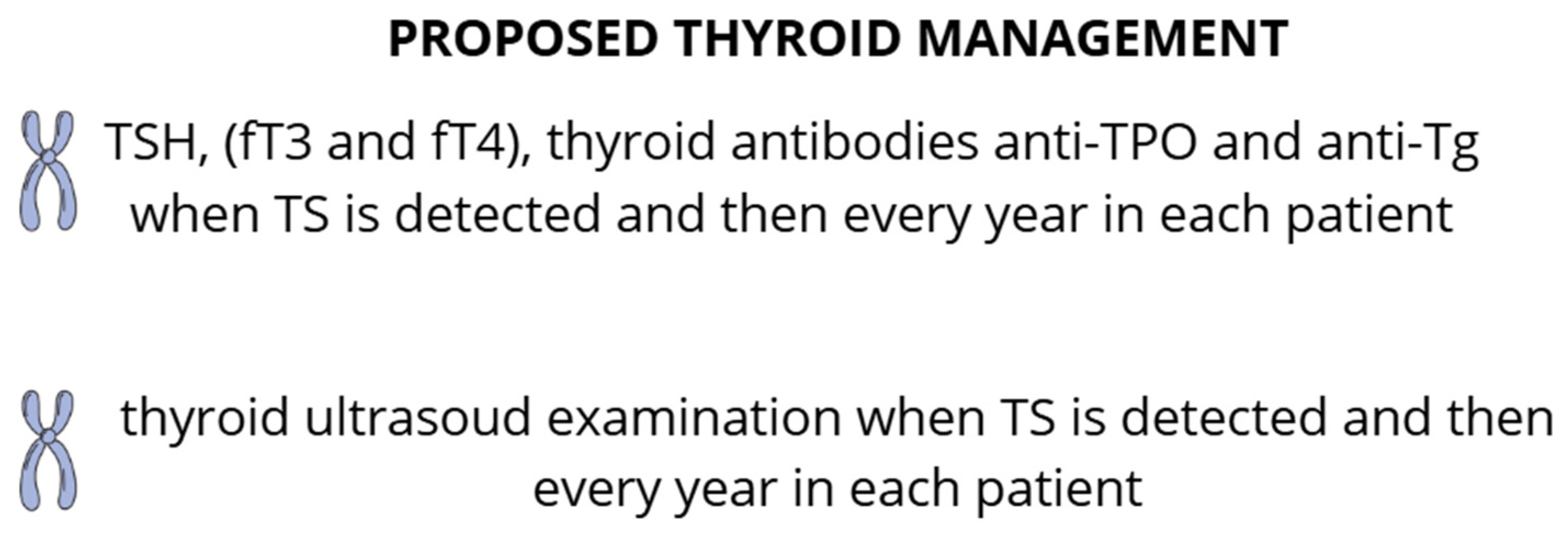
Due to the significant risk of thyroiditis in patients with TS, appropriate monitoring is essential in order to detect the disease as early as possible and to initiate treatment. According to the National Care Protocol [57], anti-TPO Ab and TSH ± fT4 should be measured at the time of diagnosis in patients older than four years of age, and subsequently the test should be repeated annually. Thyroid ultrasound should be performed when abnormalities in thyroid parameters (such as TSH, thyroid hormones, anti-thyroid antibodies) are observed. In the case of hypothyroidism, L-thyroxine replacement therapy should be introduced, and thyroid parameters should be tested every 6 months in childhood and every 12 months in adulthood. Antithyroid drugs may be used in cases of Graves’ disease [57].

2.4. Future Prospects

Given the karyotype distribution, congenital anomalies as well as comorbidities, patients with TS require close and complex medical surveillance throughout their entire lives.
Our systematic and critical review highlights the fact that thyroid diseases are common in the TS population [10]. Screening for hypothyroidism with measurement of TSH is recommended every 1 year, starting at 2 years of age and continuing through adulthood, as well as in cases when new symptoms appear. If TSH is increased, testing for anti-thyroid antibodies seems to be warranted [58].
Many of the discussed comorbidities associated with Turner syndrome appear to have a genetic basis of (at least partially) known etiology. Tests should include mutations which affect, or have a high probability of affecting, the development of diseases, such as: TIMP1 and TIMP3 [59], SHOX [42,60], CLTRN [61,62], IL3RA [39], CSF2RA [39], KDM6A [63] and BDNF [64].
Moreover, it is crucial to conduct further research into potential mechanisms involved in the development of hypothyroidism in patients with Turner syndrome and to perform larger-scale placebo-controlled trials. Figure 2 shows the proposed thyroid management for patients with TS that should facilitate early detection of thyroid diseases and, consequently, the initiation of treatment in the early stages of the disease.

2.5. Limitations

The main limitation of our analysis involves the lack of data on serum-free triiodothyronine levels, which should also be tested in patients suffering from TS. Additionally, there is no differentiation in terms of the number of patients who showed positive anti-TPO and anti-Tg levels. The sources only report the total number of antithyroid-positive patients, as well as average antibody levels.

3. Materials and Methods

3.1. Search Strategy

The presented review is designed following the PRISMA 2020 guidelines (Supplementary Materials). A search of PubMed, Scopus, Cochrane Library, and Google Scholar was conducted throughout July 2024. The search terms included “Turner syndrome”, “anti-TPO”, “anti-Tg”, “autoimmune thyroid disorders”, “TSH”, and “hypothyroidism”, which were used as isolated entries, as well as entered in various combinations. The first selection of research papers was based on the study title and the keywords. Overall, 1936 papers were found, which had been published since the beginning of TS research until July 2024. Such a broad time span stems from the fact that even the oldest manuscripts were significant for obtaining as much data as possible. The country of origin and form of the publications were irrelevant. Two analysts, working independently, verified the search engine results to select the most relevant studies. In total, the presented systematic review comprised 8 studies, and the study protocol was registered (ID: CRD42023471448).

3.2. Inclusion and Exclusion Criteria

In order to meet the inclusion criteria for our review, a given research paper had to include the mean and standard deviation for the parameters listed above. Moreover, the values had to be provided for both a study group, which was represented by female patients with Turner syndrome, and a control group, which consisted of healthy women.
In addition, our review incorporated laboratory findings regarding serum thyroid peroxidase antibodies (anti-TPO) and thyroglobulin antibodies (anti-TG), as well as thyroid function including thyrotropin (TSH), free thyroxine (fT4), total thyroxine (TT4), and total triiodothyronine (TT3) in female TS patients. We also included thyroid ultrasound findings, i.e., echogenicity and volume, as well as the diagnosis of Hashimoto’s disease, hypothyroidism, and hyperthyroidism.

3.3. Data Extraction and Quality Assessment

Two researchers independently extracted the following data from each paper: the year of publication, the study and control group size, types and results of studies performed, and clinical status of thyroid patients. Data were extracted using Microsoft Office Excel. The results are shown in Figure 3.
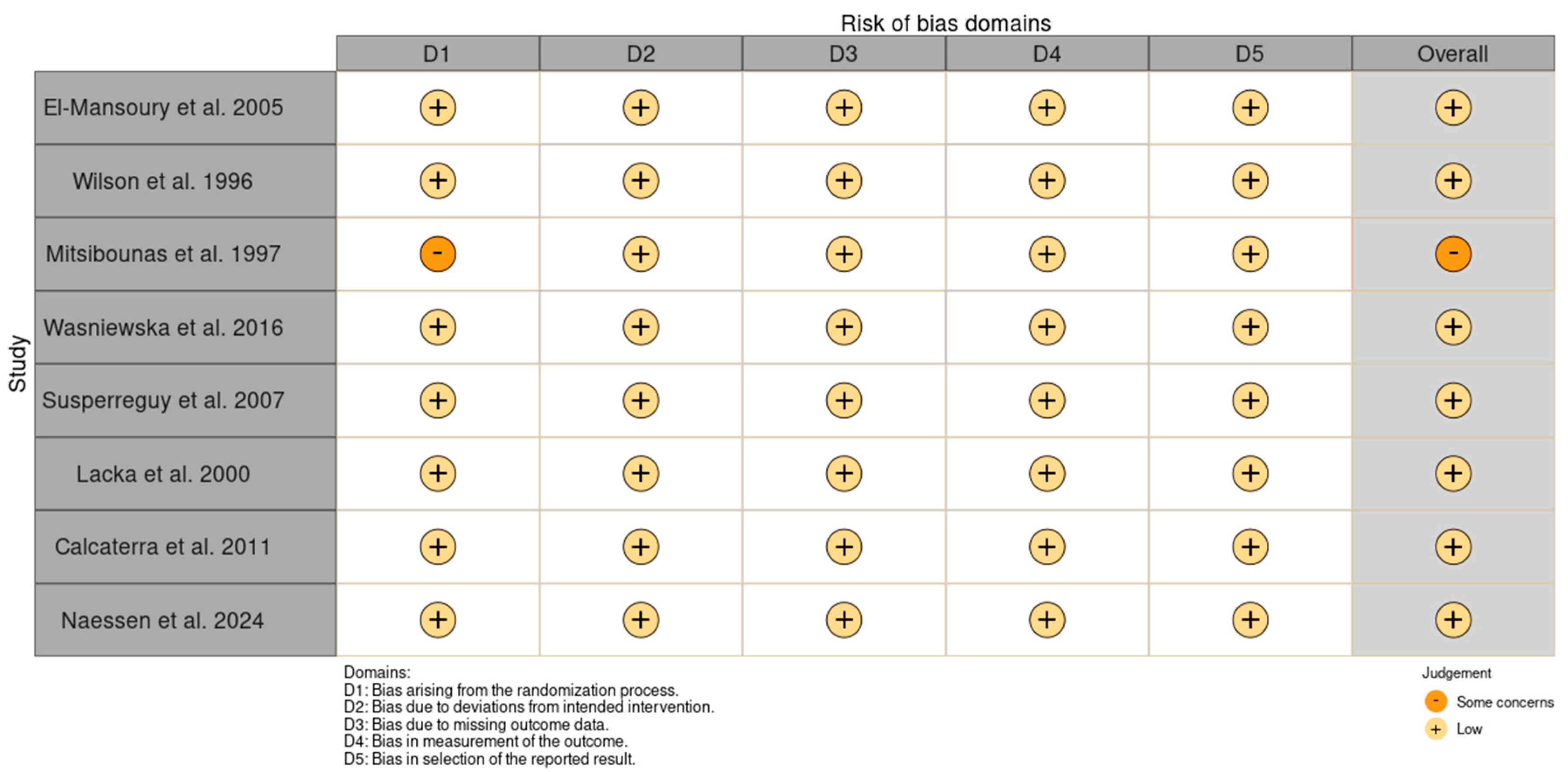
Bias assessment was conducted using the RoB 2 tool to determine the validity of the data collected from the studies. Two researchers independently answered the signaling questions for each paper. The risk of bias was used to draw conclusions from the accessed data. The results are presented in Figure 4.

4. Conclusions

Thyroiditis remains one of the most common comorbidities in patients with TS. Therefore, regular thyroid monitoring is vital in this group of patients. Since preventive screening is rarely performed, a large number of patients remain undiagnosed, or receive treatment only at a late stage of the disease. Therefore, we suggested a thyroid screening regimen in patients suffering from TS, which would allow for early detection of the disease and the implementation of relevant treatment. Simultaneously, we emphasize the significance of the genetic factors in the prevention of comorbidities, as it appears that gene mutations may account for their presence in TS women. However, further randomized studies are essential to conclusively determine whether genes play a role in the development of thyroid diseases. The abovementioned studies would be instrumental in identifying the parameters that predispose TS female patients to the disease, thus making it possible to perform active screening and to mitigate risk factors at an early stage. Consequently, the risk of the clinical consequences of hypothyroidism in this group of patients could be diminished, improving the quality of patients’ lives.

Supplementary Materials

The following supporting information can be downloaded at https://www.mdpi.com/article/10.3390/ijms252312937/s1 [65].

Author Contributions

Conceptualization, K.L.; methodology K.L., N.P., and M.M.; software, N.P. and M.M.; validation, N.P. and M.M.; formal analysis, K.L., N.P. and M.M.; investigation, K.L., N.P., A.G., A.M. and M.M.; resources, K.L. and N.P.; data curation K.L., N.P. and M.M.; writing—original draft preparation, N.P., A.G. and A.M.; writing—review and editing, K.L. and M.M.; visualization, N.P.; supervision, K.L. and M.M.; project administration, N.P.; funding acquisition, K.L. and M.M. All authors have read and agreed to the published version of the manuscript.

Funding

This research received no external funding.

Conflicts of Interest

The authors declare no conflicts of interest.

Abbreviations

anti-Tgantithyroglobulin antibody
anti-TPOthyroid peroxidase antibody
BDNFbrain-derived neurotrophic factor
BMIbody mass index
CLTRNcollectrin amino acid transport regulator
CSF2RAcolony-stimulating factor 2 receptor subunit alpha
FOXP3forkhead box P3
fT3free triiodothyronine
fT4free thyroxine
GHgrowth hormone
HDLhigh density lipoproteins
IGF-Iinsulin-like growth factor 1
IL3RAinterleukin 3 receptor alpha
KDM6Alysine demethylase 6A
L-thyroxinelevothyroxine
PTPN22protein tyrosine phosphatase non-receptor type 22
SHOXshort stature homeobox-containing gene
TIMP 1metallopeptidase inhibitor 1
TIMP 3metallopeptidase inhibitor 3
TSTurner syndrome
TSHthyroid-stimulating hormone
TT3total triiodothyronine
TT4total thyroxine
USGultrasonography

References

  1. Bondy, C.A. Care of girls and women with Turner syndrome: A guideline of the Turner Syndrome Study Group. J. Clin. Endocrinol. Metab. 2007, 92, 10–25. [Google Scholar] [CrossRef] [PubMed]
  2. Gravholt, C.H.; Andersen, N.H.; Conway, G.S.; Dekkers, O.M.; Geffner, M.E.; Klein, K.O.; Lin, A.E.; Mauras, N.; Quigley, C.A.; Rubin, K.; et al. Clinical practice guidelines for the care of girls and women with Turner syndrome: Proceedings from the 2016 Cincinnati International Turner Syndrome Meeting. Clin. Endocrinol. 2017, 55, 223–226. [Google Scholar] [CrossRef] [PubMed]
  3. Ćatović, A.; Kendić, S. Cytogenetics findings at Turner Syndrome and their correlation with clinical findings. Bosn. J. Basic Med. Sci. 2005, 5, 54–58. [Google Scholar] [CrossRef] [PubMed]
  4. Czyzyk, A.; Lacka, K.; Krawczynski, M.; Andrzejewska, J.; Wojda, A.; Skolozdrzy, J.; Latos-Bielanska, A. Analysis of phenotype and karyotype of children born from mothers with 45, X/46, XX mosaicism. Endocrinologist 2010, 20, 283–285. [Google Scholar] [CrossRef]
  5. Saenger, P. Turner’s syndrome. NEJM 1996, 335, 1749–1754. [Google Scholar] [CrossRef]
  6. Lacka, K. Turner’s Syndrome-correlation between kariotype and fenotype. Endokrynol. Pol. 2005, 6, 986–993. [Google Scholar]
  7. Turner, H.H. A syndrome of infantilism, congenital webbed neck and cubitus vulgaris. Endocrinologist 1938, 28, 566–574. [Google Scholar] [CrossRef]
  8. Morgagni, J.B. Desedibus et causis Morborum; Ex Typographia Remondiniana: Venice, Italy, 1761; Volume 6, p. 462. [Google Scholar]
  9. Gravholt, C.H. Epidemiological, endocrine and metabolic features in Turner syndrome. Arq. Bras. Endocrinol. Metabol. 2005, 49, 145–156. [Google Scholar] [CrossRef]
  10. Shankar, R.K.; Backeljauwa, P.F. Current best practice in the management of Turner syndrome. Ther. Adv. Endocrinol. Metab. 2018, 9, 33–40. [Google Scholar] [CrossRef]
  11. Chen, R.M.; Zhang, Y.; Yang, X.H.; Lin, X.Q.; Yuan, X. Thyroid disease in Chinese girls with Turner syndrome. J. Pediatr. Endocrinol. Metab. 2015, 28, 201–205. [Google Scholar] [CrossRef]
  12. Elsheikh, M.; Wass, J.A.; Conway, G.S. Autoimmune thyroid syndrome in women with Turner syndrome-the association with karyotype. Clin. Endocrinol. 2001, 55, 223–226. [Google Scholar] [CrossRef] [PubMed]
  13. Grossi, A.; Palma, A.; Zanni, G.; Novelli, A.; Loddo, S.; Cappa, M.; Fierabracci, A. Multiorgan autoimmunity in a Turner syndrome patient with partial monosomy 2q and trisomy 10p. Gene 2013, 515, 439–443. [Google Scholar] [CrossRef] [PubMed]
  14. Gawlik, A.; Gawlik, T.; Januszek-Trzciakowska, A.; Patel, H.; Małecka-Tendera, E. Incidence and dynamics of thyroid dysfunction and thyroid autoimmunity in girls with Turner’s syndrome: A long-term follow-up study. Horm. Res. Paediatr. 2011, 76, 314–320. [Google Scholar] [CrossRef] [PubMed]
  15. Aversa, T.; Messina, M.F.; Mazzanti, L.; Salerno, M.; Mussa, A.; Faienza, M.F.; Scarano, E.; De Luca, F.; Wasniewska, M. The association with Turner syndrome significantly affects the course of Hashimoto’s thyroiditis in children, irrespective of karyotype. Endocrine 2015, 50, 777–782. [Google Scholar] [CrossRef] [PubMed]
  16. Cui, X.; Cui, Y.; Shi, L.; Luan, J.; Zhou, X.; Han, J. A basic understanding of Turner syndrome: Incidence, complications, diagnosis, and treatment. Intractable Rare Dis. Res. 2018, 7, 223–228. [Google Scholar] [CrossRef]
  17. Turner, S. NORD (National Organization for Rare Disorders). Available online: https://rarediseases.org/rarediseases/turner-syndrome/ (accessed on 20 September 2024).
  18. Kikkeri, N.S.; Nagalli, S. Turner Syndrome. In StatPearls. Available online: https://www.ncbi.nlm.nih.gov/books/NBK554621/ (accessed on 20 September 2024).
  19. Sävendahl, L.; Davenport, M.L. Delayed diagnoses of Turner’s syndrome: Proposed guidelines for change. J. Pediatr. 2000, 137, 455–459. [Google Scholar] [CrossRef]
  20. Barrenäs, M.L.; Nylen, O.; Hanson, C. The influence of karyotype on the auricle, otitis media and hearing in Turner syndrome. Hear. Res. 1999, 138, 163–170. [Google Scholar] [CrossRef]
  21. Bianchi, I.; Lleo, A.; Gershwin, M.E.; Invernizzi, P. The X chromosome and immune associated genes. J. Autoimmun. 2012, 38, 87–92. [Google Scholar] [CrossRef]
  22. Gawlik, A.; Sołtysik, K.; Berdej-Szczot, E. Autoimmune disease profile in Turner syndrome patients. Endokrynol. Pediatr. 2013, 12, 57–68. [Google Scholar] [CrossRef]
  23. Libert, C.; Dejager, L.; Pinheiro, I. The X chromosome in immune functions: When a chromosome makes the difference. Nat. Rev. Immunol. 2010, 10, 594–604. [Google Scholar] [CrossRef]
  24. Gawlik, A.M.; Berdej-Szczot, E.; Blat, D.; Klekotka, R.; Gawlik, T.; Blaszczyk, E.; Hankus, M.; Malecka-Tendera, E. Immunological profile and predisposition to autoimmunity in girls with Turner syndrome. Front. Endocrinol. 2018, 9, 307. [Google Scholar] [CrossRef] [PubMed]
  25. Chiovato, L.; Larizza, D.; Bendinelli, G.; Tonacchera, M.; Marinó, M.; Mammoli, C.; Lorini, R.; Severi, F.; Pinchera, A. Autoimmune hypothyroidism and hyperthyroidism in patients with Turner’s syndrome. Eur. J. Endocrinol. 1996, 134, 568–575. [Google Scholar] [CrossRef] [PubMed]
  26. Naessén, S.; Eliasson, m.; Berntorp, K.; Kitlinski, M.; Trimpou, P.; Amundson, E.; Thunström, S.; Ekman, B.; Wahlberg, J.; Karlsson, A.; et al. Autoimmune Disease in Turner Syndrome in Sweden: An up to 25 Years’ Controlled Follow-up Study. J. Clin. Endocrinol. Metab. 2024, 109, e602–e612. [Google Scholar] [CrossRef] [PubMed]
  27. Sylvén, L.; Hagenfeldt, K.; Bröndrum-Nielsen, K.; von-Schoultz, B. Middle-aged women with Turner´s syndrome. Medical status, hormonal treatment, and social life. Acta Endocrinol. 1991, 125, 359–365. [Google Scholar]
  28. Chelikani, V.; Rao, D.N.; Balmuri, S.; Arida, A.K. A Rare Case of Hashimoto’s Encephalopathy With Mosaic Turner Syndrome. Cureus 2022, 20, e28215. [Google Scholar] [CrossRef]
  29. Pempera, N.; Miedziaszczyk, M.; Lacka, K. Difficulties in the Diagnostics and Treatment of Hashimoto’s Encephalopathy-A Systematic and Critical Review. Int. J. Mol. Sci. 2024, 25, 7101. [Google Scholar] [CrossRef]
  30. Mitsibounas, D.N.; Kosmaidou, Z.; Kontoleon, P.; Deligeoroglou, E.; Liberatou, E. Hyperlipidemia and presence of thyroid autoantibodies in girls with Turner’s syndrome or mosaic variance. J. Pediatr. Adolesc. Gynecol. 1997, 10, 133–139. [Google Scholar] [CrossRef]
  31. Wasniewska, M.; Salerno, M.; Corrias, A.; Mazzanti, L.; Matarazzo, P.; Corica, D.; Aversa, T.; Messina, M.F.; De Luca, F.; Valenzise, M. The Evolution of Thyroid Function after Presenting with Hashimoto Thyroiditis Is Different between Initially Euthyroid Girls with and Those without Turner Syndrome. Horm. Res. Paediatr. 2016, 86, 403–409. [Google Scholar] [CrossRef]
  32. Susperreguy, S.; Miras, M.B.; Montesinos, M.M.; Mascanfroni, I.D.; Muñoz, L.; Sobrero, G.; Silvano, L.; Masini-Repiso, A.M.; Coleoni, A.H.; Targovnik, H.M.; et al. Growth hormone (GH) treatment reduces peripheral thyroid hormone action in girls with Turner syndrome. Clin. Endocrinol. 2007, 67, 629–636. [Google Scholar] [CrossRef]
  33. El-Mansoury, M.; Bryman, I.; Berntorp, K.; Hanson, C.; Wilhelmsen, L.; Landin-Wilhelmsen, K. Hypothyroidism is common in turner syndrome: Results of a five-year follow-up. J. Clin. Endocrinol. Metab. 2005, 90, 2131–2135. [Google Scholar] [CrossRef]
  34. Wilson, R.; Chu, C.E.; Donaldson, M.D.C.; Thomson, J.A.; McKillop, J.H.; Connor, J.M. An increased incidence of thyroid antibodies in patients with Turner’s syndrome and their first degree relatives. Autoimmun 1996, 25, 47–52. [Google Scholar] [CrossRef] [PubMed]
  35. Łacka, K.; Wawrzyniak, M.; Andrzejewska, J.; Krysińska, I.; Ruchała, M.; Bauman-Antczak, A. Evaluation of thyroid structure and function in patients with Turner syndrome. Pol. Arch. Med. Wewn. 2000, 104, 583–589. [Google Scholar] [PubMed]
  36. Calcaterra, V.; Klersy, C.; Muratori, T.; Caramagna, C.; Brizzi, V.; Albertini, R.; Larizza, D. Thyroid ultra-sound in patients with Turner syndrome: Influence of clinical and auxological parameters. J. Endocrinol. Invest 2011, 34, 260–264. [Google Scholar] [CrossRef] [PubMed]
  37. Cogni, G.; Chiovato, L. An overview of the pathogenesis of thyroid autoimmunity. Hormones 2013, 39, 19–29. [Google Scholar] [CrossRef]
  38. Atria, A.; Sanz, R.; Donoso, S. Necropsy study of a case of Turner’s syndrome, Case report. J. Clin. Endocrinol. 1948, 8, 397–405. [Google Scholar] [CrossRef]
  39. Gravholt, C.H.; Viuff, M.H.; Brun, S.; Stochholm, K.; Andersen, N.H. Turner syndrome: Mechanisms and management. Nat. Rev. Endocrinol. 2019, 15, 601–614. [Google Scholar] [CrossRef]
  40. Sharland, M.; Burch, M.; McKenna, W.M.; Paton, M.A. A clinical study of Noonan syndrome. Arch. Dis. Child. 1992, 67, 178–183. [Google Scholar] [CrossRef]
  41. Vesterhus, P.; Aarskog, D. Noonan’s syndrome and autoimmune thyroiditis. J. Pediatr. 1973, 83, 237–240. [Google Scholar] [CrossRef]
  42. Fukami, M.; Seki, A.; Ogata, T. SHOX Haploinsufficiency as a cause of syndromic and nonsyndromic short stature. Mol. Syndromol. 2016, 7, 3–11. [Google Scholar] [CrossRef]
  43. Mohamed, S.O.O.; Elkhidir, I.H.E.; Abuzied, A.I.H.; Noureddin, A.A.M.H.; Ibrahim, G.A.A.; Mahmoud, A.A.A. Prevalence of autoimmune thyroid diseases among the Turner Syndrome patients: Meta-analysis of cross sectional studies. BMC Res. Notes 2018, 11, 842. [Google Scholar] [CrossRef]
  44. Brix, T.; Hegedüs, L. Twin studies as a model for exploring the aetiology of autoimmune thyroid disease. Clin. Endocrinol. 2012, 76, 457–464. [Google Scholar] [CrossRef] [PubMed]
  45. Fukuda, I.; Hizuka, N.; Kurimoto, M.; Morita, J.; Tanaka, S.; Yamakado, Y.; Takano, K. Autoimmune thyroid diseases in 65 Japanese women with Turner syndrome. Endocr. J. 2009, 56, 983–986. [Google Scholar] [CrossRef] [PubMed]
  46. Weeke, J.; Jens, S. Christiansen. Thyroid function during growth hormone therapy. Horm. Res. 1992, 38, 63–67. [Google Scholar]
  47. Jonklaas, J.; Bianco, A.C.; Bauer, A.J.; Burman, K.D.; Cappola, A.R.; Celi, F.S.; Cooper, D.S.; Kim, B.W.; Peeters, R.P.; Rosenthal, M.S.; et al. Guidelines for the treatment of hypothyroidism: Prepared by the american thyroid association task force on thyroid hormone replacement. Thyroid 2014, 24, 1670–1751. [Google Scholar] [CrossRef] [PubMed]
  48. Somwaru, L.L.; Rariy, C.M.; Arnold, A.M.; Cappola, A.R. The natural history of subclinical hypothyroidism in the elderly: The cardiovascular health study. J. Clin. Endocrinol. Metab. 2012, 97, 1962–1969. [Google Scholar] [CrossRef]
  49. Vanderpump, M.P.; Tunbridge, W.M.; French, J.M.; Appleton, D.; Bates, D.; Clark, F.; Grimley Evans, J.; Hasan, D.M.; Rodgers, H.; Tunbridge, F.; et al. The incidence of thyroid disorders in the community: A twenty-year follow-up of the whickham survey. Clin. Endocrinol. 1995, 43, 55–68. [Google Scholar] [CrossRef]
  50. Jorde, R.; Waterloo, K.; Storhaug, H.; Nyrnes, A.; Sundsfjord, J.; Jenssen, T.G. Neuropsychological function and symptoms in subjects with subclinical hypothyroidism and the effect of thyroxine treatment. J. Clin. Endocrinol. Metab. 2006, 91, 145–153. [Google Scholar] [CrossRef]
  51. Almandoz, J.P.; Gharib, H. Hypothyroidism: Etiology, diagnosis, and management. Med. Clin. N. Am. 2012, 96, 203–221. [Google Scholar] [CrossRef]
  52. Pearce, S.H.; Brabant, G.; Duntas, L.H.; Monzani, F.; Peeters, R.P.; Razvi, S.; Wemeau, J.L. 2013 eta guideline: Management of subclinical hypothyroidism. Eur. Thyroid J. 2013, 2, 215–228. [Google Scholar] [CrossRef]
  53. Jonklaas, J.; Razvi, S. Reference intervals in the diagnosis of thyroid dysfunction: Treating patients not numbers. Lancet Diabetes Endocrinol. 2019, 7, 473–483. [Google Scholar] [CrossRef]
  54. Razvi, S.; Korevaar, T.I.M.; Taylor, P. Trends, determinants, and associations of treated hypothyroidism in the United Kingdom, 2005–2014. Thyroid 2019, 29, 174–182. [Google Scholar] [CrossRef] [PubMed]
  55. Syamsunder, A.N.; Pal, G.K.; Pal, P.; Kamalanathan, C.S.; Parija, S.C.; Nanda, N. Association of sympathovagal imbalance with cardiovascular risks in overt hypothyroidism. N. Am. J. Med. Sci. 2013, 5, 554. [Google Scholar] [PubMed]
  56. Biondi, B.; Klein, I. Hypothyroidism as a risk factor for cardiovascular disease. Endocrine 2004, 24, 1–14. [Google Scholar] [CrossRef]
  57. Fiot, E.; Alauze, B.; Donadille, B.; Samara-Boustani, D.; Houang, M.; De Filippo, G.; Bachelot, A.; Delcour, C.; Beyler, C.; Bois, E.; et al. Turner syndrome: French National Diagnosis and Care Protocol (NDCP.; National Diagnosis and Care Protocol). Orphanet. J. Rare Dis. 2022, 17, 261. [Google Scholar] [CrossRef] [PubMed]
  58. Gravholt, C.H.; Niels, H.A.; Christin-Maitre, S.; Shanlee, M.D.; Duijnhouwer, A.; Gawlik, A.; Maciel-Guerra, A.T.; Gutmark-Little, I.; Fleischer, K.; Hong, K.; et al. Clinical practice guidelines for the care of girls and women with Turner syndrome: Proceedings from the 2023 Aarhus International Turner Syndrome Meeting. Eur. J. Endocrinol. 2023, 6, G53–G151. [Google Scholar]
  59. Corbitt, H.; Morris, S.A.; Gravholt, C.H.; Mortenson, K.H.; Tippner-Hedges, R.; Silberbach, M.; Maslen, C.L. TIMP3 and TIMP1 are risk genes for bicuspid aortic valve and aortopathy in Turner syndrome. PLoS Genet. 2018, 14, e1007692. [Google Scholar] [CrossRef]
  60. Khan, N.; Farooqui, A.; Ishrat, R. Turner Syndrome where are we? Orphanet J. Rare Dis. 2024, 19, 314. [Google Scholar] [CrossRef]
  61. Farooqui, A.; Tazyeen, S.; Ahmed, M.M.; Alam, A.; Ali, S.; Malik, M.Z.; Ali, S.; Ishrat, R. Assessment of the key regulatory genes and their Interologs for Turner Syndrome employing network approach. Sci. Rep. 2018, 8, 10091. [Google Scholar] [CrossRef]
  62. Pajenda, S.; Wagner, L.; Gerges, D.; Herkner, H.; Tevdoradze, T.; Mechtler, K.; Schmidt, A.; Winnicki, W. Urinary Collectrin (TMEM27) as Novel Marker for Acute Kidney Injury. Life 2022, 12, 1391. [Google Scholar] [CrossRef]
  63. Bonnard, Å.; Bark, R.; Hederstierna, C. Clinical update on sensorineural hearing loss in Turner syndrome and the X-chromosome. Am. J. Med. Genet. C Semin. Med. Genet. 2019, 181, 18–24. [Google Scholar] [CrossRef]
  64. Farooqui, A.; Anwer, A.; Alam, A.; Bagabir, S.A.; Haque, S.; Khadgawat, R.; Kazim, S.N.; Ali, S.; Ishrat, R. Brain-derived neurotrophic factor G196A (rs6265) gene polymorphism increases Turner syndrome susceptibility. Biotechnol. Genet. Eng. Rev. 2023, 39, 882–896. [Google Scholar] [CrossRef]
  65. Page, M.J.; McKenzie, J.E.; Bossuyt, P.M.; Boutron, I.; Hoffmann, T.C.; Mulrow, C.D.; Shamseer, L.; Tetzlaff, J.M.; Akl, E.A.; Brennan, S.E.; et al. The PRISMA 2020 statement: An updated guideline for reporting systematic reviews. Syst. Rev. 2021, 10, 89. [Google Scholar] [CrossRef]
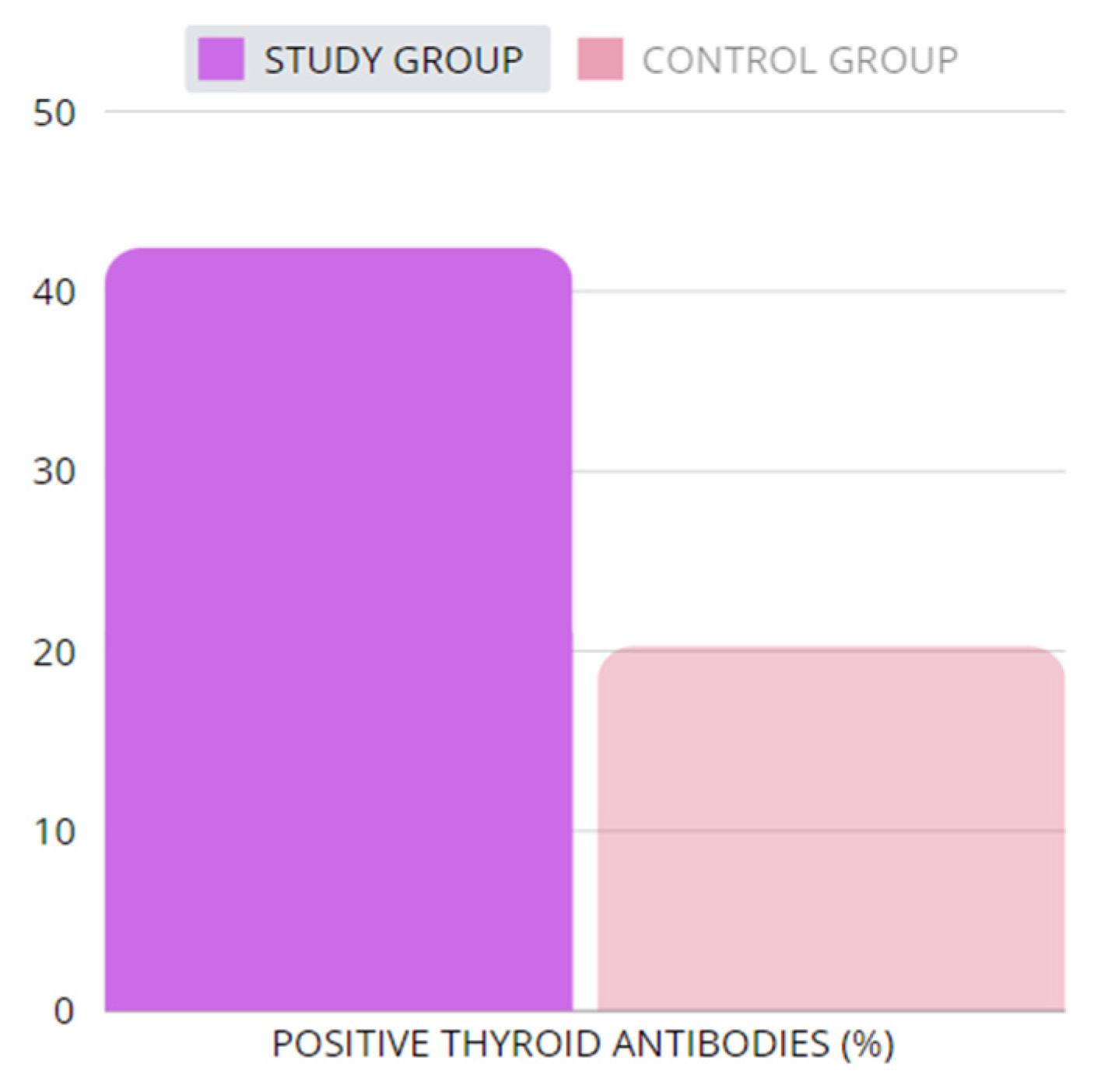
Figure 1. The percentage of patients with positive thyroid antibodies. The study group for the purpose of our review was calculated by means of adding the study groups from the relevant sources [26,30,31,32,33,34,35]; the same procedure was adopted for the control group. We excluded source [36] from this analysis, due to the lack of information regarding patients with positive thyroid antibodies. In total, 806 patients were included from the study groups (patients with TS), where in 342 cases, positive thyroid antibodies were found. The control group consisted of 887 individuals in total, among whom 180 presented with positive thyroid antibodies. The sources [26,30,31,32,33,34,35] do not provide a division according to antibody types, and hence there is no information if the anti-TPO and/or anti-Tg were elevated in patients. Table 4 presents levels of thyroid antibodies.
Ijms 25 12937 g001
Figure 2. Proposed thyroid management for patients with TS.
Ijms 25 12937 g002
Figure 3. PRISMA flowchart. We performed a PubMed, Scopus, Cochrane Library, and Google Scholar search throughout July 2024. The oldest articles were also significant in order to obtain as much data as possible. Search terms included “Turner syndrome”, “anti-TPO”, “anti-Tg”, “autoimmune thyroid disorders”, “TSH”, and “hypothyroidism”, used as isolated keywords and in combination. The flowchart was performed in order to obtain information about thyroid function in women with Turner syndrome.
Ijms 25 12937 g003
Figure 4. Bias assessment using version 2 of the Cochrane risk-of-bias tool for randomized trials (RoB 2) [26,30,31,32,33,34,35,36].
Ijms 25 12937 g004
Table 1. Presentation of cases analyzed in sources [26,30,31,32,33,34,35,36]. The table includes the number of study and control groups, information regarding Hashimoto diagnosis in the analyzed groups, age, height, weight, and calculated body mass index (BMI) characterizing each group.
Author and YearEl-Mansoury et al., 2005 [33]Wilson et al., 1996 [34]Mitsibounas et al., 1997 [30]Wasniewska et al., 2016 [31]Susperreguy et al., 2007 [32]Lacka et al., 2000 [35]Calcaterra et al., 2011 [36]Naessen et al., 2024 [26]
Group (n)study9160296620 (10 treated with GH)3773503
control2285722132103793401
Hashimoto diagnosis (n)studyNo data3No data66No dataNo data32No data
controlNo dataNo dataNo data132No dataNo data0No data
Age (y)study37.7 ± 11No data22.55 ± 6.3413.0 (4.5–17.9)9.6 ± 3.5 (TS)
10.9 ± 3.1(GH)
13.8 ± 625.01 ± 10.4327.6 ± 11.5
control37.3 ± 9No data22.05 ± 2.29910.6 (2.8–15.0)9.9 ± 2.8No data1.95 to 37.235.5 ± 5.7
Height (cm)study149.4 ± 7No data148 ± 9No dataNo dataNo dataNo data153.6 ± 7.2
control165.7 ± 6.6No data158 ± 5.5No dataNo dataNo dataNo data166.3 ± 6.6
Weight (kg)study56.4 ± 12.4No data53.16 ± 10.12No dataNo dataNo dataNo data58.9 ± 11.9
control65.4 ± 10.3No data52.86 ± 4.663No dataNo dataNo dataNo data65.2 ± 10.2
Body mass index (BMI) (kg/m2)study25.3 ± 4.7No data23.75 ± 3.64No dataNo dataNo dataNo data25.1 ± 5.7
control23.8 ± 3.7No data21.90 ± 1.681No dataNo dataNo dataNo data23.6 ± 3.4
Table 2. Parameters according to sources [26,30,31,32,33,34,35,36].
Author and YearEl-Mansoury et al., 2005 [33]Wilson et al., 1996 [34]Mitsibounas et al., 1997 [30]Wasniewska et al., 2016 [31]Susperreguy et al., 2007 [32]Lacka et al., 2000 [35]Calcaterra et al., 2011 [36]Naessen et al., 2024 [26]
Group (n)study9160296620 (10 treated with GH)3773503
control2285722132103793401
Serum TSH (U/mL)study2.5 ± 2.32.5 ± 2.52.468 ± 1.20032.8 (0.6–4.8)2.5 ± 0.63.9 ± 6.61No data3.18 ± 7.78
control1.2 ± 0.72.4 ± 1.71.95 ± 1.452.5 (0.47–4.9)2.8 ± 1.0No dataNo data1.10 ± 0.63
Serum fT4 (ng/dL)study1.07 ± 0.2No dataNo data1.173 ± 0.2251.38 ± 0.231.71 ± 2.1No data1.227 ± 0.326
control1.16 ±0.2No dataNo data1.196 ± 0.281.45 ± 0.28No dataNo data1.158 ± 0.194
Serum TT3 (nmol/L)studyNo dataNo data1.59 ± 0.35No data1.7 ± 0.32.6 ± 0.1No dataNo data
controlNo dataNo data1.88 ± 284No data1.8 ± 0.3No dataNo dataNo data
Serum TT4 (nmol/L)studyNo data104 ± 25124.131 ± 24.3063No data128 ± 23119.7 ± 46.3No dataNo data
controlNo data129 ± 44119.37 ± 15.773No data128 ± 25No dataNo dataNo data
Table 3. Hypo-/hyper/euthyroidism in patients with Turner syndrome [26,30,31,32,33,34,35,36].
Author and YearEl-Mansoury et al., 2005 [33]Wilson et al., 1996 [34]Mitsibounas et al., 1997 [30]Wasniewska
et al., 2016 [31]
Susperreguy et al., 2007 [32]Lacka et al., 2000 [35]Calcaterra et al., 2011 [36]Naessen et al., 2024 [26]
Group (n)study9160296620 (10 treated with GH)3773503
control2285722132103793401
Euthyroidism (n)studyNo data60No data66202773No data
controlNo data57No data13210No data93No data
Hyperthyroidism (n)study3000010No data
control00000No data0No data
Hypothyroidism (n)study2300008037
control50000No data00
Table 4. Thyroid antibodies in patients with TS [26,30,31,32,33,34,35,36].
Author and YearEl-Mansoury et al., 2005 [33]Wilson et al., 1996 [34]Mitsibounas et al., 1997 [30]Wasniewska et al., 2016 [31]Susperreguy et al., 2007 [32]Lacka et al., 2000 [35]Calcaterra et al., 2011 [36]Naessen et al., 2024 [26]
Group (n)study9160296620 3773503
control2285722132103793401
Positive thyroid antibodies (n)study2518466023No data206
control01013206No data41
Serum antiTPO level (mIU/L)study475 ± 960No dataNo data119 (5–6.4)No data10.9 ± 12.7No dataNo data
control<0.01No dataNo data374 (31–29.95)No dataNo dataNo dataNo data
Serum antiTg level (mIU/L)studyNo dataNo dataNo data230 (5–4.9)No data3300 ± 9100No dataNo data
controlNo dataNo dataNo data227 (10–6.4)No dataNo dataNo dataNo data
Table 5. Difference in thyroid volume in TS patients in comparison to healthy individuals [13,41].
Author and YearLacka et al., [35]Calcaterra et al., 2011 [36]
USG volume (cm3)study11.03 ± 5.07.1 ± 2.4
control16.98 ± 9.16.1 ± 2.2
Abnormal echogenicity (n)study1766
controlNo dataNo data
Disclaimer/Publisher’s Note: The statements, opinions and data contained in all publications are solely those of the individual author(s) and contributor(s) and not of MDPI and/or the editor(s). MDPI and/or the editor(s) disclaim responsibility for any injury to people or property resulting from any ideas, methods, instructions or products referred to in the content.