Article

In Silico Hydrolysis of Lupin (Lupinus angustifolius L.) Conglutins with Plant Proteases Releases Antihypertensive and Antidiabetic Peptides That Are Bioavailable, Non-Toxic, and Gastrointestinal Digestion Stable

by
Jesús Gilberto Arámburo-Gálvez
1,
Raúl Tinoco-Narez-Gil
1,
José Antonio Mora-Melgem
1,
Cesar Antonio Sánchez-Cárdenas
2,
Martina Hilda Gracia-Valenzuela
3,
Lilian Karem Flores-Mendoza
4,
Oscar Gerardo Figueroa-Salcido
2,* and
Noé Ontiveros
4,*
1
Nutrition Sciences Postgraduate Program, Faculty of Nutrition and Gastronomy Sciences, Autonomous University of Sinaloa, Culiacan 80019, Sinaloa, Mexico
2
Integral Postgraduate Program in Biotechnology, Faculty of Chemical and Biological Sciences, Autonomous University of Sinaloa, Ciudad Universitaria, Culiacan 80010, Sinaloa, Mexico
3
Laboratory for the Research and Detection of Biological Agents and Contaminants (CONAHCYT National Laboratory, LANIBIOC), Yaqui Valley Technological Institute, National Technological Institute of Mexico, Bácum 85276, Sonora, Mexico
4
Clinical and Research Laboratory (LACIUS, C.N., CONAHCYT National Laboratory, LANIBIOC), Deparment of Chemical, Biological, and Agricultural Sciences (DC-QB), Faculty of Biological and Health Sciences, University of Sonora, Navojoa 85880, Sonora, Mexico
*
Authors to whom correspondence should be addressed.
Int. J. Mol. Sci. 2024, 25(23), 12866; https://doi.org/10.3390/ijms252312866
Submission received: 2 October 2024 / Revised: 10 November 2024 / Accepted: 27 November 2024 / Published: 29 November 2024

Abstract

Lupin (Lupinus angustifolius L.) proteins are potential sources of bioactive peptides (LBPs) that can inhibit dipeptidyl peptidase IV (DPP-IV) and angiotensin I-converting enzyme (ACE-I) activity. However, the capacity of different enzymes to release LBPs, the pharmacokinetic and bioactivities of the peptides released, and their binding affinities with the active sites of DPP-IV and ECA-I are topics scarcely addressed. Therefore, we used in silico hydrolysis (BIOPEP-UWM platform) with various enzymes to predict the release of LBPs. Among the bioactive peptides identified in lupin proteins (n = 4813), 2062 and 1558 had DPP-IV and ACE-I inhibitory activity, respectively. Ficin, bromelain, and papain released the highest proportion of ACE-I (n = 433, 411, and 379, respectively) and DPP-IV (n = 556, 544, and 596, respectively) inhibitory peptides. LBPs with favorable pharmacokinetics and gastrointestinal stability tightly interacted with the active sites of ACE-I (–5.6 to –8.9 kcal/mol) and DPP-IV (–5.4 to –7.6 kcal/mol). Papain generated the most bioavailable LBPs (n = 459) with ACE-I (n = 223) and DPP-IV (n = 412) inhibitory activity. These peptides were non-toxic and gastrointestinal digestion stable. Notably, papain-based hydrolysis released some LBPs (n = 270) that inhibited both ACE-I and DPP-IV. Plant protease-based hydrolysis is a promising approach for producing lupin hydrolysates with ACE-I and DPP-IV inhibitory activities.

1. Introduction

Both type 2 Diabetes Mellitus (DM2) and hypertension commonly coexist and affect a large proportion of the adult population (up to 10.5% and 31.5%, respectively) [1,2]. Around 70% of adult diabetic patients could also have hypertension [3], increasing their risk of developing chronic heart disease and other complications [4]. In these comorbidity cases, angiotensin I-converting enzyme (ACE-I) inhibitors and angiotensin receptor blockers are the drugs of choice for treating hypertension [5]. Likewise, dipeptidyl peptidase-IV (DPP-IV) inhibitors are effective antidiabetic drugs with potential antihypertensive properties [5]. On one hand, ACE-I inhibition avoids the production of the potent vasoconstrictor angiotensin II as well as the cleaving of the vasodilator bradykinin [6], and, on the other hand, DPP-IV inhibition avoids cleaving incretin hormones such as glucose-dependent insulinotropic polypeptide and glucagon-like peptide 1, which help to control plasma glucose levels [7]. Thus, the combination of ACE-I and DPP-IV inhibitors should be considered the primary treatment for diabetic patients with hypertension. However, both ACE-I and DPP-IV inhibitor drugs can cause adverse effects in some patients [8,9], making desirable the use of side effect-free enzyme inhibitors.
DPP-IV and ACE-I inhibitory peptides have been proposed as safe therapeutic options for treating DM2 and hypertension, respectively [10,11,12]. Lupinus species are leguminous plants with high protein content [13] and are sources of ACE-I [14] and DPP-IV [15,16,17] inhibitory peptides. In fact, some peptides released after beta-conglutin hydrolysis with pepsin can inhibit up to 35% of DPP-IV activity [17]. Other lupin peptides can be released with alcalase or flavorzyme. These peptides have shown binding affinities from –8.9 to –10.7 kcal/mol and could inhibit ACE-I [18]. Beyond these findings, some issues related to lupin peptides are worth addressing in depth. Thus, the present study aimed to predict the ACE-I and DPP-IV inhibitory peptides released after conglutin hydrolysis with seven different enzymes (pepsin, trypsin, chymotrypsin, alcalase, papain, bromelain, and ficin) and gastrointestinal digestion to determine their binding affinities with the active sites of DPP-IV and ACE-I and their pharmacokinetic parameters using a comprehensive in silico approach [12,19]. The study is focused on conglutin proteins because they represent 95% of the lupin protein [20].

2. Results and Discussion

2.1. Biological Activity of Lupin Proteins

The BIOPEP tool “profiles of potential biological activity” was used to search for bioactive peptides encrypted in lupin proteins (α1-3, β1-7, δ1-4, and γ1-2 conglutin). The conglutin family accounts for around 95% of the total protein content of lupin seeds, and it is present in all species and varieties of lupin [21]. A total of 4813 lupin conglutin peptides showed biological activity (Table 1). Among these peptides, 2062 (42.8%) and 1558 (32.3%) showed the potential to inhibit DPP-IV or ACE-I, respectively. Interestingly, the set of peptides generated showed up to 23 distinct bioactivities (Table 1). Others have reported that lupin peptides have osteoprotective, anti-inflammatory, antioxidant, anti-amnesic, anticancer, antibacterial, antidiabetic, and antihypertensive properties [22,23,24]. Thus, our findings support that lupine proteins are sources of peptides with different bioactivities, including a large number of antihypertensive and antidiabetic peptides (DPP-IV and ACE-I inhibitory peptides) [25].

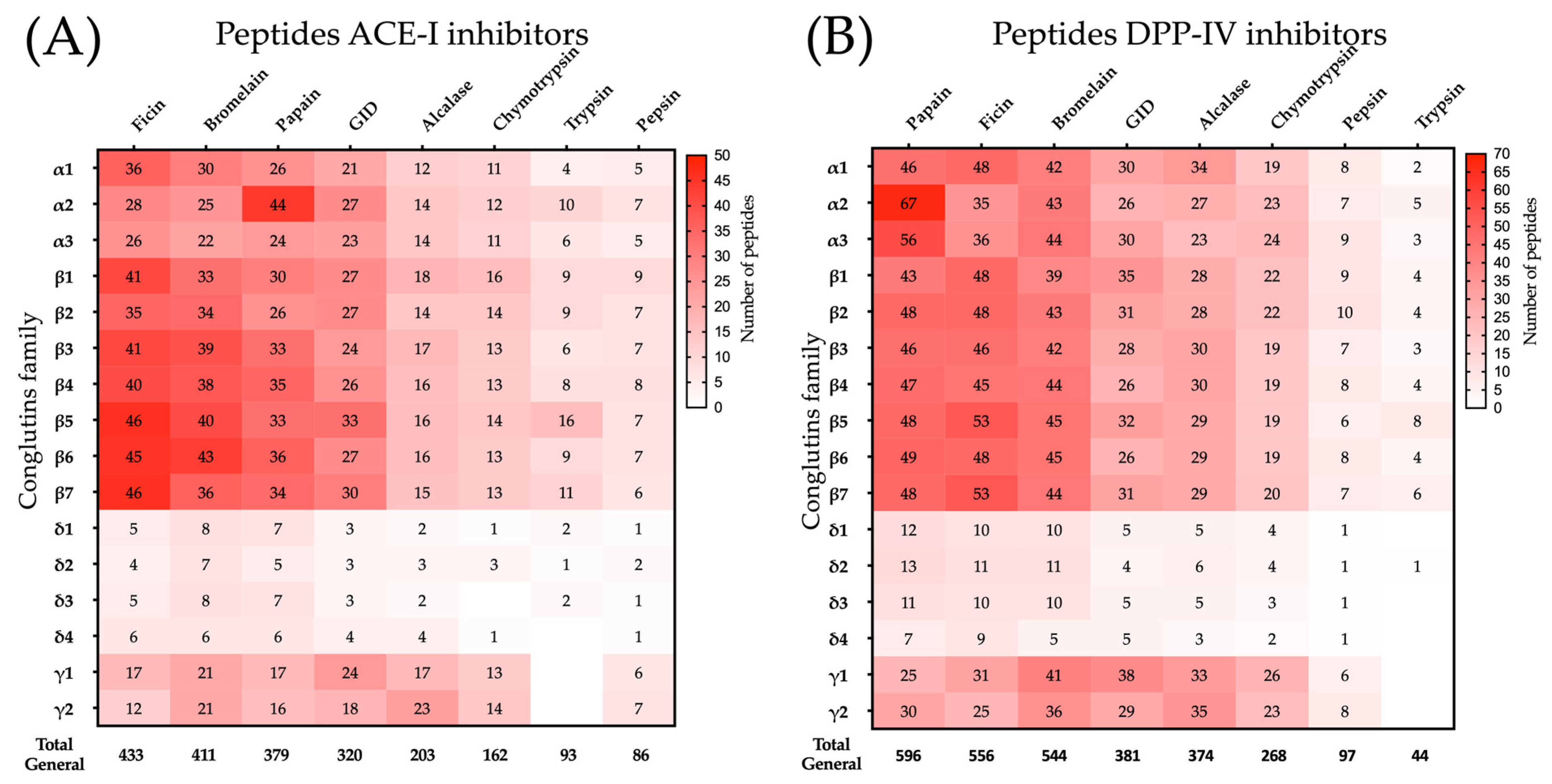
2.2. ACE-I and DPP-IV Inhibitory Peptides Released After Simulated Hydrolysis of Lupin Seed Proteins

Among the enzymes utilized to produce protein hydrolysates or peptides with bioactive properties, alcalase (EC 3.4.21.62), bromelain (EC 3.4.22.32), papain (EC 3.4.22.2), and ficin (EC 3.4.22.3) are the most reported [26,27]. These enzymes have distinct cleavage sites and can generate distinct peptide profiles with different bioactive properties [28]. To save time and resources, simulated hydrolysis can serve as a starting point to select an enzyme for searching peptides with specific bioactivities [27,29]. Certainly, bioactive peptides can be generated or broken down during human digestion, making it essential to explore this aspect in silico. In the present study, lupin proteins were hydrolyzed with 8 different enzymes to identify the enzymes with the potential to generate ACE-I or DPP-IV inhibitory peptides. Subsequently, the resulting peptides were subjected to in silico human gastrointestinal digestion (pepsin, trypsin, and chymotrypsin).
Ficin (n = 433), bromelain (n = 411), and papain (n = 379) generated the highest numbers of ACE-I inhibitory peptides after the hydrolysis of the 16 conglutins (Figure 1A). Conversely, chymotrypsin (n = 162), trypsin (n = 93), and pepsin (n = 86) released the lowest numbers of ACE-I inhibitory peptides (Figure 1A). Gastrointestinal digestion (pepsin, trypsin, and chymotrypsin) also generated ACE-I inhibitory peptides (n = 320) (Figure 1A). Similarly, the highest numbers of DPP-IV inhibitory peptides were generated after hydrolysis with papain (n = 596), ficin (n = 556), and bromelain (n = 544), and the lowest ones after hydrolysis with chymotrypsin (n = 268), pepsin (n = 97), and trypsin (n = 44) (Figure 1B). Gastrointestinal digestion also generated DPP-IV inhibitory peptides (n = 381) (Figure 1B).
Overall, the hydrolysis of the 16 conglutins generated unique ACE-I (n = 110) and DPP-IV (n = 151) inhibitory peptides. Similar results were reported using plant proteases and sources other than lupin, such as chickpeas, collagen, and meat proteins [12,19,30,31]. In fact, hydrolysates of various collagen sources obtained with bromelain, papain, or ficin have higher frequencies of DPP-IV and ACE-I inhibitory peptides than hydrolysates obtained with pepsin, trypsin, and chymotrypsin [31]. Additionally, the sequences of some DPP-IV or ACE-I inhibitory synthetic peptides were obtained after in silico digestion of food proteins with plant proteases [32,33,34]. Therefore, plant proteases have the potential to produce antihypertensive and hypoglycemic lupin hydrolysates.
Certainly, the use of synthesized or isolated food-derived peptides may not be cost-effective for treating chronic degenerative diseases, such as hypertension or diabetes [35]. In this context, protein hydrolysates with multiple bioactive peptides have gained attention for developing nutraceuticals or ingredients for functional foods. Among plant proteases, papain, bromelain, and ficin have been used to produce DPP-IV or ACE-I inhibitory food protein hydrolysates [36,37,38,39,40,41]. For instance, chickpea (Cicer arietinum L.) protein hydrolysates obtained under optimized hydrolysis conditions with papain or ficin can inhibit DPP-IV in vitro (up to 84.66% and 72.05%, respectively) [41]. Similarly, whey protein hydrolysates obtained using plant proteases (papain, ficin, or bromelain) can inhibit ACE-I in vitro (up to IC50 91.9 μg/mL) [36]. Overall, it is highlighted that plant proteases can be used for producing antidiabetic or antihypertensive food protein hydrolysates, but the DPP-IV and ACE-I inhibitory peptides generated must be stable in gastrointestinal digestion, be absorbed, and remain non-toxic to have the opportunity to be an effective and side effect-free protein hydrolysate.

2.3. ADMET Characteristics and Gastrointestinal Digestion Stability of ACE-I and DPP-IV Inhibitory Peptides

The pharmacokinetic properties of the ACE-I and DPP-IV inhibitory peptides identified were evaluated using the ADMET Lab2.0 platform. Table 2 shows the frequency of the LBPs with optimal ADMET prediction values. (For more details, see Supplementary Tables S1 and S2). The ADMET properties were evaluated in 201 unique peptides (151 inhibit DPP-IV (69.90%), 110 inhibit ACE-I (50.92%), and 60 inhibit both enzymes (27.77%)). Almost all ACE-I (94.54%) and all DPP-IV (100%) inhibitory peptides fulfill the Lipinski rule, showing the potential to exert an in vivo effect. Therapeutic compounds designed to be orally administered should be adequately absorbed at the intestinal level and reach systemic circulation. In the present study, almost all peptides identified are potentially absorbed at the intestinal level (ACE-I = 75.45%, DPP-IV = 81.45%) and can reach systemic circulation (ACE-I = 86.36%, DPP-IV = 91.39%). Similarly, almost all the ACE-I and DPP-IV inhibitory peptides identified were short-chain peptides (dipeptides = 83.79%; tripeptides = 15.27%; tetrapeptide = 0.093%), which have higher absorption and bioavailability than long-chain peptides [42]. Additionally, the results show that all the ACE-I and DPP-IV inhibitory peptides identified can be adequately distributed in the systemic circulation, showing that most of them have high half-life times (>3 h) (ACE-I = 86.36%, DPP-IV = 73.51%). Regarding toxicity, most peptides evaluated were non-toxic (ACE-I = 90.91%, DPP-IV = 92.05%).
The stability of the ACE-I and DPP-IV inhibitory peptides during in silico gastrointestinal digestion is shown in Table 2. Most peptides (n = 179; 89.05%) were stable in gastrointestinal digestion. Among these, 97 inhibit DPP-IV, and 82 inhibit ACE-I. The gastrointestinal stability of bioactive peptides depends on several factors, such as peptide length, hydrophobicity, and the presence of specific amino acids (e.g., absence of lysine, presence of proline and aliphatic amino acids) [43]. In this sense, gastrointestinal digestion-stable ACE-I and DPP-IV inhibitory peptides were mostly short-chain peptides (dipeptides = 81.75% and tripeptides = 17.51%). Furthermore, 90.51% of the peptides lack leucine, 63.50% have at least one aliphatic amino acid (e.g., valine, proline, glycine, isoleucine), and 19.70% have proline in their sequence. This explains, to some extent, the in silico gastrointestinal stability of the ACE-I and DPP-IV inhibitory peptides identified in the present study.

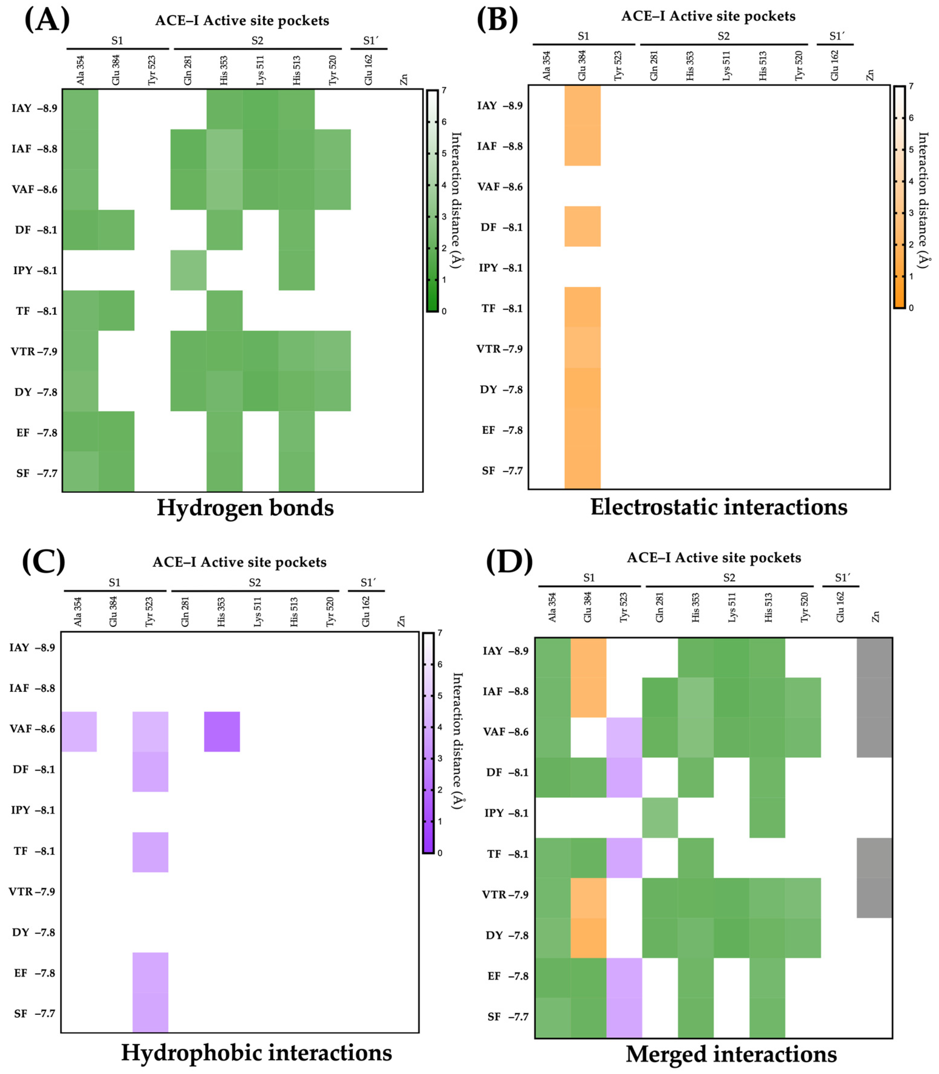
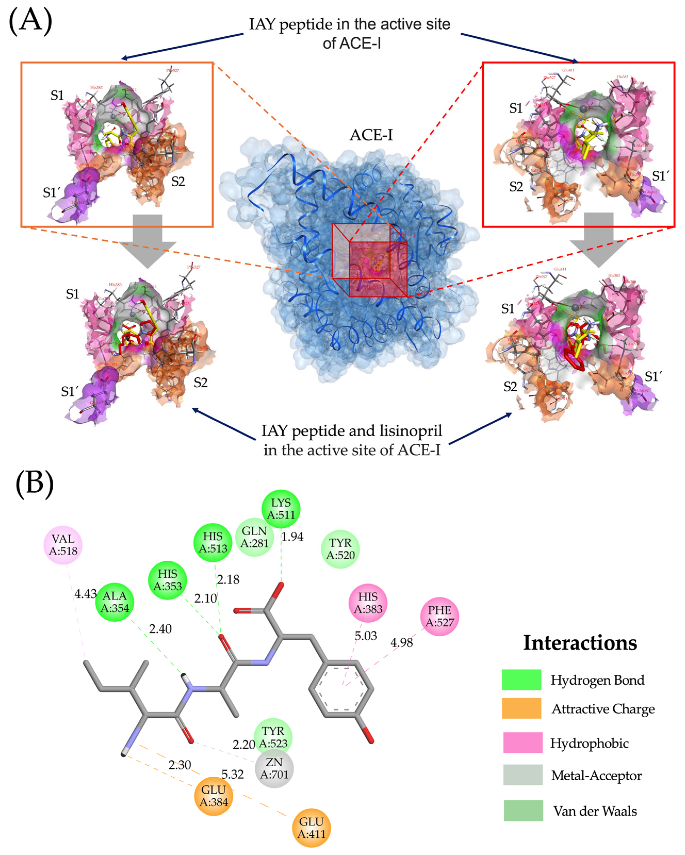
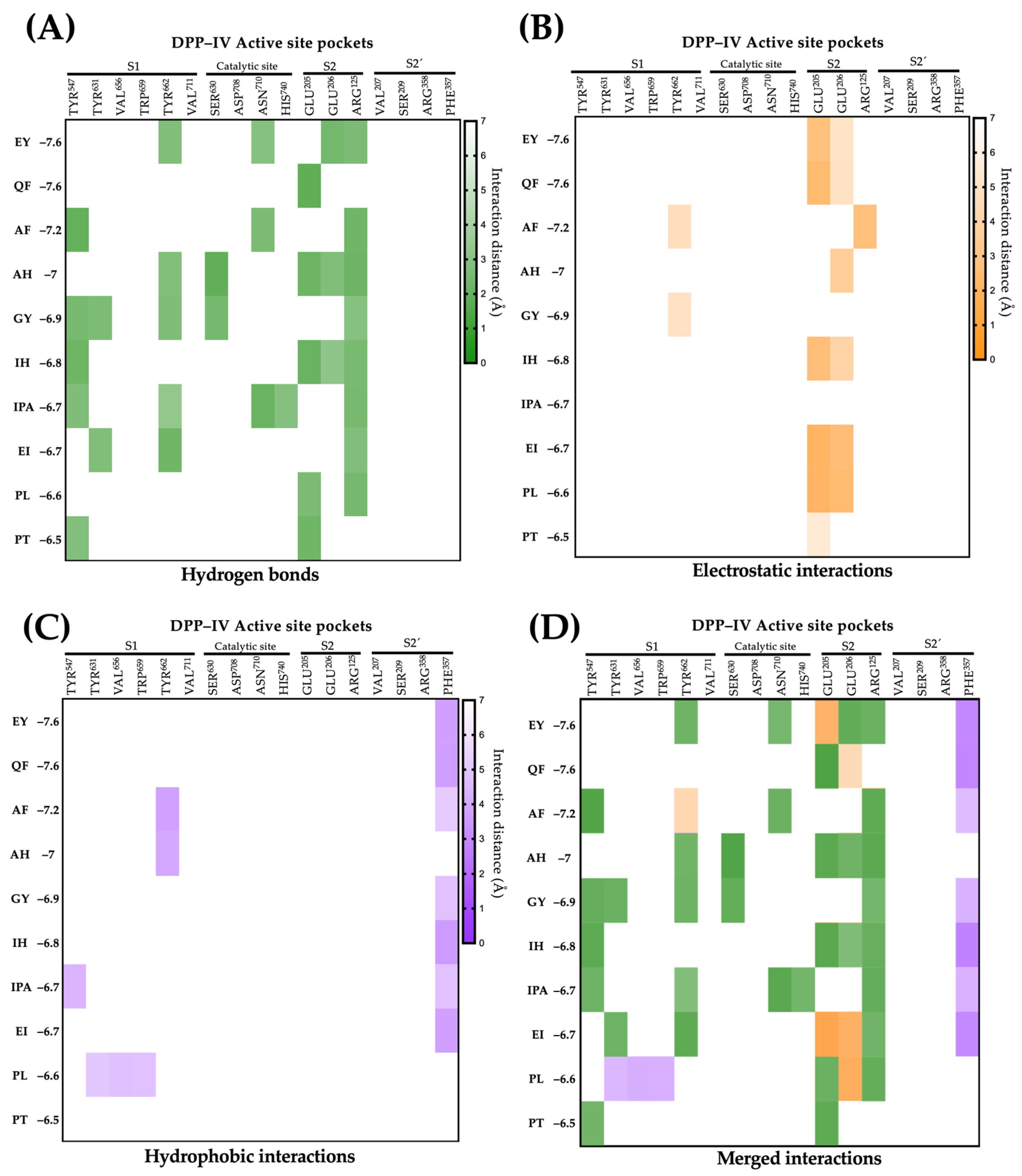
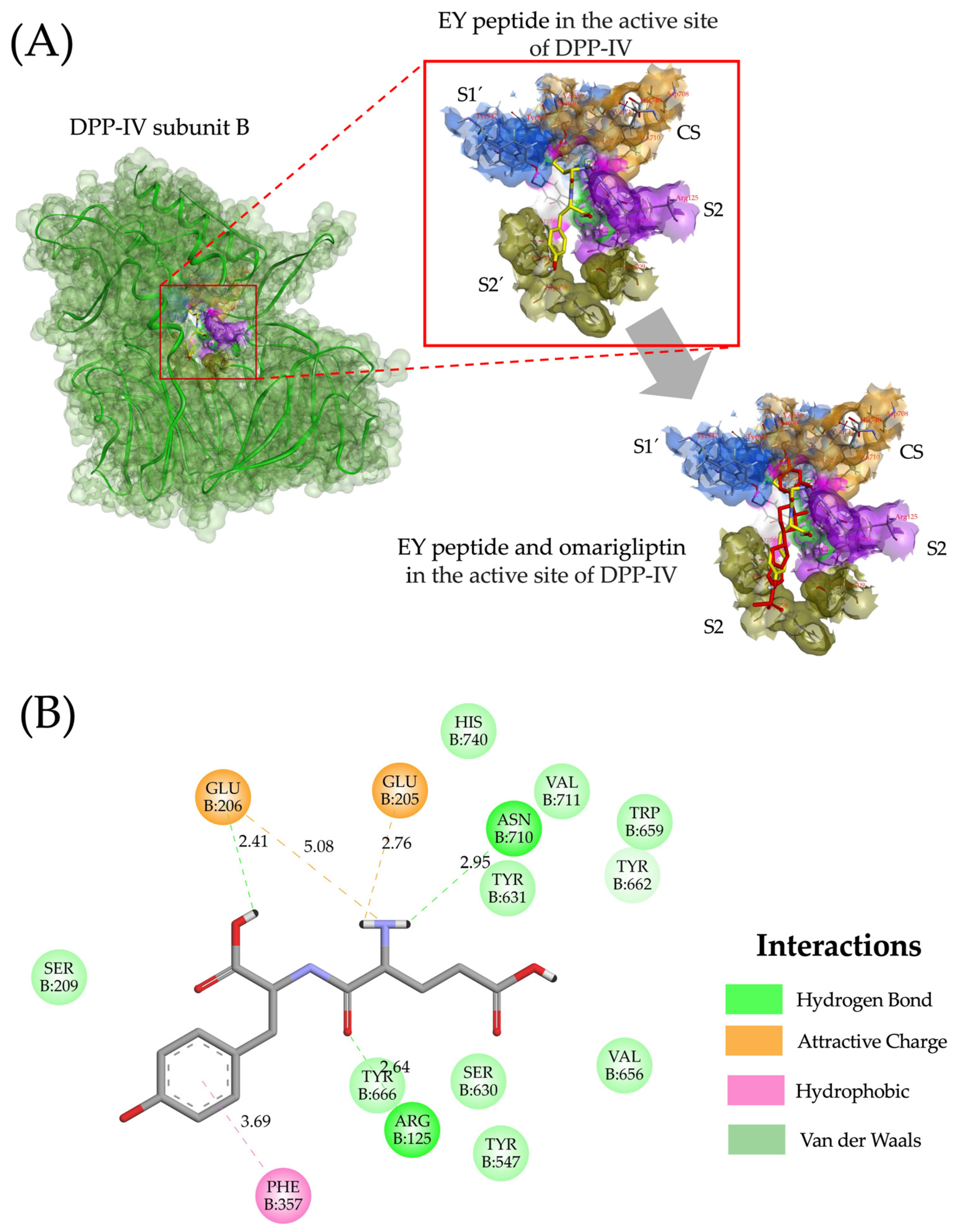
2.4. Molecular Interactions of Lupin Peptides with the Active Sites of ACE-I and DPP-IV

Molecular docking analysis was performed on all ACE-I (n = 76) and DPP-IV (n = 63) inhibitory peptides with reported PubChem or SatPDB structures. The binding energy of lupin peptides with the active site of ACE-I ranged from –5.4 kcal/mol to –9.2 kcal/mol (Supplementary Table S1). Of them, 51.31% (n = 39) were stable during in silico gastrointestinal digestion and showed optimal ADMET properties. Their binding energies ranged from –5.6 kcal/mol to –8.9 kcal/mol with distances of 1.93–4.83 Å. Among the DPP-IV inhibitory peptides (n = 63), 67.21% (n = 41) were stable in gastrointestinal digestion and had optimal ADMET properties, with binding energies ranging from –5.4 kcal/mol to –7.6 kcal/mol (distances ranged from 1.86–5.53 Å). These binding energies are similar to those reported for other ACE-I (from −5.7 to −9.2 kcal/mol) and DPP-IV (from −5.20 to −8.03 kcal/mol) inhibitory peptides derived from food sources other than lupin (chickpea, saurida elongata, α-lactalbumin, amaranth, and casein) [12,19,44,45,46,47]. Furthermore, the lupin peptides have binding energies similar to the energies reported for synthetic ACE-I (Lisinopril: –8.6 Kcal/mol) [19] and DPP-IV inhibitors (Saxagliptin: –8.4 Kcal/mol and Vildagliptin: –8.8 Kcal/mol) [48,49]. These results highlight that lupin peptides have the potential to tightly bind to the active site of ACE-I and DPP-IV and could act as potential therapeutic inhibitors.
The active site of ACE-I has three distinct pockets and one cofactor: S1 (Ala354, Glu384, and Tyr523), S2 (Gln281, His353, Lys511, His513, and Tyr520), S1′ (Gln162), and zinc as a cofactor [50]. ACE-I inhibitory peptides could interact with residues of the active site of this enzyme, competitively inhibiting ACE-I [51]. Figure 2 shows the molecular interactions of the ten lupin peptides with the lowest binding energies with the active site residues of ACE-I. ACE-I inhibitory lupin peptides mainly interact with the residues Ala354 (90%) and Glu384 (80%) of pocket S1 via conventional hydrogen bonds and electrostatic interactions at short distances (2.03–3.48 Å) (Figure 2A,B). Five out of 10 ACE-I inhibitory peptides formed hydrophobic interactions with Tyr523 of pocket S1 (distances ranging from 3.95–4.45 Å) (Figure 2C). In contrast, almost all interactions of ACE-I inhibitory peptides with S2 pocket residues were via hydrogen bonds (97.22%) (Figure 2A). Our results support the notion that hydrogen bonds are the main non-covalent interactions mediating the high affinity of ACE-I inhibitory peptides with the active site of ACE-I, but electrostatic and hydrophobic interactions aid in the stabilization of the peptide-ACE-I complex [52]. Interestingly, 5 out of 10 ACE-I inhibitory lupin peptides showed coordination with the zinc ion (Figure 2D). Zinc stabilizes the ACE-I active site, facilitating the coordination of catalytic residues and enabling peptide bonds [53]. Therefore, peptide coordination with zinc ions may induce a higher ACE-I inhibitory activity [54]. Our results show that lupin peptides could interact with residues of the active site of ACE-I, inducing a competitive inhibition.
Figure 3 shows the molecular interactions of Ile-Ala-Tyr tripeptide in the 3D structure of ACE-I and the overlapping with lisinopril in the ACE-I active site. The tripeptide Ile-Ala-Tyr had the lowest binding energy (−8.9 kcal/mol) and a docking pose comparable to lisinopril (Figure 3A), interacting with residues Ala354 and Glu384 in the S1 pocket via hydrogen bonds and electrostatic interactions (distances < 2.5 Å), as well as with residues His353, Lys511, and His513 in the S2 pocket via hydrogen bonds (Figure 3B). The high affinity of Ile-Ala-Tyr peptide for the active site of ACE-I can be attributed to the presence of Tyr in the C-terminal position. The presence of Tyr and other aromatic residues, such as Trp, Phe, Arg, and His, is associated with a higher ACE-I inhibitory potential among tripeptides, which exhibit the greatest affinity for binding in the ACE-I active site [55]. Additionally, the Ile-Ala-Tyr peptide interacts with zinc via metal coordination, contributing to its high binding affinity [54]. Furthermore, outside the active site, residues His383 and Phe527 stabilize the binding of the tripeptide via hydrophobic interactions, contributing to the negative binding energy (Figure 3B). The molecular interactions of Ile-Ala-Tyr with the active site of ACE-I are similar to those reported for ACE-I synthetic inhibitors, such as captopril and lisinopril [56], highlighting the potential of Ile-Ala-Tyr as a potent ACE-I inhibitor that is stable during in silico gastrointestinal digestion, potentially bioavailable, and non-toxic.
The DPP-IV active site is composed of four pockets: S1 (Tyr547, Tyr631, Val656, Trp659, Tyr662, and Val711), the catalytic site (Ser630, Asp708, and Asn710), S2 (Glu205, Glu206, and Arg125), and S2′ (Val207, Ser209, Arg358, and Phe357) [57]. The types of and distances of molecular interactions for the ten lupin peptides with the highest affinity for the active site of DPP-IV are shown in Figure 4. Lupin peptides mainly interact with residues of pocket S1 via hydrogen bonds (Tyr437, Tyr 631, and Tyr 662; distances ranging from 1.96 to 2.96 Å) and hydrophobic interactions (Tyr547, Tyr 631, Val656, Trp659, and Tyr662). Most peptides exhibit electrostatic interactions (80%) and hydrogen bonds (70%) with residues of pocket S2. The S1 pocket is characterized by being narrow and favoring interactions with small hydrophobic compounds, while the S2 pocket tends to interact with amino acids via electrostatic interactions [58]. The catalytic site of DPP-IV plays a pivotal role in the inhibition activity of DPP-IV synthetic drugs [59]. The present study shows that lupin peptides interact with residues of the catalytic site of DPP-IV via hydrogen bonds at short distances (1.90–2.95 Å), suggesting their high DPP-IV inhibitory potential. In general, hydrogen bonds were the most frequently non-covalent interactions between lupin peptides and the active site of DPP-IV (55.71%), followed by electrostatic (22.85%) and hydrophobic interactions (21.42%). Others also reported that hydrogen bonds are the main type of non-covalent interactions that mediate the binding of DPP-IV inhibitory lupin peptides with the enzyme’s active site [60]. Overall, the molecular interactions of DPP-IV inhibitory lupin peptides with the enzyme’s active site are similar to those reported for DPP-IV inhibitory synthetic drugs [61].
Figure 5 shows the molecular interactions of Glu-Tyr dipeptide in the 3D structure of DPP-IV and its overlapping with omarigliptin in the DPP-IV active site. The dipeptide Glu-Tyr had the lowest binding energy (−7.6 kcal/mol) among the DPP-IV inhibitory peptides and had a docking pose similar to omarigliptin (Figure 5A). The Glu-Tyr peptide forms various interactions with the DPP-IV active site: it interacts with residues Tyr662 in the S1 pocket, Asn710 in the catalytic triad, and with Glu206 and Arg125 in the S2 pocket via hydrogen bonds (Figure 5A). Additionally, it establishes electrostatic interactions with residues Glu205 and Glu206 in the S2 pocket and forms hydrophobic interactions with the residue Phe357 in the S2′ pocket (Figure 5B). These interactions contribute to explaining the low binding energy of the ligand–receptor (peptide–protein) complex [62]. Additionally, Glu-Tyr peptide shares similarities with omarigliptin, binding with residues Tyr662, Glu205, Glu206, and Arg125 [63] and interacting with the same residues (Arg125, Glu205, Glu206, Phe357, Tyr662, and Asn710) that the tripeptide IPI interacts with in the active site of DPP-IV [64], a tripeptide that has the lowest reported IC50 value (3.5 μM) [62].

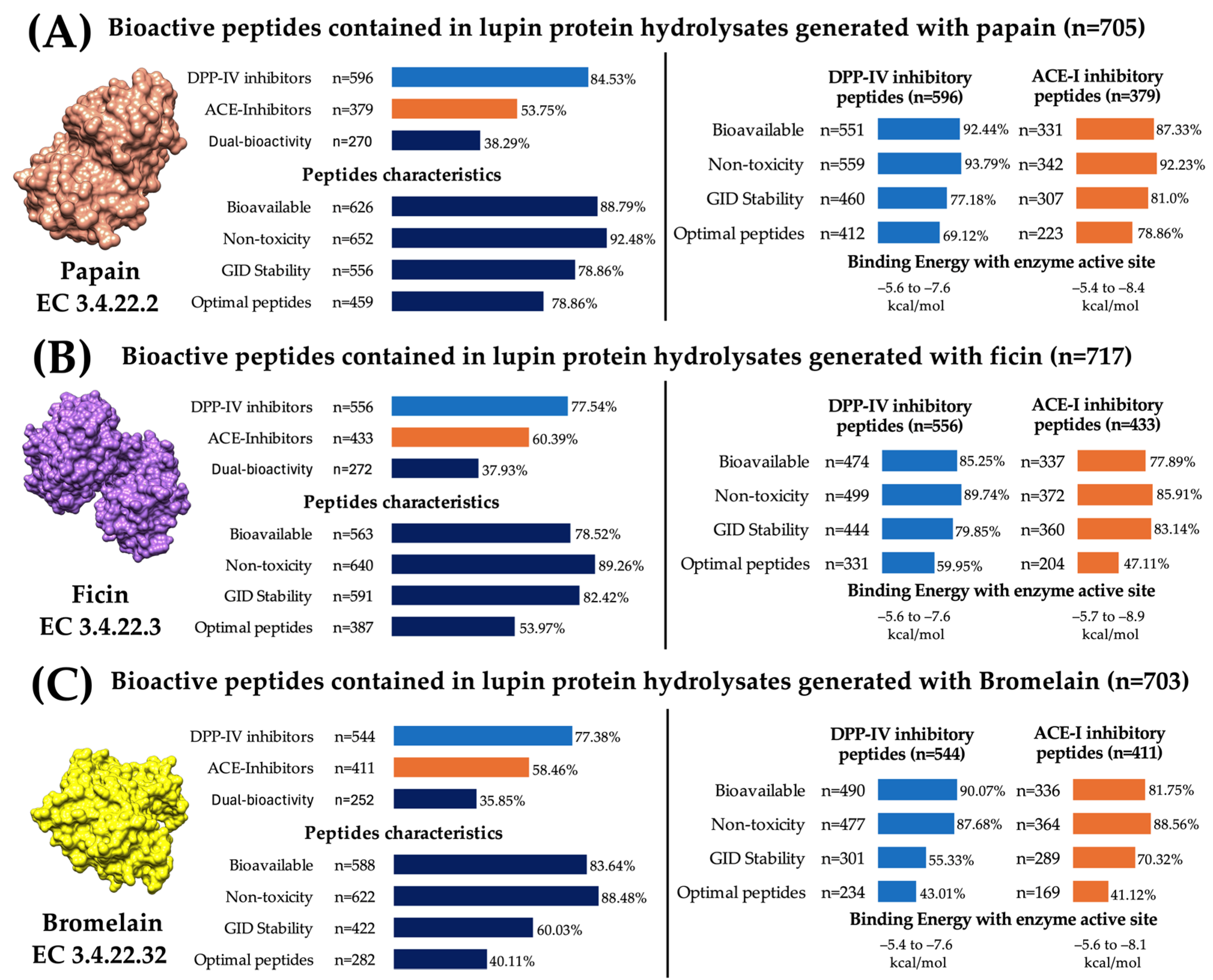
2.5. Plant Proteases Generate Lupine Protein Hydrolysates Containing Bioavailable, Non-Toxic, Digestion-Resistant, and Multi-Bioactive Peptides

Once the peptides’ pharmacokinetic properties, gastrointestinal digestion stability, and molecular interactions with the ACE-I and DPP-IV active sites were determined, the peptides were grouped according to their protein source and the enzyme that released them. This approach allows the evaluation of all peptides organized as hydrolysates. Supplementary Tables S3–S10 show the number of lupin peptides released after simulated hydrolysis. These peptides can inhibit DPP-IV, ACE-I, or both enzymes. The bioavailability, toxicity, and gastrointestinal digestion stability of the peptides from each hydrolysate are also shown in Tables S3–S10. Optimal peptides were those predicted to be bioavailable, non-toxic, and digestion resistant. Ficin (n = 717), papain (n = 705), and bromelain (n = 703) generated the hydrolysates with the highest number of ACE-I or DPP-IV inhibitory peptides. The number of optimal peptides in the other hydrolysates ranged from 115 to 547.
Despite having similar cleavage sites, plant proteases generated lupin protein hydrolysates containing peptides with different pharmacokinetic and bioactive characteristics (Figure 6). Papain generates the hydrolysate with the highest number of optimal peptides (n = 459). Among these, 89.76% (n = 412) were DPP-IV inhibitory peptides with binding energies ranging from −6.5 to −7.6 kcal/mol, 48.58% (n = 223) were ACE-I inhibitory peptides with binding energies ranging from −5.4 to −8.4. kcal/mol, and 38.34% (n = 176) had the potential to inhibit both enzymes (Figure 6A). Ficin generates hydrolysates containing 387 optimal bioactive peptides. Among these, 85.5% (n = 331) were DPP-IV inhibitory peptides with binding energies from −5.6 to 7.6, 52.71% (n = 204) were ACE-I inhibitory peptides with binding energies from −5.7 to −8.9, and 38.24% (n = 148) had the potential to inhibit both enzymes (Figure 6B). Regarding bromelain, this plant protease generates lupin protein hydrolysates with optimal peptides (n = 282). Among these, 82.97% (n = 234) were DPP-IV inhibitory peptides with binding energies from –5.4 to –7.6, 59.92% (n = 169) were ACE-I inhibitory peptides with binding energies from –5.6 to –8.1, and 42.9% (n = 121) had the potential to inhibit both enzymes (Figure 6C). These results show that lupine protein hydrolysates with antihypertensive and hypoglycemic properties can be obtained using plant proteases. These enzymes can work over wide pH and temperature ranges, but their use in producing hydrolysates containing ACE-I and DPP-IV inhibitory peptides remains largely unexplored [65]. Particularly, papain, compared to ficin and bromelain, has specific cleavage sites at both the C-terminus and N-terminus of peptide bonds [32], generating a wide variety of bioactive peptides that can not only inhibit DPP-IV and ACE-I but could also interact with other therapeutic targets.

3. Materials and Methods

3.1. Protein Sequences and Virtual Screening for Bioactive Peptides

Protein sequences of lupin storage proteins were obtained from the UniProtKB database (Conglutin alpha 1; Uni-Prot ID: F5B8V6, Conglutin alpha 2; UniProt ID: F5B8V7, Conglutin alpha 3; UniProt ID: F5B8V8, Conglutin beta 1; UniProt ID: F5B8V9, Conglutin beta 2; UniProt ID: F5B8W0, Conglutin beta 3; UniProt ID: F5B8W1, Conglutin beta 4; UniProt ID: F5B8W2, Conglutin beta 5; UniProt ID: F5B8W3, Conglutin beta 6; UniProt ID: F5B8W4, Conglutin beta 7; UniProt ID: F5B8W5, Gamma Conglutin 1; UniProt ID: Q42369, Gamma Conglutin 2; UniProt ID: F5B8W7, Conglutin Delta 1; UniProt ID: F5B8W8, Conglutin Delta 2; UniProt ID: Q99235, Conglutin Delta 3; UniProt ID: F5B8X0, Conglutin Delta 4; UniProt ID: F5B8X1).
Virtual screening for bioactive peptides was performed according to previous studies with minor modifications [12,19]. Briefly, the “Bioactive Peptides” tab was selected to perform the analyses in the BIOPEP-UWM platform (University of Warmia and Mazury in Olsztyn, Olsztyn, Poland) [66]. Afterwards, the tool “profiles of potential biological activity” available in the “Analysis” tab was utilized to determine the bioactive peptide sequences within lupin proteins. In silico enzymatic hydrolysis was performed utilizing the BIOPEP-UWM platform using the following enzymes: pepsin (EC 3.4.23.1), trypsin (EC 3.4.21.4), chymotrypsin (EC 3.4.21.1), alcalase (EC 213 3.4.21.62), papain (3.4.22.2), bromelain (3.4.22.32), and ficin (EC 3.4.22.3). Simulated gastrointestinal digestion was carried out using pepsin (EC 3.4.23.1), trypsin (EC 3.4.21.4), and chymotrypsin (EC 3.4.21.1), simultaneously. The potential biological activity of peptides released was screened using the BIOPEP-UWM database, which enables searching for 68 different biological activities of proteins/peptides.

3.2. ADMET Properties and Gastrointestinal Digestion Stability

The pharmacokinetic properties of ACE-I and DPP-IV inhibitory peptides were evaluated using the ADMETLab 2.0 platform (Zhejiang University, Hangzhou, China), as previously described [12,19]. The following pharmacokinetic properties were evaluated: (1) Lipinski’s rule, (2) human intestinal absorption, (3) volume of distribution, (4) half-life, and (5) acute oral toxicity in rats. The criteria available in the ADMETLab 2.0 platform were used to interpret the pharmacokinetic predictions. Optimal ADMET was considered when a peptide was predicted to be bioavailable (F20% < 0.3) and non-toxic (ROAT < 0.3). Gastrointestinal digestion stability of ACE-I and DPP-IV inhibitory peptides with optimal ADMET values was assessed using the BIOPEP-UWM platform (pepsin (EC 3.4.23.1), trypsin (EC 3.4.21.4), and chymotrypsin (EC 3.4.21.1)).

3.3. Molecular Docking of ACE-I and DPP-IV Inhibitory Peptides

The crystallographic structure of human ACE-I (complex with lisinopril; PDB ID: 1O86) and DPP-IV (complex with omarigliptin; PDB ID: 4PNZ) were obtained from the Protein Data Bank. Molecular docking analyses were carried out when the three-dimensional structures of ACE-I and DPP-IV inhibitory peptides were available in the PubChem Database. All structures were prepared for molecular docking (peptides: adding polar hydrogens and charges; ACE-I and DPP-IV crystallographic structures: (1) adding polar hydrogens and charges and (2) removal of water molecules and crystallographic ligands). Docking was performed using the AutoDock Vina 1.1.2. (The Scripps Research Institute, CA, USA). The coordinates selected for docking analysis covers the active site of ACE-I (x: 40.6559, y: 37.3827, z: 43.3401, radius: 20 Å) and DPP-IV (x: −6.733, y: 62.839, z: 35.416, radius: 20 Å) [12,19]. The following parameters were used for molecular docking analyses: number of binding modes per ligand = 10, exhaustiveness = 8, and maximum energy difference between models = 2 kcal/mol. The best docking pose showing the lowest binding energy for each predicted peptide was selected. Dockings between peptides and the active sites of ACE-I and DPP-IV were visualized using the Discovery Studio software v21.1.0 (Dassault Systèmes, Vélizy-Villacoublay, France). Dockings with unfavorable interactions were excluded.

4. Conclusions

Lupin conglutins are sources of ACE-I and DPP-IV inhibitory peptides. These peptides are mainly released after the hydrolysis of conglutins with plant proteases. Papain, ficin, and bromelain showed the best yields to produce ACE-I and DPP-IV inhibitory peptides. The peptides with the strongest binding energy in the active sites of DPP-IV and ECA-I were Glu-Tyr and Ile-Ala-Tyr, respectively. These peptides were bioavailable, non-toxic, and gastrointestinal digestion stable. It should be highlighted that the ACE-I and DPP-IV inhibitory potential of lupin peptides remain to be evaluated in vitro and in vivo to corroborate their bioactivity. However, our findings serve as the groundwork for future studies aimed at evaluating in vitro and in vivo the antihypertensive and hypoglycemic potential of plant protease-based lupin protein hydrolysates.

Supplementary Materials

The following supporting information can be downloaded at: https://www.mdpi.com/article/10.3390/ijms252312866/s1.

Author Contributions

Conceptualization, J.G.A.-G., O.G.F.-S. and N.O.; methodology, J.A.M.-M., R.T.-N.-G. and C.A.S.-C.; software, J.G.A.-G., O.G.F.-S. and J.A.M.-M.; investigation, J.G.A.-G., O.G.F.-S. and N.O.; data curation, R.T.-N.-G. and C.A.S.-C.; writing—original draft preparation J.G.A.-G., O.G.F.-S. and N.O.; writing—review and editing, R.T.-N.-G., J.A.M.-M., C.A.S.-C., M.H.G.-V. and L.K.F.-M.; visualization, J.G.A.-G. and J.A.M.-M.; supervision, O.G.F.-S. and N.O.; funding acquisition, N.O. All authors have read and agreed to the published version of the manuscript.

Funding

This research was funded by the Faculty of Biological and Health Sciences and the Department of Chemical, Biological, and Agricultural Sciences, Campus Navojoa, the University of Sonora. Grant number USO513008508.

Institutional Review Board Statement

Not applicable.

Informed Consent Statement

Not applicable.

Data Availability Statement

The original contributions presented in the study are included in the article/Supplementary Material, further inquiries can be directed to the corresponding author/s. PDB format of peptide-enzyme complex and 2D interactions of molecular docking for the peptides presented in Figure 2 and Figure 4 are available in a freely accessible repository (https://doi.org/10.6084/m9.figshare.27143631, accessed on 1 October 2024).

Acknowledgments

In memory of Francisco Cabrera-Chávez.

Conflicts of Interest

The authors declare no conflicts of interest.

References

  1. Sun, H.; Saeedi, P.; Karuranga, S.; Pinkepank, M.; Ogurtsova, K.; Duncan, B.B.; Stein, C.; Basit, A.; Chan, J.C.; Mbanya, J.C. IDF Diabetes Atlas: Global, Regional and Country-Level Diabetes Prevalence Estimates for 2021 and Projections for 2045. Diabetes Res. Clin. Pract. 2022, 183, 109119. [Google Scholar] [CrossRef] [PubMed]
  2. Mills, K.T.; Stefanescu, A.; He, J. The Global Epidemiology of Hypertension. Nat. Rev. Nephrol. 2020, 16, 223–237. [Google Scholar] [CrossRef] [PubMed]
  3. Naseri, M.W.; Esmat, H.A.; Bahee, M.D. Prevalence of Hypertension in Type-2 Diabetes Mellitus. Ann. Med. Surg. 2022, 78, 103758. [Google Scholar] [CrossRef] [PubMed]
  4. Farmaki, P.; Damaskos, C.; Garmpis, N.; Garmpi, A.; Savvanis, S.; Diamantis, E. Complications of the Type 2 Diabetes Mellitus. Curr. Cardiol. Rev. 2020, 16, 249–251. [Google Scholar] [CrossRef] [PubMed]
  5. Shaikh, A. A Practical Approach to Hypertension Management in Diabetes. Diabetes Ther. 2017, 8, 981–989. [Google Scholar] [CrossRef]
  6. Wong, M.K.S. Subchapter 29D—Angiotensin Converting Enzymes. In Handbook of Hormones; Takei, Y., Ando, H., Tsutsui, K., Eds.; Academic Press: San Diego, CA, USA, 2016; pp. 263–269. ISBN 978-0-12-801028-0. [Google Scholar]
  7. Boer, G.A.; Holst, J.J. Incretin Hormones and Type 2 Diabetes—Mechanistic Insights and Therapeutic Approaches. Biology 2020, 9, 473. [Google Scholar] [CrossRef]
  8. Mirabito Colafella, K.M.; Bovée, D.M.; Danser, A.H.J. The Renin-Angiotensin-Aldosterone System and Its Therapeutic Targets. Exp. Eye Res. 2019, 186, 107680. [Google Scholar] [CrossRef]
  9. Sterrett, J.J.; Bragg, S.; Weart, C.W. Type 2 Diabetes Medication Review. Am. J. Med. Sci. 2016, 351, 342–355. [Google Scholar] [CrossRef]
  10. Figueroa-Salcido, O.G.; Arámburo-Gálvez, J.G.; Mora-Melgem, J.A.; Camacho-Cervantes, D.L.; Gracia-Valenzuela, M.H.; Cuevas-Rodríguez, E.O.; Ontiveros, N. Alcalase-Based Chickpea (Cicer arietinum L.) Protein Hydrolysates Efficiently Reduce Systolic Blood Pressure in Spontaneously Hypertensive Rats. Foods 2024, 13, 1216. [Google Scholar] [CrossRef]
  11. Chávez-Ontiveros, J.; Reyes-Moreno, C.; Ramírez-Torres, G.I.; Figueroa-Salcido, O.G.; Arámburo-Gálvez, J.G.; Montoya-Rodríguez, A.; Ontiveros, N.; Cuevas-Rodríguez, E.O. Extrusion Improves the Antihypertensive Potential of a Kabuli Chickpea (Cicer arietinum L.) Protein Hydrolysate. Foods 2022, 11, 2562. [Google Scholar] [CrossRef]
  12. Mora-Melgem, J.A.; Arámburo-Gálvez, J.G.; Cárdenas-Torres, F.I.; Gonzalez-Santamaria, J.; Ramírez-Torres, G.I.; Arvizu-Flores, A.A.; Figueroa-Salcido, O.G.; Ontiveros, N. Dipeptidyl Peptidase IV Inhibitory Peptides from Chickpea Proteins (Cicer arietinum L.): Pharmacokinetics, Molecular Interactions, and Multi-Bioactivities. Pharmaceuticals 2023, 16, 1109. [Google Scholar] [CrossRef] [PubMed]
  13. Ruiz-López, M.A.; Barrientos-Ramírez, L.; García-López, P.M.; Valdés-Miramontes, E.H.; Zamora-Natera, J.F.; Rodríguez-Macias, R.; Salcedo-Pérez, E.; Bañuelos-Pineda, J.; Vargas-Radillo, J.J. Nutritional and Bioactive Compounds in Mexican Lupin Beans Species: A Mini-Review. Nutrients 2019, 11, 1785. [Google Scholar] [CrossRef] [PubMed]
  14. Boschin, G.; Scigliuolo, G.M.; Resta, D.; Arnoldi, A. ACE-Inhibitory Activity of Enzymatic Protein Hydrolysates from Lupin and Other Legumes. Food Chem. 2014, 145, 34–40. [Google Scholar] [CrossRef] [PubMed]
  15. Lammi, C.; Bollati, C.; Ferruzza, S.; Ranaldi, G.; Sambuy, Y.; Arnoldi, A. Soybean- and Lupin-Derived Peptides Inhibit DPP-IV Activity on In Situ Human Intestinal Caco-2 Cells and Ex Vivo Human Serum. Nutrients 2018, 10, 1082. [Google Scholar] [CrossRef] [PubMed]
  16. Hu, K.; Huang, H.; Li, H.; Wei, Y.; Yao, C. Legume-Derived Bioactive Peptides in Type 2 Diabetes: Opportunities and Challenges. Nutrients 2023, 15, 1096. [Google Scholar] [CrossRef]
  17. Lammi, C.; Zanoni, C.; Arnoldi, A.; Vistoli, G. Peptides Derived from Soy and Lupin Protein as Dipeptidyl-Peptidase IV Inhibitors: In Vitro Biochemical Screening and in Silico Molecular Modeling Study. J. Agric. Food Chem. 2016, 64, 9601–9606. [Google Scholar] [CrossRef]
  18. Fadimu, G.J.; Gan, C.-Y.; Olalere, O.A.; Farahnaky, A.; Gill, H.; Truong, T. Novel Antihypertensive Peptides from Lupin Protein Hydrolysate: An in-silico Identification and Molecular Docking Studies. Food Chem. 2023, 407, 135082. [Google Scholar] [CrossRef]
  19. Arámburo-Gálvez, J.G.; Arvizu-Flores, A.A.; Cárdenas-Torres, F.I.; Cabrera-Chávez, F.; Ramírez-Torres, G.I.; Flores-Mendoza, L.K.; Gastelum-Acosta, P.E.; Figueroa-Salcido, O.G.; Ontiveros, N. Prediction of ACE-I Inhibitory Peptides Derived from Chickpea (Cicer arietinum L.): In Silico Assessments Using Simulated Enzymatic Hydrolysis, Molecular Docking and ADMET Evaluation. Foods 2022, 11, 1576. [Google Scholar] [CrossRef] [PubMed]
  20. Czubinski, J.; Feder, S. Lupin Seeds Storage Protein Composition and Their Interactions with Native Flavonoids. J. Sci. Food Agric. 2019, 99, 4011–4018. [Google Scholar] [CrossRef]
  21. Foley, R.C.; Jimenez-Lopez, J.C.; Kamphuis, L.G.; Hane, J.K.; Melser, S.; Singh, K.B. Analysis of Conglutin Seed Storage Proteins across Lupin Species Using Transcriptomic, Protein and Comparative Genomic Approaches. BMC Plant Biol. 2015, 15, 106. [Google Scholar] [CrossRef]
  22. Garmidolova, A.; Desseva, I.; Mihaylova, D.; Lante, A. Bioactive Peptides from Lupinus Spp. Seed Proteins-State-of-the-Art and Perspectives. Appl. Sci. 2022, 12, 3766. [Google Scholar] [CrossRef]
  23. Magni, C.; Sessa, F.; Accardo, E.; Vanoni, M.; Morazzoni, P.; Scarafoni, A.; Duranti, M. Conglutin γ, a Lupin Seed Protein, Binds Insulin in Vitro and Reduces Plasma Glucose Levels of Hyperglycemic Rats. J. Nutr. Biochem. 2004, 15, 646–650. [Google Scholar] [CrossRef] [PubMed]
  24. Chirinos, R.; Cerna, E.; Pedreschi, R.; Calsin, M.; Aguilar-Galvez, A.; Campos, D. Multifunctional in Vitro Bioactive Properties: Antioxidant, Antidiabetic, and Antihypertensive of Protein Hydrolyzates from Tarwi (Lupinus mutabilis Sweet) Obtained by Enzymatic Biotransformation. Cereal Chem. 2021, 98, 423–433. [Google Scholar] [CrossRef]
  25. Lemes, A.C.; De Oliveira Filho, J.G.; Fernandes, S.S.; Gautério, G.V.; Egea, M.B. Bioactive Peptides from Protein-Rich Waste. In Agricultural Waste: Environmental Impact, Useful Metabolites and Energy Production; Ramawat, K.G., Mérillon, J.-M., Arora, J., Eds.; Sustainable Development and Biodiversity; Springer Nature: Singapore, 2023; Volume 31, pp. 139–166. ISBN 978-981-19877-3-1. [Google Scholar]
  26. Agyei, D.; Danquah, M.K. Industrial-Scale Manufacturing of Pharmaceutical-Grade Bioactive Peptides. Biotechnol. Adv. 2011, 29, 272–277. [Google Scholar] [CrossRef] [PubMed]
  27. Du, Z.; Li, Y. Review and Perspective on Bioactive Peptides: A Roadmap for Research, Development, and Future Opportunities. J. Agric. Food Res. 2022, 9, 100353. [Google Scholar] [CrossRef]
  28. Cruz-Casas, D.E.; Aguilar, C.N.; Ascacio-Valdés, J.A.; Rodríguez-Herrera, R.; Chávez-González, M.L.; Flores-Gallegos, A.C. Enzymatic Hydrolysis and Microbial Fermentation: The Most Favorable Biotechnological Methods for the Release of Bioactive Peptides. Food Chem. Mol. Sci. 2021, 3, 100047. [Google Scholar] [CrossRef] [PubMed]
  29. Iwaniak, A.; Darewicz, M.; Mogut, D.; Minkiewicz, P. Elucidation of the Role of in Silico Methodologies in Approaches to Studying Bioactive Peptides Derived from Foods. J. Funct. Foods 2019, 61, 103486. [Google Scholar] [CrossRef]
  30. Sharmin, K.N.; Amiza, M.A.; Ahmad, F.; Razali, S.A.; Hashim, F. In Silico Analysis of Gracilaria Changii Proteins for Potential Bioactive Peptides. IOP Conf. Ser. Earth Environ. Sci. 2022, 967, 012017. [Google Scholar] [CrossRef]
  31. Iwaniak, A.; Minkiewicz, P.; Pliszka, M.; Mogut, D.; Darewicz, M. Characteristics of Biopeptides Released In Silico from Collagens Using Quantitative Parameters. Foods 2020, 9, 965. [Google Scholar] [CrossRef]
  32. Lafarga, T.; O’Connor, P.; Hayes, M. Identification of Novel Dipeptidyl Peptidase-IV and Angiotensin-I-Converting Enzyme Inhibitory Peptides from Meat Proteins Using in Silico Analysis. Peptides 2014, 59, 53–62. [Google Scholar] [CrossRef]
  33. Bleakley, S.; Hayes, M.; O’ Shea, N.; Gallagher, E.; Lafarga, T. Predicted Release and Analysis of Novel ACE-I, Renin, and DPP-IV Inhibitory Peptides from Common Oat (Avena sativa) Protein Hydrolysates Using in Silico Analysis. Foods 2017, 6, 108. [Google Scholar] [CrossRef] [PubMed]
  34. Guo, H.; Richel, A.; Hao, Y.; Fan, X.; Everaert, N.; Yang, X.; Ren, G. Novel Dipeptidyl Peptidase-IV and Angiotensin-I-Converting Enzyme Inhibitory Peptides Released from Quinoa Protein by in Silico Proteolysis. Food Sci. Nutr. 2020, 8, 1415–1422. [Google Scholar] [CrossRef] [PubMed]
  35. Brandelli, A.; Daroit, D.J.; Corrêa, A.P.F. Whey as a Source of Peptides with Remarkable Biological Activities. Food Res. Int. 2015, 73, 149–161. [Google Scholar] [CrossRef]
  36. Peslerbes, M.; Fellenberg, A.; Jardin, J.; Deglaire, A.; Ibáñez, R.A. Manufacture of Whey Protein Hydrolysates Using Plant Enzymes: Effect of Processing Conditions and Simulated Gastrointestinal Digestion on Angiotensin-I-Converting Enzyme (ACE) Inhibitory Activity. Foods 2022, 11, 2429. [Google Scholar] [CrossRef]
  37. Salampessy, J.; Reddy, N.; Kailasapathy, K.; Phillips, M. Functional and Potential Therapeutic ACE-Inhibitory Peptides Derived from Bromelain Hydrolysis of Trevally Proteins. J. Funct. Foods 2015, 14, 716–725. [Google Scholar] [CrossRef]
  38. Osman, A.; El-Hadary, A.; Korish, A.A.; AlNafea, H.M.; Alhakbany, M.A.; Awad, A.A.; Abdel-Hamid, M. Angiotensin-I Converting Enzyme Inhibition and Antioxidant Activity of Papain-Hydrolyzed Camel Whey Protein and Its Hepato-Renal Protective Effects in Thioacetamide-Induced Toxicity. Foods 2021, 10, 468. [Google Scholar] [CrossRef]
  39. Hanafi, M.A.; Hashim, S.N.; Chay, S.Y.; Ebrahimpour, A.; Zarei, M.; Muhammad, K.; Abdul-Hamid, A.; Saari, N. High Angiotensin-I Converting Enzyme (ACE) Inhibitory Activity of Alcalase-Digested Green Soybean (Glycine max) Hydrolysates. Food Res. Int. 2018, 106, 589–597. [Google Scholar] [CrossRef]
  40. Zaharuddin, N.D.; Hanafi, M.A.; Chay, S.Y.; Hussin, F.S.; Auwal, S.M.; Zarei, M.; Sarbini, S.R.; Wan Ibadullah, W.Z.; Karim, R.; Saari, N. Multifunctional Hydrolysates from Kenaf (Hibiscus cannabinus L.) Seed Protein with High Antihypertensive Activity in Vitro and in Vivo. J. Food Meas. Charact. 2021, 15, 652–663. [Google Scholar] [CrossRef]
  41. Chandrasekaran, S.; Gonzalez de Mejia, E. Optimization, Identification, and Comparison of Peptides from Germinated Chickpea (Cicer arietinum) Protein Hydrolysates Using Either Papain or Ficin and Their Relationship with Markers of Type 2 Diabetes. Food Chem. 2022, 374, 131717. [Google Scholar] [CrossRef]
  42. Ramírez-Torres, G.I.; Ontiveros, N.; López-Teros, V.; Suarez-Jiménez, G.M.; Cabrera-Chávez, F. Food Matrices for the Delivery of Antihypertensive Peptides in Functional Foods. Biotecnia 2018, 20, 165–169. [Google Scholar] [CrossRef]
  43. Ahmed, T.; Sun, X.; Udenigwe, C.C. Role of Structural Properties of Bioactive Peptides in Their Stability during Simulated Gastrointestinal Digestion: A Systematic Review. Trends Food Sci. Technol. 2022, 120, 265–273. [Google Scholar] [CrossRef]
  44. Lan, X.; Sun, L.; Muhammad, Y.; Wang, Z.; Liu, H.; Sun, J.; Zhou, L.; Feng, X.; Liao, D.; Wang, S. Studies on the Interaction between Angiotensin-Converting Enzyme (ACE) and ACE Inhibitory Peptide from Saurida elongata. J. Agric. Food Chem. 2018, 66, 13414–13422. [Google Scholar] [CrossRef] [PubMed]
  45. Lin, K.; Zhang, L.; Han, X.; Cheng, D. Novel Angiotensin I-Converting Enzyme Inhibitory Peptides from Protease Hydrolysates of Qula Casein: Quantitative Structure-Activity Relationship Modeling and Molecular Docking Study. J. Funct. Foods 2017, 32, 266–277. [Google Scholar] [CrossRef]
  46. Gao, J.; Gong, H.; Mao, X. Dipeptidyl Peptidase-IV Inhibitory Activity and Related Molecular Mechanism of Bovine α-Lactalbumin-Derived Peptides. Molecules 2020, 25, 3009. [Google Scholar] [CrossRef]
  47. Valenzuela Zamudio, F.; Hidalgo-Figueroa, S.N.; Ortíz Andrade, R.R.; Hernández Álvarez, A.J.; Segura Campos, M.R. Identification of Antidiabetic Peptides Derived from in Silico Hydrolysis of Three Ancient Grains: Amaranth, Quinoa and Chia. Food Chem. 2022, 394, 133479. [Google Scholar] [CrossRef]
  48. Gupta, A.; Jacobson, G.A.; Burgess, J.R.; Jelinek, H.F.; Nichols, D.S.; Narkowicz, C.K.; Al-Aubaidy, H.A. Citrus Bioflavonoids Dipeptidyl Peptidase-4 Inhibition Compared with Gliptin Antidiabetic Medications. Biochem. Biophys. Res. Commun. 2018, 503, 21–25. [Google Scholar] [CrossRef]
  49. Sneha, P.; Doss, C.G.P. Gliptins in Managing Diabetes—Reviewing Computational Strategy. Life Sci. 2016, 166, 108–120. [Google Scholar] [CrossRef]
  50. Pina, A.S.; Roque, A.C.A. Studies on the Molecular Recognition between Bioactive Peptides and Angiotensin-Converting Enzyme. J. Mol. Recognit. 2009, 22, 162–168. [Google Scholar] [CrossRef]
  51. Fu, Y.; Alashi, A.M.; Young, J.F.; Therkildsen, M.; Aluko, R.E. Enzyme Inhibition Kinetics and Molecular Interactions of Patatin Peptides with Angiotensin I-Converting Enzyme and Renin. Int. J. Biol. Macromol. 2017, 101, 207–213. [Google Scholar] [CrossRef]
  52. Yu, F.; Zhang, Z.; Luo, L.; Zhu, J.; Huang, F.; Yang, Z.; Tang, Y.; Ding, G. Identification and Molecular Docking Study of a Novel Angiotensin-I Converting Enzyme Inhibitory Peptide Derived from Enzymatic Hydrolysates of Cyclina Sinensis. Mar. Drugs 2018, 16, 411. [Google Scholar] [CrossRef]
  53. Bünning, P.; Riordan, J.F. The Functional Role of Zinc in Angiotensin Converting Enzyme: Implications for the Enzyme Mechanism. J. Inorg. Biochem. 1985, 24, 183–198. [Google Scholar] [CrossRef] [PubMed]
  54. Jiang, Z.; Zhang, H.; Bian, X.; Li, J.; Li, J.; Zhang, H. Insight into the Binding of ACE-Inhibitory Peptides to Angiotensin-Converting Enzyme: A Molecular Simulation. Mol. Simul. 2019, 45, 215–222. [Google Scholar] [CrossRef]
  55. Ma, M.; Feng, Y.; Miao, Y.; Shen, Q.; Tang, S.; Dong, J.; Zhang, J.Z.H.; Zhang, L. Revealing the Sequence Characteristics and Molecular Mechanisms of ACE Inhibitory Peptides by Comprehensive Characterization of 160,000 Tetrapeptides. Foods 2023, 12, 1573. [Google Scholar] [CrossRef] [PubMed]
  56. Natesh, R.; Schwager, S.L.U.; Evans, H.R.; Sturrock, E.D.; Acharya, K.R. Structural Details on the Binding of Antihypertensive Drugs Captopril and Enalaprilat to Human Testicular Angiotensin I-Converting Enzyme. Biochemistry 2004, 43, 8718–8724. [Google Scholar] [CrossRef]
  57. Juillerat-Jeanneret, L. Dipeptidyl Peptidase IV and Its Inhibitors: Therapeutics for Type 2 Diabetes and What Else? J. Med. Chem. 2014, 57, 2197–2212. [Google Scholar] [CrossRef]
  58. Nongonierma, A.B.; FitzGerald, R.J. Features of Dipeptidyl Peptidase IV (DPP-IV) Inhibitory Peptides from Dietary Proteins. J. Food Biochem. 2019, 43, e12451. [Google Scholar] [CrossRef]
  59. Mathur, V.; Alam, O.; Siddiqui, N.; Jha, M.; Manaithiya, A.; Bawa, S.; Sharma, N.; Alshehri, S.; Alam, P.; Shakeel, F. Insight into Structure Activity Relationship of DPP-4 Inhibitors for Development of Antidiabetic Agents. Molecules 2023, 28, 5860. [Google Scholar] [CrossRef]
  60. Nongonierma, A.B.; Dellafiora, L.; Paolella, S.; Galaverna, G.; Cozzini, P.; FitzGerald, R.J. In Silico Approaches Applied to the Study of Peptide Analogs of Ile-Pro-Ile in Relation to Their Dipeptidyl Peptidase IV Inhibitory Properties. Front. Endocrinol. 2018, 9, 329. [Google Scholar] [CrossRef]
  61. Gonzatti, M.B.; Júnior, J.E.M.; Rocha, A.J.; de Oliveira, J.S.; Evangelista, A.J.d.J.; Fonseca, F.M.P.; Ceccatto, V.M.; de Oliveira, A.C.; da Cruz Freire, J.E. Mechanism of Molecular Interaction of Sitagliptin with Human DPP4 Enzyme—New Insights. Adv. Med. Sci. 2023, 68, 402–408. [Google Scholar] [CrossRef]
  62. Nongonierma, A.B.; Mooney, C.; Shields, D.C.; FitzGerald, R.J. In Silico Approaches to Predict the Potential of Milk Protein-Derived Peptides as Dipeptidyl Peptidase IV (DPP-IV) Inhibitors. Peptides 2014, 57, 43–51. [Google Scholar] [CrossRef]
  63. Lolok, N.; Ramadhan, D.S.F.; Sumiwi, S.A.; Sahidin, I.; Levita, J. Molecular Docking of β-Sitosterol and Stigmasterol Isolated from Morinda Citrifolia with α-Amylase, α-Glucosidase, Dipeptidylpeptidase-IV, and Peroxisome Proliferator-Activated Receptor-γ. Rasayan J. Chem. 2022, 15, 20. [Google Scholar] [CrossRef]
  64. You, H.; Wu, T.; Wang, W.; Li, Y.; Liu, X.; Ding, L. Preparation and Identification of Dipeptidyl Peptidase IV Inhibitory Peptides from Quinoa Protein. Food Res. Int. 2022, 156, 111176. [Google Scholar] [CrossRef] [PubMed]
  65. Matkawala, F.; Nighojkar, S.; Nighojkar, A. Next-Generation Nutraceuticals: Bioactive Peptides from Plant Proteases. BioTechnologia 2022, 103, 397–408. [Google Scholar] [CrossRef]
  66. Minkiewicz, P.; Iwaniak, A.; Darewicz, M. BIOPEP-UWM Database of Bioactive Peptides: Current Opportunities. Int. J. Mol. Sci. 2019, 20, 5978. [Google Scholar] [CrossRef] [PubMed]
Figure 1. Frequency of conglutin-derived bioactive peptides released after simulated hydrolysis. (A) ACE-I inhibitory lupin peptides. (B) DPP-IV inhibitory lupin peptides.
Ijms 25 12866 g001
Figure 2. Molecular interactions of conglutin-derived peptides with the active site of ACE-I. (A) Hydrogen bonds interactions. (B) Electrostatic interactions. (C) Hydrophobic interactions. (D) Merged interactions.
Ijms 25 12866 g002
Figure 3. Molecular interactions of the IAY peptide with the active site of ACE-I. (A) Three-dimensional representation of the molecular docking of the IAY peptide and its overlapping with lisinopril in the active site of ACE-I. (B) Two-dimensional view of the interactions between the IAY peptide and the active site of ACE-I.
Ijms 25 12866 g003
Figure 4. Molecular interactions of conglutin-derived peptides with the DPP-IV active site. (A) Hydrogen bonds interactions. (B) Electrostatic interactions. (C) Hydrophobic interactions. (D) Merged interactions.
Ijms 25 12866 g004
Figure 5. Molecular interactions of the EY peptide with the active site of DPP-IV. (A) Three-dimensional representation of the molecular docking of the EY peptide and omarigliptin in the active site of DPP-IV. (B) Two-dimensional view of the interactions between the EY peptide and the DPP-IV active site.
Ijms 25 12866 g005
Figure 6. Characteristics of plant protease-based lupin protein hydrolysates. (A) Characteristics of papain hydrolysates. (B) Characteristics of ficin hydrolysates. (C) Characteristics of bromelain hydrolysates. Bioavailable (F20% < 0.3); non-toxicity (ROAT < 0.3). GID: gastrointestinal digestion; optimal peptides: bioavailable, non-toxic, and stable in GID.
Ijms 25 12866 g006
Table 1. Number of conglutin-derived peptides with specific bioactivities.
BioactivityLupin Conglutin FamiliesTotal General
α1α2α3β1β2β3β4β5β6β7δ1δ2δ3δ4γ1γ2
DPP-IV inhibitor1481471581551511501511491491495510955401491472062
ACE-I inhibitor13211611212211911611711711711837723733921011558
DPP III inhibitor21231924232121232222711722120287
Antioxidative26202321172019191917511632121268
Stimulating111291212111111111171475108162
Renin inhibitor128811119991010121 67114
Alpha-glucosidase inhibitor3565444645 111554
Regulating3423444544121 3347
Antithrombotic5224322323242 2240
Anti-inflammatory2443333333 1 2236
Neuropeptide2333323333 11333
CaMPDE inhibitor3223333333 1231
Activating ubiquitin-mediated proteolysis222221121212113328
Anti-amnestic2212222222121 3228
Hypocholesterolemic 2 313111 12
Hypolipidemic1111111111 111
Immunomodulating 111111111 1 10
HMG-CoA reductase inhibitor 11111 1 118
Hypotensive1 1 1211 7
Anticancer 1 111 1 5
Bacterial permease ligand 1 1 1 14
Immunostimulating 1 1 13
Antibacterial11 2
Embryotoxic1 1 2
Opioid 11
Total general376358356379362355353359353356118231121883173314813
Table 2. Frequency of conglutin-derived ACE-I and DPP-IV inhibitory peptides with optimal ADMET properties and gastrointestinal digestion stability.
ParametersGeneral (n = 201)DPP-IV (n = 151)ACE-I (n = 110)
n%n%n%
HIA15778.1012381.468375.45
F20%17687.5613891.399586.36
F30%16481.5912884.779081.82
VD201100.00151100.00110100.00
T ½15677.6111173.519586.36
ROAT18491.5613992.0510090.91
LIPINSKI19195.0210468.8710494.55
GDS17989.059764.238274.54
Optimal ADMET was considered when a peptide was predicted to be bioavailable (F20% < 0.3) and non-toxic (ROAT < 0.3). HIA: human intestinal absorption; F20%: oral bioavailability 20%; F30%: oral bioavailability 30%; VD: volume of distribution; T ½: half-life time; ROAT: rat oral acute toxicity; GDS: gastrointestinal digestion stability.
Disclaimer/Publisher’s Note: The statements, opinions and data contained in all publications are solely those of the individual author(s) and contributor(s) and not of MDPI and/or the editor(s). MDPI and/or the editor(s) disclaim responsibility for any injury to people or property resulting from any ideas, methods, instructions or products referred to in the content.

