Article

Characterization and Safety Evaluation of Autoclaved Gut Commensal Parabacteroides goldsteinii RV-01

1
REVIVEBIO Co., Taipei 115, Taiwan
2
Microbio Co., Ltd., Taipei 115, Taiwan
*
Authors to whom correspondence should be addressed.
These authors contributed equally to this work.
Int. J. Mol. Sci. 2024, 25(23), 12660; https://doi.org/10.3390/ijms252312660
Submission received: 17 October 2024 / Revised: 14 November 2024 / Accepted: 19 November 2024 / Published: 25 November 2024
(This article belongs to the Section Molecular Microbiology)

Abstract

Gut commensals play important roles in maintaining the homeostasis of human health. Previous studies indicated that the abundance of P. goldsteinii in animal hosts was increased by the administration of prebiotics such as polysaccharides purified from iconic oriental medicinal fungi. Subsequently, P. goldsteinii was found to exert beneficial effects on the amelioration of multiple chronic inflammation-associated diseases. Even so, during the process of the development of P. goldsteinii as a next-generation probiotic (NGP), care has to be taken when it is used as a functional food ingredient. In this study, we isolated a novel P. goldsteinii strain, RV-01, from the feces of a healthy adult and carried out comprehensive analyses of its genomic and phenotypic characteristics. Bioinformatic analysis of P. goldsteinii RV-01 revealed the absence of potential virulence genes, as well as the presence of genes and traits potentially beneficial to human health, such as the production of short-chain fatty acids, anti-inflammatory lipopolysaccharides, and zwitterionic capsular polysaccharides, as well as immune regulatory proteins. To circumvent any potential side effects, the P. goldsteinii RV-01 was autoclaved before proceeding to the nonclinical safety assessment. The autoclaved P. goldsteinii RV-01 retained its anti-inflammatory effect in human colon epithelial cells. In addition to the three genotoxicity assays, 28-day subacute and 90-day subchronic animal toxicity studies (the highest dose tested was equivalent to 8.109 × 10¹⁰ P. goldsteinii RV-01 cells/kg body weight/day) were also implemented. The results of all studies were negative for toxicity. These results support the conclusion that autoclaved P. goldsteinii RV-01 is safe for use as a food ingredient.

1. Introduction

The homeostasis of the gut microbiota is essential for the maintenance of animal hosts’ health. Among these microbiota, Parabacteroides spp. are core members of the human gut microbiota and have an average abundance of 1.27% in humans from 12 populations [1,2]. Parabacteroides have been reported to have the physiological characteristics of carbohydrate metabolism, and they secrete short-chain fatty acids [1]. In particular, recent studies of P. goldsteinii have shown its beneficial effects in animal hosts, demonstrating that P. goldsteinii has great potential for development as a next-generation probiotic (NGP) to help with the amelioration of chronic inflammation-related diseases [1,3]. In a choline-deficient, amino acid (CDAA)-defined mouse steatohepatitis model, the abundance of P. goldsteinii was decreased [4]. Furthermore, whereas the abundance of P. goldsteinii was negatively correlated with obesity-related indexes (fasting glucose, AUC of GTT, body weight gain, serum triglyceride, and LBP), as well as inflammatory factors (TNF-α and IL-1β), it was positively correlated with the serum levels of HDL-C and IL-10 in HFD-fed mice [5].
The identification of P. goldsteinii as an NGP was originally inspired by the observation that the administration of purified high-molecular-weight polysaccharides derived from Ganoderma lucidum and Ophiocordyceps sinensis significantly increased the abundance of P. goldsteinii and ameliorated obesity and metabolic syndrome in mice fed with a high-fat diet [6,7]. To address the ameliorative function of P. goldsteinii, the clinical isolate strain P. goldsteinii (ATCC JCM13446) [8] was used as a single strain to evaluate its potential in reducing obesity and metabolic disorders in mice. Although no adverse effects were identified, oral administration of live P. goldsteinii was able to counteract body weight and fat mass gain, as well as glucose intolerance induced by high-fat feeding [7]. Furthermore, daily supplementation with live P. goldsteinii was associated with a decrease in the plasmatic level of proinflammatory lipopolysaccharides (LPSs), an increase in intestinal tight junction proteins, and an overall improvement in gut barrier function, which had deteriorated in a mouse model of diet-induced obesity [7]. Subsequently, the commensal P. goldsteinii strain MTS01 isolated from the mice was shown to ameliorate the pathology of chronic obstructive pulmonary disease (COPD) [9]. A reduction in intestinal inflammation and the enhancement of cellular mitochondrial and ribosomal activities in the colon, the systematic restoration of aberrant host amino acid metabolism in sera, and the inhibition of lung inflammation act as important ameliorative mechanisms against COPD. In addition, the LPS derived from P. goldsteinii is anti-inflammatory and ameliorates COPD by acting as an antagonist of the toll-like receptor 4 (TLR4) signaling pathway [9]. No aberrant effects were observed during this study.
Recently, an increasing number of studies have further revealed that P. goldsteinii may function as an NGP associated with or involved in the amelioration of many other chronic inflammation-related diseases [1,2]. These diseases included obesity and metabolic syndrome [10,11], murine colitis [12], Helicobacter pylori-induced gastric pathology [13], maternal immune activation-induced autism-relevant behaviors [14], anastomotic healing in colorectal cancer surgery [15], food allergies [16], aspirin-mediated intestinal damage [17], and imiquimod-induced lupus in mice [18], as well as the induction of IL-22 and the subsequent enhanced expression of the antimicrobial peptides Reg3γ and Reg3β via P. goldsteinii’s outer membrane protein A1 (OmpA1) [19]. Specifically, P. goldsteinii was shown to assist in the maturation and development of the immune system [20,21]. P. goldsteinii produced β-hexosaminidase, a conserved enzyme identified in the phylum Bacteroidota, and promoted the development of a subtype of CD4+ effector T cells (CD4IELs) that regulate adaptive immunity in the intestinal mucosa, thereby playing an important role in controlling inflammation and maintaining homeostasis of the intestinal mucosa [20]. In a cohort study focusing on the analysis of the relationship between Parabacteroides and obesity in different human subpopulations (e.g., with respect to age and sex) and its association with subsequent weight changes, P. goldsteinii was negatively correlated with blood pressure, body mass index (BMI), and waist circumference, as well as with the prevalence of obesity [22].
Although no evidence has been reported on the causality of P. goldsteinii as a disease pathogen, it is possible to isolate P. goldsteinii from clinical samples [8]. Therefore, before P. goldsteinii RV-01 can be approved as a food ingredient, a thorough safety assessment should be performed. In this study, we specifically isolated a novel P. goldsteinii strain, RV-01, from the feces of a healthy adult. Comprehensive physiological and genotypic analyses, as well as monitoring of immune modulation activity in the intestine of a germ-free mouse mono-colonized with P. goldsteinii RV-01, were performed. To circumvent any potentiality that live P. goldsteinii may cause unexpected side effects, we autoclaved the sample in order to kill P. goldsteinii RV-01 [23]. Subsequently, following the US Food and Drug Administration (FDA) and European Food Safety Authority (EFSA) guidance on toxicity testing required for the safety assessment of a new nonabsorbable food ingredient [24,25], the autoclaved P. goldsteinii RV-01 was subjected to nonclinical studies assessing its potential for genotoxicity, its subacute toxicity, and its subchronic toxicity. No adverse effects were identified. Based on these observations, the autoclaved P. goldsteinii RV-01 produced by fermentation may be considered as a safe food ingredient.

2. Results

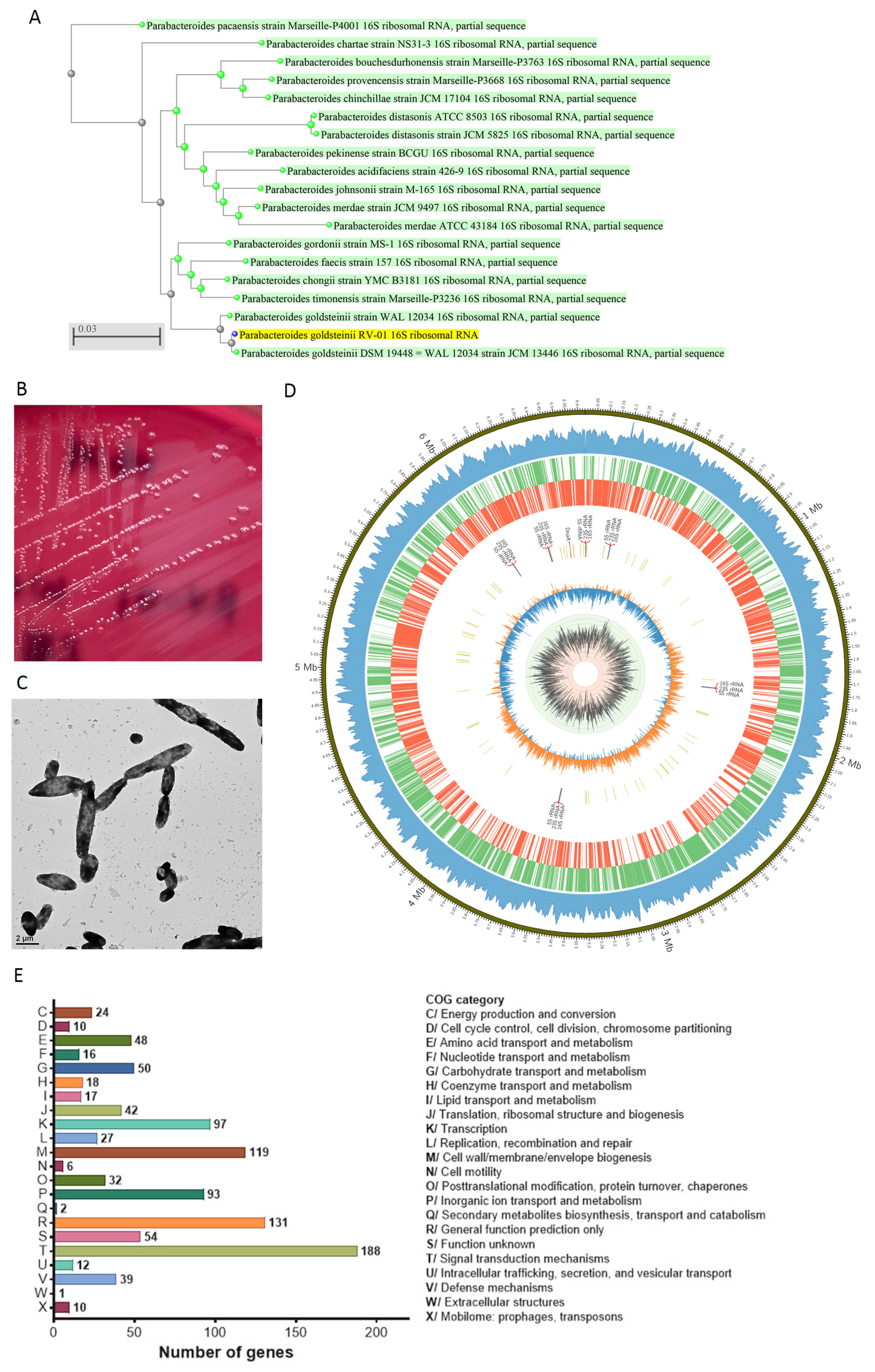
2.1. Descriptions of P. goldsteinii RV-01

Human gut commensal P. goldsteinii is a Gram-negative, rod-shaped, strictly anaerobic, and non-spore-forming bacterium belonging to the Bacteroidota phylum [26]. P. goldsteinii is involved in the metabolic regulation of the host [1,7] and grows on medium containing 20% bile [26]. The P. goldsteinii commensal strain RV-01 was originally isolated from the feces of a healthy adult. A 16S rDNA full-length sequence analysis was used to identify and group P. goldsteinii RV-01, which showed 99.71% identity to the P. goldsteinii JCM13446 in the NCBI BLAST analysis. The 16S rRNA DNA sequence of P. goldsteinii RV-01 was further analyzed by using the BLAST algorithm against the database of 16S ribosomal RNA sequences (Bacteria and Archaea). The neighbor-joining phylogenetic tree showing the relationship between P. goldsteinii RV-01 and similar species and strains indicated that P. goldsteinii RV-01 and two other P. goldsteinii strains were annotated within one cluster (Figure 1A).
When growing on CDC anaerobic blood agar plates, P. goldsteinii RV-01 colonies are smooth and circular, with a diameter of 1–2 mm, gray to off-white–gray, and slightly convex (Figure 1B). The morphology of P. goldsteinii RV-01 under a transmission electron microscope is shown in Figure 1C. The RV-01 was inactivated by heat treatment (100 °C for 15 min), autoclaving (121 °C for 15 min), or pasteurization (70 °C for 30 min). The activities of P. goldsteinii RV-01 that was inactivated at different temperatures were compared (Supplementary Figure S1). The cellular TLR2 activation activity of autoclaved RV-01 was higher than that of heat-treated or pasteurized RV-01 and was comparable with that of live RV-01. Therefore, autoclaving was adopted for the production of the P. goldsteinii RV-01 ingredient. The P. goldsteinii RV-01 ingredient was prepared as an autoclaved, lyophilized, off-white to beige, odorless powder containing more than 1.2 × 10¹⁰ total fluorescence units (TFUs)/g of powder. Proximate analysis of the ingredient is provided in Table 1. The averaged results of the proximate analyses of five batches showed that P. goldsteinii RV-01 powder is mainly composed of carbohydrates (95.1–96.3 g/100 g), protein (2.5–3.0 g/100 g), ash (0.2–0.3 g/100 g), and fat (0.4–0.9 g/100 g).

2.2. Genomic and Phenotypic Characteristics for Safety Evaluation

2.2.1. Whole Genomic Characteristics

The whole genome of P. goldsteinii RV-01 was sequenced by nanopore genome sequence analysis. The genome sequence and annotation of the P. goldsteinii RV-01 strain were deposited at NCBI under BioProject number PRJNA1153404. The completed circular chromosomal map of P. goldsteinii RV-01, with a total length of 6,631,096 bps, is shown in Figure 1D. The GC content of the genome is 43%, and no plasmid DNA was detected (Supplementary Figure S2). The nucleotide-level genomic similarity between the P. goldsteinii RV-01 genome and those of two P. goldsteinii strains (MTS01 and DSM19448), as well as of four Parabacteroides species (P. distasonis ATCC8503, P. merdae ATCC43184, P. gordonii DSM23371, and P. faecis DSM102983) was analyzed by using average nucleotide identity (ANI) analysis via the ANI calculator (https://www.ezbiocloud.net/tools/ani, accessed on 28 August 2024) using the OrthoANIu algorithm [27]. In Table 2, we can see that the ANI values between P. goldsteinii RV-01 and the two P. goldsteinii strains (MTS01 and DSM19448) are 98.17% and 98.44%, respectively. The ANI values between P. goldsteinii RV-01 and the four Parabacteroides species are less than 85% (ranging between 73.83 and 83.96%).
Annotation of the sequences of P. goldsteinii RV-01 revealed that the genome consists of 5241 coding DNA sequences, 79 tRNA genes, and 18 rRNA genes. The functional annotation results of the Clusters of Orthologous Genes (COG) database divided into 22 categories are shown in Figure 1E. Among these, the most abundant category is the signal transduction mechanisms, followed by general function and cell wall/membrane/envelope biogenesis.

2.2.2. Minimum Inhibitory Concentration (MIC) Evaluation of Antibiotics

The MIC results for the antibiotics tested are reported in Table 3. EFSA did not provide the microbiological cut-off values for Parabacteroides species [28]. Therefore, three P. goldsteinii strains and two Parabacteroides species were included for comparison. The results indicated that the observed antibiotic sensitivity of B. fragilis ATCC25285 was in compliance with the MIC QC range. Furthermore, P. goldsteinii RV-01 presented high resistance levels to aminoglycosides (gentamycin, kanamycin, streptomycin, and apramycin), vancomycin, nalidixic acid, sulfonamide, and trimethoprim. The MICs obtained for these antimicrobials tested were similar among the three P. goldsteinii strains and two Parabacteroides species, indicating that resistance to these antimicrobials may be considered intrinsic. P. goldsteinii RV-01 was susceptible to the other antibiotics (clindamycin, tetracycline, and chloramphenicol) according to the MIC breakpoints for susceptible (S) (CLSI, 2020). Taken together, the MICs of P. goldsteinii RV-01 showed a similar level among the different P. goldsteinii strains tested.

2.2.3. Bioinformatic Analyses for Antibiotic Resistance and Potential Virulence genes

Analysis of the P. goldsteinii RV-01 genome was conducted to screen for potential antibiotic resistance and virulence factor genes using homology search tools and comparing the results with the Comprehensive Antibiotic Resistance Database (CARD) and Virulence Factor DataBase (VFDB) [29,30]. To identify antimicrobial resistance sequence matches, thresholds of >60% coverage and >70% identity were applied based on relevant EFSA guidance [31]. Only one gene that showed a significant match with the tetracycline resistance gene tetQ met the threshold qualifications of >60% coverage and >70% identity (Supplementary File S1). However, intriguingly, the results of the minimal inhibitory concentration (MIC) assay showed that P. goldsteinii RV-01 was susceptible to tetracycline (Table 3). There was a possibility that the DNA fragment of the complete tetQ gene may still be present in the P. goldsteinii RV-01 ingredient. To determine whether the tetQ might be destroyed in this ingredient, PCR followed by agarose gel electrophoresis was performed to detect the presence of tetQ. The result showed that the tetQ gene was not detected in the P. goldsteinii RV-01 ingredient (Supplementary Figure S3), excluding the potential for horizontal spreading of the tetQ gene.
In silico analysis of the P. goldsteinii RV-01 genome was also conducted to screen for potential virulence factor genes; this analysis used homology search tools and compared the results with the Virulence Factor DataBase (VFDB). Thresholds of >60% coverage and >80% identity for virulence factor identity were applied based on relevant EFSA guidance [31]. No genes in the P. goldsteinii RV-01 genome demonstrating identity to known virulence factors were revealed (Supplementary File S2). Together, the bioinformatic analyses indicated that P. goldsteinii RV-01 is a non-pathogenic bacterium.

2.2.4. Bioinformatic Analyses for Allergic Proteins

A genomic and bioinformatic analysis was conducted to evaluate the allergic potential of the proteins present in P. goldsteinii RV-01. The GenBank P. goldsteinii RV-01 genome assembly, containing 5241 protein coding sequences (CDSs), was analyzed for potential allergenic proteins; sequence homology tools in Allermatch (http://allermatch.org, accessed on 30 July 2024) [32] were used for this, via the following three types of analysis: small exact word match, 80-amino-acid sliding window alignment, and full-length alignment (Supplementary Files S3–S5).
In the full-length sequence homology search, 129 hits (E-value ≤ 0.001) were identified, of which 33 hits had greater than 35% identity (ranging between 35.1 and 70.6%) (Supplementary File S3). In the 80-amino-acid sliding window alignment, 174 hits with greater than 35% identity were identified (ranging between 35.0 and 86.2%) (Supplementary File S4). An exact match search for six amino acids was also performed. Thirteen matches had an exact match, and more than six amino acids were identified (Supplementary File S5). Among these 13 matches, 11 proteins were also identified in the full-length sequence homology search and 80-amino-acid sliding window alignment. Upon closer evaluation, three proteins (endolase, adenosylhomocysteinase, and glyceraldehyde-3-phosphate dehydrogenase A) had >50% identity in both the full-length sequence homology search and 80-amino-acid sliding window alignment. The important consideration in immunogenicity and cross-reactivity is the protein structure. Therefore, although the sequence homology to known allergens was indicated, P. goldsteinii RV-01 was not expected to contain allergenic potential, as the P. goldsteinii RV-01 ingredient was prepared as an autoclaved and lyophilized powder, which resulted in protein denaturation. Taken together, P. goldsteinii RV-01 should have a low risk of allergenicity.

2.3. Bioinformatic Analysis of Genes and Traits Potentially Beneficial to Human Health

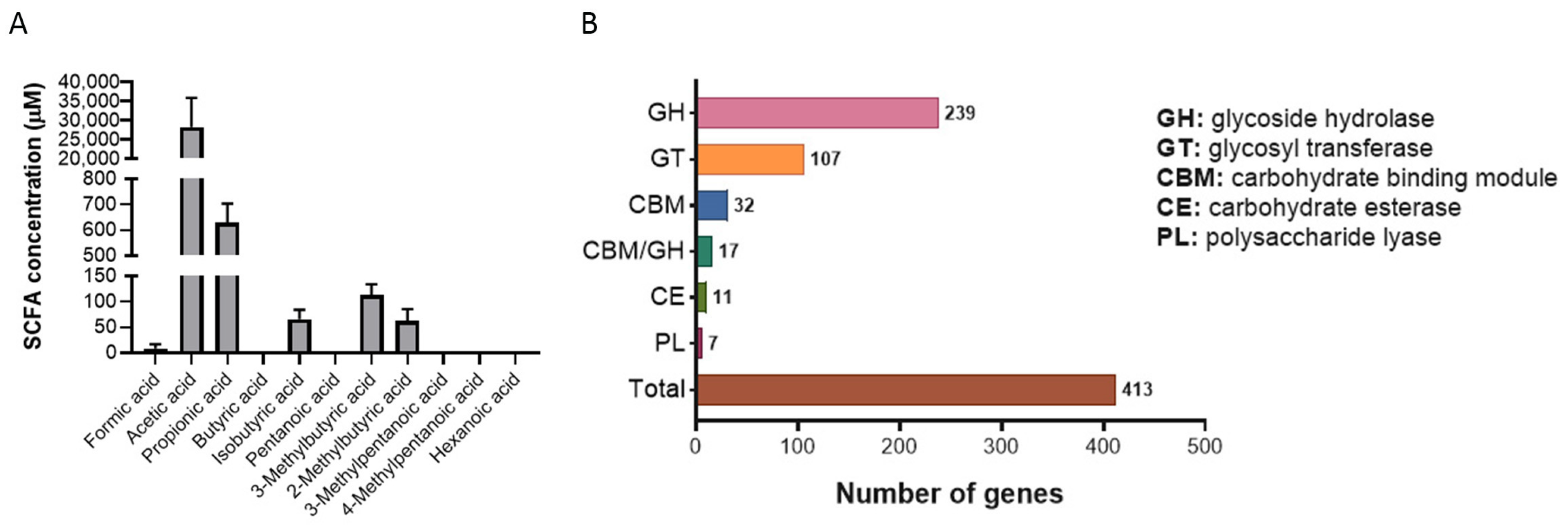
2.3.1. Production of Short-Chain Fatty Acids (SCFAs)

The SCFAs acetate, propionate, and butyrate are the main metabolites produced in the colon by bacterial fermentation of dietary fibers and resistant starch and have been shown to provide various health benefits [33]. The production of SCFAs in P. goldsteinii RV-01 was determined by mass (MS) analysis of bacterial culture supernatant. The major SCFAs found were acetic and propionic acids; minor amounts of isobutyric acid, 3-Methylbutyric acid, 2-Methylbutyric acid, and formic acid are also produced (Figure 2A). The fermentation of complex carbohydrates leads to the production of SCFAs. Therefore, the carbohydrate utilization enzymes of P. goldsteinii RV-01 were analyzed with the DIAMOND search against the Carbohydrate-Active Enzyme (CAZy) database [34,35]. A total of 413 genes (7.9% of genome) encoding CAZy, including glycoside hydrolase (GH), glycosyl transferase (GT), carbohydrate binding module (CBM), carbohydrate esterase (CE), and polysaccharide lyase (PL), were identified (Figure 2B). The major CAZy (a total of 239) was GH, which is a class of enzymes that catalyze the hydrolysis of glycosidic bonds in complex sugars. These results indicated that P. goldsteinii RV-01 is a complex carbohydrate-degrading and SCFA-producing bacterium.

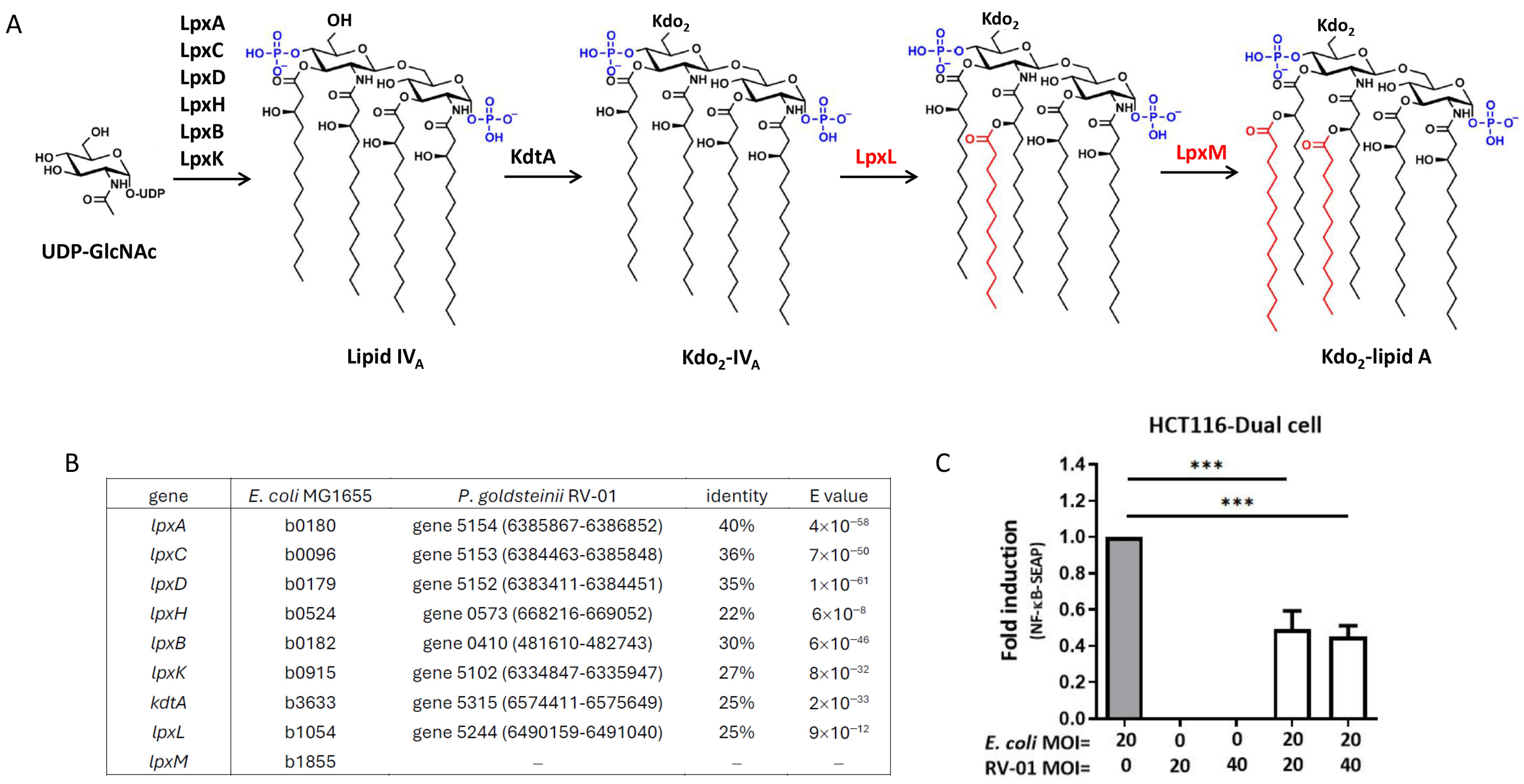
2.3.2. P. goldsteinii RV-01 Reduced TLR4-Related Inflammation

Unlike the LPS of most Gram-negative bacteria (for example, E. coli), which elicit proinflammatory responses, P. goldsteinii has been reported to produce an anti-inflammatory LPS (Pg-LPS) that antagonizes TLR4 activity. E. coli normally produce a lipid A molecule with six acyl chains [36] (Figure 3A). A BLAST analysis of the whole genome sequence of P. goldsteinii RV-01 highlighted that the genes involved in lipid A synthesis lacked LpxM, one ortholog of the acyltransferase genes that is responsible for the addition of the sixth acyl chain of LPS, and it was thus expected to produce hypo-acylated lipid A (Figure 3B). As shown in Figure 3C, autoclaved E. coli induced profound inflammation indicated by NF-κB activation in human colon epithelial HCT116 cells. In comparison, autoclaved P. goldsteinii RV-01 did not show significant activation activity. Moreover, pretreatment with autoclaved P. goldsteinii RV-01 significantly decreased the inflammation induced by autoclaved E. coli (Figure 3C). These results suggest that autoclaved P. goldsteinii RV-01 reduced the TLR4-related inflammation, where its LPS might be responsible for the TLR4 antagonization activity.

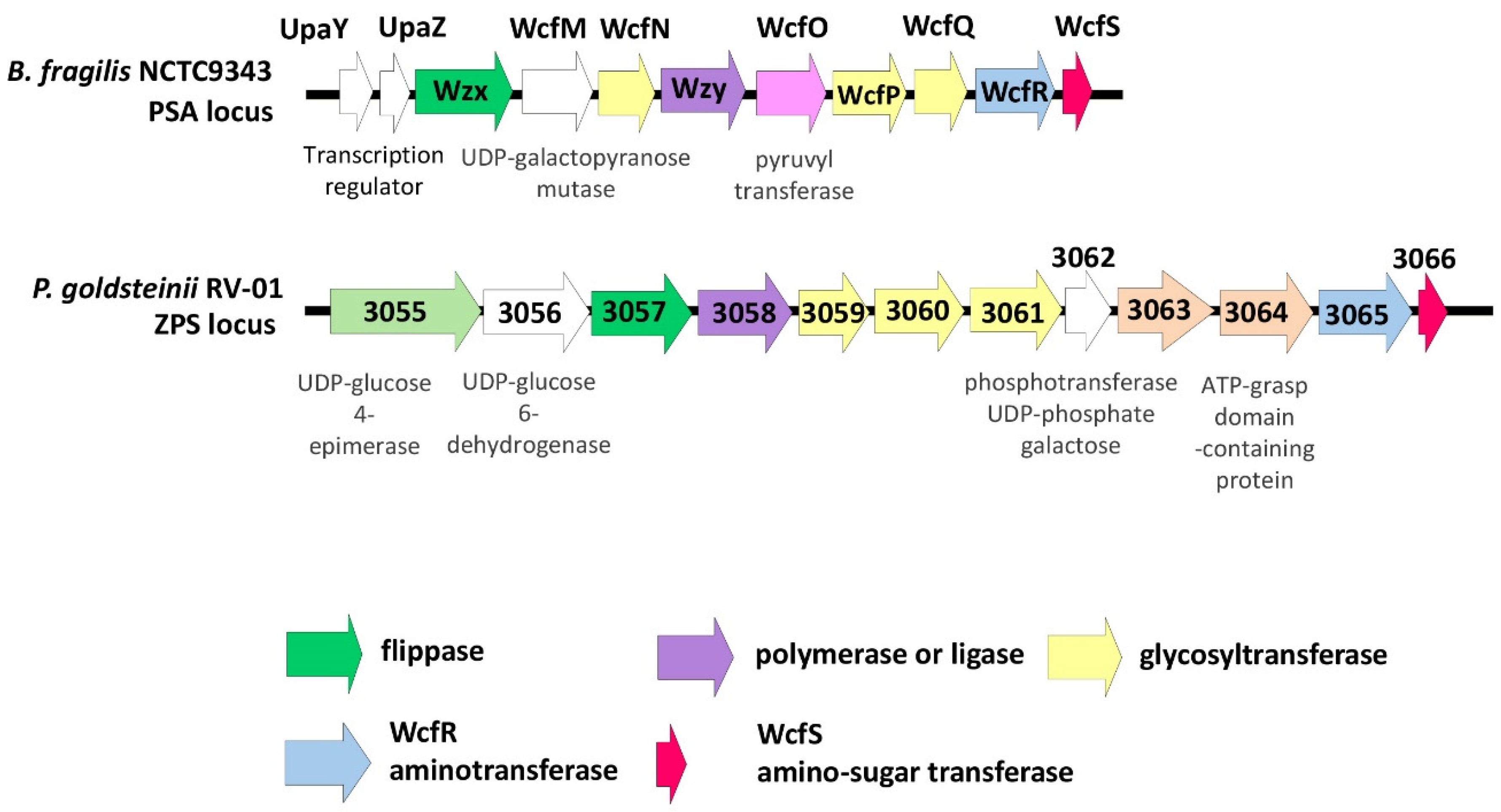
2.3.3. Bioinformatic Analyses for Zwitterionic Capsular Polysaccharide (ZPS)

Polysaccharide A (PSA), a very well-studied zwitterionic polysaccharide (ZPS) from the commensal gut bacteria Bacteroides fragilis, may induce anti-inflammatory regulatory T cells and suppress proinflammatory T helper 17 cells [37]. PSA has been shown to protect animals from colitis, encephalomyelitis, colorectal cancer, pulmonary inflammation, and asthma [37]. Previous studies demonstrated that wcfR gene homologs encoding for an amino sugar synthetase, as well as wcfS gene homologs encoding for an amino sugar transferase, are both needed in the synthesis of ZPS [38,39]. A putative ZPS biosynthesis cluster in P. goldsteinii RV-01 was identified via searching the homologs of wcfR and wcfS (Figure 4). A match with the wcfR gene (gene 3065) was found in the genome of P. goldsteinii RV-01 (38.6% protein identity with those of B. fragilis NCTC9343), and a match with the wcfS gene (gene 3066) was found in the genome of P. goldsteinii RV-01 (69.3% protein identity with those of B. fragilis NCTC9343). This putative ZPS biosynthesis cluster also carried genes responsible for the glycosyltransferase, polymerase or ligase, and flippase of capsular polysaccharides (Figure 4). Thus, P. goldsteinii RV-01 may produce ZPS with anti-inflammatory properties.

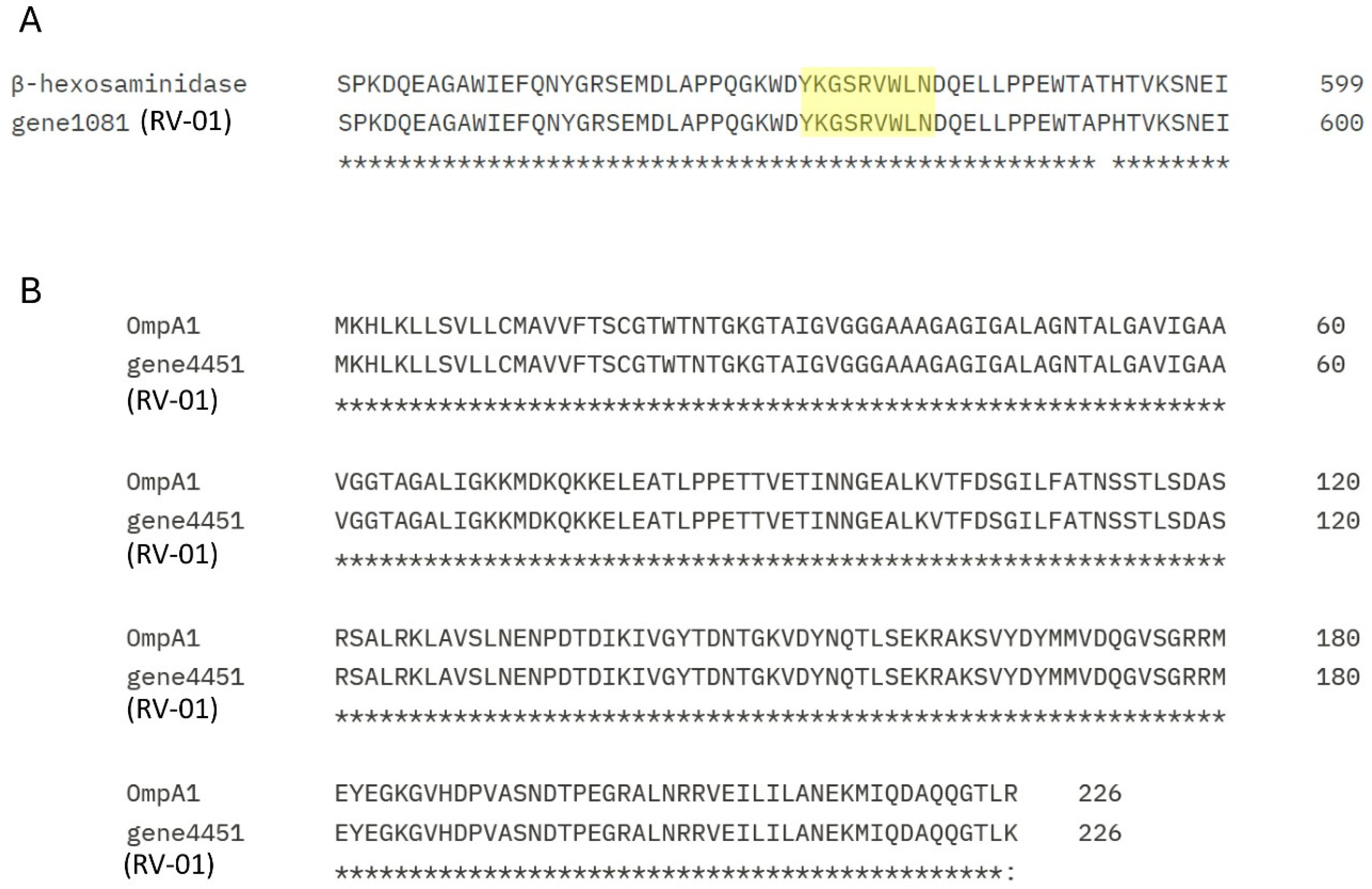
2.3.4. Bioinformatic Analyses for Immune Regulatory Proteins

A previous study has highlighted that cytoplasmic β-N-acetylhexosaminidase, after DC-T antigen presentation, enhances the cell population abundance of CD4+ CD8αα+ intraepithelial lymphocytes (CD4IELs) in the intestines [21]. In addition, an outer membrane protein, OmpA1—which increased the production of IL-22 and Reg3γ proteins that show anti-bacterial effects in the intestine—is involved in intestinal homeostasis [19]. Searching the whole genome of P. goldsteinii RV-01 revealed that a protein (gene 1081) was 97.3% identical with the hexosaminidase and harbored its immunostimulatory YKGSRVWLN epitope (Figure 5A). Moreover, a match with OmpA1 (gene 4451) was also found in the genome of the P. goldsteinii RV-01 (99.6% protein identity with those of P. goldsteinii ASF519, Figure 5B). Therefore, P. goldsteinii RV-01 was expected to have the ability to enhance the CD4IELs and to induce the production of IL-22 in an animal host.

2.4. Toxicological Studies

2.4.1. HEK293 Cytotoxicity Assay

An in vitro cytotoxicity test on human embryonic kidney 293 (HEK293) cells was first performed to see whether the autoclaved P. goldsteinii RV-01 may show any cytotoxicity potential. Different numbers of P. goldsteinii RV-01 were used to treat HEK293 cells with a multiplicity of infection (MOI) of 1, 10, 50, 100, 500, 1000, and 2000. Then, the cytotoxicity was determined by measuring the level of extracellular LDH released from damaged cells. The result shown in Supplementary Figure S4 indicates that no cytotoxicity was observed even in the MOI up to 2000.

2.4.2. Bacterial Reverse Mutation Test (Ames Test)

A Salmonella reverse mutation test (Ames test) was conducted with the P. goldsteinii RV-01 ingredient using five strains of Salmonella typhimurium (TA97a, TA98, TA100, TA102, and TA1535), in accordance with the OECD Guideline for Testing of Chemicals (Sect. 4, No. 471) (Table 4) [40]. In this study, the P. goldsteinii RV-01 ingredient at concentrations of 5, 2.5, 1.25, 0.625, and 0.3125 mg/plate was subjected to the mutagenic test. All experiments were performed in triplicate, and the mean revertant colony counts are provided in Supplementary Tables S1–S5. The overall results did not show any biologically relevant increases in the mean number of revertant colonies following exposure to the P. goldsteinii RV-01 ingredient compared with corresponding vehicle controls, in the presence or absence of metabolic activation (Supplementary Tables S1–S5). Therefore, P. goldsteinii RV-01 is non-mutagenic in the Salmonella reverse mutation assay.

2.4.3. In Vitro Mammalian Chromosomal Aberration Test

This study was performed according to the OECD Guideline for Testing of Chemicals No. 473: In Vitro Mammalian Chromosomal Aberration Test (Table 4) [41]. The results revealed that no significant chromosomal aberrations were induced by the P. goldsteinii RV-01 ingredient compared with the negative control (Supplementary Table S6). In conclusion, the P. goldsteinii RV-01 showed a negative response to inducing chromosomal aberration.

2.4.4. Rodent Micronucleus Test in Peripheral Blood

An in vivo micronucleus assay was conducted using human peripheral blood lymphocytes in accordance with the OECD Guideline for Testing of Chemicals (Sect. 4, No. 474) (Table 4) [42]. The main purpose of this study was to assess the possibility of the occurrence of micronuclei that may be caused by the P. goldsteinii RV-01 ingredient in the peripheral blood of rodents. The results for the number of micronucleated reticulocytes are provided in Supplementary Table S7. No significant difference in the number of reticulocytes with chromosomal structural aberrations (Supplementary Table S8) was observed between the treatment groups and negative control group. Therefore, the result of the micronucleus reaction of the P. goldsteinii RV-01 ingredient was considered to be negative. In brief, P. goldsteinii RV-01 did not induce structural or numerical chromosomal damage in human lymphocytes.

2.4.5. Twenty-Eight-Day Repeated Dose Oral Subacute Toxicity Study in Rats

To assess the potential adverse effects of the P. goldsteinii RV-01 ingredient, a 28-day dose range-finding oral toxicity assay in Sprague–Dawley (SD) rats (Table 4) was conducted in compliance with the Taiwan FDA GLP (2019), OECD-GLP (ENV/MC/CHEM (98) 17, 1997), and US FDA (21 CFR Part 58, 2023) on the principles of GLP for Nonclinical Laboratory Studies [43]. In this study, groups of five males and five females received 0 (control), 74 (4.0 × 10⁹ TFU/kg bw), 370 (2.0 × 10¹⁰ TFU/kg bw), or 1500 mg (8.109 × 10¹⁰ TFU/kg bw) of P. goldsteinii RV-01 once daily for 28 days. There were no deaths and no test item-related clinical signs. Animals administered the P. goldsteinii RV-01 ingredient showed similar body (Supplementary Tables S9–S11) and organ (Supplementary Tables S12–S15) weight changes. Further, no test items related to histopathological macroscopic or microscopic lesions were observed in the highest-dose group (Supplementary Table S16) nor any adverse effects in experimental animals. In conclusion, after 4 weeks of daily oral dosing of the P. goldsteinii RV-01 ingredient, the no-observed-adverse-effect level (NOAEL) was 1500 mg/kg/day in SD rats.

2.4.6. Ninety-Day Repeated Dose Oral Subchronic Toxicity Study in Rats

This study was also conducted in accordance with OECD Principles of GLP Test Guidance (TG) No. 408 for a 90-day study (Table 4) [44]. The healthy SD rats (n = 10/sex/group; 6 weeks old) were administered P. goldsteinii RV-01 via gavage in doses of 0, 4.0 × 10⁹ (low dose), 2.0 × 10¹⁰ (medium dose), or 8.109 × 10¹⁰ (high dose) TFU/kg bw per day. No positive correlations in the degree and incidence rate of the histological lesions between the test article and control groups were noted (Supplementary Tables S17 and S18). Furthermore, no clinical indications of test item-related toxicity were observed during this study. The functional observation battery did not identify any significant test item-dependent changes in behavior or in general ophthalmologic health. Based on the results of this study, the NOAEL was determined to be 8.109 × 10¹⁰ TFU/kg body weight/day, the highest dose tested in rats. The acceptable daily intake (ADI) is 900 mg/day based on an NOAEL of 1500 mg/kg/day, margin of safety (MOS) = 100, and 60 kg adult body weight. Taken together, the results of the repeat dose studies and the genotoxicity and mutagenicity studies show that the product is non-toxicogenic.

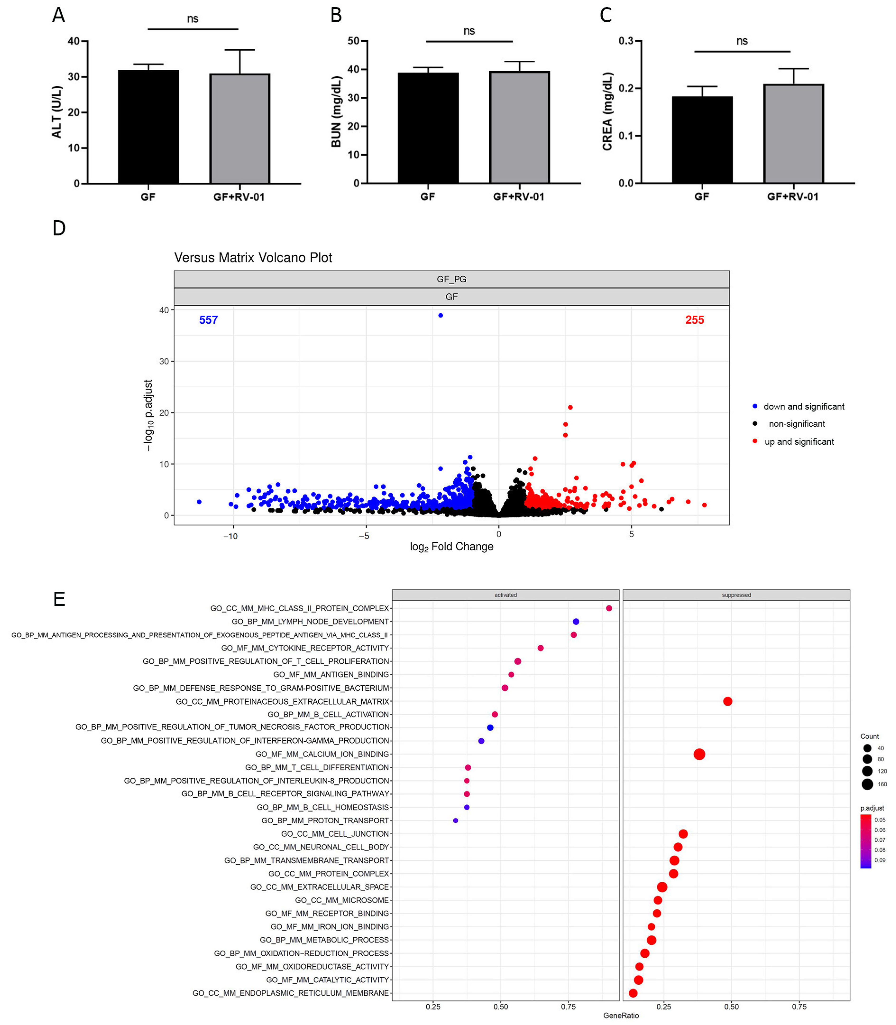
2.5. P. goldsteinii RV-01 Enhances Intestinal Immunity in Germ-Free (GF) Mice

GF mice were orally administered live P. goldsteinii RV-01 only. Then, the sera and colon tissues were collected after 2 weeks. Sera examination for liver and renal functions indicated that no adverse effects occurred (Figure 6A–C). Subsequently, the genes whose expression levels were affected by P. goldsteinii RV-01 alone in the intestinal tissue were characterized. P. goldsteinii RV-01 influenced the expression of multiple genes, with 255 upregulated genes and 557 downregulated genes identified in the colon tissue (Figure 6D). The pathways influenced by P. goldsteinii RV-01 were subsequently identified by GSEA (Figure 6E). Among these, the expression levels of genes related to the defense response to bacteria, antigen processing and presentation, T-cell differentiation, as well as B-cell homeostasis, were upregulated. By contrast, decreased expression levels of genes related to neuronal cell body and cell junction were also observed (Figure 6E). Previous results revealed that a normal immune development and a delayed neuronal response are elicited only after the microbiota accommodating homeostasis has been established in GF mice [45,46]. In this study, the administration of P. goldsteinii RV-01 significantly enhanced the immunity and significantly decreased the neuronal response in the colon, implying the involvement of P. goldsteinii in the development of immunity and neuronal signaling.

3. Discussion

The main purpose of this study was to assess the basic characteristics and evaluate the safety issue of the autoclaved bacterial strain P. goldsteinii RV-01 to be developed as a safe food supplement. Despite there being no P. goldsteinii-related adverse effects reported in previous studies, there were no data available nor any documented history of P. goldsteinii’s use as a food ingredient. Thus, given the vastly increasing amount of scientific research showing interest in proposing P. goldsteinii as a potential food ingredient, appropriate safety and toxicological evaluations were warranted. We used the safety assessment methodology following the tiered toxicity testing approach proposed by the EFSA and the US FDA guidance for assessing the safety of food ingredients. This approach involved the assessment of potential genotoxicity tests together with conducting an in vivo oral toxicity study in animals to assess the potential for subacute and subchronic toxicity. The results of the genotoxicity tests demonstrated that autoclaved P. goldsteinii is neither mutagenic (as assessed in the bacterial reverse mutation test) nor clastogenic or aneugenic (as evaluated in the mammalian cell micronucleus test and the mammalian chromosomal aberration test). Concordantly, in the 28-day and 90-day in vivo animal studies, there were no test item-related adverse effects on body and organ weights, food and water consumption, clinical observations and pathology, and neurobehavioral assessments. Furthermore, the NOAEL for the 90-day study was concluded to be 1500 mg/kg body weight/day (8.109 × 10¹⁰ cells/kg body weight/day), the highest dose tested. This NOAEL provides a safety factor of over 100-fold when compared with the worst-case anticipated exposure from use in foods and is the minimum safety requirement when applying animal experimentation data to humans [47]. The results therefore suggested that the autoclaved P. goldsteinii may have potential for application as a novel food supplement.
Currently, the majority of probiotics sold on the market mainly comprise microorganisms of the genera Lactobacillus and Bifidobacterium [48]. At the same time, continuing rapid advances in gut microbiota-related studies have led to significant progress in terms of exploring novel gut commensals’ functions as beneficial probiotics. An increasing number of new bacterial strains have been identified, isolated, and functionally characterized for the development of next-generation beneficial bacteria [49]. These NGPs might function as either novel food supplements, nutraceuticals, or as “live biotherapeutic products” [50,51]. Of these, Anaerobutyricum soehngenii (formerly Eubacterium hallii) [52], Faecalibacterium prausnitzii [53], Akkermansia muciniphila [54], Christensenella minuta [55], Roseburia intestinalis [56], Bacteroides fragilis [57], Ruminococcus bromii [50], and P. goldsteinii [3] (among others) were highlighted. These NGPs produce many postbiotic metabolites, leading to maintaining intestinal barrier integrity, modulating gastrointestinal and systemic immunity, increasing immunotherapy efficacy in cancer hosts, etc. [58]. Therefore, their presence and abundance in the gut might have impacts on maintaining health or even ameliorating various diseases [3,58].
P. goldsteinii is a gut commensal identified in the human intestines [2]. While the abundance of this bacterium is decreased by some chemicals or drugs such as aspirin [17], it was increased in animal hosts treated with prebiotic polysaccharides [6,7,19]. In recent years, an increasing amount of evidence has indicated that live P. goldsteinii is involved in maintaining the healthy gut lining and immunity, as well as decreasing low-grade smoldering inflammation in the intestines and increasing cellular mitochondrial and ribosomal activity [7,9]. Furthermore, the administration of P. goldsteinii was shown to be associated with the amelioration of several diseases [1,13,14,18]. There were some functional components identified in P. goldsteinii. In addition to SCFAs such as acetate and propionate (Figure 2A), P. goldsteinii also produced anti-inflammatory glycolipids (Pg-LPS) that antagonize TLR4 activity [9] and a cytoplasmic β-N-acetylhexosaminidase that enhances the cell abundance of CD4+ CD8αα+ intraepithelial lymphocytes (CD4IELs) in the intestines [21]. Furthermore, P. goldsteinii comprises an outer membrane protein, OmpA1, that increases the production of IL-22, leading to the production of anti-bacterial Reg3γ proteins in the intestine [19]. On the other hand, in collaboration with its hosts, P. goldsteinii produces a beneficial secondary bile acid, 7-α-LCA, that acts through antagonizing farnesoid X-receptor (FXR), leading to the facilitation of Wnt signaling and the self-renewal of intestinal stem cells [17]. In addition, P. goldsteinii and its extracellular vesicles (EVs) were reported to increase extracellular adenosine in the cecal lumen and induce IL-10-secreting dendritic cells in colonic lamina propria. Tolerogenic immunophenotypes ameliorating autoimmune disease are dependent on adenosine A2a signaling [59]. By contrast, no specific virulence- or allergy-related genes were identified, and only one TetQ gene was highlighted in genomic screening (Supplementary File S1). Interestingly, even though the TetQ gene was identified in P. goldsteinii RV-01, P. goldsteinii RV-01 is sensitive to tetracycline administration (Table 3). Further study of the underlying mechanism is warranted.
Previous studies have indicated that various live NGP strains of the same species might work like double-edged swords, with some strains being advantageous, whereas others cause infections [3]. The potential risks of live NGPs comprise (i) translocation across the intestine and causing systemic infection, especially in vulnerable patients and pediatric populations; (ii) acquiring antibiotic resistance and virulence genes, followed by spreading thereafter; and (iii) interfering with the colonization of gut commensals in neonates [60]. To avoid these risks, there is increasing interest in the development of non-viable microorganisms (paraprobiotics) that are capable of functionally replacing the live bacteria. As the probiotic bacteria that have been killed are unable to replicate or colonize the human gut, they are generally regarded as safe. For example, the administration of up to 1 × 10¹² inactivated Bacteroides xylanisolvens DSM 23964 did not cause significant clinical side effects [61]. On the other hand, the consumption of heat-killed Mycobacterium setense manresensis did not contribute to the pool of transmissible antimicrobial resistance genes [62]. Therefore, it was expected that the development of autoclaved P. goldsteinii would enforce the safety aspect. Although the bioinformatic analysis for potential virulence genes demonstrated that P. goldsteinii RV-01 is a non-pathogenic bacterium, safety concerns remained regarding the administration of viable bacteria to vulnerable individuals. Meanwhile, our results demonstrated that the cellular activity of autoclaved P. goldsteinii RV-01 was comparable with that of live bacteria (Supplementary Figure S1), and autoclaved P. goldsteinii RV-01 retained an anti-inflammatory effect (Figure 3C). Therefore, the P. goldsteinii RV-01 was autoclaved before proceeding to the nonclinical safety assessment.
Dead probiotics may still maintain their beneficial effects, as the key active components may still be preserved in bacteria that have been killed [60,63]. Previous studies showed examples of heat-killed bacteria exhibiting similar therapeutic effects compared to those of live probiotics [64]. One example of this is that Akkermansia muciniphila retained its ability to provide trained immunity and to ameliorate airway allergies after being heat-killed at 70 °C for 15 min [65,66]. Other examples include the finding that killed L. paracasei maintained anti-aging functions [67], improved physical and cognitive activities, and extended the life span of Caenorhabditis elegans [68], as well as modulated brain activities [69]. In addition, the administration of heat-inactivated Lactobacillus gasseri tablets still lowered anxiety, improved sleep, and produced more n-valeric acid in young adults [70]. Further, the administration of a heat-killed Lactobacillus helveticus strain was still able to modulate anxiety-like or depression-like phenotypes in adolescent male mice [71]. Heat-killed Enterococcus fecalis maintained its activity in decreasing depressive and anxiety-like behaviors in mice [72]. Our recent study also indicated that the phosphorylation of CagA and the pathology this initiated were increased in Helicobacter pylori-infected cells, which was significantly decreased in cells pretreated with either live or heat-inactivated P. goldsteinii [13]. Our results demonstrated that the cellular activity of autoclaved P. goldsteinii RV-01 was comparable with that of live bacteria, and autoclaved P. goldsteinii RV-01 retained its anti-inflammatory effect. Thus, anti-inflammatory lipopolysaccharide and zwitterionic polysaccharides that may survive after autoclaving were speculated to constitute active components of autoclaved P. goldsteinii RV-01 ingredients. In brief, in addition to demonstrating a reduced risk of toxicity and increased stability, heat-killed probiotics may still be functional in promoting gut homeostasis and health [73].
In conclusion, we genetically and phenotypically characterized P. goldsteinii RV-01 and conducted safety-associated studies of autoclaved P. goldsteinii RV-01. The results of in vitro, in vivo, and bioinformatic analyses indicate that autoclaved P. goldsteinii RV-01 does not have aberrant effects. Therefore, it was suggested that the autoclaved P. goldsteinii RV-01 is safe for use as a novel food ingredient.

4. Materials and Methods

4.1. Ethics Statement

The Ethics Committee approved all procedures and techniques used in this research (Taipei Medical University Hospital, IRB number N202210087). All animal experiments were conducted following the approval of the Institutional Animal Care and Use Committee (approval numbers: MG113008, MG113009, and MG113010) and performed according to the guidelines in an OECD/Good Laboratory Practice (GLP) for Nonclinical Laboratory Studies-certified facility.

4.2. Isolation of P. goldsteinii RV-01

P. goldsteinii RV-01 was isolated from the feces of a healthy adult by streaking on anaerobic blood agar plates, Bacteroides Bile Esculin agar plates, and Laked blood with kanamycin and vancomycin agar plates (Creative, New Taipei city, Taiwan). Bacteria were grown at 37 °C in a Whitley A35 anaerobic chamber (Don Whitley, Bingley, UK) with mixed anaerobic gas (5% carbon dioxide, 5% hydrogen, 90% nitrogen), and the anaerobic condition was confirmed using an anaerobic indicator (Oxoid, Hampshire, UK). Colonies were picked and screened by PCR using P. goldsteinii-specific 16S rRNA primers [7] and then confirmed by sequencing the full-length 16S rRNA gene via Sanger’s method.

4.3. Production of P. goldsteinii RV-01 Ingredient

The production of P. goldsteinii RV-01 by fermentation was conducted according to Good Manufacturing Practice (GMP) and Hazard Analysis and Critical Control Point conditions. A 16S rRNA gene sequencing-validated working cell bank (WCB) maintained at the production facility was used to store inoculum for culture initiation. Briefly, a seed culture was initiated using an aliquot from the WCB, and the seed culture was then used to inoculate the production culture in the fermentation vessel. The growth of bacteria was achieved under 37 °C for 24 h. Cells were separated from the growth medium by pellet centrifugation and then subjected to autoclaving (121 °C for 15 min). After mixing the autoclaved cells with maltodextrin (1:6, w/w), followed by freeze-drying in a lyophilizer, a cake was obtained and then ground to powder. The enumeration of P. goldsteinii RV-01 in the powder was conducted via flow cytometry using a LIVE/DEAD® BacLight™ Bacterial Viability and Counting Kit (Invitrogen, Carlsbad, CA, USA) [74,75]. This method, which is based on membrane integrity [76], can be used to monitor the number of dead and intact cells represented as total fluorescent units (TFUs). In the specific use described here, total cells of P. goldsteinii RV-01 are defined as TFUs, which represent the sum of dead cells. The TFUs/g of P. goldsteinii RV-01 ingredient for toxicologic studies was determined to be 5.406 × 10¹⁰.

4.4. Genome Sequencing and Analysis

The genomic DNA of P. goldsteinii RV-01 was extracted by using an Easy Pure Genomic DNA Spin Kit (Bioman, New Taipei City, Taiwan). The long fragment sequences generated by the third-generation Oxford Nanopore sequencing platform were initially assembled using Flye (version 2.9.1)/Canu (version 2.2) [77,78]. The preliminary Canu contigs then used Racon (version 1.5.0) for self-sequence correction [79]. Medaka (version 1.11.11.8.0) was used to correct the sequences of Flye contigs or Racon-corrected contigs again according to the machine learning model, and then homopolish (version 0.4.1) [80] was used to make the final sequence correction with the sequences of similar species in the database. After completing the sequence base correction of the contigs, Prokka (version 1.14.6) [81] was used to predict the position of the gene on the sequence. Subsequently, functional annotations were performed against gene annotation databases (COG, CARD, VFDB, dbCAN, and Allermatch). Finally, Circos (version 0.69.9) was used to draw a genome map based on the information obtained.

4.5. Determination of Minimum Inhibitory Concentration (MIC)

The MIC of the antimicrobials in P. goldsteinii was determined via the broth microdilution method and according to the internationally recognized standard of the Clinical and Laboratory Standard Institute (CLSI; www.clsi.org, accessed on 9 August 2024) for anaerobic bacteria. We tested the susceptibility of P. goldsteinii RV-01 to a selection of 14 antibiotics, including ampicillin, vancomycin, gentamycin, kanamycin, streptomycin, erythromycin, clindamycin, tetracycline, chloramphenicol, tylosine, apramycin, nalidixic acid, sulfonamide, and trimethoprim. The P. goldsteinii MTS01 strain, P. goldsteinii JCM13446 (ATCC BAA-1180) strain, P. distasonis ATCC8503 strain, and P. merdae ATCC43184 were also included as comparators. B. fragilis ATCC25285 was utilized as a positive control, as suggested by CLSI.

4.6. Quantification of Short-Chain Fatty Acid Production

Aliquots of 200 µL of culture supernatant from three independent fermentations or culture medium control were mixed with 70 µL of water, 50 µL of a 20 mM NaOH solution, and 160 µL of chloroform. The mixture was shaken vigorously for 1 min and then centrifuged at 1200× g for 15 min to obtain the upper layer. An amount of 200 µL of the sample was added to 70 µL of internal standard (C4-d8), 80 µL of isobutanol, and 100 µL of pyridine. The mixture was thoroughly shaken. Next, 50 µL of isobutyl chloroformate was added, followed by shaking for 2 min and subsequent sonication for 5 min. After esterification, 100 µL of hexane was added for extraction. The mixture was shaken and then centrifuged at 1200× g for 5 min. The upper layer was collected for analysis by using the Bruker GC-MS System consisting of a Bruker 436GC coupled to a Bruker EVOQ Mass Spectrometer (Brucker, Bremen, Germany). Then, the data were collected using Bruker MSWS 8.2 Software.

4.7. Cell Activity

HEK-blue-mTLR2 cells (InvivoGen, San Diego, CA, USA) were treated for 20 h with heat-treated (100 °C for 15 min), autoclaved (121 °C for 15 min), or pasteurized (70 °C for 30 min) P. goldsteinii RV-01 with a multiplicity of infection (MOI) of 10. The NF-κB activation activity was determined by measuring the OD630nm per the manufacturer’s instructions.
HCT116-Dual cells (InvivoGen, San Diego, CA, USA) were treated with either autoclaved E. coli ATCC25922 or P. goldsteinii RV-01 alone for 20 h. In parallel, cells were also pretreated with autoclaved P. goldsteinii RV-01 for 2 h, followed by 20 h of autoclaved E. coli ATCC25922 treatment. The NF-κB activation activity was determined by measuring the OD630nm per the manufacturer’s instructions.

4.8. Cytotoxicity Assay

Human embryonic kidney 293 (HEK293) cells were treated for 22 h with P. goldsteinii RV-01 ingredients under an MOI of 1, 10, 50, 100, 500, 1000, and 2000. The cytotoxicity was measured using a lactate dehydrogenase (LDH) assay kit (Promega, Madison, WI, USA); then, the cytotoxicity (%) was calculated per the manufacturer’s instructions.

4.9. Toxicologic Studies

The list of toxicological studies related to the safety of P. goldsteinii RV-01 is shown in Table 4. The tests, carried out on rats, mainly comprised three genotoxicity and mutagenicity studies, one in vivo subacute toxicity study, and one in vivo subchronic toxicity study. All tests were performed following the procedures provided in the standard protocols listed in Table 4 [40,41,42,43,44].

4.10. Colonization in Germ-Free Mice

Germ-free mice were orally administered live P. goldsteinii RV-01 (5 × 10⁸ CFU) once at 8 weeks old (n = 3 in GF group; n = 4 in GF + RV-01 group). After 2 weeks of mono-colonization, the mice were sacrificed, and their serum and colon tissues were collected for biochemical analysis and bulk RNA transcriptomic analysis, respectively.

4.11. Transcriptomic Analysis

Total RNA was prepared from the colon tissues of mice (n = 3 in each group) by using the GENEzol™ TriRNA Pure Kit (Geneaid, New Taipei City, Taiwan). RNA sequencing was conducted in an Illumina HiSeq4000 using a paired-end run (2 × 150 bases). The raw data were first checked for sequencing quality using FastQC (version 0.12.1). Sequences that passed quality management were mapped to the Mus musculus reference genome using HISAT2 (version 2.1.0), and featureCount (version 2.0.0) was used to count the number of sequences. DESeq2 (version 1.26.0) [82] was used for differential gene screening, and clusterProfiler (version 3.14.3) was used for enrichment analysis of GSEA for gene ontology (GO) gene sets. The results of the analysis were plotted using R software (version 4.3.1).

4.12. Statistical Analysis

Data are shown as the mean ± standard deviation (SD). For statistical analysis of studies with more than two groups, data were analyzed using the one-way ANOVA method followed by Dunnett’s test for comparison between groups. For statistical analysis of studies with two groups, data were analyzed using Student’s t-test for comparison between groups. A probability value of less than 0.05 (p < 0.05) was considered as significant.

Supplementary Materials

The following supporting information can be downloaded at: https://www.mdpi.com/article/10.3390/ijms252312660/s1.

Author Contributions

T.-L.L., W.-J.C., C.-M.H., Y.-L.W., C.-C.L. and H.-C.L. designed this work. T.-L.L., Y.-L.W., C.-C.L. and H.-C.L. performed the experimental study. T.-L.L., C.-C.L. and H.-C.L. performed data analysis and interpretation. T.-L.L., C.-C.L. and H.-C.L. wrote the manuscript. All authors have read and agreed to the published version of the manuscript.

Funding

This study was financially supported by REVIVEBIO Co.

Institutional Review Board Statement

The Ethics Committee approved all procedures and techniques used in this research (Taipei Medical University Hospital, IRB number N202210087 (approval date: 21 July 2023)). All animal experiments were conducted following the approval of the Institutional Animal Care and Use Committee (Approval numbers: MG113008 (approval date: 10 January 2024), MG113009 (approval date: 11 January 2024), and MG113010 (approval date: 11 January 2024)).

Informed Consent Statement

Informed consent was obtained from all subjects involved in the study.

Data Availability Statement

All relevant data are contained within the article. The original contributions presented in the study are included in the article/Supplementary Material. Further inquiries can be directed to the corresponding authors.

Conflicts of Interest

REVIVEBIO Co. supplied the test items and sponsored the studies reported in this research article. Microbio Co., Ltd. assisted in the large-scale fermentation of P. goldsteinii RV-01. The sponsors had no role in the design, execution, interpretation, or writing of the study. The authors declare the following potential conflicts of interest: H.-C.L. is a co-founder of REVIVEBIO Co.; T.-L.L. and C.-C.L. are employees of REVIVEBIO Co.; and W.-J.C., C.-M.H. and Y.-L.W. are employees of Microbio Co., Ltd.

References

  1. Cui, Y.; Zhang, L.; Wang, X.; Yi, Y.; Shan, Y.; Liu, B.; Zhou, Y.; Lu, X. Roles of intestinal Parabacteroides in human health and diseases. FEMS Microbiol. Lett. 2022, 369, fnac072. [Google Scholar] [CrossRef] [PubMed]
  2. Shin, J.H.; Tillotson, G.; MacKenzie, T.N.; Warren, C.A.; Wexler, H.M.; Goldstein, E.J.C. Bacteroides and related species: The keystone taxa of the human gut microbiota. Anaerobe 2024, 85, 102819. [Google Scholar] [CrossRef] [PubMed]
  3. Chang, C.J.; Lin, T.L.; Tsai, Y.L.; Wu, T.R.; Lai, W.F.; Lu, C.C.; Lai, H.C. Next generation probiotics in disease amelioration. J. Food Drug Anal. 2019, 27, 615–622. [Google Scholar] [CrossRef]
  4. Ishioka, M.; Miura, K.; Minami, S.; Shimura, Y.; Ohnishi, H. Altered Gut Microbiota Composition and Immune Response in Experimental Steatohepatitis Mouse Models. Dig. Dis. Sci. 2017, 62, 396–406. [Google Scholar] [CrossRef] [PubMed]
  5. Cai, W.; Xu, J.; Li, G.; Liu, T.; Guo, X.; Wang, H.; Luo, L. Ethanol extract of propolis prevents high-fat diet-induced insulin resistance and obesity in association with modulation of gut microbiota in mice. Food Res. Int. 2020, 130, 108939. [Google Scholar] [CrossRef]
  6. Chang, C.J.; Lin, C.S.; Lu, C.C.; Martel, J.; Ko, Y.F.; Ojcius, D.M.; Tseng, S.F.; Wu, T.R.; Chen, Y.Y.; Young, J.D.; et al. Ganoderma lucidum reduces obesity in mice by modulating the composition of the gut microbiota. Nat. Commun. 2015, 6, 7489. [Google Scholar] [CrossRef]
  7. Wu, T.R.; Lin, C.S.; Chang, C.J.; Lin, T.L.; Martel, J.; Ko, Y.F.; Ojcius, D.M.; Lu, C.C.; Young, J.D.; Lai, H.C. Gut commensal Parabacteroides goldsteinii plays a predominant role in the anti-obesity effects of polysaccharides isolated from Hirsutella sinensis. Gut 2019, 68, 248–262. [Google Scholar] [CrossRef]
  8. Awadel-Kariem, F.M.; Patel, P.; Kapoor, J.; Brazier, J.S.; Goldstein, E.J.C. First report of Parabacteroides goldsteinii bacteraemia in a patient with complicated intra-abdominal infection. Anaerobe 2010, 16, 223–225. [Google Scholar] [CrossRef]
  9. Lai, H.C.; Lin, T.L.; Chen, T.W.; Kuo, Y.L.; Chang, C.J.; Wu, T.R.; Shu, C.C.; Tsai, Y.H.; Swift, S.; Lu, C.C. Gut microbiota modulates COPD pathogenesis: Role of anti-inflammatory Parabacteroides goldsteinii lipopolysaccharide. Gut 2022, 71, 309–321. [Google Scholar] [CrossRef]
  10. Ishibashi, R.; Furusawa, Y.; Honda, H.; Watanabe, Y.; Fujisaka, S.; Nishikawa, M.; Ikushiro, S.; Kurihara, S.; Tabuchi, Y.; Tobe, K.; et al. Isoliquiritigenin Attenuates Adipose Tissue Inflammation and Metabolic Syndrome by Modifying Gut Bacteria Composition in Mice. Mol. Nutr. Food Res. 2022, 66, e2101119. [Google Scholar] [CrossRef]
  11. Ishibashi, R.; Matsuhisa, R.; Nomoto, M.; Chudan, S.; Nishikawa, M.; Tabuchi, Y.; Ikushiro, S.; Nagai, Y.; Furusawa, Y. Effect of Oral Administration of Polyethylene Glycol 400 on Gut Microbiota Composition and Diet-Induced Obesity in Mice. Microorganisms 2023, 11, 1882. [Google Scholar] [CrossRef] [PubMed]
  12. Gerkins, C.; Oliero, M.; Hajjar, R.; Vennin Rendos, H.; Fragoso, G.; Calvé, A.; Diop, K.; Routy, B.; Santos, M.M. A271 the modulation of intestinal inflammation by Parabacteroides goldsteinii in experimental murine colitis. J. Can. Assoc. Gastroenterol. 2023, 6, 86–87. [Google Scholar] [CrossRef]
  13. Lai, C.H.; Lin, T.L.; Huang, M.Z.; Li, S.W.; Wu, H.Y.; Chiu, Y.F.; Yang, C.Y.; Chiu, C.H.; Lai, H.C. Gut Commensal Parabacteroides goldsteinii MTS01 Alters Gut Microbiota Composition and Reduces Cholesterol to Mitigate Helicobacter pylori-Induced Pathogenesis. Front. Immunol. 2022, 13, 916848. [Google Scholar] [CrossRef]
  14. Lin, T.L.; Lu, C.C.; Chen, T.W.; Huang, C.W.; Lu, J.J.; Lai, W.F.; Wu, T.S.; Lai, C.H.; Lai, H.C.; Chen, Y.L. Amelioration of Maternal Immune Activation-Induced Autism Relevant Behaviors by Gut Commensal Parabacteroides goldsteinii. Int. J. Mol. Sci. 2022, 23, 13070. [Google Scholar] [CrossRef]
  15. Hajjar, R.; Gonzalez, E.; Fragoso, G.; Oliero, M.; Alaoui, A.A.; Calve, A.; Vennin Rendos, H.; Djediai, S.; Cuisiniere, T.; Laplante, P.; et al. Gut microbiota influence anastomotic healing in colorectal cancer surgery through modulation of mucosal proinflammatory cytokines. Gut 2023, 72, 1143–1154. [Google Scholar] [CrossRef] [PubMed]
  16. Takahashi, H.; Fujii, T.; Yamakawa, S.; Yamada, C.; Fujiki, K.; Kondo, N.; Funasaka, K.; Hirooka, Y.; Tochio, T. Combined oral intake of short and long fructans alters the gut microbiota in food allergy model mice and contributes to food allergy prevention. BMC Microbiol. 2023, 23, 266. [Google Scholar] [CrossRef]
  17. Li, T.; Ding, N.; Guo, H.; Hua, R.; Lin, Z.; Tian, H.; Yu, Y.; Fan, D.; Yuan, Z.; Gonzalez, F.J.; et al. A gut microbiota-bile acid axis promotes intestinal homeostasis upon aspirin-mediated damage. Cell Host Microbe 2024, 32, 191–208 e199. [Google Scholar] [CrossRef]
  18. Chang, S.H.; Ko, Y.F.; Liau, J.C.; Wu, C.Y.; Hwang, T.L.; Ojcius, D.M.; Young, J.D.; Martel, J. Hirsutella sinensis polysaccharides and Parabacteroides goldsteinii reduce lupus severity in imiquimod-treated mice. Biomed. J. 2024, 100754. [Google Scholar] [CrossRef] [PubMed]
  19. Wang, Y.; Ngo, V.L.; Zou, J.; Gewirtz, A.T. Commensal bacterial outer membrane protein A induces interleukin-22 production. Cell Rep. 2024, 43, 114292. [Google Scholar] [CrossRef]
  20. Cervantes-Barragan, L.; Colonna, M. A microbiota-derived antigen drives CD4(+) intraepithelial lymphocyte (CD4IEL) development. Trends Immunol. 2022, 43, 858–860. [Google Scholar] [CrossRef]
  21. Bousbaine, D.; Fisch, L.I.; London, M.; Bhagchandani, P.; Rezende de Castro, T.B.; Mimee, M.; Olesen, S.; Reis, B.S.; VanInsberghe, D.; Bortolatto, J.; et al. A conserved Bacteroidetes antigen induces anti-inflammatory intestinal T lymphocytes. Science 2022, 377, 660–666. [Google Scholar] [CrossRef] [PubMed]
  22. Zhang, F.; Zhang, X.; Fu, J.; Duan, Z.; Qiu, W.; Cai, Y.; Ma, W.; Zhou, H.; Chen, Y.; Zheng, J.; et al. Sex- and Age-Dependent Associations between Parabacteroides and Obesity: Evidence from Two Population Cohort. Microorganisms 2023, 11, 2087. [Google Scholar] [CrossRef] [PubMed]
  23. Bharti, B.; Li, H.; Ren, Z.; Zhu, R.; Zhu, Z. Recent advances in sterilization and disinfection technology: A review. Chemosphere 2022, 308, 136404. [Google Scholar] [CrossRef]
  24. Part 170—Food additives. §170.22—Safety factors to be considered. In US Code of Federal Regulations (CFR); Title 21: Food and drugs; Food and Drug Administration: Washington, DC, USA; US Government Printing Office (GPO): Washington, DC, USA, 2024. [Google Scholar]
  25. Directive 2010/63/EU of the European Parliament and of the Council of 22 September 2010 on the protection of animals used for scientific purposes. Off. J. Eur. Union 2020, 53, 33–79.
  26. Sakamoto, M.; Benno, Y. Reclassification of Bacteroides distasonis, Bacteroides goldsteinii and Bacteroides merdae as Parabacteroides distasonis gen. nov., comb. nov., Parabacteroides goldsteinii comb. nov. and Parabacteroides merdae comb. nov. Int. J. Syst. Evol. Microbiol. 2006, 56, 1599–1605. [Google Scholar] [CrossRef]
  27. Yoon, S.H.; Ha, S.M.; Lim, J.; Kwon, S.; Chun, J. A large-scale evaluation of algorithms to calculate average nucleotide identity. Antonie Van Leeuwenhoek 2017, 110, 1281–1286. [Google Scholar] [CrossRef]
  28. Additives, E.P.o.; Feed, P.o.S.u.i.A. Guidance on the assessment of bacterial susceptibility to antimicrobials of human and veterinary importance. EFSA J. 2012, 10, 2740. [Google Scholar] [CrossRef]
  29. Chen, L.; Yang, J.; Yu, J.; Yao, Z.; Sun, L.; Shen, Y.; Jin, Q. VFDB: A reference database for bacterial virulence factors. Nucleic Acids Res. 2005, 33, D325–D328. [Google Scholar] [CrossRef] [PubMed]
  30. Alcock, B.P.; Raphenya, A.R.; Lau, T.T.Y.; Tsang, K.K.; Bouchard, M.; Edalatmand, A.; Huynh, W.; Nguyen, A.V.; Cheng, A.A.; Liu, S.; et al. CARD 2020: Antibiotic resistome surveillance with the comprehensive antibiotic resistance database. Nucleic Acids Res. 2020, 48, D517–D525. [Google Scholar] [CrossRef]
  31. European Food Safety, A. EFSA statement on the requirements for whole genome sequence analysis of microorganisms intentionally used in the food chain. EFSA J. 2021, 19, e06506. [Google Scholar] [CrossRef]
  32. Fiers, M.W.; Kleter, G.A.; Nijland, H.; Peijnenburg, A.A.; Nap, J.P.; van Ham, R.C. Allermatch, a webtool for the prediction of potential allergenicity according to current FAO/WHO Codex alimentarius guidelines. BMC Bioinform. 2004, 5, 133. [Google Scholar] [CrossRef] [PubMed]
  33. Pascale, A.; Marchesi, N.; Marelli, C.; Coppola, A.; Luzi, L.; Govoni, S.; Giustina, A.; Gazzaruso, C. Microbiota and metabolic diseases. Endocrine 2018, 61, 357–371. [Google Scholar] [CrossRef] [PubMed]
  34. Cantarel, B.L.; Coutinho, P.M.; Rancurel, C.; Bernard, T.; Lombard, V.; Henrissat, B. The Carbohydrate-Active EnZymes database (CAZy): An expert resource for Glycogenomics. Nucleic Acids Res. 2009, 37, D233–D238. [Google Scholar] [CrossRef] [PubMed]
  35. Zhang, H.; Yohe, T.; Huang, L.; Entwistle, S.; Wu, P.; Yang, Z.; Busk, P.K.; Xu, Y.; Yin, Y. dbCAN2: A meta server for automated carbohydrate-active enzyme annotation. Nucleic Acids Res. 2018, 46, W95–W101. [Google Scholar] [CrossRef]
  36. Raetz, C.R.; Reynolds, C.M.; Trent, M.S.; Bishop, R.E. Lipid A modification systems in gram-negative bacteria. Annu. Rev. Biochem. 2007, 76, 295–329. [Google Scholar] [CrossRef]
  37. Erturk-Hasdemir, D.; Kasper, D.L. Finding a needle in a haystack: Bacteroides fragilis polysaccharide A as the archetypical symbiosis factor. Ann. N. Y. Acad. Sci. 2018, 1417, 116–129. [Google Scholar] [CrossRef]
  38. Neff, C.P.; Rhodes, M.E.; Arnolds, K.L.; Collins, C.B.; Donnelly, J.; Nusbacher, N.; Jedlicka, P.; Schneider, J.M.; McCarter, M.D.; Shaffer, M.; et al. Diverse Intestinal Bacteria Contain Putative Zwitterionic Capsular Polysaccharides with Anti-inflammatory Properties. Cell Host Microbe 2016, 20, 535–547. [Google Scholar] [CrossRef]
  39. Arnolds, K.L.; Yamada, E.; Neff, C.P.; Schneider, J.M.; Palmer, B.E.; Lozupone, C.A. Disruption of Genes Encoding Putative Zwitterionic Capsular Polysaccharides of Diverse Intestinal Bacteroides Reduces the Induction of Host Anti-Inflammatory Factors. Microb. Ecol. 2023, 85, 1620–1629. [Google Scholar] [CrossRef]
  40. OECD. Test. No. 471: Bacterial Reverse Mutation Test; OECD: Paris, France, 2020. [Google Scholar] [CrossRef]
  41. OECD. Test. No. 473: In Vitro Mammalian Chromosomal Aberration Test; OECD: Paris, France, 2016. [Google Scholar] [CrossRef]
  42. OECD. Test. No. 474: Mammalian Erythrocyte Micronucleus Test; OECD: Paris, France, 2014. [Google Scholar] [CrossRef]
  43. OECD. Test. No. 407: Repeated Dose 28-day Oral. Toxicity Study in Rodents; OECD: Paris, France, 2008. [Google Scholar] [CrossRef]
  44. OECD. Test. No. 408: Repeated Dose 90-Day Oral. Toxicity Study in Rodents; OECD: Paris, France, 2018. [Google Scholar] [CrossRef]
  45. Muller, P.A.; Schneeberger, M.; Matheis, F.; Wang, P.; Kerner, Z.; Ilanges, A.; Pellegrino, K.; Del Marmol, J.; Castro, T.B.R.; Furuichi, M.; et al. Microbiota modulate sympathetic neurons via a gut-brain circuit. Nature 2020, 583, 441–446. [Google Scholar] [CrossRef]
  46. Zheng, D.; Liwinski, T.; Elinav, E. Interaction between microbiota and immunity in health and disease. Cell Res. 2020, 30, 492–506. [Google Scholar] [CrossRef]
  47. US FDA. Guidance for Industry and Other Stakeholders: Toxicological Principles for the Safety Assessment of Food Ingredients: Redbook 2000 [Updated to July 2007]; US Food and Drug Administration (US FDA), Center for Food Safety and Applied Nutrition (CFSAN): Silver Spring, MD, USA, 2020. [Google Scholar]
  48. Douillard, F.P.; de Vos, W.M. Functional genomics of lactic acid bacteria: From food to health. Microb. Cell Fact. 2014, 13 (Suppl. S1), S8. [Google Scholar] [CrossRef] [PubMed]
  49. O’Toole, P.W.; Marchesi, J.R.; Hill, C. Next-generation probiotics: The spectrum from probiotics to live biotherapeutics. Nat. Microbiol. 2017, 2, 17057. [Google Scholar] [CrossRef] [PubMed]
  50. Kumari, M.; Singh, P.; Nataraj, B.H.; Kokkiligadda, A.; Naithani, H.; Azmal Ali, S.; Behare, P.V.; Nagpal, R. Fostering next-generation probiotics in human gut by targeted dietary modulation: An emerging perspective. Food Res. Int. 2021, 150, 110716. [Google Scholar] [CrossRef] [PubMed]
  51. Vallianou, N.G.; Kounatidis, D.; Tsilingiris, D.; Panagopoulos, F.; Christodoulatos, G.S.; Evangelopoulos, A.; Karampela, I.; Dalamaga, M. The Role of Next-Generation Probiotics in Obesity and Obesity-Associated Disorders: Current Knowledge and Future Perspectives. Int. J. Mol. Sci. 2023, 24, 6755. [Google Scholar] [CrossRef] [PubMed]
  52. Shetty, S.A.; Zuffa, S.; Bui, T.P.N.; Aalvink, S.; Smidt, H.; De Vos, W.M. Reclassification of Eubacterium hallii as Anaerobutyricum hallii gen. nov., comb. nov., and description of Anaerobutyricum soehngenii sp. nov., a butyrate and propionate-producing bacterium from infant faeces. Int. J. Syst. Evol. Microbiol. 2018, 68, 3741–3746. [Google Scholar] [CrossRef]
  53. Martin, R.; Rios-Covian, D.; Huillet, E.; Auger, S.; Khazaal, S.; Bermudez-Humaran, L.G.; Sokol, H.; Chatel, J.M.; Langella, P. Faecalibacterium: A bacterial genus with promising human health applications. FEMS Microbiol. Rev. 2023, 47, fuad039. [Google Scholar] [CrossRef]
  54. Li, L.; Li, M.; Chen, Y.; Yu, Z.; Cheng, P.; Yu, Z.; Cheng, W.; Zhang, W.; Wang, Z.; Gao, X.; et al. Function and therapeutic prospects of next-generation probiotic Akkermansia muciniphila in infectious diseases. Front. Microbiol. 2024, 15, 1354447. [Google Scholar] [CrossRef]
  55. Ignatyeva, O.; Tolyneva, D.; Kovalyov, A.; Matkava, L.; Terekhov, M.; Kashtanova, D.; Zagainova, A.; Ivanov, M.; Yudin, V.; Makarov, V.; et al. Christensenella minuta, a new candidate next-generation probiotic: Current evidence and future trajectories. Front. Microbiol. 2023, 14, 1241259. [Google Scholar] [CrossRef]
  56. Zhang, H.; Duan, Y.; Cai, F.; Cao, D.; Wang, L.; Qiao, Z.; Hong, Q.; Li, N.; Zheng, Y.; Su, M.; et al. Next-Generation Probiotics: Microflora Intervention to Human Diseases. Biomed. Res. Int. 2022, 2022, 5633403. [Google Scholar] [CrossRef]
  57. Kazmierczak-Siedlecka, K.; Skonieczna-Zydecka, K.; Hupp, T.; Duchnowska, R.; Marek-Trzonkowska, N.; Polom, K. Next-generation probiotics-do they open new therapeutic strategies for cancer patients? Gut Microbes 2022, 14, 2035659. [Google Scholar] [CrossRef]
  58. Al-Fakhrany, O.M.; Elekhnawy, E. Next-generation probiotics: The upcoming biotherapeutics. Mol. Biol. Rep. 2024, 51, 505. [Google Scholar] [CrossRef] [PubMed]
  59. Xu, T.; Zhao, L.; Islam, M.Z.; Pham, L.D.; Corsi, M.S.; Xavier, R.J.; Benoist, C.; Kostic, A.D. Extracellular Vesicles from a Gut Symbiont Mediate Adenosinergic Responses to Promote Immune Tolerance. SSRN Electron. J. 2022. [Google Scholar] [CrossRef]
  60. Pique, N.; Berlanga, M.; Minana-Galbis, D. Health Benefits of Heat-Killed (Tyndallized) Probiotics: An Overview. Int. J. Mol. Sci. 2019, 20, 2534. [Google Scholar] [CrossRef]
  61. EFSA Panel on Dietetic Products, Nutrition and Allergies (NDA). Scientific Opinion on the safety of ‘heat-treated milk products fermented with Bacteroides xylanisolvens DSM 23964’ as a novel food. EFSA J. 2015, 13, 3956. [Google Scholar] [CrossRef]
  62. EFSA Panel on Nutrition, Novel Foods and Food Allergens (NDA); Turck, D.; Castenmiller, J.; De Henauw, S.; Hirsch-Ernst, K.I.; Kearney, J.; Maciuk, A.; Mangelsdorf, I.; McArdle, H.J.; Naska, A.; et al. Safety of heat-killed Mycobacterium setense manresensis as a novel food pursuant to Regulation (EU) 2015/2283. EFSA J. 2019, 17, e05824. [Google Scholar] [CrossRef]
  63. Ghaderi, F.; Sotoodehnejadnematalahi, F.; Hajebrahimi, Z.; Fateh, A.; Siadat, S.D. Effects of active, inactive, and derivatives of Akkermansia muciniphila on the expression of the endocannabinoid system and PPARs genes. Sci. Rep. 2022, 12, 10031. [Google Scholar] [CrossRef]
  64. Salminen, S.; Collado, M.C.; Endo, A.; Hill, C.; Lebeer, S.; Quigley, E.M.M.; Sanders, M.E.; Shamir, R.; Swann, J.R.; Szajewska, H.; et al. The International Scientific Association of Probiotics and Prebiotics (ISAPP) consensus statement on the definition and scope of postbiotics. Nat. Rev. Gastroenterol. Hepatol. 2021, 18, 649–667. [Google Scholar] [CrossRef]
  65. Pena-Cearra, A.; Palacios, A.; Pellon, A.; Castelo, J.; Pasco, S.T.; Seoane, I.; Barriales, D.; Martin, J.E.; Pascual-Itoiz, M.A.; Gonzalez-Lopez, M.; et al. Akkermansia muciniphila-induced trained immune phenotype increases bacterial intracellular survival and attenuates inflammation. Commun. Biol. 2024, 7, 192. [Google Scholar] [CrossRef]
  66. Yoon, S.A.; Lim, Y.; Byeon, H.R.; Jung, J.; Ma, S.; Hong, M.G.; Kim, D.; Song, E.J.; Nam, Y.D.; Seo, J.G.; et al. Heat-killed Akkermansia muciniphila ameliorates allergic airway inflammation in mice. Front. Microbiol. 2024, 15, 1386428. [Google Scholar] [CrossRef]
  67. Maehata, H.; Arai, S.; Iwabuchi, N.; Abe, F. Immuno-modulation by heat-killed Lacticaseibacillus paracasei MCC1849 and its application to food products. Int. J. Immunopathol. Pharmacol. 2021, 35, 20587384211008291. [Google Scholar] [CrossRef]
  68. Wang, S.; Ahmadi, S.; Nagpal, R.; Jain, S.; Mishra, S.P.; Kavanagh, K.; Zhu, X.; Wang, Z.; McClain, D.A.; Kritchevsky, S.B.; et al. Lipoteichoic acid from the cell wall of a heat killed Lactobacillus paracasei D3-5 ameliorates aging-related leaky gut, inflammation and improves physical and cognitive functions: From C. elegans to mice. Geroscience 2020, 42, 333–352. [Google Scholar] [CrossRef] [PubMed]
  69. Zhang, Y.; Pu, F.; Cheng, R.; Guo, J.; Shen, X.; Wang, S.; Zhu, H.; Zhang, X.; Cheng, G.; Li, M.; et al. Effect of heat-inactivated Lactobacillus paracasei N1115 on microbiota and gut-brain axis related molecules. Biosci. Microbiota Food Health 2020, 39, 89–99. [Google Scholar] [CrossRef] [PubMed]
  70. Nishida, K.; Sawada, D.; Kuwano, Y.; Tanaka, H.; Rokutan, K. Health Benefits of Lactobacillus gasseri CP2305 Tablets in Young Adults Exposed to Chronic Stress: A Randomized, Double-Blind, Placebo-Controlled Study. Nutrients 2019, 11, 1859. [Google Scholar] [CrossRef] [PubMed]
  71. Maehata, H.; Kobayashi, Y.; Mitsuyama, E.; Kawase, T.; Kuhara, T.; Xiao, J.Z.; Tsukahara, T.; Toyoda, A. Heat-killed Lactobacillus helveticus strain MCC1848 confers resilience to anxiety or depression-like symptoms caused by subchronic social defeat stress in mice. Biosci. Biotechnol. Biochem. 2019, 83, 1239–1247. [Google Scholar] [CrossRef] [PubMed]
  72. Kambe, J.; Watcharin, S.; Makioka-Itaya, Y.; Inoue, R.; Watanabe, G.; Yamaguchi, H.; Nagaoka, K. Heat-killed Enterococcus fecalis (EC-12) supplement alters the expression of neurotransmitter receptor genes in the prefrontal cortex and alleviates anxiety-like behavior in mice. Neurosci. Lett. 2020, 720, 134753. [Google Scholar] [CrossRef]
  73. Landete, J.M.; Gaya, P.; Rodriguez, E.; Langa, S.; Peiroten, A.; Medina, M.; Arques, J.L. Probiotic Bacteria for Healthier Aging: Immunomodulation and Metabolism of Phytoestrogens. Biomed. Res. Int. 2017, 2017, 5939818. [Google Scholar] [CrossRef]
  74. ISO 19344:2015; Milk and Milk Products—Starter Cultures, Probiotics and Fermented Products—Quantification of Lactic Acid Bacteria by Flow Cytometry. International Organization for Standardization (ISO): Geneva, Switzerland, 2015.
  75. Foglia, C.; Allesina, S.; Amoruso, A.; De Prisco, A.; Pane, M. New insights in enumeration methodologies of probiotic cells in finished products. J. Microbiol. Methods 2020, 175, 105993. [Google Scholar] [CrossRef]
  76. Caron, G.N.V.; Badley, R.A. Viability assessment of bacteria in mixed populations using flow cytometry. J. Microsc. 1995, 179, 55–66. [Google Scholar] [CrossRef]
  77. Koren, S.; Walenz, B.P.; Berlin, K.; Miller, J.R.; Bergman, N.H.; Phillippy, A.M. Canu: Scalable and accurate long-read assembly via adaptive k-mer weighting and repeat separation. Genome Res. 2017, 27, 722–736. [Google Scholar] [CrossRef]
  78. Kolmogorov, M.; Yuan, J.; Lin, Y.; Pevzner, P.A. Assembly of long, error-prone reads using repeat graphs. Nat. Biotechnol. 2019, 37, 540–546. [Google Scholar] [CrossRef]
  79. Vaser, R.; Sovic, I.; Nagarajan, N.; Sikic, M. Fast and accurate de novo genome assembly from long uncorrected reads. Genome Res. 2017, 27, 737–746. [Google Scholar] [CrossRef] [PubMed]
  80. Huang, Y.T.; Liu, P.Y.; Shih, P.W. Homopolish: A method for the removal of systematic errors in nanopore sequencing by homologous polishing. Genome Biol. 2021, 22, 95. [Google Scholar] [CrossRef] [PubMed]
  81. Seemann, T. Prokka: Rapid prokaryotic genome annotation. Bioinformatics 2014, 30, 2068–2069. [Google Scholar] [CrossRef]
  82. Love, M.I.; Huber, W.; Anders, S. Moderated estimation of fold change and dispersion for RNA-seq data with DESeq2. Genome Biol. 2014, 15, 550. [Google Scholar] [CrossRef] [PubMed]
Figure 1. Characteristics of P. goldsteinii RV-01 strain. (A) The neighbor-joining phylogenetic tree between P. goldsteinii RV-01 and similar species and strains. (B) The colony morphology of P. goldsteinii RV-01 on an anaerobic blood agar plate. (C) The rod-shaped morphology of P. goldsteinii RV-01 under a transmission electron microscope. (D) The circular genome map of the P. goldsteinii RV-01 strain. The first (outermost) circle represents the locations of the chromosome. The second circle represents the read coverage. The third circle represents the predicted genes on the direct strand, and the fourth circle represents the predicted genes on the complementary strand. The fifth and sixth circles represent the labels and locations of 5S, 16S, 23S rRNA, and DnaA. The two innermost circles indicate the GC skew and GC content, respectively. (E) The functional annotation of the P. goldsteinii RV-01 genes analyzed against the COG database.
Ijms 25 12660 g001
Figure 2. Production of SCFAs and bioinformatic analysis of CAZy in P. goldsteinii RV-01. (A) The levels of SCFAs in P. goldsteinii RV-01 culture supernatant analyzed by using MS spectrometry. (B) The carbohydrate utilization enzymes of P. goldsteinii RV-01 analyzed against Carbohydrate-Active Enzyme (CAZy) database.
Ijms 25 12660 g002
Figure 3. The predicted structure and NF-κB activation of P. goldsteinii RV-01 LPS. (A) The biosynthesis pathway of kdo2-lipidA in E. coli, indicating LpxL and LpxM are responsible for adding the fifth and sixth acyl chains to E. coli lipid A. (B) The putative genes for kdo2-lipid A synthesis of P. goldsteinii RV-01 were identified by BLAST searches using the E. coli MG1655 lipid A biosynthesis genes as queries. The gene ID and locations, as well as identities, in comparison with those in E. coli MG1655 are indicated. (C) The NF-κB activation of HCT116-Dual cells treated with E. coli or P. goldsteinii RV-01 alone, and treated with E. coli after pretreatment with P. goldsteinii RV-01. MOI, multiplicity of infection. ***, p < 0.001.
Ijms 25 12660 g003
Figure 4. Genetic structure of a putative zwitterionic polysaccharide (ZPS) biosynthesis cluster in P. goldsteinii RV-01. A putative ZPS biosynthesis cluster in P. goldsteinii RV-01 was identified via searching the homologs of wcfR (indicated by a blue arrow) and wcfS (indicated by a red arrow), and the genetic structure was compared with those of B. fragilis NCTC9343. Genes important for the biosynthesis of capsular polysaccharides, such as glycosyltransferase (indicated by a yellow arrow), polymerase or ligase (indicated by a purple arrow), and flippase (indicated by a green arrow), are also shown.
Ijms 25 12660 g004
Figure 5. Bioinformatic analyses for functional proteins in P. goldsteinii RV-01. (A) Searching the whole genome of P. goldsteinii RV-01 revealed that a protein (gene 1081) was 97.3% identical with the hexosaminidase (WP_009860534.1) and harbored the hexosaminidase immunostimulatory YKGSRVWLN epitope (labeled by yellow color) [21]. (B) A match with the OmpA1 (gene 4451) was also found in the genome of P. goldsteinii RV-01 (99.6% protein identity with those of P. goldsteinii ASF519) [19].
Ijms 25 12660 g005
Figure 6. The effect of oral administration of P. goldsteinii RV-01 on GF mice. GF mice (n = 3 in GF group; n = 4 in GF + RV-01 group) were orally administered P. goldsteinii RV-01; then, the sera and colon tissues were collected after 2 weeks. (AC) Serum levels of alanine aminotransferase (ALT), blood urea nitrogen (BUN), and creatinine (CREA). (D) The influence of P. goldsteinii RV-01 on gene expression in the colon of germ-free mice is indicated by volcano plot. (E) The pathways influenced by P. goldsteinii RV-01 determined using gene set enrichment analysis (GSEA) are shown in a dot plot. ns, not significant.
Ijms 25 12660 g006
Table 1. Proximate composition of P. goldsteinii RV-01 ingredients.
ParameterTypical RangeMethod of Analysis
Protein2.5–3.0 g/100 gAOAC 992.15 (1992)
Fat0.4–0.9 g/100 gCNS5036
Ash0.2–0.3 g/100 gCNS5034
Carbohydrates95.1–96.3 g/100 g100 − (protein + fat + moisture + ash)
Energy value397.8–401.7 Kcal/100 g[4 × carbohydrates + 4 × protein + 9 × fat] Kcal
AOAC = Association of Official Analytical Chemists; CNS = Chinese National Standards.
Table 2. Average nucleotide identity (ANI) analysis between P. goldsteinii RV-01 and similar species and strains.
ANI Value (%)
P. goldsteinii MTS01P. goldsteinii DSM19448P. distasonis ATCC8503P. merdae ATCC43184P. gordonii DSM23371P. faecis DSM102983
P. goldsteinii RV-0198.1798.4473.8376.7782.5783.96
Table 3. Minimum inhibitory concentration (MIC) test results for 14 antibiotics.
Antibiotic AgentMIC Range (μg/mL)CLSI 2020 MIC Breakpoint for S (μg/mL)
B. fragilis ATCC25285P. goldsteinii RV-01P. goldsteinii MTS01P. goldsteinii JCM13446P. distasonis ATCC8503P. merdae ATCC43184
Ampicillin1284161624≤0.5
Vancomycin3216>25632>25632NA
Gentamycin>256>256>256>256>256>256NA
Kanamycin>256>256>256>256>256>256NA
Streptomycin>256>256>256>256>256>256NA
Erythromycin888818NA
Clindamycin0.5240.25<0.1250.25≤2
Tetracycline0.2540.50.250.258≤4
Chloramphenicol848844≤8
Tylosine20.510.50.50.5NA
Apramycin>256>256>256>256>256>256NA
Nalidixic acid256128128128128128NA
Sulfonamide>256>256>256>256>256>256NA
Trimethoprim32>256>256>256128>256NA
CLSI: Clinical & Laboratory Standards Institute. NA, not available.
Table 4. The list of toxicological studies with P. goldsteinii RV-01.
Test Type of StudyTest SystemDose
1Bacterial reverse mutation test (GLP, OECD test No. 471) [40]S. Typhimurium TA97a, TA98, TA100, TA102, and TA1535Up to 5 mg/plate (in absence and presence of S9 mix)
2In vitro mammalian chromosomal aberration test (GLP, OECD test No. 473) [41]Chinese hamster ovary cells (CHO-K1, BCRC No. 60006)Up to 5 mg/mL for 3 h (in the absence and presence of S9 mix); up to 5 mg/mL for 18 h (in the absence of S9 mix)
3Rodent micronucleus test in peripheral blood (GLP, OECD test No. 474) [42]Imprinting control region mice (ICR mice)Up to 2000 mg/kg
428-day repeated dose oral toxicity (GLP, OECD 407) [43]Sprague–Dawley ratsUp to 1500 mg/kg bw per day
(8.109 × 10¹⁰ TFU/kg bw per day)
590-day repeated dose oral toxicity (GLP, OECD TG408) [44]Sprague–Dawley ratsUp to 1500 mg/kg bw per day
(8.109 × 10¹⁰ TFU/kg bw per day)
Disclaimer/Publisher’s Note: The statements, opinions and data contained in all publications are solely those of the individual author(s) and contributor(s) and not of MDPI and/or the editor(s). MDPI and/or the editor(s) disclaim responsibility for any injury to people or property resulting from any ideas, methods, instructions or products referred to in the content.

