Article

MaPom1, a Dual-Specificity Tyrosine Phosphorylation-Regulated Kinase, Positively Regulates Thermal and UV-B Tolerance in Metarhizium acridum

1
School of Life Sciences, Chongqing University, Chongqing 401331, China
2
Chongqing Engineering Research Center for Fungal Insecticides, Chongqing 401331, China
3
Key Laboratory of Gene Function and Regulation Technologies Under Chongqing Municipal Education Commission, Chongqing 401331, China
4
National Engineering Research Center of Microbial Pesticides, Chongqing 401331, China
*
Author to whom correspondence should be addressed.
These authors contributed equally to this work.
Int. J. Mol. Sci. 2024, 25(22), 11860; https://doi.org/10.3390/ijms252211860
Submission received: 22 September 2024 / Revised: 1 November 2024 / Accepted: 3 November 2024 / Published: 5 November 2024
(This article belongs to the Section Biochemistry)

Abstract

:
Fungi play irreplaceable roles in the functioning of natural ecosystems, but global warming poses a significant threat to them. However, the mechanisms underlying fungal tolerance to thermal and UV-B stresses remain largely unknown. Dual-specificity tyrosine phosphorylation-regulated kinase (DYRK) Pom1 is crucial for fungal growth, conidiation, and virulence. However, its role in stress tolerance within kingdom fungi has not been explored. In this study, we analyzed the function of MaPom1 (a Pom1 homologous gene) in the entomopathogenic fungus Metarhizium acridum and its regulatory roles in stress tolerance. Conidial thermal and UV-B tolerance significantly decreased in the MaPom1 disruption strain (ΔMaPom1), whereas conidial yield and virulence were unaffected. RNA-Seq analysis indicated that the differentially expressed genes (DEGs) were primarily related to amino sugar, nucleotide sugar metabolism, cell wall components, growth and development, and stress response pathways. Under heat shock treatment, the expression levels of heat shock protein genes decreased significantly, leading to reduced thermotolerance. Moreover, under UV-B treatment, MaPom1 expression and the enzyme activity significantly changed, indicating its involvement in regulating UV-B tolerance. The percentage of nuclear damage in ΔMaPom1 under UV-B treatment was higher than that in the wild-type strain (WT) and the complementary strain (CP). Additionally, the transcription levels of DNA damage-related genes significantly decreased, whereas those of several genes involved in the DNA damage repair response increased significantly. Overall, MaPom1 contributed to thermal and UV-B tolerance by regulating the expression of heat shock protein genes and DNA damage repair genes.

1. Introduction

Global warming and climate change have resulted in significant abiotic stresses on life and significantly affect the global biogeochemical cycle [1,2]. By 2050, rising temperatures may cause changes in fungal and insect populations and reduce crop yields [3,4]. Fungi often show greater tolerance to stress, and fungal diversity in extreme environments is higher than previously thought [5]. Entomopathogenic fungi, as environment-friendly biopesticides, play crucial roles in agricultural pest control [6] and regulate insect populations [7] with low potential to induce insect resistance [8,9]. However, temperature and UV-B variations, as adverse environmental stressors, profoundly influence the efficacy of entomopathogenic fungi in pest management strategies [10,11,12,13,14] and insect population control [15]. These restrictions may render biological control strategies ineffective and limit their application in agriculture [16,17,18]. Therefore, it is essential to investigate the molecular mechanisms underlying tolerance to heat shock and UV-B for promoting the effectiveness and durability of fungal biopesticides in pest management.
Conidia are responsible for fungal dispersal and environmental persistence. In pathogenic fungi, conidial production, survival, dispersal, and pathogenicity are strongly affected by exposure to solar radiation. UV radiation can also reduce conidial germination speed and virulence [19]. Temperature directly affects fungal growth and protein dynamics, ultimately impacting overall fungal performance [20]. DNA serves as the primary chromophore for absorbing sunlight energy. The exposure of genomic DNA to ultraviolet radiation can cause DNA damage [21]. Adverse environmental temperatures further affect fungal survival by inducing protein misfolding and DNA damage [22,23]. Over the past 50 years, the mechanisms underlying fungal DNA damage repair have been extensively studied [24]. In Aspergillus nidulans, DNA repair can be triggered by responses to harmful environmental changes [25]. Heat shock proteins (HSPs) are ubiquitous and conserved protein families in organisms that play crucial roles in maintaining cellular proteostasis, protecting cells from stress [26], and facilitating processes such as transcriptional, translational, and posttranslational modification, protein conformational changes, and the prevention of protein aggregation and depolymerization [27]. Under heat shock conditions, Beauveria bassiana coordinates gene expression and chromatin structure via heat shock factor 1 (HSF1) and Hsp90, resulting in heat adaptation and changes in virulence [28]. In Aspergillus, HSF1 regulates the heat shock response and expression of HSPs while also enhancing heat tolerance by regulating cell wall biosynthesis, remodeling, and the expression of genes related to lipid balance [29]. Similar to Aspergillus, the pigment properties of the spores in Metarhizium anisopliae are related to reduced UV-B and temperature tolerance [30]. Overall, the mechanisms by which entomopathogenic fungi develop tolerance to temperature and UV-B radiation have not been extensively studied, and knowledge at the phenotypic and genetic levels remains limited.
Protein phosphorylation is a vital posttranslational modification process that involves the substrate subcellular of protein distribution [31] and the incorporation of phosphate groups into specific amino acids within protein polypeptide sequences [32], which play important roles in intracellular signal transduction [33]. Serine/threonine protein kinases use ATP as a phosphate donor to catalyze the phosphorylation of serine or threonine residues in target proteins. These kinases play crucial roles in various signal transduction pathways and cellular processes, including cellular triggering [34], amplification, apoptosis [35], programmed cell death (PCD) [36], and self-digestion [37,38,39]. The DYRK family proteins are phylogenetically conserved [40]. In humans, DYRK1A affects cancer by regulating various biological processes [40,41,42].
In fungi, the serine/threonine protein kinase Pom1 of the DYRK family has been extensively studied for cell growth and division, polar growth, and cellular localization [43]. Two MaPom1 homologous proteins have been widely explored in yeast. Yak1p in Saccharomyces cerevisiae is associated with the regulation of the cell cycle, cytokinesis, and cell differentiation [44], whereas Pom1p in Schizosaccharomyces pombe is involved in cell division and growth [45,46]. Pom1 acts as a direct sensor to control cell size at the onset of mitosis [47]. In rod-shaped fission yeast, Pom1 localizes at the cell tip, and entry into mitosis is delayed until the cell reaches a critical size [43,48,49]. Similarly, in Dictyostelium discoideum, the protein kinase YakA controls the cell cycle and regulates cell division intervals [50]. In Aspergillus niger, PomA kinase functions as an inhibitory factor for SepH (Cdc7p) kinase and negatively regulates conidial separation and formation [51]. In the phytopathogenic fungus Colletotrichum scovillei, Pom1 negatively regulates cell division. Asexual growth, surface hydrophobicity, and conidial capacity decreased under osmotic stress in ΔCsPom1. Additionally, the ΔCsPom1 mutant exhibited delayed penetration and invasive growth, leading to a significant reduction in virulence [52]. Overall, Pom1 plays crucial roles in the cell growth [53], mitosis [54], osmotic stress tolerance [52], and virulence [55] of various fungi. However, the functions of Pom1 and its regulatory mechanisms in thermotolerance and UV-B tolerance have not been investigated in Metarhizium acridum.
In this study, we examined the functions and related mechanisms of MaPom1 in M. acridum. The findings indicated that ΔMaPom1 germinated earlier and its conidiation was delayed, with a 50% reduction in mycelial branches and septa. However, conidial yield and virulence were not affected. Additionally, the thermal and UV-B tolerance of ΔMaPom1 conidia decreased significantly. Reduced thermotolerance may be related to the expression levels of heat shock protein genes, whereas decreased UV-B tolerance was induced by impaired DNA damage repair mechanisms.

2. Results

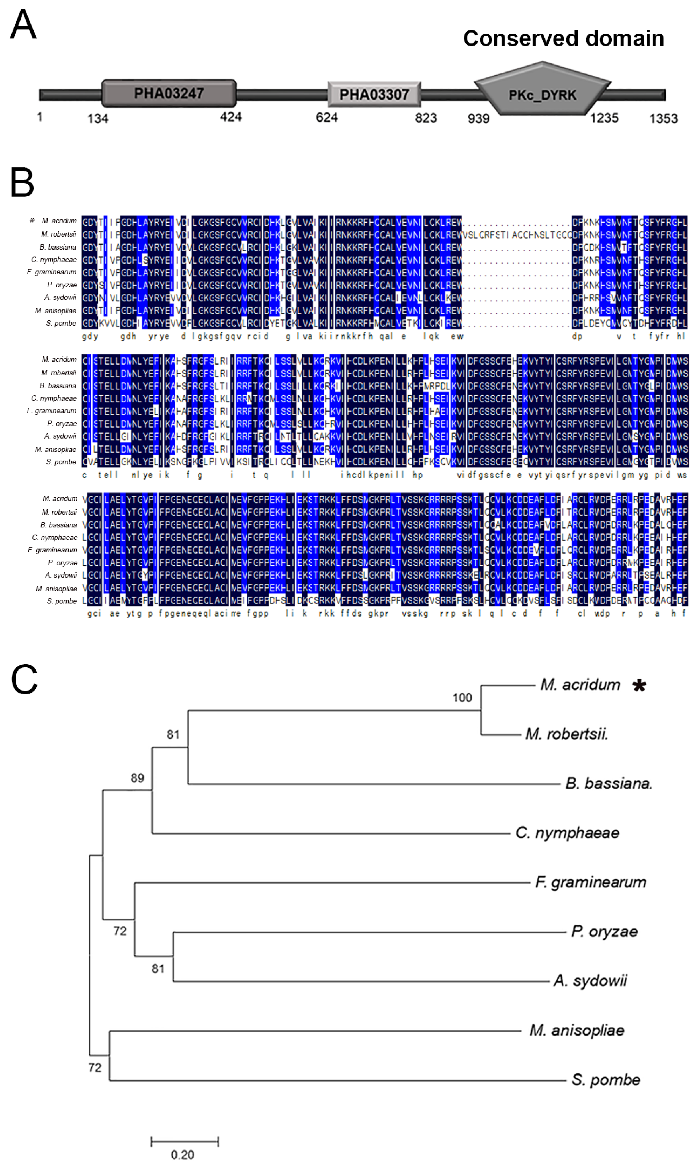
2.1. Characteristics of MaPom1

The total length of MaPom1 is 4122 bp, including a 60 bp intron. MaPom1 kinase consists of 1353 amino acids and has an isoelectric point of 10.08. The MaPom1 protein is composed of three domains: PHA03247, PHA03307, and the catalytic domain of the bispecific protein kinase Pkc_DYRK (Figure 1A). The alignment of the conserved catalytic domain (Pkc_DYRK) revealed that MaPom1 was homologous to kinases found in various fungi (Figure 1B). Phylogenetic tree analysis showed that MaPom1 in M. acridum clustered with Pom1 in M. robertsii (Figure 1C). To explore the function of MaPom1 in M. acridum, we constructed a pK2-PB-MaPom1-LR knockout vector and its corresponding complemented vector, pK2-Sur-MaPom1-eGFP (Figure 2A). Southern blotting was used to confirm the successful generation of the knockout and complemented strains (Figure 2B). To determine whether MaPom1 acts as a serine/threonine protein kinase in M. acridum, we measured the serine/threonine protein kinase activity. The results revealed that the enzyme activity of ΔMaPom1 was lower than that of the WT and CP (F2,6 = 243.3, p < 0.0001; WT vs. ΔMaPom1, p < 0.0001; WT vs. CP, p = 0.0026; ΔMaPom1 vs. CP, p < 0.0001; Tukey’s test, Figure 2C), confirming that MaPom1 serves as a serine/threonine protein kinase.

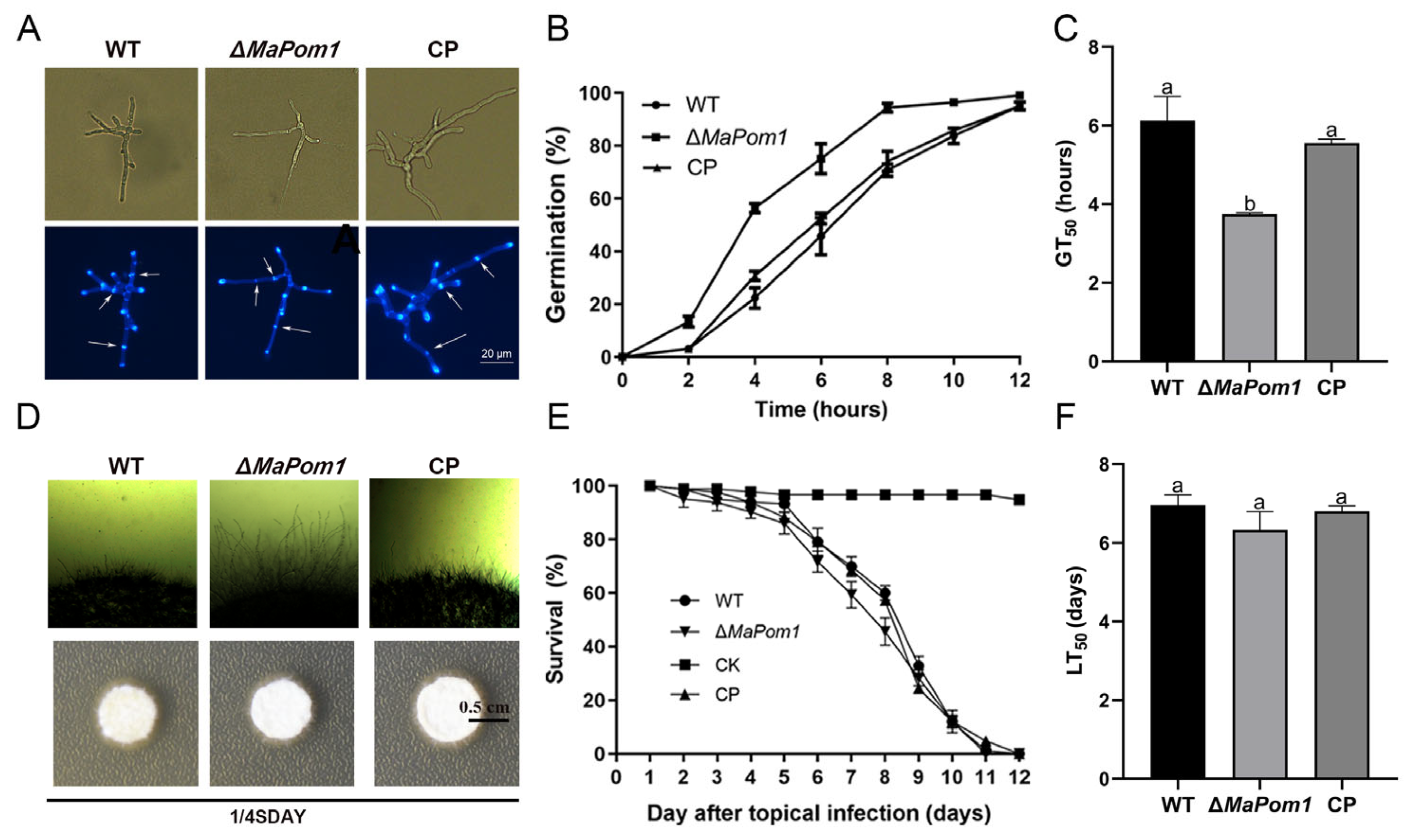
2.2. MaPom1 Contributes to Conidial Germination and Mycelial Growth with No Effect on Conidial Yield and Virulence

To investigate the impact of MaPom1 on the growth and germination of M. acridum, we observed septa and branches after 18 h of cultivation (Figure 3A). The results indicated that the number of septa in ΔMaPom1 (6.67 ± 0.58) was significantly lower than that in WT (14.00 ± 1.00) and CP (13.67 ± 1.15) (F2,6 = 57.88, p = 0.0001; WT vs. ΔMaPom1, p = 0.0002; WT vs. CP, p = 0. 9034; ΔMaPom1 vs. CP, p = 0.0002; Tukey’s test, Figure S1A). Similarly, the number of branches in ΔMaPom1 (4.00 ± 1.00) was significantly lower than that in WT (8.00 ± 1.00) and CP (7.67 ± 0.58) (F2,6 = 19.00, p = 0.0025; WT vs. ΔMaPom1, p = 0.0035; WT vs. CP, p = 0. 8906; ΔMaPom1 vs. CP, p = 0.0054; Tukey’s test, Figure S1B), indicating that MaPom1 affected mycelial division. We also evaluated germination rates at 2, 4, 6, 8, 10, and 12 h and found that ΔMaPom1 had a higher germination rate than WT and CP on 1/4 SDAY medium (Figure 3B). The half-germination time (GT50) of ΔMaPom1 (3.75 ± 0.03 h) was significantly shorter than that of WT (6.13 ± 0.62 h) and CP (5.56 ± 0.10 h) (F2,6 = 35.36, p = 0.0005; WT vs. ΔMaPom1, p = 0.0005; WT vs. CP, p = 0.2068; ΔMaPom1 vs. CP, p = 0.0021; Tukey’s test, Figure 3C), suggesting that MaPom1 regulates conidial germination and mycelial growth. Furthermore, the conidia yield of ΔMaPom1 significantly decreased by 30% compared to WT and CP on the third day, whereas no significant differences in conidial yield were detected after 3 d (Figure S1C). ΔMaPom1 also exhibited a longer mycelium (Figure 3D) that delayed conidiation (Figure S1D). Virulence assays revealed that the survival curves of locusts infected with WT, ΔMaPom1, and CP exhibited similar trends (Figure 3E), and the half-lethal time (LT50) showed no significant difference in the topical assay (F2,6 = 3.160, p = 0.1150; WT vs. ΔMaPom1, p = 0.1150; WT vs. CP, p = 0.8390; ΔMaPom1 vs. CP, p = 0.2377; Tukey’s test, Figure 3F), suggesting that MaPom1 did not influence the virulence of M. acridum.

2.3. MaPom1 Is Essential for Thermal and UV-B Tolerance

To determine whether MaPom1 affects conidial stress tolerance, we measured the conidial germination rate under heat shock (45 °C) and UV-B treatments. The results indicated a significant decrease in both thermal and UV-B tolerance (Figure 4A,D). The half-inhibition time (IT50) for ΔMaPom1 (3.89 ± 0.25 h) was significantly shorter than that of WT (6.21 ± 0.24 h) and CP (6.31 ± 0.43 h) under heat shock treatment (F2,6 = 63.40, p < 0.0001; WT vs. ΔMaPom1, p = 0.0002; WT vs. CP, p = 0.9071; ΔMaPom1 vs. CP, p = 0.0001; Tukey’s test, Figure S2A). There was no difference in the relative expression levels of MaPom1 under heat shock treatment in WT (F2,6 = 0.4471, p = 0.6592; Heat-shock-0 h vs. Heat-shock-3 h, p = 0.8223; Heat-shock-0 h vs. Heat-shock-6 h, p = 0.9437; Heat-shock-3 h vs. Heat-shock-6 h, p = 0.6420; Tukey’s test, Figure 4B). Serine/threonine protein kinase activity was significantly decreased in ΔMaPom1, although it remained unchanged after 3 and 6 h of heat shock treatment (Figure 4C), suggesting that MaPom1 may regulate thermal tolerance by affecting other factors. ΔMaPom1 also exhibited lower UV-B tolerance compared to WT and CP (Figure 4D), with IT50 values of 6.40 ± 0.11 h, 4.91 ± 0.20 h, and 6.73 ± 0.10 h, respectively (F2,6 = 74.38, p < 0.0001; WT vs. ΔMaPom1, p = 0.0002; WT vs. CP, p = 0.1713; ΔMaPom1 vs. CP, p < 0.0001; Tukey’s test, Figure S2B). MaPom1 expression was significantly upregulated in WT after UV-B irradiation (F2,6 = 412.4, p < 0.0001; UV-B-0 h vs. UV-B-3 h, p < 0.0001; UV-B-0 h vs. UV-B-4.5 h, p < 0.0001; UV-B-3 h vs. UV-B-4.5 h, p = 0.9125; Tukey’s test, Figure 4E). In contrast, serine/threonine protein kinase activity in ΔMaPom1 was notably decreased compared to WT and remained consistently lower under UV-B treatment (Figure 4F). These results indicated MaPom1 played a curial role in regulating UV-B tolerance at the transcriptional level.

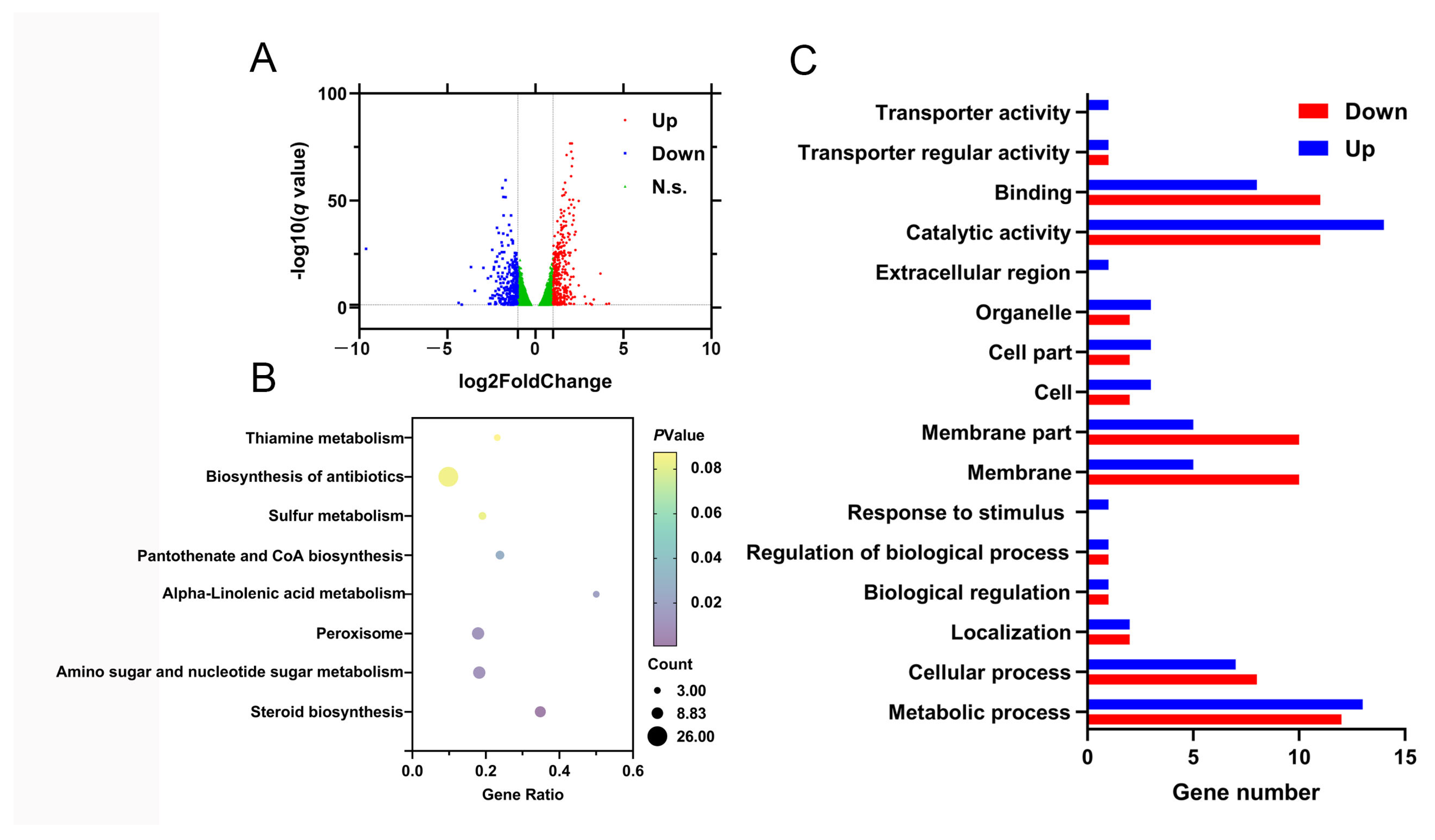
2.4. Identification of DEGs Influenced by MaPom1

To further investigate the mechanism by which MaPom1 regulates stress tolerance, RNA-seq analysis was performed to compare the DEGs in 3-day-old conidia of ΔMaPom1 and WT (the earliest time point at which stress tolerance phenotypes appeared) on 1/4 SDAY medium (Figure S2C,D). The RNA-seq results identified 85 DEGs in ΔMaPom1 compared to WT (|log2 ratio| ≥ 2, q ≤ 0.05) (Table S2), with 41 upregulated and 44 downregulated (Figure 5A). To validate the digital expression profiling, 17 DEGs were selected for expression level determination using qRT-PCR (Table S3). The qRT-PCR results confirmed that 16 of the 17 DEGs exhibited expression patterns consistent with those obtained from RNA-seq, except for MAC_09740, indicating the reliability of the RNA-seq results (Figure S3). Sugar metabolism plays a curial role in regulating the synthesis of cell wall polymers [56]. KEGG pathway analysis revealed the significant enrichment of DEGs in processes related to primary metabolisms, such as amino sugar metabolism, nucleotide sugar metabolism, steroid biosynthesis, peroxisomal functions, α-linolenic acid metabolism, and the biosynthesis pathways of pantothenate and CoA (Figure 5C), all of which may affect stress tolerance. The DEGs were also enriched in 16 GO terms, including six related to biological processes, four related to cellular components, and six related to molecular functions (Figure 5C). The cell wall is a curial component of fungal homeostasis [57]. These GO terms are associated with cell wall and membrane components as well as cell structure and function, which may contribute to reduced stress tolerance.
Further KEGG and GO pathway analyses revealed that the DEGs were primarily related to growth and development (MAC_06007, MAC_09143, and MAC_06365) and other biological activities (Table S2), which could lead to slower germination and fewer branches. Late embryogenesis abundant (LEA) domain-containing proteins provide organisms with evolutionary advantages under various stress environments [58]. Notably, the LEA domain-containing protein gene (MAC_00250) was significantly downregulated, potentially contributing to the reduced stress tolerance. The fungal cell wall, which serves as a physical barrier, is closely related to cell survival, pathogenicity, and resistance [59]. Genes related to cell wall components (MAC_03963, MAC_09810, and MAC_05133) were significantly upregulated in ΔMaPom1, suggesting that the reduced stress tolerance may not be directly related to alterations in the cell wall composition.

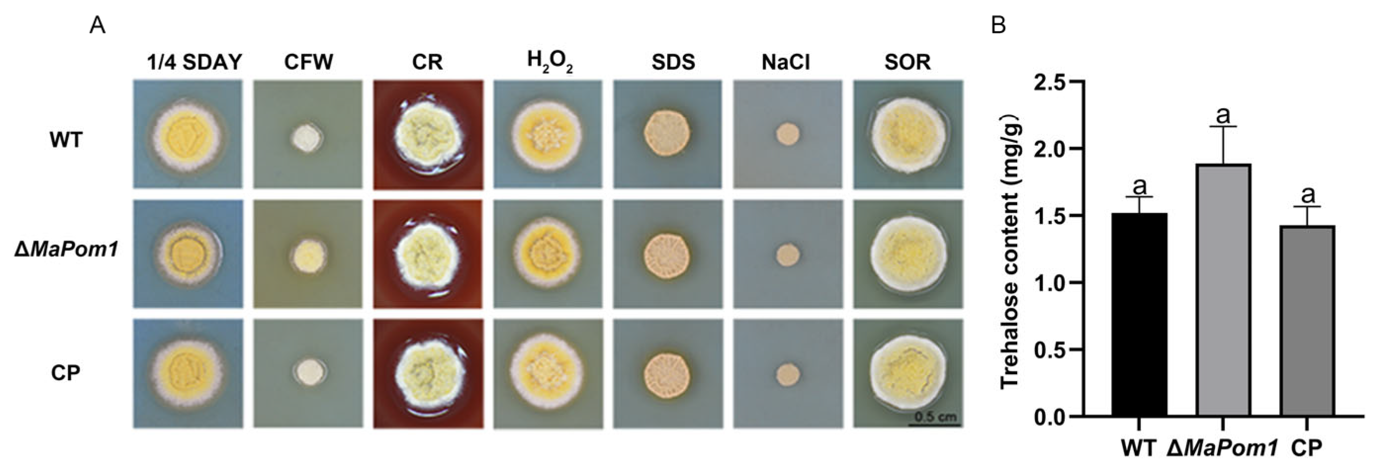
2.5. MaPom1 Contributes to Stress Tolerance Independently of Cell Wall Composition

KEGG and GO annotation analyses revealed that the DEGs were primarily related to cell wall components, including upregulated genes encoding membrane transporter proteins (MAC_09810) and cell wall proteins (MAC_05133), as well as a downregulated gene encoding hydrophobin (MAC_09507). To determine whether the reduced thermal and UV-B tolerance observed in ΔMaPom1 was linked to cell wall composition and integrity, we assessed the sensitivity of fungal strains to cell wall-disrupting agents. The results showed no significant changes in sensitivity (Figure 6A), indicating that stress tolerance reduction was not related to the cell wall. Related studies have suggested that the increased stress tolerance in conidia is associated with trehalose accumulation [60]. Although RNA-Seq data showed that putative trehalose-6-phosphate synthase/trehalose phosphatase (MAC_09143) was upregulated (Table S2), the trehalose content in ΔMaPom1 was not significantly different from that in the WT and CP (F2,6 = 4.839, p = 0.0561; WT vs. ΔMaPom1, p = 0.1234; WT vs. CP, p = 0.8315; ΔMaPom1 vs. CP, p = 0.0586; Tukey’s test, Figure 6B). These results indicated that the decreased thermal and UV-B tolerance in ΔMaPom1 was not related to trehalose metabolism.

2.6. MaPom1 Contributes to UV-B Tolerance by Regulating DNA Damage Repair

RNA-seq analysis indicated that that reduced UV-B tolerance in ΔMaPom1 may be linked to impaired DNA damage repair. To explore this further, we assessed DNA damage after UV-B and heat shock treatments. ΔMaPom1 exhibited significant nuclear dispersion after 3 and 4.5 h of UV-B exposure (Figure 7A), with nuclear dispersion rates reaching 60% and 80%, respectively, which were significantly higher than these in the WT and CP (Figure 7B). Furthermore, WT and CP demonstrated DNA repair after 3 h of recovery cultivation, whereas ΔMaPom1 showed no significant DNA repair (Figure 7C,D), indicating that the reduced UV-B tolerance of ΔMaPom1 is associated with defects in DNA damage repair. To confirm the involvement of DNA damage repair mechanisms, we measured the expression of DNA damage repair-related genes after UV-B exposure by qRT-PCR. Damage-specific DNA binding-protein 1 (DDB1) is a multifunctional protein that recognizes UV-induced DNA damage and plays a crucial role in regulating UV-induced gene transcription. Additionally, DDB1 acts as a damage sensor, helping to maintain genomic integrity and ensuring proper cell cycle progression [61]. The expression levels of a putative DNA-binding protein (MAC_06589), putative O-methyltransferase (MAC_09811), and putative NAD(P)H-dependent oxidoreductase (MAC_05938) were significantly decreased in ΔMaPom1 (Figure 7E), which contribute to its reduced UV-B tolerance. Additionally, high mobility group (HMG) box proteins [62], RNA-binding proteins [63,64], DNA PKcs [65], and flavin adenine dinucleotide (FAD) cofactors [66] are known to participate in the DNA damage response (DDR). In ΔMaPom1, the expression levels of FAD-dependent oxidoreductase family protein (MAC_00180), protein kinase with a PKc-like catalytic domain (MAC_05043), putative RNA-binding protein (MAC_05343), and putative HMG box protein (MAC_04576) were significantly increased (Figure 7E), further supporting the role of MaPom1 in regulating DNA damage repair-related genes in response to UV-B stress. However, DAPI staining after heat shock treatment revealed no significant differences in DNA damage (Figure S4A,B), suggesting that decreased thermotolerance was not related to DNA damage. Heat shock proteins (HSPs), which are highly conserved and expressed under stress conditions to protect cells [24], were significantly downregulated in ΔMaPom1 after 3 h and 6 h of heat shock treatment (Figure S4C), indicating that reduced thermotolerance may be linked to decreased HSP expression.

3. Discussion

3.1. MaPom1 Affects Conidial Germination but Not Virulence and Conidial Yield

To date, numerous studies have explored the functions of Pom1 in fungi. Pom1 regulates cell division and polar growth in yeast [46,67]. Specifically, the DYRK kinase Pom1 is involved in cell division and suppresses cellular proliferation at the cellular terminus in fission yeast [54,68]. Cytoplasmic division is essential for hyphal growth in filamentous fungi [69]. In this study, the reduced number of branches and septa in ΔMaPom1 was consistent with the phenotype observed in yeast [67]. In filamentous fungi, conidia play crucial roles in asexual reproduction and environmental dissemination [70,71,72]. Here, the absence of MaPom1 caused early germination and delayed conidiation without affecting conidial yield or virulence in M. acridum. In constrast, the absence of CsPom1 in C. scovillei significantly reduced conidial production, virulence, septum formation, conidial size and shape, and cell division [52], suggesting that the effects of Pom1 on virulence and conidial yield are not conserved across filamentous fungi.

3.2. MaPom1 Positively Regulates UV-B and Thermal Tolerance Not via Cell Wall Composition

Natural stress factors can affect the survival of entomopathogenic fungi in the environment [73]. Ultraviolet radiation can reduce fungal spore viability, and heat can lower conidial germination rates [13,74]. In this study, the conidial germination rates of ΔMaPom1 decreased significantly under heat shock and UV-B treatments compared to WT. The fungal cell wall maintains cellular homeostasis [75] and responds to environmental pressure, thereby affecting fungal stress tolerance [76]. Here, the sensitivity of ΔMaPom1 to cell wall-disrupting agents has not changed compared to WT and CP. Trehalose, a key stress metabolite, protects fungi from extreme environmental conditions [60,77,78,79] and plays an essential role in promoting productivity, enhancing stress biology, and influencing virulence [80]. However, the trehalose content in ΔMaPom1 was not significantly different from that in WT and CP, indicating that MaPom1 regulates stress tolerance not via cell wall composition. Numerous genes have been reported to influence stress tolerance in M. acridum [81,82]. For instance, MaSep1 negatively affects fungal tolerance to heat shock and UV-B irradiation by regulating the expression of genes related to DNA repair and ROS clearance [83]. In contrast, MaAts enhances fungal tolerance to UV-B irradiation and heat shock by affecting the transcription of genes involved in melanin synthesis, cell wall integrity, and various stress tolerance mechanisms [84]. These findings suggest that different genes regulate stress tolerance in M. acridum through distinct pathways.

3.3. MaPom1 Affects UV-B Tolerance via Regulating the Transcription Level of DNA Damage and Repair Genes

Environmental stressors such as heat shock and UV-B radiation, can cause DNA damage and impede DNA repair [85]. DNA is a primary target of UV-B-induced damage, and cells have developed various repair or tolerance mechanisms to counteract this damage, including light repair, excision repair, mutation repair, recombination repair, cell cycle checkpoints, and apoptosis [86]. The DNA-dependent protein kinase catalytic subunit (DNA-PKcs) is a vital component of the DNA-PK complex, which is crucial for the repair of DNA double-strand breaks via non-homologous end-joining pathways [87]. In this study, UV-B irradiation caused DNA damage in ΔMaPom1 and reduced its ability to recover from DNA damage. In addition, RNA-binding proteins (RBPs) have been shown to significantly influence the DNA damage response (DDR) and DNA repair [88,89], either by participating in DDR [69,70] or by regulating the expression of DDR-related genes [90]. High mobility group box protein 1 (HMGB1) serves as an architectural protein and plays a pivotal role in the assembly of protein–DNA complexes implicated in transcriptional processes, genetic recombination, DNA repair mechanisms, and chromatin structural modifications [91]. The expression levels of DNA damage and repair-related genes were significantly affected in ΔMaPom1 (Table S2), suggesting that the reduced UV-B tolerance of ΔMaPom1 was due to the modulation of genes related to DNA damage and repair, a mechanism similar to the regulatory mechanism in ΔMaSep1 [83]. To sum up, UV-B radiation could cause DNA damage, and the transcription level of DNA damage genes were upregulated, and DNA repair genes downregulated, in ΔMaPom1, indicating that MaPom1 regulated UV-B tolerance via the DNA damage and repair pathway.

3.4. MaPom1 Affects Thermal Tolerance via Regulating the Transcription Level of HSPs

Active fungi respond to stress by producing HSPs and chaperones that facilitate the repair of damaged functional structures [27,92]. Stress often leads to protein misfolding at the cellular level, and HSPs are activated to minimize the accumulation of denatured or abnormal proteins [93]. HSPs respond to environmental stresses by binding to heat shock elements (HSEs), promoting HSP translation, and preventing cellular damage [94]. Heat shock triggers complex cellular responses, including HSP induction, RNA and DNA damage, abnormal protein degradation, and disruptions in cell cycle progression, which ultimately cause cell death [95]. HSPs are highly conserved across organisms and expressed under stressful conditions to protect cells from damage [26,96]. They can serve as biomarkers for assessing cell damage in high-temperature environments [97]. By refolding or degrading misfolded proteins, HSPs play a critical role in repairing stress-induced protein damage [98]. For instance, in Aspergillus fumigatus, Hsp90 is transcriptionally regulated in response to heat shock [99]. In this study, the expression levels of HSPs in ΔMaPom1 were significantly decreased after heat shock treatment, suggesting that HSP gene expression contributed to the diminished heat shock tolerance of the strain. In summary, MaPom1 regulates thermal tolerance via reducing the transcription level of HSPs, which makes a small amount of HSP to protect cells from damage in high-temperature environments.

4. Materials and Methods

4.1. Strains and Cultivation Conditions

The wild-type strain (WT), designated CQMa102, was obtained from the China General Microbiological Culture Collection Center (CGMCC; No. 0877). All fungal strains used in this study were cultivated on a medium comprising one-quarter-strength Sabouraud’s dextrose agar medium (1/4 SDAY). Plasmid construction was performed using Escherichia coli BGT1 obtained from BioGround Biotechnology (Chongqing, China). Fungal transformation was performed by utilizing Agrobacterium tumefaciens AGL-1 [100], which was sourced from Weidi Biotechnology in Shanghai, China.

4.2. Bioinformatic Analysis

All homologous genes and protein sequences of MaPom1 (GenBank accession numbers: MAC_02617 (gene) and XP_007808957.1 (protein)) were downloaded from the NCBI website (https://www.ncbi.nlm.nih.gov/ (accessed on 14 February 2022)). The MaPom1 protein domain was identified using the SMART interface (http://smart.embl.de/ (accessed on 14 February 2022)). The DNAMAN software (https://www.lynnon.com/dnaman.html (accessed on 14 February 2022), Lynnon Biosoft, San Ramon, CA, USA) was used for multiple sequence alignment. A neighbor-joining phylogenetic tree was generated using MEGA 7.0 software (http://www.megasoftware.net/ (accessed on 14 February 2022)), supported by 1000 bootstrap replicates for statistical robustness [101].

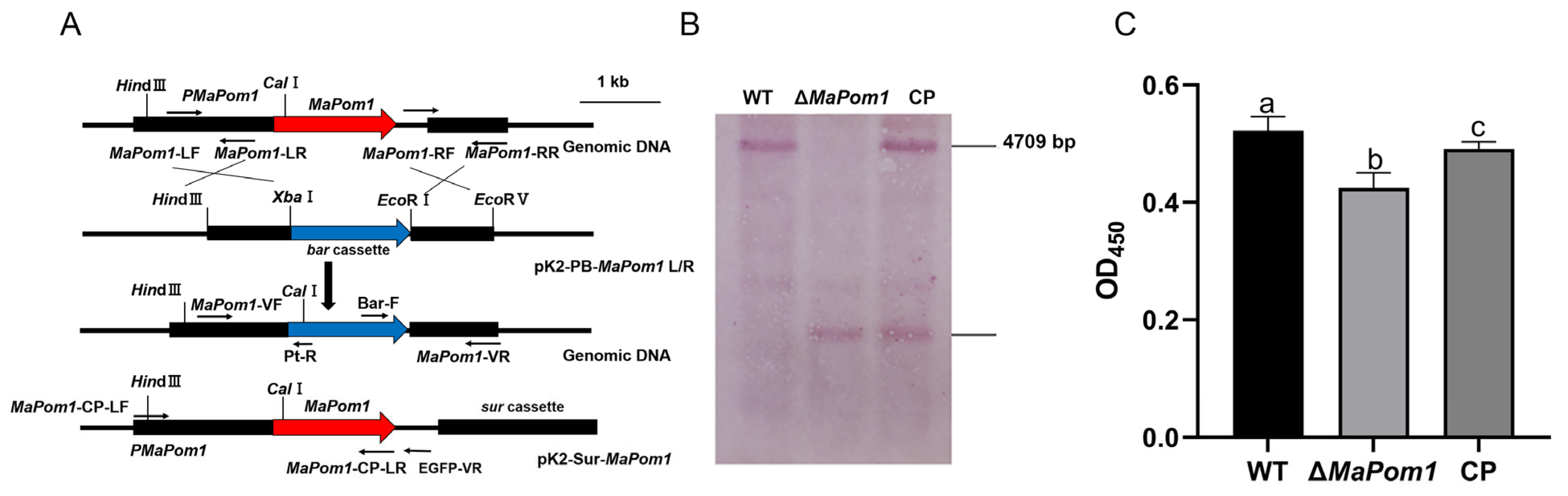
4.3. Construction of MaPom1 Mutants

The left and right arms of the MaPom1 gene were amplified using the primers MaPom1-LF/MaPom1-LR and MaPom1-RF/MaPom1-RR (Table S1), respectively, and inserted into the pK2-PB vector, which contains a phosphinothricin-resistant bar gene, to facilitate the generation of a deletion vector. The obtained vector was transformed into M. acridum via A. tumefaciens [102]. Positive transformants were screened on Czapek–Dox medium supplemented with 500 μg/mL glufosinate ammonium (Sigma-Aldrich, St. Louis, MO, USA). The MaPom1 sequence with its promoter (6111 bp in total) was amplified using the primer pair CP-F/CP-R (Table S1) and inserted into the pK2-Sur vector to yield a complementary (CP) vector. The constructed CP vector was transformed into ΔMaPom1, which was subsequently selected on Czapek–Dox agar supplemented with 20 μg/mL chlorimuron ethyl (Sigma-Aldrich, Bellefonte, PA, USA) and verified by PCR.

4.4. Southern Blotting

Genomic DNA (6 µg) extracted from each strain was subjected to enzymatic digestion with NheI and AflII restriction enzymes, and the digested DNA was separated on 1.0% agarose gel and transferred onto a nylon membrane. The specific probe, spanning 523 bp, was derived from the 5′ genomic region of MaPom1 and amplified using the TF/TR primer pair (Table S1). Subsequent experiments were conducted according to the instructions of the Roche DIG High Prime DNA Labeling and Detection Starter Kit I (Mannheim Roche, Marnheim, Germany).

4.5. Quantitative Reverse Transcription PCR (qRT-PCR)

Total RNA was extracted from conidia using an Ultrapure RNA Kit (with DNase I) (CWBIO, Beijing, China). RNA samples were reverse-transcribed using the PrimeScript RT Reagent Kit with gDNA Eraser (TaKaRa, Beijing, China) according to the manufacturer’s instructions. Gene-specific qRT-PCR primers (Table S1) were designed using the NCBI website (https://www.ncbi.nlm.nih.gov/ (accessed on 14 February 2022)). qRT-PCR was performed with the SYBR Prime qRT-PCR Set (Bio Ground, Chongqing, China) using a two-step method, as per the manufacturer’s protocol.

4.6. Serine/Threonine Kinase Activity Analysis

Total protein was extracted from 0.2 g treated conidia (heat-shock treatment for 0, 3, and 6 h and UV-B treatment for 0, 3, and 4.5 h) using a filamentous fungal protein extraction kit (Bestbio, Shenzhen, China). Protein concentration was measured using a BCA Protein Assay Kit (Beyotime Biotechnology, Nanjing, China) following the manufacturer’s guidelines. The protein concentrations of different samples were adjusted to 10 μg/μL to ensure consistency in enzyme activity measurements, which were conducted using a broad-spectrum serine/threonine kinase assay kit (ELISA, ICP0248, Immune Chem, Vancouver, BC, Canada) [103]. This detection kit is based on the principle of solid-phase enzyme-linked immunosorbent assay (ELISA), using specific peptide substrates of Serine/Threonine kinase (STK) and polyclonal antibodies that can specifically recognize phosphorylated substrates to detect STK activity in the liquid phase. The specific method is as follows: pre-coat the microplate with STK specific peptide substrate, add the test samples to the corresponding well, and start the substrate phosphorylation reaction by adding ATP. After the reaction is complete, a specific antibody is added to the well to bind to a specific phosphorylated substrate, and then the specific antibody is bound to a secondary antibody coupled with peroxidase (HRP). The coupled HRP can act on the substrate of tetramethylbenzidine (TMB) for color development. After the color development process is terminated by an acidic termination solution, the optical density (OD) is measured at 450 nm using Microplate Reader (Berthold Technologies, Winnenden, Germany). The OD450 values are directly proportional to the enzyme activity.

4.7. Microscopic Investigation and Fluorescence Staining

Conidia were stained with calcofluor white (CFW) under dark conditions [104]. Mature conidia of the WT, ΔMaPom1, and CP strains were collected to prepare spore suspensions (2 × 10⁶ conidia/mL), which were then added to 1/4 SDAY medium and incubated for approximately 20 h. DNA damage in WT, ΔMaPom1, and CP was observed using a DAPI staining protocol [105]. Conidial suspensions (50 µL aliquots, 1 × 10⁷ conidia/mL) were plated onto 1/4 SDAY plates for UV-B and heat shock treatments. All samples were observed and photographed using a fluorescence microscope (Nikon Eclipse Ci-E, Tokyo, Japan). (The percentage of cells with nuclear damage was calculated as the ratio of nuclear-damaged cells to the total number of cells).

4.8. Conidial Germination and Conidial Yield Assays

Conidial germination was performed according to a previously established protocol [106]. Aliquots of 50 µL conidial suspensions of WT, ΔMaPom1, and CP (1 × 10⁷ conidia/mL) were plated onto 1/4 SDAY medium for continuous incubation at 28 °C. Conidial germination statistics were recorded every 2 h. Conidial yield assays were performed according to previously established protocols [107]. Aliquots of 2 µL conidial suspensions (1 × 10⁶ conidia/mL) were dipped onto 1/4 SDAY medium for subsequent incubation at 28 °C. Three independent replicates of each individual strain were harvested at three-day intervals from the third day until the fifteenth day. Each experimental procedure was performed three times to ensure consistency and reliability.

4.9. Virulence Assays

Bioassays were performed according to procedures outlined in previous studies [108]. Aliquots of 4 μL conidial suspensions (1 × 10⁷ conidia/mL) prepared in paraffin oil were dipped onto the pronotum of the insects following a previously described protocol (control insects were inoculated with 4 μL paraffin oil). Each experimental group had three replicates, each with 30 insects, and the mortality rates of the locusts were recorded at 12 h intervals. The entire experiment was independently replicated three times.

4.10. Stress Tolerance Assays

Stress tolerance was measured using existing methods [109]. For the UV-B tolerance assay, aliquots of 50 μL suspensions (1 × 10⁷ conidia/mL) were evenly distributed onto 1/4 SDAY medium and subjected to UV-B irradiation (1350 mW/m2) for 1.5, 3, 4.5, and 6 h. The samples were subsequently cultured at 28 °C and harvested at the specified times to record the germination rates. For the thermotolerance assay, 50 μL aliquots of the same suspensions were exposed to 45 °C for 3, 6, 9, and 12 h, spread on 1/4 SDAY medium, and harvested at the indicated times to record the germination rates. To assess sensitivity to abiotic stress, 2 μL aliquots of suspensions (1 × 10⁶ conidia/mL) were placed onto 1/4 SDAY and 1/4 SDAY medium containing various stressors: 6 mM hydrogen peroxide (H2O2), 1 M sorbitol (SOR), 1 M sodium chloride (NaCl), 500 μg/mL Congo red (CR), 50 mg/mL calcofluor white (CFW), and 0.01% sodium dodecyl sulfate (SDS). The plates were incubated at 28 °C for 6 days [110].

4.11. Trehalose Extraction and Quantification Assays

Trehalose extraction was performed according to the protocol provided in the trehalose content assay kit (Solarbio, Beijing, China) [111]. Mature conidia (0.1 g) were extracted using 1 mL extraction solution. The fungal conidia were then crushed by ultrasonic treatment (200 W, 10 s intervals, 3 s of ultrasonication, repeated 30 times) at 4 °C. The mixture was centrifuged at 8000× g for 10 min at room temperature to obtain the supernatant. A 0.25 mL aliquot of the supernatant was transferred to an EP tube and mixed with 1 mL working solution. The samples were incubated at 95 °C for 10 min. The absorbance (A) was measured at 620 nm, and the procedure was repeated three times.

4.12. Digital Gene Expression Profiling (DGE)

Total RNA was extracted from the conidia of the WT and ΔMaPom1 strains cultured on 1/4 SDAY plates for 3 days. RNA sequencing was performed using an Illumina HiSeq 2000 platform with three biological replicates (Beijing Genomics Institute, Chongqing, China). Differentially expressed genes (DEGs) were identified based on criteria of with log2(∆MaPom1-3 d/WT-3 d) ≥ 2 and a false discovery rate (FDR) ≤ 0.001. Gene Ontology (GO) and Kyoto Encyclopedia of Genes and Genomes (KEGG) pathway enrichment analyses were conducted to classify and annotate the obtained DEGs.

4.13. Data Analysis

Data analyses were performed using the SPSS software (version 17.0) (SPSS Inc., Chicago, IL, USA). Graphs were generated with GraphPad Prism version 9.0 for Windows (GraphPad Software, San Diego, CA, USA, www.graphpad.com (accessed on 20 September 2024)). One-way analysis of variance (ANOVA) and Tukey’s test were used to assess the significance of differences between experimental groups. Significance levels were defined as p < 0.05 (*), p < 0.01 (**), and p < 0.001 (***), with “ns” indicating no significant differences.

5. Conclusions

In summary, our study revealed a new function of MaPom1 in the regulation of stress tolerance. Specifically, we identified a potential regulatory mechanism of MaPom1 in stress tolerance. MaPom1 regulated the expression of genes related to DNA damage repair, resulting in reduced UV-B tolerance, and decreased thermotolerance was related to the expression of HSP genes. Overall, MaPom1 contributes to both UV-B tolerance and thermotolerance via distinct pathways. These findings highlight the importance of MaPom1 in stress tolerance and provide new insights into the regulatory mechanisms that enable fungi to withstand abiotic stresses. This study also lays the groundwork for enhancing the efficacy of entomopathogenic fungi for insect control.

Supplementary Materials

The following supporting information can be downloaded at: https://www.mdpi.com/article/10.3390/ijms252211860/s1.

Author Contributions

All authors contributed to conceive and design this research. Material preparation, data collection and analysis were performed by Y.Z., L.S. and Y.X.; Y.Z. and L.S. wrote the first draft of the manuscript and all authors commented on previous versions of the manuscript. All authors have read and agreed to the published version of the manuscript.

Funding

The work was supported by the Technology Innovation and Application Development Project of Chongqing (CSTB2023TIAD-KPX0045), the Fundamental Research Funds for the Central Universities (2023CDJKYJH043, 2024CDJXY016).

Institutional Review Board Statement

Not applicable.

Informed Consent Statement

Not applicable.

Data Availability Statement

The authors confirm that the data supporting the findings of this study are available within this article and its Supplementary Materials. The RNA seq data has been uploaded to NCBI, the ac-cession numbers for these SRA data: SRX11118927-SRX11118932.

Acknowledgments

The authors thank the Engineering Research Center for Fungal Insecticides (School of Life Sciences, Chongqing University) providing the technical platform that supported this study. We also extend our gratitude to Kai Jin for valuable suggestions and guidance regarding this manuscript.

Conflicts of Interest

The authors declare no conflict of interest.

References

  1. Krupa, S.V.; Kickert, R.N. The Greenhouse effect: Impacts of ultraviolet-B (UV-B) radiation, carbon dioxide (CO2), and ozone (O3) on vegetation. Environ. Pollut. 1989, 61, 263–393. [Google Scholar] [CrossRef] [PubMed]
  2. Zepp, R.G.; Erickson, D.J., 3rd; Paul, N.D.; Sulzberger, B. Effects of solar UV radiation and climate change on biogeochemical cycling: Interactions and feedbacks. Photochem. Photobiol. Sci. 2011, 10, 261–279. [Google Scholar] [CrossRef] [PubMed]
  3. Deutsch, C.A.; Tewksbury, J.J.; Tigchelaar, M.; Battisti, D.S.; Merrill, S.C.; Huey, R.B.; Naylor, R.L. Increase in crop losses to insect pests in a warming climate. Science 2018, 361, 916–919. [Google Scholar] [CrossRef] [PubMed]
  4. Fadiji, A.E.; Yadav, A.N.; Santoyo, G.; Babalola, O.O. Understanding the plant-microbe interactions in environments exposed to abiotic stresses: An overview. Microbiol. Res. 2023, 271, 127368. [Google Scholar] [CrossRef]
  5. Coleine, C.; Stajich, J.E.; Selbmann, L. Fungi are key players in extreme ecosystems. Trends Ecol. Evol. 2022, 37, 517–528. [Google Scholar] [CrossRef]
  6. Deka, B.; Baruah, C.; Babu, A. Entomopathogenic microorganisms: Their role in insect pest management. Egypt. J. Biol. Pest Control 2021, 31, 121. [Google Scholar] [CrossRef]
  7. Chen, W.; Xie, W.; Cai, W.; Thaochan, N.; Hu, Q. Entomopathogenic Fungi Biodiversity in the Soil of Three Provinces Located in Southwest China and First Approach to Evaluate Their Biocontrol Potential. J. Fungi 2021, 7, 984. [Google Scholar] [CrossRef]
  8. Roberts, D.W.; St. Leger, R.J. Metarhizium spp., cosmopolitan insect-pathogenic fungi: Mycological aspects. Adv. Appl. Microbiol. 2004, 54, 1–70. [Google Scholar]
  9. Guerrero-Guerra, C.; Reyes-Montes, M.d.R.; Toriello, C.; Hernández-Velázquez, V.; Santiago-López, I.; Mora-Palomino, L.; Calderón-Segura, M.E.; Fernández, S.D.; Calderón-Ezquerro, C. Study of the persistence and viability of Metarhizium acridum in Mexico’s agricultural area. Aerobiologia 2013, 29, 249–261. [Google Scholar] [CrossRef]
  10. Fravel, D.R. Commercialization and Implementation of Biocontrol. Annu. Rev. Phytopathol. 2005, 43, 337–359. [Google Scholar] [CrossRef]
  11. Kumamoto, C.A. Molecular mechanisms of mechanosensing and their roles in fungal contact sensing. Nat. Rev. Microbiol. 2008, 6, 667–673. [Google Scholar] [CrossRef] [PubMed]
  12. Brown, A.J.; Haynes, K.; Quinn, J. Nitrosative and oxidative stress responses in fungal pathogenicity. Curr. Opin. Microbiol. 2009, 12, 384–391. [Google Scholar] [CrossRef] [PubMed]
  13. Braga, G.U.L.; Rangel, D.E.N.; Fernandes, É.K.K.; Flint, S.D.; Roberts, D.W. Molecular and physiological effects of environmental UV radiation on fungal conidia. Curr. Genet. 2015, 61, 405–425. [Google Scholar] [CrossRef] [PubMed]
  14. Papagianni, M. Fungal morphology and metabolite production in submerged mycelial processes. Biotechnol. Adv. 2004, 22, 189–259. [Google Scholar] [CrossRef] [PubMed]
  15. Chandler, D. Chapter 5—Basic and Applied Research on Entomopathogenic Fungi. In Microbial Control of Insect and Mite Pests; Lacey, L.A., Ed.; Academic Press: New York, NY, USA, 2017; pp. 69–89. [Google Scholar]
  16. Alali, S.; Mereghetti, V.; Faoro, F.; Bocchi, S.; Al Azmeh, F.; Montagna, M. Thermotolerant isolates of Beauveria bassiana as potential control agent of insect pest in subtropical climates. PLoS ONE 2019, 14, e0211457. [Google Scholar] [CrossRef]
  17. Ortiz-Urquiza, A.; Luo, Z.; Keyhani, N.O. Improving mycoinsecticides for insect biological control. Appl. Microbiol. Biotechnol. 2015, 99, 1057–1068. [Google Scholar] [CrossRef]
  18. Zhang, L.-B.; Feng, M.-G. Antioxidant enzymes and their contributions to biological control potential of fungal insect pathogens. Appl. Microbiol. Biotechnol. 2018, 102, 4995–5004. [Google Scholar] [CrossRef]
  19. Rangel, D.E.N.; Alder-Rangel, A.; Dadachova, E.; Finlay, R.D.; Kupiec, M.; Dijksterhuis, J.; Braga, G.U.L.; Corrochano, L.M.; Hallsworth, J.E. Fungal stress biology: A preface to the Fungal Stress Responses special edition. Curr. Genet. 2015, 61, 231–238. [Google Scholar] [CrossRef]
  20. Abu Bakar, N.; Karsani, S.A.; Alias, S.A. Fungal survival under temperature stress: A proteomic perspective. PeerJ 2020, 8, e10423. [Google Scholar] [CrossRef]
  21. Kciuk, M.; Marciniak, B.; Mojzych, M.; Kontek, R. Focus on UV-Induced DNA Damage and Repair-Disease Relevance and Protective Strategies. Int. J. Mol. Sci. 2020, 21, 7264. [Google Scholar] [CrossRef]
  22. Gao, L.; Liu, Y.; Sun, H.; Li, C.; Zhao, Z.; Liu, G. Advances in mechanisms and modifications for rendering yeast thermotolerance. J. Biosci. Bioeng. 2016, 121, 599–606. [Google Scholar] [CrossRef] [PubMed]
  23. Moraitis, C.; Curran, B.P. Reactive oxygen species may influence the heat shock response and stress tolerance in the yeast Saccharomyces cerevisiae. Yeast 2004, 21, 313–323. [Google Scholar] [CrossRef] [PubMed]
  24. Goldman, G.H.; McGuire, S.L.; Harris, S.D. The DNA damage response in filamentous fungi. Fungal Genet. Biol. 2002, 35, 183–195. [Google Scholar] [CrossRef] [PubMed]
  25. Goldman, G.H.; Kafer, E. Aspergillus nidulans as a model system to characterize the DNA damage response in eukaryotes. Fungal Genet. Biol. 2004, 41, 428–442. [Google Scholar] [CrossRef] [PubMed]
  26. Hu, C.; Yang, J.; Qi, Z.; Wu, H.; Wang, B.; Zou, F.; Mei, H.; Liu, J.; Wang, W.; Liu, Q. Heat shock proteins: Biological functions, pathological roles, and therapeutic opportunities. MedComm 2022, 3, e161. [Google Scholar] [CrossRef]
  27. Tiwari, S.; Thakur, R.; Shankar, J. Role of Heat-Shock Proteins in Cellular Function and in the Biology of Fungi. Biotechnol. Res. Int. 2015, 2015, 132635. [Google Scholar] [CrossRef]
  28. Xiao, W.; Zhang, J.; Huang, J.; Xin, C.; Li, M.J.; Song, Z. Response and regulatory mechanisms of heat resistance in pathogenic fungi. Appl. Microbiol. Biotechnol. 2022, 106, 5415–5431. [Google Scholar] [CrossRef]
  29. Fabri, J.; Rocha, M.C.; Fernandes, C.M.; Persinoti, G.F.; Ries, L.N.A.; da Cunha, A.F.; Goldman, G.H.; Del Poeta, M.; Malavazi, I. The Heat Shock Transcription Factor HsfA Is Essential for Thermotolerance and Regulates Cell Wall Integrity in Aspergillus fumigatus. Front. Microbiol. 2021, 12, 656548. [Google Scholar] [CrossRef]
  30. Rangel, D.E.; Butler, M.J.; Torabinejad, J.; Anderson, A.J.; Braga, G.U.; Day, A.W.; Roberts, D.W. Mutants and isolates of Metarhizium anisopliae are diverse in their relationships between conidial pigmentation and stress tolerance. J. Invertebr. Pathol. 2006, 93, 170–182. [Google Scholar] [CrossRef]
  31. Pawson, T.; Scott, J.D. Protein phosphorylation in signaling—50 years and counting. Trends Biochem. Sci. 2005, 30, 286–290. [Google Scholar] [CrossRef]
  32. Roskoski, R., Jr. A historical overview of protein kinases and their targeted small molecule inhibitors. Pharmacol. Res. 2015, 100, 1–23. [Google Scholar] [CrossRef] [PubMed]
  33. Pang, K.; Wang, W.; Qin, J.X.; Shi, Z.D.; Hao, L.; Ma, Y.Y.; Xu, H.; Wu, Z.X.; Pan, D.; Chen, Z.S.; et al. Role of protein phosphorylation in cell signaling, disease, and the intervention therapy. MedComm 2022, 3, e175. [Google Scholar] [CrossRef] [PubMed]
  34. Seumen, C.H.T.; Grimm, T.M.; Hauck, C.R. Protein phosphatases in TLR signaling. Cell Commun. Signal. 2021, 19, 45. [Google Scholar] [CrossRef] [PubMed]
  35. Cross, T.G.; Scheel-Toellner, D.; Henriquez, N.V.; Deacon, E.; Salmon, M.; Lord, J.M. Serine/threonine protein kinases and apoptosis. J. Tradit. Chin. Med. 2000, 256, 34–41. [Google Scholar] [CrossRef] [PubMed]
  36. Liu, C.Y.; Liu, W.; Zhao, W.L.; Wang, J.X.; Zhao, X.F. Upregulation of the expression of prodeath serine/threonine protein kinase for programmed cell death by steroid hormone 20-hydroxyecdysone. Apoptosis 2013, 18, 171–187. [Google Scholar] [CrossRef]
  37. Durán, R.; Villarino, A.; Bellinzoni, M.; Wehenkel, A.; Fernandez, P.; Boitel, B.; Cole, S.T.; Alzari, P.M.; Cerveñansky, C. Conserved autophosphorylation pattern in activation loops and juxtamembrane regions of Mycobacterium tuberculosis Ser/Thr protein kinases. Biochem. Biophys. Res. Commun. 2005, 333, 858–867. [Google Scholar] [CrossRef]
  38. Faucher, S.P.; Viau, C.; Gros, P.P.; Daigle, F.; Le Moual, H. The prpZ gene cluster encoding eukaryotic-type Ser/Thr protein kinases and phosphatases is repressed by oxidative stress and involved in Salmonella enterica serovar Typhi survival in human macrophages. FEMS Microbiol. Lett. 2008, 281, 160–166. [Google Scholar] [CrossRef]
  39. Pereira, S.F.; Goss, L.; Dworkin, J. Eukaryote-like serine/threonine kinases and phosphatases in bacteria. Microbiol. Mol. Biol. Rev. 2011, 75, 192–212. [Google Scholar] [CrossRef]
  40. Boni, J.; Rubio-Perez, C.; López-Bigas, N.; Fillat, C.; de la Luna, S. The DYRK Family of Kinases in Cancer: Molecular Functions and Therapeutic Opportunities. Cancers 2020, 12, 2106. [Google Scholar] [CrossRef]
  41. Rammohan, M.; Harris, E.; Bhansali, R.S.; Zhao, E.; Li, L.S.; Crispino, J.D. The chromosome 21 kinase DYRK1A: Emerging roles in cancer biology and potential as a therapeutic target. Oncogene 2022, 41, 2003–2011. [Google Scholar] [CrossRef]
  42. Laham, A.J.; Saber-Ayad, M.; El-Awady, R. DYRK1A: A down syndrome-related dual protein kinase with a versatile role in tumorigenesis. Cell. Mol. Life Sci. 2021, 78, 603–619. [Google Scholar] [CrossRef] [PubMed]
  43. Martin, S.G.; Berthelot-Grosjean, M. Polar gradients of the DYRK-family kinase Pom1 couple cell length with the cell cycle. Nature 2009, 459, 852–856. [Google Scholar] [CrossRef] [PubMed]
  44. Garrett, S.; Menold, M.M.; Broach, J.R. The Saccharomyces cerevisiae YAK1 gene encodes a protein kinase that is induced by arrest early in the cell cycle. Mol. Cell. Biol. 1991, 11, 4045–4052. [Google Scholar] [PubMed]
  45. Aranda, S.; Laguna, A.; de la Luna, S. DYRK family of protein kinases: Evolutionary relationships, biochemical properties, and functional roles. FASEB J. 2011, 25, 449–462. [Google Scholar] [CrossRef] [PubMed]
  46. Bähler, J.; Pringle, J.R. Pom1p, a fission yeast protein kinase that provides positional information for both polarized growth and cytokinesis. Genes Dev. 1998, 12, 1356–1370. [Google Scholar] [CrossRef]
  47. Wood, E.; Nurse, P. Pom1 and cell size homeostasis in fission yeast. Cell Cycle 2013, 12, 3228–3236. [Google Scholar] [CrossRef]
  48. Martin, S.G. Microtubule-dependent cell morphogenesis in the fission yeast. Trends Cell Biol. 2009, 19, 447–454. [Google Scholar] [CrossRef]
  49. Moseley, J.B.; Mayeux, A.; Paoletti, A.; Nurse, P. A spatial gradient coordinates cell size and mitotic entry in fission yeast. Nature 2009, 459, 857–860. [Google Scholar] [CrossRef]
  50. Taminato, A.; Bagattini, R.; Gorjão, R.; Chen, G.; Kuspa, A.; Souza, G.M. Role for YakA, cAMP, and Protein Kinase A in Regulation of Stress Responses of Dictyostelium discoideum Cells. Mol. Biol. Cell 2002, 13, 2266–2275. [Google Scholar] [CrossRef]
  51. Zhou, X.; Ye, J.; Zheng, L.; Jiang, P.; Lu, L. A new identified suppressor of Cdc7p/SepH kinase, PomA, regulates fungal asexual reproduction via affecting phosphorylation of MAPK-HogA. PLoS Genet. 2019, 15, e1008206. [Google Scholar] [CrossRef]
  52. Shin, J.H.; Kim, H.Y.; Fu, T.; Lee, K.H.; Kim, K.S. CsPOM1, a DYRK Family Kinase, Plays Diverse Roles in Fungal Development, Virulence, and Stress Tolerance in the Anthracnose Pathogen Colletotrichum scovillei. Front. Cell. Infect. Microbiol. 2022, 12, 861915. [Google Scholar] [CrossRef] [PubMed]
  53. Gerganova, V.; Bhatia, P.; Vincenzetti, V.; Martin, S.G. Direct and indirect regulation of Pom1 cell size pathway by the protein phosphatase 2C Ptc1. Mol. Biol. Cell 2021, 32, 703–711. [Google Scholar] [CrossRef] [PubMed]
  54. Hachet, O.; Berthelot-Grosjean, M.; Kokkoris, K.; Vincenzetti, V.; Moosbrugger, J.; Martin, S.G. A Phosphorylation Cycle Shapes Gradients of the DYRK Family Kinase Pom1 at the Plasma Membrane. Cell 2011, 145, 1116–1128. [Google Scholar] [CrossRef] [PubMed]
  55. MacAlpine, J.; Liu, Z.; Hossain, S.; Whitesell, L.; Robbins, N.; Cowen, L.E. DYRK-family kinases regulate Candida albicans morphogenesis and virulence through the Ras1/PKA pathway. mBio 2023, 14, e02183-23. [Google Scholar] [CrossRef] [PubMed]
  56. Yaakoub, H.; Latgé, J.P.; Papon, N. Central Sugar Metabolism and the Cell Wall. mBio 2022, 13, e0210422. [Google Scholar] [CrossRef]
  57. Lima, S.L.; Colombo, A.L.; de Almeida Junior, J.N. Fungal Cell Wall: Emerging Antifungals and Drug Resistance. Front. Microbiol. 2019, 10, 2573. [Google Scholar] [CrossRef]
  58. Hundertmark, M.; Hincha, D.K. LEA (Late Embryogenesis Abundant) proteins and their encoding genes in Arabidopsis thaliana. BMC Genom. 2008, 9, 118. [Google Scholar] [CrossRef]
  59. Díaz-Jiménez, D.F.; Pérez-García, L.A.; Martínez-Álvarez, J.A.; Mora-Montes, H.M. Role of the Fungal Cell Wall in Pathogenesis and Antifungal Resistance. Curr. Fungal Infect. Rep. 2012, 6, 275–282. [Google Scholar] [CrossRef]
  60. Rangel, D.E.; Anderson, A.J.; Roberts, D.W. Evaluating physical and nutritional stress during mycelial growth as inducers of tolerance to heat and UV-B radiation in Metarhizium anisopliae conidia. Mycol. Res. 2008, 112, 1362–1372. [Google Scholar] [CrossRef]
  61. Iovine, B.; Iannella, M.L.; Bevilacqua, M.A. Damage-specific DNA binding protein 1 (DDB1): A protein with a wide range of functions. Int. J. Biochem. Cell Biol. 2011, 43, 1664–1667. [Google Scholar] [CrossRef]
  62. Malarkey, C.S.; Churchill, M.E. The high mobility group box: The ultimate utility player of a cell. Trends Biochem. Sci. 2012, 37, 553–562. [Google Scholar] [CrossRef] [PubMed]
  63. Zhang, N.; Kaur, R.; Lu, X.; Shen, X.; Li, L.; Legerski, R.J. The Pso4 mRNA Splicing and DNA Repair Complex Interacts with WRN for Processing of DNA Interstrand Cross-links. J. Biol. Chem. 2005, 280, 40559–40567. [Google Scholar] [CrossRef]
  64. Mahajan, K.N.; Mitchell, B.S. Role of human Pso4 in mammalian DNA repair and association with terminal deoxynucleotidyl transferase. Proc. Natl. Acad. Sci. USA 2003, 100, 10746–10751. [Google Scholar] [CrossRef] [PubMed]
  65. Yue, X.; Bai, C.; Xie, D.; Ma, T.; Zhou, P.K. DNA-PKcs: A Multi-Faceted Player in DNA Damage Response. Front. Genet. 2020, 11, 607428. [Google Scholar] [CrossRef] [PubMed]
  66. Zhang, M.; Wang, L.; Zhong, D. Photolyase: Dynamics and electron-transfer mechanisms of DNA repair. Arch. Biochem. Biophys. 2017, 632, 158–174. [Google Scholar] [CrossRef] [PubMed]
  67. Kettenbach, A.N.; Deng, L.; Wu, Y.; Baldissard, S.; Adamo, M.E.; Gerber, S.A.; Moseley, J.B. Quantitative phosphoproteomics reveals pathways for coordination of cell growth and division by the conserved fission yeast kinase pom1. Mol. Cell. Proteom. 2015, 14, 1275–1287. [Google Scholar] [CrossRef]
  68. Padte, N.N.; Martin, S.G.; Howard, M.; Chang, F. The cell-end factor pom1p inhibits mid1p in specification of the cell division plane in fission yeast. Curr. Biol. 2006, 16, 2480–2487. [Google Scholar] [CrossRef]
  69. Harris, S.D. Septum formation in Aspergillus nidulans. Curr. Opin. Microbiol. 2001, 4, 736–739. [Google Scholar] [CrossRef]
  70. Meyer, M.; Cox, J.A.; Hitchings, M.D.T.; Burgin, L.; Hort, M.C.; Hodson, D.P.; Gilligan, C.A. Quantifying airborne dispersal routes of pathogens over continents to safeguard global wheat supply. Nat. Plants 2017, 3, 780–786. [Google Scholar] [CrossRef]
  71. Vidal, T.; Lusley, P.; Leconte, M.; de Vallavieille-Pope, C.; Huber, L.; Saint-Jean, S. Cultivar architecture modulates spore dispersal by rain splash: A new perspective to reduce disease progression in cultivar mixtures. PLoS ONE 2017, 12, e0187788. [Google Scholar] [CrossRef]
  72. Huang, M.; Hebert, A.S.; Coon, J.J.; Hull, C.M. Protein Composition of Infectious Spores Reveals Novel Sexual Development and Germination Factors in Cryptococcus. PLoS Genet. 2015, 11, e1005490. [Google Scholar] [CrossRef] [PubMed]
  73. Wong, H.J.; Mohamad-Fauzi, N.; Rizman-Idid, M.; Convey, P.; Alias, S.A. Protective mechanisms and responses of micro-fungi towards ultraviolet-induced cellular damage. Polar Sci. 2019, 20, 19–34. [Google Scholar] [CrossRef]
  74. Alves, R.T.; Bateman, R.P.; Prior, C.; Leather, S.R. Effects of simulated solar radiation on conidial germination of Metarhizium anisopliae in different formulations. Crop Prot. 1998, 17, 675–679. [Google Scholar] [CrossRef]
  75. Latgé, J.P. The cell wall: A carbohydrate armour for the fungal cell. Mol. Microbiol. 2007, 66, 279–290. [Google Scholar] [CrossRef] [PubMed]
  76. Gow, N.A.R.; Latge, J.P.; Munro, C.A. The Fungal Cell Wall: Structure, Biosynthesis, and Function. Microbiol. Spectr. 2017, 5, 28513415. [Google Scholar] [CrossRef]
  77. Estruch, F. Stress-controlled transcription factors, stress-induced genes and stress tolerance in budding yeast. FEMS Microbiol. Rev. 2000, 24, 469–486. [Google Scholar] [CrossRef]
  78. François, J.; Parrou, J.L. Reserve carbohydrates metabolism in the yeast Saccharomyces cerevisiae. FEMS Microbiol. Rev. 2001, 25, 125–145. [Google Scholar] [CrossRef]
  79. Rangel, D.E.; Braga, G.U.; Fernandes, É.K.; Keyser, C.A.; Hallsworth, J.E.; Roberts, D.W. Stress tolerance and virulence of insect-pathogenic fungi are determined by environmental conditions during conidial formation. Curr. Genet. 2015, 61, 383–404. [Google Scholar] [CrossRef]
  80. Ribeiro, G.D.; de Holanda Paranhos, L.; Eleutherio, E.C.A. Trehalose promotes biological fitness of fungi. Fungal Biol. 2024; in press. [Google Scholar] [CrossRef]
  81. Li, C.; Zhang, Q.; Xia, Y.; Jin, K. MaNmrA, a Negative Transcription Regulator in Nitrogen Catabolite Repression Pathway, Contributes to Nutrient Utilization, Stress Resistance, and Virulence in Entomopathogenic Fungus Metarhizium acridum. Biology 2021, 10, 1167. [Google Scholar] [CrossRef]
  82. Li, C.; Zhang, Q.; Xia, Y.; Jin, K. MaAreB, a GATA Transcription Factor, Is Involved in Nitrogen Source Utilization, Stress Tolerances and Virulence in Metarhizium acridum. J. Fungi 2021, 7, 512. [Google Scholar] [CrossRef] [PubMed]
  83. Song, T.; Li, C.; Jin, K.; Xia, Y. The Forkhead Box Gene, MaSep1, Negatively Regulates UV- and Thermo-Tolerances and Is Required for Microcycle Conidiation in Metarhizium acridum. J. Fungi 2024, 10, 544. [Google Scholar] [CrossRef] [PubMed]
  84. Song, L.; Xue, X.; Wang, S.; Li, J.; Jin, K.; Xia, Y. MaAts, an Alkylsulfatase, Contributes to Fungal Tolerances against UV-B Irradiation and Heat-Shock in Metarhizium acridum. J. Fungi 2022, 8, 270. [Google Scholar] [CrossRef] [PubMed]
  85. Mohammadi, B.; Safaiyan, A.; Habibi, P.; Moradi, G. Evaluation of the acoustic performance of polyurethane foams embedded with rock wool fibers at low-frequency range; design and construction. Appl. Acoust. 2021, 182, 108223. [Google Scholar] [CrossRef]
  86. Sinha, R.P.; Häder, D.P. UV-induced DNA damage and repair: A review. Photochem. Photobiol. Sci. 2002, 1, 225–236. [Google Scholar] [CrossRef]
  87. Yang, H.; Yao, F.; Marti, T.M.; Schmid, R.A.; Peng, R.-W. Beyond DNA Repair: DNA-PKcs in Tumor Metastasis, Metabolism and Immunity. Cancers 2020, 12, 3389. [Google Scholar] [CrossRef]
  88. Matsuoka, S.; Ballif, B.A.; Smogorzewska, A.; McDonald, E.R., 3rd; Hurov, K.E.; Luo, J.; Bakalarski, C.E.; Zhao, Z.; Solimini, N.; Lerenthal, Y.; et al. ATM and ATR substrate analysis reveals extensive protein networks responsive to DNA damage. Science 2007, 316, 1160–1166. [Google Scholar] [CrossRef]
  89. Meng, X.; Wang, Q.; Hao, R.; Li, X.; Li, M.; Hu, R.; Du, H.; Hu, Z.; Yu, B.; Li, S. RNA-binding protein MAC5A interacts with the 26S proteasome to regulate DNA damage response in Arabidopsis. Plant Physiol. 2023, 191, 446–462. [Google Scholar] [CrossRef]
  90. Paronetto, M.P.; Miñana, B.; Valcárcel, J. The Ewing sarcoma protein regulates DNA damage-induced alternative splicing. Mol. Cell 2011, 43, 353–368. [Google Scholar] [CrossRef]
  91. Blair, R.H.; Horn, A.E.; Pazhani, Y.; Grado, L.; Goodrich, J.A.; Kugel, J.F. The HMGB1 C-Terminal Tail Regulates DNA Bending. J. Mol. Biol. 2016, 428, 4060–4072. [Google Scholar] [CrossRef]
  92. Bai, Y.; Wang, S.; Zhong, H.; Yang, Q.; Zhang, F.; Zhuang, Z.; Yuan, J.; Nie, X.; Wang, S. Integrative analyses reveal transcriptome-proteome correlation in biological pathways and secondary metabolism clusters in A. flavus in response to temperature. Sci. Rep. 2015, 5, 14582. [Google Scholar] [CrossRef] [PubMed]
  93. Bhat, S.; Kumar, P.; Kashyap, N.; Deshmukh, B.; Dige, M.S.; Bhushan, B.; Chauhan, A.; Kumar, A.; Singh, G. Effect of heat shock protein 70 polymorphism on thermotolerance in Tharparkar cattle. Vet. World 2016, 9, 113–117. [Google Scholar] [CrossRef] [PubMed]
  94. Shan, Q.; Ma, F.; Wei, J.; Li, H.; Ma, H.; Sun, P. Physiological Functions of Heat Shock Proteins. Curr. Protein Pept. Sci. 2020, 21, 751–760. [Google Scholar] [CrossRef] [PubMed]
  95. Tabuchi, Y.; Furusawa, Y.; Kariya, A.; Wada, S.; Ohtsuka, K.; Kondo, T. Common gene expression patterns responsive to mild temperature hyperthermia in normal human fibroblastic cells. Int. J. Hyperth. 2013, 29, 38–50. [Google Scholar] [CrossRef] [PubMed]
  96. Khandia, R.; Munjal, A.K.; Iqbal, H.M.N.; Dhama, K. Heat Shock Proteins: Therapeutic Perspectives in Inflammatory Disorders. Recent Pat. Inflamm. Allergy Drug Discov. 2017, 10, 94–104. [Google Scholar] [CrossRef]
  97. Wu, T.; Chen, S.; Xiao, C.; Wang, C.; Pan, Q.; Wang, Z.; Xie, M.; Mao, Z.; Wu, Y.; Tanguay, R.M. Presence of antibody against the inducible Hsp71 in patients with acute heat-induced illness. Cell Stress Chaperones 2001, 6, 113–120. [Google Scholar] [CrossRef]
  98. Alagar Boopathy, L.R.; Jacob-Tomas, S.; Alecki, C.; Vera, M. Mechanisms tailoring the expression of heat shock proteins to proteostasis challenges. J. Biol. Chem. 2022, 298, 101796. [Google Scholar] [CrossRef]
  99. Lamoth, F.; Juvvadi, P.R.; Fortwendel, J.R.; Steinbach, W.J. Heat Shock Protein 90 Is Required for Conidiation and Cell Wall Integrity in Aspergillus fumigatus. Eukaryot. Cell 2012, 11, 1324–1332. [Google Scholar] [CrossRef]
  100. Lazo, G.R.; Stein, P.A.; Ludwig, R.A. A DNA Transformation–Competent Arabidopsis Genomic Library in Agrobacterium. Nat. Biotechnol. 1991, 9, 963–967. [Google Scholar] [CrossRef]
  101. Schwede, T.; Kopp, J.; Guex, N.; Peitsch, M.C. SWISS-MODEL: An automated protein homology-modeling server. Nucleic Acids Res. 2003, 31, 3381–3385. [Google Scholar] [CrossRef]
  102. Leclerque, A.; Wan, H.; Abschütz, A.; Chen, S.; Mitina, G.; Zimmermann, G.; Schairer, H. Agrobacterium-mediated insertional mutagenesis (AIM) of the entomopathogenic fungus Beauveria bassiana. Curr. Genet. 2004, 45, 111–119. [Google Scholar] [CrossRef] [PubMed]
  103. Drexler, C. HTRF® KinEASETM: A new solution for screening serine-threonine kinases. Nat. Methods 2006, 3, I–II. [Google Scholar] [CrossRef]
  104. Zhao, T.; Wen, Z.; Xia, Y.; Jin, K. The transmembrane protein MaSho1 negatively regulates conidial yield by shifting the conidiation pattern in Metarhizium acridum. Appl. Microbiol. Biotechnol. 2020, 104, 4005–4015. [Google Scholar] [CrossRef] [PubMed]
  105. Zhang, J.; Wang, Z.; Keyhani, N.O.; Peng, G.; Jin, K.; Xia, Y. The protein phosphatase gene MaPpt1 acts as a programmer of microcycle conidiation and a negative regulator of UV-B tolerance in Metarhizium acridum. Appl. Microbiol. Biotechnol. 2019, 103, 1351–1362. [Google Scholar] [CrossRef] [PubMed]
  106. Liu, J.; Cao, Y.; Xia, Y. Mmc, a gene involved in microcycle conidiation of the entomopathogenic fungus Metarhizium anisopliae. J. Invertebr. Pathol. 2010, 105, 132–138. [Google Scholar] [CrossRef]
  107. Li, C.; Xu, D.; Hu, M.; Zhang, Q.; Xia, Y.; Jin, K.J.M.S. MaNCP1, a C2H2 Zinc Finger Protein, Governs the Conidiation Pattern Shift through Regulating the Reductive Pathway for Nitric Oxide Synthesis in the Filamentous Fungus Metarhizium acridum. Microbiol. Spectr. 2022, 10, e00538-22. [Google Scholar] [CrossRef]
  108. Ming, Y.; Wei, Q.; Jin, K.; Xia, Y. MaSnf1, a sucrose non-fermenting protein kinase gene, is involved in carbon source utilization, stress tolerance, and virulence in Metarhizium acridum. Appl. Microbiol. Biotechnol. 2014, 98, 10153–10164. [Google Scholar] [CrossRef]
  109. Braga, G.; Flint, S.; Miller, C.; Anderson, A.; Roberts, D. Variability in Response to UV-B among Species and Strains of Metarhizium Isolated from Sites at Latitudes from 61°N to 54°S. J. Invertebr. Pathol. 2001, 78, 98–108. [Google Scholar] [CrossRef]
  110. Li, J.; Guo, M.; Cao, Y.; Xia, Y. Disruption of a C69-Family Cysteine Dipeptidase Gene Enhances Heat Shock and UV-B Tolerances in Metarhizium acridum. Front. Microbiol. 2020, 11, 849. [Google Scholar] [CrossRef]
  111. Zhou, J.; Wang, S.; Xia, Y.; Peng, G. MaAzaR, a Zn2Cys6/Fungus-Specific Transcriptional Factor, Is Involved in Stress Tolerance and Conidiation Pattern Shift in Metarhizium acridum. J. Fungi 2024, 10, 468. [Google Scholar] [CrossRef]
Figure 1. Characteristics of MaPom1. (A) Main domains of the Pom1 protein. PKc_DYRK: A serine/threonine protein kinase conserved domain. (B) Alignment of the conserved domains of the Pom1 protein with homologous proteins from other fungi, including Metarhizium acridum (XP_007808957.1), Metarhizium robertsii (EXV02279.1), Beauveria bassiana (KAF1729971.1), Colletotrichum nymphaeae (KXH60025.1), Fusarium graminearum (EYB25727.1), Pyricularia oryzae (ELQ36107.1), Aspergillus sydowii (OJJ58174.1), Metarhizium anisopliae (KAF5127571.1), and Schizosaccharomyces pombe (NP_592974.1). The asterisk represents the M. acridum used in this study. (C) Phylogenetic tree was reconstructed using the neighbor-joining method by the MEGA 7.0 software.
Ijms 25 11860 g001
Figure 2. Design and verification of MaPom1 knockout and complementary strains. (A) Construction of homologous recombination vectors and the random insertion principle in M. acridum. (B) Southern blot verification of the wild-type and mutant strains. Wild-type and mutant strains were digested with NheI and AflII. The probe sequence on the MaPom1 gene was amplified using TF/TR (Table S1), yielding a product with a length of 523 bp. WT: wild-type strain; ΔMaPom1: knockout strain; CP: complementary strain. (C) Determination of serine/threonine protein kinase activity. The OD450 values are directly proportional to the enzyme activity. Error bars represent standard deviations based on three independent replicates. The different lowercase letters indicate significant differences by one-way ANOVA and Tukey’s test.
Ijms 25 11860 g002
Figure 3. Conidial germination, mycelial growth, and virulence assays. (A) Mycelial morphology on 1/4 SDAY medium after 18 h of incubation at 28 °C. Mycelia were stained with CFW (Calcofluor white) to visualize the septa; the arrows represent hyphal septa. (B) Conidial germination rates on 1/4 SDAY medium at 28 °C. (C) Half-germination time (GT50) of each strain. (D) Observation of hyphal length and colony morphology on 1/4 SDAY medium for 3 d at 28 °C. (E) Survival rates of locust inoculated with 4 µL conidial suspensions. (F) Half-lethal time (LT50) after topical inoculation with conidia from each strain. Error bars represent standard deviations based on three independent replicates. The different lowercase letters indicate significant differences by one-way ANOVA and Tukey’s test.
Ijms 25 11860 g003
Figure 4. Stress tolerance assays. (A) Germination rates on 1/4 SDAY medium after 20 h of cultivation following heat shock treatment for 3, 6, 9, and 12 h. (B) Relative expression levels of MaPom1 after heat shock treatment (45 °C) for 0, 3, and 6 h. (C) Serine/threonine protein kinase activity after heat shock treatment for 0, 3, and 6 h. The OD450 values are directly proportional to the enzyme activity. (D) Germination rates on 1/4 SDAY medium after 20 h of cultivation following UV-B irradiation for 1.5, 3, 4.5, and 6 h. (E) Relative expression levels of MaPom1 after UV-B irradiation (1350 mW/m2) for 0, 3, and 4.5 h. (F) Serine/threonine protein kinase activity after UV-B irradiation for 0, 3, and 4.5 h. The OD450 values are directly proportional to the enzyme activity. Error bars represent standard deviations based on three independent replicates. Asterisks indicate significant differences at p < 0.05 (*), p < 0.01 (**), while “ns” indicates no significant differences, and the different lowercase letters indicate significant differences by one-way ANOVA and Tukey test.
Ijms 25 11860 g004
Figure 5. DEGs analysis of WT and ΔMaPom1. (A) Volcano plot of DEGs from ΔMaPom1-3 d vs. WT-3 d. (B) GO annotation of DEGs from ΔMaPom1-3 d vs. WT-3 d. (C) KEGG annotation of DEGs from ΔMaPom1-3 d vs. WT-3 d.
Ijms 25 11860 g005
Figure 6. Stress tolerance and trehalose content analysis. (A) Fungal colonies on 1/4 SDAY medium supplemented with 0.01% w/v SDS, 500 μg/mL CR, 1 mol/L NaCl, 50 μg/mL CFW, 6 mM H2O2, and 1 mol/L SOR. (B) Trehalose content in 15-day-old conidia. Error bars represent standard deviations based on three independent replicates. The same lowercase letters indicate no significant difference.
Ijms 25 11860 g006
Figure 7. Analysis of DNA damage and repair after UV-B irradiation. (A) DNA staining with DAPI after UV-B irradiation for different periods. “UV-0 h” denotes no UV-B treatment, “UV-3 h” denotes UV-B treatment for 3 h, and “UV-4.5 h” denotes UV-B treatment for 4.5 h. Arrows represent stained nuclear DNA. (B) Percentage of DNA damage after UV-B treatment. (C) Degree of DNA damage repair in conidia cultured on 1/4 SDAY medium for 0, 3, 6, and 9 h at 28 °C after UV-B irradiation for 4.5 h. (D) Percentage of DNA damage after 4.5 h of UV-B treatment and subsequent culture on 1/4 SDAY medium for 0, 3, 6, and 9 h at 28 °C. “UV-4.5 h-0 h” indicates conidia without recovery on 1/4 SDAY medium after 4.5 h of UV-B treatment, “UV-4.5 h-3 h” indicates conidia recovery on 1/4 SDAY medium for 3 h after UV-B treatment for 4.5 h, “UV-4.5 h-6 h” indicates conidia recovery on 1/4 SDAY medium for 6 h after UV-B treatment for 4.5 h, and “UV-4.5 h-9 h” indicates conidia recovery on 1/4 SDAY medium for 9 h after UV-B treatment for 4.5 h. (E) Relative expression levels of DNA damage repair-related genes after UV-B treatment for 3 and 4.5 h. “WT-UV-3/4.5 h” indicates conidia of WT after UV-B treatment for 3, 4.5 h. “ΔMaPom1-UV-3 h” and “ΔMaPom1-UV-4.5 h” represent conidia of WT subjected to UV-B treatment for 3 h or 4.5 h, respectively. The relative expression levels of different genes in WT were normalized to 1 after different treatments. All the images were acquired at a fixed exposure time. Error bars represent standard deviations based on three independent replicates. Asterisks indicate significant differences at p < 0.05 (*), p < 0.01 (**), p < 0.01 (***), while “ns” indicates no significant differences by one-way ANOVA and Tukey’s test.
Ijms 25 11860 g007
Disclaimer/Publisher’s Note: The statements, opinions and data contained in all publications are solely those of the individual author(s) and contributor(s) and not of MDPI and/or the editor(s). MDPI and/or the editor(s) disclaim responsibility for any injury to people or property resulting from any ideas, methods, instructions or products referred to in the content.

