

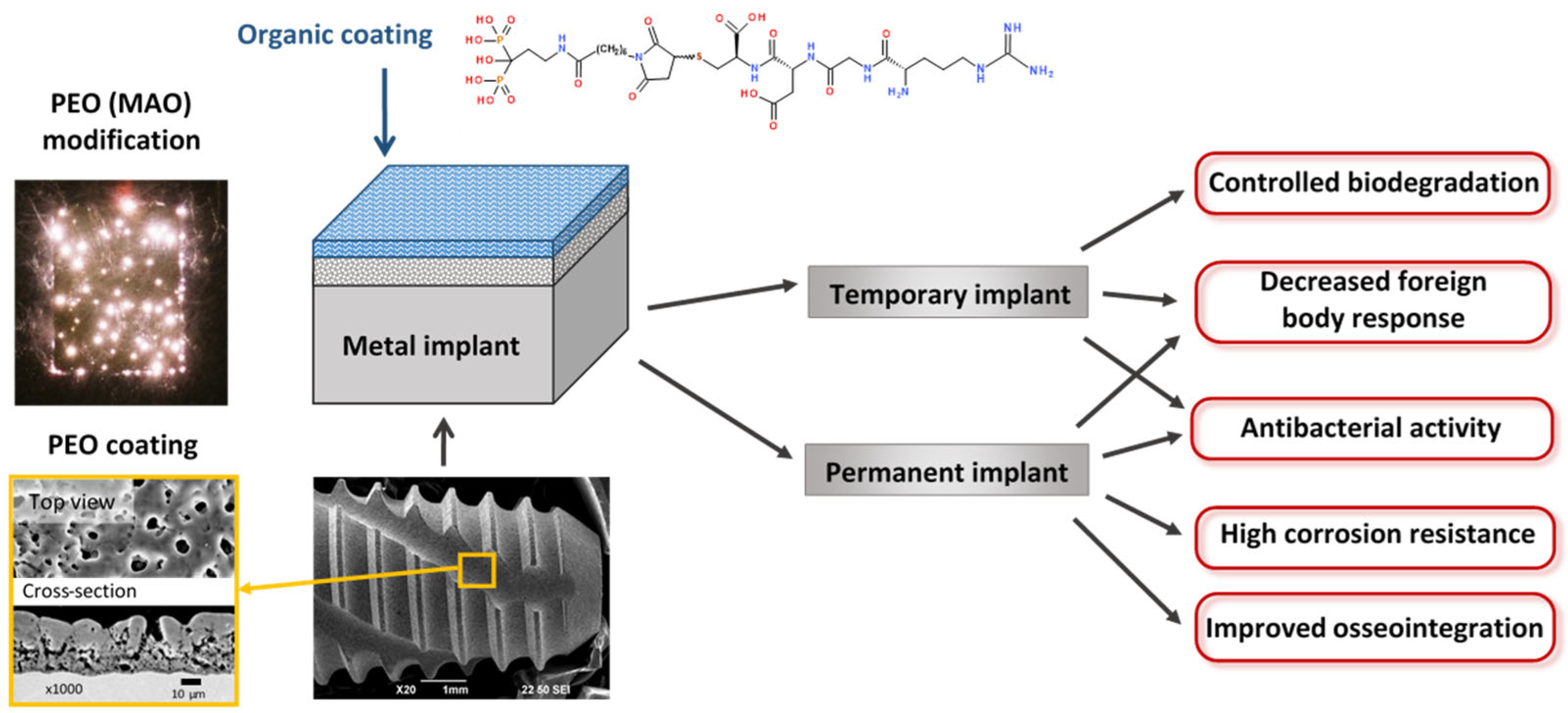
Organic-Inorganic Biocompatible Coatings for Temporary and Permanent Metal Implants


Abstract
1. Introduction
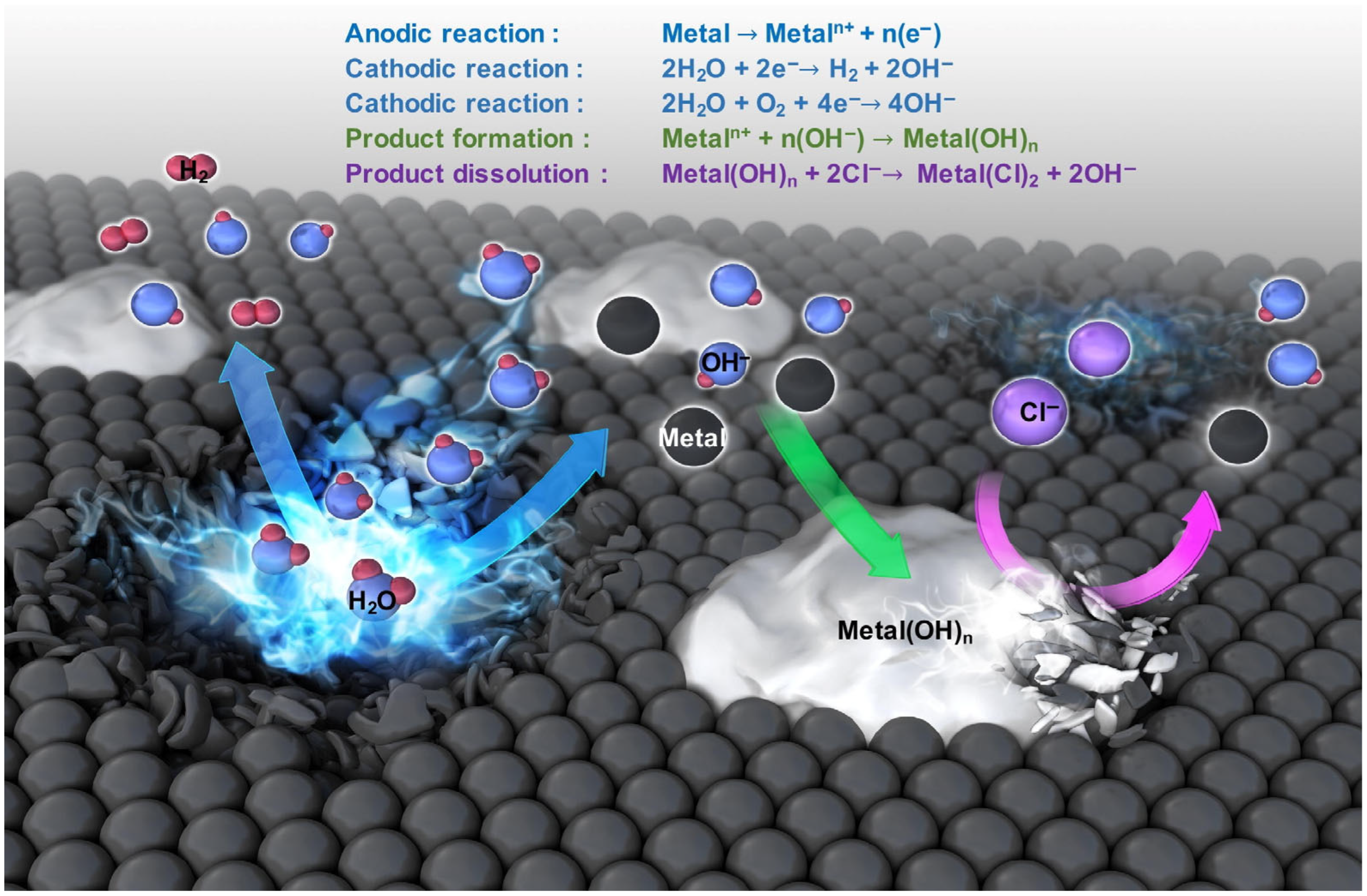
2. Temporary Implants Based on Biodegradable Metals
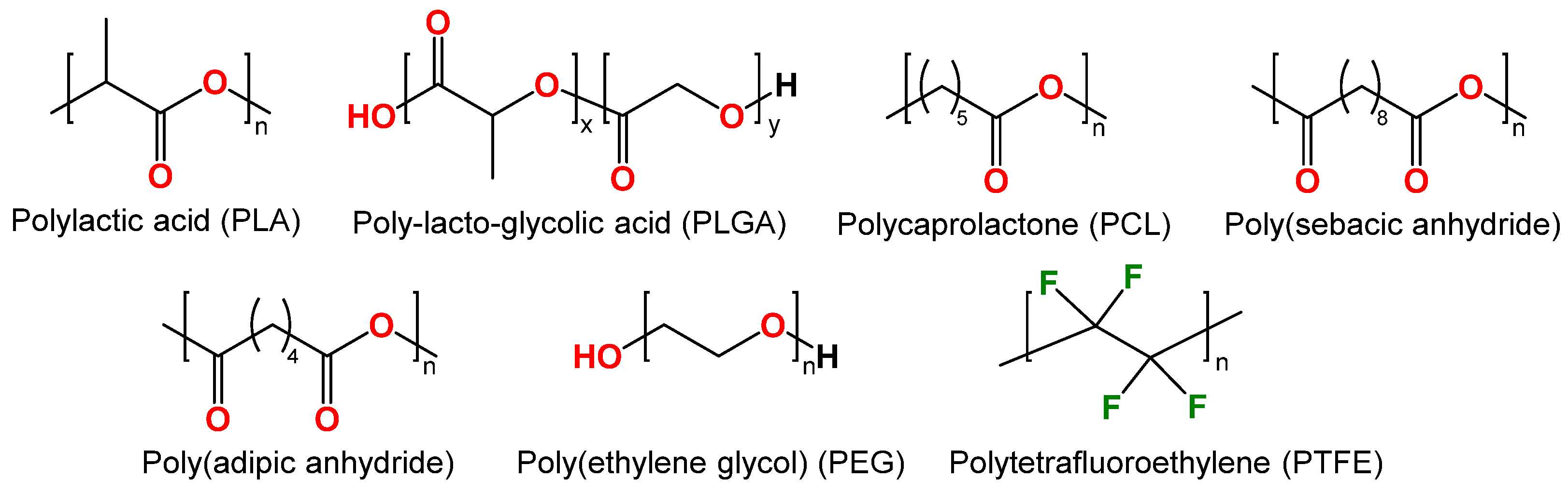
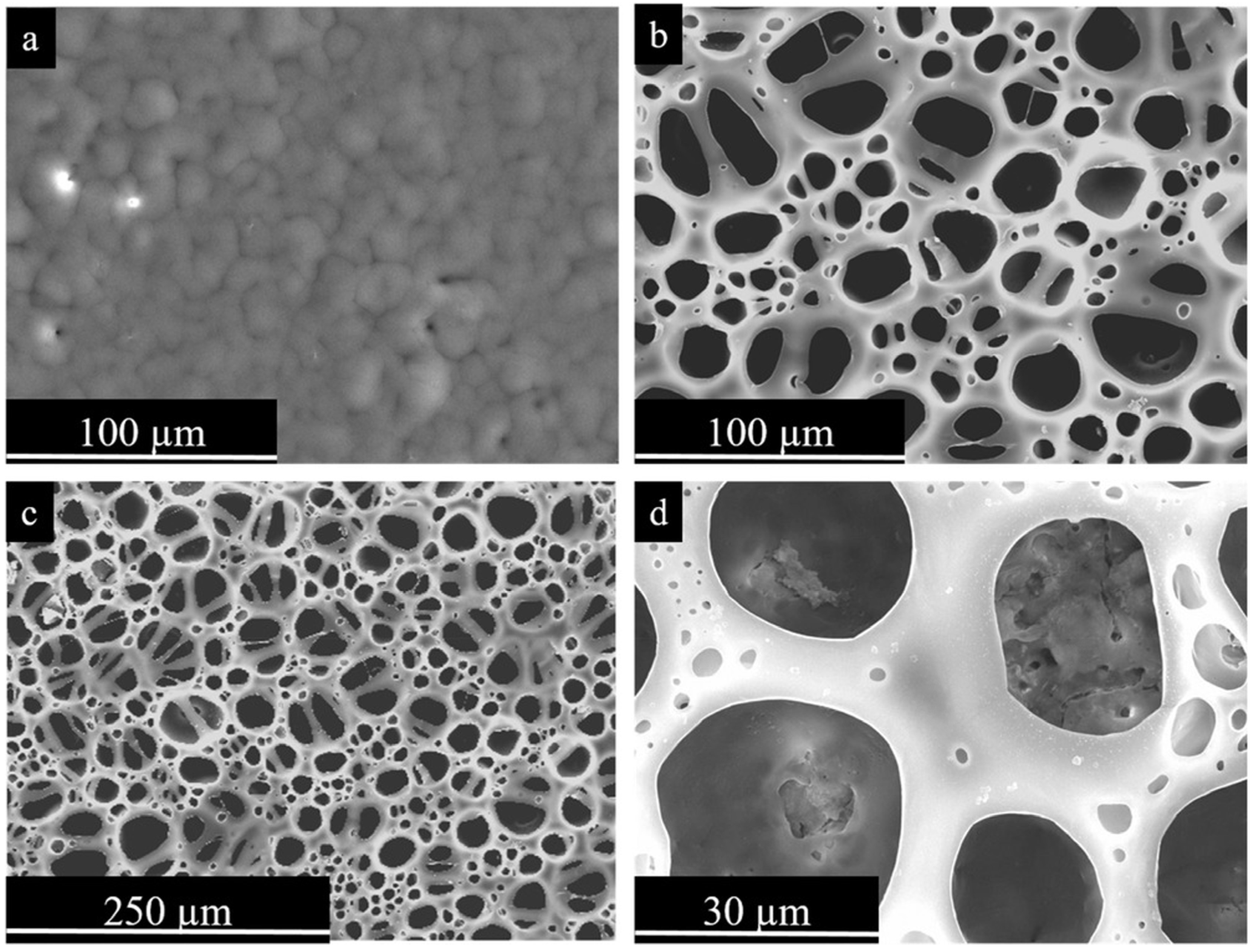
2.1. Synthetic Biocompatible Polymers
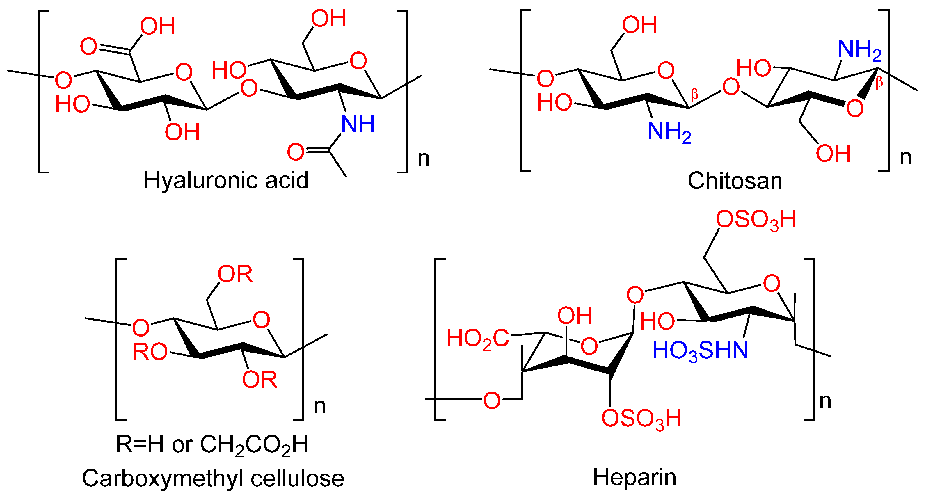
2.2. Natural Polymers for Temporary Implants
2.3. Other Polymers
3. Permanent Metal Implants with Organic Coatings
3.1. Synthetic Polymers
3.2. Natural Polymers for Permanent Implants
4. Conclusions and Outlook
Author Contributions
Funding
Conflicts of Interest
References
- Al-Shalawi, F.D.; Azmah Hanim, M.A.; Ariffin, M.K.A.; Looi Seng Kim, C.; Brabazon, D.; Calin, R.; Al-Osaimi, M.O. Biodegradable Synthetic Polymer in Orthopaedic Application: A Review. Mater. Today Proc. 2023, 74, 540–546. [Google Scholar] [CrossRef]
- Bharadwaj, A. An Overview on Biomaterials and Its Applications in Medical Science. IOP Conf. Ser. Mater. Sci. Eng. 2021, 1116, 012178. [Google Scholar] [CrossRef]
- Zhou, G.; Groth, T. Host Responses to Biomaterials and Anti-Inflammatory Design—A Brief Review. Macromol. Biosci. 2018, 18, e1800112. [Google Scholar] [CrossRef] [PubMed]
- Meyers, S.R.; Grinstaff, M.W. Biocompatible and Bioactive Surface Modifications for Prolonged In Vivo Efficacy. Chem. Rev. 2012, 112, 1615–1632. [Google Scholar] [CrossRef] [PubMed]
- Ribeiro, M.; Monteiro, F.J.; Ferraz, M.P. Infection of Orthopedic Implants with Emphasis on Bacterial Adhesion Process and Techniques Used in Studying Bacterial-Material Interactions. Biomatter 2012, 2, 176–194. [Google Scholar] [CrossRef]
- Liu, Y.; Rath, B.; Tingart, M.; Eschweiler, J. Role of Implants Surface Modification in Osseointegration: A Systematic Review. J. Biomed. Mater. Res. A 2020, 108, 470–484. [Google Scholar] [CrossRef]
- Shirazi, S.; Ravindran, S.; Cooper, L.F. Topography-mediated immunomodulation in osseointegration; Ally or Enemy. Biomaterials 2022, 291, 121903. [Google Scholar] [CrossRef]
- Kamath, S.; Bhattacharyya, D.; Padukudru, C.; Timmons, R.B.; Tang, L. Surface Chemistry Influences Implant-mediated Host Tissue Responses. J. Biomed. Mater. Res. A 2008, 86A, 617–626. [Google Scholar] [CrossRef]
- Kravanja, K.A.; Finšgar, M. A Review of Techniques for the Application of Bioactive Coatings on Metal-Based Implants to Achieve Controlled Release of Active Ingredients. Mater. Des. 2022, 217, 110653. [Google Scholar] [CrossRef]
- Predko, P.; Rajnovic, D.; Grilli, M.L.; Postolnyi, B.O.; Zemcenkovs, V.; Rijkuris, G.; Pole, E.; Lisnanskis, M. Promising Methods for Corrosion Protection of Magnesium Alloys in the Case of Mg-Al, Mg-Mn-Ce and Mg-Zn-Zr: A Recent Progress Review. Metals 2021, 11, 1133. [Google Scholar] [CrossRef]
- Yerokhin, A.; Parfenov, E.V.; Matthews, A. In Situ Impedance Spectroscopy of the Plasma Electrolytic Oxidation Process for Deposition of Ca- and P-Containing Coatings on Ti. Surf. Coat. Technol. 2016, 301, 54–62. [Google Scholar] [CrossRef]
- Gnedenkov, S.V.; Sharkeev, Y.P.; Sinebryukhov, S.L.; Khrisanfova, O.A.; Legostaeva, E.V.; Zavidnaya, A.G.; Puz’, A.V.; Khlusov, I.A.; Opra, D.P. Functional coatings formed on the titanium and magnesium alloys as implant materials by plasma electrolytic oxidation technology: Fundamental principles and synthesis conditions. Corros. Rev. 2016, 34, 65–83. [Google Scholar] [CrossRef]
- Kozelskaya, A.I.; Rutkowski, S.; Frueh, J.; Gogolev, A.S.; Chistyakov, S.G.; Gnedenkov, S.V.; Sinebryukhov, S.L.; Frueh, A.; Egorkin, V.S.; Choynzonov, E.L.; et al. Surface Modification of Additively Fabricated Titanium-Based Implants by Means of Bioactive Micro-Arc Oxidation Coatings for Bone Replacement. J. Funct. Biomater. 2022, 13, 285. [Google Scholar] [CrossRef] [PubMed]
- Sharifi, H.; Aliofkhazraei, M.; Darband, G.B.; Shrestha, S. A Review on Adhesion Strength of Peo Coatings by Scratch Test Method. Surf. Rev. Lett. 2018, 25, 1830004. [Google Scholar] [CrossRef]
- Gao, Y.; Yerokhin, A.; Matthews, A. Deposition and Evaluation of Duplex Hydroxyapatite and Plasma Electrolytic Oxidation Coatings on Magnesium. Surf. Coat. Technol. 2015, 269, 170–182. [Google Scholar] [CrossRef]
- Shishir, R.; Nasiruddin, U.; Manojkumar, P.; Ponnilavan, V.; Lokeshkumar, E.; Rama Krishna, L.; Rameshbabu, N. Development of bioactive ceramic composite coating with bactericidal property on Zn–1Mg alloy by plasma electrolytic oxidation for temporary orthopaedic implant applications. Ceram. Int. 2024, 50, 15538–15550. [Google Scholar] [CrossRef]
- Uhm, S.-H.; Kwon, J.-S.; Song, D.-H.; Lee, E.-J.; Jeong, W.-S.; OH, S.; Kim, K.-N.; Choi, E.H.; Kim, K.-M. Long-Term Antibacterial Performance and Bioactivity of Plasma-Engineered Ag-NPs/TiO2. J. Biomed. Nanotechnol. 2016, 12, 1890–1906. [Google Scholar] [CrossRef]
- Aliofkhazraei, M.; Macdonald, D.D.; Matykina, E.; Parfenov, E.V.; Egorkin, V.S.; Curran, J.A.; Troughton, S.C.; Sinebryukhov, S.L.; Gnedenkov, S.V.; Lampke, T.; et al. Review of Plasma Electrolytic Oxidation of Titanium Substrates: Mechanism, Properties, Applications and Limitations. Appl. Surf. Sci. Adv. 2021, 5, 100121. [Google Scholar] [CrossRef]
- Stewart, C.; Akhavan, B.; Wise, S.G.; Bilek, M.M.M. A Review of Biomimetic Surface Functionalization for Bone-Integrating Orthopedic Implants: Mechanisms, Current Approaches, and Future Directions. Prog. Mater. Sci. 2019, 106, 100588. [Google Scholar] [CrossRef]
- Zhang, B.; Myers, D.; Wallace, G.; Brandt, M.; Choong, P. Bioactive Coatings for Orthopaedic Implants—Recent Trends in Development of Implant Coatings. Int. J. Mol. Sci. 2014, 15, 11878–11921. [Google Scholar] [CrossRef]
- Fattah-alhosseini, A.; Chaharmahali, R.; Rajabi, A.; Babaei, K.; Kaseem, M. Performance of PEO/Polymer Coatings on the Biodegradability, Antibacterial Effect and Biocompatibility of Mg-Based Materials. J. Funct. Biomater. 2022, 13, 267. [Google Scholar] [CrossRef] [PubMed]
- Han, H.-S.; Loffredo, S.; Jun, I.; Edwards, J.; Kim, Y.-C.; Seok, H.-K.; Witte, F.; Mantovani, D.; Glyn-Jones, S. Current Status and Outlook on the Clinical Translation of Biodegradable Metals. Mater. Today 2019, 23, 57–71. [Google Scholar] [CrossRef]
- Manivasagam, G.; Suwas, S. Biodegradable Mg and Mg Based Alloys for Biomedical Implants. Mater. Sci. Technol. 2014, 30, 515–520. [Google Scholar] [CrossRef]
- Sastri, V.S.; Ghali, E.; Elboujdaini, M. Front Matter. In Corrosion Prevention and Protection; Wiley: Hoboken, NJ, USA, 2007. [Google Scholar]
- Kim, Y.-K.; Jang, Y.-S.; Kim, S.-Y.; Lee, M.-H. Functions Achieved by the Hyaluronic Acid Derivatives Coating and Hydroxide Film on Bio-Absorbed Mg. Appl. Surf. Sci. 2019, 473, 31–39. [Google Scholar] [CrossRef]
- Wang, Q.; Tu, S.; Rao, Y.; Chidambaram Seshadri, R. The Influence of Polymeric Sealing Treatment on the Wear Performance of PEO Coating Deposited on AZ31 Mg Alloy. Coatings 2022, 12, 182. [Google Scholar] [CrossRef]
- Zeng, R.-C.; Qi, W.-C.; Song, Y.-W.; He, Q.-K.; Cui, H.-Z.; Han, E.-H. In Vitro Degradation of MAO/PLA Coating on Mg-1.21Li-1.12Ca-1.0Y Alloy. Front. Mater. Sci. 2014, 8, 343–353. [Google Scholar] [CrossRef]
- Wei, Z.; Tian, P.; Liu, X.; Zhou, B. In Vitro Degradation, Hemolysis, and Cytocompatibility of PEO/PLLA Composite Coating on Biodegradable AZ31 Alloy. J. Biomed. Mater. Res. B Appl. Biomater. 2015, 103, 342–354. [Google Scholar] [CrossRef]
- Zeng, R.-C.; Cui, L.; Jiang, K.; Liu, R.; Zhao, B.-D.; Zheng, Y.-F. In Vitro Corrosion and Cytocompatibility of a Microarc Oxidation Coating and Poly(l-Lactic Acid) Composite Coating on Mg–1Li–1Ca Alloy for Orthopedic Implants. ACS Appl. Mater. Interfaces 2016, 8, 10014–10028. [Google Scholar] [CrossRef]
- Muñoz, M.; Torres, B.; Mohedano, M.; Matykina, E.; Arrabal, R.; López, A.J.; Rams, J. PLA Deposition on Surface Treated Magnesium Alloy: Adhesion, Toughness and Corrosion Behaviour. Surf. Coat. Technol. 2020, 388, 125593. [Google Scholar] [CrossRef]
- Li, L.-H.; Sankara Narayanan, T.S.N.; Kim, Y.K.; Kong, Y.-M.; Park, I.S.; Bae, T.S.; Lee, M.H. Deposition of Microarc Oxidation–Polycaprolactone Duplex Coating to Improve the Corrosion Resistance of Magnesium for Biodegradable Implants. Thin Solid Film. 2014, 562, 561–567. [Google Scholar] [CrossRef]
- Kim, Y.K.; Lee, K.B.; Kim, S.Y.; Jang, Y.S.; Kim, J.H.; Lee, M.H. Improvement of Osteogenesis by a Uniform PCL Coating on a Magnesium Screw for Biodegradable Applications. Sci. Rep. 2018, 8, 13264. [Google Scholar] [CrossRef] [PubMed]
- Tian, P.; Xu, D.; Liu, X. Mussel-Inspired Functionalization of PEO/PCL Composite Coating on a Biodegradable AZ31 Magnesium Alloy. Colloids Surf. B Biointerfaces 2016, 141, 327–337. [Google Scholar] [CrossRef]
- Santos-Coquillat, A.; Martínez-Campos, E.; Vargas-Alfredo, N.; Arrabal, R.; Rodríguez-Hernández, J.; Matykina, E. Hierarchical Functionalized Polymeric-Ceramic Coatings on Mg-Ca Alloys for Biodegradable Implant Applications. Macromol. Biosci. 2019, 19, e1900179. [Google Scholar] [CrossRef] [PubMed]
- Moreno, L.; Wang, C.; Lamaka, S.V.; Zheludkevich, M.L.; Rodríguez-Hernández, J.; Arrabal, R.; Matykina, E. Ciprofloxacin Release and Corrosion Behaviour of a Hybrid PEO/PCL Coating on Mg3Zn0.4Ca Alloy. J. Funct. Biomater. 2023, 14, 65. [Google Scholar] [CrossRef] [PubMed]
- Nicolao-Gómez, A.; Martínez-Campos, E.; Moreno, L.; Rodríguez-Hernández, J.; Matykina, E. Hierarchical Hybrid Coatings with Drug-Eluting Capacity for Mg Alloy Biomaterials. Materials 2023, 16, 7688. [Google Scholar] [CrossRef] [PubMed]
- Usmaniya, N.; Radhakrishna Pillai, S.; Palanivel, M.; Edalacheruvu, L.; Chennampalli, P.; Vaithiyanathan, P.; Parfenov, E.; Lingamaneni, R.K.; Nagumothu, R. Effect of Polycaprolactone Coating on the Corrosion and Biological Characteristics of Plasma Electrolytic Oxidised ZM21 Magnesium Alloy. Surf. Coat. Technol. 2023, 471, 129915. [Google Scholar] [CrossRef]
- Chen, Y.; Wang, J.; Dou, J.; Yu, H.; Chen, C. Layer by Layer Assembled Chitosan (TiO2)-Heparin Composite Coatings on MAO-Coated Mg Alloys. Mater. Lett. 2020, 281, 128640. [Google Scholar] [CrossRef]
- Imshinetsky, I.M.; Mashtalyar, D.V.; Nadaraia, K.V.; Sinebryukhov, S.L.; Gnedenkov, S.V. Fluoropolymer-Containing Layer Formed on MA8 Magnesium Alloy. Mater. Today Proc. 2019, 19, 1887–1890. [Google Scholar] [CrossRef]
- Gnedenkov, A.S.; Lamaka, S.V.; Sinebryukhov, S.L.; Mashtalyar, D.V.; Egorkin, V.S.; Imshinetskiy, I.M.; Zheludkevich, M.L.; Gnedenkov, S.V. Control of the Mg Alloy Biodegradation via PEO and Polymer-Containing Coatings. Corros. Sci. 2021, 182, 109254. [Google Scholar] [CrossRef]
- Gnedenkov, A.S.; Sinebryukhov, S.L.; Filonina, V.S.; Plekhova, N.G.; Gnedenkov, S.V. Smart Composite Antibacterial Coatings with Active Corrosion Protection of Magnesium Alloys. J. Magnes. Alloys 2022, 10, 3589–3611. [Google Scholar] [CrossRef]
- Mashtalyar, D.V.; Nadaraia, K.V.; Belov, E.A.; Imshinetskiy, I.M.; Sinebrukhov, S.L.; Gnedenkov, S.V. Features of Composite Layers Created Using an Aqueous Suspension of a Fluoropolymer. Polymers 2022, 14, 4667. [Google Scholar] [CrossRef] [PubMed]
- Ghanbari, A.; Bordbar-Khiabani, A.; Warchomicka, F.; Sommitsch, C.; Yarmand, B.; Zamanian, A. PEO/Polymer Hybrid Coatings on Magnesium Alloy to Improve Biodegradation and Biocompatibility Properties. Surf. Interfaces 2023, 36, 102495. [Google Scholar] [CrossRef]
- Moreno, L.; Mohedano, M.; Arrabal, R.; Rodríguez-Hernández, J.; Matykina, E. Development of Hybrid Hierarchical Coatings on Mg3Zn0.4Ca Alloy for Orthopaedic Implants. J. Mater. Res. Technol. 2023, 24, 5823–5838. [Google Scholar] [CrossRef]
- Gnedenkov, A.S.; Filonina, V.S.; Sinebryukhov, S.L.; Gnedenkov, S.V. A Superior Corrosion Protection of Mg Alloy via Smart Nontoxic Hybrid Inhibitor-Containing Coatings. Molecules 2023, 28, 2538. [Google Scholar] [CrossRef] [PubMed]
- Ranakoti, L.; Gangil, B.; Mishra, S.K.; Singh, T.; Sharma, S.; Ilyas, R.A.; El-Khatib, S. Critical Review on Polylactic Acid: Properties, Structure, Processing, Biocomposites, and Nanocomposites. Materials 2022, 15, 4312. [Google Scholar] [CrossRef] [PubMed]
- Singhvi, M.S.; Zinjarde, S.S.; Gokhale, D.V. Polylactic Acid: Synthesis and Biomedical Applications. J. Appl. Microbiol. 2019, 127, 1612–1626. [Google Scholar] [CrossRef]
- Li, X.; Chu, C.L.; Liu, L.; Liu, X.K.; Bai, J.; Guo, C.; Xue, F.; Lin, P.H.; Chu, P.K. Biodegradable Poly-Lactic Acid Based-Composite Reinforced Unidirectionally with High-Strength Magnesium Alloy Wires. Biomaterials 2015, 49, 135–144. [Google Scholar] [CrossRef]
- Modi, V.K.; Desai, D.A. Review of Taguchi Method, Design of Experiment (DOE) & Analysis of Variance (ANOVA) for Quality Improvements through Optimization in Foundry. J. Emerg. Technol. Innov. Res. 2018, 5, 184–194. [Google Scholar]
- Ali, W.; Echeverry-Rendón, M.; Kopp, A.; González, C.; LLorca, J. Effect of Surface Modification on Interfacial Behavior in Bioabsorbable Magnesium Wire Reinforced Poly-Lactic Acid Polymer Composites. npj Mater. Degrad. 2023, 7, 65. [Google Scholar] [CrossRef]
- Azimi, B.; Nourpanah, P.; Rabiee, M.; Arbab, S. Poly (∊-Caprolactone) Fiber: An Overview. J. Eng. Fibers Fabr. 2014, 9, 155892501400900. [Google Scholar] [CrossRef]
- Bai, H.; Du, C.; Zhang, A.; Li, L. Breath Figure Arrays: Unconventional Fabrications, Functionalizations, and Applications. Angew. Chem. Int. Ed. 2013, 52, 12240–12255. [Google Scholar] [CrossRef] [PubMed]
- Sanjanwala, D.; Londhe, V.; Trivedi, R.; Bonde, S.; Sawarkar, S.; Kale, V.; Patravale, V. Polysaccharide-Based Hydrogels for Medical Devices, Implants and Tissue Engineering: A Review. Int. J. Biol. Macromol. 2024, 256, 128488. [Google Scholar] [CrossRef] [PubMed]
- Schanté, C.E.; Zuber, G.; Herlin, C.; Vandamme, T.F. Chemical Modifications of Hyaluronic Acid for the Synthesis of Derivatives for a Broad Range of Biomedical Applications. Carbohydr. Polym. 2011, 85, 469–489. [Google Scholar] [CrossRef]
- Bastow, E.R.; Byers, S.; Golub, S.B.; Clarkin, C.E.; Pitsillides, A.A.; Fosang, A.J. Hyaluronan Synthesis and Degradation in Cartilage and Bone. Cell. Mol. Life Sci. 2008, 65, 395–413. [Google Scholar] [CrossRef]
- Ariyoshi, W.; Takahashi, T.; Kanno, T.; Ichimiya, H.; Takano, H.; Koseki, T.; Nishihara, T. Mechanisms Involved in Enhancement of Osteoclast Formation and Function by Low Molecular Weight Hyaluronic Acid. J. Biol. Chem. 2005, 280, 18967–18972. [Google Scholar] [CrossRef]
- Falconi, D.; Aubin, J.E. LIF Inhibits Osteoblast Differentiation at Least in Part by Regulation of HAS2 and Its Product Hyaluronan. J. Bone Miner. Res. 2007, 22, 1289–1300. [Google Scholar] [CrossRef]
- Mero, A.; Campisi, M. Hyaluronic Acid Bioconjugates for the Delivery of Bioactive Molecules. Polymers 2014, 6, 346–369. [Google Scholar] [CrossRef]
- Cui, F.Z.; Tian, W.M.; Hou, S.P.; Xu, Q.Y.; Lee, I.-S. Hyaluronic Acid Hydrogel Immobilized with RGD Peptides for Brain Tissue Engineering. J. Mater. Sci. Mater. Med. 2006, 17, 1393–1401. [Google Scholar] [CrossRef]
- Federer, C.; Kurpiers, M.; Bernkop-Schnürch, A. Thiolated Chitosans: A Multi-Talented Class of Polymers for Various Applications. Biomacromolecules 2021, 22, 24–56. [Google Scholar] [CrossRef]
- Geetha, M.; Singh, A.K.; Asokamani, R.; Gogia, A.K. Ti Based Biomaterials, the Ultimate Choice for Orthopaedic Implants—A Review. Prog. Mater. Sci. 2009, 54, 397–425. [Google Scholar] [CrossRef]
- Prado da Silva, M.H. Biomaterials Concepts. In Reference Module in Materials Science and Materials Engineering; Elsevier: Amsterdam, The Netherlands, 2016. [Google Scholar]
- Odekerken, J.C.; Welting, T.J.; Arts, J.J.; Walenkamp GH, I.M.; Emans, P.J. Modern Orthopaedic Implant Coatings—Their Pro’s, Con’s and Evaluation Methods. In Modern Surface Engineering Treatments; InTech: Silverwater, Australia, 2013. [Google Scholar]
- Valiev, R.Z.; Zhilyaev, A.P.; Langdon, T.G. Bulk Nanostructured Materials; Wiley: Hoboken, NJ, USA, 2013; ISBN 9781118095409. [Google Scholar]
- Valiev, R.Z.; Parfenov, E.V.; Parfenova, L.V. Developing Nanostructured Metals for Manufacturing of Medical Implants with Improved Design and Biofunctionality. Mater. Trans. 2019, 60, 1356–1366. [Google Scholar] [CrossRef]
- Valiev, R.Z.; Zheng, Y.; Edalati, K. Review: NanoSPD-Produced Metallic Materials for Advanced Medical Devices. J. Mater. Sci. 2024, 59, 5681–5697. [Google Scholar] [CrossRef]
- Prosolov, K.A.; Komarova, E.G.; Kazantseva, E.A.; Luginin, N.A.; Kashin, A.D.; Uvarkin, P.V.; Sharkeev, Y.P. Enhanced Corrosion Resistance and Mechanical Durability of the Composite PLGA/CaP/Ti Scaffolds for Orthopedic Implants. Polymers 2024, 16, 826. [Google Scholar] [CrossRef] [PubMed]
- Kazek-Kęsik, A.; Jaworska, J.; Krok-Borkowicz, M.; Gołda-Cępa, M.; Pastusiak, M.; Brzychczy-Włoch, M.; Pamuła, E.; Kotarba, A.; Simka, W. Hybrid Oxide-Polymer Layer Formed on Ti-15Mo Alloy Surface Enhancing Antibacterial and Osseointegration Functions. Surf. Coat. Technol. 2016, 302, 158–165. [Google Scholar] [CrossRef]
- Kazek-Kęsik, A.; Nosol, A.; Płonka, J.; Śmiga-Matuszowicz, M.; Gołda-Cępa, M.; Krok-Borkowicz, M.; Brzychczy-Włoch, M.; Pamuła, E.; Simka, W. PLGA-Amoxicillin-Loaded Layer Formed on Anodized Ti Alloy as a Hybrid Material for Dental Implant Applications. Mater. Sci. Eng. C 2019, 94, 998–1008. [Google Scholar] [CrossRef]
- Kazek-Kęsik, A.; Nosol, A.; Płonka, J.; Śmiga-Matuszowicz, M.; Student, S.; Brzychczy-Włoch, M.; Krok-Borkowicz, M.; Pamuła, E.; Simka, W. Physico-Chemical and Biological Evaluation of Doxycycline Loaded into Hybrid Oxide-Polymer Layer on Ti–Mo Alloy. Bioact. Mater. 2020, 5, 553–563. [Google Scholar] [CrossRef]
- Leśniak, K.; Płonka, J.; Śmiga-Matuszowicz, M.; Brzychczy-Włoch, M.; Kazek-Kęsik, A. Functionalization of PEO Layer Formed on Ti-15Mo for Biomedical Application. J. Biomed. Mater. Res. B Appl. Biomater. 2020, 108, 1568–1579. [Google Scholar] [CrossRef]
- Leśniak-Ziółkowska, K.; Śmiga-Matuszowicz, M.; Blacha-Grzechnik, A.; Student, S.; Brzychczy-Włoch, M.; Krok-Borkowicz, M.; Pamuła, E.; Simka, W.; Kazek-Kęsik, A. Antibacterial and Cytocompatible Coatings Based on Poly(Adipic Anhydride) for a Ti Alloy Surface. Bioact. Mater. 2020, 5, 709–720. [Google Scholar] [CrossRef]
- Al-Hawary, S.I.S.; Habash, R.T.; Abosaooda, M.; Hjazi, A.; Saleh, E.A.M.; Hassan, Z.F.; Bathaei, M.S. TiO2/PEG as Smart Anticorrosion and Drug-Eluting Platforms in Inflammatory Conditions. Heliyon 2024, 10, e25605. [Google Scholar] [CrossRef]
- Zhang, T.; Zhou, W.; Yang, W.; Bi, J.; Li, H.; Gao, X.; Zhang, B.; Shi, G.; Li, K.; Wei, Z.; et al. Vancomycin-Encapsulated Hydrogel Loaded Microarc-Oxidized 3D-Printed Porous Ti6Al4V Implant for Infected Bone Defects: Reconstruction, Anti-Infection, and Osseointegration. Bioact. Mater. 2024, 42, 18–31. [Google Scholar] [CrossRef]
- Neupane, M.P.; Park, I.S.; Lee, M.H. Surface Characterization and Corrosion Behavior of Micro-Arc Oxidized Ti Surface Modified with Hydrothermal Treatment and Chitosan Coating. Thin Solid Film. 2014, 550, 268–271. [Google Scholar] [CrossRef]
- Wang, X.; Li, B.; Liu, S.; Zhang, C.; Hao, J. Antibacterial and Biological Properties of a Micro-Structured BMP-2/Chitosan/Hydroxyapatite Hybrid Coating on Ti Surface. J. Hard Tissue Biol. 2019, 28, 303–314. [Google Scholar] [CrossRef]
- Yılmaz, E. Modification of the Micro Arc-Oxidized Ti Surface for Implant Applications. J. Bionic Eng. 2021, 18, 1391–1399. [Google Scholar] [CrossRef]
- Chai, M.; An, M.; Zhang, X. Construction of a TiO2/MoSe2/CHI Coating on Dental Implants for Combating Streptococcus mutans Infection. Mater. Sci. Eng. C 2021, 129, 112416. [Google Scholar] [CrossRef]
- Zhou, R.; Zhou, Y.; Cheng, J.; Cao, J.; Li, M.; Yu, H.; Wei, D.; Li, B.; Wang, Y.; Zhou, Y. Surface Configuration of Microarc Oxidized Ti with Regionally Loaded Chitosan Hydrogel Containing Ciprofloxacin for Improving Biological Performance. Mater. Today Bio. 2022, 16, 100380. [Google Scholar] [CrossRef]
- Parfenova, L.V.; Galimshina, Z.R.; Gil’fanova, G.U.; Alibaeva, E.I.; Danilko, K.V.; Pashkova, T.M.; Kartashova, O.L.; Farrakhov, R.G.; Mukaeva, V.R.; Parfenov, E.V.; et al. Hyaluronic Acid Bisphosphonates as Antifouling Antimicrobial Coatings for PEO-Modified Titanium Implants. Surf. Interfaces 2022, 28, 101678. [Google Scholar] [CrossRef]
- Parfenov, E.V.; Parfenova, L.V.; Dyakonov, G.S.; Danilko, K.V.; Mukaeva, V.R.; Farrakhov, R.G.; Lukina, E.S.; Valiev, R.Z. Surface Functionalization via PEO Coating and RGD Peptide for Nanostructured Titanium Implants and Their in Vitro Assessment. Surf. Coat. Technol. 2019, 357, 669–683. [Google Scholar] [CrossRef]
- Parfenova, L.V.; Lukina, E.S.; Galimshina, Z.R.; Gil’fanova, G.U.; Mukaeva, V.R.; Farrakhov, R.G.; Danilko, K.V.; Dyakonov, G.S.; Parfenov, E.V. Biocompatible Organic Coatings Based on Bisphosphonic Acid RGD-Derivatives for PEO-Modified Titanium Implants. Molecules 2020, 25, 229. [Google Scholar] [CrossRef]
- Parfenov, E.; Parfenova, L.; Mukaeva, V.; Farrakhov, R.; Stotskiy, A.; Raab, A.; Danilko, K.; Rameshbabu, N.; Valiev, R. Biofunctionalization of PEO Coatings on Titanium Implants with Inorganic and Organic Substances. Surf. Coat. Technol. 2020, 404, 126486. [Google Scholar] [CrossRef]
- Parfenova, L.V.; Galimshina, Z.R.; Gil’fanova, G.U.; Alibaeva, E.I.; Danilko, K.V.; Aubakirova, V.R.; Farrakhov, R.G.; Parfenov, E.V.; Valiev, R.Z. Modeling of Biological Activity of PEO-Coated Titanium Implants with Conjugates of Cyclic RGD Peptide with Amino Acid Bisphosphonates. Materials 2022, 15, 8120. [Google Scholar] [CrossRef]
- Parfenova, L.V.; Galimshina, Z.R.; Gil’fanova, G.U.; Alibaeva, E.I.; Pashkova, T.M.; Kartashova, O.L.; Farrakhov, R.G.; Aubakirova, V.R.; Parfenov, E.V. Hybrid Antimicrobial Coating Based on Conjugate of Hyaluronic Acid with LL-37 Peptide for PEO-Modified Titanium Implants. Russ. J. Bioorg. Chem. 2024, 50, 500–507. [Google Scholar] [CrossRef]
- Wlodarczyk, J.; Musial-Kulik, M.; Jelonek, K.; Stojko, M.; Karpeta-Jarzabek, P.; Pastusiak, M.; Janeczek, H.; Dobrzynski, P.; Sobota, M.; Kasperczyk, J. Dual-Jet Electrospun PDLGA/PCU Nonwovens as Promising Mesh Implant Materials with Controlled Release of Sirolimus and Diclofenac. Int. J. Pharm. 2022, 625, 122113. [Google Scholar] [CrossRef] [PubMed]
- Sanchez-Cano, C.; Carril, M. Recent Developments in the Design of Non-Biofouling Coatings for Nanoparticles and Surfaces. Int. J. Mol. Sci. 2020, 21, 1007. [Google Scholar] [CrossRef] [PubMed]
- Salim, M.; Mishra, G.; Fowler, G.J.S.; O’Sullivan, B.; Wright, P.C.; McArthur, S.L. Non-Fouling Microfluidic Chip Produced by Radio Frequency Tetraglyme Plasma Deposition. Lab Chip 2007, 7, 523. [Google Scholar] [CrossRef]
- Menzies, D.J.; Cowie, B.; Fong, C.; Forsythe, J.S.; Gengenbach, T.R.; McLean, K.M.; Puskar, L.; Textor, M.; Thomsen, L.; Tobin, M.; et al. One-Step Method for Generating PEG-Like Plasma Polymer Gradients: Chemical Characterization and Analysis of Protein Interactions. Langmuir 2010, 26, 13987–13994. [Google Scholar] [CrossRef]
- Thierry, B.; Winnik, F.M.; Merhi, Y.; Griesser, H.J.; Tabrizian, M. Biomimetic Hemocompatible Coatings through Immobilization of Hyaluronan Derivatives on Metal Surfaces. Langmuir 2008, 24, 11834–11841. [Google Scholar] [CrossRef]
- Dalsin, J.L.; Hu, B.-H.; Lee, B.P.; Messersmith, P.B. Mussel Adhesive Protein Mimetic Polymers for the Preparation of Nonfouling Surfaces. J. Am. Chem. Soc. 2003, 125, 4253–4258. [Google Scholar] [CrossRef]
- Kang, C.-K.; Lee, Y.-S. The Surface Modification of Stainless Steel and the Correlation between the Surface Properties and Protein Adsorption. J. Mater. Sci. Mater. Med. 2007, 18, 1389–1398. [Google Scholar] [CrossRef]
- Lih, E.; Oh, S.H.; Joung, Y.K.; Lee, J.H.; Han, D.K. Polymers for Cell/Tissue Anti-Adhesion. Prog. Polym. Sci. 2015, 44, 28–61. [Google Scholar] [CrossRef]
- Reyes, C.D.; Petrie, T.A.; Burns, K.L.; Schwartz, Z.; García, A.J. Biomolecular Surface Coating to Enhance Orthopaedic Tissue Healing and Integration. Biomaterials 2007, 28, 3228–3235. [Google Scholar] [CrossRef]
- Joner, M.; Cheng, Q.; Schönhofer-Merl, S.; Lopez, M.; Neubauer, S.; Mas-Moruno, C.; Laufer, B.; Kolodgie, F.D.; Kessler, H.; Virmani, R. Polymer-free Immobilization of a Cyclic RGD Peptide on a Nitinol Stent Promotes Integrin-dependent Endothelial Coverage of Strut Surfaces. J. Biomed. Mater. Res. B Appl. Biomater. 2012, 100B, 637–645. [Google Scholar] [CrossRef] [PubMed]
- Mas-Moruno, C.; Dorfner, P.M.; Manzenrieder, F.; Neubauer, S.; Reuning, U.; Burgkart, R.; Kessler, H. Behavior of Primary Human Osteoblasts on Trimmed and Sandblasted Ti6Al4V Surfaces Functionalized with Integrin Avβ3-selective Cyclic RGD Peptides. J. Biomed. Mater. Res. A 2013, 101A, 87–97. [Google Scholar] [CrossRef] [PubMed]
- Mas-Moruno, C. Surface Functionalization of Biomaterials for Bone Tissue Regeneration and Repair. In Peptides and Proteins as Biomaterials for Tissue Regeneration and Repair; Elsevier: Amsterdam, The Netherlands, 2018; pp. 73–100. [Google Scholar]
- Queffélec, C.; Petit, M.; Janvier, P.; Knight, D.A.; Bujoli, B. Surface Modification Using Phosphonic Acids and Esters. Chem. Rev. 2012, 112, 3777–3807. [Google Scholar] [CrossRef]
- Goura, J.; Chandrasekhar, V. Molecular Metal Phosphonates. Chem. Rev. 2015, 115, 6854–6965. [Google Scholar] [CrossRef]
- Bujoli, B.; Queffelec, C. Fine-Tuning the Functionality of Inorganic Surfaces Using Phosphonate Chemistry. In Tailored Organic-Inorganic Materials; Wiley: Hoboken, NJ, USA, 2015; pp. 299–318. [Google Scholar]
- Lecollinet, G.; Delorme, N.; Edely, M.; Gibaud, A.; Bardeau, J.-F.; Hindré, F.; Boury, F.; Portet, D. Self-Assembled Monolayers of Bisphosphonates: Influence of Side Chain Steric Hindrance. Langmuir 2009, 25, 7828–7835. [Google Scholar] [CrossRef]
- Portet, D.; Denizot, B.; Rump, E.; Lejeune, J.-J.; Jallet, P. Nonpolymeric Coatings of Iron Oxide Colloids for Biological Use as Magnetic Resonance Imaging Contrast Agents. J. Colloid Interface Sci. 2001, 238, 37–42. [Google Scholar] [CrossRef]
- Mookherjee, N.; Brown, K.L.; Bowdish, D.M.E.; Doria, S.; Falsafi, R.; Hokamp, K.; Roche, F.M.; Mu, R.; Doho, G.H.; Pistolic, J.; et al. Modulation of the TLR-Mediated Inflammatory Response by the Endogenous Human Host Defense Peptide LL-37. J. Immunol. 2006, 176, 2455–2464. [Google Scholar] [CrossRef]
- Duplantier, A.J.; van Hoek, M.L. The Human Cathelicidin Antimicrobial Peptide LL-37 as a Potential Treatment for Polymicrobial Infected Wounds. Front. Immunol. 2013, 4, 143. [Google Scholar] [CrossRef]
- Neshani, A.; Zare, H.; Akbari Eidgahi, M.R.; Kamali Kakhki, R.; Safdari, H.; Khaledi, A.; Ghazvini, K. LL-37: Review of Antimicrobial Profile against Sensitive and Antibiotic-Resistant Human Bacterial Pathogens. Gene Rep. 2019, 17, 100519. [Google Scholar] [CrossRef]


| Substrate | PEO Coating (Electrolyte Composition and Processing Mode) | Physicochemical Properties of PEO Coating (Porosity, Corr. Resistance, Thickness, etc.) | Organic Coating | Physicochemical Properties of Hybrid Coating PEO + Organic Molecules | Biological Effect of Hybrid Coating | Refs. |
|---|---|---|---|---|---|---|
| Mg-1.21Li-1.12Ca-1.0Y | NaOH, Na2SiO3, NaB4O7, Na3C6H5O7, phytic acid; constant voltage 130–140 V, t = 10 min, room temperature | dmean = 8.61 μm, number of pores (NP): 15,332, corrosion current density icorr = 6.313 × 10⁻⁶ A·cm−2, charge transfer resistance Rct = 354.8 Ω/cm2 | PLA (Mw = 60,000) | dmean = 2.26 μm, NP: 3489, icorr = 1.702 × 10⁻⁶ A·cm−2, Rct = 1894.0 Ω/cm2 | - | [27] |
| AZ31 | Na2SiO3·9H2O, NaOH, NaF; constant current density 50 mA·cm−2, frequency 800 Hz, duty cycle 20%, t = 15 min, room temperature | Ecorr = −1.606 V, icorr = 1.1 μA·cm−2, Rc = 4888 Ω/cm2, Rct = 58,295 Ω/cm2 | PLA | Ecorr = −1.317 V, icorr = 0.00024 μA·cm−2 Rc = 32,892 Ω/cm2, Rct = 895,900 Ω/cm2 | Hemolysis ratio (%): 0.806 ± 0.771 (<5%, excellent blood compatibility). Adhesion (1 day): The MC3T3-E1 cells exhibited good adhesion and displayed numerous filopodia extensions. Proliferation and vitality (3 and 7 days): MC3T3-E1 cells on PEO/PLA coating showed higher proliferation rate and vitality than that on PEO coating. | [28] |
| Mg-1Li-1Ca | NaOH, phytic acid; breakdown voltage 170–180 V, duty cycle 50%, frequency 100 Hz, t = 600 s | Ecorr = −1.62 V, icorr = 1.03 × 10⁻⁵ A·cm−2 (sample immersion 140 h) | PLA (Mw = 200,000) | Ecorr = −1.60 V, icorr = 4.19 × 10⁻⁶ A·cm−2 | Hemolysis ratio (%): 0.17 ± 0.04 (<5%, excellent blood compatibility). Cytotoxicity: MC3T3-E1 cells, MTT assay, relative growth ratio (RGR) of all samples is <100% after 24 h of culture, RGR of MAO/PLA coating >100% after 72 h. Proliferation: MAO/PLA showed higher proliferation rate and vitality comparing with Mg-1Li-1Ca substrates and MAO coating. Alkaline Phosphatase (ALP) activity (osteoblastic differentiation): MAO/PLLA coatings displayed a highest ALP expression of MC3T3 cells | [29] |
| AZ31 | Na3PO4·12H2O, NaF, KOH, CaO; peak-to-peak voltage 480 V and direct current offset 190 V, duty cycle 50%, frequency 50 Hz, current density limit 138 mA·cm−2 | average bonding strength 5.2 ± 0.4 MPa, Ecorr = −1.23 V, icorr = 6.5 × 10⁻⁸ A·cm−2 | PLA | average bonding strength 4.0 ± 0.7 MPa, Ecorr = −1.13 V, icorr = 1.7 × 10⁻⁸ A·cm−2 | - | [30] |
| Mg | Na2SiO3·9H2O, NaF; current density limit 50 mA/cm2, limiting voltage 250 V, t = 3 min | Ecorr = −1.65 V, icorr = 1.14 μA·cm−2 | PCL (Mw = 70,000–90,000) | Ecorr = −1.53 V, icorr = 0.0045 μA·cm−2 | - | [31] |
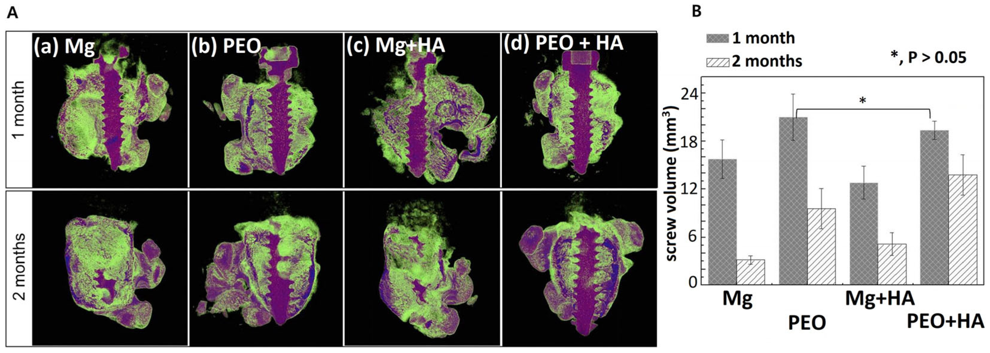
| Mg, screw | NaOH, Na3PO4, glycerol; constant current 300 mA/cm2, pulse width 100 ms, duty cycle 50%, t = 3 min | - | PCL (Mw = 70,000–90,000) | - | In vivo: Sprague Dawley rats, stable and compact new bone formed on the PCL coating. The hybrid coating reduced the release of magnesium ions and degradation rate of magnesium. Denser and thicker bone formed around the PEO/PCL-coated screw than the PEO-coated screw after 2 months of implantation. | [32] |
| AZ31 | Na2SiO3·9H2O, KOH, KF·2H2O; current density 50 mA/cm2, frequency 300 Hz, duty cycle 10%, t = 15 min | Ecorr = −1.640 V, icorr = 4.738 × 10⁻⁷ A·cm−2, Rp = 1.930 × 10⁵ Ω | PCL, PCL/PDAM, PCL/PDAM/ PHMB | PCL: Ecorr = −1342 V, icorr = 3.260 × 10⁻¹⁰ A·cm−2, Rp = 2.686 × 10⁸ Ω; PCL/PDAM: Ecorr = −1.303 V, icorr = 1.843 × 10⁻¹⁰ A·cm−2, Rp = 4.920 × 10⁸ Ω | Cytotoxicity: MC3T3-E1 cells, viability >70%. Adhesion (1 day): cells were fully spread and displayed polygon shapes with a large number of filopodia and lamellipodia on PEO/PCL and PEO/PCL/PDAM surfaces compared to AZ31 and AZ31-PEO. Proliferation (7 days): cells on PEO/PCL and PEO/PCL/PDAM formed a layer, the amounts of cells on the AZ31 alloy and PEO were small. Antibacterial efficacy: S. aureus, E. coli, PEO/PCL and PEO/PCL/PDAM did not show antibacterial effect. The effect was achieved after PHMB immobilization on the surface. | [33] |
| Mg0.8Ca | Na3PO4·12H2O, Na2SiO3·5H2O, KOH, CaO, NaF; peak-to-peak voltage 490 V, direct current offset 190 V, duty cycle 50%, frequency 50 Hz, current density limit 138 mA·cm−2, t = 300 s | Thickness ≈ 13 µm, Roughness Ra = 0.84 ± 0.01 µm, Rz = 5.6 ± 0.3 µm, contact angle 34.1 ± 4.6° | PCL/BF | Thickness ≈ 35 µm, Ra = 8 ± 0.30 µm, Rz = 36 ± 0.6 µm, contact angle 106.3 ± 1.8° | Adhesion and proliferation: C2C12-GFP (ATCC CRL-1772) mouse premyoblast cell line, cells can colonize the inner PEO ceramic coating structure where higher amount of bioelements are present. The Mg/PEO/PCL/BF scaffolds exhibit equally good or better cell adhesion and proliferation compared with Ti CP control. | [34] |
| Mg3Zn0.4Ca | Na3PO4·12H2O, Na2SiO3·5H2O, KOH, CaO; peak-to-peak voltage 400 V, frequency 50 Hz, current density limit 100 mA·cm−2, t = 300 s | - | PCL/BF, PCL/BF/CIP | PCL/BF: pore size 8.5 ± 2.8 µm, contact angle 88.9 ± 5.0° PCL/BF/CIP: pore size 12.2 ± 1.1 µm, contact angle 96.1 ± 3.0° | PCL/BF/CIP system avoided burst release and ensured gradual drug elution (64% over 240 h). Over 11 days of immersion in pseudo-physiological conditions (at constant pH 7.4 under CO2 flow), the PCL/BF/CIP system revealed 74% reduction of the degradation rate of Mg alloy compared to the PCL/BF. | [35] |
| Mg3Zn0.4Ca | Na2SiO3·5H2O, Na3PO4·12H2O, CaO, KOH; peak-to-peak voltage 400 V, frequency 50 Hz, current density limit 100 mA·cm−2, t = 300 s | - | PCL/BF/CIP PCL/BF/PAR | Drug release: for the loaded Mg-HHC system, a gradual elution with 20 and 40% of PAR, and CIP was observed after 10 days. Cytotoxicity: C2C12-GFP (ATCC CRL-1772) mouse premyoblast cell line and C166-GFP (ATCC CRL-2583) mouse endothelial cell line, the addition of PAR did not prevent the growth of endothelial cells and premyoblasts in the range of concentrations up to 120 μg/mL. PAR and CIP release from a complete Mg-HHC system showed a lower concentration range, suggesting that drug addition would not constitute a drawback in the biomaterial’s cytocompatibility. | [36] | |
| ZM21 | Na2SiO3·9H2O, KOH, Na2B4O7·10H2O; current density 60 mA/cm2, duty cycle 50%, frequency 1000 Hz, t = 10 min | Thickness 28 ± 2 μm, Ra = 2.085 μm, contact angle 40.2 ± 1.5°, Ecorr = −549.1 ± 38.2 mV, icorr = (1.48 ± 0.13) × 10−5 mA·cm−2, corrosion rate (3.81 ± 0.33) × 10−4 mm/y | PCL (Mw = 80,000) | PCL5: Thickness 68 ± 5 μm, Ra = 1.529 μm, contact angle 63.2 ± 1.3°, Ecorr = 543.2 ± 45.3 mV, icorr = (6.47 ± 0.52) × 10−9 mA·cm−2, corrosion rate (1.67 ± 0.14) × 10−7 mm/y | Cytotoxicity: mouse fibroblast cells L929. The ZM21/PEO/PCL5 material exhibited the highest biological activity with a significantly lower number of dead cells even at 100% extract concentration. The sample demonstrated superior cell growth and proliferation in the direct contact assay. | [37] |
| Mg | NaOH, Na3PO4, glycerol; unipolar pulse current 300 mA/cm2, duty cycle 50%, t = 3 min | surface roughness Rq = 109.1 ± 8.69 nm, contact angle 28.601 ± 0.68°, Ecorr = −1.55 V, icorr = 5.362 × 10⁻⁶ A·cm−2 | HYA/CMC | Rq = 180 ± 31.01 nm, contact angle 16.192 ± 0.46°, Ecorr = −1.034 V, icorr = 5.362 × 10⁻⁷ A·cm−2 | In vitro: Osteoblast cells (MC3T3-E1), HYA/CMC significantly increased the cell proliferation and viability in 3-day test. In vivo: Sprague Dawley rats, HYA/CMC loading of the PEO layer self-heals localized damage and stable osteocytes in long-term bone regeneration; the layer reduced osteoblast proliferation and cell stress, and this stably promotes bone formation around the implant. | [25] |
| Mg-3Zn-0.5Sr | Na5P3O10, NaOH, C3H8O3, NaF, K2TiF6,CH3COOAg | Ecorr = −1.681 V vs. SCE, icorr = 7.659 × 10⁻⁴ A·cm−2, polarization resistance Rp = 219.6 Ω·cm2 | nano-TiO2/CS/GEL/HEP | Ecorr = −1.505 V vs. SCE, icorr = 2.776 × 10⁻⁷ A·cm−2, Rp = 130,830.6 Ω·cm2 | Immobilized heparin layer effectively inhibited the adhesion of platelets and reduced the hemolysis rate. | [38] |
| MA8 | Na4SiO4, NaF; 1st step: voltage 30 to 240 V, sweep rate 1.05 V/s, t = 200 s; 2nd step: voltage 240 to 200 V, sweep rate 0.07 V/s, t = 600 s, duty cycle 50%, frequency 300 Hz | contact angle 50° | SPTFE | contact angle 155° | - | [39] |
| MA8 | C3H7O6PCa, NaF, Na2SiO3; pulsed bipolar mode, voltage 420 V, duty cycle 50%, frequency 100 Hz, t = 110 s | Ecorr = −1.57 V vs. SCE, icorr = 5.4 × 10⁻⁶ A·cm−2 | SPTFE | Ecorr = −0.18 V vs. SCE, icorr = 7.6 × 10⁻¹⁰ A·cm−2 | - | [40] |
| MA8 | (1) SiF electrolyte: Na2SiO3·5H2O, NaF; pulsed bipolar mode, voltage 30 to 300 V (anode) and 30 V (cathode), sweep rate 0.45 V·s−1, t = 10 min (2) GP electrolyte: (C3H7O6P)Ca2·H2O, NaF, Na2SiO3·5H2O; pulsed bipolar mode, voltage 380 V (anode), current density 1.3 to 0.8 A·cm−2, sweep rate—4.5 mA·cm−2·s−1, t = 110 s | SiF electrolyte: Ecorr = −1.48 V vs. SCE, icorr = 1.5 × 10⁻⁷ A·cm−2, GP electrolyte: Ecorr= −1.62 V vs. SCE, icorr = 2.2 × 10⁻⁷ A·cm−2 | 8-HQ/SPTFE, 8-HQ/PVDF | SiF electrolyte: 8-HQ/SPTFE, Ecorr = −1.42 V vs. SCE, icorr = 7.7 × 10⁻¹² A·cm−2; 8-HQ/PVDF, Ecorr = −1.22 V vs. SCE, icorr = 1.6 × 10⁻¹¹ A·cm−2; GP electrolyte: 8-HQ/SPTFE, Ecorr = −1.24 V vs. SCE, icorr = 9.2 × 10⁻¹² A·cm−2; 8-HQ/PVDF, Ecorr = −1.19 V vs. SCE, icorr = 1.5 × 10⁻¹¹ A·cm−2; | Antibacterial efficacy: 8-HQ coating killed S. aureus (MRSA) within 24 h. | [41] |
| MA8 | Na4SiO4, NaF; 1st step: voltage 20 to 240 V, t = 200 s; 2nd step: voltage 240 to 200 V, sweep rate 0.07 V/s, t = 600 s, duty cycle 50%, frequency 300 Hz | Ecorr = −1.43 V, icorr = 2.4 × 10⁻⁷ A·cm−2, contact angle 45.3 ± 1.2° | SPTFE | Ecorr = 0.12 V, icorr = 7.7 × 10⁻¹¹ A·cm−2, contact angle 152.3 ± 0.9 | - | [42] |
| AZ31B | KOH, Na3PO4·12H2O, current density 50 mA·cm−2, duty cycle 25%, t = 15 min | Ecorr = −1.81 V, icorr = 4.31 × 10⁻⁵ A·cm−2, porosity 1.06 × 10⁻¹%, contact angle 65 ± 6.4° | DFP (dopamine functionalized PTMC polymer) | Ecorr = −1.58 V, icorr = 0.56 × 10⁻⁷ A·cm−2, porosity 2.53 × 10⁻⁴%, contact angle 41 ± 3.6° | Cell viability: osteoblast-like cells (G292), PEO/DFP sample showed the highest cell viability, indicating no significant toxicity in time intervals (1, 3, and 8 days). Proliferation: the highest cell spreading as well as confluence was observed in the case of PEO/DFP coating. | [43] |
| Substrate | PEO Coating (Electrolyte Composition and Processing Mode) | Physicochemical Properties of PEO Coating (Porosity, Corr. Resistance, Thickness, etc.) | Organic Coating | Physicochemical Properties of Hybrid Coating PEO + Organic Molecules | Biological Effect of Hybrid Coating | Refs. |
|---|---|---|---|---|---|---|
| Ti (Grade 2) | Nanosized hydroxyapatite (Ca10(PO4)6(OH)2), CaCO3, H3PO4; voltage 200 V, frequency 50 Hz, pulse duration 100 μs, t = 10 min | 0.9% NaCl solution: Ecorr = −0.065 mV, icorr = 228.2 × 10⁻⁹ A·cm−2 PBS solution: Ecorr = −0.088 mV, icorr = 355.0 × 10⁻⁹ A·cm−2, adhesion strength 20.1 ± 1.6 MPa, roughness Ra = 3.3 ± 0.4 μm | 5%, 8%, 10% PLGA | 0.9% NaCl solution (8% PLGA): Ecorr = −0.508 mV, icorr = 0.3 × 10⁻⁹ A·cm−2; PBS solution (10% PLGA): Ecorr = −0.416 mV, icorr = 0.1 × 10⁻⁹ A·cm−2, adhesion strength 9.8 ± 3.8 MPa (8% PLGA), Ra = 3.0 ± 0.3 μm (8% PLGA) | - | [67] |
| Ti-15Mo | Ca(H2PO2)2, CaSiO3; voltage 300 V, current density 100 mA·cm−2 | Ecorr = 178.7 ± 8.1 mV, icorr = (6.7 ± 0.4) × 10−6 A·cm−2 | PLGA, PLGA/ gentamicin | PLGA: Ecorr = −168.8 ± 4.3 mV, icorr = (3.5 ± 0.1) × 10−5 A·cm−2 | Cell viability: MG-63, PEO/PLGA layers were cytocompatible, and cells were well-adhered to the modified surfaces. Antibacterial efficacy: S. aureus (DSM 24167), for the PEO/PLGA samples, slightly higher adhesion area of S. aureus was observed, gentamicin-loaded PLGA samples showed no adhesion of bacteria. | [68] |
| Ti-15Mo | Ca(H2PO2)2; voltage 300 V, current density 100 mA·cm−2, t = 5 min | Ra = 1.20 μm, contact angle 44.7 ± 5.9° | PLGA, PLGA/AMX | PLGA: Ra = 1.46 μm, contact angle 91.4 ± 4.3°; PLGA/AMX: Ra = 1.71 μm, contact angle 96.5 ± 2.2° | Cell viability: MG-63, after 7 days of culture, the highest level of cell viability was observed for Ti-15Mo and Ti-15Mo/PEO. PLGA and PLGA/AMX layers provided a slightly lower number of cells compared to Ti-15Mo and Ti-15Mo/PEO. Antibacterial efficacy: S. aureus (DSM 24167), S. epidermidis (ATCC 700296, the concentration of released drug during 1 h was enough to inhibit growth of both bacteria strains. | [69] |
| Ti-15Mo | Ca(H2PO2)2; voltage 300 V, current density 100 mA·cm−2 t = 5 min | surface roughness Rz = 1.5 ± 0.3 μm, Ra = 0.5 ± 0.09 μm | PLGA (Mn 19,000), PLGA/DOX | PLGA: Rz = 2.3 ± 0.9 μm, Ra = 1.2 ± 0.2 μm; PLGA/DOX: Rz = 4.9 ± 0.9 μm, Ra = 1.2 ± 0.1 μm | Cell viability: MG-63, after 7 days, the highest increase in the cell number was observed for PEO/PLGA–21.17%. For PEO/PLGA/DOX the proliferation decreased to 10.30%, but it was higher than in the case of Ti-15Mo and Ti-15Mo/PEO. Antibacterial efficacy: S. aureus (ATCC 25923), S. epidermidis (ATCC 700256), the amount of loaded doxycycline was sufficient to inhibit bacterial growth. | [70] |
| Ti-15Mo | Ca(H2PO2)2; voltage 300 V, current density 100 mA·cm−2, t = 5 min | Ra = 1.21 ± 0.15 μm, contact angle 47.6 ± 5.7° | PSBA, PSBA/AMX, PSBA/CEF, PSBA/VANC | PSBA: Ra = 0.82 ± 0.17 μm, contact angle 80.6 ± 2.1°; PSBA/AMX: Ra = 0.81 ± 0.09 μm, contact angle 62.9 ± 3.9°; PSBA/CEF: Ra = 0.93 ± 0.19 μm, contact angle 74.6 ± 3.7°; PSBA/VANC: Ra = 1.2 ± 0.05 μm, contact angle 79.0 ± 2.2° | PSBA undergoes rapid hydrolysis (77.8 mol% after 7 days). Antibacterial efficacy: S. aureus (DSM 24167) and S. epidermidis (ATCC 700296), all of the coatings significantly decreased the number of bacteria, and the concentration of the drugs was suitable for septic treatment around the material. The inhibition of bacteria growth depends on the concentration of drug released from the coatings. Amoxicillin showed the best results for the artificial saliva. | [71] |
| Ti-2Ta-3Zr-36Nb | Ca(H2PO2)2; voltage 300 V, current density 150 mA·cm−2, t = 5 min | Ra = 1.24 ± 0.35 μm, contact angle 61.31° | PADA, PADA/AMX, PADA/CEF, PADA/VANC | PADA: Ra = 1.29 ± 0.05 μm, contact angle 41.69°; PADA/AMX: Ra = 1.53 ± 0.26 μm, contact angle 63.02°; PADA/CEF: Ra = 1.92 ± 0.28 μm, contact angle 51.24°; PADA/VANC: Ra = 2.06 ± 0.36 μm, contact angle 39.51° | PADA is the fast-degrading polymer (80% after 48 h). Cell viability: MG-63, the viability of cells on the hybrid layers was slightly higher than on the PEO surface. After 7 days PEO/PADA/AMX showed the highest number of cells in comparison to other drug-loaded surfaces. Antibacterial efficacy: S. aureus (ATCC 25923) and S. aureus (MRSA 1030). Surfaces loaded with drug exhibited stronger bacteriostatic effects than those without antibiotics. The minimal number of adhered bacteria was observed for PEO/PADA/CEF. | [72] |
| Ti-6Al-4V | K2SiO3, KOH; constant current density 75 mA·cm−2, duty cycle 50%, t = 60 min | test’s solution, normal (pH 7.2): Ecorr = −206 mV, icorr = 2.14 μA·cm−2; inflammatory (pH 5.0): Ecorr = −329 mV, icorr = 8.87 μA·cm−2 | PEG (Mw = 1000 g·mol−1)/BET | normal (pH 7.2): Ecorr = −54 mV, icorr = 0.081 μA·cm−2; inflammatory (pH 5.0): Ecorr = −154 mV, icorr = 1.75 μA·cm−2 | - | [73] |
| Ti-6Al-4V (3D printed) | Ca(CH3COO)2·H2O, NaH2PO4, EDTA-2Na, NaOH | - | PEG hydrogel, PEG-VANC | - | Cell adhesion and proliferation: hMSCs, no significant difference was found in proliferative activity between PEO, PEO/PEG, PEO/PEG-VANC. PEO/PEG showed higher ALP activity compared to the other two groups, VANC reduced hMSC osteo-differentiation. Antibacterial efficacy: S. aureus, PEO/PEG-VANC implants showed significantly greater effect comparing with PEO and PEO/PEG. In vivo: male New Zealand white rabbits, after 6 weeks intraporous bone volume fraction (BVF) of the PEO/PEG-VANC group (49.2% ± 3.3%), was significantly higher than that of the other two groups (PEO 14.6% ± 2.2%, PEO/PEG 16.4% ± 3.2%). Histological studies of infected sites in the PEO and PEO/PEG groups showed typical signs of chronic bone infection, including inflammatory cell (mononuclear cell and granulocyte) infiltrate, bone necrosis, and bone erosion. These features were notably diminished in the PEO/PEG-VANC group. | [74] |
| Ti | (1) PEO: Ca(CH3COO)2, calcium glycerophosphate; voltage 350 V, current density 50 mA·cm−2, t = 10 min; (2) hydrothermal treatment, T = 250 °C, t = 3 h in an autoclave | (1) Ra = 0.328 ± 0.012 μm, Ecorr = −0.288 V, icorr = 2.93 × 10⁻⁷ A·cm−2; (2) Ra = 0.513 ± 0.011 μm, Ecorr = −0.229 V, icorr = 2.33 × 10⁻⁷ A·cm−2 | Chitosan (Mw = 200 kDa, ≥93% deacetylated) | (1) Ra = 0.422 ± 0.016 μm, Ecorr = −0.194 V, icorr = 1.96 × 10⁻⁷ A·cm−2; (2) Ra = 0.649 ± 0.029 μm, Ecorr = −0.182 V, icorr = 1.83 × 10⁻⁸ A·cm−2 | - | [75] |
| Ti | Ca(COOH)2, NaH2PO4; voltage 360 V, frequency 100 Hz, t = 5 min | HA/PEO: Ra = 0.774 ± 0.023 μm, contact angle 16.4 ± 1.5° | BMP-2, Chitosan, BMP-2/Chitosan | BMP-2: Ra = 0.759 ± 0.016° μm, contact angle 17.1 ± 1.2; Chitosan: Ra = 0.634 ± 0.017° μm, contact angle 21.7 ± 0.8; BMP-2/Chitosan Ra = 0.622 ± 0.026 μm, contact angle 22.1 ± 1.0° | Chitosan completely degrades on the surface within 4 weeks in a PBS—lysozyme solution. BMP-2/HA/PEO surfaces released about 95% of the BMP-2 within 2 days. BMP-2 released from BMP-2/Chi/HA/PEO for 4 weeks in a PBS—lysozyme solution and somewhat slower in PBS solution. Cell proliferation: MC3T3-E1, BMP-2/Chi/HA/PEO showed highest cell proliferation in a 7-day test. Antibacterial efficacy: E. coli, Chi/HA/PEO exhibited strong antibacterial activity (70% decrease after 24 h incubation). | [76] |
| Ti | (1) PEO: Na2SiO3, TiO2; voltage 400 V, t = 5 min. (2) HA deposition: Ca(OH)2 + H3PO4. | Chitosan | Antibacterial efficacy: E. coli, more intense bacteria growth was obtained on the PEO/HA surface compared to that of PEO/HA/Chi. | [77] | ||
| Ti | (1) PEO: Ca(CH3COO)2, Na3PO4·12H2O, current density 20 A·dm−2, t = 5 min, pulse frequency 800 Hz, duty ratio 30%, 25 °C (2) MoSe2 deposition: Na2MoO4·2H2O, Se powder, hydrothermal treatment | PEO/MoS2: contact angle 130° | Chitosan | contact angle 40° | Cell viability: MC3T3-E1, hydrophobic PEO/MoS2 is not favorable to the cell adhesion and spreading. The cells regained the vitality and spread again on PEO/MoSe2/Chi surface. Antibacterial efficacy: S. mutans, PEO/MoSe2/Chi exhibited excellent antibacterial activity in vitro and in vivo (82.03%) upon illumination with 808 nm NIR light. The coating promoted new bone formation in the presence of infection in vivo under NIR light irradiation. | [78] |
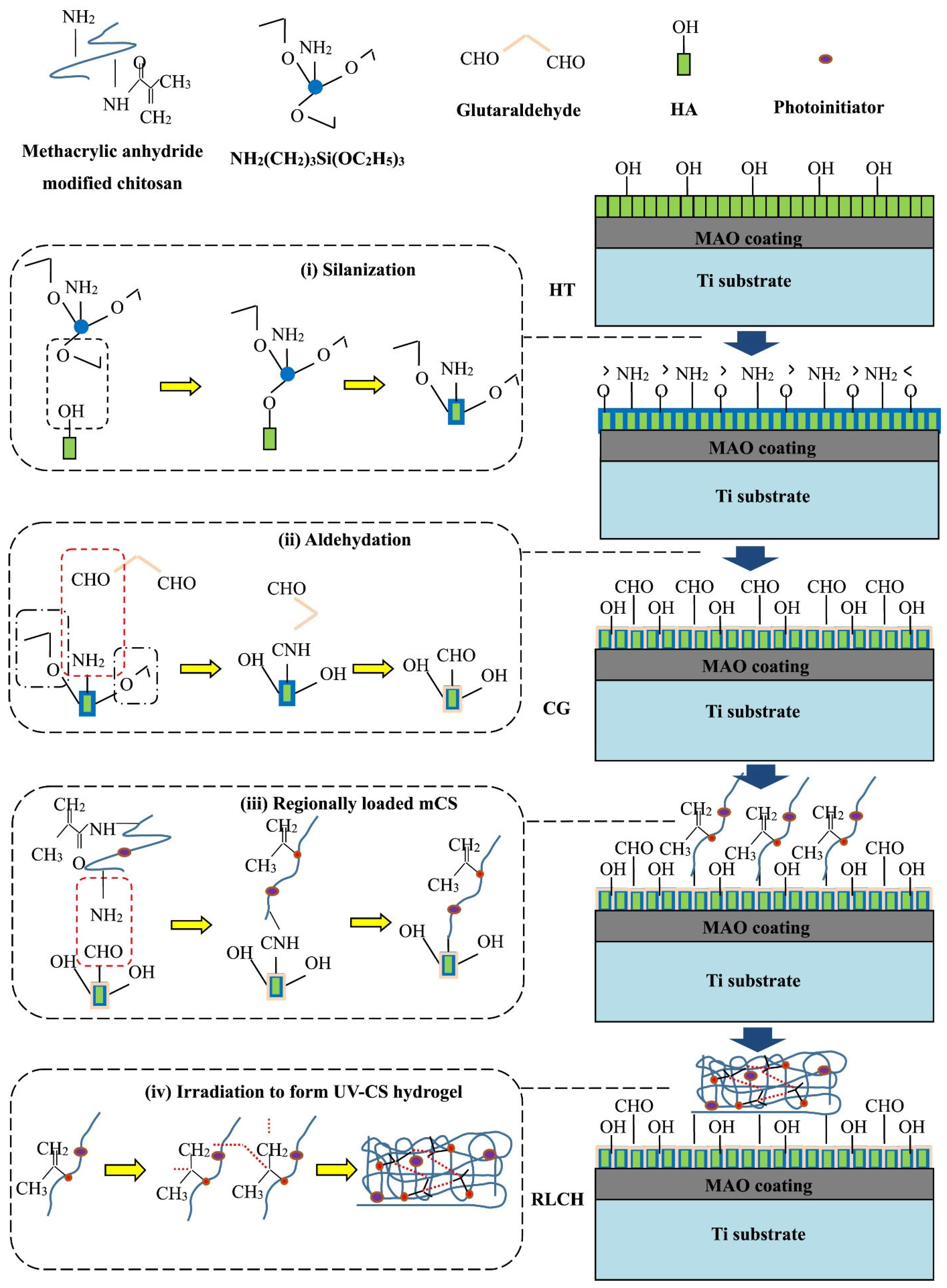
| Ti | (1) PEO: EDTA-2Na, Ca(H2PO4)2·H2O, NaOH; voltage 350 V, frequency 1 kHz, duty ratio 10%, t = 5 min; (2) HA nanodots: NaOH, hydrothermal treatment | (1) Ra = 0.102 ± 0.014 μm; (2) Ra = 0.140 ± 0.008 μm | Surface silanization, aldehydation (CG), Chitosan (UV-CS)/ciprofloxacin | PEO/HT/CG Ra = 0.133 ± 0.004 μm | Cell viability: hBMSCs, regional loading chitosan hydrogel with ciprofloxacin does not show any biological toxicity. Antibacterial test: E. coli, S. aureus, the synergistic effect of ciprofloxacin and modified chitosan allows to effectively inhibit bacterial growth with low drug loadings. | [79] |
| CG-Ti, nano-Ti | Na3PO4·12H2O; pulsed bipolar mode, positive pulse: voltage 470 V, duty cycle 51%, negative pulse: voltage 40 V, duty cycle 26%, frequency 300 Hz, 20 °C, t = 5 min | CG-Ti/PEO: Ra = 2.6 ± 0.13 μm, porosity 7.6 ± 1.4%, average pore size 1.44 ± 0.17 μm, contact angle 67°; nano-Ti/PEO: Ra = 2.5 ± 0.05 μm, porosity 7.6 ± 1.5%, average pore size 0.61 ± 0.11 μm, contact angle 68° | HYA, HYA-PH | CG-Ti/PEO/HYA-PH: contact angle 60°; nano-Ti/PEO/HYA-PH: contact angle 51° | Cytotoxicity: human adipose tissue MSC, HYA and all HYA-PH are non-toxic, some HYA-PH promote cell growth. Cell viability: PEO/HYA and PEO/HYA-PH provided a reduction in the viability of fibroblasts (by 20–40%), MG-63 (by 30–60%), and MSC (by more than 60%) on the surface (7-day test). Antibacterial efficacy: P. aeruginosa, S. aureus, E. faecium. The significant decrease in the adhesion of bacteria on PEO/HYA and PEO/HYA-PH surface was found. Use of nano-Ti as a substrate resulted in a reduction of pathogen adhesion by up to 84%. | [80] |
| CG-Ti, nano-Ti | Na3PO4·12H2O, Ca(CH3COO)2, pulsed bipolar mode, positive pulse: voltage 470 V, duty cycle 51%, negative pulse: voltage 40 V, duty cycle 26%, frequency 300 Hz, 20 °C, t = 10 min | CG-Ti/PEO: Ra = 1.13 ± 0.09 μm, porosity 8.8 ± 0.5%, average pore size 3.3 ± 0.6 μm, Ecorr = 0.033 ± 0.029 V, icorr = (7.7 ± 1.2) × 10−9 A·cm−2; nano-Ti/PEO: Ra = 0.75 ± 0.11 μm, porosity 10.2 ± 0.5%, average pore size 3.1 ± 0.6 μm, Ecorr = −0.169 ± 0.022 V, icorr = (35.1± 2.2) × 10−9 A·cm−2 | RGD-PH | CG-Ti/PEO/RGD-PH: Ecorr = −0.338 ± 0.031 V, icorr = (176 ± 32.1) × 10−9 A·cm−2; nano-Ti/PEO/RGD-PH: Ecorr = −0.419 ± 0.026 V, icorr = (168 ± 49.1) × 10−9 A·cm−2 | Cell viability: human embryonic lung fibroblasts (FLECH-104). PEO coating on nano-Ti gives 43% increase in the number of cells compared to CG-Ti/PEO; nano-Ti/PEO/RGD-PH gives 45% increase in the number of cells compared to nano-Ti, and 66% compared to both uncoated and coated CG-Ti. | [81] |
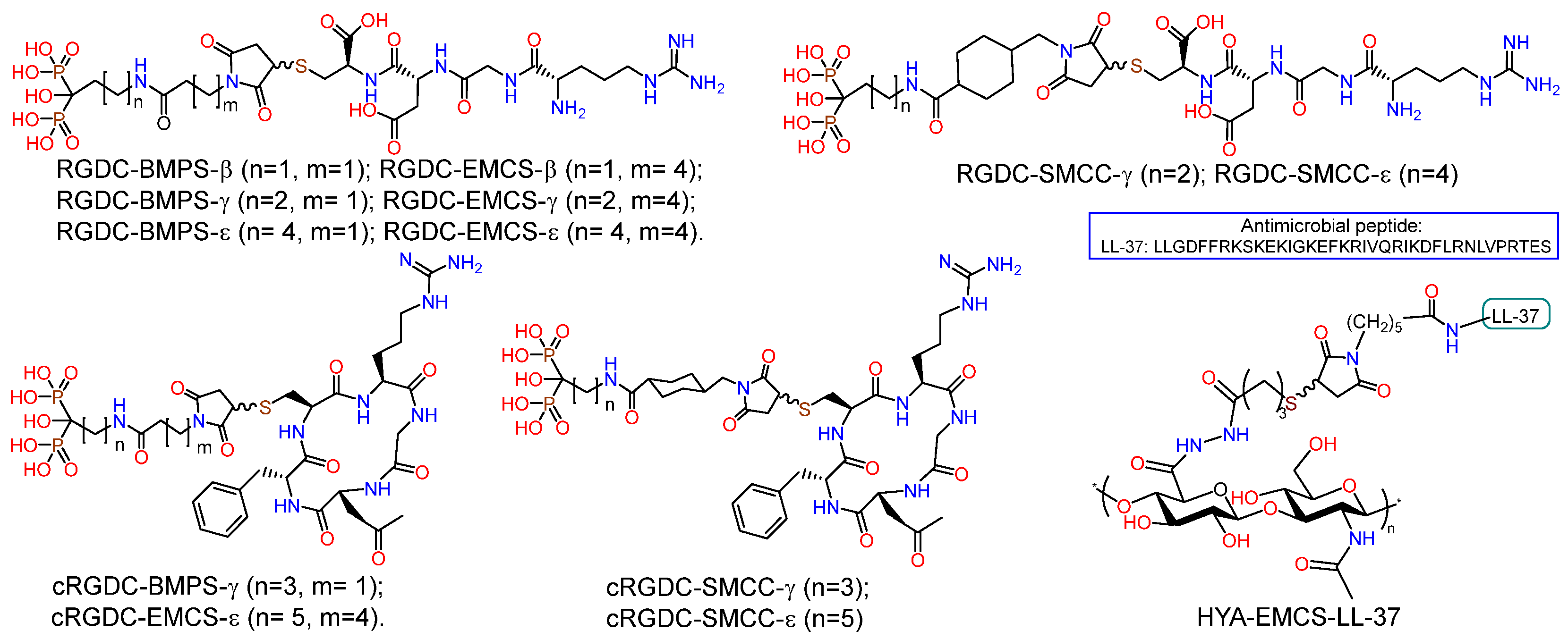
| Ti | Na3PO4·12H2O; pulsed bipolar mode, positive pulse: voltage 470 V, duty cycle 51%, negative pulse: voltage 40 V, duty cycle 26%, frequency 300 Hz, 20 °C, t = 5 min | Ecorr = −0.238 V, icorr = 0.019 μA·cm−2 | RGDC, RGD-PH | Ecorr = −0.218 – −0.180 V, icorr = 0.179 – 0.401 μA·cm−2 | Cell viability: FLECH-104, MSC, MG-63. RGDC does not influence cell viability on PEO surface. RGD-PH with relatively short bisphosphonate anchors and BMPS linker, as well as molecules containing a linker with a cyclohexyl fragment, increase cell viability on the surface of PEO-modified titanium. | [82] |
| Ti | E1: Na3PO4·12H2O; E2: Na3PO4·12H2O, Ca(CH3COO)2; pulsed bipolar mode, positive pulse: voltage 470 V, duty cycle 51%, negative pulse: voltage 40 V, duty cycle 26%, frequency 300 Hz, 20 °C, t = 5 min | E1: Ra = 2.1 ± 0.4 μm, porosity 6.8 ± 0.4%, average pore size 0.82 ± 0.17 μm, Ecorr = 0.1024 ± 0.005 V, icorr = (9.45 ± 0.8) × 10−9 A·cm−2; E2: Ra = 3.1 ± 0.5 μm, porosity 14.5 ± 0.4%, average pore size 0.77 ± 0.16 μm, Ecorr = 0.0331 ± 0.029 V, icorr (A·cm−2) = (7.67 ± 1.2) × 10−9 A·cm−2 | RGD-PH | PEO(E1)/RGD-PH Ecorr = −0.218 ± 0.001 V, icorr = (2.16 ± 1.12) × 10−7 A·cm−2 | Cell viability: MG-63, cell viability decreased by 15% over 7 days on the surface of sample PEO(E2) compared to sample PEO(E1); the bioactivity of PEO(E1)/RGD-PH is 37% higher compared to PEO(E1). | [83] |
| CG-Ti, nano-Ti | Na3PO4·12H2O, pulsed bipolar mode, positive pulse: voltage 470 V, duty cycle 51%, negative pulse: voltage 40 V, duty cycle 26%, frequency 300 Hz, 20 °C, t = 5 min | CG-Ti/PEO: Ra = 2.6 ± 0.13 μm, porosity 7.6 ± 1.4%, average pore size 1.44 ± 0.17 μm; nano-Ti/PEO: Ra = 2.5 ± 0.05 μm, porosity 7.6 ± 1.5%, average pore size 0.61 ± 0.11 μm | c(RGDfC), cPGD-PH | - | Cell viability: FLECH-104, MSC, MG-63. c(RGDfC) reduces cell viability due to the toxicity. The appearance of a linker and a bisphosphonate anchor reduced the toxicity of cyclo-RGD, ε-aminocaproic acid derivative with an SMCC linker increased the degree of fibroblasts and MSC cell proliferation on the CG-Ti/PEO and nano-Ti/PEO. | [84] |
| Ti | Na3PO4·12H2O, pulsed bipolar mode, positive pulse: voltage 470 V, duty cycle 51%, negative pulse: voltage 40 V, duty cycle 26%, frequency 300 Hz, 20 °C, t = 5 min | - | HYA–LL37 | - | Antibacterial efficacy: S. aureus, P. aeruginosa, E. faecium, E. coli. PEO/HYA–LL37 demonstrated a significant (p < 0.05) suppression of the ability of bacteria to form biofilms. | [85] |
Disclaimer/Publisher’s Note: The statements, opinions and data contained in all publications are solely those of the individual author(s) and contributor(s) and not of MDPI and/or the editor(s). MDPI and/or the editor(s) disclaim responsibility for any injury to people or property resulting from any ideas, methods, instructions or products referred to in the content. |
© 2024 by the authors. Licensee MDPI, Basel, Switzerland. This article is an open access article distributed under the terms and conditions of the Creative Commons Attribution (CC BY) license (https://creativecommons.org/licenses/by/4.0/).
Share and Cite
Parfenova, L.V.; Galimshina, Z.R.; Parfenov, E.V. Organic-Inorganic Biocompatible Coatings for Temporary and Permanent Metal Implants. Int. J. Mol. Sci. 2024, 25, 11623. https://doi.org/10.3390/ijms252111623
Parfenova LV, Galimshina ZR, Parfenov EV. Organic-Inorganic Biocompatible Coatings for Temporary and Permanent Metal Implants. International Journal of Molecular Sciences. 2024; 25(21):11623. https://doi.org/10.3390/ijms252111623
Chicago/Turabian StyleParfenova, Lyudmila V., Zulfiya R. Galimshina, and Evgeny V. Parfenov. 2024. "Organic-Inorganic Biocompatible Coatings for Temporary and Permanent Metal Implants" International Journal of Molecular Sciences 25, no. 21: 11623. https://doi.org/10.3390/ijms252111623
APA StyleParfenova, L. V., Galimshina, Z. R., & Parfenov, E. V. (2024). Organic-Inorganic Biocompatible Coatings for Temporary and Permanent Metal Implants. International Journal of Molecular Sciences, 25(21), 11623. https://doi.org/10.3390/ijms252111623