Article

Assessing the Potential of 1,2,3-Triazole-Dihydropyrimidinone Hybrids Against Cholinesterases: In Silico, In Vitro, and In Vivo Studies

by
Carlos M. Gastalho
1,2,3,
Ana M. Sena
4,
Óscar López
5,
José G. Fernández-Bolaños
5,
Alfonso T. García-Sosa
6,
Florbela Pereira
7,
Célia M. Antunes
2,3,8,
Ana R. Costa
2,3,8,
Anthony J. Burke
1,9,10,11 and
Elisabete P. Carreiro
1,*
1
LAQV-REQUIMTE, Institute for Research and Advanced Training (IIFA), University of Évora, Rua Romão Ramalho, 59, 7000-671 Évora, Portugal
2
Institute of Earth Sciences, Institute of Research and Advanced Training, University of Évora, 7000-671 Évora, Portugal
3
Academic Clinical Center of Alentejo, C-TRAIL, Rua Romão Ramalho, 59, 7000-671 Évora, Portugal
4
Department of Chemistry and Biochemistry, School of Sciences and Technologies, University of Évora, Rua Romão Ramalho, 59, 7000-671 Évora, Portugal
5
Departamento de Química Orgánica, Facultad de Química, Universidad de Sevilla, Apartado 1203, E-41071 Seville, Spain
6
Institute of Chemistry, University of Tartu, Ravila 14 A, 50411 Tartu, Estonia
7
LAQV-REQUIMTE, Department of Chemistry, NOVA School of Science and Technology, Universidade Nova de Lisboa, 2829-516 Caparica, Portugal
8
Department of Medical and Health Sciences, School of Health and Human Development, University of Évora, Rua Romão Ramalho, 59, 7000-671 Évora, Portugal
9
Faculty Pharmacy, University of Coimbra, Pólo das Ciências da Saúde, Azinhaga de Santa Comba, 3000-548 Coimbra, Portugal
10
Departamento de Química, Coimbra Chemistry Centre-Institute of Molecular Sciences (CQC-IMS), University of Coimbra, 3004-535 Coimbra, Portugal
11
Center for Neurosciences and Cellular Biology (CNC), Polo I, Universidade de Coimbra Rua Larga Faculdade de Medicina, Polo I, 1ºandar, 3004-504 Coimbra, Portugal
*
Author to whom correspondence should be addressed.
Int. J. Mol. Sci. 2024, 25(20), 11153; https://doi.org/10.3390/ijms252011153
Submission received: 19 September 2024 / Revised: 8 October 2024 / Accepted: 13 October 2024 / Published: 17 October 2024
(This article belongs to the Special Issue Molecular Research on Neurodegenerative Diseases 4.0)

Abstract

Combining the pharmacological properties of the 1,2,3-triazole and dihydropyrimidinone classes of compounds, two small families of mono- and di(1,2,3-triazole)-dihydropyrimidinone hybrids, A and B, were previously synthesized. The main objective of this work was to investigate the potential anti-Alzheimer effects of these hybrids. The inhibitory activities of cholinesterases (AChE and BuChE), antioxidant activity, and the inhibitory mechanism through in silico (molecular docking) and in solution (STD-NMR) experiments were evaluated. The 1,2,3-triazole-dihydropyrimidinone hybrids (A and B) showed moderate in vitro inhibitory activity on eqBuChE (IC50 values between 1 and 58.4 μM). The best inhibitor was the hybrid B4, featuring two 1,2,3-triazole cores, which exhibited stronger inhibition than galantamine, with an IC50 of 1 ± 0.1 μM for eqBuChE, through a mixed inhibition mechanism. Among the hybrids A, the most promising inhibitor was A1, exhibiting an IC50 of 12 ± 2 µM, similar to that of galantamine. Molecular docking and STD-NMR experiments revealed the key binding interactions of these promising inhibitors with BuChE. Hybrids A and B did not display Artemia salina toxicity below 100 μM.

1. Introduction

Alzheimer’s disease (AD) is known as a neurodegenerative disorder often characterized both by abnormal activities and intellectual impairment. Considered one of the main public health conditions, it currently affects more than 55 million people worldwide, particularly the elderly in low-and middle-income countries (WHO, 2023). It is predicted that, over the years, more people will suffer from the AD condition at some point and the population’s average age will also increase [1].
The pathophysiology of AD remains an enigma and without a cure, despite all the efforts of researchers in recent decades. AD is a multifactorial disease, and its causes could be associated with oxidative stress, metal ion dyshomeostasis, mitochondrial dysfunction, Aβ aggregation, hyperphosphorylation of Tau, and cholinergic dysfunction [2]. Oxidative stress and cholinergic dysfunction stand as central pillars in the pathogenesis of Alzheimer’s disease, profoundly influencing its onset and progression. The imbalance of oxidative stress is implicated in the onset of Alzheimer’s disease. Normally, the brain produces free radicals, which are regulated by antioxidative mechanisms. However, under pathological conditions, this balance is disrupted, leading to an overproduction of oxidizing species, including alterations in the balance of iron (Fe) and copper (Cu) [3,4].
The cholinergic mechanism involves the decline of the neurotransmitter acetylcholine (ACh) due to its hydrolysis by cholinesterases (ChEs), namely, acetylcholinesterase (AChE) and butyrylcholinesterase (BuChE); this process plays a direct role in cognitive deterioration. Additionally, it has been found that amyloid protein plaques are produced in the ACh deficiency state and that these can be reduced through the use of ChE inhibitors [5]. Neurotransmitter ACh can be hydrolyzed by both ChEs, even though the AChE is mostly neural in origin, while BuChE is mostly glial. ACh is preferentially catalyzed by AChE rather than BuChE [6]. Cholinesterase inhibitors (ChEIs) are considered the main symptomatic treatment for AD. For example, Donepezil, Galantamine, and Rivastigmine approved by the FDA [6] are ChEIs, which, in clinical practice, are often coupled with other molecules with a different mechanism of action (i.e., Memantine, Lecanemab, Aducanumab) for outcome improving.
A large number of heterocyclic compounds are currently available that feature the 1,2,3-triazole and the dihydropyrimidinone rings (DHPM), both pharmaceutically relevant, displaying a variety of biological, antitumor, antiviral, and anti-inflammatory activities and anti-Alzheimer’s, amongst others [7,8,9,10,11,12]. Pairing these two different rings in a single molecule enables it to target multiple molecular pathways simultaneously. The heterocyclic 1,2,3-triazole possesses robustness and resistance to hydrolysis, reduction, and oxidation, allowing it to serve as both a hydrogen bond donor and acceptor. This versatility has garnered significant interest among scientific researchers. Their growing use in medical fields is due to their stability and low toxicity. Indeed, they exhibit enhanced water solubility compared to many aromatic compounds and demonstrate greater stability in biological systems, making them particularly promising for drug development [9].
DHPMs are a major class of heterocyclic compounds, considering the different pharmacotherapeutic properties of these structures and their derivatives, with relevant anti-inflammatory, anti-hypertensive, antibacterial, antiviral, and anticancer properties [10]. In the literature, there are several examples of compounds containing the 1,2,3-triazole (I–IV) and DHPM (V and VI) rings in their structures, which exhibit anticholinesterase activity (Figure 1).
Hybrids comprising the 1,2,3-triazole ring linked to at least one heterocyclic ring exhibit high biological activity against AD [8,12]. In Figure 1, the compounds feature the 1,2,3-triazole ring linked to various heterocyclics: paenol (I) [13], coumarin and tacrine (II) [14], isatin (III) [15], and quercetin (IV) [16]. This linkage is pivotal in enhancing their biological activity owing to their structural characteristics. Conversely, DHPMs have received limited attention for their anticholinesterase activity, with few examples reported in the literature. Figure 2 depicts DHPM derivatives (V) [17] and (VI) [18], wherein the DHPM ring is functionalized with aromatic substituents (V) and selenium element (VI). Although several 1,2,3-triazole-DHPM hybrids have been reported in the literature, none have been evaluated for their anticholinesterase activity [19].
Carreiro et al. synthesized two types of hybrids: the mono- and di(1,4-disubstituted-1,2,3-triazole)-DHPM, hybrids A and B, respectively (Figure 2) [19]. With the aim of creating more potent inhibitors, these hybrids were designated based on their structural characteristics, i.e., polarity, rigidity, ability to establish hydrogen bonds, and π–π interactions with a wide range of molecular targets.
The hybrids A15 contain the 1,2,3-triazole unit at the C-5 position of the DHPM ring and the other hybrids B116 contain two 1,2,3-triazole rings linked at the C-5 and C-6 positions of the methyl group of DHPM [19]. These new hybrids A and B were evaluated for their anticancer activity in vitro against six cancer cell lines: A549 and SW1573 (non-small cell lung), HBL-100 and T-47D (breast), HeLa (cervix) and WiDr (colon), and some of them have shown promising anticancer activity and therefore can be considered possible candidates as chemotherapeutic agents [19].
The aim of this work was to evaluate the cholinesterase inhibitory properties of hybrids A13 and B15, as well as their antioxidant activity using the DPPH, ABTS, and FRAP colorimetric methods. Additionally, we investigated their toxicity. To obtain information on the inhibitory mechanism of the most promising hybrids, we conducted both in silico (Molecular Docking) and in solution (STD-NMR) studies.

2. Results and Discussion

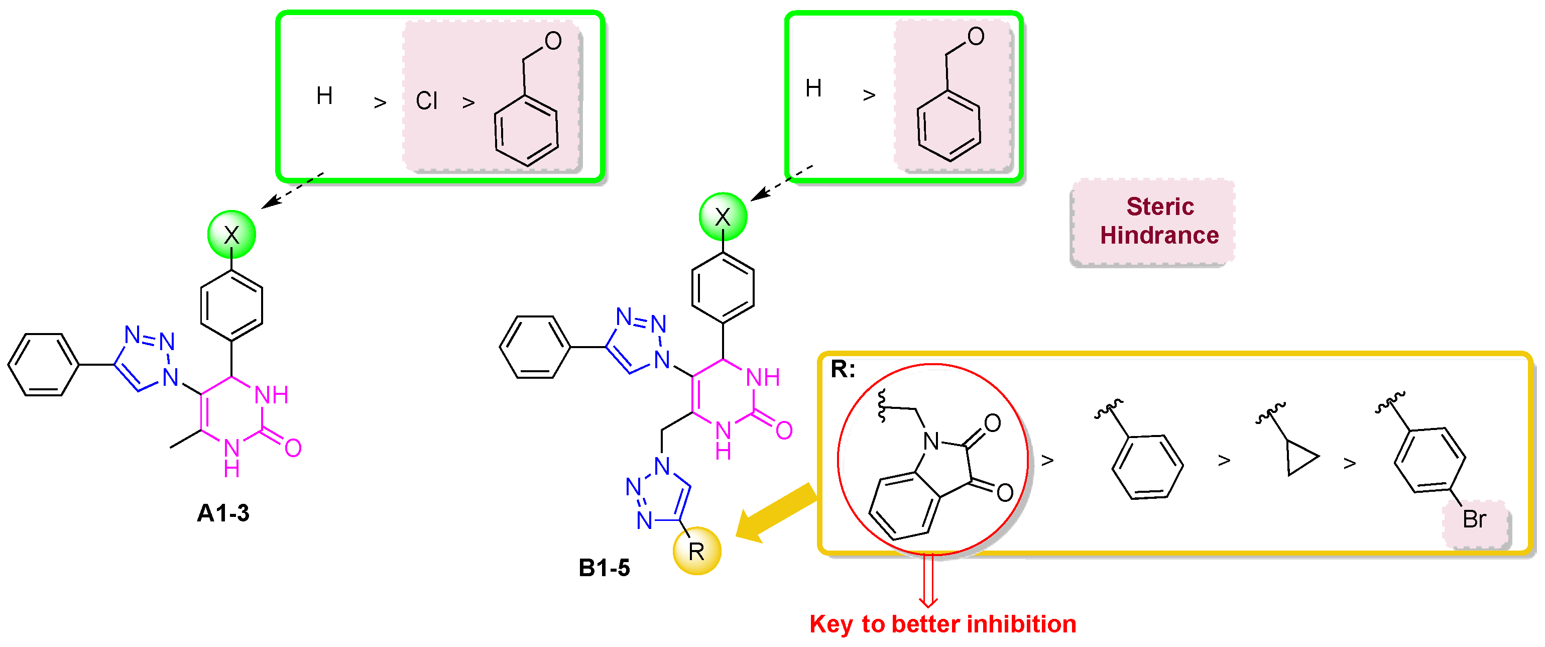
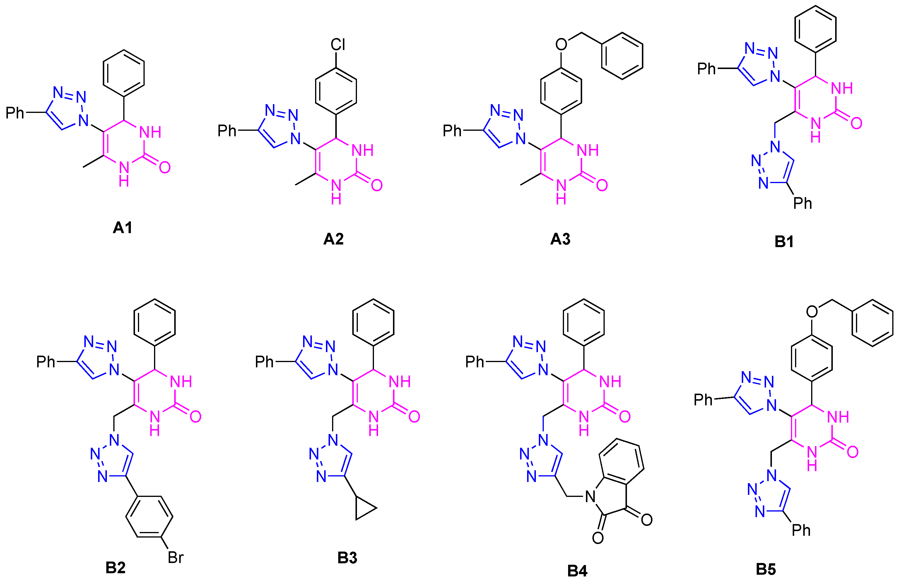
2.1. Anticholinesterase Activity

In order to evaluate the inhibitory activity against cholinesterase enzymes (AChE and BuChE), well-established in vitro models were used—purified enzymes of animal origin, namely electrophorus electricus AChE (eeAChE) and equine serum BuChE (eqBuChE) [20,21,22,23,24,25,26]. Hybrids A13 and B15 (Figure 3) were evaluated in vitro for cholinesterase inhibition using Ellman’s colorimetric method [27]. Table 1 shows the results obtained for the 50% inhibitory concentration (IC50) of the studied hybrids against the enzymes eeAChE and eqBuChE. The values are expressed as the mean ± standard deviation (SD). By analyzing the results obtained for the IC50 (Table 1), it was found that none of the hybrids studied were effective inhibitors of eeAChE, as they presented IC50 levels higher than 100 µM.
Although the hybrids generally showed weak inhibition of eqBuChE, two compounds demonstrated significant activity: hybrid A1 (with one 1,2,3-triazole ring) exhibited an IC50 = 12 µM, while hybrid B4 (with two 1,2,3-triazole rings and a heterocyclic isatin) showed an IC50 = 1.0 µM. Additionally, three other compounds (A2, B1, and B3) exhibited moderate inhibition, with IC50 values ranging from 23 to 58 µM. Comparing the anticholinesterase activities of hybrids A1 and A2 with B1 and B3 suggests that hybrids A (with a single 1,2,3-triazole ring) are slightly more active than hybrids B (with two 1,2,3-triazole rings), with the exception of B4 (see below). Nonetheless, hybrid B4, featuring two 1,2,3-triazole rings, emerged as the most potent eqBuChE inhibitor investigated in this study. Moreover, despite possessing two 1,2,3-triazole rings in its structure, it also has an isatin unit (with renowned biological activity), which is probably responsible for the high inhibition activity achieved. Hybrid B4 was a better inhibitor than galantamine (IC50 = 10 µM).
In the case of the A1-3 hybrids, IC50 values were determined to be 12 ± 2 (A1), 24 ± 1 (A2), and >100 (A3) µM, the structure–activity relationship suggested that the unsubstituted phenyl ring in the 4-position of the DHPM core was the most active. However, the presence of the chlorine or benzyloxy groups in the para-position of the phenyl ring decreased the activity of the compounds, possibly due to the steric hindrance promoted by these groups. On the other hand, these groups also impart hydrophobicity to the hybrids, which could potentially interfere with their binding to the enzyme (Figure 4). In addition, hybrid A1 showed the best inhibitory activity, with an IC50 value very similar to that of galantamine (IC50 = 10 µM).
Figure 4. Structure−activity relationship of hybrids A13 and B15 in eqBuChE inhibition.
Ijms 25 11153 g004
Table 1. IC50 values for hybrids A13 e B15 against cholinesterases (eeAChE and eqBuChE).
IC50 ± SD (µM) a
CompoundeeAChEeqBuChE
A1>10012 ± 2
A2>10024 ± 1
A3>100>100
B1>10053 ± 4
B2>100>100
B3>10058 ± 3
B4>1001 ± 0.1
Mixed inhibition b
Kia = 1.1 ± 0.3 µM
Kib = 1.4 ± 0.2 µM
B5>100>100
Galantamine2.7 ± 0.210.0
a [S] = 121 µM for eeAChE, 112 µM for eqBuChE; b methods of Dixon and Cornish-Bowden (Figure 5 and Figure 6) [28].
Regarding hybrids B15, these furnished IC50 values of 1 ± 0.1 (B4), 53 ± 4 (B1), 58 ± 3 (B3), >100 (B2 and B5) µM. By comparing hybrids B14, the structure–activity relationship suggested that the best inhibitor is that which possesses an isatin moiety linked to the 1,2,3-triazole unit, while the phenyl and cyclopropane rings decreased the activity of the hybrids. Hybrids B2 (with a 4-bromophenyl group linked to the 1,2,3-triazole) and B5 (with a benzyloxy group in the para-position of the phenyl ring linked to the 4-position of the DHPM core) were the poorest inhibitors, possibly due to the presence of bulky groups in their structures, which could have compromised their binding to the enzyme’s active site. Notably, compound B4 displayed very good anticholinesterase activity [15,29]. Furthermore, hybrid B4 was a stronger inhibitor than galantamine by one order of magnitude.
A study of the enzymatic kinetics for the inhibition of eqBuChE was carried out in order to determine the type of inhibition exhibited by the best inhibitor, notably, hybrid B4. The calculation of the kinetic parameters (non-linear regression, GraphPad 8.0) indicated a slight modification of KM values and a decrease in Vmax upon the increase in inhibitor concentration. Such observations are compatible with a mixed mode of inhibition, where the inhibitor binds the free enzyme and the E-S complex (Kia, Kib, respectively). This was further demonstrated using the Cornish-Bowden method [28], which considers two graphs: 1/V vs. [I] and [S]/V vs. [I], shown in Figure 5 and Figure 6, respectively. An analysis of the graphs reveals that a single intercept between the lines indicates the mixed mode of inhibition. The inhibition constants Kia and Kib were calculated, where Kia represents the inhibition constant of the inhibitor binding to the enzyme-free complex (EF) and Kib represents the inhibition constant of the inhibitor binding to the enzyme–substrate complex (ES). The value obtained for both constants was quite similar (Kia = 1.1 µM, Kib = 1.4 µM), confirming that the inhibitor binds both the free enzyme and the E-S complex, with analogous potency, leading to a change in the enzyme’s structure, altering the configuration of its active center, and thus preventing any enzyme activity (see below for a further discussion on this in the context of the docking and NMR studies).

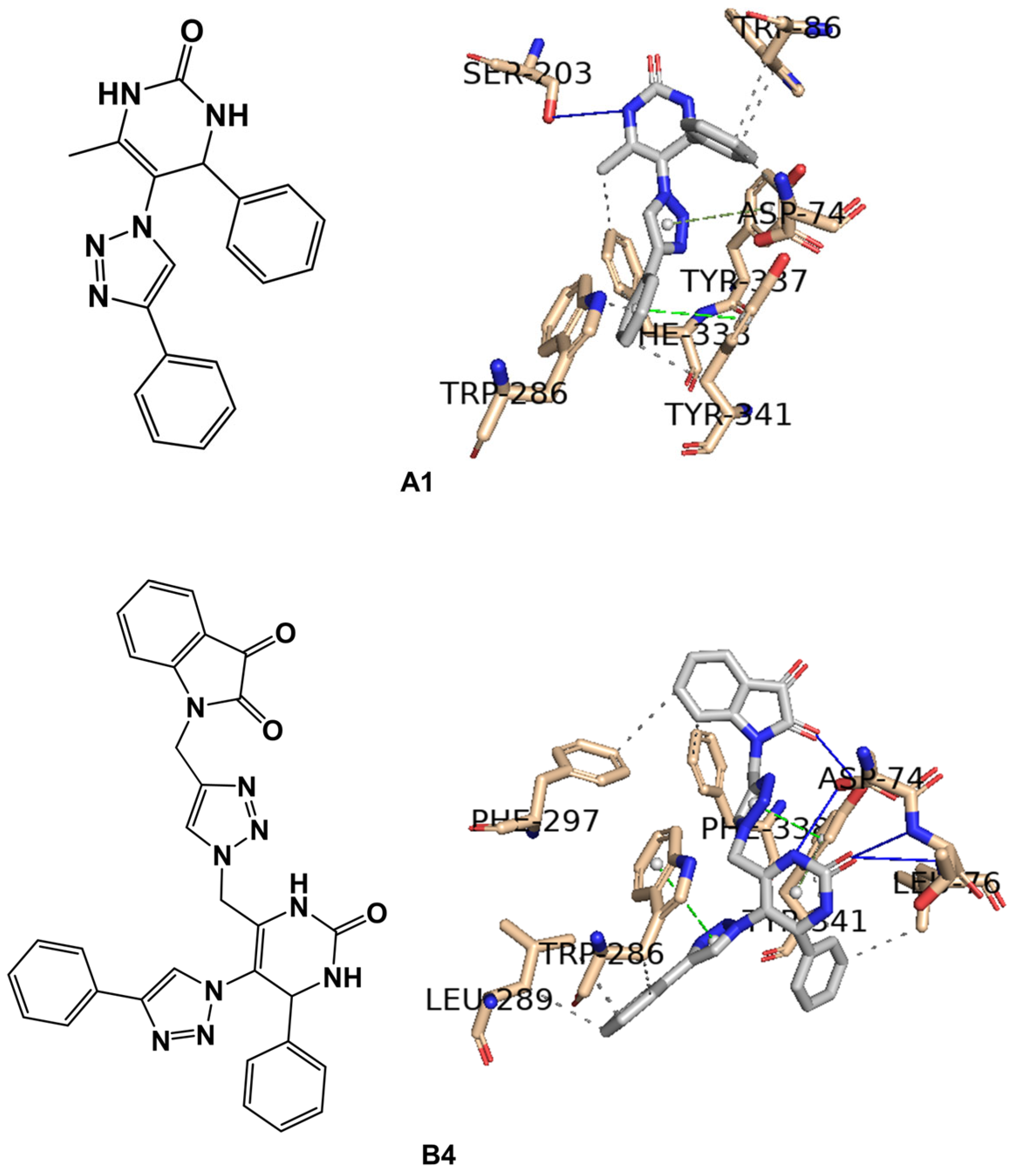
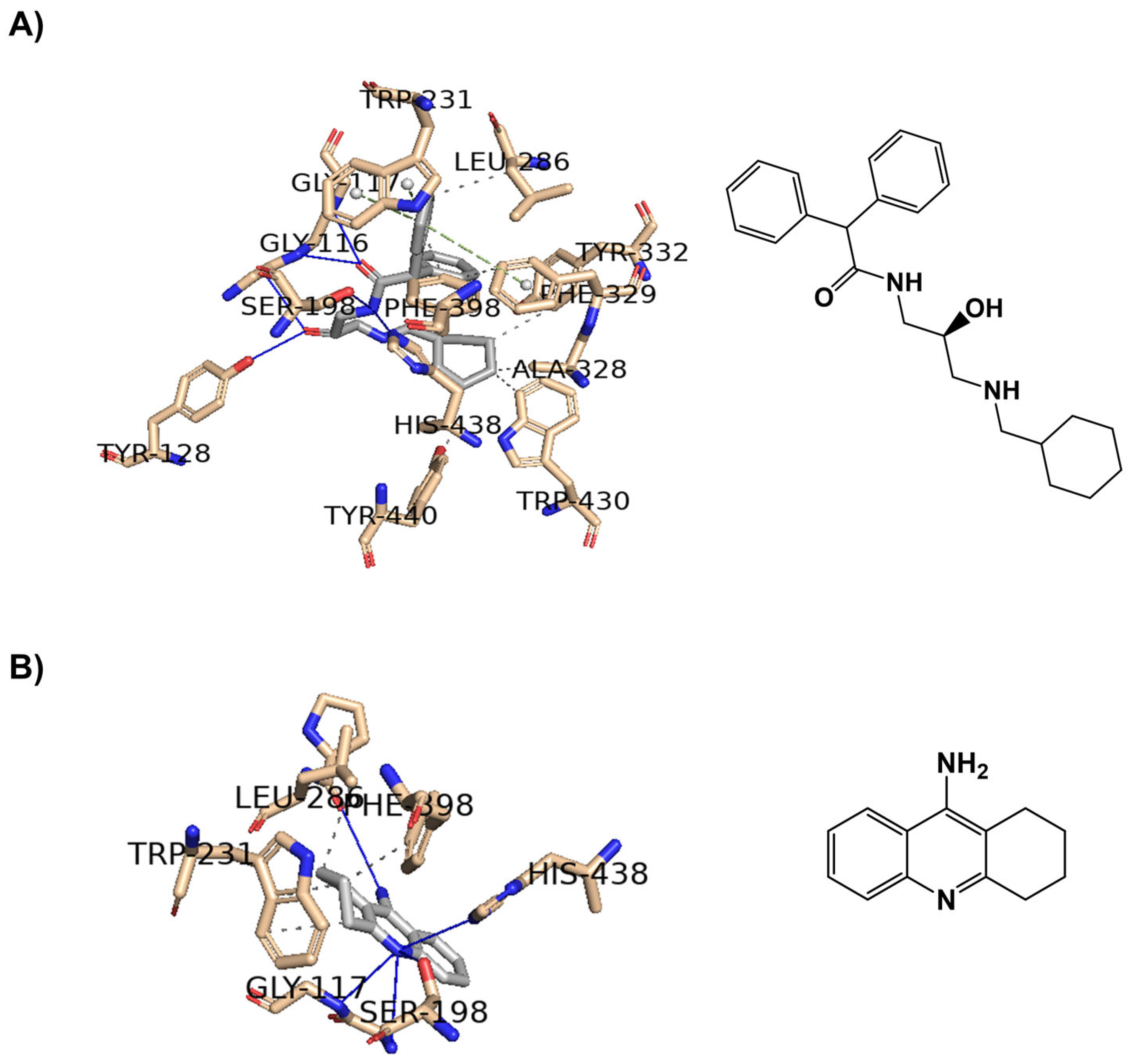
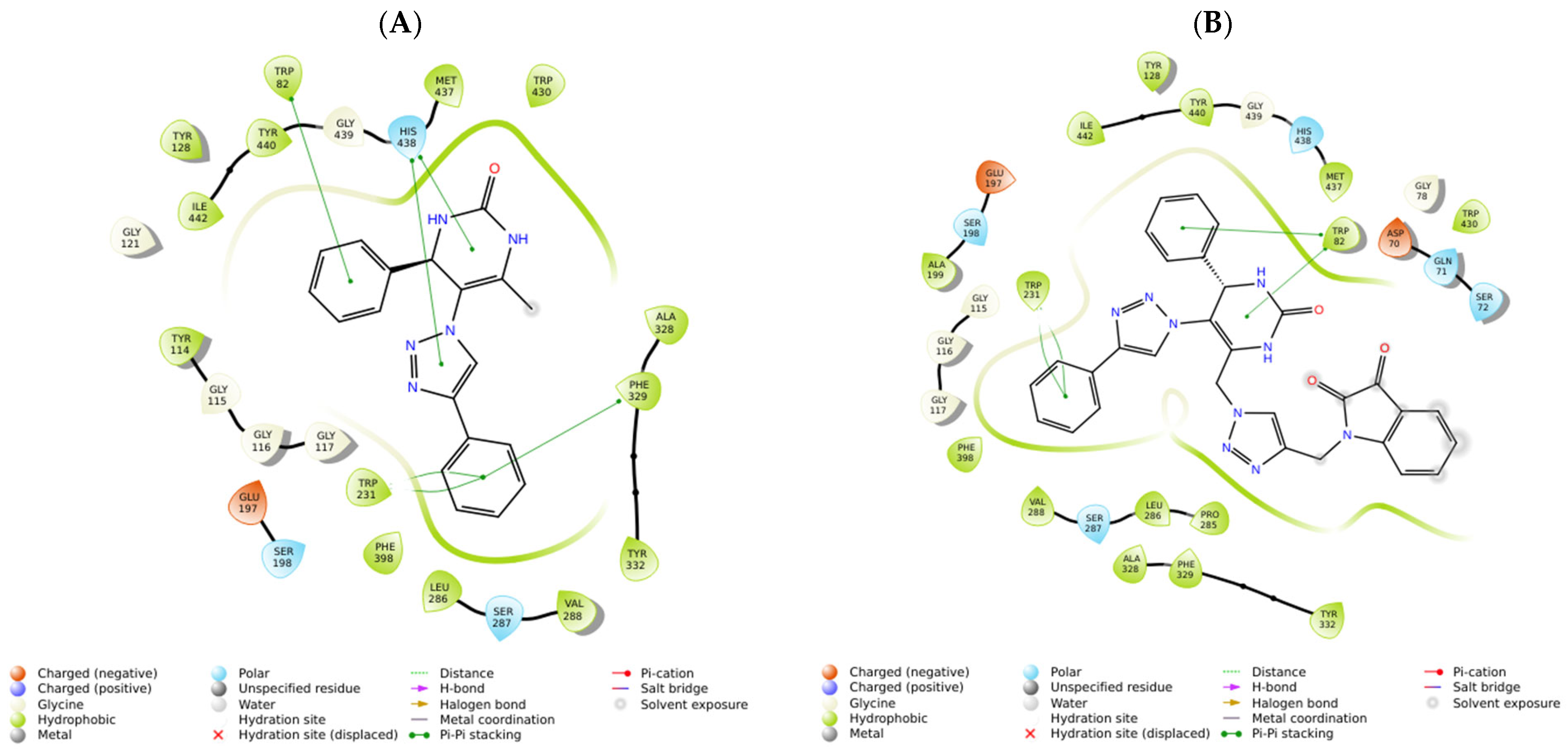
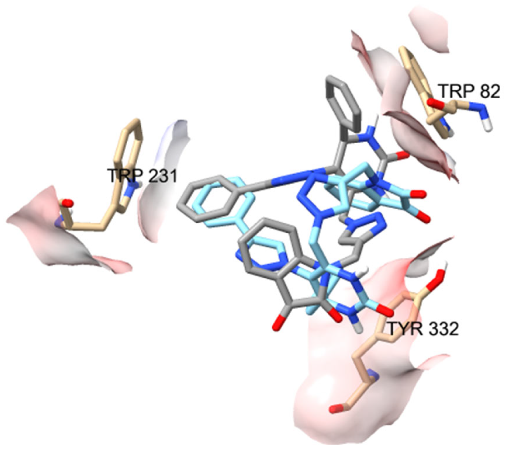
2.2. In Silico Studies

The binding of the most promising inhibitors, A1 and B4, to ChEs was explored and predicted through molecular docking using two software programs, Glide XP and AutoDock Vina. This study utilized several human protein X-ray crystal structures: AChE (PDB IDs 6O4W and 4EY7, both co-crystallized with donepezil) and BuChE (PDB IDs 4AQD, 7Q1M, and 4BDS, co-crystallized with β-alanine, a hydroxypropyl derivative, and tacrine, respectively). The calculated binding affinities obtained through molecular docking of the most promising ChE inhibitors identified are presented in Table 2. As shown in Table 2 using the Glide program, the known inhibitor donepezil exhibited a stronger predicted interaction with AChE compared to BuChE as expected. Conversely, B4 exhibited stronger predicted interactions with BuChE than with AChE across all experiments, and, in each case, B4 was predicted to interact more strongly than the co-crystallized ligands β-alanine (PDB ID 4AQD), the hydroxypropyl derivative, HPD, (PDB ID 7Q1M), and tacrine (PDB ID 4BDS).
As observed experimentally, galantamine interacts with both AChE and BuChE simultaneously; however, gratifyingly, its interaction is predicted to be weaker than that of the B4 derivative against BuChE. As shown in Figure 7, the best-docked pose for the positive control, donepezil, on the AChE enzyme was achieved using PDB IDs 6O4W (A) and 4EY7 (B).
The active site of human AChE has a long gorge approximately 20 Å in length (Figure 7, [30]), primarily composed of the catalytic active (or anionic) site (CAS) at the bottom of the gorge (His447, Ser203, Trp86, Tyr337) and the peripheral anionic site (PAS) near the entrance (His287, Ser293, Trp286, Tyr72). These two sites are connected by a narrow groove (Tyr124, Phe295, Tyr341). Compounds that can interact with both CAS and PAS are desirable due to their potential to exert multiple therapeutic effects [30,31], as exemplified by donepezil in Figure 7.
To validate the re-docking process, PyMOL software was used to superimpose the docked complexes of AChE (A) and (B) with donepezil onto the solved structures (PDB IDs 6O4W and 4EY7, respectively). The root-mean-square displacement (RMSD) between complex (A) and the solved structure was 0.161 Å, and between complex (B) and the solved structure was 0.225 Å, indicating high structural similarity in both cases.
Figure 8 presents the best-docked poses for the two most promising AChE inhibitors, A1 and B4, docked on PDB ID 6O4W.
Considering the calculation of the binding affinities for the hybrid A1 against AChE using Glide and AutoDock Vina, which resulted in −3.72 kcal/mol and −11.75 kcal/mol, respectively, only the Glide docking score seems to justify the experimentally observed IC50 of >100 μM (Table 1 and Table 2). Moreover, the best-docked poses shown in Figure 8 for the hybrids A1 and B4 only seem to justify the lack of activity demonstrated against AChE for the hybrid B4 (Table 1), as in this case, interactions are only observable with the PAS residue, specifically Trp286. In the case of hybrid A1, interactions are observable with the CAS residues (Trp86, Ser203, and Tyr337) as well as PAS (Trp286), as shown in Figure 8.
As with AChE, in BuChE, compounds capable of interacting with CAS residues (Trp82, Phe398, His438) and PAS residues (Asp70, Ser198, Phe329) appear to be more promising [31], as demonstrated for the hydroxypropyl and tacrine derivatives in Figure 9.
Similar to the approach used with AChE, the re-docking process was validated by using PyMOL software to superimpose the docked complexes of BuChE (A) and (B) with the hydroxypropyl derivative and tacrine onto the solved structures (PDB IDs 7Q1M and 4BDS, respectively). The root-mean-square deviation (RMSD) between complex (A) and the solved structure was 0.475 Å, and between complex (B) and the solved structure was 0.361 Å, indicating a high degree of structural similarity in both cases.
In the case of our stronger inhibitor compounds A1 and B4, an in silico study was carried out to predict the binding poses in the binding site of BuChE (Figure 10), which showed good affinity. Regarding hybrid A1 binding to the enzyme, the major interactions are through π–π stacking interactions, such as Trp82 from CAS with phenyl ring linked to the 4-position of the DHPM core, His438 from catalytic triad in the CAS with DHPM and 1,2,3-triazole cores, and Trp231, Phe329, and Tyr332 (from PAS) with a phenyl ring linked to 1,2,3-triazole moiety. Compound B4 binds the amino acid residues of the enzyme via π–π stacking, such as Trp231 with a phenyl ring linked to the 1,2,3-triazole in the 5-position of the DHPM core and Trp82 from CAS with DHPM and its phenyl substituent of the 4-position. Asp70 and Tyr332 from PAS and Val288 from the acyl pocket bind to the isatin core. Ser72 and/or Gln71 could establish an H-bond with the carbonyl group of the isatin unit. Additionally, general Van der Waals interactions were also observed between the inhibitors and the enzyme. Hybrid B4 exhibits mixed inhibition, binding at both the CAS and the PAS sites but with an apparent higher affinity for the PAS of BuChE rather than th CAS of BuChE.
However, even more relevant seems to be the π–π stacking interactions with the CAS residue (Trp82) and with the two PAS residues (Trp231 and Tyr332). It is observed that in the ten best poses of B4 against the BuChE enzyme (PDB ID 4AQD), these poses oscillate between the pose represented in gray (five conformers) and in blue (five conformers) in Figure 11. As can be seen in Figure 11, for the Trp82 residue in the pose represented in gray, the mentioned interaction occurs with the phenyl ring attached to the DHPM core at position 4, while, in the blue pose, the interaction occurs with the isatin moiety. The opposite occurs with the Tyr332 residue in the two poses represented, gray and blue, in Figure 11.

2.3. STD-NMR Studies

In order to gain an insight into the interaction of our 1,2,3-triazole-DHPM hybrids with eqBuChE, we performed an STD-NMR study on the most potent compounds A1 and B4. STD-NMR is a very useful validation technique for docking studies, which we have used previously with success [15,29,32].

2.3.1. STD-NMR of Hybrid A1

The results obtained in the STD-NMR experiment of hybrid A1 with the eqBuChE enzyme are shown in Figure 12 (the graph of the STD amplification factor as a function of saturation time (s) is shown in the Supplementary Materials, Figure S1); moreover, it can be seen that all the hydrogens interact with the amino acid residues in the enzyme’s binding pocket, with the exception of H1 and H2 of the N-H of the DHPM core. The aromatic hydrogens of the two phenyl rings show an attenuation of around 89–100%, showing a strong interaction with the enzyme. These results are completely in line with the prediction of the docking study, which shows the interaction of these aromatic rings with Trp82 (CAS), Tyr332 (PAS), Trp231, and Phe329. The H3 and H7 hydrogens of the main nuclei 1,2,3-triazole and DHPM showed very good attenuation values of 87 and 88%, respectively. This confirms the predicted interaction of His438 from the catalytic trio in the CAS with these cores.

2.3.2. STD-NMR of Hybrid B4

The results obtained for the STD-NMR experiment for the most potent inhibitor against eqBuChE are shown in Figure 13 (the graph of the STD amplification factor as a function of saturation time (s) is shown in the Supplementary Materials, Figure S2). All the hydrogens of the hybrid interact with the amino acid residues of the enzyme; however, the H11 and H13 of the CH2 groups showed a moderate attenuation value of 33%. Once again, and according to the epitope intensities shown in Figure 10, the aromatic hydrogens are the most affected by the amino acid residues, showing high attenuations of around 93–96%; as suggested by the docking study, these aromatic hydrogens interact with Trp231 and Trp82 in PAS and CAS, respectively. The H1–H3 hydrogens from the DHPM core showed 59–71% attenuation and may interact with Trp82, as predicted. The H15–H17 hydrogens show 93–100% attenuation, which indicates that they are very close to the enzyme; in fact, the docking study suggests that these hydrogens are close to the Asp70 and Tyr332 in PAS and Val288 in the acyl pocket.

2.4. In Vitro Antioxidant Assays

To assess the antioxidant activity of compounds A13 and B15, the in vitro methods DPPH, ABTS, and FRAP were used.
All compounds evaluated at concentrations between 6 and 200 µM presented null values of antioxidant activity when using the DPPH and ABTS methods. It is thus concluded that, at least in this concentration range, these compounds do not have the capacity to scavenge free radicals (nor DPPH or ABTS radicals).
In the FRAP method, compounds A13, B1, B2, and B4 also have null values of antioxidant capacity. On the other hand, the compounds B3 and B5 presented Trolox Equivalent Antioxidant Capacity (TEAC) in the FRAP method, although much lower than the positive control Trolox (a soluble analog of Vitamin E), and the results can be observed in Figure 14. At 200 μM, the highest concentration tested, TEACs of 0.257 and 0.216 were found for B3 and B5, respectively, compared to Trolox, revealing that these compounds have some ability to reduce Fe(III) to Fe(II).

2.5. Toxicity Assay In Vivo

Artemia salina was used for in vivo general toxicity assessment of the compounds A13 and B15. The results are shown in Table 3.
The test with K2Cr2O7 showed an LC50 of 23.6 mg/L. This validates the method as it is within the reference range (CI = 95% {20.7–38.3 mg/L}). Relative to the 1,2,3-triazole-dihydropyrimidinone hybrids tested, the compounds A13, B1, and B35 showed no toxicity to Artemia salina in the concentration range tested. It should be noted that, for these compounds, no toxicity (NOEC) was observed at 200 μM concentration. The compound B1 presented 37 ± 3% mortality at 200 μM concentration (data showed in Supplementary Materials, Figures S3–S11), which means that the LC50 will be >200 μM, also revealing a low toxicity.

2.6. SwissADME Calculations

Our study involved predicting the physicochemical properties of all examined hybrids using SwissADME [33]. Remarkably, all calculated results adhered to Lipinski’s rules [34], except for hybrids B2, B4, and B5, which slightly exceeded the specified MW limit. Encouragingly, none of these compounds were flagged as Pan-Assay Interference Compounds (PAINS), indicating no adverse interactions with multiple targets that could affect screening experiments negatively (Table 4). While all evaluated hybrids were predicted to be suitable for gastrointestinal absorption (GI), only hybrids A1 and A2 were anticipated to penetrate the blood–brain barrier (BBB). Most products exhibited moderate water solubility, with hybrid A1 being notably soluble, while B5 displayed poor solubility (Table 4). In fact, our best inhibitor B4 does not permeate the BBB; however, this situation can be overcome by encapsulating it in nanoparticles of various types [35]. All hybrids, except A3, are estimated to be non-inhibitors of cytochrome P450 isoforms (CYP2D6 and CYP3A4), an important class of detoxification enzymes primarily found in the liver.

3. Materials and Methods

3.1. General Remarks

For carrying out this work, reagents were used as received. Iodide acetylthiocholine (ATCI), S-butyrylthiocholine iodide (BTCI), and 5,5′-dithiobis(2-nitrobenzoic acid) (DTNB) (Ellman’s reagent) were obtained from Sigma–Aldrich (St. Louis, MO, USA) and Alfa Aesar (Haverhill, MA, USA). The selected enzymes were purchased from Sigma–Aldrich: electric eel acetylcholinesterase (eeAChE) (E.C.3.1.1.7, Type VI-S, lyophilized powder, 500 U/2 mg), and equine serum butyrylcholinesterase (eqBuChE) (E.C. 3.1.1.8, lyophilized powder, 10.9 U/mg). The commercial inhibitors used were galantamine (TCI). 1,1-Diphenyl-2-picrylhydrazyl (DPPH), (2,2′-Azino-bis(3-ethylbenzothiazoline-6-sulfonic acid) diammonium salt) ABTS used to determine the radical-scavenging activity, was purchased from Fluka. TPTZ (2,4,6-Tris(2-pyridyl)-s-triazine) and Trolox (6-Hydroxy-2,5,7,8-tetramethylchroman-2-carboxylic acid) was purchased from BLDPharma. All other reagents and solvents used in the experiments were of analytical grade.

3.2. 1,2,3-Triazole-Dihydropyrimidinone Hybrids A and B

1,2,3-Triazole-Dihydropyrimidinone hybrids A and B were previously reported by Carreiro et al. [19]. 1H spectra data are included in the Supplementary Materials. The 8 hybrids evaluated were:
6-Methyl-4-phenyl-5-(4-phenyl-1,2,3-triazol-1-yl)-3,4-dihydropyrimidin-2-one (A1); 4-(4-Chlorophenyl)-6-methyl-5-(4-phenyl-1,2,3-triazol-1-yl)-3,4-dihydropyrimidin-2-one (A2); 4-(4-(Benzyloxy)phenyl)-6-methyl-5-(4-phenyl-1,2,3-triazol-1-yl)-3,4-dihydropyrimidin-2-one (A3); 4-Phenyl-5-(4-phenyl-1,2,3-triazol-1-yl)-6-((4-phenyl-1,2,3-triazol-1-yl)methyl)-3,4-dihydropyrimidin-2-one (B1); 6-((4-(4-Bromophenyl)-1,2,3-triazol-1-yl)methyl)-4-phenyl-5-(4-phenyl-1,2,3-triazol-1-yl)-3,4-dihydropyrimidin-2-one (B2); 6-((4-Cyclopropyl-1H-1,2,3-triazol-1-yl)methyl)-4-phenyl-5-(4-phenyl-1,2,3-triazol-1-yl)-3,4-dihydropyrimidin-2-one (B3); 1-((1-((2-Oxo-6-phenyl-5-(4-phenyl-1,2,3-triazol-1-yl)-1,2,3,6-tetrahydropyrimidin-4-yl)methyl)-1,2,3-triazol-4-yl)methyl)indoline-2,3-dione (B4); 4-(4-(Benzyloxy)phenyl)-5-(4-phenyl-1,2,3-triazol-1-yl)-6-((4-phenyl-1,2,3-triazol-1-yl)methyl)-3,4-dihydropyrimidin-2-one (B5).

3.3. Cholinesterase Inhibitory Assays

The enzymatic activity of electrophorus electricus AChE (eeAChE) and equine serum BuChE (eqBuCHE) was assessed using the Ellman colorimetric assay, with minor modifications. DMSO (final concentration of 1.25% in the cuvette) was used to prepare the stock solutions of the inhibitors (A13 and B15). Enzyme kinetics were monitored using UV-Vis spectroscopy using 0.875 mM 5,5′-dithiobis(2-nitrobenzoic acid) (DTNB) (Ellman’s reagent) as a chromogenic agent. The reaction took place in a buffered medium (0.1 M phosphate buffer, pH 8.0), T = 25 °C, and was monitored for 125 s. To determine the percentage of inhibition ([I] = 100 µM), the substrate concentration (acetylthiocholine iodide for AChE and butyrylthiocholine iodide for BuChE) was set at 121 µM for AChE and 112 µM for BuChE. The results are shown in Table 1.
For compounds showing strong inhibition at 100 µM inhibition concentration, IC50 values were obtained. For this purpose, GraphPad Prism 8.02 software was used to plot %I vs. [I] (5 different concentrations) via a non-linear regression.
Methods of Dixon (Figure 5) and Cornish-Bowden (Figure 6) (1/V vs. [I] and [S]/V vs. [I]) were used for the visualization of the type of inhibition of the most active compound, hybrid B4. Calculation of the kinetic parameters (KM, Vmax) was accomplished using a nonlinear regression analysis (least squares fit) implemented in GraphPad Prism 8.01 software. The following equations were used for calculating the inhibition constants of the inhibitor obtained herein:
K m , a p p = K M 1 + [ I ] K i a 1 + [ I ] K i b
V m a x , a p p = V m a x 1 + [ I ] K i a
Kia: Inhibition constant for the interaction of the inhibitor with the free enzyme (E).
Kib: Inhibition constant for the interaction of the inhibitor with the complex enzyme-substrate (E-S).

3.4. In Silico Studies

Protein X-ray crystal structures were obtained from the Protein Data Bank (https://www.rcsb.org/, accessed on 7 October 2024), 6O4W and 4EY7 for AChE and 4AQD, 7Q1M and 4BDS for BuChE. All protein structures were determined at high resolution. The structures were inspected and assessed as adequate for docking, with resolutions of 2.35 Å, 2.35 Å, 2.5 Å, 2.79 Å, and 2.10 Å, respectively. The optimization of the 3D structures of the most promising inhibitors, A1 and B4, as well as the positive controls (donepezil (PDB IDs 6O4W and 4EY7), β-alanine (PDB ID 4AQD), 3-(cyclohexylmethyl)amino-2-hydroxypropyl (PDB ID 7Q1M), tacrine (PDB ID 4BDS), and galantamine (PDB IDs 6O4W, 4EY7, 4AQD, 7Q1M and 4BDS) was carried out using the RDKit function MMFFOptimizeMolecule with the arguments mmffVariant = ‘MMFF94’ and maxIters = 5000 in Python (cite: Landrum, G. RDKit: Open-Source Cheminformatics Software. http://www.rdkit.org (2016)). Hydrogen atoms were added with Maestro software [Schrödinger, 2020]. Docking was then performed using extra precision Glide XP [Schrödinger, 2020] with extended sampling and the OPLS3e forcefield [36]. The software program OpenBabel (version 2.3.1) [37] was used to convert the mol2 files to PDBQT files. PDBQT files were used for docking to AChE (PDB IDs 604W and 4EY7) and BuChE (PDB IDs 4AQD, 7Q1M, and 4BDS) enzymes with AutoDock Vina (version 1.2.3) [38,39]. Water molecules, ions, and ligands were removed from all enzymes (604W, EY7, 4AQD, 7Q1M, 4BDS) prior to docking using the AutoDockTools (http://mgltools.scripps.edu/, accessed on 29 August 2024). The search space coordinates were AChE enzyme; 6O4W—Centre X: 89.341 Y: 84.453 Z: −5.628, and 4EY7—Centre X: −15.834 Y: −43.535 Z: 25.391; and BuChE enzyme; 4AQD—Centre X: 7.0 Y: −10.639 Z: −12.236, 7Q1M—Centre X: 19.553 Y: 42.576 Z: 41.06, and 4BDS—Centre X: 131.866 Y: 112.975 Z: −44.529, Dimensions X: 20.000 Y: 20.000 Z: 20.000. Ligand tethering of the AChE and BuChE enzymes was performed by regulating the genetic algorithm (GA) parameters, using 10 runs of the GA criteria. The docking binding poses were visualized with PyMOL Molecular Graphics System, Version 2.0 Schrödinger, LLC (New York, NY, USA), UCSF Chimera [40], and the Protein–Ligand Interaction Profiler (PLIP) web tool (https://plip-tool.biotec.tu-dresden.de/plip-web/plip, accessed on 7 October 2024) [41].

3.5. STD-NMR Experiments

The NMR spectroscopy experiments were performed on a Bruker Avance III 400 MHz spectrometer equipped with a 5 mm broadband (PABBO BB/19F-1H/D Z-GRD) resonance probe head. The STD-NMR experiments were realized according to our established method [29,32]. Basically, NMR and STD-NMR experiments were carried out with solvent suppression and a 10 ms spinlock filter after the 90◦ pulse to reduce residual signals from the protein. For selective saturation, cascades of Gaussian pulses with a length of 50 ms and 40–60 dB of attenuation were employed, with an interpulse delay of 1 ms [29,32]. The on-resonance and off-resonance frequencies were set to 0 and 12,000 Hz, respectively. STD-NMR controls were performed using the ligand itself. Blank experiments were performed to guarantee the absence of direct saturation of the ligand proton signals. The relaxation delay was properly adjusted so that the experiment time length was kept constant at 6.5 s. Water suppression at 1880 Hz (4.7 ppm) was conducted. Specifically, the saturation time to obtain the STD buildup curves was recorded at 0.25, 0.5, 1, 2, 3, 4, and 5 s [29,32]. A 5 μM eqBuChE solution was prepared in a D2O. Five mM stock solutions were prepared for hybrids A1 and B4. Samples for NMR analysis were prepared by adding 300 μL of the ligand to a 300 μL enzyme solution. The final concentrations of ligand and enzyme were 2.5 µM and 2.5 mM, respectively.

3.6. Antioxidant Assays

3.6.1. DPPH Antioxidant Assay

In the DPPH method, the antioxidants react with DPPH (2,2-diphenyl-1-picrylhydrazyl) (which has a strong violet color) and convert it into 2,2-diphenyl-1-picrylhydrazine (yellow in color). The colorimetric DPPH assay was carried out as described in [29]. The compounds A13 and B15 were evaluated through the DPPH assay against positive control, ascorbic acid.
In brief, 30 μL of the tested compounds (6–200 µM in MeOH + DMSO 0.5%) was mixed with 200 μL of DPPH (0.03 g/L in MeOH) in a 96-well plate in triplicate. After 30 min incubation at room temperature in the absence of light, the absorbance was measured at 517 nm using Microplate Spectrophotometry (TriStar® S LB 942 model instrument, Berthold Technologies GmbH & Co.KG, Bad Wildbad, Germany). The free radical scavenging activity (DPPH) results are expressed as a percentage of DPPH inhibition according to the following formula:
I n h i b i t i o n   ( % ) = A b l a n k A s a m p l e A b l a n k × 100
where Ablank consists of MeOH + DMSO 0.5%) (30 μL) mixed with DPPH (200 μL) absorbance, and Asample is the absorbance value for the added sample concentration compound mixed with DPPH.

3.6.2. ABTS Antioxidant Assay

The ABTS (2,2′-Azino-bis(3-ethylbenzothiazoline-6-sulfonic acid) diammonium salt) test measures the relative capacity of antioxidants (hydrophilic and lipophilic) to eliminate ABTS generated in the aqueous phase (light green). ABTS is generated by the reaction of a strong oxidizing agent (e.g., potassium persulfate, K2S2O8) with the ABTS salt (dark green color). The assay was carried out using the modified method described in [42]. The compounds A13 and B15 were selected through the ABTS assay against the positive control, ascorbic acid.
In brief, 30 μL of the tested compounds (6–200 µM in MeOH + DMSO 0.5%) was mixed with 200 μL of ABTS (prepared by mixing 0.373mmol of ABTS and 0.125 mmol of K2S2O8 in MeOH over 24H stirring) in a 96-well plate in triplicate.
After 30 min incubation at room temperature in the absence of light, the absorbances were measured at 517 nm using Microplate Spectrophotometry (TriStar® S LB 942 model instrument, Berthold Technologies GmbH & Co.KG, Bad Wildbad, Germany). The free radical scavenging activity (ABTS) results are expressed as a percentage of ABTS inhibition according to the following formula:
I n h i b i t i o n   ( % ) = A b l a n k A s a m p l e A b l a n k × 100
where Ablank consists of MeOH + DMSO 0.5% (30 μL) mixed with ABTS (200 μL) absorbance, and Asample is the absorbance value for the added sample concentration compound mixed with ABTS.

3.6.3. FRAP Antioxidant Assay

The Ferric Reducing Antioxidant Power (FRAP assay) evaluates antioxidant capacity by quantifying the reduction in the Fe(III)-2,4,6-Tris(2-pyridyl)-s-triazine (TPTZ) complex (light blue in color) to Fe(II)-TPTZ (dark blue in color), in an acidic medium (HCl), compared to a Trolox (6-hydroxy-2,5,7,8-tetramethylchroman-2-carboxylic acid) standard (analog of vitamin E).
The assay was carried out as described in [43]. The compounds A13 and B15 were selected through the FRAP assay against positive control and water-soluble analogs of vitamin E and Trolox.
In brief, 30 μL of the tested compounds (6–200 µM in MeOH + DMSO 0.5%) was mixed with 200 μL of the FRAP reagent (obtained from a mixture of 0.3 M acetate buffer, 10 mM TPTZ solution, and 20 mM ferric chloride solution in a ratio of 10:1:1 (V/V), respectively) in a 96-well plate in triplicate. After 10 min incubation at room temperature in the absence of light, the absorbances were measured at 593 nm using Microplate Spectrophotometry (TriStar® S LB 942 model instrument, Berthold Technologies GmbH & Co.KG, Bad Wildbad, Germany). The results of the Fe(II)-TPTZ complex formation are expressed as Trolox Equivalent Antioxidant Capacity (TEAC) according to the following formula:
T E A C = c ( T E ) c ( c o m p o u n d )
where c(TE) is the concentration of Trolox and c(compound) is the concentration of compound tested, the c(TE) was determined according to the following formula:
c ( T E ) = A 1 A 0 S l o p e
where A0 consists of the solvent (MeOH + DMSO 0.5%) (30 μL) mixed with FRAP solution (200 μL) absorbance, A1 is the absorbance value for the added sample concentration compound mixed with FRAP solution, and the slope was obtained from the calibration curve of Trolox (positive control) (slope = 0.0029 with R2 = 0.99), Figure 14.

3.7. Artemia Salina Lethal Toxicity Assay

The ARTOXKIT M protocol was used. The percentage of dyed nauplii of Artemia salina, grown in the presence of variable concentrations of the inhibitor compounds (A13 and B15) (0.01 to 100 µM).
Saline water with 1% DMSO was used as a blank. After 24 h incubation at 25 °C, the LD50 values were determined.
In parallel, a K2Cr2O7 test was performed as quality control.

3.8. Statistical Analysis

All experimental results are shown as the mean ± SD. Origin 9.0 software (OriginLab, Northampton, MA, USA), GraphPrism® software (version 9.2, 64-bit) and Microsoft® Excel® (for Microsoft 365 MSO, version 2307 Build 16.0.16626.20198, 64-bit) were used for drawing and data analysis in this paper.

4. Conclusions

In conclusion, hybrids A and B were evaluated as inhibitors of eeAChE and eqBuChE and did not inhibit eeAChE (IC50 values of >100.0 µM). Five (A1, A2, B1, B3, and B4) of the eight hybrids evaluated proved to be eqBuChE inhibitors displaying IC50 values in the range of 1.0–58.0 µM. Hybrid B4 was the most potent inhibitor, probably due to the presence of two 1,2,3-triazole rings linked to the core DHPM unit, in addition to an isatin ring as a substituent (both 1,2,3-triazole and isatin are potent pharmacophores). However, hybrid analogs B1 and B3 showed weaker inhibition than hybrids A1 and A2 (having only a single 1,2,3-triazole ring linked to the DHPM). Additionally, the latter compounds were predicted using SwissADME to be capable of permeating the BBB. Hybrid A1 showed an IC50 of 12 ± 2 µM, which is very similar to galantamine. Hybrid B4 showed the most potent inhibitory activity, with an IC50 value = 1.0 ± 0.1 μM, more active than the approved drug galantamine (IC50 = 10 μM). It can be concluded that the isatin substituent is the key to obtaining good anticholinesterase activity. Using the Cornish-Bowden method, it was concluded that hybrid B4 shows mixed inhibition.
Both in silico and STD NMR studies confirm the strong binding of the inhibitors to the enzyme (BuChE) through π–π stacking and other interactions (such as Van der Waals) between the amino acid residues and the aromatic and heterocyclic rings of the inhibitors. Additionally, B4 presented no toxicity in Artemia salina in the concentration range tested. This makes the B4 hybrid the one with the best potential for future pharmacological application in Alzheimer’s disease.
The compounds A13 and B15 did not show significant antioxidant activity for DPPH and ABTS, with no capacity for radical scavenging. The compounds B3 and B5 showed some potential to reduce Fe(III) to Fe(II) using the FRAP method. Despite their low antioxidant and inhibitory activities, these compounds may hold potential as pharmacological agents for other therapeutic applications due to their low toxicity.

Supplementary Materials

The following supporting information can be downloaded at: https://www.mdpi.com/article/10.3390/ijms252011153/s1.

Author Contributions

Conceptualization E.P.C.; methodology, A.M.S., C.M.G., Ó.L., A.T.G.-S., A.R.C. and E.P.C.; validation, A.R.C. and E.P.C.; formal analysis, Ó.L., A.T.G.-S., A.R.C., F.P. and E.P.C.; investigation, A.R.C., C.M.G., F.P. and E.P.C.; writing—original draft preparation, A.R.C., C.M.G., F.P. and E.P.C.; writing—review and editing, J.G.F.-B., C.M.A., A.J.B., A.R.C. and E.P.C. All authors have read and agreed to the published version of the manuscript.

Funding

E.P.C., A.J.B. and F.P. acknowledge the Fundação para a Ciência e a Tecnologia (FCT) for funding through the strategic project to LAQV-REQUIMTE (FCT/MCTES; UIDB/50006/2020|UIDP/50006/2020). A.J.B. thanks FCT for funding through the strategic project UIDB/00313/2020|UIDP/00313/2020 to the Coimbra Chemistry Centre—Institute of Molecular Sciences (CQC-IMS). A.T.G.-S. thanks the Estonian Research Council (grant PRG1509) for financing. O.L. and J.G.F.B. thanks the Grant PID2020-116460RB-I00 funded by MCIN/AEI/10.13039/501100011033. C.M.A. and A.R.C. thank FCT for funding through the strategic projects to ICT (projects UIBD/04683/2020 and UIDP/04683/2020). F.P. gratefully acknowledges FCT, I.P., for an Assistant Research Position (CEECIND/01649/2021).

Institutional Review Board Statement

Artemia salina, a crustacean, is not considered an endangered or protected species in any international species catalog, including the CITES list (www.cites.org, accessed in March 2024), and is not included in the list of species regulated by EC Directive 2010/63/EU.

Informed Consent Statement

Not applicable.

Data Availability Statement

The original contributions presented in the study are included in the article/Supplementary Materials, and further inquiries can be directed to the corresponding author/s.

Conflicts of Interest

The authors declare no conflicts of interest.

References

  1. Alzheimer’s Association. 2019 Alzheimer’s Disease Facts and Figures. Alzheimer’s Dement. 2019, 15, 321–387. [Google Scholar] [CrossRef]
  2. Monteiro, A.R.; Barbosa, D.J.; Remião, F.; Silva, R. Alzheimer’s disease: Insights and new prospects in disease pathophysiology, biomarkers and disease-modifying drugs. Biochem. Pharmacol. 2023, 211, 115522. [Google Scholar] [CrossRef]
  3. Smith, M.A.; Rottkamp, C.A.; Nunomura, A.; Raina, A.K.; Perry, G. Oxidative stress in Alzheimer’s disease. Biochim. Biophys. Acta 2000, 1502, 139–144. [Google Scholar] [CrossRef]
  4. Pritam, P.; Deka, R.; Bhardwaj, A.; Srivastava, R.; Kumar, D.; Jha, A.K.; Jha, N.K.; Villa, C.; Jha, S.K. Antioxidants in Alzheimer’s Disease: Current Therapeutic Significance and Future Prospects. Biology 2022, 11, 212. [Google Scholar] [CrossRef]
  5. Stanciu, G.D.; Luca, A.; Rusu, R.N.; Bild, V.; Chiriac, S.I.B.; Solcan, C.; Bild, W.; Ababei, D.C. Alzheimer’s Disease Pharmacotherapy in Relation to Cholinergic System Involvement. Biomolecules 2020, 10, 40. [Google Scholar] [CrossRef]
  6. Dorababu, A. Critical evaluation of current Alzheimer’s drug discovery (2018–2019) & futuristic Alzheimer drug model approach. Bioorg. Chem. 2019, 93, 103299. [Google Scholar] [CrossRef]
  7. Matos, L.H.S.; Masson, F.T.; Simeoni, L.A.; Homem-de-Mello, M. Biological activity of dihydropyrimidinone (DHPM) derivatives: A systematic review. Eur. J. Med. Chem. 2018, 143, 1779. [Google Scholar] [CrossRef]
  8. Dheer, D.; Singh, V.; Shankar, R. Medicinal attributes of 1,2,3-triazoles: Current developments. Bioorg. Chem. 2017, 71, 30. [Google Scholar] [CrossRef]
  9. Singh, A.; Singh, K.; Kaur, J.; Kaur, R.; Sharma, A.; Kaur, J.; Kaur, U.; Chadha, R.; Bedi, P.M.S. Pathogenesis of Alzheimer’s Disease and Diversity of 1,2,3-Triazole Scaffold in Drug Development: Design Strategies, Structural Insights, and Therapeutic Potential. ACS Chem. Neurosci. 2023, 14, 3291–3317. [Google Scholar] [CrossRef]
  10. Bozorov, K.; Zhao, J.; Aisa, H.A. 1,2,3-Triazole-containing hybrids as leads in medicinal chemistry: A recent overview. Bioorg. Med. Chem. 2019, 27, 3477–3744. [Google Scholar] [CrossRef]
  11. Kaur, R.; Chaudhary, S.; Kumar, K.; Gupta, M.K.; Rawal, R.K. Recent synthetic and medicinal perspectives of dihydropyrimidinones: A review. Eur. J. Med. Chem. 2017, 132, 108–134. [Google Scholar] [CrossRef]
  12. Xu, M.; Peng, Y.; Zhu, L.; Wang, S.; Ji, J.; Rakesh, K.P. Triazole derivatives as inhibitors of Alzheimer’s disease: Current developments and structure-activity relationships. Eur. J. Med. Chem. 2019, 180, 656–672. [Google Scholar] [CrossRef]
  13. Oh, J.M.; Kang, Y.; Hwang, J.H.; Park, J.-H.; Shin, W.-H.; Mun, S.-K.; Lee, J.U.; Yee, S.-T.; Kim, H. Synthesis of 4-substituted benzyl-2-triazole-linked-tryptamine-paeonol derivatives and evaluation of their selective inhibitions against butyrylcholinesterase and monoamine oxidase-B. Int. J. Biol. Macromol. 2022, 217, 910–921. [Google Scholar] [CrossRef]
  14. Najafi, Z.; Mahdavi, M.; Saeedi, M.; Karimpour-Razkenari, E.; Edraki, N.; Sharifzadeh, M.; Khanavi, M.; Akbarzadeh, T. Novel tacrine-coumarin hybrids linked to 1,2,3-triazole as anti-Alzheimer’s compounds: In vitro and in vivo biological evaluation and docking study. Bioorg. Chem. 2019, 83, 303–316. [Google Scholar] [CrossRef]
  15. Marques, C.S.; López, Ó.; Bagetta, D.; Carreiro, E.P.; Petralla, S.; Alcaro, S.; Monti, B.; Bartolini, M.; Hoffmann, M.; Bolognesi, M.L.; et al. N-1,2,3-triazole-isatin derivatives for cholinesterase and β-amyloid aggregation inhibition: A comprehensive bioassay study. Bioorg. Chem. 2020, 98, 103753. [Google Scholar] [CrossRef]
  16. Carreiro, E.P.; Costa, A.R.; Antunes, C.M.; Ernesto, S.; Pinto, F.; Rodrigues, B.; Burke, A.J. Quercetin-1,2,3-Triazole Hybrids as Multifunctional Anti-Alzheimer’s Agents. Molecules 2023, 28, 7495. [Google Scholar] [CrossRef]
  17. Arunkhamkaew, S.; Athipornchai, A.; Apiratikul, N.; Suksamrarn, A.; Ajavakom, V. Novel racemic tetrahydrocurcuminoid dihydropyrimidinone analogues as potent acetylcholinesterase inhibitors. Bioorg. Med. Chem. Lett. 2013, 23, 2880–2882. [Google Scholar] [CrossRef]
  18. Barbosa, F.A.R.; Canto, R.F.S.; Saba, S.; Rafique, J.; Braga, A.L. Synthesis and evaluation of dihydropyrimidinone-derived selenoesters as multi-targeted directed compounds against Alzheimer’s disease. Bioorg. Med. Chem. 2016, 24, 5762–5770. [Google Scholar] [CrossRef]
  19. Carreiro, E.P.; Sena, A.M.; Puerta, A.; Padrón, J.M.; Burke, A.J. Synthesis of Novel 1,2,3-Triazole-Dihyropyrimidinone Hybrids using Multicomponent 1,3-Dipolar Cycloaddition(Click)-Biginelli Reactions. Synlett 2020, 31, 615–621. [Google Scholar] [CrossRef]
  20. Laghchioua, F.E.; da Silva, C.F.M.; Pinto, D.C.G.A.; Cavaleiro, J.A.S.; Mendes, R.F.; Paz, F.A.A.; Faustino, M.A.F.; Rakib, E.M.; Neves, M.G.P.M.S.; Pereira, F.; et al. Design of Promising Thiazoloindazole-Based Acetylcholinesterase Inhibitors Guided by Molecular Docking and Experimental Insights. ACS Chem. Neurosci. 2024, 15, 2853–2869. [Google Scholar] [CrossRef]
  21. Costa, L.D.; Silva, C.F.M.; Pinto, D.C.G.A.; Silva, A.M.S.; Pereira, F.; Faustino, M.A.F.; Tomé, A.C. Discovery of thiazolo [5,4-c]isoquinoline based compounds as acetylcholinesterase inhibitors through computational target prediction, molecular docking and bioassay. J. Mol. Struct. 2023, 1291, 136088. [Google Scholar] [CrossRef]
  22. Malafaia, D.; Oliveira, A.; Fernandes, P.A.; Ramos, M.J.; Albuquerque, H.M.T.; Silva, A.M.S. Chromeno [3,4-b]xanthones as First-in-Class AChE and Aβ Aggregation Dual-Inhibitors. Int. J. Mol. Sci. 2021, 22, 4145. [Google Scholar] [CrossRef]
  23. Hameed, A.; Zehra, S.T.; Shah, S.J.A.; Khan, K.M.; Alharthy, R.D.; Furtmann, N.; Bajorath, J.; Tahir, M.N.; Iqbal, J. Syntheses, Cholinesterases Inhibition, and Molecular Docking Studies of Pyrido[2,3-b]pyrazine Derivatives. Chem. Biol. Drug Des. 2015, 86, 1115–1120. [Google Scholar] [CrossRef]
  24. Başaran, E.; Çakmak, R.; Şentürk, M.; Taskin-Tok, T. Biological activity and molecular docking studies of some N-phenylsulfonamides against cholinesterases and carbonic anhydrase isoenzymes. J. Mol. Recognit. 2022, 35, e2982. [Google Scholar] [CrossRef]
  25. Zilbeyaz, K.; Stellenboom, N.; Guney, M.; Oztekin, A.; Senturk, M. Effects of aryl methanesulfonate derivatives on acetylcholinesterase and butyrylcholinesterase. J. Biochem. Mol. Toxicol. 2018, 32, e22210. [Google Scholar] [CrossRef]
  26. Dighe, S.N.; Deora, G.S.; De la Mora, E.; Nachon, F.; Stephen Chan, S.; Parat, M.-O.; Brazzolotto, X.; Ross, B.P. Discovery and Structure–Activity Relationships of a Highly Selective Butyrylcholinesterase Inhibitor by Structure-Based Virtual Screening. J. Med. Chem. 2016, 59, 7683–7689. [Google Scholar] [CrossRef]
  27. Ellman, G.; Courtney, K.; Andres, V.; Featherstone, R. A new and rapid colorimetric determination of acetylcholinesterase activity. Biochem. Pharmacol. 1961, 7, 88–95. [Google Scholar] [CrossRef]
  28. Cornish-Bowden, A. A simple graphical method for determining the inhibition constants of mixed, uncompetitive and non-competitive inhibitors. Biochem. J. 1974, 137, 143–144. [Google Scholar] [CrossRef]
  29. Hofmanova, T.; Marques, C.; García-Sosa, A.T.; López, Ó.; Leitzbach, L.; Carreiro, E.P.; González-Bakker, A.; Puerta, A.; Stark, H.; Padrón, J.M.; et al. N-Substituted 3-Aminooxindoles and N-Propargyl Derivatives: Potential Biological Activities against Alzheimer’s Disease. Results Chem. 2023, 6, 101032. [Google Scholar] [CrossRef]
  30. Asghar, A.; Yousuf, M.; Fareed, G.; Nazir, R.; Hassan, A.; Maalik, A.; Noor, T.; Iqbal, N.; Rasheed, L. Synthesis, acetylcholinesterase (AChE) and butyrylcholinesterase (BuChE) activities, and molecular docking studies of a novel compound based on combination of flurbiprofen and isoniazide. RSC Adv. 2020, 10, 19346–19352. [Google Scholar] [CrossRef]
  31. Pourshojaei, Y.; Abiri, A.; Eskandari, K.; Haghighijoo, Z.; Najmeh Edraki, N.; Asadipour, A. Phenoxyethyl Piperidine/Morpholine Derivatives as PAS and CAS Inhibitors of Cholinesterases: Insights for Future Drug Design. Sci. Rep. 2019, 9, 19855. [Google Scholar] [CrossRef]
  32. Bacalhau, P.; Fernandes, L.; Martins, M.R.; Candeias, F.; Carreiro, E.P.; Lopez, O.; Caldeira, A.T.; Totobenazara, J.; Guedes, R.C.; Burke, A.J. In silico, NMR and pharmacological evaluation of an hydroxyoxindole cholinesterase inhibitor. Bioorg. Med. Chem. 2019, 27, 354–363. [Google Scholar] [CrossRef]
  33. Daina, A.; Michielin, O.; Zoete, V. SwissADME: A free web tool to evaluate pharmacokinetics, drug-likeness and medicinal chemistry friendliness of small molecules. Sci. Rep. 2017, 7, 42717. [Google Scholar] [CrossRef]
  34. Lipinski, C.A.; Lombardo, F.; Dominy, B.W.; Feeney, P.J. Experimental and computational approaches to estimate solubility and permeability in drug discovery and development settings. Adv. Drug Deliv. Rev. 1997, 23, 3–25. [Google Scholar] [CrossRef]
  35. Ayub, A.; Wettig, S. An Overview of Nanotechnologies for Drug Delivery to the Brain. Pharmaceutics 2022, 14, 224. [Google Scholar] [CrossRef]
  36. Roos, K.; Wu, C.; Damm, W.; Reboul, M.; Stevenson, J.M.; Lu, C.; Dahlgren, M.K.; Mondal, S.; Chen, W.; Wang, L.; et al. OPLS3e: Extending force field coverage for drug-like small molecules. J. Chem. Theory Comput. 2019, 15, 1863–1874. [Google Scholar] [CrossRef]
  37. O’Boyle, N.M.; Banck, M.; James, C.A.; Morley, C.; Vandermeersch, T.; Hutchison, G.R. Open Babel: An open chemical toolbox. J. Cheminform. 2011, 3, 33. [Google Scholar] [CrossRef]
  38. Eberhardt, J.; Santos-Martins, D.; Tillack, A.F.; Forli, S. AutoDock Vina 1.2.0: New Docking Methods, Expanded Force Field, and Python Bindings. J. Chem. Inf. Model. 2021, 61, 3891–3898. [Google Scholar] [CrossRef]
  39. Trott, O.; Olson, A.J. AutoDock Vina: Improving the speed and accuracy of docking with a new scoring function, efficient optimization, and multithreading. J. Comput. Chem. 2010, 31, 455–461. [Google Scholar] [CrossRef]
  40. Pettersen, E.F.; Goddard, T.D.; Huang, C.C.; Couch, G.S.; Greenblatt, D.M.; Meng, E.C.; Ferrin, T.E. UCSF Chimera—A visualization system for exploratory research and analysis. J. Comput. Chem. 2004, 25, 1605–1612. [Google Scholar] [CrossRef]
  41. Adasme, M.F.; Linnemann, K.L.; Bolz, S.N.; Kaiser, F.; Salentin, S.; Haupt, V.J.; Schroeder, M. PLIP 2021: Expanding the scope of the protein-ligand interaction profiler to DNA and RNA. Nucleic Acids Res. 2021, 49, W530–W534. [Google Scholar] [CrossRef]
  42. Wootton-Beard, P.C.; Moran, A.; Ryan, L. Stability of the total antioxidant capacity and total polyphenol content of 23 commercially available vegetable juices before and after in vitro digestion measured by FRAP, DPPH, ABTS and Folin–Ciocalteu methods. Food Res. Int. 2011, 44, 217–224. [Google Scholar] [CrossRef]
  43. Rumpf, J.; Burger, R.; Schulze, M. Statistical evaluation of DPPH, ABTS, FRAP, and Folin-Ciocalteu assays to assess the antioxidant capacity of lignins. Int. J. Biol. Macromol. 2023, 233, 123470. [Google Scholar] [CrossRef]
Figure 1. Compounds with anticholinesterase activity containing the 1,2,3-triazole (IIV) and DHPM (V,VI) rings.
Ijms 25 11153 g001
Figure 2. Structures of hybrids A and B [19].
Ijms 25 11153 g002
Figure 3. Structures of the hybrids evaluated in the cholinesterase inhibition assays.
Ijms 25 11153 g003
Figure 5. Graph 1/V vs. [I] study, where [I] is the B4 inhibitor concentration.
Ijms 25 11153 g005
Figure 6. Graph [S]/V vs. [I] study, where [I] is the inhibitor B4 concentration.
Ijms 25 11153 g006
Figure 7. Interaction profile of the best-docked pose for the positive control, donepezil, against the AChE enzyme using (A) PDB ID 6O4W and (B) PDB ID 4EY7. Hydrophobic interactions are shown as black dashed lines while π-stacking interactions are depicted as green (parallel) and gray (perpendicular) dashed lines.
Ijms 25 11153 g007
Figure 8. Interaction profiles of the best-docked poses for the A1 and B4 against the AChE enzyme. The hydrophobic interactions are shown as black dash lines and the π-stacking interactions are in green (parallel) and gray (perpendicular) dash lines. H-bond interactions are shown as blue continuous lines.
Ijms 25 11153 g008
Figure 9. Interaction profiles of the best-docked poses for the two positive controls (A) 3-(cyclohexylmethyl)amino-2-hydroxypropyl (PDB ID 7Q1M) and (B) tacrine (PDB ID 4BDS) against BuChE enzyme. The hydrophobic interactions are shown as black dash lines and the π-stacking interactions are in green (parallel) and gray (perpendicular) dash lines. H-bond interactions are shown as blue continuous lines.
Ijms 25 11153 g009
Figure 10. Interaction profiles of the best-docked poses for the (A) A1 and (B) B4 against BuChE enzyme (PDB ID 4AQD).
Ijms 25 11153 g010
Figure 11. Interaction profiles of the two types of ten best-docked poses for B4 against the BuChE enzyme (PDB ID 4AQD). The gray pose corresponds to the 1st, 2nd, 5th, 6th, and 10th best-docked poses, while the blue pose corresponds to the 3rd, 4th, 7th, 8th, and 9th.
Ijms 25 11153 g011
Figure 12. (Black line) Reference 1H NMR spectrum of hybrid A1 (2.5 mM) with eqBuChE enzyme (2.5 μM). (Blue line): the corresponding STD-NMR spectrum with 3 s of saturation. The H9 proton was set to 100%. The NMR spectra were carried out in the mixture of solvents, DMSO-d6 and D2O, at 25 °C.
Ijms 25 11153 g012
Figure 13. (Black line) reference 1H NMR spectrum of hybrid B4 (2.5 mM) with eqBuChE enzyme (2.5 μM). (Blue line): the corresponding STD-NMR spectrum with 4 s of saturation. The H16 proton was set to 100%. The NMR spectra were carried out in the mixture of solvents, DMSO-d6 and D2O, at 25 °C.
Ijms 25 11153 g013
Figure 14. (A) Calibration curve of Trolox; (B) TEAC obtained from FRAP method for B3 and B5 compounds at 200 µM.
Ijms 25 11153 g014
Table 2. Calculated binding affinities (Score) upon docking the selected compounds, A1 and B4, and positive controls against AChE and BuChE.
CompoundsPositive Controls
A1B4Donepezil 2β-alanine 3HPD 3Tacrine 3Galantamine 2,3
Software 1ChE (PDB ID)Score (kcal/mol) 4
Glide XpAChE (6O4W)−3.72−8.99−15.08−5.29---
BuChE (4AQD)−7.80−9.95−7.04−5.00---
AutoDock VinaAChE (6O4W)−11.75−11.13−11.07---−9.89
AChE (4EY7)−11.63−11.21−11.63---−9.76
BuChE (4AQD)−10.58−12.57-−3.967--−8.60
BuChE (7Q1M)−10.62−13.28--−9.81-−8.72
BuChE (4BDS)−10.96−12.57---−8.28−8.98
1 Used for molecular docking. 2 Against AChE. 3 Against BuChE. 4 Calculated binding affinity.
Table 3. Artemia salina results for the compounds A13 and B15 expressed as LC50, NOEC (No Observed Effect Concentration), and LOEC (Lowest Observed Effect Concentration) parameters. Reference test: K2Cr2O7 (mg/L).
K2Cr2O7 A1A2A3B1B2B3B4B5
LC5023.6 mg/L---->200 μM---
NOEC10 mg/L200 μM200 μM200 μM200 μM1 μM200 μM200 μM200 μM
LOEC18 mg/L----100 μM---
Table 4. Calculated ADME properties for all hybrids.
HybridMW 1 (g/mol)MLOGPLogSHBA 2HBD 3TPSA 4GIBBBPAINS #
Alerts
CYP2D6/CYP3A4 5
A1331.372.36−3.623271.84HighYes0No 6
A2365.822.86−4.213271.84highyes0No 6
A3437.493.13−5.014281.07highno0Yes 7
B1474.522.95−4.7952102.55highno0No 6
B2553.413.51−5.7052102.55highno0No 6
B3438.482.59−3.8052102.55highno0No 6
B4557.561.79−4.4372139.93highno0No 6
B5580.643.59−6.1762111.78highno0No 6
1 Molecular weight. 2 Number of hydrogen bond acceptors. 3 Number of hydrogen bond donors. 4 Topological surface area (Å2). 5 Cytochrome P450 isoform inhibitors. 6 Non-inhibitor. 7 Inhibitor.
Disclaimer/Publisher’s Note: The statements, opinions and data contained in all publications are solely those of the individual author(s) and contributor(s) and not of MDPI and/or the editor(s). MDPI and/or the editor(s) disclaim responsibility for any injury to people or property resulting from any ideas, methods, instructions or products referred to in the content.

