
Novel Pitolisant-Derived Sulfonyl Compounds for Alzheimer Disease

 , ,
, ,  ,
,  and
and 
Abstract
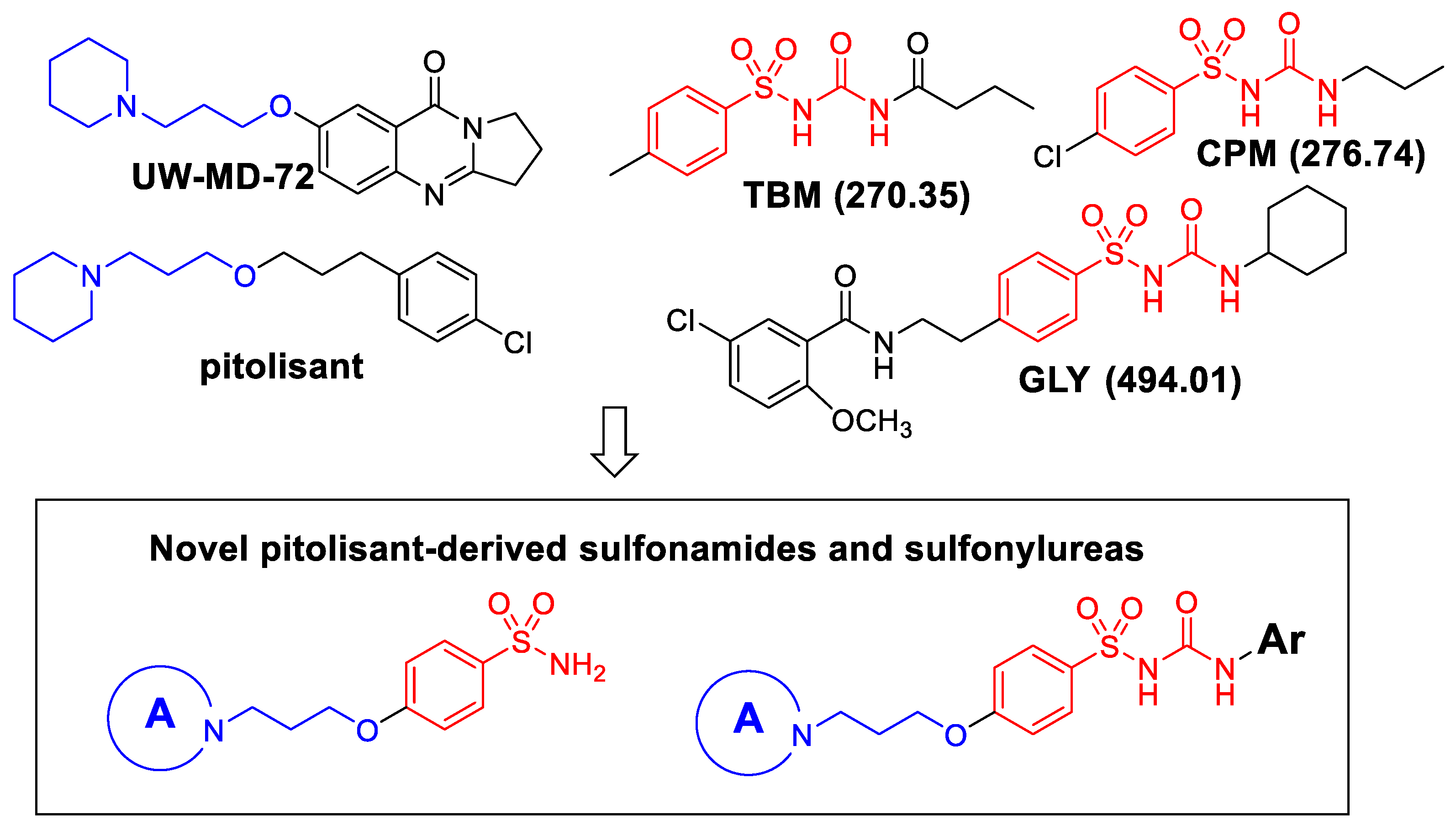
1. Introduction
2. Results
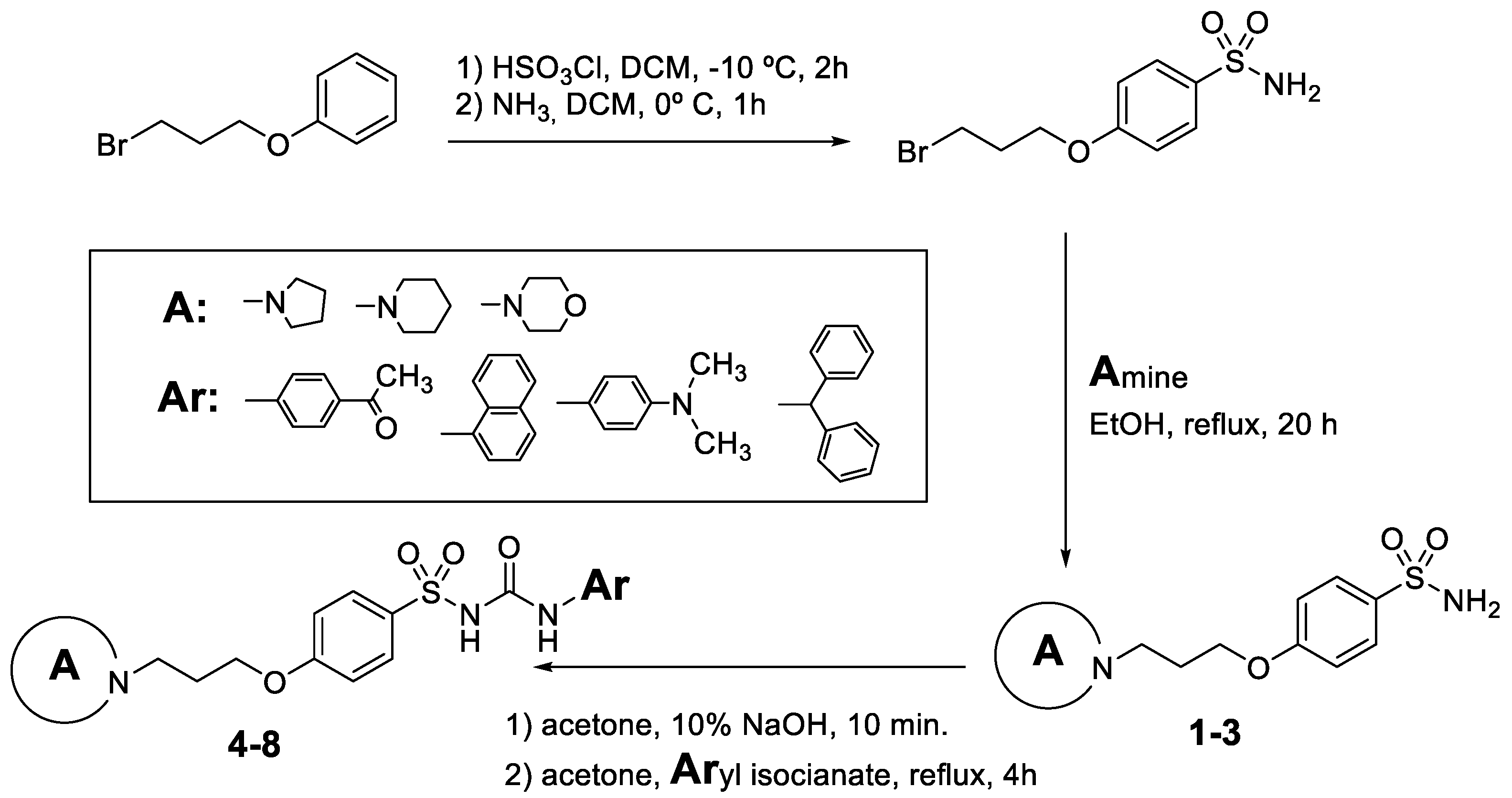
2.1. Chemistry
2.2. Biological Evaluation
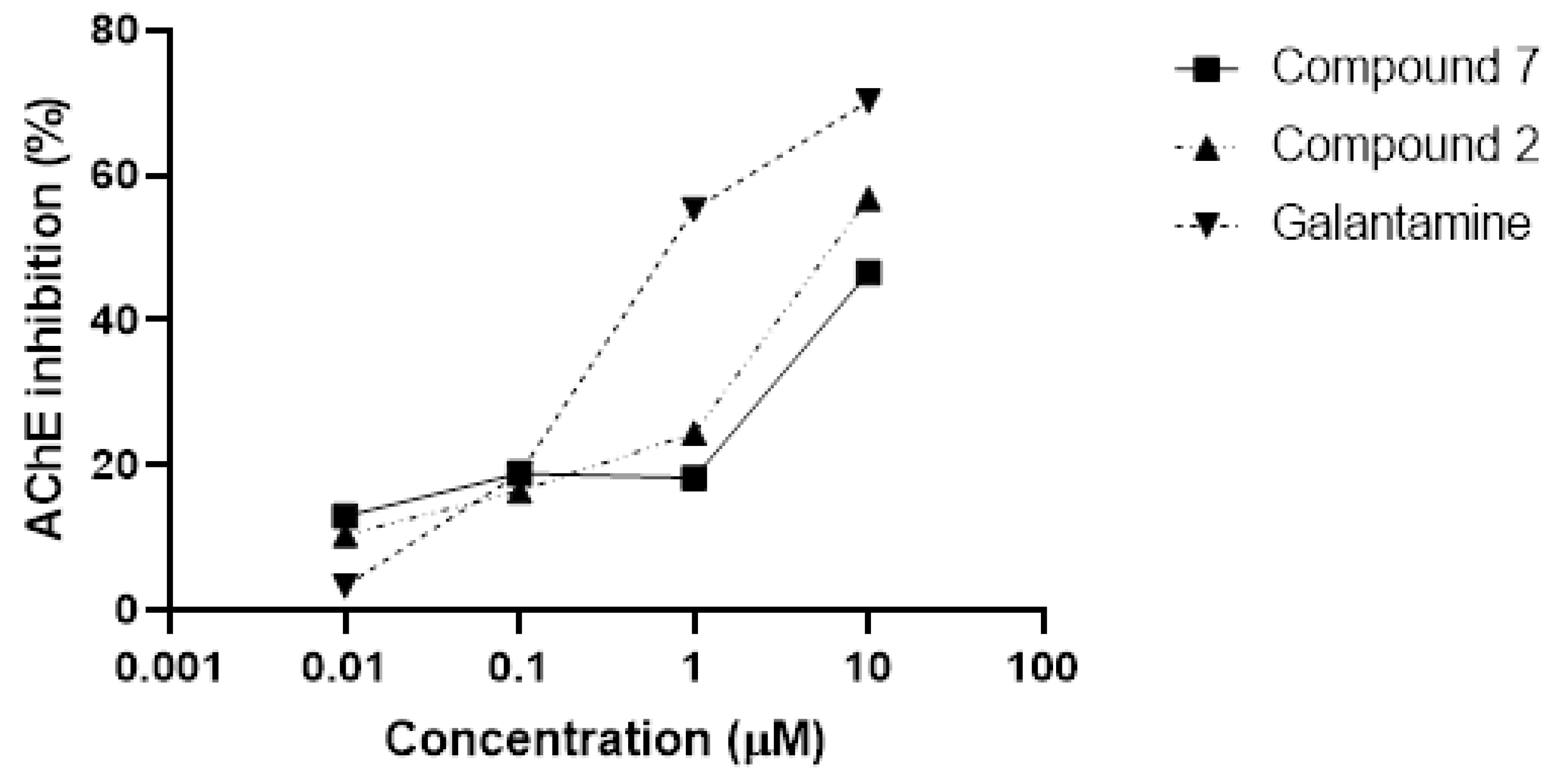
2.2.1. Acetylcholinesterase Inhibition
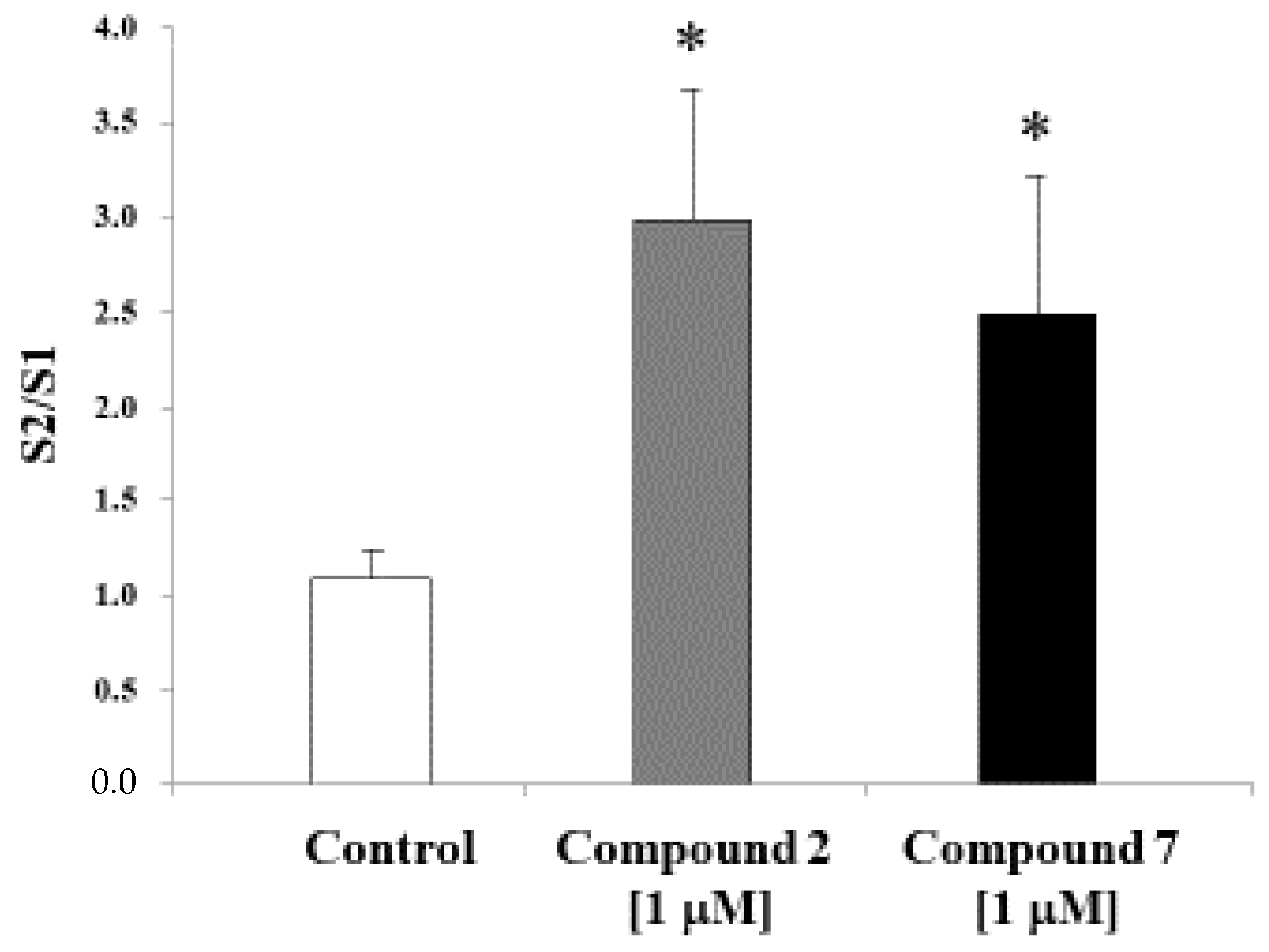
2.2.2. Acetylcholine Release
2.2.3. In Vitro Blood-Brain Barrier Permeation Assay (PAMPA-BBB Assay)
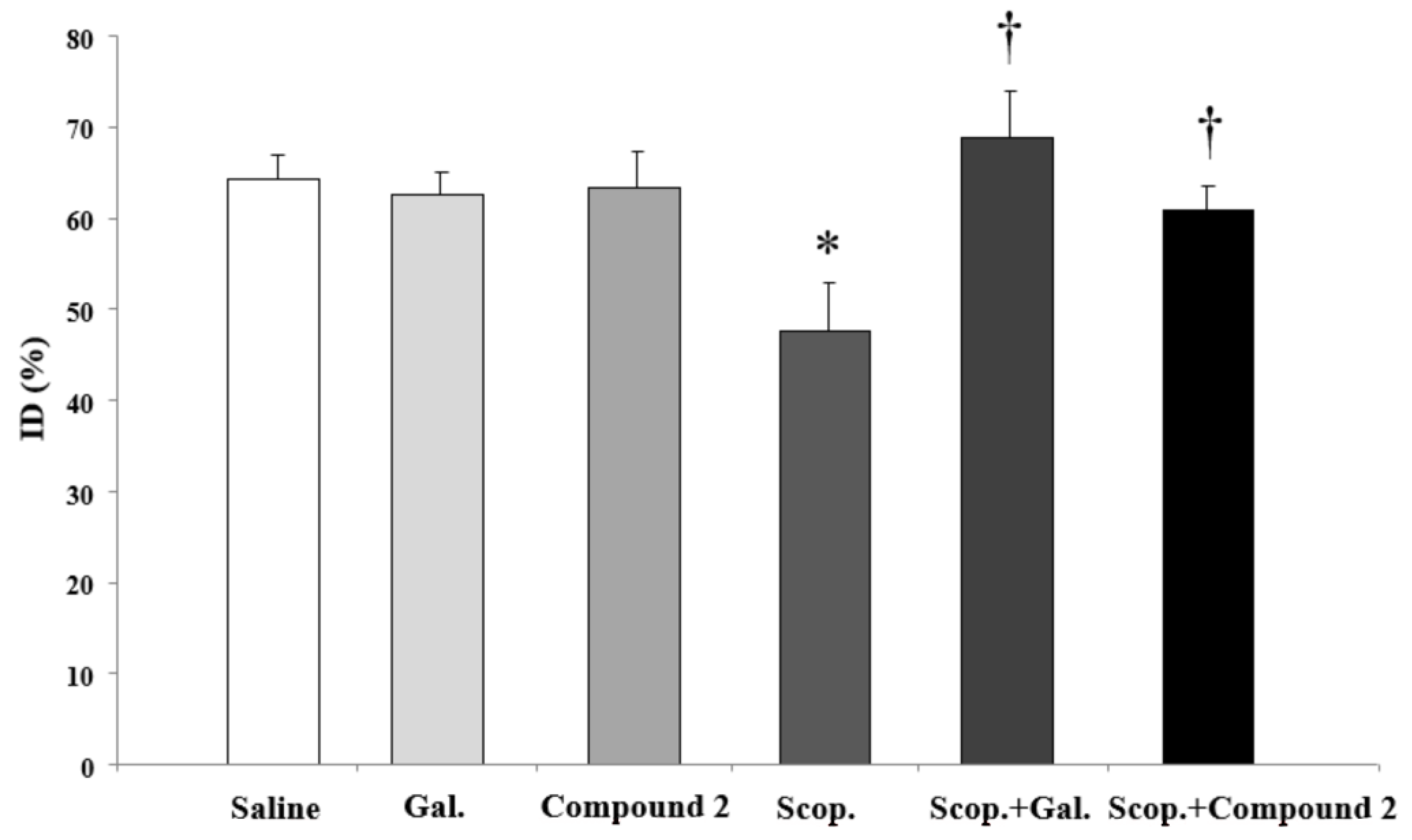
2.2.4. Novel Object Recognition Test (NORT)
2.2.5. ADME Properties Prediction
3. Discussion
4. Materials and Methods
4.1. Chemistry
4.2. Biological Evaluation
4.2.1. Acetylcholinesterase Activity
4.2.2. In Vitro ACh Release
4.2.3. In Vitro Blood-Brain Barrier Permeation Assay
4.2.4. Cognitive Evaluation: Novel Object Recognition Test
4.2.5. ADME Prediction Properties
4.2.6. Data Analysis
Author Contributions
Funding
Institutional Review Board Statement
Informed Consent Statement
Data Availability Statement
Conflicts of Interest
References
- Mensah-Kane, P.; Sumien, N. The Potential of Hyperbaric Oxygen as a Therapy for Neurodegenerative Diseases. Geroscience 2023, 45, 747–756. [Google Scholar] [CrossRef] [PubMed]
- Rajan, K.B.; Weuve, J.; Barnes, L.L.; McAninch, E.A.; Wilson, R.S.; Evans, D.A. Population Estimate of People with Clinical Alzheimer’s Disease and Mild Cognitive Impairment in the United States (2020–2060). Alzheimers Dement. 2021, 17, 1966–1975. [Google Scholar] [CrossRef]
- Viña, J.; Lloret, A. Why Women Have More Alzheimer’s Disease than Men: Gender and Mitochondrial Toxicity of Amyloid-Beta Peptide. J. Alzheimers Dis. 2010, 20 (Suppl. 2), S527–S533. [Google Scholar] [CrossRef] [PubMed]
- Jutkowitz, E.; Kane, R.L.; Gaugler, J.E.; MacLehose, R.F.; Dowd, B.; Kuntz, K.M. Societal and Family Lifetime Cost of Dementia: Implications for Policy. J. Am. Geriatr. Soc. 2017, 65, 2169–2175. [Google Scholar] [CrossRef] [PubMed]
- Mosconi, L.; Rahman, A.; Diaz, I.; Wu, X.; Scheyer, O.; Hristov, H.W.; Vallabhajosula, S.; Isaacson, R.S.; de Leon, M.J.; Brinton, R.D. Increased Alzheimer’s Risk during the Menopause Transition: A 3-Year Longitudinal Brain Imaging Study. PLoS ONE 2018, 13, e0207885. [Google Scholar] [CrossRef] [PubMed]
- Rahman, A.; Schelbaum, E.; Hoffman, K.; Diaz, I.; Hristov, H.; Andrews, R.; Jett, S.; Jackson, H.; Lee, A.; Sarva, H.; et al. Sex-Driven Modifiers of Alzheimer Risk: A Multimodality Brain Imaging Study. Neurology 2020, 95, e166–e178. [Google Scholar] [CrossRef]
- Lin, K.A.; Choudhury, K.R.; Rathakrishnan, B.G.; Marks, D.M.; Petrella, J.R.; Doraiswamy, P.M.; Alzheimer’s Disease Neuroimaging Initiative. Marked Gender Differences in Progression of Mild Cognitive Impairment over 8 Years. Alzheimers Dement. 2015, 1, 103–110. [Google Scholar] [CrossRef]
- Sundermann, E.E.; Biegon, A.; Rubin, L.H.; Lipton, R.B.; Landau, S.; Maki, P.M.; Alzheimer’s Disease Neuroimaging Initiative. Does the Female Advantage in Verbal Memory Contribute to Underestimating Alzheimer’s Disease Pathology in Women versus Men? J. Alzheimers Dis. 2017, 56, 947–957. [Google Scholar] [CrossRef]
- Rabarison, K.M.; Bouldin, E.D.; Bish, C.L.; McGuire, L.C.; Taylor, C.A.; Greenlund, K.J. The Economic Value of Informal Caregiving for Persons with Dementia: Results From 38 States, the District of Columbia, and Puerto Rico, 2015 and 2016 BRFSS. Am. J. Public Health 2018, 108, 1370–1377. [Google Scholar] [CrossRef]
- Chêne, G.; Beiser, A.; Au, R.; Preis, S.R.; Wolf, P.A.; Dufouil, C.; Seshadri, S. Gender and Incidence of Dementia in the Framingham Heart Study from Mid-Adult Life. Alzheimers Dement. 2015, 11, 310–320. [Google Scholar] [CrossRef]
- Gurvich, C.; Hoy, K.; Thomas, N.; Kulkarni, J. Sex Differences and the Influence of Sex Hormones on Cognition through Adulthood and the Aging Process. Brain Sci. 2018, 8, 163. [Google Scholar] [CrossRef] [PubMed]
- Paganini-Hill, A.; Henderson, V.W. Estrogen Deficiency and Risk of Alzheimer’s Disease in Women. Am. J. Epidemiol. 1994, 140, 256–261. [Google Scholar] [CrossRef] [PubMed]
- NIH State-of-the-Science Conference Statement on Management of Menopause-Related Symptoms. NIH Consens. State Sci. Statements 2005, 22, 1–38.
- Kripke, D.F.; Brunner, R.; Freeman, R.; Hendrix, S.L.; Jackson, R.D.; Masaki, K.; Carter, R.A. Sleep Complaints of Postmenopausal Women. Clin. J. Womens Health 2001, 1, 244–252. [Google Scholar] [CrossRef] [PubMed]
- Mosconi, L.; Berti, V.; Dyke, J.; Schelbaum, E.; Jett, S.; Loughlin, L.; Jang, G.; Rahman, A.; Hristov, H.; Pahlajani, S.; et al. Menopause Impacts Human Brain Structure, Connectivity, Energy Metabolism, and Amyloid-Beta Deposition. Sci. Rep. 2021, 11, 10867. [Google Scholar] [CrossRef] [PubMed]
- Schelbaum, E.; Loughlin, L.; Jett, S.; Zhang, C.; Jang, G.; Malviya, N.; Hristov, H.; Pahlajani, S.; Isaacson, R.; Dyke, J.P.; et al. Association of Reproductive History With Brain MRI Biomarkers of Dementia Risk in Midlife. Neurology 2021, 97, e2328–e2339. [Google Scholar] [CrossRef] [PubMed]
- Mosconi, L.; Berti, V.; Quinn, C.; McHugh, P.; Petrongolo, G.; Varsavsky, I.; Osorio, R.S.; Pupi, A.; Vallabhajosula, S.; Isaacson, R.S.; et al. Sex Differences in Alzheimer Risk: Brain Imaging of Endocrine vs. Chronologic Aging. Neurology 2017, 89, 1382–1390. [Google Scholar] [CrossRef] [PubMed]
- Alhazmi, H.A.; Albratty, M. An Update on the Novel and Approved Drugs for Alzheimer Disease. Saudi Pharm. J. 2022, 30, 1755–1764. [Google Scholar] [CrossRef]
- Falkenstein, M.; Reiner-Link, D.; Zivkovic, A.; Gering, I.; Willbold, D.; Stark, H. Histamine H3 Receptor Antagonists with Peptidomimetic (Keto)Piperazine Structures to Inhibit Aβ Oligomerisation. Bioorg. Med. Chem. 2021, 50, 116462. [Google Scholar] [CrossRef]
- Harwell, V.; Fasinu, P.S. Pitolisant and Other Histamine-3 Receptor Antagonists-An Update on Therapeutic Potentials and Clinical Prospects. Medicines 2020, 7, 55. [Google Scholar] [CrossRef]
- Keam, S.J. Pitolisant: Pediatric First Approval. Paediatr. Drugs 2023, 25, 483–488. [Google Scholar] [CrossRef] [PubMed]
- Shan, L.; Swaab, D.F. Changes in Histaminergic System in Neuropsychiatric Disorders and the Potential Treatment Consequences. Curr. Neuropharmacol. 2022, 20, 403–411. [Google Scholar] [CrossRef] [PubMed]
- Baruah, P.; Das, A.; Paul, D.; Chakrabarty, S.; Aguan, K.; Mitra, S. Sulfonylurea Class of Antidiabetic Drugs Inhibit Acetylcholinesterase Activity: Unexplored Auxiliary Pharmacological Benefit toward Alzheimer’s Disease. ACS Pharmacol. Transl. Sci. 2021, 4, 193–205. [Google Scholar] [CrossRef] [PubMed]
- Sadek, B.; Khan, N.; Darras, F.H.; Pockes, S.; Decker, M. The Dual-Acting AChE Inhibitor and H3 Receptor Antagonist UW-MD-72 Reverses Amnesia Induced by Scopolamine or Dizocilpine in Passive Avoidance Paradigm in Rats. Physiol. Behav. 2016, 165, 383–391. [Google Scholar] [CrossRef] [PubMed]
- Łażewska, D.; Bajda, M.; Kaleta, M.; Zaręba, P.; Doroz-Płonka, A.; Siwek, A.; Alachkar, A.; Mogilski, S.; Saad, A.; Kuder, K.; et al. Rational Design of New Multitarget Histamine H3 Receptor Ligands as Potential Candidates for Treatment of Alzheimer’s Disease. Eur. J. Med. Chem. 2020, 207, 112743. [Google Scholar] [CrossRef] [PubMed]
- Łażewska, D.; Kaleta, M.; Zaręba, P.; Godyń, J.; Dubiel, M.; Honkisz-Orzechowska, E.; Doroz-Płonka, A.; Więckowska, A.; Stark, H.; Kieć-Kononowicz, K. Multitargeting Histamine H3 Receptor Ligands among Acetyl- and Propionyl-Phenoxyalkyl Derivatives. Molecules 2023, 28, 2349. [Google Scholar] [CrossRef]
- Ceras, J.; Cirauqui, N.; Pérez-Silanes, S.; Aldana, I.; Monge, A.; Galiano, S. Novel Sulfonylurea Derivatives as H3 Receptor Antagonists. Preliminary SAR Studies. Eur. J. Med. Chem. 2012, 52, 1–13. [Google Scholar] [CrossRef]
- Medhurst, A.D.; Atkins, A.R.; Beresford, I.J.; Brackenborough, K.; Briggs, M.A.; Calver, A.R.; Cilia, J.; Cluderay, J.E.; Crook, B.; Davis, J.B.; et al. GSK189254, a Novel H3 Receptor Antagonist That Binds to Histamine H3 Receptors in Alzheimer’s Disease Brain and Improves Cognitive Performance in Preclinical Models. J. Pharmacol. Exp. Ther. 2007, 321, 1032–1045. [Google Scholar] [CrossRef]
- Lipinski, C.A.; Lombardo, F.; Dominy, B.W.; Feeney, P.J. Experimental and Computational Approaches to Estimate Solubility and Permeability in Drug Discovery and Development Settings. Adv. Drug Deliv. Rev. 2001, 46, 3–26. [Google Scholar] [CrossRef]
- Heemels, M.-T. Neurodegenerative Diseases. Nature 2016, 539, 179. [Google Scholar] [CrossRef]
- Hurd, M.D.; Martorell, P.; Delavande, A.; Mullen, K.J.; Langa, K.M. Monetary Costs of Dementia in the United States. N. Engl. J. Med. 2013, 368, 1326–1334. [Google Scholar] [CrossRef] [PubMed]
- Hickman, R.A.; Faustin, A.; Wisniewski, T. Alzheimer Disease and Its Growing Epidemic: Risk Factors, Biomarkers, and the Urgent Need for Therapeutics. Neurol. Clin. 2016, 34, 941–953. [Google Scholar] [CrossRef] [PubMed]
- Cummings, J.; Lai, T.-J.; Hemrungrojn, S.; Mohandas, E.; Yun Kim, S.; Nair, G.; Dash, A. Role of Donepezil in the Management of Neuropsychiatric Symptoms in Alzheimer’s Disease and Dementia with Lewy Bodies. CNS Neurosci. Ther. 2016, 22, 159–166. [Google Scholar] [CrossRef] [PubMed]
- Weller, J.; Budson, A. Current Understanding of Alzheimer’s Disease Diagnosis and Treatment. F1000Research 2018, 7, 1161. [Google Scholar] [CrossRef]
- Selkoe, D.J. Resolving Controversies on the Path to Alzheimer’s Therapeutics. Nat. Med. 2011, 17, 1060–1065. [Google Scholar] [CrossRef] [PubMed]
- Rösler, M.; Anand, R.; Cicin-Sain, A.; Gauthier, S.; Agid, Y.; Dal-Bianco, P.; Stähelin, H.B.; Hartman, R.; Gharabawi, M. Efficacy and Safety of Rivastigmine in Patients with Alzheimer’s Disease: International Randomised Controlled Trial. BMJ 1999, 318, 633–638. [Google Scholar] [CrossRef]
- Whitehouse, P.J.; Price, D.L.; Clark, A.W.; Coyle, J.T.; DeLong, M.R. Alzheimer Disease: Evidence for Selective Loss of Cholinergic Neurons in the Nucleus Basalis. Ann. Neurol. 1981, 10, 122–126. [Google Scholar] [CrossRef]
- Cummings, J.L.; Tong, G.; Ballard, C. Treatment Combinations for Alzheimer’s Disease: Current and Future Pharmacotherapy Options. J. Alzheimers Dis. 2019, 67, 779–794. [Google Scholar] [CrossRef]
- Ramírez, M.J.; Cenarruzabeitia, E.; Lasheras, B.; Del Río, J. Involvement of GABA Systems in Acetylcholine Release Induced by 5-HT3 Receptor Blockade in Slices from Rat Entorhinal Cortex. Brain Res. 1996, 712, 274–280. [Google Scholar] [CrossRef]
- Astrain-Redin, N.; Talavera, I.; Moreno, E.; Ramírez, M.J.; Martínez-Sáez, N.; Encío, I.; Sharma, A.K.; Sanmartín, C.; Plano, D. Seleno-Analogs of Scaffolds Resembling Natural Products a Novel Warhead toward Dual Compounds. Antioxidants 2023, 12, 139. [Google Scholar] [CrossRef]
- Zengin Kurt, B. Synthesis and Anticholinesterase Activity of Novel Non-Hepatotoxic Naphthyridine-11-Amine Derivatives. Mol. Divers. 2019, 23, 625–638. [Google Scholar] [CrossRef] [PubMed]
- Aisa, B.; Tordera, R.; Lasheras, B.; Del Río, J.; Ramírez, M.J. Cognitive Impairment Associated to HPA Axis Hyperactivity after Maternal Separation in Rats. Psychoneuroendocrinology 2007, 32, 256–266. [Google Scholar] [CrossRef] [PubMed]

| Comp. | Structure | MW (g/mol) | H3R IC50 (μM) | AChE Inhibition (%) ± SEM |
|---|---|---|---|---|
| 1 |  | 284 | 0.25 | I |
| 2 | 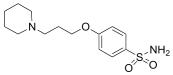 | 298 | 0.13 | 37.06 ± 4.02 |
| 3 |  | 300 | 0.40 | I |
| 4 |  | 445 | 0.40 | 20.82 ± 4.98 |
| 5 |  | 453 | 0.08 | 25.22 ± 7.70 |
| 6 |  | 446 | 0.50 | 29.91 ± 3.63 |
| 7 | 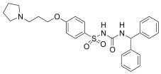 | 493 | 0.50 | 37.02 ± 4.61 |
| 8 | 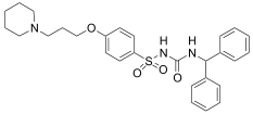 | 507 | 0.50 | 30.17 ± 4.86 |
| Compound | PAMPA-BBB Assay | |
|---|---|---|
| Pe (×10−6 cm/s) | Classification | |
| 2 | 2.23 ± 0.31 | CNS +/− |
| 6 | 0.32 ± 0.01 | CNS − |
| 7 | 0.47 ± 0.06 | CNS − |
| Locomotor Activity | Discrimination Index (%) | |
|---|---|---|
| SALINE | 20,397.14 ± 1590.99 | 51.88 ± 1.34 |
| GAL | 18,785.69 ± 1597.80 | 50.21 ± 5.52 |
| SCOP | 21,676.27 ± 1871.73 | 54.58 ± 2.54 |
| 2 | 20,036.75 ± 2200.76 | 52.07 ± 2.37 |
| SCOP + GAL | 19,806.37 ± 2050.06 | 51.77 ± 3.15 |
| SCOP + 2 | 21,487.37 ± 1531.95 | 50.61 ± 2.92 |
| Absorption | ||||||||
|---|---|---|---|---|---|---|---|---|
| Ref. | Molecular Weight (g/mol) | Log P | H-Bond Donor | H-Bond Acceptor | Lipinski Violation | GI Absorption | Water Solubility | Caco2 |
| 2 | 298.40 | 1.589 | 1 | 4 | 0 | 91.4% | −1.418 | 0.875 |
| 7 | 507.22 | 4.719 | 2 | 5 | 1 | 88.5 | −4.748 | 0.632 |
Disclaimer/Publisher’s Note: The statements, opinions and data contained in all publications are solely those of the individual author(s) and contributor(s) and not of MDPI and/or the editor(s). MDPI and/or the editor(s) disclaim responsibility for any injury to people or property resulting from any ideas, methods, instructions or products referred to in the content. |
© 2024 by the authors. Licensee MDPI, Basel, Switzerland. This article is an open access article distributed under the terms and conditions of the Creative Commons Attribution (CC BY) license (https://creativecommons.org/licenses/by/4.0/).
Share and Cite
Pérez-Silanes, S.; Martisova, E.; Moreno, E.; Solas, M.; Plano, D.; Sanmartin, C.; Ramírez, M.J. Novel Pitolisant-Derived Sulfonyl Compounds for Alzheimer Disease. Int. J. Mol. Sci. 2024, 25, 799. https://doi.org/10.3390/ijms25020799
Pérez-Silanes S, Martisova E, Moreno E, Solas M, Plano D, Sanmartin C, Ramírez MJ. Novel Pitolisant-Derived Sulfonyl Compounds for Alzheimer Disease. International Journal of Molecular Sciences. 2024; 25(2):799. https://doi.org/10.3390/ijms25020799
Chicago/Turabian StylePérez-Silanes, Silvia, Eva Martisova, Esther Moreno, Maite Solas, Daniel Plano, Carmen Sanmartin, and María Javier Ramírez. 2024. "Novel Pitolisant-Derived Sulfonyl Compounds for Alzheimer Disease" International Journal of Molecular Sciences 25, no. 2: 799. https://doi.org/10.3390/ijms25020799
APA StylePérez-Silanes, S., Martisova, E., Moreno, E., Solas, M., Plano, D., Sanmartin, C., & Ramírez, M. J. (2024). Novel Pitolisant-Derived Sulfonyl Compounds for Alzheimer Disease. International Journal of Molecular Sciences, 25(2), 799. https://doi.org/10.3390/ijms25020799