Article

Improved Solubility and Stability of a Thermostable Carbonic Anhydrase via Fusion with Marine-Derived Intrinsically Disordered Solubility Enhancers

Division of Life Science, Research Institute of Life Science, and Anti-Aging Bio Cell Factory Regional Leading Research Center (ABC-RLRC), Gyeongsang National University, Jinju 52828, Republic of Korea
Int. J. Mol. Sci. 2024, 25(2), 1139; https://doi.org/10.3390/ijms25021139
Submission received: 13 December 2023 / Revised: 12 January 2024 / Accepted: 16 January 2024 / Published: 17 January 2024
(This article belongs to the Special Issue Protein Stability Research)

Abstract

:
Carbonic anhydrase (CA), an enzyme catalyzing the reversible hydration reaction of carbon dioxide (CO2), is considered a promising biocatalyst for CO2 reduction. The α-CA of Thermovibrio ammonificans (taCA) has emerged as a compelling candidate due to its high thermostability, a critical factor for industrial applications. However, the low-level expression and poor in vitro solubility have hampered further utilization of taCA. Recently, these limitations have been addressed through the fusion of the NEXT tag, a marine-derived, intrinsically disordered small peptide that enhances protein expression and solubility. In this study, the solubility and stability of NEXT-taCA were further investigated. When the linker length between the NEXT tag and the taCA was shortened, the expression level decreased without compromising solubility-enhancing performance. A comparison between the NEXT tag and the NT11 tag demonstrated the NEXT tag’s superiority in improving both the expression and solubility of taCA. While the thermostability of taCA was lower than that of the extensively engineered DvCA10, the NEXT-tagged taCA exhibited a 30% improvement in long-term thermostability compared to the untagged taCA, suggesting that enhanced solubility can contribute to enzyme thermostability. Furthermore, the bioprospecting of two intrinsically disordered peptides (Hcr and Hku tags) as novel solubility-enhancing fusion tags was explored, demonstrating their performance in improving the expression and solubility of taCA. These efforts will advance the practical application of taCA and provide tools and insights for enzyme biochemistry and bioengineering.

1. Introduction

Carbonic anhydrase (CA, EC 4.2.1.1) is a widespread enzyme that holds great importance for physiological processes such as carbon dioxide (CO2) transport, various CO2/HCO3-related metabolisms, photosynthesis, pH homeostasis, and biocalcification, by catalyzing the reversible hydration of CO2: CO2 + H2O ↔ HCO3 + H+ [1]. CA enzymes are classified into eight distinct types, among which α-type CAs are the best-known and the most studied [2]. Due to the ultrafast kinetics of CA exhibiting a kcat of up to 4.4 × 10⁶ s−1 and its proteinaceous nature that can be produced via renewable biological routes, CA has been considered a promising biocatalyst for enzyme-based CO2 capture, utilization, and storage (CCUS) technologies [3]. By accelerating CO2/HCO3 interconversion and thereby overcoming the low CO2 solubility in aqueous conditions, CA can improve CO2 absorption/desorption [4], mineral carbonation [5], C1-platform chemical synthesis [6,7,8,9], and photoautotrophic microbial growth [10,11,12]. Despite these strengths, two primary challenges limit its practical application: low stability under high-temperature conditions required for efficient CCUS and high enzyme production costs [3,13].
The α-type CA (taCA) of Thermovibrio ammonificans type strain HB-1T, isolated from a deep-sea hydrothermal vent, has been the most thermostable CA among naturally occurring bacterial CAs known to date, supported by both experimental results and computational simulations [14,15]. Due to its outstanding and promising thermostability, taCA has emerged as a compelling candidate for further enzyme engineering to improve its thermostability [15,16,17]. A large amount of enzymes should be obtained to accelerate protein engineering research and the practical utilization of the enzymes. However, the expression of taCA has been reported to be insufficient in microbial Escherichia coli hosts. Moreover, due to its intrinsically poor solubility, taCA is susceptible to aggregation and precipitation under low-salt conditions [14].
The NEXT tag has been employed as a fusion tag to address the challenges associated with both the low-level expression and poor solubility of taCA [18,19]. The NEXT tag, a 53 amino acid-length peptide with a molecular mass of 5.5 kDa, originated from the N-terminal extension sequence of α-type CA (hmCA) in the marine bacterium Hydrogenovibrio marinus [18,20]. Despite its small size, the NEXT tag outperforms commercially available fusion tags, such as E. coli maltose-binding protein (MBP) and Schistosoma japonicum glutathione S-transferase (GST), which have molecular masses of 40 kDa and 26 kDa, respectively. This superior performance is attributed to a distinctive feature of the NEXT tag: it is an intrinsically disordered protein (IDP) that can entropically exclude neighboring macromolecules, thereby preventing protein aggregation [18,21]. However, no comprehensive investigation has been conducted to date regarding the solubility and stability of the NEXT-tagged taCA.
This study examined the length effect of a flexible linker on the expression and solubility of taCA. A comparative analysis was conducted between the NEXT tag and the previously known NT11 tag [22], assessing their influence on the expression and solubility of taCA. Notably, the impact of the NEXT tag on the long-term thermal stability of taCA was explored. Additionally, novel IDP-based solubility-enhancing tags were discovered, and their potential was investigated to enhance the expression and solubility of taCA.

2. Results and Discussion

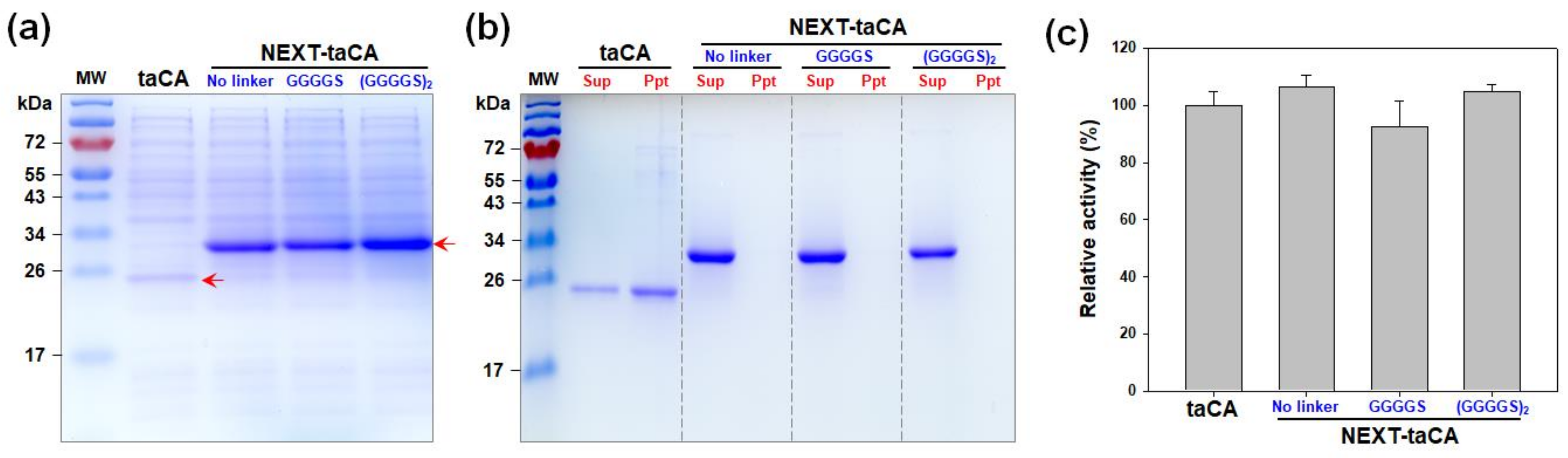
2.1. Effects of Length of Flexible Linker

In the previous study, the NEXT tag was fused to the taCA along with the flexible linker (GGGGS)×2 [18]. The fusion of the NEXT tag significantly improved the expression level and the solubility of taCA, minimally affecting the enzymatic properties such as activity and stability [18]. However, the effect of linker length on the expression level, solubility, and activity of NEXT-taCA has not been investigated. As optimization of linker length may enhance protein expression yield [23], two additional fusion proteins featuring distinct linker lengths were subsequently constructed: one where the NEXT tag and taCA were directly attached without a linker and the other where they were fused via a shorter linker (one GGGGS). All variants of NEXT-tagged taCA exhibited high expression levels in soluble forms, whereas, as previously demonstrated, the soluble expression of untagged taCA was considerably lower (Figure 1a). Notably, the original construct with the (GGGGS)×2 linker exhibited the highest expression, showing approximately 50% and 37% higher levels than those without a linker and with the shorter linker, respectively.
Despite a protein being expressed in a soluble and folded state, the protein in its isolated state may still form aggregates when possessing low intrinsic, in vitro solubility [24,25]. The taCA enzyme, known for its low in vitro solubility, aggregates under low-salt buffer conditions [18]. The low solubility of taCA appears to be correlated with its positively charged surface [26,27]. To assess the impact of the linker length on the in vitro solubility of NEXT-taCA, the purified proteins were exposed to a low-salt buffer (20 mM sodium phosphate, pH 7.5) without additional supplementation of NaCl, and any resulting protein precipitates were analyzed via SDS–PAGE. As previously demonstrated, the untagged taCA displayed significant precipitates, whereas all the NEXT-tagged taCA enzymes remained entirely soluble, irrespective of the linker length (Figure 1b). This result indicates that the linker length does not influence the solubility of NEXT-taCA. Furthermore, the linker length did not significantly affect the specific enzyme activity (Figure 1c). Consequently, the NEXT-taCA with the (GGGGS)×2 linker was selected for all subsequent experiments.

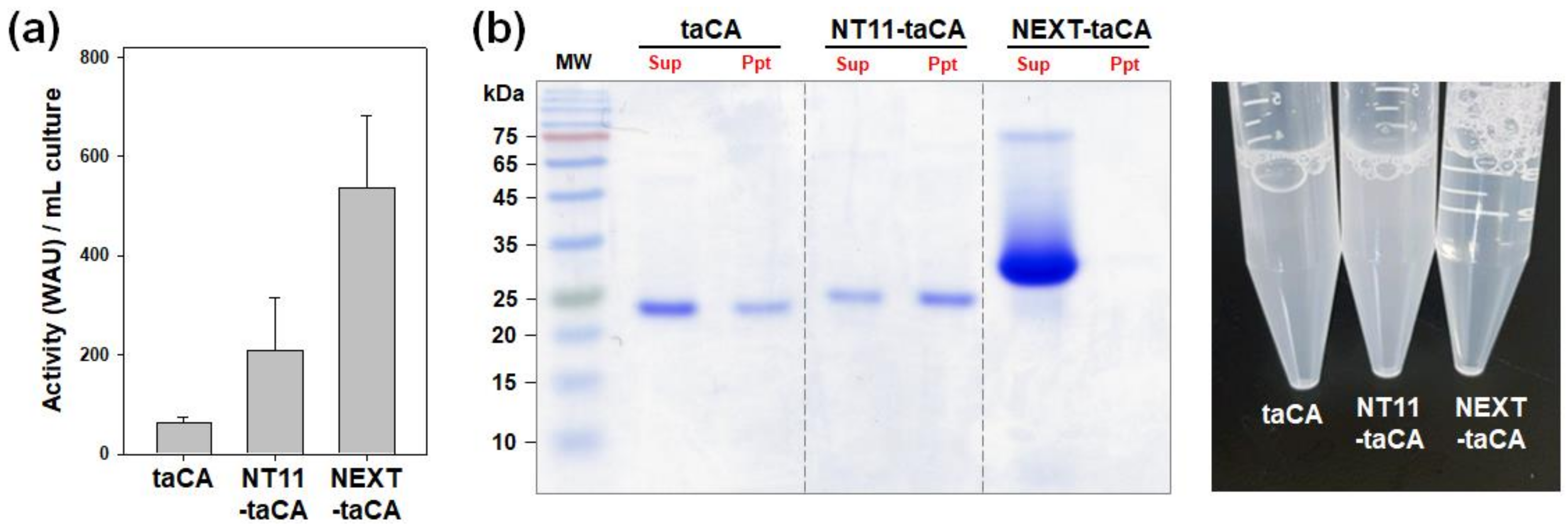
2.2. Comparison of NEXT Tag with NT11 Tag

The NT11 tag, consisting of 11 amino acids (VSEPHDYNYEK), is derived from the N-terminal domain of a carbonic anhydrase found in Dunaliella species. Due to its small size and notable efficacy, the NT11 tag has gained recognition as a promising tag for the expression of recombinant proteins, including taCA [22]. In this study, the performance of the NT11 tag was assessed and compared with that of the NEXT tag in enhancing protein expression and in vitro solubility of taCA.
First, protein expression levels were roughly assessed by quantifying the enzymatic activities within total cell lysates (Figure 2a) based on the fact that the fusion of either the NT11 tag or the NEXT tag did not result in a statistically significant impact on the enzymatic activity of taCA [14,22]. The CO2 hydration activity of the NT11-taCA lysate measured 207.8 WAU mL−1, exhibiting a 3.3-fold increase compared to the untagged taCA lysate (63.8 WAU mL−1). The activity of the NEXT-taCA lysate (536.2 WAU mL−1) was 8.4-fold higher than that of the untagged taCA lysate, which agrees well with the reported value (8-fold) determined through densitometric analysis of band intensities on protein gels [18]. The production yield of NEXT-taCA was estimated to be 104 ± 28 mg L−1 by calculation based on the specific activity (5175 WAU mg−1) of the purified NEXT-taCA. These findings substantiate that the NEXT tag surpasses the NT11 tag in enhancing the functional expression level of taCA in E. coli.
Next, the in vitro solubilities of the tagged proteins were compared by observing protein precipitation following dialysis against a low-salt buffer. Unexpectedly, the NT11-taCA exhibited a significant amount of protein precipitation, rendering the enzyme solution turbid (Figure 2b). The precipitation of NT11-taCA appeared to be more pronounced than that of the untagged taCA. This result suggests that the NT11 tag is ineffective in improving the solubility of taCA. In contrast, as previously shown, no precipitation was evident when assessing the NEXT-taCA, resulting in a visually clear enzyme solution (Figure 2b).

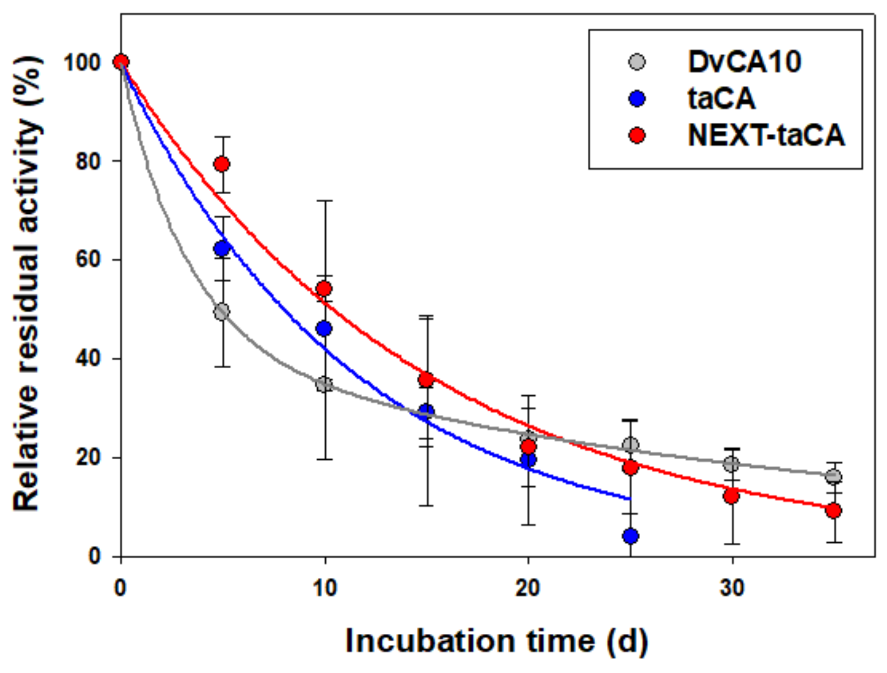
2.3. Improved Long-Term Thermal Stability of taCA by the Fusion of NEXT Tag

To evaluate the impact of the NEXT tag fusion on the long-term thermal stability of taCA, the taCA enzymes were incubated at 70 °C in a phosphate buffer supplemented with 300 mM NaCl for up to 35 days, and their residual activities were measured (Figure 3). DvCA10, an ultrastable CA engineered by directed evolution, was also tested under the same condition for comparison [28]. The two enzymes, taCA and NEXT-taCA, showed the general first-order inactivation kinetics that can be described as follows:
Residual   activity % = e k t × 100
where k is the inactivation rate constant, and t is the incubation time. In the case of DvCA10, the data were better fitted to the biphasic three-parameter kinetic model. It is known that enzyme inactivation kinetics can be described by the simple three-parameter model regardless of the underlying mechanism for the inactivation [29]. The three-parameter model can be expressed by the sum of two first-order equations as follows:
Residual   activity % = x 1 e k 1 t + x 2 e k 2 t × 100
where x1 and x2 are pre-exponential parameters; x1 + x2 = 1, k1 and k2 are apparent rate constants, and t is the incubation time. Considering only the apparent rate constant k2, DvCA10 was 3.2-fold more stable than taCA under the experimental condition (Table 1).
Remarkably, the fusion of the NEXT tag resulted in a 30% enhancement in the long-term thermal stability of taCA (Figure 3 and Table 1). This result seems contradictory to the previous study’s findings, where the thermal stability of NEXT-taCA was reported to be almost the same as that of the untagged taCA when their stability was assessed after heat exposure for 1 h at 90 °C [18]. This apparent discrepancy might arise from the different timescales for the inactivation kinetics and the aggregation kinetics of the soluble enzymes under exceptionally high-temperature conditions; the short-term exposure to 90 °C might induce rapid enzyme denaturation before enzyme aggregation significantly influences the overall enzyme stability, resulting in the seemingly similar stabilities between the NEXT-taCA and the untagged taCA. On the other hand, under the relatively mild condition of 70 °C, the inactivation and aggregation kinetics might have similar timescales. Since enzyme aggregation and precipitation contribute to a reduction in enzymatic activity, the precipitation of the untagged taCA before heat-induced denaturation could expedite the overall enzyme deactivation. These results underscore that the long-term thermal stability of taCA can benefit from the enhanced enzyme solubility conferred by the fusion of the NEXT tag.

2.4. Bioprospecting of Novel IDP-Based Solubility Enhancers

As previously mentioned, the NEXT tag originated from the N-terminal extension sequence of hmCA. Notably, genome sequencing has revealed that the presence of the unusual N-terminal extension is not exclusive to hmCA but is a shared characteristic among various α-CAs identified in widely distributed marine chemolithoautotrophic γ-Proteobacteria belonging to the genera Hydrogenovibrio, Thiomicrorhabdus, and Thiomicrospira [30]. These N-terminal sequences are unique in that they are absent in α-CAs from eukaryotes and other bacterial species (Figure 4a), and no sequence has been known with homology to them. Similar to the case of the NEXT tag, it was speculated that these unique N-terminal sequences may also be used as solubility-enhancing fusion tags.
To this end, the N-terminal extension sequences were selected from H. crunogena XCL-2 and H. kuenenii and were designated as Hcr tag and Hku tag, respectively. These were predicted as almost entirely IDP, along with the NEXT tag (Figure 4b). All other sequence properties, such as the sequence length, molecular mass, pI, and hydrophobicity, were similar (Table 2). When the novel IDP tags were fused to the taCA, the expression level of taCA was improved by both the Hcr and Hku tags. Notably, the performance of the Hcr tag was comparable to that of the NEXT tag (Figure 4c). The tagged taCA variants were purified along with the untagged taCA, and their in vitro solubility was examined. All of the tagged taCA enzymes exhibited no protein precipitation, while significant precipitation occurred with the untagged counterpart, demonstrating the effectiveness of the novel IDP-based solubility enhancers (Figure 4d). In addition, the activity changes of taCA caused by the fusion of the Hcr and Hku tags were marginal (Figure 4e), showing that these IDP-based fusion tags exert minimal effects on the taCA enzyme. These results suggest that the novel Hcr and Hku tags have the potential to be powerful solubility enhancers. Meanwhile, future analysis of the unique N-terminal extension sequences may reveal the sequence characteristics of IDPs that are essential for use as solubility-enhancing tags.

3. Materials and Methods

3.1. Strains and Construction of Expression Vectors

The strains, plasmids, and oligonucleotide primers used in this study are listed in Table 3. E. coli TOP10 (Thermo Fisher Scientific, Waltham, MA, USA) was used for the construction of plasmid vectors, and E. coli BL21(DE3) (Novagen, Madison, WI, USA) was used for recombinant protein expression. The cells were routinely cultivated in Luria–Bertani (LB) medium supplemented with appropriate antibiotics (50 μg/mL of ampicillin for recombinant strains or 10 μg/mL of streptomycin for wild-type E. coli TOP10) at 37 °C and 220 rpm in a shaking incubator (Jeiotech, Daejeon, Korea). The genes for NEXT tags with different linker lengths were amplified by polymerase chain reaction (PCR) using the primers listed in Table 3 and the previously constructed pET-NEXT-taCA [18] as the template. The PCR products were ligated into the pGEM-T Easy vector (Promega, Madison, WI, USA), and the insert sequences were confirmed by Sanger sequencing (Genotech, Daejeon, Korea). The genes were subcloned into pET-NEXT-taCA treated by NdeI and NcoI restriction enzymes by replacing the original NEXT tag sequence, resulting in pET-NEXT-taCAno link and pET-NEXT-taCAshort link. The DvCA10 gene [28] was codon-optimized, synthesized (Genscript, Piscataway, NJ, USA), and subcloned into pET-22b(+) vector (Novagen, Madison, WI, USA) using NdeI and XhoI sites, resulting in pET-DvCA10. The genes for Hcr tag and Hku tag were chemically synthesized (Genotech, Daejeon, Korea) and subcloned into pET-NEXT-taCA using NdeI and NcoI sites by replacing the NEXT tag sequence, resulting in pET-Hcr-taCA and pET-Hku-taCA. The vector pET-NT11-taCA was kindly gifted by Professor Seung Pil Pack (Korea University, Republic of Korea) [22]. The recombinant genes had a hexahistidine (His6)-tag encoding sequence at their 3′ terminus.

3.2. Purification of Recombinant Proteins

The recombinant protein was purified by immobilized metal affinity chromatography via His6-tag. After cell lysis, the soluble fraction was mixed with Ni2+-nitrilotriacetic acid agarose beads (Qiagen, Germantown, MD, USA), and the recombinant protein was purified according to the manufacturer’s instructions. The protein was eluted using elution buffer (50 mM of sodium phosphate, 300 mM of NaCl, and 250 mM of imidazole; pH 8.0). The eluate was thoroughly dialyzed against enzyme buffer (20 mM of sodium phosphate buffer and 300 mM of NaCl; pH 7.5) at 4 °C. After dialysis, any protein precipitates were removed by centrifugation at 10,000× g at 4 °C for 10 min. The supernatants were used for subsequent activity and stability tests.

3.3. In Vitro Solubility Test

The eluate was dialyzed against low-salt buffer (20 mM of sodium phosphate; pH 7.5) at 4 °C. After dialysis was completed, protein precipitates were separated by centrifugation at 10,000× g at 4 °C for 10 min. The precipitate fraction was resuspended in the same buffer and analyzed by SDS–PAGE along with the supernatant fraction.

3.4. Protein Analyses

For protein quantification, the purified protein was denatured in a denaturing buffer (6 M of guanidine hydrochloride GuHCl/20 mM of sodium phosphate buffer; pH 7.5), and the absorbance of the denatured protein was measured at 280 nm in a quartz crystal cuvette (Hellma Analytics, Müllheim, Germany). The protein concentration was determined using the measured absorbance and the calculated molar extinction coefficient at 280 nm [31]. Protein samples were separated by SDS–PAGE on 15% PAGE gel and visualized by Coomassie blue R-250 (Bio-Rad, Hercules, CA, USA) staining.

3.5. CO2 Hydration Assay

Enzyme activity was measured via a colorimetric CO2 hydration assay modified from the Wilbur–Anderson method [32,33]. The assay was performed at 0 °C inside the spectrophotometer equipped with a temperature-controllable cell holder. Briefly, 10 μL of the sample was added to a disposable cuvette containing 600 μL of 20 mM Tris buffer (pH 8.3) supplemented with 100 μM of phenol red (Sigma-Aldrich, St. Louis, MO, USA). The CO2 hydration reaction was initiated by adding 400 μL of CO2-saturated water prepared in ice-cold water. The absorbance change was monitored at 570 nm, and the time (t) required for the pH drop from 7.5 to 6.5 was obtained. The time (t0) for the uncatalyzed reaction was also measured by adding the corresponding blank buffer. The Wilbur–Anderson unit (WAU) was calculated as (t0t)/t.

3.6. Thermal Inactivation Test

The concentration of purified enzyme was adjusted to 10 μM. The enzymes (taCA, NEXT-taCA, and DvCA10) were incubated at 70 °C in a water bath (Jeiotech, Daejeon, Korea). They were then stored at 4 °C until their activities were measured. The CO2 hydration activities of the incubated samples were measured and compared with the activity of the nonincubated sample. The relative residual activity was calculated as follows:
Relative   residual   activity % = Activity   of   heat treated   sample Activity   of   untreated   sample × 100

3.7. In Silico Analyses

Densitometric analysis of the protein band on the gel was performed using ImageJ 1.50i [34]. Protein parameters, including molar extinction coefficient, molecular mass, number of charged amino acids, and isoelectric point (pI), were calculated by ProtParam (http://web.expasy.org/protparam/ (accessed on 3 December 2023)) [35]. The Kyte–Doolittle hydropathy index was calculated by ProtScale (https://web.expasy.org/protscale (accessed on 3 December 2023)) using a window size of 5, and the values were averaged to obtain a mean hydropathy index [35,36]. Signal peptide cleavage sites were predicted by the SignalP 6.0 server (https://services.healthtech.dtu.dk/services/SignalP-6.0/ (accessed on 2 December 2023)) [37]. Multiple sequence alignment was performed using ClustalX 2.0 [38], and the result was visualized by ESPript 3.0 (https://espript.ibcp.fr/ESPript/ESPript/ (accessed on 2 December 2023)) [39]. Disordered propensity was predicted by IUPred2A (https://iupred2a.elte.hu (accessed on 1 December 2023)) [40], PONDR (http://www.pondr.com (accessed on 1 December 2023)) [41], and DISpro (http://scratch.proteomics.ics.uci.edu (accessed on 1 December 2023)) [42].

4. Conclusions

The length of the linker influenced the expression level but did not appear to be a critical factor for improving the solubility of taCA. The NEXT tag proved effective in improving both the expression and solubility of taCA, whereas the NT11 improved only the expression level. The enhanced solubility by the fusion of the NEXT tag contributed to the improved thermostability of taCA, presumably by limiting enzyme aggregation and thereby alleviating activity loss. Two novel solubility-enhancing tags were discovered and examined for their impact on the improved expression and solubility of taCA. This study sheds light on the diverse aspects of the NEXT tag and its related sequences, providing valuable tools and insights for the recombinant expression of enzymes.

Funding

This work was supported by the National Research Foundation of Korea (NRF) grant funded by the Ministry of Science & ICT (2021R1F1A1057310, 2021R1A5A8029490, and RS-2023-00235511) and by the Korea Basic Science Institute (National Research Facilities and Equipment Center) grant funded by the Ministry of Education (2022R1A6C101B724).

Institutional Review Board Statement

Not applicable.

Informed Consent Statement

Not applicable.

Data Availability Statement

Data will be made available on request.

Conflicts of Interest

The author declares no conflicts of interest.

References

  1. Smith, K.S.; Ferry, J.G. Prokaryotic carbonic anhydrases. FEMS Microbiol. Rev. 2000, 24, 335–366. [Google Scholar] [CrossRef] [PubMed]
  2. Nocentini, A.; Supuran, C.T.; Capasso, C. An overview on the recently discovered iota-carbonic anhydrases. J. Enzyme Inhib. Med. Chem. 2021, 36, 1988–1995. [Google Scholar] [CrossRef] [PubMed]
  3. Talekar, S.; Jo, B.H.; Dordick, J.S.; Kim, J. Carbonic anhydrase for CO2 capture, conversion and utilization. Curr. Opin. Biotechnol. 2022, 74, 230–240. [Google Scholar] [CrossRef] [PubMed]
  4. Qi, G.J.; Liu, K.; House, A.; Salmon, S.; Ambedkar, B.; Frimpong, R.A.; Remias, J.E.; Liu, K.L. Laboratory to bench-scale evaluation of an integrated CO2 capture system using a thermostable carbonic anhydrase promoted K2CO3 solvent with low temperature vacuum stripping. Appl. Energy 2018, 209, 180–189. [Google Scholar] [CrossRef]
  5. Power, I.M.; Harrison, A.L.; Dipple, G.M. Accelerating mineral carbonation using carbonic anhydrase. Environ. Sci. Technol. 2016, 50, 2610–2618. [Google Scholar] [CrossRef] [PubMed]
  6. Ji, X.Y.; Su, Z.G.; Wang, P.; Ma, G.H.; Zhang, S.P. Tethering of nicotinamide adenine dinucleotide inside hollow nanofibers for high-yield synthesis of methanol from carbon dioxide catalyzed by coencapsulated multienzymes. ACS Nano 2015, 9, 4600–4610. [Google Scholar] [CrossRef]
  7. Gao, S.; Mohammad, M.; Yang, H.C.; Xu, J.; Liang, K.; Hou, J.W.; Chen, V. Janus reactors with highly efficient enzymatic CO2 nanocascade at air-liquid interface. ACS Appl. Mater. Interfaces 2017, 9, 42806–42815. [Google Scholar] [CrossRef]
  8. Yu, S.S.; Lv, P.F.; Xue, P.; Wang, K.; Yang, Q.; Zhou, J.H.; Wang, M.; Wang, L.; Chen, B.Q.; Tan, T.W. Light-driven enzymatic nanosystem for highly selective production of formic acid from CO2. Chem. Eng. J. 2021, 420, 127649. [Google Scholar] [CrossRef]
  9. Liu, G.H.; Wang, L.R.; Yan, L.H.; Zhao, H.; Li, Y.X.; Zhou, L.Y.; He, Y.; Ma, L.; Liu, Y.T.; Gao, J.; et al. A dual-enzyme microreactor based on encapsulation and covalent bond for enzymatic electrocatalytic CO2 reduction. Chem. Eng. J. 2023, 475, 146186. [Google Scholar] [CrossRef]
  10. Xu, X.Y.; Kentish, S.E.; Martin, G.J.O. Direct air capture of CO2 by microalgae with buoyant beads encapsulating carbonic anhydrase. ACS Sustain. Chem. Eng. 2021, 9, 9698–9706. [Google Scholar] [CrossRef]
  11. Jun, S.H.; Yang, J.; Jeon, H.; Kim, H.S.; Pack, S.P.; Jin, E.; Kim, J. Stabilized and immobilized carbonic anhydrase on electrospun nanofibers for enzymatic CO2 conversion and utilization in expedited microalgal growth. Environ. Sci. Technol. 2020, 54, 1223–1231. [Google Scholar] [CrossRef] [PubMed]
  12. You, S.K.; Ko, Y.J.; Shin, S.K.; Hwang, D.H.; Kang, D.H.; Park, H.M.; Han, S.O. Enhanced CO2 fixation and lipid production of Chlorella vulgaris through the carbonic anhydrase complex. Bioresour. Technol. 2020, 318, 124072. [Google Scholar] [CrossRef] [PubMed]
  13. Nguyen, K.; Iliuta, I.; Bougie, F.; Pasquier, L.C.; Iliuta, M.C. Techno-economic assessment of enzymatic CO2 capture in hollow fiber membrane contactors with immobilized carbonic anhydrase. Sep. Purif. Technol. 2023, 307, 122702. [Google Scholar] [CrossRef]
  14. Jo, B.H.; Seo, J.H.; Cha, H.J. Bacterial extremo-α-carbonic anhydrases from deep-sea hydrothermal vents as potential biocatalysts for CO2 sequestration. J. Mol. Catal. B-Enzym. 2014, 109, 31–39. [Google Scholar] [CrossRef]
  15. Parra-Cruz, R.; Jager, C.M.; Lau, P.L.; Gomes, R.L.; Pordea, A. Rational design of thermostable carbonic anhydrase mutants using molecular dynamics simulations. J. Phys. Chem. B 2018, 122, 8526–8536. [Google Scholar] [CrossRef]
  16. Parra-Cruz, R.; Lau, P.L.; Loh, H.S.; Pordea, A. Engineering of Thermovibrio ammonificans carbonic anhydrase mutants with increased thermostability. J. CO2 Util. 2020, 37, 1–8. [Google Scholar] [CrossRef]
  17. Voyer, N.; Daigle, R.; Madore, É.; Fradette, S. Variants of Thermovibrio ammonificans Carbonic Anhydrase and CO2 Capture Methods Using Thermovibrio ammonificans Carbonic Anhydrase Variants. U.S. Patent US10415028B2, 17 September 2019. [Google Scholar]
  18. Jo, B.H. An intrinsically disordered peptide tag that confers an unusual solubility to aggregation-prone proteins. Appl. Environ. Microbiol. 2022, 88, e00097-22. [Google Scholar] [CrossRef]
  19. Hwang, I.S.; Kim, J.H.; Jo, B.H. Enhanced production of a thermostable carbonic anhydrase in Escherichia coli by using a modified NEXT tag. Molecules 2021, 26, 5830. [Google Scholar] [CrossRef]
  20. Jo, B.H.; Im, S.K.; Cha, H.J. Halotolerant carbonic anhydrase with unusual N-terminal extension from marine Hydrogenovibrio marinus as novel biocatalyst for carbon sequestration under high-salt environments. J. CO2 Util. 2018, 26, 415–424. [Google Scholar] [CrossRef]
  21. Santner, A.A.; Croy, C.H.; Vasanwala, F.H.; Uversky, V.N.; Van, Y.Y.; Dunker, A.K. Sweeping away protein aggregation with entropic bristles: Intrinsically disordered protein fusions enhance soluble expression. Biochemistry 2012, 51, 7250–7262. [Google Scholar] [CrossRef]
  22. Nguyen, T.K.M.; Ki, M.R.; Son, R.G.; Pack, S.P. The NT11, a novel fusion tag for enhancing protein expression in Escherichia coli. Appl. Microbiol. Biotechnol. 2019, 103, 2205–2216. [Google Scholar] [CrossRef] [PubMed]
  23. Chen, X.; Zaro, J.L.; Shen, W.C. Fusion protein linkers: Property, design and functionality. Adv. Drug Deliv. Rev. 2013, 65, 1357–1369. [Google Scholar] [CrossRef]
  24. Trevino, S.R.; Scholtz, J.M.; Pace, C.N. Measuring and increasing protein solubility. J. Pharm. Sci. 2008, 97, 4155–4166. [Google Scholar] [CrossRef] [PubMed]
  25. Golovanov, A.P.; Hautbergue, G.M.; Wilson, S.A.; Lian, L.Y. A simple method for improving protein solubility and long-term stability. J. Am. Chem. Soc. 2004, 126, 8933–8939. [Google Scholar] [CrossRef] [PubMed]
  26. Kramer, R.M.; Shende, V.R.; Motl, N.; Pace, C.N.; Scholtz, J.M. Toward a molecular understanding of protein solubility: Increased negative surface charge correlates with increased solubility. Biophys. J. 2012, 102, 1907–1915. [Google Scholar] [CrossRef]
  27. Chan, P.; Curtis, R.A.; Warwicker, J. Soluble expression of proteins correlates with a lack of positively-charged surface. Sci. Rep. 2013, 3, 3333. [Google Scholar] [CrossRef]
  28. Alvizo, O.; Nguyen, L.J.; Savile, C.K.; Bresson, J.A.; Lakhapatri, S.L.; Solis, E.O.P.; Fox, R.J.; Broering, J.M.; Benoit, M.R.; Zimmerman, S.A.; et al. Directed evolution of an ultrastable carbonic anhydrase for highly efficient carbon capture from flue gas. Proc. Natl. Acad. Sci. USA 2014, 111, 16436–16441. [Google Scholar] [CrossRef] [PubMed]
  29. Aymard, C.; Belarbi, A. Kinetics of thermal deactivation of enzymes: A simple three parameters phenomenological model can describe the decay of enzyme activity, irrespectively of the mechanism. Enzyme Microb. Technol. 2000, 27, 612–618. [Google Scholar] [CrossRef]
  30. Scott, K.M.; Williams, J.; Porter, C.M.B.; Russel, S.; Harmer, T.L.; Paul, J.H.; Antonen, K.M.; Bridges, M.K.; Camper, G.J.; Campla, C.K.; et al. Genomes of ubiquitous marine and hypersaline Hydrogenovibrio, Thiomicrorhabdus and Thiomicrospira spp. encode a diversity of mechanisms to sustain chemolithoautotrophy in heterogeneous environments. Environ. Microbiol. 2018, 20, 2686–2708. [Google Scholar] [CrossRef]
  31. Gill, S.C.; von Hippel, P.H. Calculation of protein extinction coefficients from amino acid sequence data. Anal. Biochem. 1989, 182, 319–326. [Google Scholar] [CrossRef]
  32. Wilbur, K.M.; Anderson, N.G. Electrometric and colorimetric determination of carbonic anhydrase. J. Biol. Chem. 1948, 176, 147–154. [Google Scholar] [CrossRef] [PubMed]
  33. Kim, J.H.; Jo, B.H. A colorimetric CO2 hydration assay for facile, accurate, and precise determination of carbonic anhydrase activity. Catalysts 2022, 12, 1391. [Google Scholar] [CrossRef]
  34. Schneider, C.A.; Rasband, W.S.; Eliceiri, K.W. NIH Image to ImageJ: 25 years of image analysis. Nat. Methods 2012, 9, 671–675. [Google Scholar] [CrossRef]
  35. Wilkins, M.R.; Gasteiger, E.; Bairoch, A.; Sanchez, J.C.; Williams, K.L.; Appel, R.D.; Hochstrasser, D.F. Protein identification and analysis tools in the ExPASy server. Methods Mol. Biol. 1999, 112, 531–552. [Google Scholar] [PubMed]
  36. Kyte, J.; Doolittle, R.F. A simple method for displaying the hydropathic character of a protein. J. Mol. Biol. 1982, 157, 105–132. [Google Scholar] [CrossRef] [PubMed]
  37. Teufel, F.; Almagro Armenteros, J.J.; Johansen, A.R.; Gislason, M.H.; Pihl, S.I.; Tsirigos, K.D.; Winther, O.; Brunak, S.; von Heijne, G.; Nielsen, H. SignalP 6.0 predicts all five types of signal peptides using protein language models. Nat. Biotechnol. 2022, 40, 1023–1025. [Google Scholar] [CrossRef]
  38. Larkin, M.A.; Blackshields, G.; Brown, N.P.; Chenna, R.; McGettigan, P.A.; McWilliam, H.; Valentin, F.; Wallace, I.M.; Wilm, A.; Lopez, R.; et al. Clustal W and Clustal X version 2.0. Bioinformatics 2007, 23, 2947–2948. [Google Scholar] [CrossRef] [PubMed]
  39. Robert, X.; Gouet, P. Deciphering key features in protein structures with the new ENDscript server. Nucleic Acids Res. 2014, 42, W320–W324. [Google Scholar] [CrossRef]
  40. Meszaros, B.; Erdos, G.; Dosztanyi, Z. IUPred2A: Context-dependent prediction of protein disorder as a function of redox state and protein binding. Nucleic Acids Res. 2018, 46, W329–W337. [Google Scholar] [CrossRef]
  41. Romero, P.; Obradovic, Z.; Li, X.; Garner, E.C.; Brown, C.J.; Dunker, A.K. Sequence complexity of disordered protein. Proteins 2001, 42, 38–48. [Google Scholar] [CrossRef]
  42. Cheng, J.; Sweredoski, M.J.; Baldi, P. Accurate prediction of protein disordered regions by mining protein structure data. Data Min. Knowl. Discov. 2005, 11, 213–222. [Google Scholar] [CrossRef]
Figure 1. Effect of length of flexible linker on NEXT-taCA. (a) Soluble expression level. Protein expression was conducted at 37 °C, and the soluble fraction was analyzed by SDS–PAGE followed by Coomassie blue staining. The arrow indicates the band positions of recombinant proteins. (b) In vitro solubility of purified enzyme. After dialysis against 20 mM sodium phosphate buffer (pH 7.5), protein precipitates were separated from soluble supernatants via centrifugation and analyzed by SDS–PAGE. Lanes: MW, molecular mass marker; Sup, supernatant; Ppt, precipitate. (c) Activity of purified enzyme. The activities were measured by CO2 hydration assay and normalized to the activity of the untagged taCA. Error bars represent standard deviations from two independent experiments.
Ijms 25 01139 g001
Figure 2. Comparison of NEXT-taCA with NT11-taCA. (a) Activity of cell lysate. Error bars represent standard deviations from three independent experiments. (b) In vitro solubility of purified enzyme. After dialysis against 20 mM sodium phosphate buffer (pH 7.5), protein precipitates were analyzed by SDS–PAGE followed by Coomassie blue staining (left) and by photograph (right). Lanes: MW, molecular mass marker; Sup, supernatant; Ppt, precipitate.
Ijms 25 01139 g002
Figure 3. Time-course enzyme inactivation. Enzymes were incubated at 70 °C in 20 mM phosphate buffer (pH 7.5) supplemented with 300 mM NaCl, and the residual activities were measured by CO2 hydration assay. Solid lines represent the fitted regression curves. Error bars represent standard deviations from two independent experiments.
Ijms 25 01139 g003
Figure 4. Bioprospecting of novel IDP-based solubility enhancers. (a) Multiple sequence alignment of α-type CAs. Only the N-terminal parts of the entire sequences are shown. The predicted signal sequences are boxed in black. The unique N-terminal extension sequences are enclosed in green boxes. Blue boxes indicate columns with strictly (red background) or 70% (red characters) conserved sequences across all the aligned protein sequences. (b) Intrinsic disorder propensities. Position-dependent prediction of disordered regions was performed using three different methods (IUPred2A, PONDR, and DISpro). The dashed line corresponds to the cutoff threshold score for the determination of the disordered region. (c) Expression analysis by SDS–PAGE followed by Coomassie blue staining. Lanes: MW, molecular mass marker; Sol, soluble fraction; Ins, insoluble fraction. (d) In vitro solubility of purified enzyme. After dialysis against 20 mM of sodium phosphate buffer (pH 7.5), protein precipitates were analyzed by SDS–PAGE followed by Coomassie blue staining. Lanes: MW, molecular mass marker; Sup, supernatant; Ppt, precipitate. (e) Activity of purified enzyme. The activities were measured by CO2 hydration assay and normalized to the activity of the untagged taCA. Error bars represent standard deviations from two independent experiments.
Ijms 25 01139 g004
Table 1. Kinetic values for thermal inactivation of enzyme at 70 °C.
Kinetic ModePre-Exponential ParametersInactivation Rate Constants (Half-Life in Day)
DvCA10Biphasicx1: 0.58k1: 0.3064 d−1 (2.3 d)
x2: 0.42k2: 0.0272 d−1 (25.5 d)
taCAMonophasic1k: 0.0867 d−1 (8.0 d)
NEXT-taCAMonophasic1k: 0.0667 d−1 (10.4 d)
Table 2. Sequence properties of IDP-based solubility-enhancing tags used in this study.
Fusion Tag Amino Acid LengthMolecular Mass (kDa)pICharged Amino AcidsNet ChargeMean Hydropathy
NEXT535.58.128%+10.397
Hcr515.59.039%+20.367
Hku575.89.221%+20.406
Table 3. Strains, plasmids, and oligonucleotide primers used in this study.
Strains, Plasmids, or PrimersGenotypes, Relevant Characteristics, or SequencesSource or References
Strains
E. coli TOP10F mcrA Δ(mrr-hsdRMS-mcrBC) Φ80lacZΔM15 ΔlacX74 recA1 araD139 Δ(ara-leu)7697 galU galK rpsL(Strr) endA1 nupGThermo Fisher Scientific, Waltham, MA, USA
E. coli BL21(DE3)F ompT hsdSB(rB mB) gal dcm lon λ(DE3), carrying T7 RNA polymerase geneNovagen, Madison, WI, USA
Plasmids
pGEM-T EasypUC ori, Ampr, TA cloning vector,Promega, Madison, WI, USA
pET-22b(+)T7lac promoter, pBR322 ori, Ampr, parental expression vector harboring PelB signal sequenceNovagen, Madison, WI, USA
pET-taCAExpression plasmid carrying taCA gene[18]
pET-NEXT-taCAExpression plasmid carrying NEXT-tagged taCA gene with the (GGGGS)2 linker[18]
pET-NEXT-taCAno linkExpression plasmid carrying NEXT-tagged taCA gene without linkerThis study
pET-NEXT-taCAshort linkExpression plasmid carrying NEXT-tagged taCA gene with the GGGGS linkerThis study
pET-NT11-taCAExpression plasmid carrying NT11-tagged taCA gene[22]
pET-DvCA10Expression plasmid carrying DvCA10 geneThis study
pET-Hcr-taCAExpression plasmid carrying Hcr-tagged taCA gene with the (GGGGS)2 linkerThis study
pET-Hku-taCAExpression plasmid carrying Hku-tagged taCA gene with the (GGGGS)2 linkerThis study
Primers a
NEXT-ForwardCATATGGCTGTTCAACATAGCAATGCCCC[18]
NEXT-no link-ReverseCCATGGCCACAACGGGTTTTGGTTTAGThis study
NEXT-short link-ReverseCCATGGAGCCTCCACCGCCCACAACGGGTTTTGGTTTAGThis study
a Restriction sites are underlined.
Disclaimer/Publisher’s Note: The statements, opinions and data contained in all publications are solely those of the individual author(s) and contributor(s) and not of MDPI and/or the editor(s). MDPI and/or the editor(s) disclaim responsibility for any injury to people or property resulting from any ideas, methods, instructions or products referred to in the content.

