
Effect of the Lys62Ala Mutation on the Thermal Stability of BstHPr Protein by Molecular Dynamics


Abstract

1. Introduction
2. Results
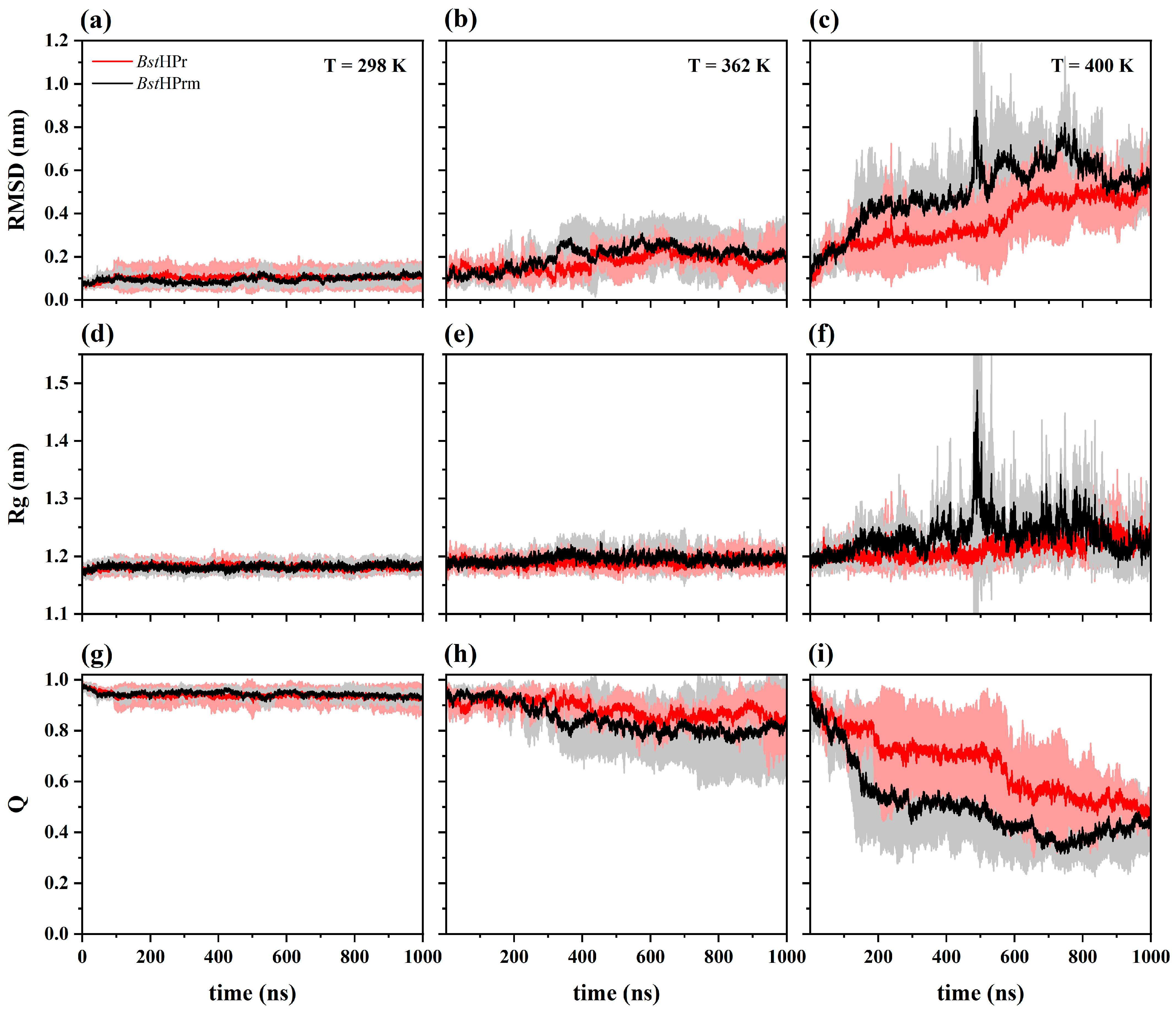
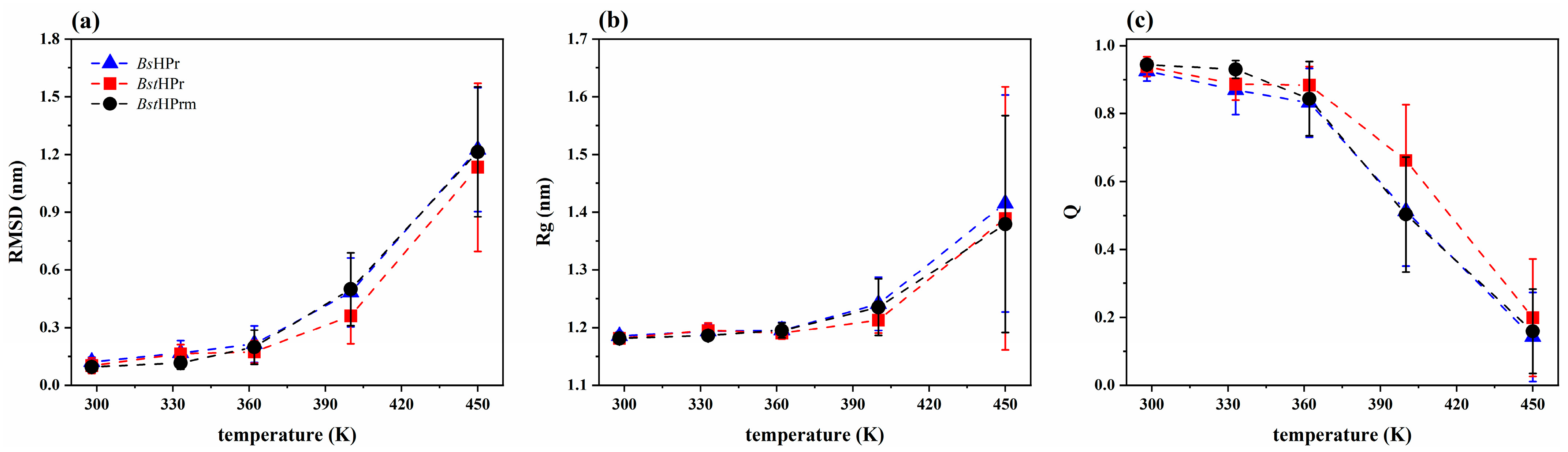
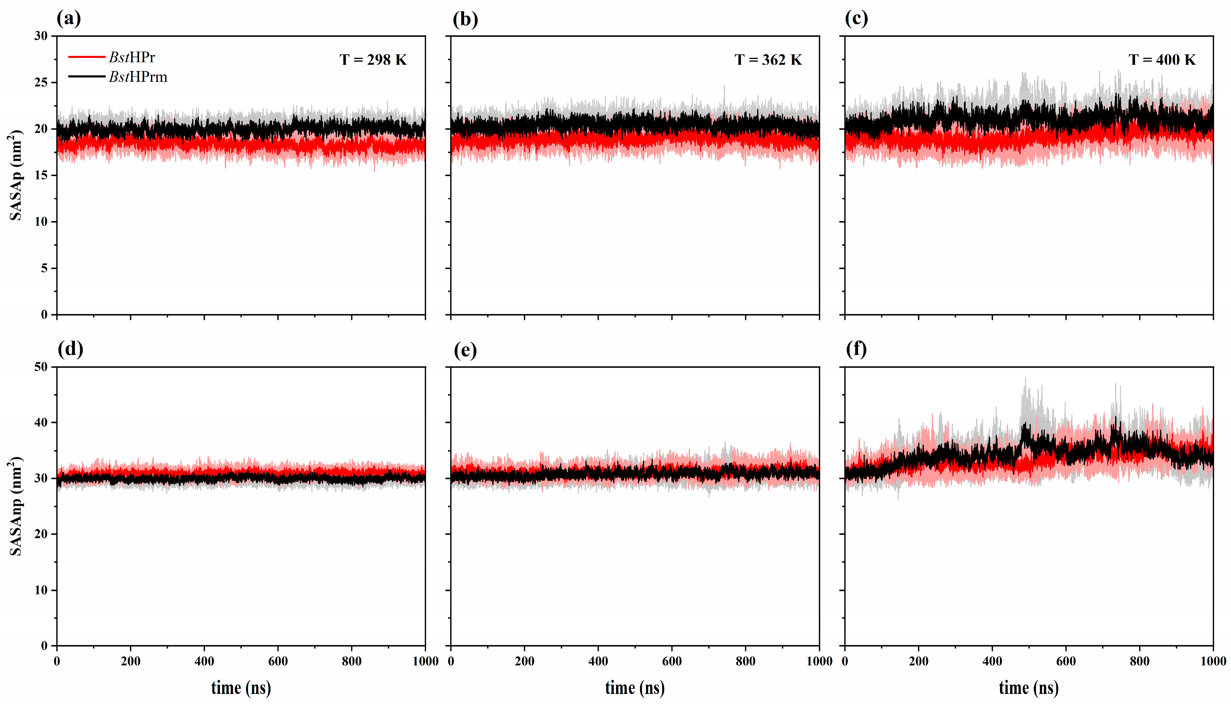
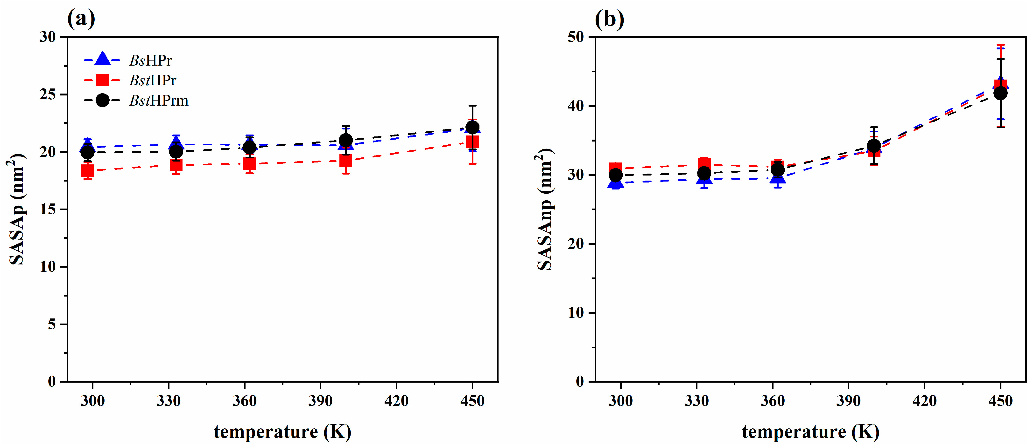
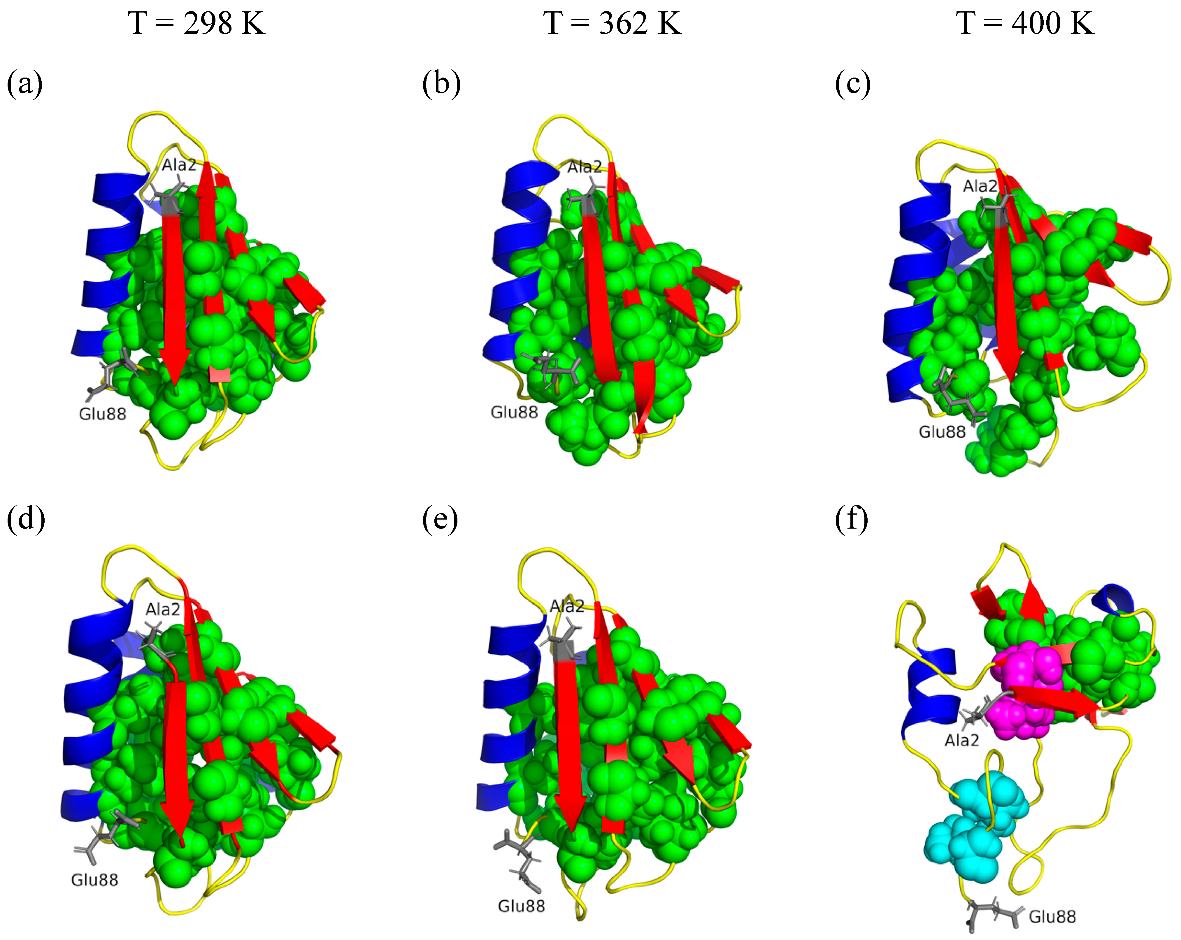
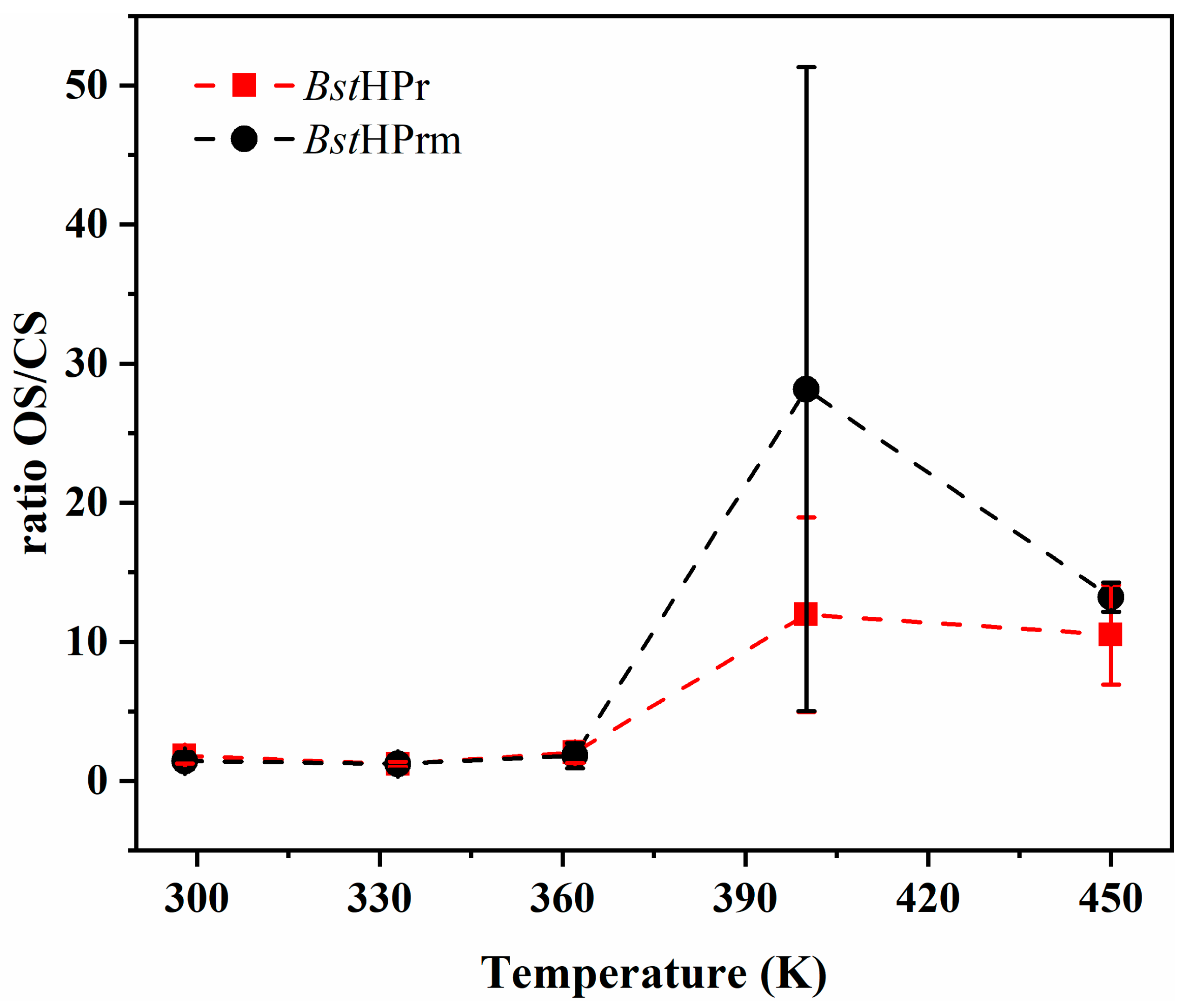
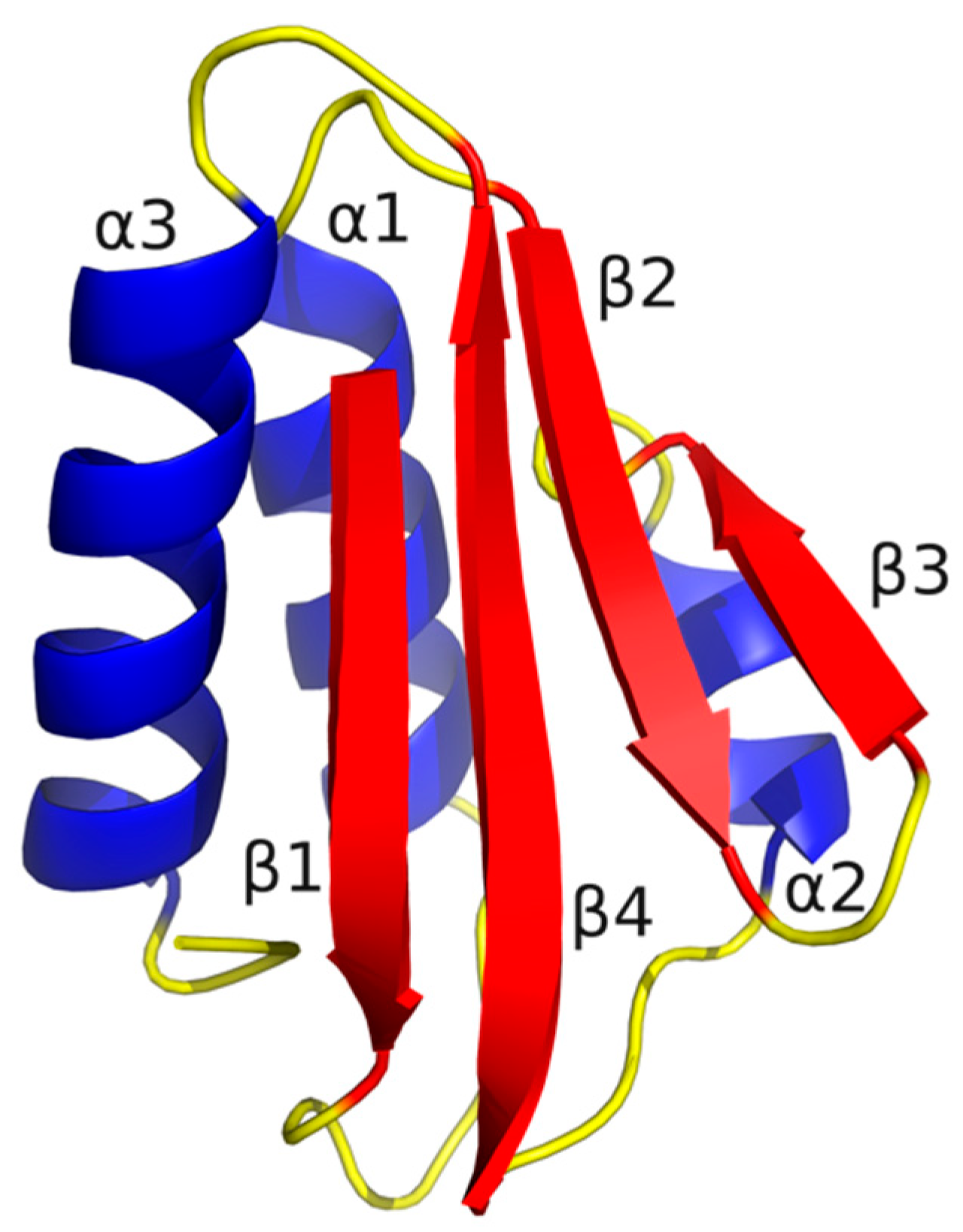
2.1. Structural Behavior
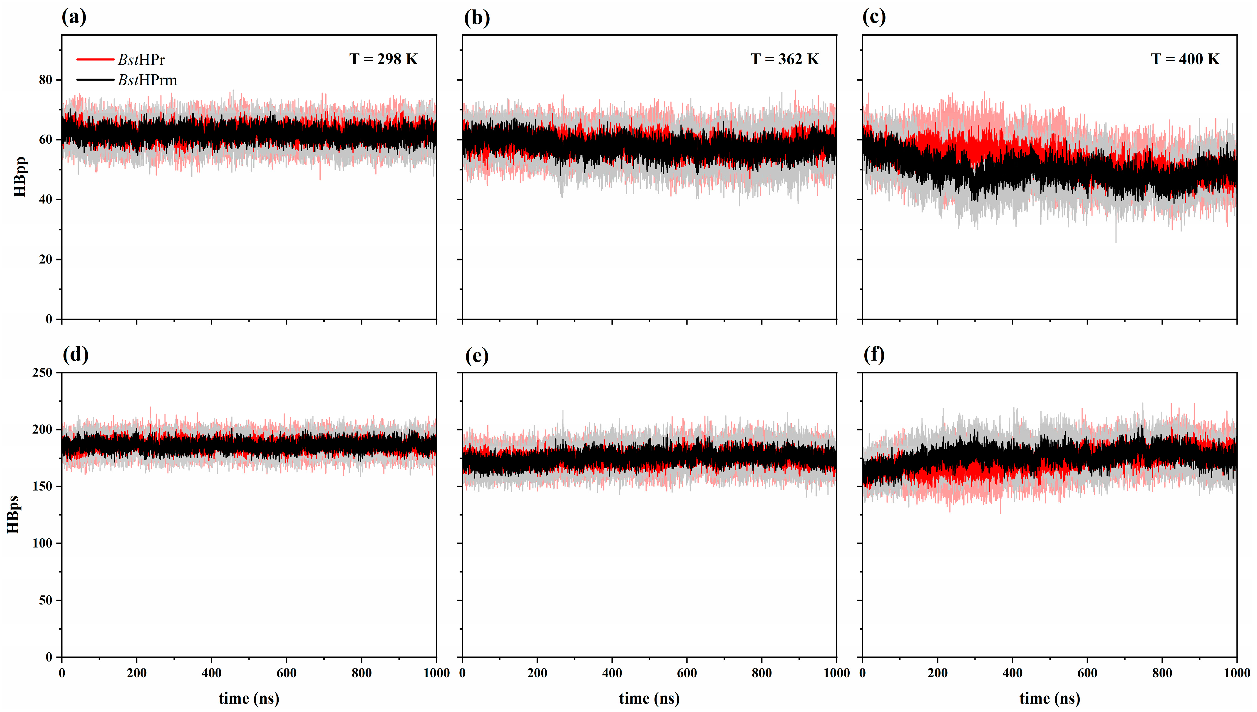
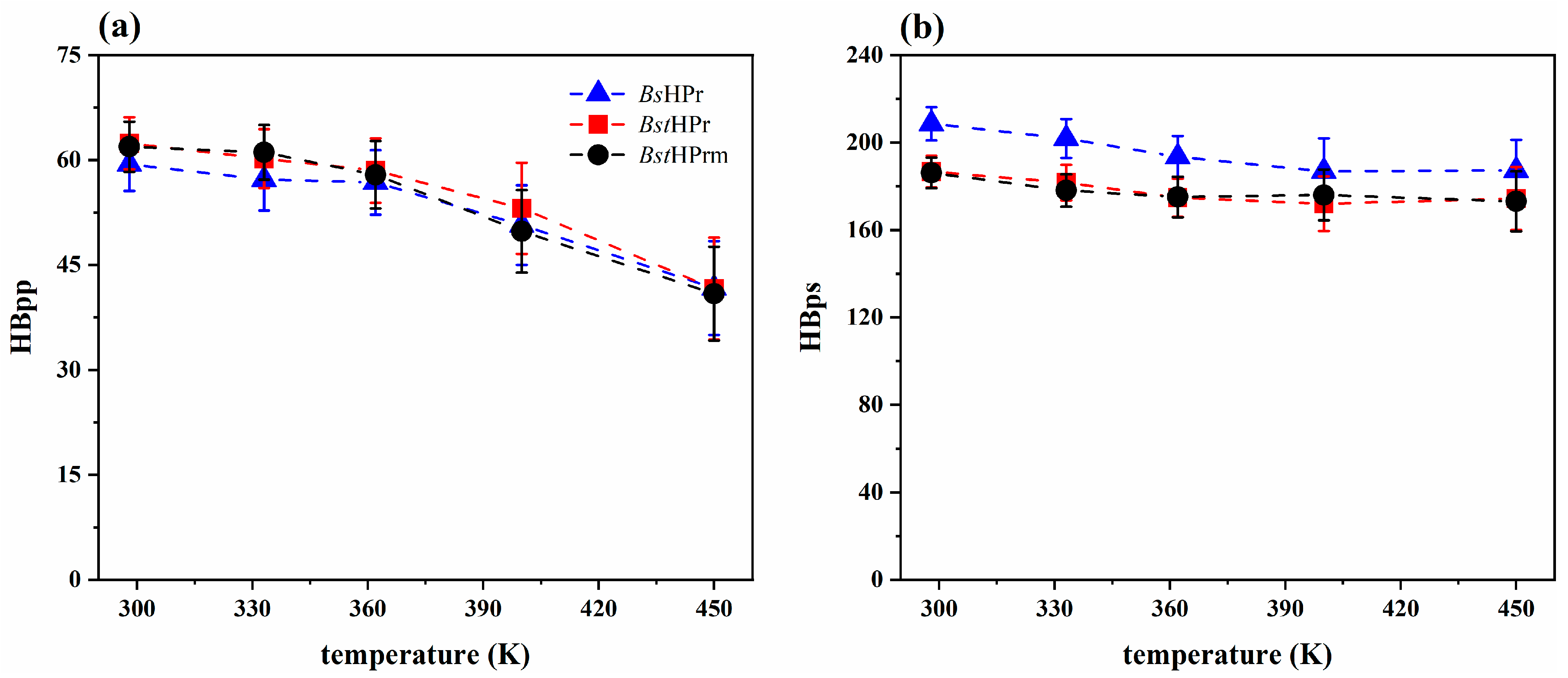
2.2. Molecular Interactions
- (a)
- The Asp79–Lys83 salt bridge, located on the α3-helix, does not undergo meaningful changes; only its frequency is slightly higher at 333 K.
- (b)
- The Glu84–Arg17 salt bridge, formed between the α3-helix and α1-helix, undergoes significant changes, since it presents lower frequencies at all temperatures, causing the secondary structures to be less stable.
- (c)
- The Asp11–Lys57 salt bridge, formed between two loops (that are in the β1-strand/α1-helix and the α2-helix/β4-strand structures), increases its frequency at 298 and 333 K but decreases for the other temperatures. In particular, it decreases drastically at 400 K.
- (d)
- The Glu32–Lys45 salt bridge is formed between the β2-strand and the first residue of the loop after the β3-strand. This interaction is very weak, as its frequency is less than 0.3 at 298 and 333 K. It reaches values slightly higher than 0.3 at 362 and 400 K, but it is almost lost at 450 K.
3. Discussion
4. Materials and Methods
4.1. Molecular Models
4.2. Molecular Dynamic Simulations
4.3. Simulation Analysis
- (a)
- Root mean square deviation: The first structural conformation of the DM simulation is used as the reference structure (t = 0 ns).
- (b)
- Radius of gyration: This parameter is calculated from the protein center of mass.
- (c)
- Fraction of native contacts: This indicator is determined using the Best–Hummer–Eaton model [35]. For this calculation, the first conformation of the simulations is defined as the native structure (t = 0 ns). The total number of contacts in the native structure is taken as Q = 1, and from this reference, the contacts for the remaining conformations of the trajectory are obtained.
- (d)
- Secondary structure profiles: SS assignment is performed using the define secondary structure of proteins (DSSP) algorithm [36]. This algorithm considers 8 types of SS: α-helix, π-helix, 310-helix, β-strand, β-bridge, random coil, bend, and turn. After this calculation, Micsonai et al. classified these structures in three different groups [37], that is, in the α-helix SS the 3 helix structures (α-helix, π-helix, and 310-helix) are included, in the β-strand SS only the β-strand is considered, and in the random coil the remaining structures (β-bridge, random coil, bend, and turn) are included. Micsonai et al. proposed this classification from protein structure data and their respective circular dichroism spectra. Therefore, the set of the three classifications (α-helix, β-strand, and random coil) is considered 100% of the secondary structure.
- (e)
- Hydrogen bonds: For this calculation, the distance r and the angle θ between the mass centers of the acceptor (A) and donor (D) atoms of the proton (H) are considered (rAD ≤ 3.5 Å and θAD ≤ 30°).
- (f)
- Solvent accessible surface area: This parameter is determined using the Lee and Richards approximation: one solvent sphere with a radius of 1.4 Å is used [38].
- (g)
- Cluster ILV: The contacts of structural units (CSU) algorithm is used to find the groupings of isoleucine, leucine, and valine residues within proteins [39]. This methodology analyzes atoms as spheres with a van der Waals radius. The contact of two atoms, A and B, is considered, i.e., a test sphere on the surface of atom A must overlap at least 10 Å with the surface of the sphere of atom B. If this contact occurs between the atoms of the residues Ile, Leu, and Val, they are considered part of a cluster. Therefore, different ILV clusters can be expected in the proteins.
- (h)
- Salt bridges: The Barlow and Thornton criterion is taken to measure SB formation [40], i.e., rSB ≤ 0.4 nm. In addition, the ionic pairs were calculated using the GetContacts program (https://getcontacts.github.io/ (accessed on 22 February 2024)), taking as a criterion of formation that the average frequency must be equal to 0.3 during the trajectories of the three replicas for each temperature.
5. Conclusions
- (a)
- Global fluctuations increase, compaction/expansion processes increase, topological native contacts decrease, ordered secondary structures are lost, and disordered structures increase;
- (b)
- Buried hydrogen bonds decrease and those formed with the solvent increase, while the non-polar residues are more exposed to the solvent, causing a loss of hydrophobic interactions.
Supplementary Materials
Author Contributions
Funding
Institutional Review Board Statement
Informed Consent Statement
Data Availability Statement
Acknowledgments
Conflicts of Interest
References
- Fersht, A. Structure and Mechanism in Protein Science: A Guide to Enzyme Catalysis and Protein Folding, 1st ed.; W. H. Freeman and Company: New York, NY, USA, 1999; ISBN 0-7167-3268-8. [Google Scholar]
- Rangwala, H.; Karypis, G. (Eds.) Introduction to Protein Structure Prediction: Methods and Algorithms, 1st ed.; John Wiley & Sons, Inc.: Hoboken, NJ, USA, 2010; ISBN 978-0-470-47059-6. [Google Scholar]
- Beltrán, H.I.; Alas-Guardado, S.J.; González-Pérez, P.P. Improving Coarse-Grained Models of Protein Folding through Weighting of Polar-Polar/Hydrophobic-Hydrophobic Interactions into Crowded Spaces. J. Mol. Model. 2022, 28, 87. [Google Scholar] [CrossRef] [PubMed]
- Sen, S.; Nilsson, L. (Eds.) Thermostable Proteins: Structural Stability and Design, 1st ed.; Taylor & Francis Group: Boca Raton, FL, USA, 2012; ISBN 978-1-4398-3913-3. [Google Scholar]
- Shriver, J.W. Protein Structure, Stability, and Interactions, 1st ed.; Humana Press: New York, NY, USA, 2009; ISBN 978-1-58829-954-3. [Google Scholar] [CrossRef]
- Nikam, R.; Kulandaisamy, A.; Harini, K.; Sharma, D.; Gromiha, M.M. ProThermDB: Thermodynamic Database for Proteins and Mutants Revisited After 15 Years. Nucl. Acids Res. 2021, 49, D420–D424. [Google Scholar] [CrossRef] [PubMed]
- Rigoldi, F.; Donini, S.; Redaelli, A.; Parisini, E.; Gautieri, A. Review: Engineering of Thermostable Enzymes for Industrial Applications. APL Bioeng. 2018, 2, 011501. [Google Scholar] [CrossRef]
- Finch, A.J.; Kim, J.R. Thermophilic Proteins as Versatile Scaffolds for Protein Engineering. Microorganisms 2018, 6, 97. [Google Scholar] [CrossRef]
- Xu, Z.; Cen, Y.-K.; Zou, S.-P.; Xue, Y.-P.; Zheng, Y.-G. Recent Advances in the Improvement of Enzyme Thermostability by Structure Modification. Crit. Rev. Biotechnol. 2020, 40, 83–98. [Google Scholar] [CrossRef] [PubMed]
- Merino, N.; Aronson, H.S.; Bojanova, D.P.; Feyhl-Buska, J.; Wong, M.L.; Zhang, S.; Giovannelli, D. Living at the Extremes: Extremhopiles and the Limits of Life in a Planetary Context. Front. Microbiol. 2019, 10, 780. [Google Scholar] [CrossRef] [PubMed]
- Qu, Z.; Zhang, L.; Sun, Y. Molecular Insights into the Enhanced Activity and/or Thermostability of PET Hydrolase by D186 Mutations. Molecules 2024, 29, 1338. [Google Scholar] [CrossRef] [PubMed]
- Razvi, A. A Comparative Study of HPr Proteins from Extremophilic Organisms. Ph.D. Thesis, Texas A&M University, College Station, TX, USA, 2005. [Google Scholar]
- Razvi, A.; Scholtz, J.M. Lessons in Stability from Thermophilic Proteins. Protein Sci. 2006, 15, 1569–1578. [Google Scholar] [CrossRef] [PubMed]
- Sridharan, S.; Razvi, A.; Scholtz, J.M.; Sacchettini, J.C. The HPr Proteins from the Thermophile Bacillus stearothermophilus can Form Domain-Swapped Dimers. J. Mol. Biol. 2005, 346, 919–931. [Google Scholar] [CrossRef]
- Razvi, A.; Scholtz, J.M. A Thermodynamic Comparison of HPr Proteins from Extremophilic Organisms. Biochemistry 2006, 45, 4084–4092. [Google Scholar] [CrossRef]
- Feng, C.Y.; Gao, F.; Liu, Y.W.; Wang, G.H.; Peng, H.; Ma, Y.H.; Yan, J.H.; Gao, G.F. Crystal Structure of Histidine-Containing Phosphocarrier Protein from Thermoanaerobacter tengcongensis MB4 and the Implications for Thermostability. Sci. China Life Sci. 2011, 54, 513–519. [Google Scholar] [CrossRef]
- Szilágyi, A.; Závodszky, P. Structural Differences Between Mesophilic, Moderately Thermophilic and Extremely Thermophilic Protein Subunits: Results of a Comprehensive Survey. Structure 2000, 8, 493–504. [Google Scholar] [CrossRef] [PubMed]
- Deutscher, J.; Francke, C.; Postma, P.W. How Phosphotransferase System-Related Protein Phosphorylation Regulates Carbohydrate Metabolism in Bacteria. Microbiol. Mol. Biol. Rev. 2006, 70, 939–1031. [Google Scholar] [CrossRef] [PubMed]
- Homeyer, N.; Essigke, T.; Ullmann, G.M.; Sticht, H. Effects of Histidine Protonation and Phosphorylation on Histidine-Containing Phosphocarrier Protein Structure, Dynamics, and Physicochemical Properties. Biochemistry 2007, 46, 12314–12326. [Google Scholar] [CrossRef] [PubMed]
- Gómez-Flores, A.K.; López-Pérez, E.; Alas-Guardado, S.J. Molecular Dynamics Simulations of HPr Proteins from a Thermophilic and a Mesophilic Organism: A Comparative Thermal Study. Int. J. Mol. Sci. 2023, 24, 9557. [Google Scholar] [CrossRef] [PubMed]
- Ferruz, N.; Schmidt, S.; Höcker, B. ProteinTools: A Toolkit to Analyze Protein Structures. Nucleic Acids Res. 2021, 49, W559–W566. [Google Scholar] [CrossRef] [PubMed]
- López-Chávez, E.; Pérez-Hernández, G.; Aparicio, F.; Alas, S.J. On the Thermal Stability of O6-Methylguanine-DNA Methyltransferase from Archaeon Pyrococcus kodakaraensis by Molecular Dynamics Simulations. J. Chem. Inf. Model. 2020, 60, 2138–2154. [Google Scholar] [CrossRef]
- Herzberg, O.; Reddy, P.; Sutrina, S.; Saier, M.; Reizer, J.; Kapadia, G. Structure of the Histidine-Containing Phosphocarrier Protein HPr from Bacillus Subtilis at 2.0 Å Resolution. Proc. Natl. Acad. Sci. USA 1992, 89, 2499–2503. [Google Scholar] [CrossRef] [PubMed]
- Dolinsky, T.J.; Nielsen, J.E.; McCammon, J.A.; Baker, N.A. PDB2PQR: An Automated Pipeline for the Setup of Poisson–Boltzmann Electrostatics Calculations. Nucleic Acids Res. 2004, 32, W665–W667. [Google Scholar] [CrossRef]
- Marabotti, A.; Del Prete, E.; Scafuri, B.; Facchiano, A. Performance of Web Tools for Predicting Changes in Protein Stability Caused by Mutations. BMC Bioinform. 2021, 22 (Suppl. S7), 345. [Google Scholar] [CrossRef]
- Abraham, M.J.; van der Spoel, D.; Lindahl, E.; Hess, B. GROMACS User Manual, Version 2024.1. Available online: www.gromacs.org (accessed on 24 March 2024).
- Abraham, M.J.; Murtola, T.; Schulz, R.; Páll, S.; Smith, J.C.; Hess, B.; Lindahl, E. GROMACS: High Performance Molecular Simulations Through Multi-Level Parallelism from Laptops to Supercomputers. SoftwareX 2015, 1–2, 19–25. [Google Scholar] [CrossRef]
- Hornak, V.; Abel, R.; Okur, A.; Strockbine, B.; Roitberg, A.; Simmerling, C. Comparison of Multiple Amber Force Fields and Development of Improved Protein Backbone Parameters. Proteins Struct. Funct. Bioinform. 2006, 65, 712–725. [Google Scholar] [CrossRef] [PubMed]
- Berendsen, H.J.C.; Grigera, J.R.; Straatsma, T.P. The Missing Term in Effective Pair Potentials. J. Phys. Chem. 1987, 91, 6269–6271. [Google Scholar] [CrossRef]
- Darden, T.; York, D.; Pedersen, L. Particle Mesh Ewald: An N⋅log(N) Method for Ewald Sums in Large Systems. J. Chem. Phys. 1993, 98, 10089–10092. [Google Scholar] [CrossRef]
- Hess, B.; Bekker, H.; Berendsen, H.J.C.; Fraaije, J.G.E.M. LINCS: A Linear Constraint Solver for Molecular Simulations. J. Comput. Chem. 1997, 18, 1463–1472. [Google Scholar] [CrossRef]
- Zimmermann, K. ORAL: All Purpose Molecular Mechanics Simulator and Energy Minimizer. J. Comput. Chem. 1991, 12, 310–319. [Google Scholar] [CrossRef]
- Bussi, G.; Donadio, D.; Parrinello, M. Canonical Sampling through Velocity Rescaling. J. Chem. Phys. 2007, 126, 014101. [Google Scholar] [CrossRef] [PubMed]
- Parrinello, M.; Rahman, A. Polymorphic Transitions in Single Crystals: A New Molecular Dynamics Method. J. Appl. Phys. 1981, 52, 7182–7190. [Google Scholar] [CrossRef]
- Best, R.B.; Hummer, G.; Eaton, W.A. Native Contacts Determine Protein Folding Mechanisms in Atomistic Simulations. Proc. Natl. Acad. Sci. USA 2013, 110, 17874–17879. [Google Scholar] [CrossRef]
- Kabsch, W.; Sander, C. Dictionary of Protein Secondary Structure: Pattern Recognition of Hydrogen-Bonded and Geometrical Features. Biopolymers 1983, 22, 2577–2637. [Google Scholar] [CrossRef]
- Micsonai, A.; Wien, F.; Kernya, L.; Lee, Y.-H.; Goto, Y.; Réfrégiers, M.; Kardos, J. Accurate Secondary Structure Prediction and Fold Recognition for Circular Dichroism Spectroscopy. Proc. Natl. Acad. Sci. USA 2015, 112, E3095–E3103. [Google Scholar] [CrossRef] [PubMed]
- Lee, B.; Richards, F.M. The Interpretation of Protein Structures: Estimation of Static Accessibility. J. Mol. Biol. 1971, 55, 379–400. [Google Scholar] [CrossRef] [PubMed]
- Wołek, K.; Gómez-Sicilia, A.; Cieplak, M. Determination of Contact Maps in Proteins: A Combination of Structural and Chemical Approaches. J. Chem. Phys. 2015, 143, 243105. [Google Scholar] [CrossRef] [PubMed]
- Barlow, D.J.; Thornton, J.M. Ion-Pairs in Proteins. J. Mol. Biol. 1983, 168, 867–885. [Google Scholar] [CrossRef] [PubMed]


| Protein | TS (°C) | Tm (°C) | ∆GS (kcal/mol) | ∆Cp (kcal/mol K) | ∆H (kcal/mol) |
|---|---|---|---|---|---|
| BstHPr | 24.8 | 88.9 | 8.2 | 1.37 | 98.6 |
| BsHPr | 24.1 | 74.4 | 5.2 | 1.33 | 76.7 |
| Temperature Range | BstHPr | BstHPrm | ||
|---|---|---|---|---|
| Number | Percentage | Number | Percentage | |
| 298–333 | 2 | 3.5 | 1 | 1.3 |
| 298–362 | 4 | 6.2 | 4 | 6.5 |
| 298–400 | 9 | 14.9 | 12 | 19.5 |
| 298–450 | 21 | 33.3 | 21 | 33.9 |
| Temperature Range | BstHPr | BstHPrm | ||
|---|---|---|---|---|
| Number | Percentage | Number | Percentage | |
| 298–333 | 5 | 2.7 | 8 | 4.3 |
| 298–362 | 12 | 6.4 | 11 | 5.9 |
| 298–400 | 15 | 7.9 | 10 | 5.4 |
| 298–450 | 12 | 6.6 | 13 | 7.0 |
| Temperature | SASAp | SASAnp | ||
|---|---|---|---|---|
| Area (nm2) | Percentage | Area (nm2) | Percentage | |
| 298 | 1.59 | 8.7 | 0.93 | 3.0 |
| 333 | 1.15 | 6.1 | 1.26 | 4.0 |
| 362 | 1.42 | 7.5 | 0.42 | 1.3 |
| 400 | 1.77 | 9.2 | 0.77 * | 2.3 * |
| 450 | 1.24 | 5.9 | 1.08 | 2.5 |
| Residue Pairs | Temperature (K) | ||||
|---|---|---|---|---|---|
| 298 | 333 | 362 | 400 | 450 | |
| Asp79–Lys83 | 0.822 | 0.773 | 0.773 | 0.527 | 0.341 |
| Glu84–Arg17 | 0.579 | 0.495 | 0.526 | 0.292 | 0.104 |
| Asp11–Lys57 | 0.412 | 0.397 | 0.396 | 0.220 | 0.081 |
| Glu3–Lys62 | 0.620 | 0.617 | 0.633 | 0.597 | 0.088 |
| Glu36–Lys62 | 0.380 | 0.403 | 0.526 | 0.553 | 0.203 |
| Residue Pairs | Temperature (K) | ||||
|---|---|---|---|---|---|
| 298 | 333 | 362 | 400 | 450 | |
| Asp79–Lys83 | 0.810 | 0.804 | 0.733 | 0.526 | 0.347 |
| Glu84–Arg17 | 0.422 | 0.476 | 0.406 | 0.245 | 0.055 |
| Asp11–Lys57 | 0.474 | 0.436 | 0.336 | 0.057 | 0.055 |
| Glu32–Lys45 | 0.246 | 0.244 | 0.334 | 0.351 | 0.149 |
Disclaimer/Publisher’s Note: The statements, opinions and data contained in all publications are solely those of the individual author(s) and contributor(s) and not of MDPI and/or the editor(s). MDPI and/or the editor(s) disclaim responsibility for any injury to people or property resulting from any ideas, methods, instructions or products referred to in the content. |
© 2024 by the authors. Licensee MDPI, Basel, Switzerland. This article is an open access article distributed under the terms and conditions of the Creative Commons Attribution (CC BY) license (https://creativecommons.org/licenses/by/4.0/).
Share and Cite
Martínez-Zacarias, A.C.; López-Pérez, E.; Alas-Guardado, S.J. Effect of the Lys62Ala Mutation on the Thermal Stability of BstHPr Protein by Molecular Dynamics. Int. J. Mol. Sci. 2024, 25, 6316. https://doi.org/10.3390/ijms25126316
Martínez-Zacarias AC, López-Pérez E, Alas-Guardado SJ. Effect of the Lys62Ala Mutation on the Thermal Stability of BstHPr Protein by Molecular Dynamics. International Journal of Molecular Sciences. 2024; 25(12):6316. https://doi.org/10.3390/ijms25126316
Chicago/Turabian StyleMartínez-Zacarias, Aranza C., Edgar López-Pérez, and Salomón J. Alas-Guardado. 2024. "Effect of the Lys62Ala Mutation on the Thermal Stability of BstHPr Protein by Molecular Dynamics" International Journal of Molecular Sciences 25, no. 12: 6316. https://doi.org/10.3390/ijms25126316
APA StyleMartínez-Zacarias, A. C., López-Pérez, E., & Alas-Guardado, S. J. (2024). Effect of the Lys62Ala Mutation on the Thermal Stability of BstHPr Protein by Molecular Dynamics. International Journal of Molecular Sciences, 25(12), 6316. https://doi.org/10.3390/ijms25126316