
The Influence of Genetic Polymorphisms on the Expression of Interleukin-1beta, Prostaglandin E2 and Tumor Necrosis Factor Alpha in Peri-Implant Crevicular Fluid: A Cross-Sectional Study

 ,
,  ,
,  ,
,  ,
, 
Abstract
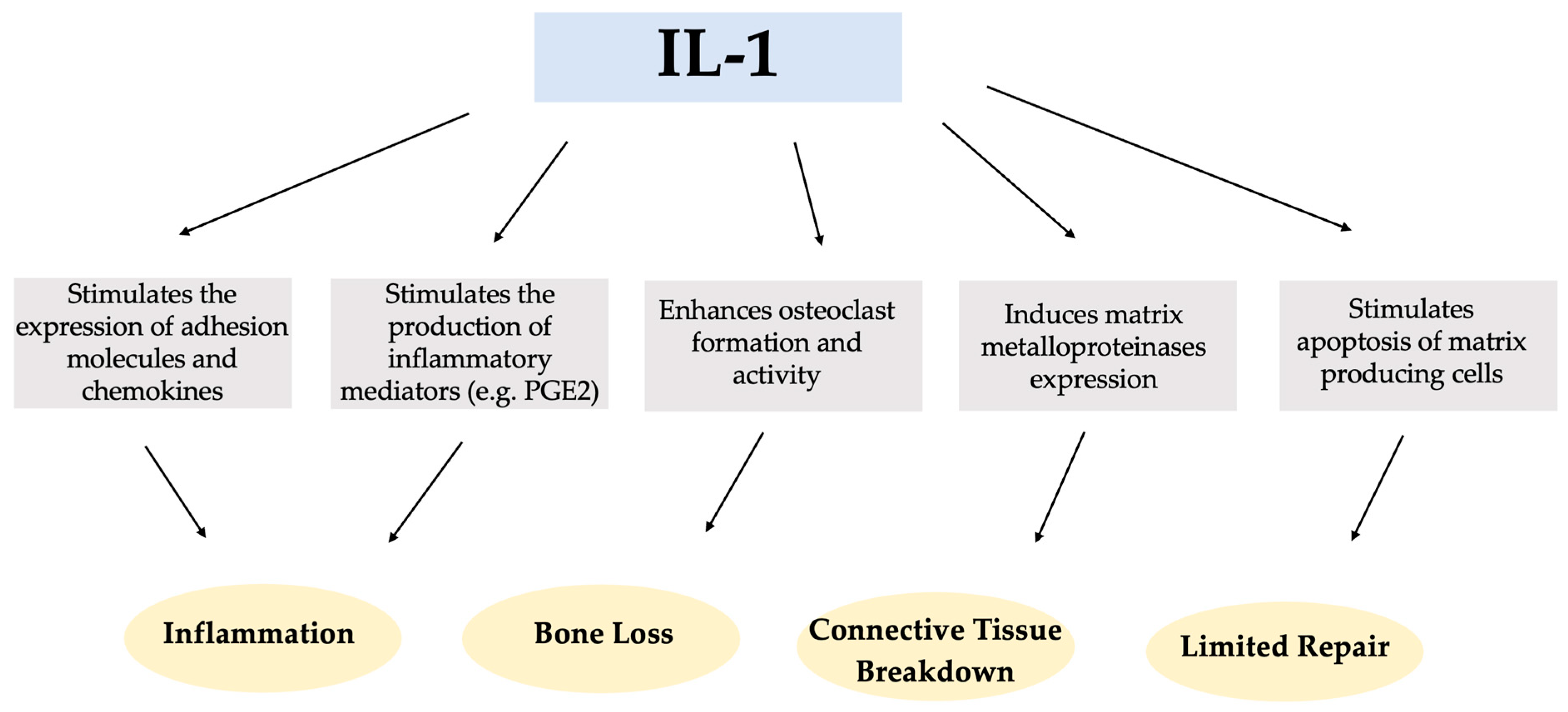
1. Introduction
2. Results
2.1. Demographic and Clinical Characteristics of the Study Group
2.2. Genotype Frequencies and Genetic Models
2.3. Influence of IL-1A -889, IL-1B +3954, and IL-1RN (VNTR) Polymorphisms on PICF Volume and Selected Biochemical Markers of the Peri-Implant Immune Response
2.4. Correlation between Concentrations of Biochemical Markers (IL-1β, TNF-α, and PGE2)
3. Discussion
4. Materials and Methods
4.1. Study Design and Population
4.2. Socio-Demographic and Clinical Variables
4.3. Genetic Analysis
4.4. Biochemical Analysis
4.5. Statistical Analysis
5. Conclusions
Supplementary Materials
Author Contributions
Funding
Institutional Review Board Statement
Informed Consent Statement
Data Availability Statement
Conflicts of Interest
Abbreviations
| CD14 | cluster of differentiation 14 |
| CI | confidence interval |
| ELISA | enzyme-linked immunosorbent assay |
| EMDC | Egas Moniz Dental Clinic |
| FMPS | full-mouth plaque score |
| IL-1 | interleukin-1 |
| IL-1α | interleukin-1 alpha |
| IL-1β | interleukin-1 beta |
| IL-1Ra | interleukin-1 receptor antagonist |
| OPG | osteoprotegerin |
| OR | odds ratio |
| PAI-2 | plasminogen activator inhibitor type 2 |
| PCR | polymerase chain reaction |
| PD | probing depth |
| PGE2 | prostaglandin E2 |
| PICF | peri-implant crevicular fluid |
| RANKL | receptor activator of nuclear factor-Kappa B ligand |
| SD | standard deviation |
| TNF-α | tumor necrosis factor alpha |
| VNTR | variable number of tandem repeats |
References
- Pjetursson, B.E.; Thoma, D.; Jung, R.; Zwahlen, M.; Zembic, A. A Systematic Review of the Survival and Complication Rates of Implant-Supported Fixed Dental Prostheses (FDPs) after a Mean Observation Period of at Least 5 Years. Clin. Oral. Implants Res. 2012, 23 (Suppl. S6), 22–38. [Google Scholar] [CrossRef] [PubMed]
- Romanos, G.E.; Delgado-Ruiz, R.; Sculean, A. Concepts for Prevention of Complications in Implant Therapy. Periodontol. 2000 2019, 81, 7–17. [Google Scholar] [CrossRef] [PubMed]
- Jepsen, S.; Berglundh, T.; Genco, R.; Aass, A.M.; Demirel, K.; Derks, J.; Figuero, E.; Giovannoli, J.L.; Goldstein, M.; Lambert, F.; et al. Primary Prevention of Peri-Implantitis: Managing Peri-Implant Mucositis. J. Clin. Periodontol. 2015, 42 (Suppl. S16), S152–S157. [Google Scholar] [CrossRef] [PubMed]
- Berglundh, T.; Armitage, G.; Araujo, M.G.; Avila-Ortiz, G.; Blanco, J.; Camargo, P.M.; Chen, S.; Cochran, D.; Derks, J.; Figuero, E.; et al. Peri-Implant Diseases and Conditions: Consensus Report of Workgroup 4 of the 2017 World Workshop on the Classification of Periodontal and Peri-Implant Diseases and Conditions. J. Clin. Periodontol. 2018, 45 (Suppl. S20), S286–S291. [Google Scholar] [CrossRef] [PubMed]
- Lang, N.P.; Berglundh, T.; Working Group 4 of Seventh European Workshop on Periodontology. Periimplant Diseases: Where Are We Now?—Consensus of the Seventh European Workshop on Periodontology. J. Clin. Periodontol. 2011, 38 (Suppl. S11), 178–181. [Google Scholar] [CrossRef] [PubMed]
- Fourmousis, I.; Vlachos, M. Genetic Risk Factors for the Development of Periimplantitis. Implant. Dent. 2019, 28, 103–114. [Google Scholar] [CrossRef]
- Schwarz, F.; Derks, J.; Monje, A.; Wang, H.-L. Peri-Implantitis. J. Periodontol. 2018, 89 (Suppl. S1), S267–S290. [Google Scholar] [CrossRef]
- Herrera, D.; Berglundh, T.; Schwarz, F.; Chapple, I.; Jepsen, S.; Sculean, A.; Kebschull, M.; Papapanou, P.N.; Tonetti, M.S.; Sanz, M.; et al. Prevention and Treatment of Peri-Implant Diseases-The EFP S3 Level Clinical Practice Guideline. J. Clin. Periodontol. 2023, 50, 4–76. [Google Scholar] [CrossRef]
- Madi, M.; Smith, S.; Alshehri, S.; Zakaria, O.; Almas, K. Influence of Smoking on Periodontal and Implant Therapy: A Narrative Review. Int. J. Environ. Res. Public. Health 2023, 20, 5368. [Google Scholar] [CrossRef]
- Wach, T.; Hadrowicz, P.; Trybek, G.; Michcik, A.; Kozakiewicz, M. Is Corticalization in Radiographs Related to a Higher Risk of Bone Loss around Dental Implants in Smoking Patients? A 5-Year Observation of Radiograph Bone-Texture Changes. J. Clin. Med. 2023, 12, 5351. [Google Scholar] [CrossRef]
- Sgolastra, F.; Petrucci, A.; Severino, M.; Gatto, R.; Monaco, A. Smoking and the Risk of Peri-Implantitis. A Systematic Review and Meta-Analysis. Clin. Oral. Implants Res. 2015, 26, e62–e67. [Google Scholar] [CrossRef] [PubMed]
- Farronato, D.; Azzi, L.; Giboli, L.; Maurino, V.; Tartaglia, G.M.; Farronato, M. Impact of Smoking Habit on Peri-Implant Indicators Following Different Therapies: A Systematic Review. Bioengineering 2022, 9, 569. [Google Scholar] [CrossRef] [PubMed]
- Spinell, T.; Kröger, A.; Freitag, L.; Würfl, G.; Lauseker, M.; Hickel, R.; Kebschull, M. Dental Implant Material Related Changes in Molecular Signatures in Peri-Implantitis—A Systematic Review of Omics in-Vivo Studies. Dent. Mater. 2023, 39, 1150–1158. [Google Scholar] [CrossRef] [PubMed]
- Oh, J.-M.; Kim, Y.; Son, H.; Kim, Y.H.; Kim, H.-J. Comparative Transcriptome Analysis of Periodontitis and Peri-Implantitis in Human Subjects. J. Periodontol. 2023. [Google Scholar] [CrossRef] [PubMed]
- Mohammadi, H.; Roochi, M.M.; Sadeghi, M.; Garajei, A.; Heidar, H.; Meybodi, A.A.; Dallband, M.; Mostafavi, S.; Mostafavi, M.; Salehi, M.; et al. Association between Interleukin-1 Polymorphisms and Susceptibility to Dental Peri-Implant Disease: A Meta-Analysis. Pathogens 2021, 10, 1600. [Google Scholar] [CrossRef] [PubMed]
- Saremi, L.; Shafizadeh, M.; Esmaeilzadeh, E.; Ghaffari, M.E.; Mahdavi, M.H.; Amid, R.; Kadkhodazadeh, M. Assessment of IL-10, IL-1ß and TNF-α Gene Polymorphisms in Patients with Peri-Implantitis and Healthy Controls. Mol. Biol. Rep. 2021, 48, 2285–2290. [Google Scholar] [CrossRef]
- Okada, H.; Murakami, S. Cytokine Expression in Periodontal Health and Disease. Crit. Rev. Oral. Biol. Med. 1998, 9, 248–266. [Google Scholar] [CrossRef]
- Arend, W.P. The Balance between IL-1 and IL-1Ra in Disease. Cytokine Growth Factor. Rev. 2002, 13, 323–340. [Google Scholar] [CrossRef]
- Laine, M.L.; Leonhardt, A.; Roos-Jansåker, A.-M.; Peña, A.S.; van Winkelhoff, A.J.; Winkel, E.G.; Renvert, S. IL-1RN Gene Polymorphism Is Associated with Peri-Implantitis. Clin. Oral. Implants Res. 2006, 17, 380–385. [Google Scholar] [CrossRef]
- Salcetti, J.M.; Moriarty, J.D.; Cooper, L.F.; Smith, F.W.; Collins, J.G.; Socransky, S.S.; Offenbacher, S. The Clinical, Microbial, and Host Response Characteristics of the Failing Implant. Int. J. Oral. Maxillofac. Implant. 1997, 12, 32–42. [Google Scholar]
- Duarte, P.M.; de Mendonça, A.C.; Máximo, M.B.B.; Santos, V.R.; Bastos, M.F.; Nociti Júnior, F.H. Differential Cytokine Expressions Affect the Severity of Peri-Implant Disease. Clin. Oral. Implants Res. 2009, 20, 514–520. [Google Scholar] [CrossRef] [PubMed]
- Faot, F.; Nascimento, G.G.; Bielemann, A.M.; Campão, T.D.; Leite, F.R.M.; Quirynen, M. Can Peri-Implant Crevicular Fluid Assist in the Diagnosis of Peri-Implantitis? A Systematic Review and Meta-Analysis. J. Periodontol. 2015, 86, 631–645. [Google Scholar] [CrossRef] [PubMed]
- Zani, S.R.; Moss, K.; Shibli, J.A.; Teixeira, E.R.; de Oliveira Mairink, R.; Onuma, T.; Feres, M.; Teles, R.P. Peri-Implant Crevicular Fluid Biomarkers as Discriminants of Peri-Implant Health and Disease. J. Clin. Periodontol. 2016, 43, 825–832. [Google Scholar] [CrossRef] [PubMed]
- Ghassib, I.; Chen, Z.; Zhu, J.; Wang, H.-L. Use of IL-1 β, IL-6, TNF-α, and MMP-8 Biomarkers to Distinguish Peri-Implant Diseases: A Systematic Review and Meta-Analysis. Clin. Implant. Dent. Relat. Res. 2019, 21, 190–207. [Google Scholar] [CrossRef] [PubMed]
- Graves, D.T.; Cochran, D. The Contribution of Interleukin-1 and Tumor Necrosis Factor to Periodontal Tissue Destruction. J. Periodontol. 2003, 74, 391–401. [Google Scholar] [CrossRef]
- Deo, V.; Bhongade, M.L. Pathogenesis of Periodontitis: Role of Cytokines in Host Response. Dent. Today 2010, 29, 60–62, 64–66, quiz 68–69. [Google Scholar]
- Chen, Z.; Yan, Q.; Zhang, R.; Li, Y.; Huang, S. Identification of Novel Candidate Biomarkers Related to Immune Cell Infiltration in Peri-Implantitis. Oral. Dis. 2023, 1–11. [Google Scholar] [CrossRef]
- Dinarello, C.A. Proinflammatory Cytokines. Chest 2000, 118, 503–508. [Google Scholar] [CrossRef]
- Möller, B.; Villiger, P.M. Inhibition of IL-1, IL-6, and TNF-Alpha in Immune-Mediated Inflammatory Diseases. Springer Semin. Immunopathol. 2006, 27, 391–408. [Google Scholar] [CrossRef] [PubMed]
- Zhao, B. Intrinsic Restriction of TNF-Mediated Inflammatory Osteoclastogenesis and Bone Resorption. Front. Endocrinol. 2020, 11, 583561. [Google Scholar] [CrossRef]
- Graves, D.T.; Oskoui, M.; Volejnikova, S.; Naguib, G.; Cai, S.; Desta, T.; Kakouras, A.; Jiang, Y. Tumor Necrosis Factor Modulates Fibroblast Apoptosis, PMN Recruitment, and Osteoclast Formation in Response to P. Gingivalis Infection. J. Dent. Res. 2001, 80, 1875–1879. [Google Scholar] [CrossRef]
- Yalçn, S.; Baseğmez, C.; Mijiritsky, E.; Yalçn, F.; Isik, G.; Onan, U. Detection of Implant Crevicular Fluid Prostaglandin E2 Levels for the Assessment of Peri-Implant Health: A Pilot Study. Implant. Dent. 2005, 14, 194–200. [Google Scholar] [CrossRef] [PubMed]
- Basegmez, C.; Yalcin, S.; Yalcin, F.; Ersanli, S.; Mijiritsky, E. Evaluation of Periimplant Crevicular Fluid Prostaglandin E2 and Matrix Metalloproteinase-8 Levels from Health to Periimplant Disease Status: A Prospective Study. Implant. Dent. 2012, 21, 306–310. [Google Scholar] [CrossRef] [PubMed]
- Hart, T.C.; Kornman, K.S. Genetic Factors in the Pathogenesis of Periodontitis. Periodontol. 2000 1997, 14, 202–215. [Google Scholar] [CrossRef] [PubMed]
- Lachmann, S.; Kimmerle-Müller, E.; Axmann, D.; Scheideler, L.; Weber, H.; Haas, R. Associations between Peri-Implant Crevicular Fluid Volume, Concentrations of Crevicular Inflammatory Mediators, and Composite IL-1A -889 and IL-1B +3954 Genotype. A Cross-Sectional Study on Implant Recall Patients with and without Clinical Signs of Peri-Implantitis. Clin. Oral. Implants Res. 2007, 18, 212–223. [Google Scholar] [CrossRef]
- Hamdy, A.A.E.-M.M.; Ebrahem, M.A.E.-M. The Effect of Interleukin-1 Allele 2 Genotype (IL-1a(-889) and IL-1b(+3954)) on the Individual’s Susceptibility to Peri-Implantitis: Case-Control Study. J. Oral. Implantol. 2011, 37, 325–334. [Google Scholar] [CrossRef]
- He, K.; Jian, F.; He, T.; Tang, H.; Huang, B.; Wei, N. Analysis of the Association of TNF-α, IL-1A, and IL-1B Polymorphisms with Peri-Implantitis in a Chinese Non-Smoking Population. Clin. Oral. Investig. 2020, 24, 693–699. [Google Scholar] [CrossRef]
- Gruica, B.; Wang, H.-Y.; Lang, N.P.; Buser, D. Impact of IL-1 Genotype and Smoking Status on the Prognosis of Osseointegrated Implants. Clin. Oral. Implant. Res. 2004, 15, 393–400. [Google Scholar] [CrossRef]
- García-Delaney, C.; Sánchez-Garcés, M.-Á.; Figueiredo, R.; Sánchez-Torres, A.; Gay-Escoda, C. Clinical Significance of Interleukin-1 Genotype in Smoking Patients as a Predictor of Peri-Implantitis: A Case-Control Study. Med. Oral. Patol. Oral. Cir. Bucal 2015, 20, e737–e743. [Google Scholar] [CrossRef]
- Nicklin, M.J.; Weith, A.; Duff, G.W. A Physical Map of the Region Encompassing the Human Interleukin-1 Alpha, Interleukin-1 Beta, and Interleukin-1 Receptor Antagonist Genes. Genomics 1994, 19, 382–384. [Google Scholar] [CrossRef]
- Dominici, R.; Cattaneo, M.; Malferrari, G.; Archi, D.; Mariani, C.; Grimaldi, L.M.E.; Biunno, I. Cloning and Functional Analysis of the Allelic Polymorphism in the Transcription Regulatory Region of Interleukin-1 Alpha. Immunogenetics 2002, 54, 82–86. [Google Scholar] [CrossRef] [PubMed]
- Tountas, N.A.; Casini-Raggi, V.; Yang, H.; Di Giovine, F.S.; Vecchi, M.; Kam, L.; Melani, L.; Pizarro, T.T.; Rotter, J.I.; Cominelli, F. Functional and Ethnic Association of Allele 2 of the Interleukin-1 Receptor Antagonist Gene in Ulcerative Colitis. Gastroenterology 1999, 117, 806–813. [Google Scholar] [CrossRef] [PubMed]
- Santtila, S.; Savinainen, K.; Hurme, M. Presence of the IL-1RA Allele 2 (IL1RN*2) Is Associated with Enhanced IL-1beta Production in Vitro. Scand. J. Immunol. 1998, 47, 195–198. [Google Scholar] [CrossRef] [PubMed]
- Vaz, P.; Gallas, M.M.; Braga, A.C.; Sampaio-Fernandes, J.C.; Felino, A.; Tavares, P. IL1 Gene Polymorphisms and Unsuccessful Dental Implants. Clin. Oral. Implants Res. 2012, 23, 1404–1413. [Google Scholar] [CrossRef] [PubMed]
- Melo, R.F.; Lopes, B.M.V.; Shibli, J.A.; Marcantonio, E.; Marcantonio, R.A.C.; Galli, G.M.T. Interleukin-1β and Interleukin-6 Expression and Gene Polymorphisms in Subjects with Peri-Implant Disease. Clin. Implant. Dent. Relat. Res. 2012, 14, 905–914. [Google Scholar] [CrossRef] [PubMed]
- Petkovic-Curcin, A.; Zeljic, K.; Cikota-Aleksic, B.; Dakovic, D.; Tatic, Z.; Magic, Z. Association of Cytokine Gene Polymorphism with Peri-Implantitis Risk. Int. J. Oral. Maxillofac. Implant. 2017, 32, e241–e248. [Google Scholar] [CrossRef] [PubMed]
- Karimbux, N.Y.; Saraiya, V.M.; Elangovan, S.; Allareddy, V.; Kinnunen, T.; Kornman, K.S.; Duff, G.W. Interleukin-1 Gene Polymorphisms and Chronic Periodontitis in Adult Whites: A Systematic Review and Meta-Analysis. J. Periodontol. 2012, 83, 1407–1419. [Google Scholar] [CrossRef] [PubMed]
- da Silva, F.-R.-P.; Guimarães-Vasconcelos, A.-C.-C.; de-Carvalho-França, L.-F.; di-Lenardo, D.; Rodrigues, L.-S.; Barreto-do-Nascimento, M.-L.-L.; Vasconcelos, D.-F.-P. Relationship between -889 C/T Polymorphism in Interleukin-1A Gene and Risk of Chronic Periodontitis: Evidence from a Meta-Analysis with New Published Findings. Med. Oral. Patol. Oral. Cir. Bucal 2017, 22, e7–e14. [Google Scholar] [CrossRef]
- da Silva, F.R.P.; Vasconcelos, A.C.C.G.; de Carvalho França, L.F.; Di Lenardo, D.; Nascimento, H.M.S.; Vasconcelos, D.F.P. Association between the Rs1143634 Polymorphism in Interleukin-1B and Chronic Periodontitis: Results from a Meta-Analysis Composed by 54 Case/Control Studies. Gene 2018, 668, 97–106. [Google Scholar] [CrossRef]
- Kornman, K.S.; Crane, A.; Wang, H.Y.; di Giovine, F.S.; Newman, M.G.; Pirk, F.W.; Wilson, T.G.; Higginbottom, F.L.; Duff, G.W. The Interleukin-1 Genotype as a Severity Factor in Adult Periodontal Disease. J. Clin. Periodontol. 1997, 24, 72–77. [Google Scholar] [CrossRef]
- Belibasakis, G.N. Microbiological and Immuno-Pathological Aspects of Peri-Implant Diseases. Arch. Oral. Biol. 2014, 59, 66–72. [Google Scholar] [CrossRef] [PubMed]
- Cardoso, J.M.; Ribeiro, A.C.; Palos, C.; Proença, L.; Noronha, S.; Alves, R.C. Association between IL-1A and IL-1B Gene Polymorphisms with Peri-Implantitis in a Portuguese Population—A Pilot Study. PeerJ 2022, 10, e13729. [Google Scholar] [CrossRef] [PubMed]
- Wang, H.-L.; Garaicoa-Pazmino, C.; Collins, A.; Ong, H.-S.; Chudri, R.; Giannobile, W.V. Protein Biomarkers and Microbial Profiles in Peri-Implantitis. Clin. Oral. Implants Res. 2016, 27, 1129–1136. [Google Scholar] [CrossRef] [PubMed]
- Alassy, H.; Parachuru, P.; Wolff, L. Peri-Implantitis Diagnosis and Prognosis Using Biomarkers in Peri-Implant Crevicular Fluid: A Narrative Review. Diagnostics 2019, 9, 214. [Google Scholar] [CrossRef] [PubMed]
- Engebretson, S.P.; Lamster, I.B.; Herrera-Abreu, M.; Celenti, R.S.; Timms, J.M.; Chaudhary, A.G.; di Giovine, F.S.; Kornman, K.S. The Influence of Interleukin Gene Polymorphism on Expression of Interleukin-1beta and Tumor Necrosis Factor-Alpha in Periodontal Tissue and Gingival Crevicular Fluid. J. Periodontol. 1999, 70, 567–573. [Google Scholar] [CrossRef] [PubMed]
- Pani, P.; Tsilioni, I.; McGlennen, R.; Brown, C.A.; Hawley, C.E.; Theoharides, T.C.; Papathanasiou, E. IL-1B(3954) Polymorphism and Red Complex Bacteria Increase IL-1β (GCF) Levels in Periodontitis. J. Periodontal. Res. 2021, 56, 501–511. [Google Scholar] [CrossRef]
- Chapple, I.L.C.; Mealey, B.L.; Van Dyke, T.E.; Bartold, P.M.; Dommisch, H.; Eickholz, P.; Geisinger, M.L.; Genco, R.J.; Glogauer, M.; Goldstein, M.; et al. Periodontal Health and Gingival Diseases and Conditions on an Intact and a Reduced Periodontium: Consensus Report of Workgroup 1 of the 2017 World Workshop on the Classification of Periodontal and Peri-Implant Diseases and Conditions. J. Clin. Periodontol. 2018, 45 (Suppl. S20), S68–S77. [Google Scholar] [CrossRef]
- Papapanou, P.N.; Sanz, M.; Buduneli, N.; Dietrich, T.; Feres, M.; Fine, D.H.; Flemmig, T.F.; Garcia, R.; Giannobile, W.V.; Graziani, F.; et al. Periodontitis: Consensus Report of Workgroup 2 of the 2017 World Workshop on the Classification of Periodontal and Peri-Implant Diseases and Conditions. J. Periodontol. 2018, 89 (Suppl. S1), S173–S182. [Google Scholar] [CrossRef]
- O’Leary, T.J.; Drake, R.B.; Naylor, J.E. The Plaque Control Record. J. Periodontol. 1972, 43, 38. [Google Scholar] [CrossRef]

| Genetic Polymorphisms Evaluated in the Studies |
|---|
| IL-1A -889 |
| IL-1A +4845 |
| IL-1B -511 |
| IL-1B +3954 |
| IL-1RN +2018 |
| IL-1RN (VNTR) |
| IL-6 -174 |
| IL-17 (rs10484879) |
| IL-17R (rs879576) |
| TNF-α -308 |
| OPG T950C |
| OPG G1181C |
| RANKL (rs9533156) |
| RANKL (rs2277438) |
| CD-14 -159 |
| Peri-Implant Health (n = 27) | Peri-Implantitis (n = 24) | p | |
|---|---|---|---|
| Age, | 0.865 | ||
| Mean (SD) (years) | 58.74 (8.72) | 59.17 (12.40) | |
| Median (years) | 60 | 57.5 | |
| Range (years) | 41–74 | 38–90 | |
| Sex, n (%) | 0.121 | ||
| Male | 11 (40.7) | 15 (62.5) | |
| Female | 16 (59.3) | 9 (37.5) | |
| Smoking, n (%) | 0.134 | ||
| No | 18 (66.7) | 11 (45.8) | |
| Yes | 9 (33.3) | 13 (54.2) | |
| History of Periodontitis, n (%) | 0.585 | ||
| No | 11 (40.7) | 8 (33.3) | |
| Yes | 16 (59.3) | 16 (66.7) | |
| FMPS (%) | 0.234 | ||
| ≤15% | 11 (40.7) | 6 (25) | |
| >15% | 16 (59.3) | 18 (75.0) | |
| Implant function time | 0.960 | ||
| Mean (SD) (years) | 8.6 (4.8) | 8.7 (5.8) | |
| Median (years) | 9 | 6.5 | |
| Range (years) | 1–20 | 1–20 |
| Genotype, n (%) | Peri-Implant Health (n = 27) | Peri-Implantitis (n = 24) | Total (n = 51) | p |
|---|---|---|---|---|
| IL-1A -889 | 0.714 | |||
| CC | 16 (59.3) | 13 (54.2) | 29 (56.9) | |
| CT/TT | 11 (40.7) | 11 (45.8) | 22 (43.1) | |
| IL-1A -889 | - | |||
| CC | 16 (59.3) | 13 (54.2) | 29 (56.9) | |
| CT | 10 (37.0) | 6 (25.0) | 16 (31.4) | |
| TT | 1 (3.7) | 5 (20.8) | 6 (11.7) | |
| IL-1B +3954 | 0.749 | |||
| CC | 18 (66.7) | 17 (70.8) | 35 (68.6) | |
| CT/TT | 9 (33.3) | 7 (29.2) | 16 (31.4) | |
| IL-1B +3954 | - | |||
| CC | 18 (66.7) | 17 (70.8) | 35 (68.6) | |
| CT | 9 (33.3) | 6 (25.0) | 15 (29.4) | |
| TT | 0 (0.0) | 1 (4.2) | 1 (2.0) | |
| IL-1RN (VNTR) | 0.811 | |||
| 1/1 | 23 (85.2) | 21 (87.5) | 44 (86.3) | |
| 1/5, 2/2 | 4 (14.8) | 3 (12.5) | 7 (13.7) | |
| IL-1RN (VNTR) | - | |||
| 1/1 | 23 (85.2) | 21 (87.5) | 44 (86.3) | |
| 1/5 | 1 (3.7) | 0 (0.0) | 1 (1.9) | |
| 2/2 | 3 (11.1) | 3 (12.5) | 6 (11.8) |
| Genetic Model IL-1A -889 | Peri-Implant Health (n = 27) | Peri-Implantitis (n = 24) | OR (95% CI) * | p | ||
|---|---|---|---|---|---|---|
| Events | Total | Events | Total | |||
| Dominant TT + CT vs. CC | 11 | 27 | 11 | 24 | 0.81 (0.27–2.47) | 0.934 |
| Recessive TT vs. CC + CT | 1 | 27 | 5 | 24 | 0.15 (0.02–1.36) | 0.148 |
| Genetic Model IL-1B +3954 | Peri-Implant Health (n = 27) | Peri-Implantitis (n = 24) | OR (95% CI) * | p | ||
|---|---|---|---|---|---|---|
| Events | Total | Events | Total | |||
| Dominant TT + CT vs. CC | 9 | 27 | 7 | 24 | 1.21 (0.37–3.99) | 0.986 |
| Recessive TT vs. CC + CT | 0 | 27 | 1 | 24 | - | - |
| Genotype | Number of Individuals | IL-1β (pg/μL) Mean (SD) | TNF-α (pg/μL) Mean (SD) | PGE2 (pg/μL) Mean (SD) |
|---|---|---|---|---|
| IL-1A -889 | ||||
| CC | 29 | 19.3 (21.9) | 1.2 (1.1) | 258.5 (332.3) |
| CT/TT | 22 | 29.0 (59.0) | 1.6 (3.5) | 177.7 (221.9) |
| p | 0.518 | 0.313 | 0.231 | |
| IL-1B +3954 | ||||
| CC | 35 | 26.1 (48.7) | 1.2 (1.3) | 262.0 (339.6) |
| CT/TT | 16 | 18.0 (20.4) | 1.6 (3.9) | 139.7 (91.3) |
| p | 0.776 | 0.490 | 0.543 | |
| IL-1RN (VNTR) | ||||
| 1/1 | 44 | 23.4 (44.2) | 0.9 (1.2) | 193.8 (265.5) |
| 1/5, 2/2 | 7 | 24.1 (24.7) | 3.9 (5.5) | 411.0 (384.8) |
| p | 0.339 | 0.002 | 0.049 | |
| IL-1 positive composite Genotype | ||||
| Yes | 13 | 18.5 (22.1) | 1.8 (4.3) | 145.0 (96.9) |
| No | 38 | 25.2 (46.9) | 1.2 (1.3) | 250.5 (328.2) |
| p | 0.713 | 0.364 | 0.634 |
| Patients | PICF Mean Volume (μL) (SD) | IL-1β (pg/μL) Mean (SD) | TNF-α (pg/μL) Mean (SD) | PGE2 (pg/μL) Mean (SD) |
|---|---|---|---|---|
| IL-1 positive composite genotype patients | ||||
| Healthy patients (n = 8) | 0.51 (0.28) | 15.31 (18.31) | 2.59 (5.53) | 119.06 (65.74) |
| Patients with peri-implantitis (n = 5) | 0.66 (0.44) | 23.65 (28.81) | 0.60 (0.50) | 186.54 (130.81) |
| p | 0.042 | 0.612 | 0.499 | 0.043 |
| Patients with IL-1RN (VNTR) polymorphism | ||||
| Healthy patients (n = 4) | 0.41 (0.56) | 31.28 (29.31) | 5.56 (7.20) | 365.81 (346.89) |
| Patients with peri-implantitis (n = 3) | 0.48 (0.29) | 14.60 (17.30) | 1.68 (0.15) | 471.32 (504.10) |
| p | 0.660 | 0.464 | 0.558 | 0.464 |
| Correlation (Rho, Spearman) | ||
|---|---|---|
| Rho | p | |
| IL-1β vs. TNF-α | 0.480 | <0.001 |
| IL-1β vs. PGE2 | 0.382 | 0.006 |
| TNF-α vs. PGE2 | 0.528 | <0.001 |
Disclaimer/Publisher’s Note: The statements, opinions and data contained in all publications are solely those of the individual author(s) and contributor(s) and not of MDPI and/or the editor(s). MDPI and/or the editor(s) disclaim responsibility for any injury to people or property resulting from any ideas, methods, instructions or products referred to in the content. |
© 2024 by the authors. Licensee MDPI, Basel, Switzerland. This article is an open access article distributed under the terms and conditions of the Creative Commons Attribution (CC BY) license (https://creativecommons.org/licenses/by/4.0/).
Share and Cite
Cardoso, J.M.; Ribeiro, A.C.; Botelho, J.; Proença, L.; Noronha, S.; Alves, R.C. The Influence of Genetic Polymorphisms on the Expression of Interleukin-1beta, Prostaglandin E2 and Tumor Necrosis Factor Alpha in Peri-Implant Crevicular Fluid: A Cross-Sectional Study. Int. J. Mol. Sci. 2024, 25, 651. https://doi.org/10.3390/ijms25010651
Cardoso JM, Ribeiro AC, Botelho J, Proença L, Noronha S, Alves RC. The Influence of Genetic Polymorphisms on the Expression of Interleukin-1beta, Prostaglandin E2 and Tumor Necrosis Factor Alpha in Peri-Implant Crevicular Fluid: A Cross-Sectional Study. International Journal of Molecular Sciences. 2024; 25(1):651. https://doi.org/10.3390/ijms25010651
Chicago/Turabian StyleCardoso, José Maria, Ana Clara Ribeiro, João Botelho, Luís Proença, Susana Noronha, and Ricardo Castro Alves. 2024. "The Influence of Genetic Polymorphisms on the Expression of Interleukin-1beta, Prostaglandin E2 and Tumor Necrosis Factor Alpha in Peri-Implant Crevicular Fluid: A Cross-Sectional Study" International Journal of Molecular Sciences 25, no. 1: 651. https://doi.org/10.3390/ijms25010651
APA StyleCardoso, J. M., Ribeiro, A. C., Botelho, J., Proença, L., Noronha, S., & Alves, R. C. (2024). The Influence of Genetic Polymorphisms on the Expression of Interleukin-1beta, Prostaglandin E2 and Tumor Necrosis Factor Alpha in Peri-Implant Crevicular Fluid: A Cross-Sectional Study. International Journal of Molecular Sciences, 25(1), 651. https://doi.org/10.3390/ijms25010651