Review

Regulation of Satellite Cells Functions during Skeletal Muscle Regeneration: A Critical Step in Physiological and Pathological Conditions

by
Giorgia Careccia
1,
Laura Mangiavini
2,3 and
Federica Cirillo
4,5,*
1
Department of Biosciences, University of Milan, 20133 Milan, Italy
2
IRCCS Istituto Ortopedico Galeazzi, 20161 Milan, Italy
3
Department of Biomedical Sciences for Health, University of Milan, 20133 Milan, Italy
4
IRCCS Policlinico San Donato, 20097 San Donato Milanese, Italy
5
Institute for Molecular and Translational Cardiology (IMTC), 20097 San Donato Milanese, Italy
*
Author to whom correspondence should be addressed.
Int. J. Mol. Sci. 2024, 25(1), 512; https://doi.org/10.3390/ijms25010512
Submission received: 26 October 2023 / Revised: 20 December 2023 / Accepted: 28 December 2023 / Published: 29 December 2023
(This article belongs to the Special Issue Myogenesis and Muscular Diseases)

Abstract

:
Skeletal muscle regeneration is a complex process involving the generation of new myofibers after trauma, competitive physical activity, or disease. In this context, adult skeletal muscle stem cells, also known as satellite cells (SCs), play a crucial role in regulating muscle tissue homeostasis and activating regeneration. Alterations in their number or function have been associated with various pathological conditions. The main factors involved in the dysregulation of SCs’ activity are inflammation, oxidative stress, and fibrosis. This review critically summarizes the current knowledge on the role of SCs in skeletal muscle regeneration. It examines the changes in the activity of SCs in three of the most common and severe muscle disorders: sarcopenia, muscular dystrophy, and cancer cachexia. Understanding the molecular mechanisms involved in their dysregulations is essential for improving current treatments, such as exercise, and developing personalized approaches to reactivate SCs.

1. Introduction

Skeletal muscle is composed of syncytial muscle fibers, each characterized by hundreds of myonuclei that support efficient contractility by releasing mRNA and promoting protein synthesis [1,2]. Skeletal muscle requires large amounts of energy and proteins for its functions but is also a source of energy during starvation and disease and, therefore, shows high plasticity. Cell size generally results from the balance between catabolism and anabolism, which are equally represented in homeostasis.
On the other hand, the well-being of skeletal muscle also depends on the ability to repair and regenerate new muscle fibers after traumatic events, thanks to the presence of adult stem cells, the satellite cells (SCs), first identified by Alexander Mauro [3]. SCs have two essential functions: (i) the ability to self-renew, which is essential to maintain and replenish the stem cell pool, and (ii) the capacity to differentiate into myogenic progenitor cells [4]. Defects in self-renewal ability result in a depleted SCs pool, impairing muscle regeneration capacity.
After injury, SCs undergo asymmetric division necessary to preserve the SCs pool, maintaining an undifferentiated mother cell that returns to a quiescent state and providing committed progenies [5]. During this phase, asymmetric co-segregation of immortal and younger DNA strands occurs in different daughter cells [6]. Specifically, the immortal DNA strand is co-segregated in daughter cells expressing the stem cell marker Sca1. In contrast, the younger strand is allocated to differentiated daughter cells expressing the differentiation marker Desmin [6]. The committed SCs become myoblasts, which further proliferate and eventually differentiate into postmitotic myocytes that fuse with existing fibers or each other to form new myofibers [7].
Nevertheless, some physiological and pathological conditions, such as aging, muscular dystrophy, and cancer cachexia, affect the ability of SCs to restore skeletal muscle tissue after damage, resulting in impaired regenerative machinery [8,9,10]. The molecular and biochemical mechanisms activated in these diseases are diverse and influence the number and functionality of SCs. In particular, age-related sarcopenia is associated with a decrease in SCs during aging, especially those associated with fibers II [11].
On the other hand, Duchenne muscular dystrophy is characterized by a progressive decline in functional SCs and their myogenic capacity, leading to the loss of myofibers and their replacement by fibrotic tissue [12,13]. Last but not least, cancer cachexia is a severe condition characterized by progressive body wasting and a remarkable weight loss of skeletal muscle [14].
In this review, we will discuss the role of SCs in controlling the skeletal muscle regeneration process, focusing on their functional and regulatory mechanisms, dysfunction, and therapeutic implications.

2. Biology of Satellite Cell in Regulating Muscle Homeostasis and Regeneration

The ability of skeletal muscle tissue to maintain its uniform architecture and functions is due to SCs, which reside in a defined anatomical hypoxic niche [15]. Upon injury, the oxygen concentration in muscle tissue and niches changes, and so does the expression of hypoxia-inducible factors (HIFs) [16,17]. In particular, HIF1α levels rapidly increase after cardiotoxin (CTX)-induced muscle injury, while HIF2α levels drop sharply after injury and gradually return to the baseline level after muscle regeneration [16]. Moreover, pharmacological activation of the HIF1α improves skeletal muscle regeneration and leads to the formation of bigger fibers [18,19].
In hypoxic niches, quiescent SCs express the paired-box transcription factors PAX7 and PAX3; the latter is particularly abundant in the diaphragm but much less in hindlimb muscles [20,21,22]. The differential expression of PAX3 in the tissues is probably due to the assumption that it plays a minor role in myogenesis in hindlimb muscles. On the other hand, PAX7 has been described as a master regulator of SCs’ function because it controls the expression of genes involved in SCs’ survival and proliferation and inhibits genes that trigger differentiation [23,24]. In addition to its role as a regulator, its activity is also modulated by post-translational modifications such as acetylation [25]. Although the expression of PAX7 is crucial to identify SCs in skeletal muscle tissue, it is now accepted that SCs are a heterogeneous population of stem cells. Indeed, in vivo experiments have demonstrated that SCs are characterized by the expression of PAX7 at different levels, which reflect multiple subpopulations of SCs with other functions. In particular, SCs characterized by a high expression of PAX7 (PAX7High) require a longer time to undergo first mitosis compared to SCs with a low expression of PAX7 (PAX7Low), which is also the reason why some SCs lose their ability to return to a quiescent state and become more committed to differentiation [26].
Moreover, SCs segregate their DNA asymmetrically, so the daughter cells that receive the template strand also maintain the expression of stem cell markers [27,28]. In addition, recent articles have identified different populations of SCs based on the expression of Gli factors. Gli1-positive SCs are more prone to proliferate and differentiate and contribute to the establishment of GAlert [29]. Gli2 is expressed during muscle stem cell activation, while Gli3 plays a crucial role in establishing quiescence of muscle stem cells [30,31]. Therefore, the stemness of SCs is characterized by a complex mechanism controlled by multiple factors.
Myogenic progenitor cells, often called myoblasts, are highly proliferative cells characterized by the expression of myogenic regulatory factors (MRFs), especially MYOD, which is crucial for adult muscle regeneration [32,33]. Indeed, MYOD knockout (KO) mice show a delay in the onset of myogenic differentiation and an increased propensity for self-renewal, suggesting its role in the balance between self-renewal and differentiation during regeneration [34,35,36,37]. In this context, Fujita, R. and colleagues have recently published that SCs with a low expression level of MYOD are undifferentiated. In contrast, SCs with a high expression of MYOD showed commitment and differentiation signatures [38]. Moreover, several studies have shown that the over-expression of MYOD can reprogram the fate of many non-muscle cell types, such as fibroblasts, to a myogenic lineage when specifically over-expressed [39,40,41]. MYOD has been reported to regulate CDC6 expression early after activation and induce SCs to re-enter the cell cycle [42]. Depending on the level and activity of MYOD, activated SCs have two different fates: they can reduce MYOD level and self-renew, guaranteeing the maintenance of a pool of quiescent PAX7+ SCs; alternatively, SCs can maintain MYOD expression, leading to the downregulation of PAX7 and determining the commitment to differentiation [38,43]. Therefore, the tight balance between PAX7 and MYOD determines the fate of SCs.
Once SCs begin to differentiate into myoblasts, other specific differentiation markers are upregulated, such as MYOGENIN, different isoforms of myosin heavy chain (MHC), slow-twitch skeletal muscle troponin T (TNNT1), cardiac and slow-twitch skeletal muscle Ca2+-ATPase (ATP2A2), insulin-like growth factor-2 (IGF-2), fibroblast growth factor receptor 4 (FGFR4), nicotinic cholinergic receptor alpha polypeptide 1 (CHRNA1), and cardiac/slow-twitch skeletal muscle troponin C (TNCC) [44,45]. Noteworthy, MYOGENIN is a direct target of MYOD and promotes terminal differentiation of myogenic progenitor cells, which is associated with the downregulation of MYOD [20,32,46]. The involvement of several factors in the differentiation phase suggests that skeletal muscle regeneration is controlled by a complex mechanism in which each element plays a crucial role.
The final phase of this process requires the elongation of myocytes into multinucleated myotubes, their maturation into contractile myofibers, and the expression of adult MHC isoforms with temporally and spatially regulated expression patterns. In particular, skeletal muscles of different mammalian species contain three major myosin heavy-chain (MHC) isoforms: one “slow”, I-MHC, and three “fast” IIa-, IIb-, and IIx-MHCs [47]. The differential distribution of MHCs defines four major fiber types containing a single MHC isoform and several intermediate hybrid fiber populations containing both slow- and IIa-MHC, IIa- and IIx-MHC, or IIx- and IIb-MHC, characterized by different metabolic features. For example, the slow-oxidative unit mainly expresses a slow MHC gene known as slow type I. The fast-oxidative unit expresses a combination of the fast type IIa and IIx MHC genes. In contrast, the fast-glycolytic unit combines the fast IIb and IIx MHC genes [48]. The amount of MHC and its isoforms was associated with the type of innervation. Indeed, following denervation injury, slow fibers show a decrease in the content of I-MHC while the amount of IIa-MHC increases [49]. These changes are reversed 10 days after the reinnervation but with some significant differences depending on the fiber type [49]. In fast fibers, denervation leads to an enhancement in IIa-MHC and a reduction in IIb-MHC. In this case, reinnervation restores the ratio between the two fiber types as the IIa isoform continues to increase [49].
Several characters are involved in the proliferation and differentiation of SCs, including (i) fibro-adipogenic progenitor cells (FAPs), which have been defined as multipotent progenitor cells able to differentiate into fibroblasts, adipocytes, and possibly osteoblasts and chondrocytes, but not myoblasts, and (ii) inflammatory and immune cells, that secrete cytokines and factors determining the timing of regeneration [50]. In the quiescent state, FAPs are often localized near blood vessels outside the capillary basement membrane. In contrast, during muscle injury, FAPs proliferate and expand, creating a transient favorable environment that promotes SCs regeneration [50]. The deposition and secretion of type I and III collagen, fibronectin, elastin, proteoglycans, and laminin are essential for maintaining the overall integrity of the tissue and providing a scaffold for newly formed fibers [51]. Expansion of FAPs is critical during regeneration to sustain SCs differentiation in a paracrine manner and to maintain the SCs pool [52]. Among the molecules secreted by FAPs, inducible signaling pathway protein 1 (WISP1) of Wnt family 1 (WNT1) plays an essential role in regulating SCs expansion and asymmetric commitment to myogenic differentiation [53].
On the other hand, almost all phases of muscle regeneration are tightly regulated by acute inflammation and immune cells, and changes in the inflammatory response modify the course of muscle regeneration. Indeed, inhibition of inflammation destroys muscle repair. In contrast, a stronger initial inflammatory response results in accelerated skeletal muscle regeneration, demonstrating that the inflammatory factors also support the activities of SCs [54,55]. Along this line, macrophages play a central role in regulating skeletal muscle regeneration by giving rise to M1 and M2 macrophages [56]. The M1 macrophages are pro-inflammatory, while the M2 macrophages are tissue-healing [57]. M1 macrophages are the predominant type of macrophages in the early phase of muscle regeneration. They remove muscle debris resulting from trauma and secrete cytokines, such as tumor necrosis factor α (TNF-α) and Interleukin 6 (IL-6), which amplify the inflammatory status [58,59]. TNF-α attracts SCs to the injured muscle site and promotes the proliferation of SCs by activating the transcription factor nuclear factor-kappa B (NF-κB) [60]. In addition, TNF-α activates the p38 signaling pathway and stimulates SCs to differentiate [61]. Blocking TNF-α action by anti-TNF-α or inhibiting p38 kinase activity leads to downregulating the expression of muscle differentiation markers such as MYOD, MYOGENIN, or MHC [61,62]. IL-6 stimulates the migration, proliferation, and differentiation of myoblasts [63]. A reduction in IL-6 expression impairs differentiation, whereas overexpression of IL-6 enhances muscle stem cell differentiation [63]. Besides macrophages, T cells are the major cell population recruited to the lesion in the second wave of immune cell infiltration. Recently, it has been demonstrated that IL-1α, IL-13, TNF-α, and IFN-γ secreted by T cells are sufficient to promote SCs’ expansion both in vivo and in vitro [64]. Differentiation of SCs and maturation of newly formed myofibres is supported by the switch to an anti-inflammatory microenvironment promoted by M2 macrophages [65]. M2 macrophages produce anti-inflammatory cytokines, including IL-4, IL-10, and IL-13, to suppress the local inflammatory response at the injury site [66]. The conversion of M1 to M2 macrophages is facilitated by cytokines, such as IL-10, secreted by Regulatory T cells (Tregs) [67,68]. There is now evidence that M2 macrophages promote SCs’ differentiation into myotubes and thus support the late stage of myogenesis and regeneration [69,70]. The absence of M2 macrophages induces a delay in muscle growth and blocks muscle differentiation and regeneration [71]. Thus, this transition in macrophage phenotype is an essential component of muscle regeneration in vivo after acute or chronic muscle injury [68].
The regeneration process is a tightly regulated process in which the functions of SCs are regulated by factors secreted by other cells, such as fibroblasts and immune cells, giving these populations the same importance as SCs.

3. Skeletal Muscle Regeneration in Pathological Conditions

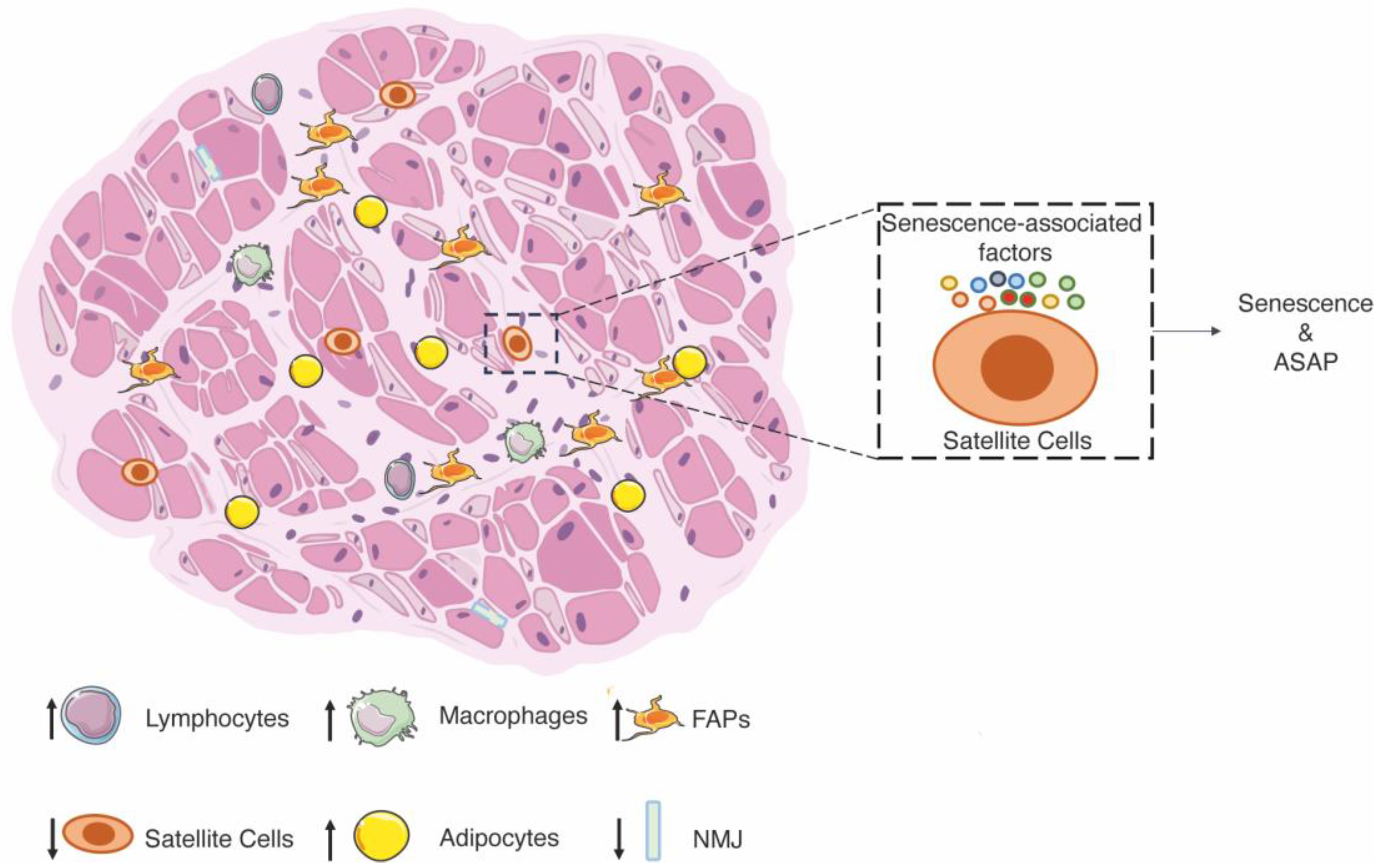
3.1. Age-Related Sarcopenia

In recent decades, the scientific community has begun to use the term “frailty” to refer to a progressive loss of functions, resulting in an inability to respond to physical or psychosocial stress [72,73,74]. One of the critical mechanisms of frailty is age-related sarcopenia, characterized by low muscle mass, low physical function (e.g., gait speed), and low muscle strength (e.g., grip strength) due to no other specific causes [75]. Possible explanations for the decline in muscle quality include (i) infiltration of fat in the muscles or myosteatosis [76], (ii) altered deposition of collagen and other non-contractile tissue in the muscles [77], and (iii) progressive loss of individual muscle fibers [6], including a decrease in the proportion of fast-twitch type II muscle fibers, while the size of type I muscle fibers remain largely unaffected during aging [78]. Finally, sarcopenia is associated with a reduced number of SCs, especially in type II fibers (fast), which is considered a major cause of physical disability and loss of independence in the geriatric population of humans and mice [79,80,81]. Indeed, if the number of SCs decreases, fewer stem cells are responsive in the tissue to activate the regeneration process and repair muscle injuries after trauma. In particular, Pax7+ SCs decrease during aging as they cycle more frequently during homeostasis and spend less time in a quiescent state, decreasing their ability to self-renew [82,83,84,85]. In this context, basal macroautophagy is a key system for maintaining organelle and protein homeostasis [86]. This activity gradually decreases during aging, leading to the accumulation of toxic cellular debris and dysfunctions [87]. In geriatric mice, the decrease in quiescent SCs has been associated with increased expression of the master regulator of senescence p16, leading to impairment of the regenerative apparatus [85]. An increase in p16 has been associated with a decrease in AUF1, an RNA-binding protein/cytokine mRNA destabilizing protein, which regulates several aging-associated pathways [88]. Analysis of EDL myofibers shows a significant age effect on the number of SCs per myofiber, with an age-related shift towards myofibers with fewer SCs [83]. The number of SCs per myofiber has been described to be significantly higher in two younger groups of mice (i.e., 3–4 and 7–10 months), but the median value of SCs per myofiber declined nearly to that of senile mice by 1 year of age (i.e., in the 11–13 months group) [83]. To confirm this aspect, aged GFP-SCs were transplanted into adult mice, and the SCs pool showed a decrease of 60% compared to young cells, suggesting age-dependent cellular dysfunction [80,84]. On the other hand, transplanting young SCs into the muscle of progeroid mice prolongs lifespan and ameliorates the alterations observed in skeletal muscle [89]. However, the reduced number of SCs is not the only cause of the decline in skeletal muscle regeneration with age (Figure 1).
In addition to the cellular dysfunctions of aged SCs, the muscle environment also modulates the activity of SCs. To this purpose, hetero-parabiosis and hetero-transplantation analyses have shown that the decline in muscle regeneration during aging can be partially reversed if aged SCs are transplanted into a youthful external environment [90]. This finding suggests that the dysfunctions of progenitor cell activity may be modulated by systemic factors that change with age. In this line, aged SCs show significant changes in TGF-β signaling, resulting in an increase in fibrotic phenotype and a decrease in myogenic differentiation through various mechanisms, including suppression of MYOD transcription [91]. Myostatin, also known as growth and differentiation factor 8 (GDF8), belongs to the TGF-β superfamily and inhibits muscle growth/development. Notably, myostatin serum levels increase with age, and muscle mass is inversely correlated with myostatin serum levels. This suggests a link between myostatin and age-related sarcopenia and opens up the possibility of developing therapeutic strategies to stimulate muscle growth and prevent muscle wasting [92]. In contrast, the pathway of the insulin-like growth factor 1 (IGF-1), which plays a central role in muscle growth, differentiation, and regeneration, is lower in the serum of aged individuals with poorer muscle strength, walking speed, mobility tasks, and various physical performances [93]. Indeed, overexpression of the IGF-1 isoform in muscle led to muscle hypertrophy in adulthood and guaranteed the maintenance of muscle mass and functionality during aging and in animal models of neuromuscular diseases [94]. Finally, aging is generally associated with a chronic state of slightly elevated plasma levels of pro-inflammatory effectors [95,96]. This condition is often referred to as low-grade inflammation (LGI), characterized by pro-inflammatory cytokines (TNF-α, IL-6, and NF-κB overactivation), which impair the functions of SCs and the efficacy of the regeneration process [97]. The alterations in the secretion of systemic factors may help to develop preventive approaches to monitor the progression of sarcopenia and intervene promptly.
In addition to changes in the microenvironment, another critical element responsible for SCs’ fate is the structural component of the stem cell niche. Stearns-Reider KM et al. have shown that aging is associated with increased muscle stiffness due to a pathogenic extracellular matrix (ECM) architecture, resulting in altered myogenic progression of SCs and affecting their behavior [98]. In particular, the niches of SCs are dramatically altered by abnormal deposition of ECM, leading to changes in the dynamics of cell support, resulting in a loss of function of SCs and, consequently, a decline in the regenerative capacity of skeletal muscle tissue [99]. In this context, some ECM components, including laminin, biglycan, and testican-1, are expressed at higher levels in old muscles than in young muscles under homeostatic conditions [99]. Moreover, lower fibronectin levels were found in old tissues than in young ones after injury [99]. The different effects on ECM composition could be due to the localization of individual components in the niches of SCs. Interestingly, the disruptions in the ECM converge on a few significant axes of intercellular communication, including integrin signaling, confirming previous findings demonstrating dysfunctional Itgb1 signaling in aging SCs [100]. The results showed that several signaling pathways are altered in aged skeletal muscle, leading to increased ligands and cell surface receptors on SCs [101].
FAPs in skeletal muscle tissue produce most of the ECM proteins, which show age-related differences. Since the number of FAPs does not increase with age, several groups have speculated that the gene expression programs of FAPs change, resulting in altered abundance of specific ECM molecules [53,101]. In particular, it has recently been shown that the ECM molecule Wisp1 is significantly reduced in the aged ECM of skeletal muscle and that this decrease is due to changes in aged FAPs [53]. At the same time, other mechanisms that are not dependent on transcriptional changes, such as altered protein turnover due to reduced degradation or increased protein synthesis and secretion, could also lead to protein accumulation in the ECM of aged skeletal muscle. In addition to the role that FAPs play in altering the composition of the extracellular matrix, their differentiation into adipocytes is enhanced, leading to the accumulation of ectopic fat in the interstitial space of muscle tissue, which contributes to the deterioration of muscle function with age. In particular, FAPs have been shown to respond to a gastric inhibitory polypeptide (GIP), a 42-amino-acid hormone released in response to the ingestion of nutrients, including fat or glucose. The inhibition of this metabolism reduces the accumulation of the intramuscular adipose tissue and improves sarcopenia [102] (Figure 1).
Finally, the inability to regenerate in a sarcopenic phenotype has been linked to the deterioration of neuromuscular junctions (NMJs), leading to chronic denervation. Presynaptic nerve terminals from mice at 29 months of age were disorganized and exhibited extensive sprouting compared to mice at 3 months of age. Postsynaptic endplates appeared diffuse, irregular, and with granular fragmentation [103]. The persistent changes in NMJs in aged mice reflect an alteration of the SCs pool, leading to impaired regenerative machinery. In particular, uncontrolled proliferation of SCs occurs after denervation injury, which could be responsible firstly for the exhaustion and then for the depletion of the SCs pool [104]. Indeed, SCs are reduced by one-third after 7 months of denervation compared to uninjured mice [104]. The reduction of SCs in denervated muscle was directly related to injury due to an increased susceptibility to apoptotic cell death [105] (Figure 1).
Based on these data, it is clear that a tight balance of several players modulates the activity of SCs. If one of them does not correctly work, the whole machinery can be impaired.
In recent years, several clinical trials on the treatment of sarcopenia have shown that exercise is the only effective strategy to counteract sarcopenia [106]. In particular, resistance and eccentric training are recommended as first-line therapy for the treatment of sarcopenia, as they improve muscle mass, strength, and physical performance in older people [107,108,109,110]. The beneficial effects of resistance training on muscle regeneration are associated with an increase in the number of SCs in humans [111]. The same results have been shown in mouse and rat models [112,113]. Specifically, a 13-week moderate-intensity run in rats of both sexes leads to an increase in the number of SCs per myofiber [83]. Exercise increases the number of SCs and enhances their ability to differentiate into structural and functional myotubes in vitro [112]. Current understanding of the factors that activate SCs during exercise is limited. Some results show that SCs activation during exercise is mediated by elements released by myofibers and interstitial cell populations, including IGF-1 and molecules secreted by FAPs supporting SCs in the regeneration process [114,115]. Based on these findings, the World Health Organization and, in particular, the Global Recommendations on Physical Activity for Health, recommend that adults 65 years and older should perform 150 min of moderate or 75 min of vigorous-intensity aerobic activity and two or more days of muscle-strengthening activities per week [116].

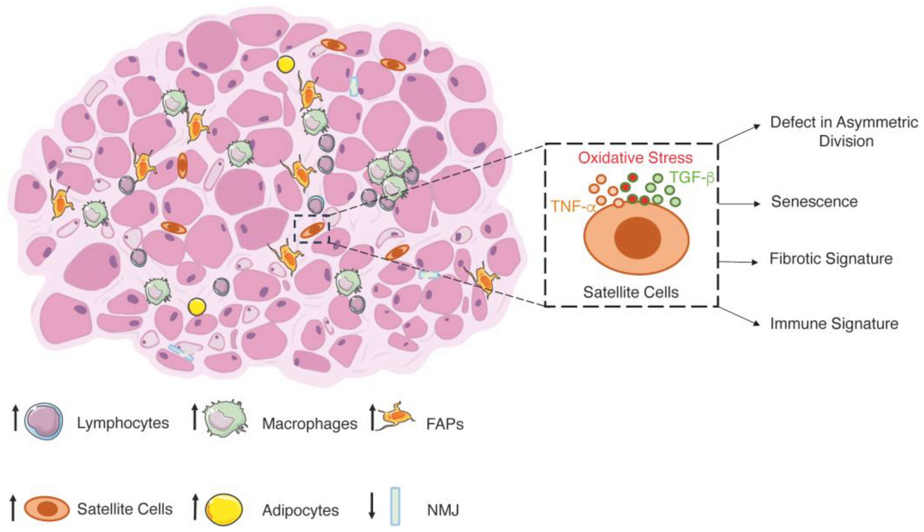
3.2. Duchenne Muscular Dystrophy
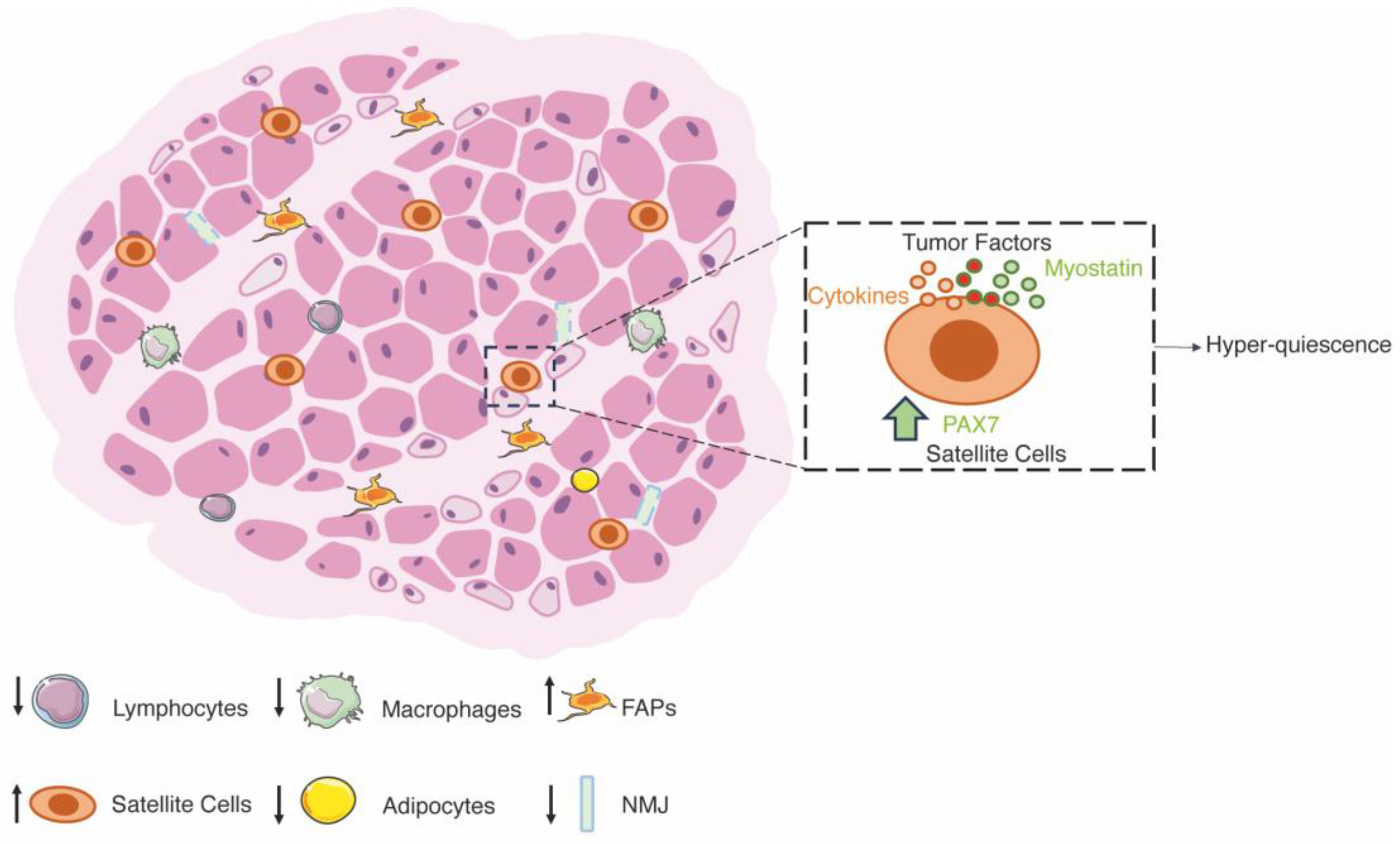
Muscular dystrophies are a heterogeneous family of neuromuscular disorders characterized by muscle wasting. Different dominant and recessive genetic mutations lead to dystrophies with different prognoses and phenotypes [117]. In most diseases, a mutation is present in genes encoding proteins of the dystrophin-associated glycoprotein complex that stabilizes muscle fibers during contraction by acting as a mechanical link between the cytoskeleton and the extracellular matrix [117]. Duchenne muscular dystrophy (DMD) is the most severe and common form of muscular dystrophy, affecting 1 out of 3500 male newborns [118]. The primary cause of the disease is a mutation in the gene encoding for dystrophin, resulting in a dysfunctional or absent protein [119,120]. Dystrophin links actin to the dystrophin-associated protein complex, and its lack leads to tears in the sarcolemma membrane and myofibers damage [121].
In addition to the primary cause, DMD is associated with high levels of oxidative stress and inflammation, which actively exacerbate the dystrophic phenotype. Although oxidative stress and inflammation are crucial for tissue regeneration after initial damage, their constant presence in skeletal muscles leads to further necrosis, injury, and accumulation of fibrosis [122,123,124,125,126,127]. Sustained and excessive infiltration of macrophages, as described in dystrophic muscles, disrupts redox homeostasis and causes muscle damage [128].
Among the causes of muscle wasting in muscular dystrophy, the dysfunctionality of SCs plays a crucial role. Understanding if this dysfunctionality can be counted among the secondary causes or if the dysfunctionality of SCs is strictly dependent on inflammation and oxidative stress, resulting in tertiary causes of DMD, is still unclear.
After an injury, SCs should be activated and differentiated to replenish damaged myofibers. However, the accumulation of fibrotic and adipose tissue may indicate that SCs cannot adequately replace damaged myofibers. The contribution of SCs to the progression of DMD has been extensively investigated. SCs’ dysfunction has been described since the 1980s [12,129], and a widely accepted model is the SC exhaustion caused by repeated cycles of muscle degeneration and regeneration leading to depletion of the regenerative capacity of SCs [130]. However, recent studies have shown that the number of SCs is increased in DMD patients and dystrophin-deficient mdx mice, suggesting that the defective regenerative capacity of dystrophic muscles is not exclusively due to a lack of SCs [131,132,133]. Genome-wide metabolic modeling and functional analyses have demonstrated defects in myoblast commitment. Indeed, 170 genes showed altered expression in mdx myoblasts compared to controls, including MYOD, MYOGENIN, Myomaker, Myomixer, epigenetic regulators, extracellular matrix interactors, calcium signaling, and fibrosis genes [134]. Functionally, increased myoblast proliferation, altered chemotaxis, and accelerated differentiation have been described in myoblasts isolated from dystrophic mice and patients [134]. In vitro differentiation of human pluripotent stem cells lacking dystrophin and DMD myoblasts displayed mis-organization of sarcomeres, contributing to myofibers instability, mis-localization of proteins belonging to the dystrophin-associated complex, myotubes branching, contraction defects and hyperactivation of calcium signaling [135,136,137].
The changes in the phenotype of SCs have been associated with both intrinsic and extrinsic defects. In particular, inflammation and oxidative stress trigger senescence and alter the plasticity of muscle stem cells [138,139,140]. Senescence can also be triggered by several cytokines, including TNF-α and TGF-β, elevated in the skeletal muscle of DMD patients [141]. TGF-β is a key mediator of fibrogenesis and acts as an inhibitor of SCs’ proliferation, activation, and differentiation [142,143]. TNF-α has bimodal effects on myogenesis depending on the concentration. Lower doses (0.05 ng/mL) of TNF-α stimulate myogenesis, while under pathological conditions, TNF-α levels between 0.5 and 5 ng/mL inhibit myogenesis [61].
Moreover, telomere shortening is one of the causes of senescence, and ablation of telomerase in dystrophic mice resulted in a more severe phenotype caused by impaired proliferation and a reduction in the number of SCs [130]. The accumulation of senescent cells expressing CDKN2A, p16, and p19 has been described to contribute to fibrosis and inflammation [144]. Along this line, the depletion of senescent cells by senolytic drugs improves muscle regeneration in dystrophic mice by decreasing the expression of IL-6, TGF-β, and IL-1β [144,145]. Furthermore, preventing senescence increases the regenerative potential of SCs and improves muscle functions [146]. In addition to senescence, altered plasticity of SCs has also been demonstrated over the years. In response to increased TGF-β signaling, myogenic cells adopt fibrogenic properties, with a decrease in PAX7 and MYF5 expression and an increase in fibrogenic genes expression (i.e., collagen I, fibronectin, and α-smooth muscle actin), leading to being more prone to undergo osteogenic or adipogenic transformations and produce extracellular matrix proteins [147,148]. On the same line, the inhibition of the TGF-β signaling pathway improves the expression of PAX7 and MYOD in muscles [147]. Moreover, a recent publication found that a group of muscle stem cells that possess an immune signature (C1qa/b, Lyz2, and Cd53) is present in severe dystrophic muscles [149] (Figure 2). Functional validation must be performed to understand their role in dystrophy. Besides the functions of inflammation and oxidative stress triggering SCs dysfunctions, a recent article has shown that the defects in SCs phenotype are also related to myofiber stability. Indeed, in transplantation experiments, Hicks and collaborators have demonstrated that skeletal muscle progenitor cells (SMPCs) require the support of ACTC1+ fibers to sustain the functional engraftment of SMPCs and to support the stem cell state [150]. Therefore, secondary players of muscular dystrophy trigger senescence and altered plasticity of SCs. Still, the senescence itself can exacerbate the effects of inflammation and oxidative stress, rendering SCs senescence a secondary cause of DMD.
On the other hand, intrinsic defects of SCs are the leading cause of SCs’ dysfunctionality. Dystrophin is expressed in SCs and is associated with Par1b, an important regulator of cell polarity. Its absence in DMD results in a 5-fold reduction in the proportion of asymmetric division, leading to a lower number of myogenic progenitor cells available for efficient muscle regeneration [133]. Intrinsic defects decrease the generation of myogenic progenitor cells required for proper muscle regeneration after tissue damage. The rescue of asymmetric division in dystrophin-deficient satellite stem cells, with EGF treatment, enhances regeneration in skeletal muscle from mdx [151]. This limitation highly impacts muscle regeneration, leading to the identification of intrinsic defects as a secondary cause of DMD.
Besides the destabilization of muscle myofibers due to the destabilization of the dystrophin–glycoprotein complex, dystrophin and the related structural proteins are required for synaptic homeostasis [152]. Abnormal neuromuscular junction (NMJ) morphology has been described in dystrophic patients, and the muscle weakness of mouse models for DMD is associated with changes in NMJ and neuromuscular transmission failure [153,154,155,156,157,158]. However, changes in NMJ morphology result from cycles of degeneration and regeneration of the tissue rather than a secondary cause of muscular dystrophies [158]. Still, this hypothesis remains to be fully elucidated (Figure 2).
Although a consensus has emerged over the years to enhance the functions of SCs and stimulate regeneration in dystrophy to improve the dystrophic phenotype, recent evidence has shown that slowing down the degeneration–regeneration cycles may also help to preserve skeletal muscle in dystrophies [4,126,151,159,160,161,162,163,164]. Despite this debate, the scientific community agrees that skeletal muscle must be preserved by correcting the genetic defect of DMD. Any proposal to restore dystrophin expression by gene editing in vivo or stem cell transplantation requires a synergistic approach to preserve skeletal muscle in order to cure the disease [165,166,167,168].

3.3. Cachexia

Cachexia, from the Greek kakos “bad” and hexis “condition,” is an unintentional body weight loss of more than 5% in the past 6–12 months or the acquirement of a low body mass index that occurs in association with various chronic diseases, including cancer, diabetes, rheumatoid arthritis, kidney disease, heart failure, and chronic infections (such as HIV and tuberculosis) [14,169,170,171]. Cancer cachexia affects 50–85% of cancer patients, depending on the type and stage of cancer: a higher incidence has been described in patients with lung, gastrointestinal, and pancreatic cancers, and overall, most advanced cancer patients suffer from emaciation [172]. In parallel with tumor growth, most organs and muscles experience a decline and loss of functions. This mechanism is driven by systemic inflammation, which is one of the first central players in triggering cachexia [173].
Over the years, a long list of cytokines has been described to increase in the blood of cachectic patients and animal models. These include TNF-α, interleukins (IL-6; IL-1β and IL-1α), interferon-gamma (IFN-γ), leukemia inhibitory factor (LIF), and the TGF-β superfamily have been shown to mediate cancer-induced muscle wasting [174,175,176,177,178]. Cytokines and tumor factors affect different organs and tissues, leading to their wasting and modifying their functionality. In chronological order, after the affection of the immune system, the brain, pancreas, liver, adipose tissue, and gut are severely involved before the skeletal muscles [173]. However, skeletal muscle wasting and dysfunctionality highly impact the quality of life and survival of patients, rendering the wasting of skeletal muscle central in the diagnosis of cachexia.
Over the years, the importance of the imbalance between protein synthesis and degradation has been described as a primary cause for the establishment of cachectic muscles. In this context, the two major pathways controlling protein synthesis are the Akt-mTOR pathway, which acts as a positive regulator of protein synthesis, and the myostatin-Smad2/3 pathway, a negative regulator of the IGF-1/Akt/mTOR pathway [179]. Conversely, the main processes controlling protein degradation are the ubiquitin-proteasome (UPS) and the autophagy-lysosome systems. In cancer cachexia, the loss of skeletal muscle is due to both a downregulation of protein synthesis, mainly due to the decrease in IGF-1 and the increase in myostatin pathway, and an upregulation of protein degradation due to the overexpression of atrogenes and the hyperactivation of the autophagy and proteasome system [180]. Specifically, atrophy occurs in type II fibers with a 40% reduction in diameter, while type I fibers remain largely unchanged [181], similar to fasting [152]. In addition, recent evidence has shown that the bone morphogenetic protein (BMP) pathway in cancer cachexia disrupts neuromuscular junction and denervation, contributing to muscle wasting [182]. IL-6 and Activin A have a synergic role in inducing Noggin overexpression that causes NMJ impairment and atrophy, whereas treatment aimed to increase BMP signaling attenuate muscle atrophy during cancer cachexia [182] (Figure 3).
In addition to the contribution of the balance between protein synthesis and degradation, several articles have demonstrated a central role of impaired regeneration in the establishment of cachexia in skeletal muscle, pointing to new important mechanisms in cachexia. Under atrophic conditions, the muscle membrane is damaged, as evidenced by alteration of the sarcolemma and basal lamina, which can be observed by increased Evans blue dye penetration, decreased expression of extracellular matrix genes, and increased IgG and diffuse laminin staining [181,183]. After the establishment of muscle damage, the regeneration program begins, as evidenced by myofibers with centrally located nuclei, leading to the expression of embryonic MHC and the formation of small muscle fibers [104,184,185]. However, clear evidence of defect of regeneration was demonstrated after acute injury, where regeneration in cachectic muscle is highly impaired, as evidenced by patches of necrotic fibers surrounded by few inflammatory cells [183,186,187]. Moreover, it has been demonstrated that fibrosis and collagen deposition increase in cachectic muscles in patients and mice to replace myofibers, suggesting that the progressive development of fibrosis is a consequence of the expansion and differentiation of FAPs progenitors in cachectic muscles [188,189,190,191]. This is also accompanied by no inflammatory infiltration in the muscle of tumor-bearing mice [192,193]. The infiltration of immune cells is fundamental for the proper timing of regeneration, and its deficiency in cachectic muscles could be one of the causes of poor regeneration [54]. Accordingly, IL-4 administration improves regeneration by impacting SCs’ numbers and functions [194]. The impaired regeneration is due to different cell populations whose functionality is strongly modified by tumor growth (Figure 3).
In addition to the impairment of homeostasis of myofibers, immune cells, and FAPs, attention has also focused on the effects of cachexia on the expression of PAX7. Indeed, results have shown that PAX7 expression is increased 10-fold in cachectic muscles compared to control subjects [183]. Independent of the atrophic injury, an increase in PAX7+ cells is associated with a higher number of SCs [183,195]. Moreover, interstitial localized cells, such as FAPs and pericytes, begin to express PAX7 and contribute to its overexpression [183]. The overexpression of PAX7 and an increase in the number of SCs without an increase in MYOD and MYOGENIN demonstrate the inability of SCs to differentiate into muscle fibers and repair the damage caused by atrophy while promoting self-renewal [183,196,197]. In addition, the accumulation of SCs is central to establishing the atrophic phenotype of muscles, and it is considered to be one of the main leading causes of cachexia [187]. Indeed, the administration of MEK inhibitors attenuates atrophy by restoring the myogenic potential of myoblasts [187].
Next, the question of the mechanism of the SCs’ dysfunction arises. Indeed, the dysfunction of SCs is closely related to the tumor because SCs readily fuse into muscle fibers after resection of the tumor [183]. Moreover, isolated SCs from tumor-bearing mice cultured in a differentiation medium without prior expansion showed a higher fusion index than cells isolated from control mice, suggesting that myogenic cells exposed to cachectic factors retain the ability to differentiate [183]. In vitro studies suggest that tumor factors can induce apoptosis and thus modulate the pool of functional SCs [198,199]. The mechanism by which SCs experience impaired differentiation capacity is due to the overexpression of PAX7 through the activation of the canonical NF-κB pathway [183]. Indeed, overexpression of PAX7 in vivo results in decreased muscle mass and increased muscle wasting in cancer cachexia [183]. Moreover, cachectic muscles depleted of PAX7 retain more muscle mass than control subjects [183]. PAX7 overexpression inhibits differentiation by suppressing MYOD and MYOGENIN, limiting myoblast fusion with damaged myofibers to complete regeneration [196]. Activation of NF-κB in myoblasts has been shown to inhibit MYOD synthesis and upregulate PAX7 [200,201]. In addition, inhibition of NF-κB signaling rescues cancer-related atrophy associated with increased fusion of SCs into muscle fibers [183]. At the same time, selective activation of NF-κB exacerbates muscle mass decline and increases PAX7 expression [183]. However, the mechanism by which NF-κB signaling is activated has not been fully elucidated, as serum from cachectic mice and patients induce PAX7 expression, whereas cytokines (TNF-α, IL-1β, IL-6, and IFN-γ, administered either singly or as a cocktail) or C-26 or LLC conditioned medium do not recapitulate the effect on PAX7 and further analyses are required to address this point. Another signaling pathway that negatively regulates the differentiation of SCs is the myostatin/ActRIIB pathway [183,202]. Blockade of the ActRIIB pathway reverses muscle wasting in tumor-bearing mice by attenuating protein catabolism and stimulating the growth of SCs [202]. In addition, the state of quiescence or hyper-quiescence of SCs could be beneficial to circumvent death. Increased expression of CCAAT/enhancer binding protein beta (C/EBPβ), an inhibitor of myogenesis, was found in SCs from cachectic animals. Genetic depletion of C/EBPβ leads to increased apoptosis and further impairment of regeneration. Taken together, the inhibition of muscle regeneration during cancer cachexia could be the result of tumor factors stimulating muscle wasting and degeneration, but also a mechanism of muscle to maintain viability and avoid death (Figure 3).
Recently, exercise, especially a combination of endurance and resistance training, has been shown to counteract or limit skeletal muscle atrophy in cachectic conditions [203,204,205,206]. The beneficial effects of moderate exercise have been explained by reduced inflammation, oxidative stress, and improved antioxidant capacity [204,207,208]. Moreover, physical training promotes hypertrophy mainly through the activation, proliferation, and differentiation of SCs [209]. In summary, various processes occur in skeletal muscle during cancer cachexia. In addition to modulation of myofibers size, deregulation of SCs functions has been described recently and can be exploited therapeutically. In particular, strategies aimed at restoring regeneration may also prove effective in restoring muscle mass in cancer patients.

4. Summary and Future Prospective

Muscle regeneration is a multi-faceted and well-orchestrated phenomenon in adulthood that regulates skeletal muscle tissue homeostasis and ensures the restoration of mass and function. Under physiological conditions, SCs play an essential role in providing the progeny required for regenerating all damaged tissue by switching from a quiescent to an active state. Each phase of stepwise muscle healing, including the expansion of SCs (PAX7+/MYOD), differentiation into myoblasts (PAX7/MYOD+), and the formation of new fibers, is characterized by the combination of activation and recruitment of cells and molecular signals playing specific roles in the complex framework of regeneration [20]. It is well known that changes in the number and function of SCs affect skeletal muscle regeneration in various pathological conditions. Based on these premises, the identification of new therapeutic approaches that can counteract the loss of SCs and reestablish their function is a strategy for treating various muscle diseases. In particular, a reduced number of SCs has been associated with the aging process leading to sarcopenia [11,83]. Indeed, SCs are lower in older people than in young controls in this condition. However, the effects of sarcopenia are related to the number of SCs and to a change in their function, leading to the wasting of skeletal muscle tissue [91]. The impairment of SCs’ function also plays a crucial role in muscular dystrophy, where the number of SCs seems to increase without compensating for the loss of muscle fibers replaced by an enhancement of adipose and fibrotic tissue deposition [131,132,133]. Along this line, cachectic patients also show an increase in SCs number without an increase in MYOD and MYOGENIN, demonstrating the inability of SCs to differentiate into muscle fibers, leading to skeletal muscle atrophy [183,195].
The alteration in the functions of SCs is mainly due to an increased inflammatory state in skeletal muscle tissue. In particular, the low chronic inflammation during aging induces SCs to move toward a senescence phenotype that increases the secretion of inflammatory cytokines such as TNF-α and IL-6 [95,96]. On the other hand, skeletal muscle dystrophy is characterized by an increased number of inflammatory cells, while cancer cachexia does not affect the cellular component but induces a significant enhancement of secreted cytokines, aggravating the pathological phenotype by increasing autoinflammation and fibrotic tissue deposition [123,124,175,176]. Another factor secreted by inflammatory cells that plays a central role in altering muscle regeneration is TGF-β. In this context, TGF-β promotes fibrogenesis by inhibiting the proliferation, activation, and differentiation of SCs and inducing FAPs cells to adopt a fibrogenic phenotype that promotes the deposition of ECM proteins [91,147,148].
In recent years, skeletal muscle tissue has been described as an autocrine, paracrine, and endocrine organ that regulates the functions of other organs [210,211]. Physical activity, in particular, stimulates this special feature through the release of proteins known as myokines. According to current knowledge, the primary physiological role of myokines is to protect the functionality of skeletal muscles [210,211]. Myokines control adaptive processes in skeletal muscle by acting as paracrine regulators of fuel oxidation, hypertrophy, angiogenesis, inflammatory processes, and regulation of the extracellular matrix [211]. The endocrine functions attributed to myokines regulate body weight, low-grade inflammation, insulin sensitivity, tumor growth suppression, and cognitive function improvement [211]. For these reasons, it is clear that physical activity can counteract muscle wasting in various skeletal muscle diseases. In addition to the classic approaches to reduce muscle atrophy, such as nutritional integration or pharmacological treatment, the development of therapeutic methods based on the identification of individualized exercise programs has become more attractive in the treatment of any pathological condition. In particular, several studies have investigated the effects of different types of exercise on the activation of SCs. In particular, eccentric exercise has been shown to stimulate the activation and proliferation of SCs in the elderly [110]. Along this line, acute activation of SCs after exercise leads to a regenerative response associated with a subsequent return to a quiescent state. This notion is supported by studies showing benefits to overall muscle health in the elderly who perform resistance training that stimulates SCs activation through the release of myokines from myofibers and interstitial cell populations (FAPs), which reduces chronic low-level inflammation and promotes SCs functions [212]. The role of exercise in reducing inflammatory levels and preserving SCs’ function is also critical in other pathological conditions. Indeed, aerobic exercise in muscular dystrophy and moderate training in cachexia counteract the inflammatory state, activating SCs and promoting skeletal muscle hypertrophy [204,207,208,209,213,214].
Currently, the understanding of SCs’ behaviors after exercise is relatively limited. Future studies need to precisely define the mechanisms involved in the changes of skeletal muscle regeneration under physiological and pathological conditions in order to design more appropriate training to counteract skeletal muscle atrophy and any systemic side effects on the organism.

Author Contributions

Conceptualization, F.C., L.M. and G.C.; investigation, F.C., L.M. and G.C.; writing—original draft preparation, F.C. and G.C.; writing—review and editing, F.C., L.M. and G.C.; funding acquisition, F.C. and L.M. All authors have read and agreed to the published version of the manuscript.

Funding

This work was partially supported by Ricerca Corrente funding from the Italian Ministry of Health to IRCCS Policlinico San Donato and to IRCCS Ospedale Galeazzi-Sant’Ambrogio.

Conflicts of Interest

The authors declare no conflict of interest.

References

  1. Bruusgaard, J.C.; Liestol, K.; Ekmark, M.; Kollstad, K.; Gundersen, K. Number and spatial distribution of nuclei in the muscle fibres of normal mice studied in vivo. J. Physiol. 2003, 551, 467–478. [Google Scholar] [CrossRef] [PubMed]
  2. Roman, W.; Pinheiro, H.; Pimentel, M.R.; Segales, J.; Oliveira, L.M.; Garcia-Dominguez, E.; Gomez-Cabrera, M.C.; Serrano, A.L.; Gomes, E.R.; Munoz-Canoves, P. Muscle repair after physiological damage relies on nuclear migration for cellular reconstruction. Science 2021, 374, 355–359. [Google Scholar] [CrossRef] [PubMed]
  3. Mauro, A. Satellite cell of skeletal muscle fibers. J. Biophys. Biochem. Cytol. 1961, 9, 493–495. [Google Scholar] [CrossRef] [PubMed]
  4. Kodippili, K.; Rudnicki, M.A. Satellite cell contribution to disease pathology in Duchenne muscular dystrophy. Front. Physiol. 2023, 14, 1180980. [Google Scholar] [CrossRef] [PubMed]
  5. Wang, Y.X.; Rudnicki, M.A. Satellite cells, the engines of muscle repair. Nat. Rev. Mol. Cell Biol. 2011, 13, 127–133. [Google Scholar] [CrossRef] [PubMed]
  6. Conboy, M.J.; Karasov, A.O.; Rando, T.A. High incidence of non-random template strand segregation and asymmetric fate determination in dividing stem cells and their progeny. PLoS Biol. 2007, 5, e102. [Google Scholar] [CrossRef]
  7. Kuang, S.; Gillespie, M.A.; Rudnicki, M.A. Niche regulation of muscle satellite cell self-renewal and differentiation. Cell Stem Cell 2008, 2, 22–31. [Google Scholar] [CrossRef] [PubMed]
  8. Huo, F.; Liu, Q.; Liu, H. Contribution of muscle satellite cells to sarcopenia. Front. Physiol. 2022, 13, 892749. [Google Scholar] [CrossRef]
  9. Arneson, P.C.; Doles, J.D. Impaired Muscle Regeneration in Cancer-Associated Cachexia. Trends Cancer 2019, 5, 579–582. [Google Scholar] [CrossRef]
  10. Saleh, K.K.; Switzler, C.; Hicks, M.R.; Gane, L.; Gibbs, D.E.; Pyle, A.D. Duchenne muscular dystrophy disease severity impacts skeletal muscle progenitor cells systemic delivery. Front. Physiol. 2023, 14, 1190524. [Google Scholar] [CrossRef]
  11. Verdijk, L.B.; Koopman, R.; Schaart, G.; Meijer, K.; Savelberg, H.H.; van Loon, L.J. Satellite cell content is specifically reduced in type II skeletal muscle fibers in the elderly. Am. J. Physiol.-Endocrinol. Metab. 2007, 292, E151–E157. [Google Scholar] [CrossRef] [PubMed]
  12. Blau, H.M.; Webster, C.; Pavlath, G.K. Defective myoblasts identified in Duchenne muscular dystrophy. Proc. Natl. Acad. Sci. USA 1983, 80, 4856–4860. [Google Scholar] [CrossRef] [PubMed]
  13. Decary, S.; Hamida, C.B.; Mouly, V.; Barbet, J.P.; Hentati, F.; Butler-Browne, G.S. Shorter telomeres in dystrophic muscle consistent with extensive regeneration in young children. Neuromuscul. Disord. 2000, 10, 113–120. [Google Scholar] [CrossRef] [PubMed]
  14. Fearon, K.; Strasser, F.; Anker, S.D.; Bosaeus, I.; Bruera, E.; Fainsinger, R.L.; Jatoi, A.; Loprinzi, C.; MacDonald, N.; Mantovani, G.; et al. Definition and classification of cancer cachexia: An international consensus. Lancet Oncol. 2011, 12, 489–495. [Google Scholar] [CrossRef] [PubMed]
  15. Lepper, C.; Partridge, T.A.; Fan, C.M. An absolute requirement for Pax7-positive satellite cells in acute injury-induced skeletal muscle regeneration. Development 2011, 138, 3639–3646. [Google Scholar] [CrossRef] [PubMed]
  16. Yang, X.; Yang, S.; Wang, C.; Kuang, S. The hypoxia-inducible factors HIF1alpha and HIF2alpha are dispensable for embryonic muscle development but essential for postnatal muscle regeneration. J. Biol. Chem. 2017, 292, 5981–5991. [Google Scholar] [CrossRef] [PubMed]
  17. Huang, L.E.; Arany, Z.; Livingston, D.M.; Bunn, H.F. Activation of hypoxia-inducible transcription factor depends primarily upon redox-sensitive stabilization of its alpha subunit. J. Biol. Chem. 1996, 271, 32253–32259. [Google Scholar] [CrossRef]
  18. Cirillo, F.; Resmini, G.; Angelino, E.; Ferrara, M.; Tarantino, A.; Piccoli, M.; Rota, P.; Ghiroldi, A.; Monasky, M.M.; Ciconte, G.; et al. HIF-1alpha Directly Controls WNT7A Expression During Myogenesis. Front. Cell Dev. Biol. 2020, 8, 593508. [Google Scholar] [CrossRef]
  19. Cirillo, F.; Resmini, G.; Ghiroldi, A.; Piccoli, M.; Bergante, S.; Tettamanti, G.; Anastasia, L. Activation of the hypoxia-inducible factor 1alpha promotes myogenesis through the noncanonical Wnt pathway, leading to hypertrophic myotubes. FASEB J. 2017, 31, 2146–2156. [Google Scholar] [CrossRef]
  20. Von Maltzahn, J.; Bentzinger, C.F.; Rudnicki, M.A. Characteristics of Satellite Cells and Multipotent Adult Stem Cells in the Skeletal Muscle; Hayat, M.A., Ed.; Springer: Dordrecht, The Netherlands, 2013; Volume 12. [Google Scholar]
  21. Bentzinger, C.F.; Wang, Y.X.; Rudnicki, M.A. Building muscle: Molecular regulation of myogenesis. Cold Spring Harb. Perspect. Biol. 2012, 4, a008342. [Google Scholar] [CrossRef]
  22. Relaix, F.; Rocancourt, D.; Mansouri, A.; Buckingham, M. Divergent functions of murine Pax3 and Pax7 in limb muscle development. Genes Dev. 2004, 18, 1088–1105. [Google Scholar] [CrossRef] [PubMed]
  23. Soleimani, V.D.; Punch, V.G.; Kawabe, Y.; Jones, A.E.; Palidwor, G.A.; Porter, C.J.; Cross, J.W.; Carvajal, J.J.; Kockx, C.E.; van IJcken, W.F.; et al. Transcriptional dominance of Pax7 in adult myogenesis is due to high-affinity recognition of homeodomain motifs. Dev. Cell 2012, 22, 1208–1220. [Google Scholar] [CrossRef] [PubMed]
  24. Sincennes, M.C.; Brun, C.E.; Rudnicki, M.A. Concise Review: Epigenetic Regulation of Myogenesis in Health and Disease. STEM Cells Transl. Med. 2016, 5, 282–290. [Google Scholar] [CrossRef] [PubMed]
  25. Sincennes, M.C.; Brun, C.E.; Lin, A.Y.T.; Rosembert, T.; Datzkiw, D.; Saber, J.; Ming, H.; Kawabe, Y.I.; Rudnicki, M.A. Acetylation of PAX7 controls muscle stem cell self-renewal and differentiation potential in mice. Nat. Commun. 2021, 12, 3253. [Google Scholar] [CrossRef]
  26. Rocheteau, P.; Gayraud-Morel, B.; Siegl-Cachedenier, I.; Blasco, M.A.; Tajbakhsh, S. A subpopulation of adult skeletal muscle stem cells retains all template DNA strands after cell division. Cell 2012, 148, 112–125. [Google Scholar] [CrossRef]
  27. Shinin, V.; Gayraud-Morel, B.; Tajbakhsh, S. Template DNA-strand co-segregation and asymmetric cell division in skeletal muscle stem cells. Methods Mol. Biol. 2009, 482, 295–317. [Google Scholar] [CrossRef]
  28. Shinin, V.; Gayraud-Morel, B.; Gomes, D.; Tajbakhsh, S. Asymmetric division and cosegregation of template DNA strands in adult muscle satellite cells. Nat. Cell Biol. 2006, 8, 677–687. [Google Scholar] [CrossRef]
  29. Peng, J.; Han, L.; Liu, B.; Song, J.; Wang, Y.; Wang, K.; Guo, Q.; Liu, X.; Li, Y.; Zhang, J.; et al. Gli1 marks a sentinel muscle stem cell population for muscle regeneration. Nat. Commun. 2023, 14, 6993. [Google Scholar] [CrossRef]
  30. Palla, A.R.; Hilgendorf, K.I.; Yang, A.V.; Kerr, J.P.; Hinken, A.C.; Demeter, J.; Kraft, P.; Mooney, N.A.; Yucel, N.; Burns, D.M.; et al. Primary cilia on muscle stem cells are critical to maintain regenerative capacity and are lost during aging. Nat. Commun. 2022, 13, 1439. [Google Scholar] [CrossRef]
  31. Brun, C.E.; Sincennes, M.C.; Lin, A.Y.T.; Hall, D.; Jarassier, W.; Feige, P.; Le Grand, F.; Rudnicki, M.A. GLI3 regulates muscle stem cell entry into G(Alert) and self-renewal. Nat. Commun. 2022, 13, 3961. [Google Scholar] [CrossRef]
  32. Singh, K.; Dilworth, F.J. Differential modulation of cell cycle progression distinguishes members of the myogenic regulatory factor family of transcription factors. FEBS J. 2013, 280, 3991–4003. [Google Scholar] [CrossRef] [PubMed]
  33. Chang, N.C.; Rudnicki, M.A. Satellite cells: The architects of skeletal muscle. Curr. Top. Dev. Biol. 2014, 107, 161–181. [Google Scholar] [CrossRef] [PubMed]
  34. Megeney, L.A.; Kablar, B.; Garrett, K.; Anderson, J.E.; Rudnicki, M.A. MyoD is required for myogenic stem cell function in adult skeletal muscle. Genes Dev. 1996, 10, 1173–1183. [Google Scholar] [CrossRef] [PubMed]
  35. Yablonka-Reuveni, Z.; Rudnicki, M.A.; Rivera, A.J.; Primig, M.; Anderson, J.E.; Natanson, P. The transition from proliferation to differentiation is delayed in satellite cells from mice lacking MyoD. Dev. Biol. 1999, 210, 440–455. [Google Scholar] [CrossRef] [PubMed]
  36. White, J.D.; Scaffidi, A.; Davies, M.; McGeachie, J.; Rudnicki, M.A.; Grounds, M.D. Myotube formation is delayed but not prevented in MyoD-deficient skeletal muscle: Studies in regenerating whole muscle grafts of adult mice. J. Histochem. Cytochem. 2000, 48, 1531–1544. [Google Scholar] [CrossRef]
  37. Sabourin, L.A.; Girgis-Gabardo, A.; Seale, P.; Asakura, A.; Rudnicki, M.A. Reduced differentiation potential of primary MyoD-/- myogenic cells derived from adult skeletal muscle. J. Cell Biol. 1999, 144, 631–643. [Google Scholar] [CrossRef]
  38. Fujita, R.; Mizuno, S.; Sadahiro, T.; Hayashi, T.; Sugasawa, T.; Sugiyama, F.; Ono, Y.; Takahashi, S.; Ieda, M. Generation of a MyoD knock-in reporter mouse line to study muscle stem cell dynamics and heterogeneity. iScience 2023, 26, 106592. [Google Scholar] [CrossRef]
  39. Weintraub, H.; Dwarki, V.J.; Verma, I.; Davis, R.; Hollenberg, S.; Snider, L.; Lassar, A.; Tapscott, S.J. Muscle-specific transcriptional activation by MyoD. Genes Dev. 1991, 5, 1377–1386. [Google Scholar] [CrossRef]
  40. Weintraub, H.; Davis, R.; Tapscott, S.; Thayer, M.; Krause, M.; Benezra, R.; Blackwell, T.K.; Turner, D.; Rupp, R.; Hollenberg, S.; et al. The myoD gene family: Nodal point during specification of the muscle cell lineage. Science 1991, 251, 761–766. [Google Scholar] [CrossRef]
  41. Tapscott, S.J.; Weintraub, H. MyoD and the regulation of myogenesis by helix-loop-helix proteins. J. Clin. Investig. 1991, 87, 1133–1138. [Google Scholar] [CrossRef]
  42. Zhang, K.; Sha, J.; Harter, M.L. Activation of Cdc6 by MyoD is associated with the expansion of quiescent myogenic satellite cells. J. Cell Biol. 2010, 188, 39–48. [Google Scholar] [CrossRef] [PubMed]
  43. Relaix, F.; Zammit, P.S. Satellite cells are essential for skeletal muscle regeneration: The cell on the edge returns centre stage. Development 2012, 139, 2845–2856. [Google Scholar] [CrossRef] [PubMed]
  44. Yan, Z.; Choi, S.; Liu, X.; Zhang, M.; Schageman, J.J.; Lee, S.Y.; Hart, R.; Lin, L.; Thurmond, F.A.; Williams, R.S. Highly coordinated gene regulation in mouse skeletal muscle regeneration. J. Biol. Chem. 2003, 278, 8826–8836. [Google Scholar] [CrossRef] [PubMed]
  45. Musarò, A. The Basis of Muscle Regeneration. Adv. Biol. 2014, 2014, 16. [Google Scholar] [CrossRef]
  46. Hernandez-Hernandez, J.M.; Garcia-Gonzalez, E.G.; Brun, C.E.; Rudnicki, M.A. The myogenic regulatory factors, determinants of muscle development, cell identity and regeneration. Semin. Cell Dev. Biol. 2017, 72, 10–18. [Google Scholar] [CrossRef] [PubMed]
  47. Schiaffino, S.; Reggiani, C. Myosin isoforms in mammalian skeletal muscle. J. Appl. Physiol. 1994, 77, 493–501. [Google Scholar] [CrossRef] [PubMed]
  48. Larsson, L.; Edstrom, L.; Lindegren, B.; Gorza, L.; Schiaffino, S. MHC composition and enzyme-histochemical and physiological properties of a novel fast-twitch motor unit type. Am. J. Physiol. 1991, 261, C93–C101. [Google Scholar] [CrossRef]
  49. Jakubiec-Puka, A.; Kordowska, J.; Catani, C.; Carraro, U. Myosin heavy chain isoform composition in striated muscle after denervation and self-reinnervation. Eur. J. Biochem. 1990, 193, 623–628. [Google Scholar] [CrossRef]
  50. Joe, A.W.; Yi, L.; Natarajan, A.; Le Grand, F.; So, L.; Wang, J.; Rudnicki, M.A.; Rossi, F.M. Muscle injury activates resident fibro/adipogenic progenitors that facilitate myogenesis. Nat. Cell Biol. 2010, 12, 153–163. [Google Scholar] [CrossRef]
  51. Mann, C.J.; Perdiguero, E.; Kharraz, Y.; Aguilar, S.; Pessina, P.; Serrano, A.L.; Munoz-Canoves, P. Aberrant repair and fibrosis development in skeletal muscle. Skelet. Muscle 2011, 1, 21. [Google Scholar] [CrossRef]
  52. Wosczyna, M.N.; Konishi, C.T.; Perez Carbajal, E.E.; Wang, T.T.; Walsh, R.A.; Gan, Q.; Wagner, M.W.; Rando, T.A. Mesenchymal Stromal Cells Are Required for Regeneration and Homeostatic Maintenance of Skeletal Muscle. Cell Rep. 2019, 27, 2029–2035.e2025. [Google Scholar] [CrossRef] [PubMed]
  53. Lukjanenko, L.; Karaz, S.; Stuelsatz, P.; Gurriaran-Rodriguez, U.; Michaud, J.; Dammone, G.; Sizzano, F.; Mashinchian, O.; Ancel, S.; Migliavacca, E.; et al. Aging Disrupts Muscle Stem Cell Function by Impairing Matricellular WISP1 Secretion from Fibro-Adipogenic Progenitors. Cell Stem Cell 2019, 24, 433–446.e437. [Google Scholar] [CrossRef] [PubMed]
  54. Tu, H.; Li, Y.L. Inflammation balance in skeletal muscle damage and repair. Front. Immunol. 2023, 14, 1133355. [Google Scholar] [CrossRef] [PubMed]
  55. Tarban, N.; Papp, A.B.; Deak, D.; Szentesi, P.; Halasz, H.; Patsalos, A.; Csernoch, L.; Sarang, Z.; Szondy, Z. Loss of adenosine A3 receptors accelerates skeletal muscle regeneration in mice following cardiotoxin-induced injury. Cell Death Dis. 2023, 14, 706. [Google Scholar] [CrossRef] [PubMed]
  56. Biswas, S.K.; Mantovani, A. Macrophage plasticity and interaction with lymphocyte subsets: Cancer as a paradigm. Nat. Immunol. 2010, 11, 889–896. [Google Scholar] [CrossRef] [PubMed]
  57. Mantovani, A.; Sica, A.; Sozzani, S.; Allavena, P.; Vecchi, A.; Locati, M. The chemokine system in diverse forms of macrophage activation and polarization. Trends Immunol. 2004, 25, 677–686. [Google Scholar] [CrossRef] [PubMed]
  58. Miyazaki, A.; Kawashima, M.; Nagata, I.; Miyoshi, M.; Miyakawa, M.; Sugiyama, M.; Sakuraya, T.; Sonomura, T.; Arakawa, T. Icing after skeletal muscle injury decreases M1 macrophage accumulation and TNF-alpha expression during the early phase of muscle regeneration in rats. Histochem. Cell Biol. 2023, 159, 77–89. [Google Scholar] [CrossRef]
  59. Zhang, C.; Li, Y.; Wu, Y.; Wang, L.; Wang, X.; Du, J. Interleukin-6/signal transducer and activator of transcription 3 (STAT3) pathway is essential for macrophage infiltration and myoblast proliferation during muscle regeneration. J. Biol. Chem. 2013, 288, 1489–1499. [Google Scholar] [CrossRef]
  60. Peterson, J.M.; Bakkar, N.; Guttridge, D.C. NF-kappaB signaling in skeletal muscle health and disease. Curr. Top. Dev. Biol. 2011, 96, 85–119. [Google Scholar] [CrossRef]
  61. Chen, S.E.; Jin, B.; Li, Y.P. TNF-alpha regulates myogenesis and muscle regeneration by activating p38 MAPK. Am. J. Physiol.-Cell Physiol. 2007, 292, C1660–C1671. [Google Scholar] [CrossRef]
  62. Zhan, M.; Jin, B.; Chen, S.E.; Reecy, J.M.; Li, Y.P. TACE release of TNF-alpha mediates mechanotransduction-induced activation of p38 MAPK and myogenesis. J. Cell Sci. 2007, 120, 692–701. [Google Scholar] [CrossRef] [PubMed]
  63. Munoz-Canoves, P.; Scheele, C.; Pedersen, B.K.; Serrano, A.L. Interleukin-6 myokine signaling in skeletal muscle: A double-edged sword? FEBS J. 2013, 280, 4131–4148. [Google Scholar] [CrossRef] [PubMed]
  64. Fu, X.; Xiao, J.; Wei, Y.; Li, S.; Liu, Y.; Yin, J.; Sun, K.; Sun, H.; Wang, H.; Zhang, Z.; et al. Combination of inflammation-related cytokines promotes long-term muscle stem cell expansion. Cell Res. 2015, 25, 655–673. [Google Scholar] [CrossRef] [PubMed]
  65. Stout, R.D.; Suttles, J. Functional plasticity of macrophages: Reversible adaptation to changing microenvironments. J. Leukoc. Biol. 2004, 76, 509–513. [Google Scholar] [CrossRef] [PubMed]
  66. Biswas, S.K.; Gangi, L.; Paul, S.; Schioppa, T.; Saccani, A.; Sironi, M.; Bottazzi, B.; Doni, A.; Vincenzo, B.; Pasqualini, F.; et al. A distinct and unique transcriptional program expressed by tumor-associated macrophages (defective NF-kappaB and enhanced IRF-3/STAT1 activation). Blood 2006, 107, 2112–2122. [Google Scholar] [CrossRef]
  67. Fontenot, J.D.; Gavin, M.A.; Rudensky, A.Y. Foxp3 programs the development and function of CD4+CD25+ regulatory T cells. Nat. Immunol. 2003, 4, 330–336. [Google Scholar] [CrossRef] [PubMed]
  68. Villalta, S.A.; Rinaldi, C.; Deng, B.; Liu, G.; Fedor, B.; Tidball, J.G. Interleukin-10 reduces the pathology of mdx muscular dystrophy by deactivating M1 macrophages and modulating macrophage phenotype. Hum. Mol. Genet. 2011, 20, 790–805. [Google Scholar] [CrossRef]
  69. Arnold, L.; Henry, A.; Poron, F.; Baba-Amer, Y.; van Rooijen, N.; Plonquet, A.; Gherardi, R.K.; Chazaud, B. Inflammatory monocytes recruited after skeletal muscle injury switch into antiinflammatory macrophages to support myogenesis. J. Exp. Med. 2007, 204, 1057–1069. [Google Scholar] [CrossRef]
  70. Ruffell, D.; Mourkioti, F.; Gambardella, A.; Kirstetter, P.; Lopez, R.G.; Rosenthal, N.; Nerlov, C. A CREB-C/EBPbeta cascade induces M2 macrophage-specific gene expression and promotes muscle injury repair. Proc. Natl. Acad. Sci. USA 2009, 106, 17475–17480. [Google Scholar] [CrossRef]
  71. Tidball, J.G.; Wehling-Henricks, M. Macrophages promote muscle membrane repair and muscle fibre growth and regeneration during modified muscle loading in mice in vivo. J. Physiol. 2007, 578, 327–336. [Google Scholar] [CrossRef]
  72. Fried, L.P.; Tangen, C.M.; Walston, J.; Newman, A.B.; Hirsch, C.; Gottdiener, J.; Seeman, T.; Tracy, R.; Kop, W.J.; Burke, G.; et al. Frailty in older adults: Evidence for a phenotype. J. Gerontol. Ser. A Biol. Sci. Med. Sci. 2001, 56, M146–M156. [Google Scholar] [CrossRef] [PubMed]
  73. Clegg, A.; Young, J.; Iliffe, S.; Rikkert, M.O.; Rockwood, K. Frailty in elderly people. Lancet 2013, 381, 752–762. [Google Scholar] [CrossRef] [PubMed]
  74. Sukkriang, N.; Punsawad, C. Comparison of geriatric assessment tools for frailty among community elderly. Heliyon 2020, 6, e04797. [Google Scholar] [CrossRef] [PubMed]
  75. Cruz-Jentoft, A.J.; Bahat, G.; Bauer, J.; Boirie, Y.; Bruyere, O.; Cederholm, T.; Cooper, C.; Landi, F.; Rolland, Y.; Sayer, A.A.; et al. Sarcopenia: Revised European consensus on definition and diagnosis. Age Ageing 2019, 48, 16–31. [Google Scholar] [CrossRef] [PubMed]
  76. Correa-de-Araujo, R.; Addison, O.; Miljkovic, I.; Goodpaster, B.H.; Bergman, B.C.; Clark, R.V.; Elena, J.W.; Esser, K.A.; Ferrucci, L.; Harris-Love, M.O.; et al. Myosteatosis in the Context of Skeletal Muscle Function Deficit: An Interdisciplinary Workshop at the National Institute on Aging. Front. Physiol. 2020, 11, 963. [Google Scholar] [CrossRef] [PubMed]
  77. Zullo, A.; Fleckenstein, J.; Schleip, R.; Hoppe, K.; Wearing, S.; Klingler, W. Structural and Functional Changes in the Coupling of Fascial Tissue, Skeletal Muscle, and Nerves During Aging. Front. Physiol. 2020, 11, 592. [Google Scholar] [CrossRef] [PubMed]
  78. Akasaki, Y.; Ouchi, N.; Izumiya, Y.; Bernardo, B.L.; Lebrasseur, N.K.; Walsh, K. Glycolytic fast-twitch muscle fiber restoration counters adverse age-related changes in body composition and metabolism. Aging Cell 2014, 13, 80–91. [Google Scholar] [CrossRef]
  79. Roth, S.M.; Martel, G.F.; Ivey, F.M.; Lemmer, J.T.; Metter, E.J.; Hurley, B.F.; Rogers, M.A. Skeletal muscle satellite cell populations in healthy young and older men and women. Anat. Rec. 2000, 260, 351–358. [Google Scholar] [CrossRef]
  80. Day, K.; Shefer, G.; Shearer, A.; Yablonka-Reuveni, Z. The depletion of skeletal muscle satellite cells with age is concomitant with reduced capacity of single progenitors to produce reserve progeny. Dev. Biol. 2010, 340, 330–343. [Google Scholar] [CrossRef]
  81. Renault, V.; Thornell, L.E.; Eriksson, P.O.; Butler-Browne, G.; Mouly, V. Regenerative potential of human skeletal muscle during aging. Aging Cell 2002, 1, 132–139. [Google Scholar] [CrossRef]
  82. Conboy, I.M.; Conboy, M.J.; Smythe, G.M.; Rando, T.A. Notch-mediated restoration of regenerative potential to aged muscle. Science 2003, 302, 1575–1577. [Google Scholar] [CrossRef] [PubMed]
  83. Shefer, G.; Van de Mark, D.P.; Richardson, J.B.; Yablonka-Reuveni, Z. Satellite-cell pool size does matter: Defining the myogenic potency of aging skeletal muscle. Dev. Biol. 2006, 294, 50–66. [Google Scholar] [CrossRef] [PubMed]
  84. Chakkalakal, J.V.; Jones, K.M.; Basson, M.A.; Brack, A.S. The aged niche disrupts muscle stem cell quiescence. Nature 2012, 490, 355–360. [Google Scholar] [CrossRef] [PubMed]
  85. Sousa-Victor, P.; Gutarra, S.; Garcia-Prat, L.; Rodriguez-Ubreva, J.; Ortet, L.; Ruiz-Bonilla, V.; Jardi, M.; Ballestar, E.; Gonzalez, S.; Serrano, A.L.; et al. Geriatric muscle stem cells switch reversible quiescence into senescence. Nature 2014, 506, 316–321. [Google Scholar] [CrossRef] [PubMed]
  86. Cuervo, A.M.; Bergamini, E.; Brunk, U.T.; Droge, W.; Ffrench, M.; Terman, A. Autophagy and aging: The importance of maintaining “clean” cells. Autophagy 2005, 1, 131–140. [Google Scholar] [CrossRef] [PubMed]
  87. Garcia-Prat, L.; Martinez-Vicente, M.; Perdiguero, E.; Ortet, L.; Rodriguez-Ubreva, J.; Rebollo, E.; Ruiz-Bonilla, V.; Gutarra, S.; Ballestar, E.; Serrano, A.L.; et al. Autophagy maintains stemness by preventing senescence. Nature 2016, 529, 37–42. [Google Scholar] [CrossRef]
  88. Pont, A.R.; Sadri, N.; Hsiao, S.J.; Smith, S.; Schneider, R.J. mRNA decay factor AUF1 maintains normal aging, telomere maintenance, and suppression of senescence by activation of telomerase transcription. Mol. Cell 2012, 47, 5–15. [Google Scholar] [CrossRef]
  89. Lavasani, M.; Robinson, A.R.; Lu, A.; Song, M.; Feduska, J.M.; Ahani, B.; Tilstra, J.S.; Feldman, C.H.; Robbins, P.D.; Niedernhofer, L.J.; et al. Muscle-derived stem/progenitor cell dysfunction limits healthspan and lifespan in a murine progeria model. Nat. Commun. 2012, 3, 608. [Google Scholar] [CrossRef]
  90. Conboy, I.M.; Conboy, M.J.; Wagers, A.J.; Girma, E.R.; Weissman, I.L.; Rando, T.A. Rejuvenation of aged progenitor cells by exposure to a young systemic environment. Nature 2005, 433, 760–764. [Google Scholar] [CrossRef]
  91. Beggs, M.L.; Nagarajan, R.; Taylor-Jones, J.M.; Nolen, G.; Macnicol, M.; Peterson, C.A. Alterations in the TGFbeta signaling pathway in myogenic progenitors with age. Aging Cell 2004, 3, 353–361. [Google Scholar] [CrossRef]
  92. Yarasheski, K.E.; Bhasin, S.; Sinha-Hikim, I.; Pak-Loduca, J.; Gonzalez-Cadavid, N.F. Serum myostatin-immunoreactive protein is increased in 60-92 year old women and men with muscle wasting. J. Nutr. Health Aging 2002, 6, 343–348. [Google Scholar] [PubMed]
  93. Scicchitano, B.M.; Rizzuto, E.; Musaro, A. Counteracting muscle wasting in aging and neuromuscular diseases: The critical role of IGF-1. Aging 2009, 1, 451–457. [Google Scholar] [CrossRef] [PubMed]
  94. Barton-Davis, E.R.; Shoturma, D.I.; Musaro, A.; Rosenthal, N.; Sweeney, H.L. Viral mediated expression of insulin-like growth factor I blocks the aging-related loss of skeletal muscle function. Proc. Natl. Acad. Sci. USA 1998, 95, 15603–15607. [Google Scholar] [CrossRef] [PubMed]
  95. Bruunsgaard, H.; Skinhoj, P.; Pedersen, A.N.; Schroll, M.; Pedersen, B.K. Ageing, tumour necrosis factor-alpha (TNF-alpha) and atherosclerosis. Clin. Exp. Immunol. 2000, 121, 255–260. [Google Scholar] [CrossRef]
  96. Harris, T.B.; Ferrucci, L.; Tracy, R.P.; Corti, M.C.; Wacholder, S.; Ettinger, W.H., Jr.; Heimovitz, H.; Cohen, H.J.; Wallace, R. Associations of elevated interleukin-6 and C-reactive protein levels with mortality in the elderly. Am. J. Med. 1999, 106, 506–512. [Google Scholar] [CrossRef] [PubMed]
  97. Tchkonia, T.; Zhu, Y.; van Deursen, J.; Campisi, J.; Kirkland, J.L. Cellular senescence and the senescent secretory phenotype: Therapeutic opportunities. J. Clin. Investig. 2013, 123, 966–972. [Google Scholar] [CrossRef] [PubMed]
  98. Stearns-Reider, K.M.; D’Amore, A.; Beezhold, K.; Rothrauff, B.; Cavalli, L.; Wagner, W.R.; Vorp, D.A.; Tsamis, A.; Shinde, S.; Zhang, C.; et al. Aging of the skeletal muscle extracellular matrix drives a stem cell fibrogenic conversion. Aging Cell 2017, 16, 518–528. [Google Scholar] [CrossRef]
  99. Lukjanenko, L.; Jung, M.J.; Hegde, N.; Perruisseau-Carrier, C.; Migliavacca, E.; Rozo, M.; Karaz, S.; Jacot, G.; Schmidt, M.; Li, L.; et al. Loss of fibronectin from the aged stem cell niche affects the regenerative capacity of skeletal muscle in mice. Nat. Med. 2016, 22, 897–905. [Google Scholar] [CrossRef]
  100. Rozo, M.; Li, L.; Fan, C.M. Targeting beta1-integrin signaling enhances regeneration in aged and dystrophic muscle in mice. Nat. Med. 2016, 22, 889–896. [Google Scholar] [CrossRef]
  101. Schuler, S.C.; Kirkpatrick, J.M.; Schmidt, M.; Santinha, D.; Koch, P.; Di Sanzo, S.; Cirri, E.; Hemberg, M.; Ori, A.; von Maltzahn, J. Extensive remodeling of the extracellular matrix during aging contributes to age-dependent impairments of muscle stem cell functionality. Cell Rep. 2021, 35, 109223. [Google Scholar] [CrossRef]
  102. Takahashi, Y.; Fujita, H.; Seino, Y.; Hattori, S.; Hidaka, S.; Miyakawa, T.; Suzuki, A.; Waki, H.; Yabe, D.; Seino, Y.; et al. Gastric inhibitory polypeptide receptor antagonism suppresses intramuscular adipose tissue accumulation and ameliorates sarcopenia. J. Cachexia Sarcopenia Muscle 2023, 14, 2703–2718. [Google Scholar] [CrossRef] [PubMed]
  103. Chai, R.J.; Vukovic, J.; Dunlop, S.; Grounds, M.D.; Shavlakadze, T. Striking denervation of neuromuscular junctions without lumbar motoneuron loss in geriatric mouse muscle. PLoS ONE 2011, 6, e28090. [Google Scholar] [CrossRef] [PubMed]
  104. Rodrigues Ade, C.; Schmalbruch, H. Satellite cells and myonuclei in long-term denervated rat muscles. Anat. Rec. 1995, 243, 430–437. [Google Scholar] [CrossRef] [PubMed]
  105. Jejurikar, S.S.; Marcelo, C.L.; Kuzon, W.M., Jr. Skeletal muscle denervation increases satellite cell susceptibility to apoptosis. Plast. Reconstr. Surg. 2002, 110, 160–168. [Google Scholar] [CrossRef] [PubMed]
  106. Tezze, C.; Sandri, M.; Tessari, P. Anabolic Resistance in the Pathogenesis of Sarcopenia in the Elderly: Role of Nutrition and Exercise in Young and Old People. Nutrients 2023, 15, 4073. [Google Scholar] [CrossRef] [PubMed]
  107. Dent, E.; Morley, J.E.; Cruz-Jentoft, A.J.; Arai, H.; Kritchevsky, S.B.; Guralnik, J.; Bauer, J.M.; Pahor, M.; Clark, B.C.; Cesari, M.; et al. International Clinical Practice Guidelines for Sarcopenia (ICFSR): Screening, Diagnosis and Management. J. Nutr. Health Aging 2018, 22, 1148–1161. [Google Scholar] [CrossRef]
  108. Borde, R.; Hortobagyi, T.; Granacher, U. Dose-Response Relationships of Resistance Training in Healthy Old Adults: A Systematic Review and Meta-Analysis. Sports Med. 2015, 45, 1693–1720. [Google Scholar] [CrossRef]
  109. Yoshimura, Y.; Wakabayashi, H.; Yamada, M.; Kim, H.; Harada, A.; Arai, H. Interventions for Treating Sarcopenia: A Systematic Review and Meta-Analysis of Randomized Controlled Studies. J. Am. Med. Dir. Assoc. 2017, 18, 553.e1–553.e16. [Google Scholar] [CrossRef]
  110. Darr, K.C.; Schultz, E. Exercise-induced satellite cell activation in growing and mature skeletal muscle. J. Appl. Physiol. 1987, 63, 1816–1821. [Google Scholar] [CrossRef]
  111. Walker, D.K.; Fry, C.S.; Drummond, M.J.; Dickinson, J.M.; Timmerman, K.L.; Gundermann, D.M.; Jennings, K.; Volpi, E.; Rasmussen, B.B. PAX7+ satellite cells in young and older adults following resistance exercise. Muscle Nerve 2012, 46, 51–59. [Google Scholar] [CrossRef]
  112. Cisterna, B.; Giagnacovo, M.; Costanzo, M.; Fattoretti, P.; Zancanaro, C.; Pellicciari, C.; Malatesta, M. Adapted physical exercise enhances activation and differentiation potential of satellite cells in the skeletal muscle of old mice. J. Anat. 2016, 228, 771–783. [Google Scholar] [CrossRef] [PubMed]
  113. Shefer, G.; Rauner, G.; Yablonka-Reuveni, Z.; Benayahu, D. Reduced satellite cell numbers and myogenic capacity in aging can be alleviated by endurance exercise. PLoS ONE 2010, 5, e13307. [Google Scholar] [CrossRef] [PubMed]
  114. Lee, S.; Barton, E.R.; Sweeney, H.L.; Farrar, R.P. Viral expression of insulin-like growth factor-I enhances muscle hypertrophy in resistance-trained rats. J. Appl. Physiol. 2004, 96, 1097–1104. [Google Scholar] [CrossRef] [PubMed]
  115. Serrano, A.L.; Baeza-Raja, B.; Perdiguero, E.; Jardi, M.; Munoz-Canoves, P. Interleukin-6 is an essential regulator of satellite cell-mediated skeletal muscle hypertrophy. Cell Metab. 2008, 7, 33–44. [Google Scholar] [CrossRef] [PubMed]
  116. World Health Organization. WHO Guidelines on Physical Activity and Sedentary Behaviour; World Health Organization: Geneva, Switzerland, 2020. [Google Scholar]
  117. Carter, J.C.; Sheehan, D.W.; Prochoroff, A.; Birnkrant, D.J. Muscular Dystrophies. Clin. Chest Med. 2018, 39, 377–389. [Google Scholar] [CrossRef]
  118. Flanigan, K.M. Duchenne and Becker muscular dystrophies. Neurol. Clin. 2014, 32, 671–688. [Google Scholar] [CrossRef]
  119. Hoffman, E.P.; Monaco, A.P.; Feener, C.C.; Kunkel, L.M. Conservation of the Duchenne muscular dystrophy gene in mice and humans. Science 1987, 238, 347–350. [Google Scholar] [CrossRef]
  120. Koenig, M.; Hoffman, E.P.; Bertelson, C.J.; Monaco, A.P.; Feener, C.; Kunkel, L.M. Complete cloning of the Duchenne muscular dystrophy (DMD) cDNA and preliminary genomic organization of the DMD gene in normal and affected individuals. Cell 1987, 50, 509–517. [Google Scholar] [CrossRef]
  121. Constantin, B. Dystrophin complex functions as a scaffold for signalling proteins. Biochim. Biophys. Acta 2014, 1838, 635–642. [Google Scholar] [CrossRef]
  122. Kozakowska, M.; Pietraszek-Gremplewicz, K.; Jozkowicz, A.; Dulak, J. The role of oxidative stress in skeletal muscle injury and regeneration: Focus on antioxidant enzymes. J. Muscle Res. Cell Motil. 2015, 36, 377–393. [Google Scholar] [CrossRef]
  123. Tulangekar, A.; Sztal, T.E. Inflammation in Duchenne Muscular Dystrophy-Exploring the Role of Neutrophils in Muscle Damage and Regeneration. Biomedicines 2021, 9, 1366. [Google Scholar] [CrossRef] [PubMed]
  124. Farini, A.; Villa, C.; Tripodi, L.; Legato, M.; Torrente, Y. Role of Immunoglobulins in Muscular Dystrophies and Inflammatory Myopathies. Front. Immunol. 2021, 12, 666879. [Google Scholar] [CrossRef] [PubMed]
  125. Renjini, R.; Gayathri, N.; Nalini, A.; Srinivas Bharath, M.M. Oxidative damage in muscular dystrophy correlates with the severity of the pathology: Role of glutathione metabolism. Neurochem. Res. 2012, 37, 885–898. [Google Scholar] [CrossRef] [PubMed]
  126. Careccia, G.; Saclier, M.; Tirone, M.; Ruggieri, E.; Principi, E.; Raffaghello, L.; Torchio, S.; Recchia, D.; Canepari, M.; Gorzanelli, A.; et al. Rebalancing expression of HMGB1 redox isoforms to counteract muscular dystrophy. Sci. Transl. Med. 2021, 13, eaay8416. [Google Scholar] [CrossRef] [PubMed]
  127. Pelosi, L.; Forcina, L.; Nicoletti, C.; Scicchitano, B.M.; Musaro, A. Increased Circulating Levels of Interleukin-6 Induce Perturbation in Redox-Regulated Signaling Cascades in Muscle of Dystrophic Mice. Oxidative Med. Cell. Longev. 2017, 2017, 1987218. [Google Scholar] [CrossRef] [PubMed]
  128. Kumar, A.; Boriek, A.M. Mechanical stress activates the nuclear factor-kappaB pathway in skeletal muscle fibers: A possible role in Duchenne muscular dystrophy. FASEB J. 2003, 17, 386–396. [Google Scholar] [CrossRef] [PubMed]
  129. Webster, C.; Blau, H.M. Accelerated age-related decline in replicative life-span of Duchenne muscular dystrophy myoblasts: Implications for cell and gene therapy. Somat. Cell Mol. Genet. 1990, 16, 557–565. [Google Scholar] [CrossRef]
  130. Sacco, A.; Mourkioti, F.; Tran, R.; Choi, J.; Llewellyn, M.; Kraft, P.; Shkreli, M.; Delp, S.; Pomerantz, J.H.; Artandi, S.E.; et al. Short telomeres and stem cell exhaustion model Duchenne muscular dystrophy in mdx/mTR mice. Cell 2010, 143, 1059–1071. [Google Scholar] [CrossRef]
  131. Kottlors, M.; Kirschner, J. Elevated satellite cell number in Duchenne muscular dystrophy. Cell Tissue Res. 2010, 340, 541–548. [Google Scholar] [CrossRef]
  132. Bankole, L.C.; Feasson, L.; Ponsot, E.; Kadi, F. Fibre type-specific satellite cell content in two models of muscle disease. Histopathology 2013, 63, 826–832. [Google Scholar] [CrossRef]
  133. Dumont, N.A.; Wang, Y.X.; von Maltzahn, J.; Pasut, A.; Bentzinger, C.F.; Brun, C.E.; Rudnicki, M.A. Dystrophin expression in muscle stem cells regulates their polarity and asymmetric division. Nat. Med. 2015, 21, 1455–1463. [Google Scholar] [CrossRef] [PubMed]
  134. Gosselin, M.R.F.; Mournetas, V.; Borczyk, M.; Verma, S.; Occhipinti, A.; Rog, J.; Bozycki, L.; Korostynski, M.; Robson, S.C.; Angione, C.; et al. Loss of full-length dystrophin expression results in major cell-autonomous abnormalities in proliferating myoblasts. Elife 2022, 11, e75521. [Google Scholar] [CrossRef] [PubMed]
  135. Barthelemy, F.; Santoso, J.W.; Rabichow, L.; Jin, R.; Little, I.; Nelson, S.F.; McCain, M.L.; Miceli, M.C. Modeling Patient-Specific Muscular Dystrophy Phenotypes and Therapeutic Responses in Reprogrammed Myotubes Engineered on Micromolded Gelatin Hydrogels. Front. Cell Dev. Biol. 2022, 10, 830415. [Google Scholar] [CrossRef] [PubMed]
  136. Shoji, E.; Sakurai, H.; Nishino, T.; Nakahata, T.; Heike, T.; Awaya, T.; Fujii, N.; Manabe, Y.; Matsuo, M.; Sehara-Fujisawa, A. Early pathogenesis of Duchenne muscular dystrophy modelled in patient-derived human induced pluripotent stem cells. Sci. Rep. 2015, 5, 12831. [Google Scholar] [CrossRef] [PubMed]
  137. Al Tanoury, Z.; Zimmerman, J.F.; Rao, J.; Sieiro, D.; McNamara, H.M.; Cherrier, T.; Rodriguez-delaRosa, A.; Hick-Colin, A.; Bousson, F.; Fugier-Schmucker, C.; et al. Prednisolone rescues Duchenne muscular dystrophy phenotypes in human pluripotent stem cell-derived skeletal muscle in vitro. Proc. Natl. Acad. Sci. USA 2021, 118, e2022960118. [Google Scholar] [CrossRef] [PubMed]
  138. Hernandez-Segura, A.; Nehme, J.; Demaria, M. Hallmarks of Cellular Senescence. Trends Cell Biol. 2018, 28, 436–453. [Google Scholar] [CrossRef]
  139. Braumuller, H.; Wieder, T.; Brenner, E.; Assmann, S.; Hahn, M.; Alkhaled, M.; Schilbach, K.; Essmann, F.; Kneilling, M.; Griessinger, C.; et al. T-helper-1-cell cytokines drive cancer into senescence. Nature 2013, 494, 361–365. [Google Scholar] [CrossRef]
  140. Reimann, M.; Lee, S.; Loddenkemper, C.; Dorr, J.R.; Tabor, V.; Aichele, P.; Stein, H.; Dorken, B.; Jenuwein, T.; Schmitt, C.A. Tumor stroma-derived TGF-beta limits myc-driven lymphomagenesis via Suv39h1-dependent senescence. Cancer Cell 2010, 17, 262–272. [Google Scholar] [CrossRef]
  141. Song, Y.; Yao, S.; Liu, Y.; Long, L.; Yang, H.; Li, Q.; Liang, J.; Li, X.; Lu, Y.; Zhu, H.; et al. Expression levels of TGF-beta1 and CTGF are associated with the severity of Duchenne muscular dystrophy. Exp. Ther. Med. 2017, 13, 1209–1214. [Google Scholar] [CrossRef]
  142. Allen, R.E.; Boxhorn, L.K. Inhibition of skeletal muscle satellite cell differentiation by transforming growth factor-beta. J. Cell. Physiol. 1987, 133, 567–572. [Google Scholar] [CrossRef]
  143. Carlson, M.E.; Hsu, M.; Conboy, I.M. Imbalance between pSmad3 and Notch induces CDK inhibitors in old muscle stem cells. Nature 2008, 454, 528–532. [Google Scholar] [CrossRef] [PubMed]
  144. Sugihara, H.; Teramoto, N.; Nakamura, K.; Shiga, T.; Shirakawa, T.; Matsuo, M.; Ogasawara, M.; Nishino, I.; Matsuwaki, T.; Nishihara, M.; et al. Cellular senescence-mediated exacerbation of Duchenne muscular dystrophy. Sci. Rep. 2020, 10, 16385. [Google Scholar] [CrossRef] [PubMed]
  145. Moiseeva, V.; Cisneros, A.; Sica, V.; Deryagin, O.; Lai, Y.; Jung, S.; Andres, E.; An, J.; Segales, J.; Ortet, L.; et al. Senescence atlas reveals an aged-like inflamed niche that blunts muscle regeneration. Nature 2023, 613, 169–178. [Google Scholar] [CrossRef] [PubMed]
  146. Taglietti, V.; Kefi, K.; Rivera, L.; Bergiers, O.; Cardone, N.; Coulpier, F.; Gioftsidi, S.; Drayton-Libotte, B.; Hou, C.; Authier, F.J.; et al. Thyroid-stimulating hormone receptor signaling restores skeletal muscle stem cell regeneration in rats with muscular dystrophy. Sci. Transl. Med. 2023, 15, eadd5275. [Google Scholar] [CrossRef] [PubMed]
  147. Biressi, S.; Miyabara, E.H.; Gopinath, S.D.; Carlig, P.M.; Rando, T.A. A Wnt-TGFbeta2 axis induces a fibrogenic program in muscle stem cells from dystrophic mice. Sci. Transl. Med. 2014, 6, 267ra176. [Google Scholar] [CrossRef] [PubMed]
  148. Pessina, P.; Kharraz, Y.; Jardi, M.; Fukada, S.; Serrano, A.L.; Perdiguero, E.; Munoz-Canoves, P. Fibrogenic Cell Plasticity Blunts Tissue Regeneration and Aggravates Muscular Dystrophy. Stem Cell Rep. 2015, 4, 1046–1060. [Google Scholar] [CrossRef] [PubMed]
  149. Saleh, K.K.; Xi, H.; Switzler, C.; Skuratovsky, E.; Romero, M.A.; Chien, P.; Gibbs, D.; Gane, L.; Hicks, M.R.; Spencer, M.J.; et al. Single cell sequencing maps skeletal muscle cellular diversity as disease severity increases in dystrophic mouse models. iScience 2022, 25, 105415. [Google Scholar] [CrossRef]
  150. Hicks, M.R.; Saleh, K.K.; Clock, B.; Gibbs, D.E.; Yang, M.; Younesi, S.; Gane, L.; Gutierrez-Garcia, V.; Xi, H.; Pyle, A.D. Regenerating human skeletal muscle forms an emerging niche in vivo to support PAX7 cells. Nat. Cell Biol. 2023, 25, 1758–1773. [Google Scholar] [CrossRef]
  151. Wang, Y.X.; Feige, P.; Brun, C.E.; Hekmatnejad, B.; Dumont, N.A.; Renaud, J.M.; Faulkes, S.; Guindon, D.E.; Rudnicki, M.A. EGFR-Aurka Signaling Rescues Polarity and Regeneration Defects in Dystrophin-Deficient Muscle Stem Cells by Increasing Asymmetric Divisions. Cell Stem Cell 2019, 24, 419–432.e416. [Google Scholar] [CrossRef]
  152. Ieko, T.; Fujiki, J.; Hasegawa, Y.; Iwasaki, T.; Iwano, H.; Maeda, N. Mechanism of skeletal muscle atrophy by muscle fiber types in male rats under long-term fasting stress. Steroids 2023, 200, 109328. [Google Scholar] [CrossRef]
  153. Priez, A.; Duchene, J.; Goubel, F. Duchenne muscular dystrophy quantification: A multivariate analysis of surface EMG. Med. Biol. Eng. Comput. 1992, 30, 283–291. [Google Scholar] [CrossRef] [PubMed]
  154. Theroux, M.C.; Olivant, A.; Akins, R.E. C Histomorphology of neuromuscular junction in Duchenne muscular dystrophy. Pediatr. Anesth. 2008, 18, 256–259. [Google Scholar] [CrossRef] [PubMed]
  155. Banks, G.B.; Chamberlain, J.S.; Froehner, S.C. Truncated dystrophins can influence neuromuscular synapse structure. Mol. Cell. Neurosci. 2009, 40, 433–441. [Google Scholar] [CrossRef] [PubMed]
  156. Pisani, C.; Strimpakos, G.; Gabanella, F.; Di Certo, M.G.; Onori, A.; Severini, C.; Luvisetto, S.; Farioli-Vecchioli, S.; Carrozzo, I.; Esposito, A.; et al. Utrophin up-regulation by artificial transcription factors induces muscle rescue and impacts the neuromuscular junction in mdx mice. Biochim. Biophys. Acta Mol. Basis Dis. 2018, 1864, 1172–1182. [Google Scholar] [CrossRef] [PubMed]
  157. Pratt, S.J.P.; Shah, S.B.; Ward, C.W.; Inacio, M.P.; Stains, J.P.; Lovering, R.M. Effects of in vivo injury on the neuromuscular junction in healthy and dystrophic muscles. J. Physiol. 2013, 591, 559–570. [Google Scholar] [CrossRef]
  158. Pratt, S.J.P.; Shah, S.B.; Ward, C.W.; Kerr, J.P.; Stains, J.P.; Lovering, R.M. Recovery of altered neuromuscular junction morphology and muscle function in mdx mice after injury. Cell. Mol. Life Sci. 2015, 72, 153–164. [Google Scholar] [CrossRef] [PubMed]
  159. Hindi, S.M.; Kumar, A. TRAF6 regulates satellite stem cell self-renewal and function during regenerative myogenesis. J. Clin. Investig. 2016, 126, 151–168. [Google Scholar] [CrossRef] [PubMed]
  160. Reano, S.; Angelino, E.; Ferrara, M.; Malacarne, V.; Sustova, H.; Sabry, O.; Agosti, E.; Clerici, S.; Ruozi, G.; Zentilin, L.; et al. Unacylated Ghrelin Enhances Satellite Cell Function and Relieves the Dystrophic Phenotype in Duchenne Muscular Dystrophy mdx Model. Stem Cells 2017, 35, 1733–1746. [Google Scholar] [CrossRef]
  161. Consalvi, S.; Saccone, V.; Giordani, L.; Minetti, G.; Mozzetta, C.; Puri, P.L. Histone deacetylase inhibitors in the treatment of muscular dystrophies: Epigenetic drugs for genetic diseases. Mol. Med. 2011, 17, 457–465. [Google Scholar] [CrossRef]
  162. Pelosi, L.; Berardinelli, M.G.; De Pasquale, L.; Nicoletti, C.; D’Amico, A.; Carvello, F.; Moneta, G.M.; Catizone, A.; Bertini, E.; De Benedetti, F.; et al. Functional and Morphological Improvement of Dystrophic Muscle by Interleukin 6 Receptor Blockade. EBioMedicine 2015, 2, 285–293. [Google Scholar] [CrossRef]
  163. Boyer, J.G.; Huo, J.; Han, S.; Havens, J.R.; Prasad, V.; Lin, B.L.; Kass, D.A.; Song, T.; Sadayappan, S.; Khairallah, R.J.; et al. Depletion of skeletal muscle satellite cells attenuates pathology in muscular dystrophy. Nat. Commun. 2022, 13, 2940. [Google Scholar] [CrossRef] [PubMed]
  164. Rossi, G.; Bonfanti, C.; Antonini, S.; Bastoni, M.; Monteverde, S.; Innocenzi, A.; Saclier, M.; Taglietti, V.; Messina, G. Silencing Nfix rescues muscular dystrophy by delaying muscle regeneration. Nat. Commun. 2017, 8, 1055. [Google Scholar] [CrossRef]
  165. Chen, G.; Wei, T.; Yang, H.; Li, G.; Li, H. CRISPR-Based Therapeutic Gene Editing for Duchenne Muscular Dystrophy: Advances, Challenges and Perspectives. Cells 2022, 11, 2964. [Google Scholar] [CrossRef] [PubMed]
  166. Kwon, J.B.; Ettyreddy, A.R.; Vankara, A.; Bohning, J.D.; Devlin, G.; Hauschka, S.D.; Asokan, A.; Gersbach, C.A. In Vivo Gene Editing of Muscle Stem Cells with Adeno-Associated Viral Vectors in a Mouse Model of Duchenne Muscular Dystrophy. Mol. Ther. Methods Clin. Dev. 2020, 19, 320–329. [Google Scholar] [CrossRef] [PubMed]
  167. Nance, M.E.; Shi, R.; Hakim, C.H.; Wasala, N.B.; Yue, Y.; Pan, X.; Zhang, T.; Robinson, C.A.; Duan, S.X.; Yao, G.; et al. AAV9 Edits Muscle Stem Cells in Normal and Dystrophic Adult Mice. Mol. Ther. 2019, 27, 1568–1585. [Google Scholar] [CrossRef] [PubMed]
  168. Domenig, S.A.; Bundschuh, N.; Lenardic, A.; Ghosh, A.; Kim, I.; Qabrati, X.; D’Hulst, G.; Bar-Nur, O. CRISPR/Cas9 editing of directly reprogrammed myogenic progenitors restores dystrophin expression in a mouse model of muscular dystrophy. Stem Cell Rep. 2022, 17, 321–336. [Google Scholar] [CrossRef]
  169. Baracos, V.E.; Martin, L.; Korc, M.; Guttridge, D.C.; Fearon, K.C.H. Cancer-associated cachexia. Nat. Rev. Dis. Primers 2018, 4, 17105. [Google Scholar] [CrossRef]
  170. Keithley, J.K.; Swanson, B. HIV-associated wasting. J. Assoc. Nurses AIDS Care 2013, 24, S103–S111. [Google Scholar] [CrossRef]
  171. Cheung, W.W.; Paik, K.H.; Mak, R.H. Inflammation and cachexia in chronic kidney disease. Pediatr. Nephrol. 2010, 25, 711–724. [Google Scholar] [CrossRef]
  172. Biswas, A.K.; Acharyya, S. Cancer-Associated Cachexia: A Systemic Consequence of Cancer Progression. Annu. Rev. Cancer Biol. 2020, 4, 391–411. [Google Scholar] [CrossRef]
  173. Ferrara, M.; Samaden, M.; Ruggieri, E.; Venereau, E. Cancer cachexia as a multiorgan failure: Reconstruction of the crime scene. Front. Cell Dev. Biol. 2022, 10, 960341. [Google Scholar] [CrossRef] [PubMed]
  174. Tracey, K.J.; Lowry, S.F.; Cerami, A. Cachectin: A hormone that triggers acute shock and chronic cachexia. J. Infect. Dis. 1988, 157, 413–420. [Google Scholar] [CrossRef] [PubMed]
  175. Hayden, M.S.; Ghosh, S. Regulation of NF-kappaB by TNF family cytokines. Semin. Immunol. 2014, 26, 253–266. [Google Scholar] [CrossRef] [PubMed]
  176. Narsale, A.A.; Carson, J.A. Role of interleukin-6 in cachexia: Therapeutic implications. Curr. Opin. Support. Palliat. Care 2014, 8, 321–327. [Google Scholar] [CrossRef] [PubMed]
  177. Zhang, D.; Zheng, H.; Zhou, Y.; Tang, X.; Yu, B.; Li, J. Association of IL-1beta gene polymorphism with cachexia from locally advanced gastric cancer. BMC Cancer 2007, 7, 45. [Google Scholar] [CrossRef] [PubMed]
  178. Costelli, P.; Llovera, M.; Carbo, N.; Garcia-Martinez, C.; Lopez-Sorianoq, F.J.; Argiles, J.M. Interleukin-1 receptor antagonist (IL-1ra) is unable to reverse cachexia in rats bearing an ascites hepatoma (Yoshida AH-130). Cancer Lett. 1995, 95, 33–38. [Google Scholar] [CrossRef] [PubMed]
  179. Murgia, M.; Serrano, A.L.; Calabria, E.; Pallafacchina, G.; Lomo, T.; Schiaffino, S. Ras is involved in nerve-activity-dependent regulation of muscle genes. Nat. Cell Biol. 2000, 2, 142–147. [Google Scholar] [CrossRef]
  180. Schiaffino, S.; Dyar, K.A.; Ciciliot, S.; Blaauw, B.; Sandri, M. Mechanisms regulating skeletal muscle growth and atrophy. FEBS J. 2013, 280, 4294–4314. [Google Scholar] [CrossRef]
  181. Acharyya, S.; Butchbach, M.E.; Sahenk, Z.; Wang, H.; Saji, M.; Carathers, M.; Ringel, M.D.; Skipworth, R.J.; Fearon, K.C.; Hollingsworth, M.A.; et al. Dystrophin glycoprotein complex dysfunction: A regulatory link between muscular dystrophy and cancer cachexia. Cancer Cell 2005, 8, 421–432. [Google Scholar] [CrossRef]
  182. Sartori, R.; Hagg, A.; Zampieri, S.; Armani, A.; Winbanks, C.E.; Viana, L.R.; Haidar, M.; Watt, K.I.; Qian, H.; Pezzini, C.; et al. Perturbed BMP signaling and denervation promote muscle wasting in cancer cachexia. Sci. Transl. Med. 2021, 13, eaay9592. [Google Scholar] [CrossRef]
  183. He, W.A.; Berardi, E.; Cardillo, V.M.; Acharyya, S.; Aulino, P.; Thomas-Ahner, J.; Wang, J.; Bloomston, M.; Muscarella, P.; Nau, P.; et al. NF-kappaB-mediated Pax7 dysregulation in the muscle microenvironment promotes cancer cachexia. J. Clin. Investig. 2013, 123, 4821–4835. [Google Scholar] [CrossRef] [PubMed]
  184. Talbert, E.E.; Metzger, G.A.; He, W.A.; Guttridge, D.C. Modeling human cancer cachexia in colon 26 tumor-bearing adult mice. J. Cachexia Sarcopenia Muscle 2014, 5, 321–328. [Google Scholar] [CrossRef] [PubMed]
  185. Borisov, A.B.; Dedkov, E.I.; Carlson, B.M. Differentiation of activated satellite cells in denervated muscle following single fusions in situ and in cell culture. Histochem. Cell Biol. 2005, 124, 13–23. [Google Scholar] [CrossRef] [PubMed]
  186. Ramamoorthy, S.; Donohue, M.; Buck, M. Decreased Jun-D and myogenin expression in muscle wasting of human cachexia. Am. J. Physiol.-Endocrinol. Metab. 2009, 297, E392–E401. [Google Scholar] [CrossRef] [PubMed]
  187. Penna, F.; Costamagna, D.; Fanzani, A.; Bonelli, G.; Baccino, F.M.; Costelli, P. Muscle wasting and impaired myogenesis in tumor bearing mice are prevented by ERK inhibition. PLoS ONE 2010, 5, e13604. [Google Scholar] [CrossRef] [PubMed]
  188. Judge, S.M.; Nosacka, R.L.; Delitto, D.; Gerber, M.H.; Cameron, M.E.; Trevino, J.G.; Judge, A.R. Skeletal Muscle Fibrosis in Pancreatic Cancer Patients with Respect to Survival. JNCI Cancer Spectr. 2018, 2, pky043. [Google Scholar] [CrossRef] [PubMed]
  189. Banduseela, V.; Ochala, J.; Lamberg, K.; Kalimo, H.; Larsson, L. Muscle paralysis and myosin loss in a patient with cancer cachexia. Acta Myol. 2007, 26, 136–144. [Google Scholar]
  190. Mu, X.; Agarwal, R.; March, D.; Rothenberg, A.; Voigt, C.; Tebbets, J.; Huard, J.; Weiss, K. Notch Signaling Mediates Skeletal Muscle Atrophy in Cancer Cachexia Caused by Osteosarcoma. Sarcoma 2016, 2016, 3758162. [Google Scholar] [CrossRef]
  191. Nosacka, R.L.; Delitto, A.E.; Delitto, D.; Patel, R.; Judge, S.M.; Trevino, J.G.; Judge, A.R. Distinct cachexia profiles in response to human pancreatic tumours in mouse limb and respiratory muscle. J. Cachexia Sarcopenia Muscle 2020, 11, 820–837. [Google Scholar] [CrossRef]
  192. Berardi, E.; Aulino, P.; Murfuni, I.; Toschi, A.; Padula, F.; Scicchitano, B.M.; Coletti, D.; Adamo, S. Skeletal muscle is enriched in hematopoietic stem cells and not inflammatory cells in cachectic mice. Neurol. Res. 2008, 30, 160–169. [Google Scholar] [CrossRef]
  193. Inaba, S.; Hinohara, A.; Tachibana, M.; Tsujikawa, K.; Fukada, S.I. Muscle regeneration is disrupted by cancer cachexia without loss of muscle stem cell potential. PLoS ONE 2018, 13, e0205467. [Google Scholar] [CrossRef] [PubMed]
  194. Costamagna, D.; Duelen, R.; Penna, F.; Neumann, D.; Costelli, P.; Sampaolesi, M. Interleukin-4 administration improves muscle function, adult myogenesis, and lifespan of colon carcinoma-bearing mice. J. Cachexia Sarcopenia Muscle 2020, 11, 783–801. [Google Scholar] [CrossRef] [PubMed]
  195. Ferreira, R.; Neuparth, M.J.; Ascensao, A.; Magalhaes, J.; Vitorino, R.; Duarte, J.A.; Amado, F. Skeletal muscle atrophy increases cell proliferation in mice gastrocnemius during the first week of hindlimb suspension. Eur. J. Appl. Physiol. 2006, 97, 340–346. [Google Scholar] [CrossRef] [PubMed]
  196. Olguin, H.C.; Olwin, B.B. Pax-7 up-regulation inhibits myogenesis and cell cycle progression in satellite cells: A potential mechanism for self-renewal. Dev. Biol. 2004, 275, 375–388. [Google Scholar] [CrossRef] [PubMed]
  197. Olguin, H.C.; Pisconti, A. Marking the tempo for myogenesis: Pax7 and the regulation of muscle stem cell fate decisions. J. Cell. Mol. Med. 2012, 16, 1013–1025. [Google Scholar] [CrossRef]
  198. Jejurikar, S.S.; Kuzon, W.M., Jr. Satellite cell depletion in degenerative skeletal muscle. Apoptosis 2003, 8, 573–578. [Google Scholar] [CrossRef]
  199. He, W.A.; Calore, F.; Londhe, P.; Canella, A.; Guttridge, D.C.; Croce, C.M. Microvesicles containing miRNAs promote muscle cell death in cancer cachexia via TLR7. Proc. Natl. Acad. Sci. USA 2014, 111, 4525–4529. [Google Scholar] [CrossRef]
  200. Guttridge, D.C.; Mayo, M.W.; Madrid, L.V.; Wang, C.Y.; Baldwin, A.S., Jr. NF-kappaB-induced loss of MyoD messenger RNA: Possible role in muscle decay and cachexia. Science 2000, 289, 2363–2366. [Google Scholar] [CrossRef]
  201. Tisdale, M.J. Mechanisms of cancer cachexia. Physiol. Rev. 2009, 89, 381–410. [Google Scholar] [CrossRef]
  202. Zhou, X.; Wang, J.L.; Lu, J.; Song, Y.; Kwak, K.S.; Jiao, Q.; Rosenfeld, R.; Chen, Q.; Boone, T.; Simonet, W.S.; et al. Reversal of cancer cachexia and muscle wasting by ActRIIB antagonism leads to prolonged survival. Cell 2010, 142, 531–543. [Google Scholar] [CrossRef]
  203. Ballaro, R.; Penna, F.; Pin, F.; Gomez-Cabrera, M.C.; Vina, J.; Costelli, P. Moderate Exercise Improves Experimental Cancer Cachexia by Modulating the Redox Homeostasis. Cancers 2019, 11, 285. [Google Scholar] [CrossRef] [PubMed]
  204. Tsitkanou, S.; Murach, K.A.; Washington, T.A.; Greene, N.P. Exercise Counteracts the Deleterious Effects of Cancer Cachexia. Cancers 2022, 14, 2512. [Google Scholar] [CrossRef] [PubMed]
  205. Montero-Bullon, J.F.; Melo, T.; Ferreira, R.; Padrao, A.I.; Oliveira, P.A.; Domingues, M.R.M.; Domingues, P. Exercise training counteracts urothelial carcinoma-induced alterations in skeletal muscle mitochondria phospholipidome in an animal model. Sci. Rep. 2019, 9, 13423. [Google Scholar] [CrossRef] [PubMed]
  206. Vina, J.; Sanchis-Gomar, F.; Martinez-Bello, V.; Gomez-Cabrera, M.C. Exercise acts as a drug; the pharmacological benefits of exercise. Br. J. Pharmacol. 2012, 167, 1–12. [Google Scholar] [CrossRef] [PubMed]
  207. Murphy, E.A.; Davis, J.M.; Barrilleaux, T.L.; McClellan, J.L.; Steiner, J.L.; Carmichael, M.D.; Pena, M.M.; Hebert, J.R.; Green, J.E. Benefits of exercise training on breast cancer progression and inflammation in C3(1)SV40Tag mice. Cytokine 2011, 55, 274–279. [Google Scholar] [CrossRef] [PubMed]
  208. Mader, T.; Chaillou, T.; Alves, E.S.; Jude, B.; Cheng, A.J.; Kenne, E.; Mijwel, S.; Kurzejamska, E.; Vincent, C.T.; Rundqvist, H.; et al. Exercise reduces intramuscular stress and counteracts muscle weakness in mice with breast cancer. J. Cachexia Sarcopenia Muscle 2022, 13, 1151–1163. [Google Scholar] [CrossRef]
  209. Solsona, R.; Pavlin, L.; Bernardi, H.; Sanchez, A.M. Molecular Regulation of Skeletal Muscle Growth and Organelle Biosynthesis: Practical Recommendations for Exercise Training. Int. J. Mol. Sci. 2021, 22, 2741. [Google Scholar] [CrossRef]
  210. Giudice, J.; Taylor, J.M. Muscle as a paracrine and endocrine organ. Curr. Opin. Pharmacol. 2017, 34, 49–55. [Google Scholar] [CrossRef]
  211. Hoffmann, C.; Weigert, C. Skeletal Muscle as an Endocrine Organ: The Role of Myokines in Exercise Adaptations. Cold Spring Harb. Perspect. Med. 2017, 7, a029793. [Google Scholar] [CrossRef]
  212. Chen, W.; Datzkiw, D.; Rudnicki, M.A. Satellite cells in ageing: Use it or lose it. Open Biol. 2020, 10, 200048. [Google Scholar] [CrossRef]
  213. Ljubicic, V.; Miura, P.; Burt, M.; Boudreault, L.; Khogali, S.; Lunde, J.A.; Renaud, J.M.; Jasmin, B.J. Chronic AMPK activation evokes the slow, oxidative myogenic program and triggers beneficial adaptations in mdx mouse skeletal muscle. Hum. Mol. Genet. 2011, 20, 3478–3493. [Google Scholar] [CrossRef] [PubMed]
  214. Kotelnikova, E.; Shkrob, M.A.; Pyatnitskiy, M.A.; Ferlini, A.; Daraselia, N. Novel approach to meta-analysis of microarray datasets reveals muscle remodeling-related drug targets and biomarkers in Duchenne muscular dystrophy. PLoS Comput. Biol. 2012, 8, e1002365. [Google Scholar] [CrossRef] [PubMed]
Figure 1. Schematic representation of sarcopenia. Sarcopenic skeletal muscle is characterized by the loss of myofibers, which are replaced by fibrous and fatty tissue. The number of SCs decreases in sarcopenia. The tissue is more heavily infiltrated with inflammatory cells (lymphocytes and macrophages). Fast fibers (light pink) are severely impacted by mass loss, while slow fibers are not affected (dark pink). The neuromuscular connections are impaired. On the right side, the SCs’ dysfunctions are caused by senescence-associated factors (higher concentration of cytokines and myostatin; decrease in IGF1), which stimulate senescence and the senescence-associated secretory phenotype.
Ijms 25 00512 g001
Figure 2. Schematic representation of muscular dystrophy. Dystrophic muscles are characterized by cycles of degeneration and regeneration, which, over time, lead to the loss of myofibers with an accumulation of mostly fibrotic tissue. The fast fibers (light pink) are the most affected by the cycles of regeneration and regeneration, causing an abnormal morphology of the neuromuscular junctions, while slow fibers are not affected (dark pink). High infiltration of inflammatory cells (lymphocytes and macrophages) leads to an increase in the number of SCs, which, however, show a dysfunctional phenotype. The dysfunctions observed in the SCs are triggered by extracellular factors (cytokines) but also by intracellular defects (lack of dystrophin) that lead to a disruption of asymmetric division as well as senescence and changes in the myogenic signature towards a fibrotic and immunological signature.
Ijms 25 00512 g002
Figure 3. Schematic representation of cancer cachexia. A cachectic muscle is characterized by a decrease in muscle mass due to changes in metabolism but also by changes in the function of the SCs. The skeletal muscle shows a reduced number of infiltrating cells (lymphocytes and macrophages), although it is degenerated in places. The fast fibers (light pink) are most severely affected and show a high degree of denervation, while slow fibers are not affected (dark pink). The main change is a state of hyper-quiescence of the SCs, which leads to overexpression of PAX7 via the NF-κB and myostatin pathways.
Ijms 25 00512 g003
Disclaimer/Publisher’s Note: The statements, opinions and data contained in all publications are solely those of the individual author(s) and contributor(s) and not of MDPI and/or the editor(s). MDPI and/or the editor(s) disclaim responsibility for any injury to people or property resulting from any ideas, methods, instructions or products referred to in the content.

