Article

Novel Susceptibility Genes Drive Familial Non-Medullary Thyroid Cancer in a Large Consanguineous Kindred

1
The Shraga Segal Department of Microbiology, Immunology & Genetics, Faculty of Health Sciences, Ben-Gurion University of the Negev, Beer-Sheva 84105, Israel
2
The National Institute for Biotechnology in the Negev, Ben-Gurion University of the Negev, Beer-Sheva 84105, Israel
3
Endocrinology Unit, Soroka University Medical Center and Faculty of Health Sciences, Ben-Gurion University of the Negev, Beer-Sheva 84101, Israel
4
Internal Medicine A, Soroka University Medical Center and Faculty of Health Sciences, Ben-Gurion University of the Negev, Beer-Sheva 84101, Israel
5
Pediatric Endocrinology Unit, Soroka University Medical Center and Faculty of Health Sciences, Ben-Gurion University of the Negev, Beer-Sheva 84101, Israel
6
Department of Life Sciences, Faculty of Natural Sciences, Ben-Gurion University of the Negev, Beer-Sheva 84101, Israel
*
Author to whom correspondence should be addressed.
These authors contributed equally to this work.
Int. J. Mol. Sci. 2023, 24(9), 8233; https://doi.org/10.3390/ijms24098233
Submission received: 30 March 2023 / Revised: 30 April 2023 / Accepted: 1 May 2023 / Published: 4 May 2023
(This article belongs to the Section Molecular Biology)

Abstract

:
Familial non-medullary thyroid cancer (FNMTC) is a well-differentiated thyroid cancer (DTC) of follicular cell origin in two or more first-degree relatives. Patients typically demonstrate an autosomal dominant inheritance pattern with incomplete penetrance. While known genes and chromosomal loci account for some FNMTC, the molecular basis for most FNMTC remains elusive. To identify the variation(s) causing FNMTC in an extended consanguineous family consisting of 16 papillary thyroid carcinoma (PTC) cases, we performed whole exome sequence (WES) analysis of six family patients. We demonstrated an association of ARHGEF28, FBXW10, and SLC47A1 genes with FNMTC. The variations in these genes may affect the structures of their encoded proteins and, thus, their function. The most promising causative gene is ARHGEF28, which has high expression in the thyroid, and its protein-protein interactions (PPIs) suggest predisposition of PTC through ARHGEF28-SQSTM1-TP53 or ARHGEF28-PTCSC2-FOXE1-TP53 associations. Using DNA from a patient’s thyroid malignant tissue, we analyzed the possible cooperation of somatic variations with these genes. We revealed two somatic heterozygote variations in XRCC1 and HRAS genes known to implicate thyroid cancer. Thus, the predisposition by the germline variations and a second hit by somatic variations could lead to the progression to PTC.

1. Background

Differentiated thyroid cancer (DTC), the most common endocrine cancer, is derived from follicular cells, as opposed to the rarer medullary thyroid cancer (MTC) of thyroid C-cell origin. DTC has three histological subtypes: papillary thyroid carcinoma (PTC), follicular thyroid carcinoma (FTC), and Hürthle-cell carcinoma, accounting for at least 80% of the total and approximately 90% of all new cases [1,2]. According to the Surveillance, Epidemiology, and End Results (SEER) database (https://seer.cancer.gov/statfacts/html/thyro.html, accessed on 7 July 2022), its yearly incidence in the United States is 14 new cases per 100,000 persons with approximately 3:1 female-to-male ratio, with the lifetime risk of developing DTC being ~1.2%. Family history is a well-known risk factor for DTC [3,4]. Using the Swedish Cancer Registry from 1958 to 2002, it was found that the standardized incidence ratio (the ratio of the observed to the expected number of cases) for PTC was 3.21 and 6.24 when a parent and a sibling, respectively, were diagnosed previously with DTC [5]. Comparable results were presented more recently in a nationwide cohort study from Taiwan [6].
DTC in two or more first-degree relatives without other predisposing hereditary or environmental causes defines familial non-medullary thyroid cancer (FNMTC) [7]. FNMTC accounts for 3.2–9.6% (and in some surveys may reach 15%) of all DTC cases typically presented with an autosomal dominant pattern of inheritance with incomplete penetrance [8,9]. As with sporadic cases of DTC, PTC is the most prevalent histological subtype, and the female-to-male ratio is 3:1 [10,11]. There are some uncertainties regarding the clinical presentation of FNMTC. Most studies demonstrate younger age at presentation, multifocality, and more aggressive histopathological features compared to sporadic DTC [7,8,12,13,14].
In families with three to five affected FNMTC members, the likelihood of a familial trait is 96% [15]. Genetic evaluation is complex because family members of FNMTC patients are at increased risk of developing benign thyroid diseases, including follicular adenoma, Hashimoto’s thyroiditis, and multinodular goiter (MNG) [6]. This variable expression of FNMTC suggests that the responsible gene(s) may lead to predisposition or susceptibility to thyroid cancer and not directly to cancer development.
Several chromosomal loci (1p13.2–1q22, 2q21, 14q32, 19p13.2, and 8p23.1–p22) [16,17,18,19] and low-penetrance mutations in NKX2.1, FOXE1, HABP2, RTFC, MYH9, and CHEK2 have been identified to confer susceptibility to non-syndromic FNMTC [20,21,22,23,24,25]. MYO1F and DICER1 were proposed to be the causative genes in 19p13.2 and the 14q32 loci, respectively [26,27,28,29]. Recent data have also shown that dysregulation of miRNA expression is a hallmark of thyroid cancer [30]. Predisposing risk variants were found in non-coding genes, including miRNAs [31] and a long non-coding RNA gene PTCSC2 [32]. Altered splicing regulation has been reported in FNMTC patients carrying a germline mutation in the SRRM2 gene, encoding a splicing machinery subunit [33]. Germline mutations were also identified as activating tumorigenic signaling pathways in MAP2K5 [34] and SPRY4 [34,35] and in genes encoding RAS pathway regulators, such as RASAL1 and SRGAP1 [36,37]. Studies conducted in patients with DTC and FNMTC identified germline mutations in DNA repair genes (e.g., BRCA1/2, ATM, and XRCC1) [38,39,40,41]. These data indicate that the genetic predisposition to FNMTC is characterized by a high degree of heterogeneity, and the genetic and molecular basis for most FNMTC remains elusive.
Herein we report the results of a detailed clinical evaluation and exome sequencing analysis of a large consanguineous Bedouin family, with many cases of PTC and MNG, to identify the germline variations that may generate the susceptibility for their neoplastic thyroid diseases.

2. Results

2.1. Clinical Findings

The proband (III8) was evaluated at the endocrine unit of Soroka University Medical Center (SUMC) following thyroidectomy, with a final pathology report of PTC. During this index visit, he reported that three siblings and a few other second-degree relatives were diagnosed with PTC. Accordingly, we offered full clinical assessment for all family members of the large Bedouin kindred. The clinical assessment included thorough medical history, physical examination, biochemical evaluation, neck ultrasound (US), and thyroid US-guided fine needle aspiration (FNA) for cytological biopsy evaluation when thyroid nodules were found.
Fourteen patients with PTC or a multinodular goiter (MNG) in the kindred were evaluated. We mainly focused on the two left branches (according to the pedigree, Figure 1), which showed the most significant number of family members with thyroid pathology (Table 1). In the third generation (III in the pedigree) of the left branch, four out of ten siblings were diagnosed with PTC and two others with benign MNG, while three had a normal thyroid gland per the US. In the second branch from the left, three out of five siblings of generation III had PTC. In addition, they reported that their mother died of thyroid cancer at a young age (objective data are not available). According to our knowledge, only one patient in generation IV was diagnosed with PTC. Of the eight patients with documented PTC, four were males and four were females. The age at the time of PTC diagnosis ranged from 22 to 46. All five patients with available full pathology descriptions had multifocal PTC; in four of them, the histology reported a follicular variant of PTC. At least five patients had clinically significant lymph node metastasis, and seven out of eight were treated with radioactive iodine (at least six with high activity) following thyroidectomy. Despite relatively aggressive disease at presentation, the response to treatment was favorable in all patients, and all patients were free of disease at the last follow-up.

2.2. Evaluating and Prioritizing Potential Candidate Variants

As described in the clinical findings, we recruited members of a large Bedouin family, providing a Lod Score of 2.8 for dominant incomplete penetrance (90%) for the two left branches (Figure 1). Both recessive and dominant patterns are feasible because of the high consanguinity of the family. For the recessive mode, an affected person (the mother (II2) of patients XFIII1, XFIII4, and XFIII5 was reported to have died of thyroid cancer) could have siblings with a heterozygous mate, thus showing a pseudo-dominant pattern. A healthy parent could have the mutation without presenting a disease-related phenotype due to incomplete penetrance for the dominant model. In the case of dominant inheritance, all patients should present the mutation in heterozygosity. Still, healthy individuals will not negate a putative-causing mutation because they could be too young to show the FNMTC or non-penetrant. Similarly, those with MNG may have the mutation, but FNMTC has not been developed yet, or the mutation is not causing MNG. Thus, for the patients with MNG, we considered their clinical status regarding FNMTC as unknown, and they were not used for approval or negation in the segregation analysis.
A whole exome sequence (WES) analysis was performed on six patients (Figure 1). No mutations in the genes previously associated with FNMTC [17,20,25,33,35,42,43,44,45,46] were present (Supplementary Table S1). We also verified and negated mutations in the transcription factors PAX8 and HHEX, which are crucial for thyroid morphogenesis during embryogenesis and maintaining normal thyroid architecture, differentiation, and function [27].
We employed the Omicia, Franklin, and Ingenuity pipelines to search for shared homozygous or heterozygous variants in the exomes of five patients (III8, III10, IVA5, XFIII4, and XFIII5). We found the three pipelines’ usage necessary since their predictions and filtration tools differ. We looked for shared variations between the five patients of the two left branches: III8, III10, IVA5, XFIII4, and XFIII5 (Figure 1). We used several negation criteria to prioritize the possible candidate variants, detailed in Table 2. After the filtration process, the segregation of the 54 remaining variants was verified by PCR amplification of the DNA containing the variation, followed by Sanger sequencing. Fifty variants were negated by the segregation in the family (Supplementary Table S2). Assuming an autosomal dominant pattern of inheritance with incomplete penetrance [8,47], four variants segregated as expected, all with incomplete penetrance (Table 3).
The thyroid expression (according to GTEx Portal), prediction for damage, and frequency in the population of origin for the four remaining candidate variants are detailed in Table 4. The splice region variant in Solute Carrier Family 24 Member 4 (SLC24A4, Gene ID: 10978) is less probable for causing FNMTC since its prediction for altering the splice site is benign by splicing prediction models (dbscSNV Ada: <0.01 and splice AI: 0.17) and by the ACMG guidelines (Table 4) [48]. This leaves three potential candidate variants presenting as heterozygous (Table 4): (1) an in-frame deletion in F-Box And WD Repeat Domain Containing 10 (FBXW10, Gene ID: 1211), (2) the missense variant in Solute Carrier Family 47 Member 1 (SLC47A1, Gene ID: 25588), and (3) the missense variant in Rho Guanine Nucleotide Exchange Factor 28 gene (ARHGEF28, Gene ID: 30322).

2.3. Evolutionary Conservation and the Effects of the Amino Acid Changes on Protein Conformation

To understand the effect of the amino acid alterations, we used the relevant alpha fold models and analyzed them using PyMOL software. ARHGEF28 Asn108 is not evolutionarily conserved (Figure 2A). To verify the effect of the variation on the protein structure, we used the human ARHGEF28 alpha fold model (accession code—Q8N1W1). The domain surrounding the variation has a confidence level of 70 < pLDDT < 90 (Per-residue Confident Score). The amino acid Asn108 is predicted to locate at the tip of an alpha helix (part B1 of Figure 2). This residue stabilizes the domain fold via a network of hydrogen bonds (dashed lines); thus, the Asn108Ser amino acid change will most likely alter this hydrogen bond network. FBXW10 Ile440 is conserved evolutionarily down to placental mammals (Figure 2A). No further orthologs were found in the National Center for Biotechnology Information database (NCBI). The domain surrounding the variation, predicted by the human FBXW10 alpha fold model (accession code—Q5XX13), has a very high confidence level of pLDDT > 90. The amino acid Ile440 (part B2 of Figure 2) is located in the beta-hairpin of one of the propeller blades. Deleting the Ile440 amino acid will most likely cause the alteration of the beta-hairpin due to possible disruption of the surrounding hydrogen bond network (dashed lines).
SLC47A1 Gly288 is conserved evolutionarily down to fish (Figure 2A). The domain surrounding the variation, predicted by the human SLC47A1 alpha fold model (accession code—Q96FL8), has a very high confidence level of pLDDT > 90. The amino acid Gly288 (part B3 of Figure 2) is located between two alpha-helices. This residue most likely allows the formation of a turn during the transporter folding. Thus, the change of glycine by serine may prevent the needed flexibility. Glycine is known as a helix-deforming residue, and its alteration may cause an extended helix during protein folding.

2.4. Analysis of Protein Interactions

We looked for protein–protein interactions (PPIs) that could reveal a biological network between the susceptible variations in the three genes ARHGEF28, FBXW10, and SLC47A1 and genes previously related to FNMTC. Using the BioGrid database, we found a significant PPI only for the ARHGEF28 gene, with Sequestosome 1 (SQSTM1) gene detected by affinity chromatography technology [51]. In turn, the SQSTM1 gene had a PPI with the Tumor Protein P53 (TP53) gene, detected by affinity capture (identified by Western blot) and reconstituted complex (an interaction detected between purified proteins in vitro) [52,53,54,55,56]. TP53 is among the genes which show many genetic alterations in excised malignant thyroid nodules [57] (Supplementary Table S3).
In addition, another high-interaction-weight PPI of the ARHGEF28 gene was with Myosin Heavy Chain 9 (MYH9) gene, identified by cross-linking mass spectrometry [58]. MYH9 was previously suggested as a germline genetic risk factor for the development of non-medullary thyroid cancer [25]. MYH9 binds to lncRNA gene PTCSC2 (Papillary Thyroid Carcinoma Susceptibility Candidate 2) and regulates FOXE1 (Forkhead box protein E1) in the 9q22 thyroid cancer risk locus [59] (Supplementary Tables S1 and S3).

2.5. Evaluating Somatic Candidate Variants

We hypothesized that the research’s germline variation serves as susceptibility and another variation should occur for the malignant progression. To find the somatic variations, we performed WES analysis for the DNA purified from the malignant thyroid tissue of patient III8 (Figure 1). Assuming a two-hit hypothesis that requires both alleles to be inactivated in tumor suppressor genes, we looked for additional variations in ARHGEF28, FBXW10, and SLC47A1 genes. We found no homozygote, heterozygote, or compound heterozygote second-hit variation in these genes. Next, we searched for possible causative variations in other genes. We applied the Franklin pipeline to search for homozygous or heterozygous variants in the exome sequences of the malignant tissue that do not appear in the gDNA exome sequence of the same patient. We looked for alleles with less than 5% frequency in the public databases (gnomAD browser, 1000 Genomes, ExAC, and EVS) in the malignant thyroid exome sequence (see Table 5 for negation criteria). We identified two variations in genes in which germline variations cause susceptibility to FNMTC (Supplementary Table S1) and in 13 somatic genes with genetic alterations in malignant thyroid nodules (NRAS, HRAS, KRAS, THADA, PIK3CA, BRAF, TERT, PAX/PPARG, PTEN, DICER, E1F1AX, TSHR, and TP53) [57].
The two remaining variations were as follows:
  • A heterozygote missense variation in X-Ray Repair Cross Complementing 1 gene (XRCC1, Gene ID: 12828) on chromosome 19:44,047,825 (GRCh37/hg19), c.1727A > C (NM_006297.2), p.Asn576Thr. This gene was reported to have mutations causing FNMTC [38]. Patient IVA5 was the only family member to present heterozygosity of the variation in his gDNA sequence. This variant was present in approximately 4% of our collection of Bedouin exomes. No clinical evidence was found for this variant, and it is classified as a benign variant (BS1 and BS2) according to ACMG [48].
  • A heterozygote missense variation in HRAS proto-oncogene (Gene ID: 5173) on chromosome 11:533,875 (GRCh37/hg19), c.181C > A (NM_005343.4), p.Gln61Lys. It is one of the 13 genes with genetic alterations in excised malignant thyroid nodules [57]. None of the family patients presented the variation in their gDNA sequence. Moreover, the variation was absent in the public databases (gnomAD browser, 1000 Genomes, ExAC, and EVS), our collection of Bedouin exomes, the database of 77 Bedouins exomes [49], and the Qatari genome of more than 1000 exomes, with a majority of the Bedouin population [50]. According to ACMG, HRAS gene variation c.181C > A is classified as a pathogenic variant (PM1, PP2, PM2, PM5, and PP5). The variation is linked to DTC, specifically to FTC, and the follicular variant of PTC, the prominent histopathological variant among the PTC patients in the described family [60,61]. In addition, it was classified as a pathogenic variant in ClinVar and UniProt classification.

3. Discussion

To identify the genetic causes of FNMTC, we recruited an extended Bedouin family (Figure 1) with ten family members diagnosed with PTC and four with MNG. We focused mainly on two branches in the family pedigree presenting a similar pattern of inheritance and consisting of the most significant number of family members with PTC and MNG (Table 1). Using WES analysis, we did not identify a promising homozygote candidate variant. Assuming an autosomal dominant inheritance pattern with incomplete penetrance [8,47], we identified four heterozygote potential candidate variants which segregated as expected in the family. The splice region variant in the SLC24A4 gene had benign splice-altering scores according to different tools. Thus, it can probably be considered a chance variation, not contributing to the causation of FNMTC. The variants in FBXW10 and SLC47A1 genes were a second priority because the expression of both genes is very low in normal thyroid tissue. However, they cannot be negated since the expression level could change in thyroid malignancy, and the variations appear to affect the protein’s structure and function. The FBXW10 gene is a member of the F-box protein family that acts as a protein-ubiquitin ligase. The SLC47A1 gene encodes a carrier protein of unknown function. Among its related pathways are the transport of glucose and other sugars, bile salts and organic acids, metal ions and amine compounds, and glucose/energy metabolism. PPI analysis could not link FBXW10 or SLC47A1 genes to a pathway that contributes to thyroid disorders or malignancies.
Thus, the most promising candidate for the FNMTC causative variation is in the ARHGEF28 gene. The missense heterozygote variation c.323A > G causes the amino acid change in position 108 from asparagine to serine, with damaging prediction scores of SIFT 0.14 (tolerate prediction), CADD > 10 (10% most deleterious), and PolyPhen 0.02 (benign). The ARHGEF28 gene is highly expressed in the normal thyroid tissue sample, and the variant found in the gene is extremely rare.
The ARHGEF28 gene was the only candidate gene with a subnetwork that connects it with genes associated with PTC [57] (Supplementary Table S1). The most noticeable was the PPI between ARHGEF28-SQSTM1-TP53 genes. Several findings indicate that alterations in the p53 sequence play a role in the early stages of thyroid carcinogenesis. Indeed, p53 mutations were recently found in up to 40% of PTCs and 22% of oncocytic follicular thyroid carcinomas [62]. However, we did not detect any variation in p53 by the WES analysis of the malignant tissue of patient III8. Another association of the ARHGEF28 gene with PTC was through the PPI of ARHGEF28-MYH9 genes. The MYH9 gene binds to the lncRNA gene, PTCSC2, and suppresses the expression of FOXE1, which regulates the p53 pathway in thyroid cells [59]. Multiple GWAS and familial studies, including functional analyses, strongly support the involvement of FOXE1 variations in FNMTC etiology [21,63,64].
Our WES analysis of the DNA extracted from the malignant thyroid tissue of patient III8 identified somatic heterozygote missense candidate variations in two genes: XRCC1 and HRAS, reported to have a role in thyroid cancer [38,57]. The XRCC1 gene encodes a protein with an essential role in the base excision repair (BER) pathway for single-strand DNA break repair and maintenance of genetic stability. It was also demonstrated to co-localize with aprataxin, PARP-1, and p53 on chromatin [65]. The HRAS proto-oncogene variation is extremely rare and classified as a pathogenic variant according to ACMG, ClinVar, and UniProt classification. In a study that demonstrated the variety in the molecular profile of 96 surgically resected thyroid nodules, RAS (HRAS, NRAS, KRAS) oncogene mutations were present, either alone or with other mutations in almost one-half of cases (46 of 96 cases; 48%) [57].
In summary, ARHGEF28 could harbor the germline mutation predisposing to PTC. It may affect two pathways: the p53 pathway by binding to SQSTM1, and the regulation of FOXE1 expression by binding to MYH9. The additional second hits in the HRAS proto-oncogene and XRCC1, which is essential for DNA break repair and maintenance of genetic stability, could lead to PTC progression. Additional studies on the molecular function of the genes suggested by our research, mainly the ARGHEF28 gene, will be beneficial for understanding the molecular mechanism of DTC.

4. Patients and Methods

4.1. Family Member Evaluation

The local ethical committee approved the current research on the 26th of January 2016 (approval number 0185-15-SOR). Many patients from the same family, followed by the endocrine unit of SUMC, have thyroid pathology (mainly DTC and MNG). Thus, we invited all family members to participate in this study. All participants were evaluated phenotypically by an endocrinologist. All computerized data of family members included in the study (patients followed for DTC and/or MNG and those with anatomically and functionally normal thyroid glands) were available for the clinicians of the research team. The clinical evaluation included medical history, physical examination, a routine neck US according to the current accepted clinical guidelines [3], cytological results whenever FNA was conducted, histopathological results for those who were operated on, and follow-up data when appropriate. The biochemical evaluation included a thyroid function test, thyroglobulin concentration, and the presence of anti-thyroglobulin and anti-thyroid-peroxidase antibodies.

4.2. Exome Capture and Sequencing

gDNA was extracted from participants’ blood and submitted to a commercial company for exome capture and sequencing. Three companies were used: (1) Otogenetics Corporation (Norcross, GA, USA). Illumina libraries were made from qualified fragmented gDNA using NEBNext reagents (New England Biolabs, Ipswich, MA, USA, catalog# E6040), and the resulting libraries were subjected to exome enrichment using NimbleGen SeqCap EZ Human Exome Library v2.0 (Roche NimbleGen, Inc., Madison, WI, USA, catalog# 05860482001) following manufacturer’s instructions. Enriched libraries were tested for enrichment by qPCR and size distribution and concentration by an Agilent Bioanalyzer 2100. The samples were then sequenced on an Illumina HiSeq2000. The average depth of sequence was X30. Data were analyzed for quality, exome coverage, and exome-wide SNP/InDel using the platform provided by DNAnexus (DNAnexus, Inc, Mountain View, CA, USA). (2) Theragene. SureSelect XT Human all exon V6 kit was used for library preparation, the target size was 58 Mb, and the coverage uniformity at X10 coverage was ≥90%. Results were analyzed using QIAGEN’s Ingenuity Variant Analysis software (www.qiagen.com/ingenuity, QIAGEN Redwood City). The depth of the sequences was X30, X50, and X100. (3) Macrogen Humanizing Genomics. Agilent SureSelect V5 was used for library preparation, and the depth of the sequences was X50.
For the DNA purified from the formalin-fixed, paraffin-embedded (FFPE) tissue, WES analysis was carried out at Theragene using Illumina NovaSeq 6000 sequencing system. SureSelect XT Human all exon V6 kit was used for library preparation, the target size was 58 Mb, and the coverage uniformity at X10 coverage was ≥90%. Results were analyzed using QIAGEN’s Ingenuity Variant Analysis software (www.qiagen.com/ingenuity, accessed on 1 July 2020, QIAGEN Redwood City).

4.3. DNA Purification from the FFPE Tissue

DNA from a patient’s extracted thyroid cancer preserved in formalin was purified using TAIGEN LabTurbo Handbook kit version 2.4 D (https://labturbo.com/wp-content/uploads/2021/08/LabTurbo-Kit-Handbook-48C_2020-10-17_ENCE.pdf, accessed on 10 June 2020).

4.4. Genetic Analysis

gDNA was extracted from the blood of 17 individuals (Figure 1), most of them from the third generation. For exome capture and sequencing, gDNA of patients III8, XSIII5, and IVA5 were submitted to Otogenetics Corporation, III10 and XFIII5 to Theragene, and XFIII4 to Macrogen (Figure 1). Furthermore, DNA of patient III8 was also extracted from his FFPE thyroid cancer tissue and submitted to Theragene corporation. The results were analyzed using the Fabric Genomics, Franklin, and Ingenuity pipelines. ClinVar, UniProt, American College of Medical Genetics (ACMG) criteria, SIFT, CADD, PolyPhen, splice AI, and dbscSNV Ada tools were used to evaluate the pathogenicity of the variants. The public databases used to exclude variants based on frequency in the general population were gnomAD browser, 1000 genomes, EVS, and ExAC. Our in-house Bedouin exome sequence database, created while looking for variations in this population, excluding thyroid patients, included 780 sequences. An additional database for healthy Bedouins [49] and more than 1000 exomes of Qatari genomes whose majority is of the Bedouin population [50] were used to exclude frequent variants. The Biological General Repository for Interaction Datasets (BioGRID) was used to find genetic and protein interaction data for the susceptible variations.

4.5. Verification of the Variations

The primers used in this research for PCR amplification of the different genes’ loci are detailed in Supplementary Table S4. Direct sequencing of the PCR products was performed as detailed in [66].

Supplementary Materials

The following supporting information can be downloaded at: https://www.mdpi.com/article/10.3390/ijms24098233/s1.

Author Contributions

P.M. performed the bioinformatics and molecular biology analyses and experiments, analyzed the data, generated the figures and tables, and wrote the first draft of the manuscript. U.Y. conducted the clinical study and contributed the clinical results and description to the manuscript. P.M. and U.Y. contributed equally to this work. T.N. recruited and evaluated the family members. M.F., A.H. and N.L. contributed to the clinical results. R.Z. analyzed the effect of amino acid replacement on the proteins’ structures. E.H. contributed to the clinical study and results. R.P. designed the genetics and molecular biology analyses, coordinated the research, and wrote the final version of the manuscript. All authors have read and agreed to the published version of the manuscript.

Funding

This work was partly supported by an internal grant from Ben-Gurion University of the Negev, Faculty of Health Sciences, and by the Israel Cancer Association, grant No. 8756622.

Institutional Review Board Statement

The study was carried out according to the guidelines of the Declaration of Helsinki and was approved by the Institutional Review Board of Soroka University Medical Center.

Informed Consent Statement

Informed consent was obtained from all subjects involved in the study.

Data Availability Statement

The data will be made available upon request. Web sources: GTEx portal: https://gtexportal.org/home/gene/ARHGEF28, accessed on 7 September 2022; BioGRID Database: https://thebiogrid.org/122127/summary/homo-sapiens/arhgef28.html, accessed on 20 March 2023.

Acknowledgments

We are grateful to the affected individuals and their families whose cooperation made this study possible.

Conflicts of Interest

The authors declare no conflict of interest.

References

  1. Seib, C.D.; Sosa, J.A. Evolving Understanding of the Epidemiology of Thyroid Cancer. Endocrinol. Metab. Clin. North Am. 2018, 48, 23–35. [Google Scholar] [CrossRef] [PubMed]
  2. Shaha, A.R.; Migliacci, J.C.; Nixon, I.; Wang, L.Y.; Wong, R.J.; Morris, L.G.; Patel, S.G.; Shah, J.P.; Tuttle, R.M.; Ganly, I. Stage migration with the new American Joint Committee on Cancer (AJCC) staging system (8th edition) for differentiated thyroid cancer. Surgery 2019, 165, 6–11. [Google Scholar] [CrossRef] [PubMed]
  3. Haugen, B.R.; Alexander, E.K.; Bible, K.C.; Doherty, G.M.; Mandel, S.J.; Nikiforov, Y.E.; Pacini, F.; Randolph, G.W.; Sawka, A.M.; Schlumberger, M.; et al. 2015 American Thyroid Association Management Guidelines for Adult Patients with Thyroid Nodules and Differentiated Thyroid Cancer: The American Thyroid Association Guidelines Task Force on Thyroid Nodules and Differentiated Thyroid Cancer. Thyroid 2016, 26, 1–133. [Google Scholar] [CrossRef] [PubMed]
  4. Lamartina, L.; Grani, G.; Durante, C.; Filetti, S.; Cooper, D.S. Screening for differentiated thyroid cancer in selected populations. Lancet Diabetes Endocrinol. 2020, 8, 81–88. [Google Scholar] [CrossRef] [PubMed]
  5. Hemminki, K.; Eng, C.; Chen, B. Familial risks for nonmedullary thyroid cancer. J. Clin. Endocrinol. Metab. 2005, 90, 5747–5753. [Google Scholar] [CrossRef]
  6. Lin, H.-T.; Liu, F.-C.; Lin, S.-F.; Kuo, C.-F.; Chen, Y.-Y.; Yu, H.-P. Familial Aggregation and Heritability of Nonmedullary Thyroid Cancer in an Asian Population: A Nationwide Cohort Study. J. Clin. Endocrinol. Metab. 2020, 105, e2521–e2530. [Google Scholar] [CrossRef]
  7. Ammar, S.A.; Alobuia, W.M.; Kebebew, E. An update on familial nonmedullary thyroid cancer. Endocrine 2020, 68, 502–507. [Google Scholar] [CrossRef]
  8. Moses, W.; Weng, J.; Kebebew, E. Prevalence, Clinicopathologic features, and somatic genetic mutation profile in familial versus sporadic nonmedullary thyroid cancer. Thyroid 2011, 21, 367–371. [Google Scholar] [CrossRef]
  9. Kamani, T.; Charkhchi, P.; Zahedi, A.; Akbari, M.R. Genetic susceptibility to hereditary non-medullary thyroid cancer. Hered. Cancer Clin. Pract. 2022, 20, 9. [Google Scholar] [CrossRef]
  10. Mazeh, H.; Sippel, R.S. Familial nonmedullary thyroid carcinoma. Thyroid 2013, 23, 1049–1056. [Google Scholar] [CrossRef]
  11. Capezzone, M.; Sagnella, A.; Cantara, S.; Fralassi, N.; Maino, F.; Forleo, R.; Brilli, L.; Pilli, T.; Cartocci, A.; Castagna, M.G. Risk of Second Malignant Neoplasm in Familial Non-Medullary Thyroid Cancer Patients. Front. Endocrinol. 2022, 13, 845954. [Google Scholar] [CrossRef]
  12. Park, Y.J.; Ahn, H.Y.; Choi, H.S.; Kim, K.W.; Park, D.J.; Cho, B.Y. The long-term outcomes of the second generation of familial nonmedullary thyroid carcinoma are more aggressive than sporadic cases. Thyroid 2012, 22, 356–362. [Google Scholar] [CrossRef]
  13. Robenshtok, E.; Tzvetov, G.; Grozinsky-Glasberg, S.; Shraga-Slutzky, I.; Weinstein, R.; Lazar, L.; Serov, S.; Singer, J.; Hirsch, D.; Shimon, I.; et al. Clinical characteristics and outcome of familial nonmedullary thyroid cancer: A retrospective controlled study. Thyroid 2011, 21, 43–48. [Google Scholar] [CrossRef]
  14. Cirello, V. Familial non-medullary thyroid carcinoma: Clinico-pathological features, current knowledge and novelty regarding genetic risk factors. Minerva Endocrinol. 2020, 46, 5–20. [Google Scholar] [CrossRef]
  15. Charkes, N.D. On the Prevalence of Familial Nonmedullary Thyroid Cancer in Multiply Affected Kindreds. Thyroid 2006, 16, 181–186. [Google Scholar] [CrossRef]
  16. Cavaco, B.M.; Batista, P.F.; Sobrinho, L.G.; Leite, V. Mapping a New Familial Thyroid Epithelial Neoplasia Susceptibility Locus to Chromosome 8p23.1-p22 by High-Density Single-Nucleotide Polymorphism Genome-Wide Linkage Analysis. J. Clin. Endocrinol. Metab. 2008, 93, 4426–4430. [Google Scholar] [CrossRef]
  17. Yang, S.P.; Ngeow, J. Familial non-medullary thyroid cancer: Unraveling the genetic maze. Endocr. Relat. Cancer 2016, 23, R577–R595. [Google Scholar] [CrossRef]
  18. McKay, J.D.; Lesueur, F.; Jonard, L.; Pastore, A.; Williamson, J.; Hoffman, L.; Burgess, J.; Duffield, A.; Papotti, M.; Stark, M.; et al. Localization of a Susceptibility Gene for Familial Nonmedullary Thyroid Carcinoma to Chromosome 2q. Am. J. Hum. Genet. 2001, 69, 440–446. [Google Scholar] [CrossRef]
  19. Malchoff, C.D.; Sarfarazi, M.; Tendler, B.; Forouhar, F.; Whalen, G.; Joshi, V.; Malchoff, D.M. Papillary Thyroid Carcinoma Associated with Papillary Renal Neoplasia: Genetic Linkage Analysis of a Distinct Heritable Tumor Syndrome. J. Clin. Endocrinol. Metab. 2000, 85, 1758–1764. [Google Scholar]
  20. Gara, S.K.; Jia, L.; Merino, M.J.; Agarwal, S.K.; Zhang, L.; Cam, M.; Patel, D.; Kebebew, E. Germline HABP2 Mutation Causing Familial Nonmedullary Thyroid Cancer. New Engl. J. Med. 2015, 373, 448–455. [Google Scholar] [CrossRef]
  21. Pereira, J.S.; Da Silva, J.G.; Tomaz, R.A.; Pinto, A.E.; Bugalho, M.J.; Leite, V.; Cavaco, B.M. Identification of a novel germline FOXE1 variant in patients with familial non-medullary thyroid carcinoma (FNMTC). Endocrine 2014, 49, 204–214. [Google Scholar] [CrossRef] [PubMed]
  22. Ngan, E.S.W.; Lang, B.H.; Liu, T.; Shum, C.K.; So, M.T.; Lau, D.K.; Garcia-Barceló, M. A Germline Mutation (A339V) in Thyroid Transcription Factor-1 (TITF-1/NKX2.1) in Patients with Multinodular Goiter and Papillary Thyroid Carcinoma. JNCI J. Natl. Cancer Inst. 2009, 101, 162–175. [Google Scholar] [CrossRef] [PubMed]
  23. Liu, C.; Yu, Y.; Yin, G.; Zhang, J.; Wen, W.; Ruan, X.; Li, D.; Zhang, S.; Cai, W.; Gao, M.; et al. C14orf93 ( RTFC ) is identified as a novel susceptibility gene for familial nonmedullary thyroid cancer. Biochem. Biophys. Res. Commun. 2017, 482, 590–596. [Google Scholar] [CrossRef] [PubMed]
  24. Zhao, Y.; Yu, T.; Chen, L.; Xie, D.; Wang, F.; Fu, L.; Cheng, C.; Li, Y.; Zhu, X.; Miao, G. A Germline CHEK2 Mutation in a Family with Papillary Thyroid Cancer. Thyroid 2020, 30, 924–930. [Google Scholar] [CrossRef]
  25. Hińcza, K.; Kowalik, A.; Kowalska, A. Current Knowledge of Germline Genetic Risk Factors for the Development of Non-Medullary Thyroid Cancer. Genes 2019, 10, 482. [Google Scholar] [CrossRef]
  26. Diquigiovanni, C.; Bergamini, C.; Evangelisti, C.; Isidori, F.; Vettori, A.; Tiso, N.; Argenton, F.; Costanzini, A.; Iommarini, L.; Anbunathan, H.; et al. Mutant MYO1F alters the mitochondrial network and induces tumor proliferation in thyroid cancer. Int. J. Cancer 2018, 143, 1706–1719. [Google Scholar] [CrossRef]
  27. Frio, T.R.; Bahubeshi, A.; Kanellopoulou, C.; Hamel, N.; Niedziela, M.; Sabbaghian, N.; Pouchet, C.; Gilbert, L.; O’Brien, P.K.; Serfas, K.; et al. DICER1 Mutations in Familial Multinodular Goiter With and Without Ovarian Sertoli-Leydig Cell Tumors. JAMA 2011, 305, 68–77. [Google Scholar] [CrossRef]
  28. Sauer, M.; Barletta, J.A. Proceedings of the North American Society of Head and Neck Pathology, Los Angeles, CA, March 20, 2022: DICER1-Related Thyroid Tumors. Head Neck Pathol. 2022, 16, 190–199. [Google Scholar] [CrossRef]
  29. Onder, S.; Mete, O.; Yilmaz, I.; Bayram, A.; Bagbudar, S.; Altay, A.Y.; Issin, G.; Terzi, N.K.; Iscan, Y.; Sormaz, I.C.; et al. DICER1 Mutations Occur in More Than One-Third of Follicular-Patterned Pediatric Papillary Thyroid Carcinomas and Correlate with a Low-Risk Disease and Female Gender Predilection. Endocr. Pathol. 2022, 33, 437–445. [Google Scholar] [CrossRef]
  30. Santiago, K.; Chen Wongworawat, Y.; Khan, S. Differential MicroRNA-Signatures in Thyroid Cancer Subtypes. J. Oncol. 2020, 2020, 2052396. [Google Scholar] [CrossRef]
  31. Swierniak, M.; Wojcicka, A.; Czetwertynska, M.; Stachlewska, E.; Maciag, M.; Wiechno, W.; Gornicka, B.; Bogdanska, M.; Koperski, L.; De La Chapelle, A.; et al. In-Depth Characterization of the MicroRNA Transcriptome in Normal Thyroid and Papillary Thyroid Carcinoma. J. Clin. Endocrinol. Metab. 2013, 98, E1401–E1409. [Google Scholar] [CrossRef]
  32. Lin, R.-X.; Yang, S.-L.; Jia, Y.; Wu, J.-C.; Xu, Z.; Zhang, H. Epigenetic regulation of papillary thyroid carcinoma by long non-coding RNAs. Semin. Cancer Biol. 2022, 83, 253–260. [Google Scholar] [CrossRef]
  33. Tomsic, J.; He, H.; Akagi, K.; Liyanarachchi, S.; Pan, Q.; Bertani, B.; Nagy, R.; Symer, D.E.; Blencowe, B.J.; de la Chapelle, A. A germline mutation in SRRM2, a splicing factor gene, is implicated in papillary thyroid carcinoma predisposition. Sci. Rep. 2015, 5, 10566. [Google Scholar] [CrossRef]
  34. Ye, F.; Gao, H.; Xiao, L.; Zuo, Z.; Liu, Y.; Zhao, Q.; Chen, H.; Feng, W.; Fu, B.; Sun, L.; et al. Whole exome and target sequencing identifies MAP2K5 as novel susceptibility gene for familial non-medullary thyroid carcinoma. Int. J. Cancer 2018, 144, 1321–1330. [Google Scholar] [CrossRef]
  35. Marques, I.J.; Gomes, M.I.; Pojo, M.; Pires, M.C.; Moura, M.M.; Cabrera, R.; Santos, C.; van Ijcken, W.F.J.; Teixeira, M.R.; Ramalho, J.S.; et al. Identification of SPRY4 as a Novel Candidate Susceptibility Gene for Familial Nonmedullary Thyroid Cancer. Thyroid 2021, 31, 1366–1375. [Google Scholar] [CrossRef]
  36. He, H.; Bronisz, A.; Liyanarachchi, S.; Nagy, R.; Li, W.; Huang, Y.; Akagi, K.; Saji, M.; Kula, D.; Wojcicka, A.; et al. SRGAP1Is a Candidate Gene for Papillary Thyroid Carcinoma Susceptibility. J. Clin. Endocrinol. Metab. 2013, 98, E973–E980. [Google Scholar] [CrossRef]
  37. Liu, D.; Yang, C.; Bojdani, E.; Murugan, A.K.; Xing, M. Identification of RASAL1 as a Major Tumor Suppressor Gene in Thyroid Cancer. Gynecol. Oncol. 2013, 105, 1617–1627. [Google Scholar] [CrossRef]
  38. A Ryu, R.; Tae, K.; Min, H.J.; Jeong, J.H.; Cho, S.H.; Lee, S.H.; Ahn, Y.H. XRCC1 Polymorphisms and Risk of Papillary Thyroid Carcinoma in a Korean Sample. J. Korean Med. Sci. 2011, 26, 991–995. [Google Scholar] [CrossRef]
  39. Yu, Y.; Dong, L.; Li, D.; Chuai, S.; Wu, Z.; Zheng, X.; Cheng, Y.; Han, L.; Yu, J.; Gao, M. Targeted DNA Sequencing Detects Mutations Related to Susceptibility among Familial Non-medullary Thyroid Cancer. Sci. Rep. 2015, 5, 16129. [Google Scholar] [CrossRef]
  40. Srivastava, A.; Kumar, A.; Giangiobbe, S.; Bonora, E.; Hemminki, K.; Försti, A.; Bandapalli, O. Whole Genome Sequencing of Familial Non-Medullary Thyroid Cancer Identifies Germline Alterations in MAPK/ERK and PI3K/AKT Signaling Pathways. Biomolecules 2019, 9, 605. [Google Scholar] [CrossRef]
  41. Fagin, J.A.; Wells, S.A., Jr. Biologic and Clinical Perspectives on Thyroid Cancer. New Engl. J. Med. 2016, 375, 1054–1067. [Google Scholar] [CrossRef] [PubMed]
  42. He, J.; Zhou, M.; Li, X.; Gu, S.; Cao, Y.; Xing, T.; Chen, W.; Chu, C.; Gu, F.; Zhou, J.; et al. SLC34A2 simultaneously promotes papillary thyroid carcinoma growth and invasion through distinct mechanisms. Oncogene 2020, 39, 2658–2675. [Google Scholar] [CrossRef] [PubMed]
  43. Hwangbo, Y.; Lee, E.K.; Son, H.-Y.; Im, S.-W.; Kwak, S.-J.; Yoon, J.W.; Kim, M.J.; Kim, J.; Choi, H.S.; Ryu, C.H.; et al. Genome-Wide Association Study Reveals Distinct Genetic Susceptibility of Thyroid Nodules From Thyroid Cancer. J. Clin. Endocrinol. Metab. 2018, 103, 4384–4394. [Google Scholar] [CrossRef] [PubMed]
  44. Kandoth, C.; Schultz, N.; Cherniack, A.D.; Akbani, R.; Liu, Y.; Shen, H.; Levine, D.A. Integrated Genomic Characterization of Papillary Thyroid Carcinoma. Cell 2014, 159, 676–690. [Google Scholar] [CrossRef]
  45. Sarquis, M.; Moraes, D.C.; Bastos-Rodrigues, L.; Azevedo, P.G.; Ramos, A.V.; Reis, F.V.; Dande, P.V.; Paim, I.; Friedman, E.; De Marco, L. Germline Mutations in Familial Papillary Thyroid Cancer. Endocr. Pathol. 2020, 31, 14–20. [Google Scholar] [CrossRef]
  46. Zhao, Y.; Yu, T.; Sun, J.; Wang, F.; Cheng, C.; He, S.; Chen, L.; Xie, D.; Fu, L.; Guan, X.; et al. Germ-line mutations in WDR77 predispose to familial papillary thyroid cancer. Proc. Natl. Acad. Sci. USA 2021, 118, e2026327118. [Google Scholar] [CrossRef]
  47. Vriens, M.R.; Suh, I.; Moses, W.; Kebebew, E. Clinical Features and Genetic Predisposition to Hereditary Nonmedullary Thyroid Cancer. Thyroid 2009, 19, 1343–1349. [Google Scholar] [CrossRef]
  48. Miller, D.T.; Lee, K.; Chung, W.K.; Gordon, A.S.; Herman, G.E.; Klein, T.E. ACMG SF v3.0 list for reporting of secondary findings in clinical exome and genome sequencing: A policy statement of the American College of Medical Genetics and Genomics (ACMG). Genet. Med. 2021, 23, 1381–1390. [Google Scholar] [CrossRef]
  49. AlSalem, A.B.; Halees, A.S.; Anazi, S.; Alshamekh, S.; Alkuraya, F.S. Autozygome Sequencing Expands the Horizon of Human Knockout Research and Provides Novel Insights into Human Phenotypic Variation. PLOS Genet. 2013, 9, e1004030. [Google Scholar] [CrossRef]
  50. A Fakhro, K.; Staudt, M.R.; Ramstetter, M.D.; Robay, A.; A Malek, J.; Badii, R.; Al-Marri, A.A.-N.; Khalil, C.A.; Al-Shakaki, A.; Chidiac, O.; et al. The Qatar genome: A population-specific tool for precision medicine in the Middle East. Hum. Genome Var. 2016, 3, 16016. [Google Scholar] [CrossRef]
  51. Keller, B.A.; Volkening, K.; Droppelmann, C.A.; Ang, L.C.; Rademakers, R.; Strong, M.J. Co-aggregation of RNA binding proteins in ALS spinal motor neurons: Evidence of a common pathogenic mechanism. Acta Neuropathol. 2012, 124, 733–747. [Google Scholar] [CrossRef]
  52. Horikawa, I.; Fujita, K.; Jenkins, L.M.M.; Hiyoshi, Y.; Mondal, A.M.; Vojtesek, B.; Lane, D.P.; Appella, E.; Harris, C.C. Autophagic degradation of the inhibitory p53 isoform Δ133p53α as a regulatory mechanism for p53-mediated senescence. Nat. Commun. 2014, 5, 4706. [Google Scholar] [CrossRef]
  53. Di Lello, P.; Jenkins LM, M.; Jones, T.N.; Nguyen, B.D.; Hara, T.; Yamaguchi, H.; Omichinski, J.G. Structure of the Tfb1/p53 Complex: Insights into the Interaction between the p62/Tfb1 Subunit of TFIIH and the Activation Domain of p53. Mol. Cell 2006, 22, 731–740. [Google Scholar] [CrossRef]
  54. Yuan, F.; Sun, Q.; Zhang, S.; Ye, L.; Xu, Y.; Deng, G.; Xu, Z.; Zhang, S.; Liu, B.; Chen, Q. The dual role of p62 in ferroptosis of glioblastoma according to p53 status. Cell Biosci. 2022, 12, 20. [Google Scholar] [CrossRef]
  55. Kang, J.H.; Lee, J.-S.; Hong, D.; Lee, S.-H.; Kim, N.; Lee, W.-K.; Sung, T.-W.; Gong, Y.-D.; Kim, S.-Y. Renal cell carcinoma escapes death by p53 depletion through transglutaminase 2-chaperoned autophagy. Cell Death Dis. 2016, 7, e2163. [Google Scholar] [CrossRef]
  56. Mukherjee, S.; Maddalena, M.; Lü, Y.; Martinez, S.; Nataraj, N.B.; Noronha, A.; Oren, M. Cross-talk between mutant p53 and p62/SQSTM1 augments cancer cell migration by promoting the degradation of cell adhesion proteins. Proc. Natl. Acad. Sci. USA 2022, 119, e2119644119. [Google Scholar] [CrossRef]
  57. Desai, D.; Lepe, M.; Baloch, Z.W.; Mandel, S.J. ThyroSeq v3 for Bethesda III and IV: An institutional experience. Cancer Cytopathol. 2021, 129, 164–170. [Google Scholar] [CrossRef]
  58. Fasci, D.; van Ingen, H.; Scheltema, R.A.; Heck, A.J.R. Histone Interaction Landscapes Visualized by Crosslinking Mass Spectrometry in Intact Cell Nuclei. Mol. Cell Proteom. 2018, 17, 2018–2033. [Google Scholar] [CrossRef]
  59. Wang, Y.; He, H.; Li, W.; Phay, J.; Shen, R.; Yu, L.; Hancioglu, B.; de la Chapelle, A. MYH9 binds to lncRNA gene PTCSC2 and regulates FOXE1 in the 9q22 thyroid cancer risk locus. Proc. Natl. Acad. Sci. USA 2017, 114, 474–479. [Google Scholar] [CrossRef]
  60. Nikiforova, M.N.; Lynch, R.A.; Biddinger, P.W.; Alexander, E.K.; Dorn, G.W., 2nd; Tallini, G.; Kroll, T.G.; Nikiforov, Y.E. RAS Point Mutations and PAX8-PPARγ Rearrangement in Thyroid Tumors: Evidence for Distinct Molecular Pathways in Thyroid Follicular Carcinoma. J. Clin. Endocrinol. Metab. 2003, 88, 2318–2326. [Google Scholar] [CrossRef]
  61. Buhrman, G.; Wink, G.; Mattos, C. Transformation Efficiency of RasQ61 Mutants Linked to Structural Features of the Switch Regions in the Presence of Raf. Structure 2007, 15, 1618–1629. [Google Scholar] [CrossRef] [PubMed]
  62. Manzella, L.; Stella, S.; Pennisi, M.S.; Tirrò, E.; Massimino, M.; Romano, C.; Puma, A.; Tavarelli, M.; Vigneri, P. New Insights in Thyroid Cancer and p53 Family Proteins. Int. J. Mol. Sci. 2017, 18, 1325. [Google Scholar] [CrossRef] [PubMed]
  63. He, H.; Huiling, H.; Liyanarachchi, S.; Jendrzejewski, J.; Srinivas, M.; Davuluri, R.V.; Nagy, R.; De La Chapelle, A. Genetic Predisposition to Papillary Thyroid Carcinoma: Involvement of FOXE1, TSHR, and a Novel lincRNA Gene, PTCSC. J. Clin. Endocrinol. Metab. 2015, 100, E164–E172. [Google Scholar] [CrossRef] [PubMed]
  64. Hwangbo, Y.; Park, Y.J. Genome-Wide Association Studies of Autoimmune Thyroid Diseases, Thyroid Function, and Thyroid Cancer. Endocrinol. Metab. 2018, 33, 175–184. [Google Scholar] [CrossRef] [PubMed]
  65. Gueven, N.; Becherel, O.J.; Kijas, A.W.; Chen, P.; Howe, O.; Rudolph, J.H.; Gatti, R.; Date, H.; Onodera, O.; Taucher-Scholz, G.; et al. Aprataxin, a novel protein that protects against genotoxic stress. Hum. Mol. Genet. 2004, 13, 1081–1093. [Google Scholar] [CrossRef]
  66. Muhammad, E.; Levitas, A.; Singh, S.R.; Braiman, A.; Ofir, R.; Etzion, S.; Sheffield, V.C.; Etzion, Y.; Carrier, L.; Parvari, R. PLEKHM2mutation leads to abnormal localization of lysosomes, impaired autophagy flux and associates with recessive dilated cardiomyopathy and left ventricular noncompaction. Hum. Mol. Genet. 2015, 24, 7227–7240. [Google Scholar] [CrossRef]
Figure 1. Simplified pedigree of the Bedouin kindred. The two left branches are our current focus. Black symbols denote FNMTC; grey symbols denote MNG. * Available DNA, ** whole exome sequence available. Ijms 24 08233 i001
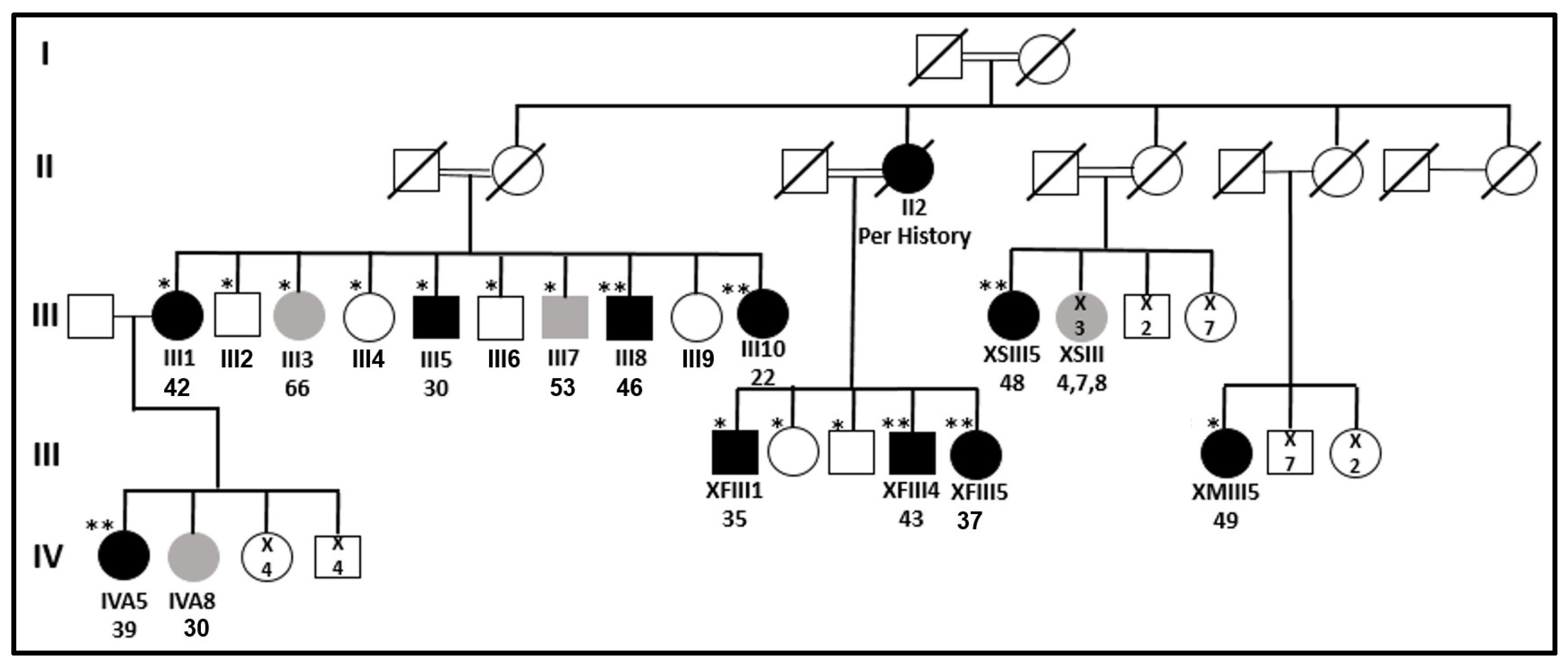
: top—patient’s code; bottom—patient’s age at diagnosis. Ijms 24 08233 i002: number of individuals.
Ijms 24 08233 g001
Figure 2. Evolutionary conservation and 3D structure of ARHGEF28, FBXW10, and SLC47A1 proteins. (A) The evolutionary conservation in the region of the proteins containing the variations. An * (asterisk) indicates a fully conserved residue. A : (colon) indicates conservation between groups of strongly similar properties. A . (period) indicates conservation between groups of weakly similar properties. (B) The predicted 3D structure of the proteins. Part 1—ARHGEF28, the left side represents residues 1–285 of the overall 3D structure of the ARHGEF28 protein, based on the alpha fold model (accession code—Q8N1W1). The right side presents a zoomed-in view of the site of the affected amino acid, Asn108, and its hydrogen bonds (dashed lines) connecting it to the Ser154 and Arg163 amino acids. Part 2—FBXW10, the left side represents the propellor domain, residues 339–693 of the overall 3D structure of the FBXW10 protein, based on the alpha fold model (accession code—Q5XX13). The right side presents a zoomed-in view of the site of the affected amino acid, Ile440, and its hydrogen bonds connecting it to the Asp438 and Lys443 amino acids. Part 3—SLC47A1, zoomed-in view of the site of the affected amino acid, Gly288 of the SLC47A1 protein, based on the alpha fold model (accession code—Q96FL8).
Ijms 24 08233 g002
Table 1. Clinical characteristics of all family members of the two branches of main interest.
IDGender M/FCurrent AgeAge at DiagnosisPathology or US Results ¥Largest DiameterVariant #Multifocality #LN MTS #RAI #Status (Year) §
III1F7942PTCNANANANANAFree of disease (2016)
III2M7669NT NT (2016)
III3F7366Benign MNG ˄2 cm Benign MNG (2016)
III4F7063NT NT (2016)
III5M6330PTCNANANAYesYesLost to follow-up
III6M6154NT NT (2016)
III7M6053Benign MNG ~4 cm Benign MNG (2016)
III8M5746PTC1.6 cmFollicularYes, 5 fociNo100Free of disease (2021)
III9F54NA
III10F5022PTC2.5 cmFollicularYes, 3 fociNoYes- TwiceFree of disease (2021)
XFIII1M5835PTC1.5 cmFollicularYes, 3 fociYes250 *Lost to follow-up
XFIII2FNA
XFIII3MNA
XFIII4M5143PTC2.1 cmClassic/follicularYes, “many foci”Yes100Free of disease (2022)
XFIII5F5037PTCNANANAYes150Free of disease (2022)
IVA5 @F5339PTC1.4 cmClassicYes, at least 4 fociYes150Free of disease (2011) @
IVA8F4530Benign MNG3 cm Lost to follow-up
At the time of diagnosis or clinical evaluation, ¥ papillary thyroid carcinoma, multinodular goiter, or normal thyroid by ultrasound, # for family members with PTC, § last follow-up (year of last visit), ˄ benign ultrasound features—there was no indication for fine-needle aspiration biopsy, ~ fine needle aspiration reported benign nodule(s), * in 2 separate doses, @ at 12/2020 (when she was 50 years old) IVA5 was diagnosed with adenocarcinoma of the lung. Abbreviations: LN MTS—lymph node metastasis; RAI—treatment with radioactive iodine (activity in millicurie if the information was available); F—female; M—male; PTC—papillary thyroid carcinoma; NA—not available (patient refused clinical and genetic evaluation); NT—normal thyroid; MNG—multinodular goiter. The family members’ annotations are according to Figure 1.
Table 2. Evaluating and prioritizing potential candidate variants in patients III8, III10, IVA5, XFIII4, and XFIII5 based on several criteria.
Number of Variants after FiltrationFiltration Criteria for Homozygous or Heterozygous Variations
644,298 → 12,626Shared variations between the WESs of patients III8, III10, IVA5, XFIII4, and XFIII5
12,626 → 84Presence in the general databases—1KG, EVS, ExAC, and gnomAD—at frequencies < 1%
84 → 54Presence in our internal laboratory exome database of the Bedouin population at frequencies < 1%
54 → 4Familial segregation analysis (detailed in Supplementary Table S2)
Table 3. Familial segregation analysis for the four potential candidate variants compatible with the association of a causative variant.
Gene Symbol and PositionRef/AltIII1
A
III2
H
III4
H
III5
A
III6
H
III8
A
III10
A
IVA5
A
XFIII1
A
XFIII4
A
XFIII5
A
ARHGEF28
chr5:73048875
A > G−/++/++/+−/+−/+−/+−/+−/+−/+−/+−/−
FBXW10
chr17:18661699
delCAT−/+−/++/+−/++/+−/+−/+−/+−/+−/+−/+
SLC24A4
chr14:92953131
A > G−/++/+−/+−/++/+−/+−/+−/+−/+−/+−/+
SLC47A1
chr17:19459316
G > A−/+−/++/+−/−+/+−/+−/+−/+−/+−/+−/+
The family members’ annotations are according to Figure 1. “A” represents affected PTC individuals, and “H” represents healthy individuals. Underlined font: incomplete penetrance. −/−: homozygote for the variation; −/+: heterozygote; +/+: normal. Positions on chromosomes are according to GRCh37/hg19.
Table 4. Expression value, frequency in the population of origin, and predictions for damage analysis of the four potential candidate variants compatible with the association of a causative variant.
Gene Symbol
Position
Ref/Alt
cDNA
Protein
GTEx
(TPM) @
Prevalence in the Population of OriginPrediction ToolsACMGClinVar
Internal Lab Frequency *Saudi’s Frequency &Qatari’s Allele Number $
Het.Hom.Het.Hom.Het.Hom.SIFTCADDPolyPhen
ARHGEF28
chr5:73048875
A > G
c.323A > G
p.Asn108Ser
19.440.00570.0006000.00300.14 (tolerated)10.710.02
(benign)
BS2, BP4
(likely benign)
VUS
FBXW10
chr17:18661699
delCAT
c.1317_
1319delCAT
p.Ile440del
1.220.012800000---PM2, PM4 (VUS)-
SLC24A4
chr14:92953131
A > G
c.1532A > G
-
0.20--000.00650-4.68-BS2, BP4, BP6 (benign)Benign
SLC47A1
chr17:19459316
G > A
c.862G > A
p.Gly288Ser
7.450.01470.00060.0260.0130.01200.83 (tolerated)220.73 (possibly damaging)BP4, PM2 (VUS)-
The family members’ annotations are according to Figure 1. @ Expression value according to GTEx portal. * Variant frequency in our collection of 780 individual Bedouin exomes (without any thyroid disorders). & Variant frequency in Saudi’s Bedouin database of 77 exomes [49]. $ Variant frequency in Qatari genome of ~1000 exomes, with a majority of the Bedouin population [50]. Het, heterozygote variant. Hom, homozygote variant. VUS, a variant of uncertain significance. Positions on chromosomes are according to GRCh37/hg19.
Table 5. Evaluating and prioritizing potential candidate variants in the malignant tissue of patient III8 based on several criteria.
Number of Variants after FiltrationFiltration Criteria
217,476 → 6697Variations that exist in the WES of the malignant thyroid tissue and are absent in the gDNA of the same patient—III8
6697 → 1527Presence in the general databases—1KG, EVS, ExAC and gnomAD—at frequencies < 5%
1527 → 2Applying a case panel that presents only variations in genes associated with PTC—see Supplementary Table S1 and list of 13 somatic genes [57]
Disclaimer/Publisher’s Note: The statements, opinions and data contained in all publications are solely those of the individual author(s) and contributor(s) and not of MDPI and/or the editor(s). MDPI and/or the editor(s) disclaim responsibility for any injury to people or property resulting from any ideas, methods, instructions or products referred to in the content.

