
The Immunohistochemical Expression of Epithelial–Mesenchymal Transition Markers in Precancerous Lesions and Cervical Cancer

 , , ,
, , ,
Abstract
1. Introduction
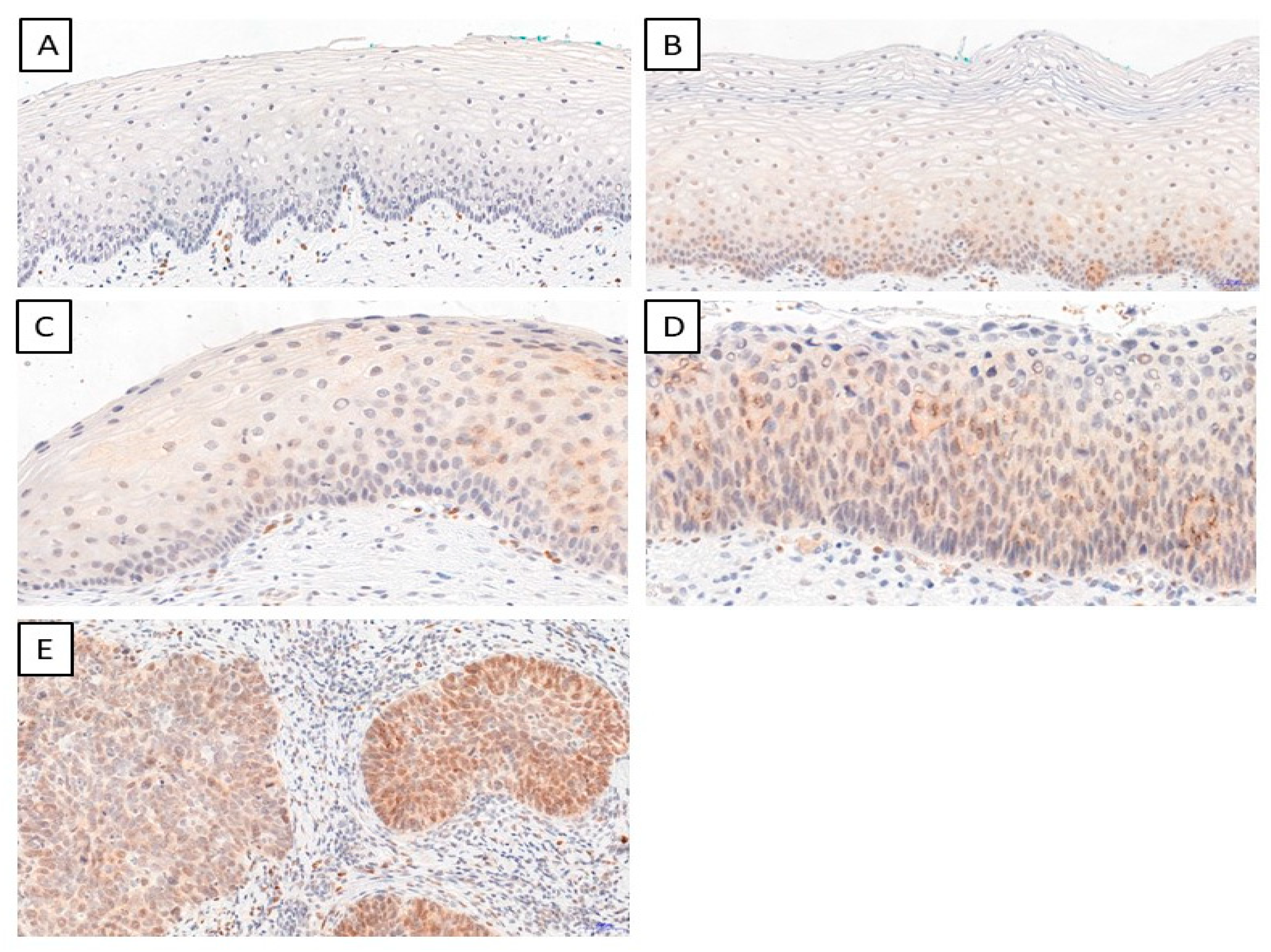
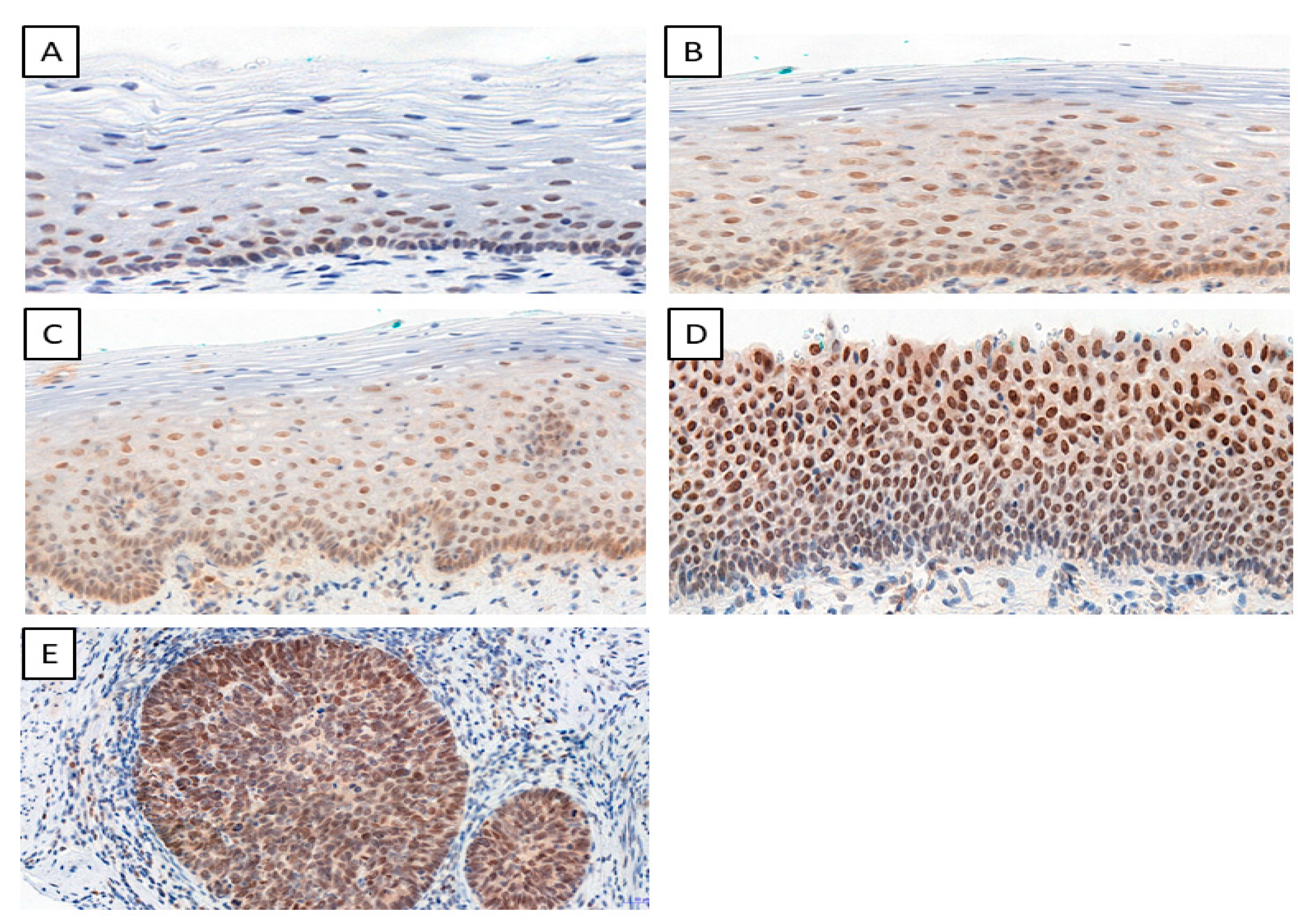
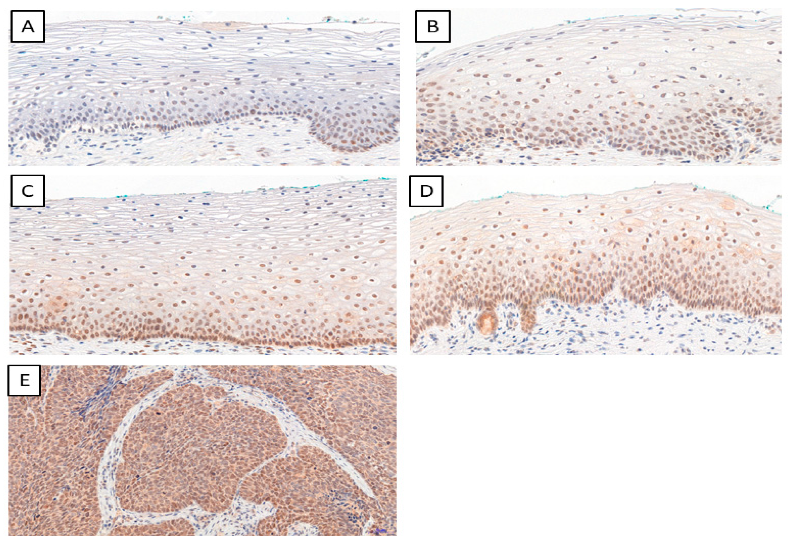
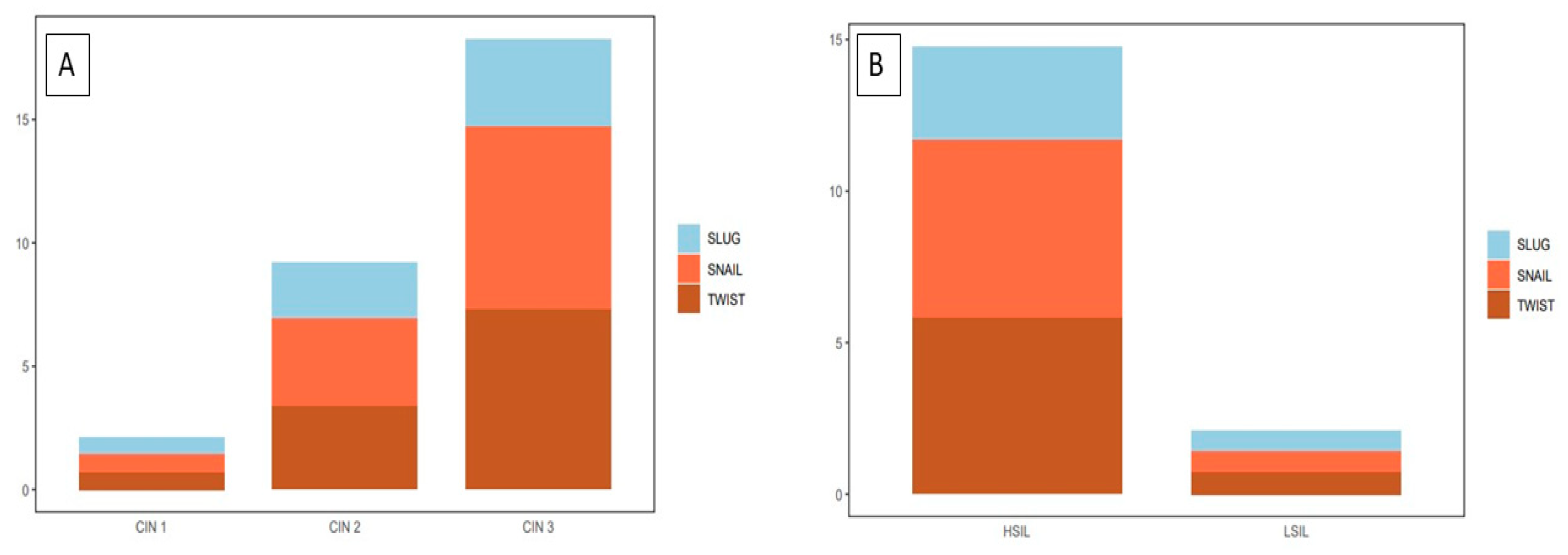
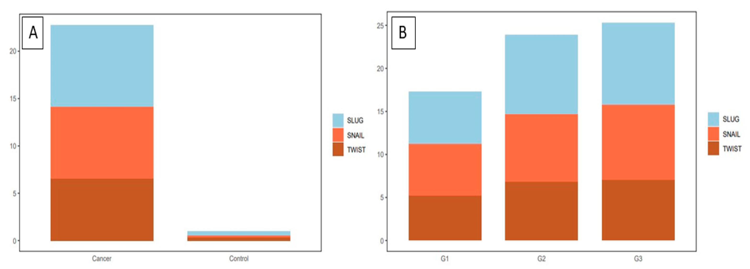
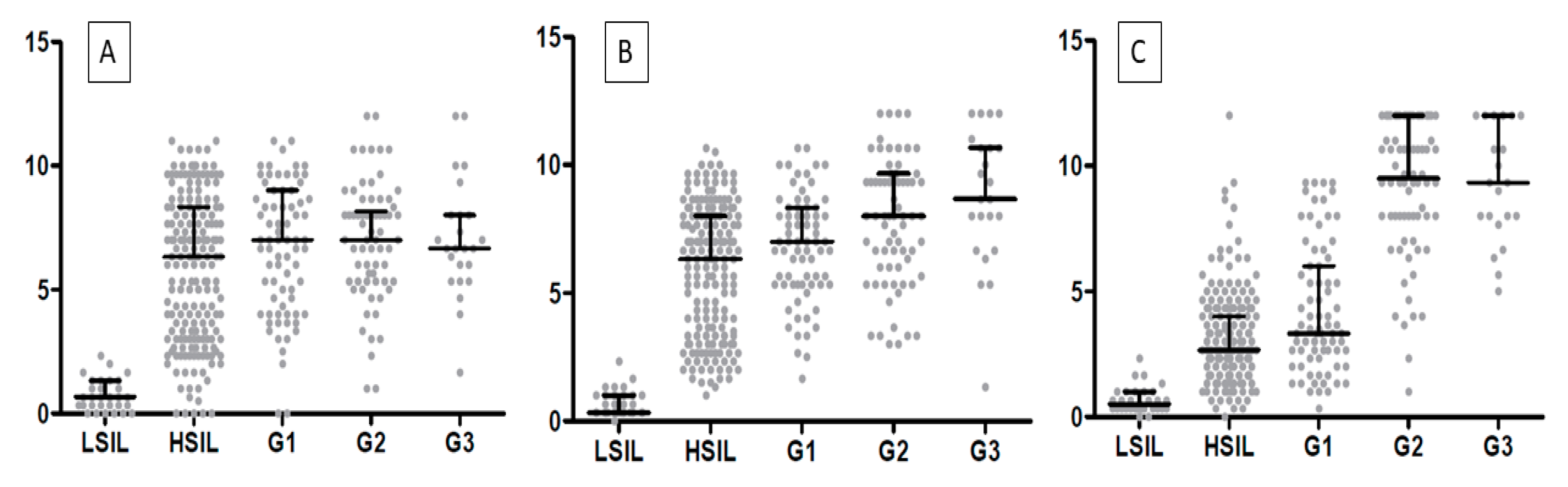
2. Results
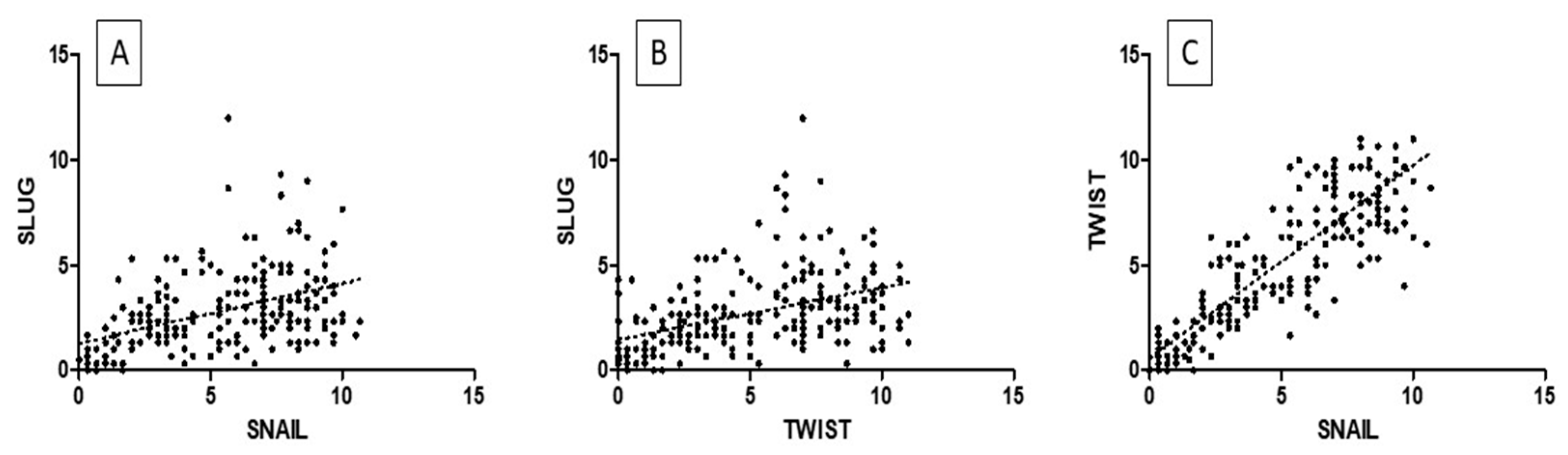
2.1. Precancerous Lesions
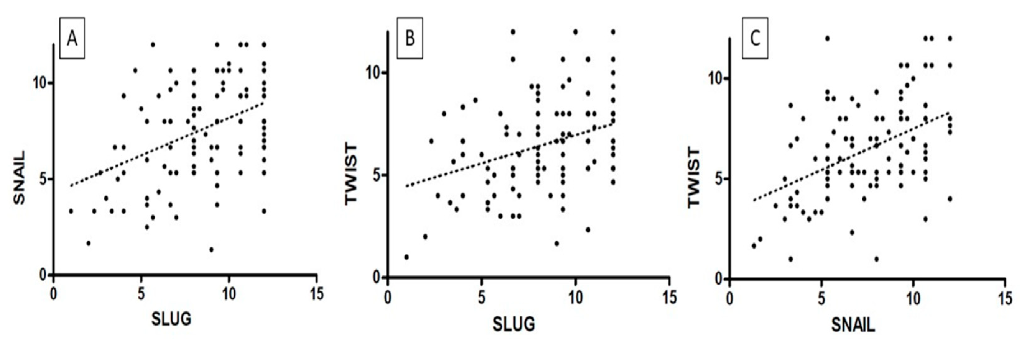
2.2. Cervical Cancer
3. Discussion
4. Materials and Methods
4.1. TMA Construction
4.2. Immunohistochemistry (IHC)
4.3. Statistical Analysis
5. Conclusions
Author Contributions
Funding
Institutional Review Board Statement
Informed Consent Statement
Data Availability Statement
Conflicts of Interest
References
- Li, Y.; Fu, Y.; Cheng, B.; Xie, X.; Wang, X. A Comparative Study on the Accuracy and Efficacy Between Dalton and CINtec® PLUS p16/Ki-67 Dual Stain in Triaging HPV-Positive Women. Front. Oncol. 2021, 11, 815213. [Google Scholar] [CrossRef]
- Cervical Cancer—Cancer Stat Facts. Available online: https://seer.cancer.gov/statfacts/html/cervix.html (accessed on 22 May 2022).
- Popiel, A.; Kobierzycki, C.; Dzięgiel, P. The Role of Testin in Human Cancers. Pathol. Oncol. Res. 2019, 25, e12770. [Google Scholar] [CrossRef]
- Cervical Cancer: Statistics|Cancer.Net. Available online: https://www.cancer.net/cancer-types/cervical-cancer/statistics (accessed on 13 June 2022).
- Landoni, F.; Maneo, A.; Colombo, A.; Placa, F.; Milani, R.; Perego, P.; Favini, G.; Ferri, L.; Mangioni, C. Randomised study of radical surgery versus radiotherapy for stage Ib-IIa cervical cancer. Lancet 1997, 350, 535–540. [Google Scholar] [CrossRef]
- Rotman, M.; Sedlis, A.; Piedmonte, M.R.; Bundy, B.; Lentz, S.S.; Muderspach, L.I.; Zaino, R.J. A phase III randomized trial of postoperative pelvic irradiation in stage IB cervical carcinoma with poor prognostic features: Follow-up of a gynecologic oncology group study. Int. J. Radiat. Oncol. Biol. Phys. 2006, 65, 169–176. [Google Scholar] [CrossRef]
- Delgado, G.; Bundy, B.; Zaino, R.; Sevin, B.-U.; Creasman, W.T.; Major, F. Prospective surgical-pathological study of disease-free interval in patients with stage IB squamous cell carcinoma of the cervix: A Gynecologic Oncology Group study. Gynecol. Oncol. 1990, 38, 352–357. [Google Scholar] [CrossRef]
- SEER Cancer Statistics Review 1975–2003—Previous Version—SEER Cancer Statistics. Available online: https://seer.cancer.gov/archive/csr/1975_2003/ (accessed on 13 June 2022).
- Lee, M.-Y.; Chou, C.-Y.; Tang, M.-J.; Shen, M.-R. Epithelial-Mesenchymal Transition in Cervical Cancer: Correlation with Tumor Progression, Epidermal Growth Factor Receptor Overexpression, and Snail Up-Regulation. Clin. Cancer Res. 2008, 14, 4743–4750. [Google Scholar] [CrossRef]
- Li, J.; Zhou, B.P. Activation of β-catenin and Akt pathways by Twist are critical for the maintenance of EMT associated cancer stem cell-like characters. BMC Cancer 2011, 11, 49. [Google Scholar] [CrossRef] [PubMed]
- Lee, M.-Y.; Shen, M.-R. Epithelial-mesenchymal transition in cervical carcinoma. Am. J. Transl. Res. 2012, 4, 1–13. [Google Scholar] [PubMed]
- Shibata, K.; Kajiyama, H.; Ino, K.; Terauchi, M.; Yamamoto, E.; Nawa, A.; Nomura, S.; Kikkawa, F. Twist expression in patients with cervical cancer is associated with poor disease outcome. Ann. Oncol. 2008, 19, 81–85. [Google Scholar] [CrossRef] [PubMed]
- Zhu, K.; Chen, L.; Han, X.; Wang, J.; Wang, J. Short hairpin RNA targeting Twist1 suppresses cell proliferation and improves chemosensitivity to cisplatin in HeLa human cervical cancer cells. Oncol. Rep. 2012, 27, 1027–1034. [Google Scholar] [CrossRef] [PubMed]
- Wang, T.; Li, Y.; Wang, W.; Tuerhanjiang, A.; Wu, Z.; Yang, R.; Yuan, M.; Ma, D.; Wang, W.; Wang, S. Twist2, the key Twist isoform related to prognosis, promotes invasion of cervical cancer by inducing epithelial-mesenchymal transition and blocking senescence. Hum. Pathol. 2014, 45, 1839–1846. [Google Scholar] [CrossRef]
- Fan, Q.; Qiu, M.-T.; Zhu, Z.; Zhou, J.-H.; Chen, L.; Zhou, Y.; Gu, W.; Wang, L.-H.; Li, Z.-N.; Xu, Y.; et al. Twist induces epithelial-mesenchymal transition in cervical carcinogenesis by regulating the TGF-β/Smad3 signaling pathway. Oncol. Rep. 2015, 34, 1787–1794. [Google Scholar] [CrossRef]
- Kyo, S.; Sakaguchi, J.; Ohno, S.; Mizumoto, Y.; Maida, Y.; Hashimoto, M.; Nakamura, M.; Takakura, M.; Nakajima, M.; Masutomi, K. High Twist expression is involved in infiltrative endometrial cancer and affects patient survival. Hum. Pathol. 2006, 37, 431–438. [Google Scholar] [CrossRef] [PubMed]
- Baygi, M.E.; Soheili, Z.S.; Schmitz, I.; Sameie, S.; Schulz, W.A. Snail regulates cell survival and inhibits cellular senescence in human metastatic prostate cancer cell lines. Cell Biol. Toxicol. 2010, 26, 553–567. [Google Scholar] [CrossRef]
- Zha, Y.-H.; He, J.-F.; Mei, Y.-W.; Yin, T.; Mao, L. Zinc-finger transcription factor Snail accelerates survival, migration and expression of matrix metalloproteinase-2 in human bone mesenchymal stem cells. Cell Biol. Int. 2007, 31, 1089–1096. [Google Scholar] [CrossRef]
- Vega, S.; Morales, A.V.; Ocaña, O.H.; Valdés, F.; Fabregat, I.; Nieto, M.A. Snail blocks the cell cycle and confers resistance to cell death. Genes Dev. 2004, 18, 1131–1143. [Google Scholar] [CrossRef] [PubMed]
- Kudo-Saito, C.; Shirako, H.; Takeuchi, T.; Kawakami, Y. Cancer Metastasis Is Accelerated through Immunosuppression during Snail-Induced EMT of Cancer Cells. Cancer Cell 2009, 15, 195–206. [Google Scholar] [CrossRef] [PubMed]
- Wu, Y.; Zhou, B.P. Snail: More than EMT. Cell Adhes. Migr. 2010, 4, 199–203. [Google Scholar] [CrossRef] [PubMed]
- Jiang, B.; Sun, R.; Fang, S.; Qin, C.; Pan, X.; Peng, L.; Li, Y.; Li, G. Lnc-CC3 increases metastasis in cervical cancer by increasing Slug expression. Oncotarget 2016, 7, 41650–41661. [Google Scholar] [CrossRef] [PubMed]
- Lai, S.-Y.; Guan, H.-M.; Liu, J.; Huang, L.-J.; Hu, X.-L.; Chen, Y.-H.; Wu, Y.-H.; Wang, Y.; Yang, Q.; Zhou, J.-Y. Long noncoding RNA SNHG12 modulated by human papillomavirus 16 E6/E7 promotes cervical cancer progression via ERK/Slug pathway. J. Cell. Physiol. 2020, 235, 7911–7922. [Google Scholar] [CrossRef]
- Liu, X.; Feng, Q.; Zhang, Y.; Zheng, P.; Cui, N. Absence of EpCAM in cervical cancer cells is involved in slug induced epithelial-mesenchymal transition. Cancer Cell Int. 2021, 21, 163. [Google Scholar] [CrossRef] [PubMed]
- Cui, N.; Yang, W.-T.; Zheng, P.-S. Slug inhibits the proliferation and tumor formation of human cervical cancer cells by up-regulating the p21/p27 proteins and down-regulating the activity of the Wnt/β-catenin signaling pathway via the trans-suppression Akt1/p-Akt1 expression. Oncotarget 2016, 7, 26152–26167. [Google Scholar] [CrossRef] [PubMed]
- Liao, Y.; Huang, J.; Liu, P.; Zhang, C.; Liu, J.; Xia, M.; Shang, C.; Ooi, S.; Chen, Y.; Qin, S.; et al. Downregulation of LNMAS orchestrates partial EMT and immune escape from macrophage phagocytosis to promote lymph node metastasis of cervical cancer. Oncogene 2022, 41, 1931–1943. [Google Scholar] [CrossRef] [PubMed]
- Hugo, H.J.; Kokkinos, M.I.; Blick, T.; Ackland, M.L.; Thompson, E.W.; Newgreen, D.F. Defining the E-Cadherin Repressor Interactome in Epithelial-Mesenchymal Transition: The PMC42 Model as a Case Study. Cells Tissues Organs 2011, 193, 23–40. [Google Scholar] [CrossRef] [PubMed]
- Onder, T.T.; Gupta, P.B.; Mani, S.A.; Yang, J.; Lander, E.S.; Weinberg, R.A. Loss of E-Cadherin Promotes Metastasis via Multiple Downstream Transcriptional Pathways. Cancer Res. 2008, 68, 3645–3654. [Google Scholar] [CrossRef]
- Bourboulia, D.; Han, H.; Jensen-Taubman, S.; Gavil, N.; Isaac, B.; Wei, B.; Neckers, L.; Stetler-Stevenson, W.G. TIMP-2 modulates cancer cell transcriptional profile and enhances E-cadherin/beta-catenin complex expression in A549 lung cancer cells. Oncotarget 2013, 4, 166–176. [Google Scholar] [CrossRef]
- Rosty, C.; Hewett, D.G.; Brown, I.S.; Leggett, B.A.; Whitehall, V.L.J. Serrated polyps of the large intestine: Current understanding of diagnosis, pathogenesis, and clinical management. J. Gastroenterol. 2012, 48, 287–302. [Google Scholar] [CrossRef] [PubMed]
- Ge, Y.; Christensen, P.A.; Luna, E.; Armylagos, D.; Xu, J.; Hsu, W.-C.; Zhou, H.; Schwartz, M.R.; Mody, D.R. Age-specific 3-year cumulative risk of cervical cancer and high-grade dysplasia on biopsy in 9434 women who underwent HPV cytology cotesting. Cancer Cytopathol. 2019, 127, 757–764. [Google Scholar] [CrossRef]
- Raab, S.S.; Bishop, N.S.; Zaleski, M.S. Long-term outcome and relative risk in women with atypical squamous cells of undetermined significance. Am. J. Clin. Pathol. 1999, 112, 57–62. [Google Scholar] [CrossRef] [PubMed]
- Zhao, W.; Zhou, Y.; Xu, H.; Cheng, Y.; Kong, B. Snail family proteins in cervical squamous carcinoma: Expression and significance. Clin. Investig. Med. 2013, 36, E223–E233. [Google Scholar] [CrossRef]
- Qureshi, R.; Arora, H.; Rizvi, M. EMT in cervical cancer: Its role in tumour progression and response to therapy. Cancer Lett. 2015, 356, 321–331. [Google Scholar] [CrossRef] [PubMed]
- Campo, L.; Zhang, C.; Breuer, E.K. EMT-Inducing Molecular Factors in Gynecological Cancers. Biomed Res Int. 2015, 2015, 420891. [Google Scholar] [CrossRef]
- Tian, Y.; Qi, P.; Niu, Q.; Hu, X. Combined Snail and E-cadherin Predicts Overall Survival of Cervical Carcinoma Patients: Comparison Among Various Epithelial-Mesenchymal Transition Proteins. Front. Mol. Biosci. 2020, 7, 22. [Google Scholar] [CrossRef] [PubMed]
- Dai, B.; Yu, R.; Fan, M.; Yang, T.; Wang, B.; Zhang, Y. HMQ-T-F2 suppresses migration of the human cervical cancer HeLa cells by reversing EMT via the PI3K/Akt signaling pathway. Oncol. Rep. 2019, 42, 1451–1458. [Google Scholar] [CrossRef]
- Weadick, B.; Nayak, D.; Persaud, A.K.; Hung, S.W.; Raj, R.; Campbell, M.J.; Chen, W.; Li, J.; Williams, T.M.; Govindarajan, R. EMT-Induced Gemcitabine Resistance in Pancreatic Cancer Involves the Functional Loss of Equilibrative Nucleoside Transporter 1. Mol. Cancer Ther. 2021, 20, 410–422. [Google Scholar] [CrossRef]
- Alafate, W.; Li, X.; Zuo, J.; Zhang, H.; Xiang, J.; Wu, W.; Xie, W.; Bai, X.; Wang, M.; Wang, J. Elevation of CXCL1 indicates poor prognosis and radioresistance by inducing mesenchymal transition in glioblastoma. CNS Neurosci. Ther. 2020, 26, 475–485. [Google Scholar] [CrossRef]
- Zhang, J.; Si, J.; Gan, L.; Guo, M.; Yan, J.; Chen, Y.; Wang, F.; Xie, Y.; Wang, Y.; Zhang, H. Inhibition of Wnt signalling pathway by XAV939 enhances radiosensitivity in human cervical cancer HeLa cells. Artif. Cells Nanomed. Biotechnol. 2020, 48, 479–487. [Google Scholar] [CrossRef]
- Chen, S.; Yuan, X.; Xu, H.; Yi, M.; Liu, S.; Wen, F. WNT974 Inhibits Proliferation, Induces Apoptosis, and Enhances Chemosensitivity to Doxorubicin in Lymphoma Cells by Inhibiting Wnt/β-Catenin Signaling. Experiment 2020, 26, e923799. [Google Scholar] [CrossRef]
- Popiel-Kopaczyk, A.; Grzegrzolka, J.; Piotrowska, A.; Olbromski, M.; Smolarz, B.; Romanowicz, H.; Rusak, A.; Mrozowska, M.; Dziegiel, P.; Podhorska-Okolow, M.; et al. The Expression of Testin, Ki-67 and p16 in Cervical Cancer Diagnostics. Curr. Issues Mol. Biol. 2023, 45, 490–500. [Google Scholar] [CrossRef] [PubMed]
- Chen, T.; Liu, Y.; Huang, L. ImageGP: An easy-to-use data visualization web server for scientific researchers. Imeta 2022, 1, e5. [Google Scholar] [CrossRef]


| CIN | Cervical Cancer | |||
|---|---|---|---|---|
| SLUG | TWIST | SLUG | TWIST | |
| SNAIL | r = 0.4787 p < 0.0001 | r = 0.8470 p < 0.0001 | r = 0.3764 p < 0.0001 | r = 0.4338 p < 0.0001 |
| TWIST | r = 0.2157 p < 0.0021 | NA | r = 0.1066 p < 0.2483 | NA |
Disclaimer/Publisher’s Note: The statements, opinions and data contained in all publications are solely those of the individual author(s) and contributor(s) and not of MDPI and/or the editor(s). MDPI and/or the editor(s) disclaim responsibility for any injury to people or property resulting from any ideas, methods, instructions or products referred to in the content. |
© 2023 by the authors. Licensee MDPI, Basel, Switzerland. This article is an open access article distributed under the terms and conditions of the Creative Commons Attribution (CC BY) license (https://creativecommons.org/licenses/by/4.0/).
Share and Cite
Popiel-Kopaczyk, A.; Piotrowska, A.; Sputa-Grzegrzolka, P.; Smolarz, B.; Romanowicz, H.; Dziegiel, P.; Podhorska-Okolow, M.; Kobierzycki, C. The Immunohistochemical Expression of Epithelial–Mesenchymal Transition Markers in Precancerous Lesions and Cervical Cancer. Int. J. Mol. Sci. 2023, 24, 8063. https://doi.org/10.3390/ijms24098063
Popiel-Kopaczyk A, Piotrowska A, Sputa-Grzegrzolka P, Smolarz B, Romanowicz H, Dziegiel P, Podhorska-Okolow M, Kobierzycki C. The Immunohistochemical Expression of Epithelial–Mesenchymal Transition Markers in Precancerous Lesions and Cervical Cancer. International Journal of Molecular Sciences. 2023; 24(9):8063. https://doi.org/10.3390/ijms24098063
Chicago/Turabian StylePopiel-Kopaczyk, Aneta, Aleksandra Piotrowska, Patrycja Sputa-Grzegrzolka, Beata Smolarz, Hanna Romanowicz, Piotr Dziegiel, Marzenna Podhorska-Okolow, and Christopher Kobierzycki. 2023. "The Immunohistochemical Expression of Epithelial–Mesenchymal Transition Markers in Precancerous Lesions and Cervical Cancer" International Journal of Molecular Sciences 24, no. 9: 8063. https://doi.org/10.3390/ijms24098063
APA StylePopiel-Kopaczyk, A., Piotrowska, A., Sputa-Grzegrzolka, P., Smolarz, B., Romanowicz, H., Dziegiel, P., Podhorska-Okolow, M., & Kobierzycki, C. (2023). The Immunohistochemical Expression of Epithelial–Mesenchymal Transition Markers in Precancerous Lesions and Cervical Cancer. International Journal of Molecular Sciences, 24(9), 8063. https://doi.org/10.3390/ijms24098063