Review

Cancer Stem Cells and Prostate Cancer: A Narrative Review

1
Urology Unit, Department of Medico-Surgical Sciences & Biotechnologies, Faculty of Pharmacy & Medicine, Sapienza University of Rome, 04100 Latina, Italy
2
Urology Unit, Sant’Andrea Hospital, Sapienza University of Rome, 00189 Rome, Italy
*
Author to whom correspondence should be addressed.
Int. J. Mol. Sci. 2023, 24(9), 7746; https://doi.org/10.3390/ijms24097746
Submission received: 3 March 2023 / Revised: 17 April 2023 / Accepted: 21 April 2023 / Published: 24 April 2023
(This article belongs to the Special Issue Molecular Biology of Prostate Cancer)

Abstract

:
Cancer stem cells (CSCs) are a small and elusive subpopulation of self-renewing cancer cells with the remarkable ability to initiate, propagate, and spread malignant disease. In the past years, several authors have focused on the possible role of CSCs in PCa development and progression. PCa CSCs typically originate from a luminal prostate cell. Three main pathways are involved in the CSC development, including the Wnt, Sonic Hedgehog, and Notch signaling pathways. Studies have observed an important role for epithelial mesenchymal transition in this process as well as for some specific miRNA. These studies led to the development of studies targeting these specific pathways to improve the management of PCa development and progression. CSCs in prostate cancer represent an actual and promising field of research.

1. Introduction

Prostate cancer (PCa) represents the most common solid tumor among men in Western countries and the fifth cause of cancer mortality worldwide [1]. According to the actual statistics, the worldwide prostate cancer burden is expected to grow to almost 2 million new cases and 700,000 deaths in the next 20 years [2]. Age, race, genetics, family history, and lifestyle (e.g., obesity, smoking, etc.) represent risk factors for higher incidence of PCa [1]. Early detection of PCA is possible through routine checkups, with the determination of prostate specific antigen (PSA), through digital rectal examination, mpMRI, and prostate biopsy [3]. Several risk calculators/apps are available in order to estimate the risk of PCa and significant PCa [4,5].
Several studies have investigated the etiology and pathophysiology of PCa, but the exact mechanisms are still unknown [6]. Important roles are played by the genetic characteristics of the patients as well as its immune system [7,8,9]. The heterogeneous biological nature of the disease brings great challenges during treatment, highlighting the need for better definition of the pathophysiology of PCa [10].
In the past years, several authors have focused on the possible role of stem cells in prostate cancer. Stem cells have the ability to copiously proliferate (self-renewal) starting from a single cell (clonal) and to differentiate into various kinds of cells and tissue (potent) [10]. Self-renewal and potency characteristics are very similar to those of cancer cells [10]. This led to the hypothesis that tumors may arise from the transformation of stem cells and that cancer stem cells (CSC) found inside tumor tissues may be responsible for tumorigenesis [11].
Prostate cancer stem cells (PCSCs) were discovered for the first time by Collins et al. in 2005 in human prostate tumors, and the authors enhanced an increased ability of proliferation in PCSCs, highlighting the idea that they may play an important role in the pathogenesis of prostate cancer. PCSCs have been isolated in metastatic PCa cell lines [11]. Since their discovery, PCSCs have been of great interest in the study of the PC. Today, different and various antigens are known to be responsible for the carcinogenesis of PCa as well as the formation of metastasis and the resistance to pharmacological treatment [12].
The CSC model hypothesis explains tumor heterogeneity, initiating ability, and therapeutic resistance [13]. In addition, a small number of CSCs are needed to recapitulate the tumor and its initial heterogeneity [13]. Cancer cells are organized in hierarchical order and CSCs stem on top of the pyramid, having stem-like characteristics such as self-renewal, pluripotency, and plasticity, that evolve during the lifetime of a tumor [14]. CSCs may be involved in PCa development and may solve the unmet needs in PCa prevention and diagnosis [13]. Their possible role in PCa progression may also improve the current gaps in PCa treatment, especially in the castration-resistant stages of the disease when androgen sensitivity is compromised [15]. Although several treatments have been introduced in this setting, there are still several unmet needs [16,17].
An overview of the most important evidence on this complex topic is essential, especially for clinical urologists. The aim of the present review is to summarize in a narrative fashion the most relevant data on CSCs and PCa.

2. Evidence Synthesis

2.1. Prostate Cancer Stem Cells Origin and Phenotype

PCa is an heterogeneous disease where different grades may coexist [18,19]. Cell heterogeneity, upon progression and treatment, tends to accentuate [18,19]. The best example of this process is the expression of the androgen receptor (AR), where AR−PCa cells gradually become, in high-grade untreated tumors, the predominant cell population in castration-resistant PCa (CRPC) [18,19].
The first prospective studies aimed to identify PCSCs markers in patient-derived PCa cell lines [11]. These PCa cells expressed surface markers such as CD44+/α2β1hi/CD133+ and the expression of transporter protein ATP-binding cassette subfamily G member 2 (ABCG2) was identified [18].
Since 2005, several markers have been isolated for PCSCs, such as CD44/CD133, aldehyde dehydrogenase (ALDH), CD166, and CD44+/CD24−. PCa cells expressing these markers can self-renew and generate heterogeneous cell subpopulations [20,21]. Furthermore, markers such as CD151, CD160, and podocalyxin TRA-60–1 are associated with PCSCs that give birth to tumors with hierarchical organization cell organization [22]. Expression of SOX2, Notch, and Oct4 are associated with epithelium–mesenchymal transition, which promotes PCa cell migrations [23,24].
PCSCs correlated markers are shown in Table 1.
The origin of PCSCs is debated. In a normal prostate, the stem cell population resides in the proximal duct, originating from the basal cell layer of the epithelium [25]. Moad M. et al. proved in a recent gene-tracing study that luminal cells have a stem cell population, though less potent than the basal stem cell population [26]. PCSCs might derive from the basal cell layer because p63 and other basal markers are expressed in tissue-derived tumor-initiating cells in immunocompromised mice but did not express the AR or other markers of luminal differentiation (PSA and PAP) [27]. PCSCs might originate from normal prostate stem cells after epigenetic modifications created by the tumor microenvironment [28]. MYC activation and PTEN loss of function added to mutations to DNA repair genes such as BRCA2 create instability to the genome and cause tumor heterogeneity and progression [29,30]. It is well known that common gene fusion in differentiated PCa is represented by TMPRSS2: ERG. Recent studies by Polson and colleagues also showed that PCSCs had high-frequency of TMPRSS2:ERG [31]. ERG is a transcription factor under the control of TMPRSS2. TMPRSS2 is an androgen-regulated, prostate-specific gene promoter, which is upregulated in CSCs. The result of its activation is differentiation, self-renewal, and maintenance of SCs [32]. Another important marker of CSCs lies in the Homebox gene Nkx 3.1 during prostate regeneration. More specifically, animal studies demonstrate that Nkx 3.1 is required for CSC maintenance and that the deletion of PTEN in these cells results in a rapid transformation in castration-resistant carcinoma [33,34].

2.2. PCSC Molecular Pathways and Metabolism

In order to better uncover new and improved therapeutic strategies, molecular pathways and PCSC metabolism have to be analyzed. The Wnt, Sonic Hedgehog, and Notch signaling pathways have been found to be crucial for CSC development [35,36].

2.2.1. Wnt Pathway

The normal Wnt pathway affects cell survival; Wnt ligands bind to Frizzled and low-density lipoprotein receptor-related protein (LRP) 5/6, activating the downstream of the subsequent molecular targets, accumulating β-catenin and mediating its translocation in the cell nucleus [34]. Wnt activates downstream effectors and activates targeted gene expression and cytoskeleton rearrangement, resulting in altered cell survival in noncanonical pathways [34]. In PCA, not only elevated β-catenin expression is found in the cancer cell nucleus [35], but Wnt signal promotes cell self-renewal in several cell models including LNCaP, C42B, and PC3 cells in an AR-independent way, increasing expression of CD133 and CD44 [36]. The downregulated Wnt/β-catenin pathway significantly suppresses stem cell-like properties instead [37]. Furthermore, Wnt3 has been shown to increase the expression of its downstream effectors and CSC markers, including CD133 and CD44, thus playing an important role in CSC development in CRPC (Figure 1).

2.2.2. Sonic Hedgehog Pathway

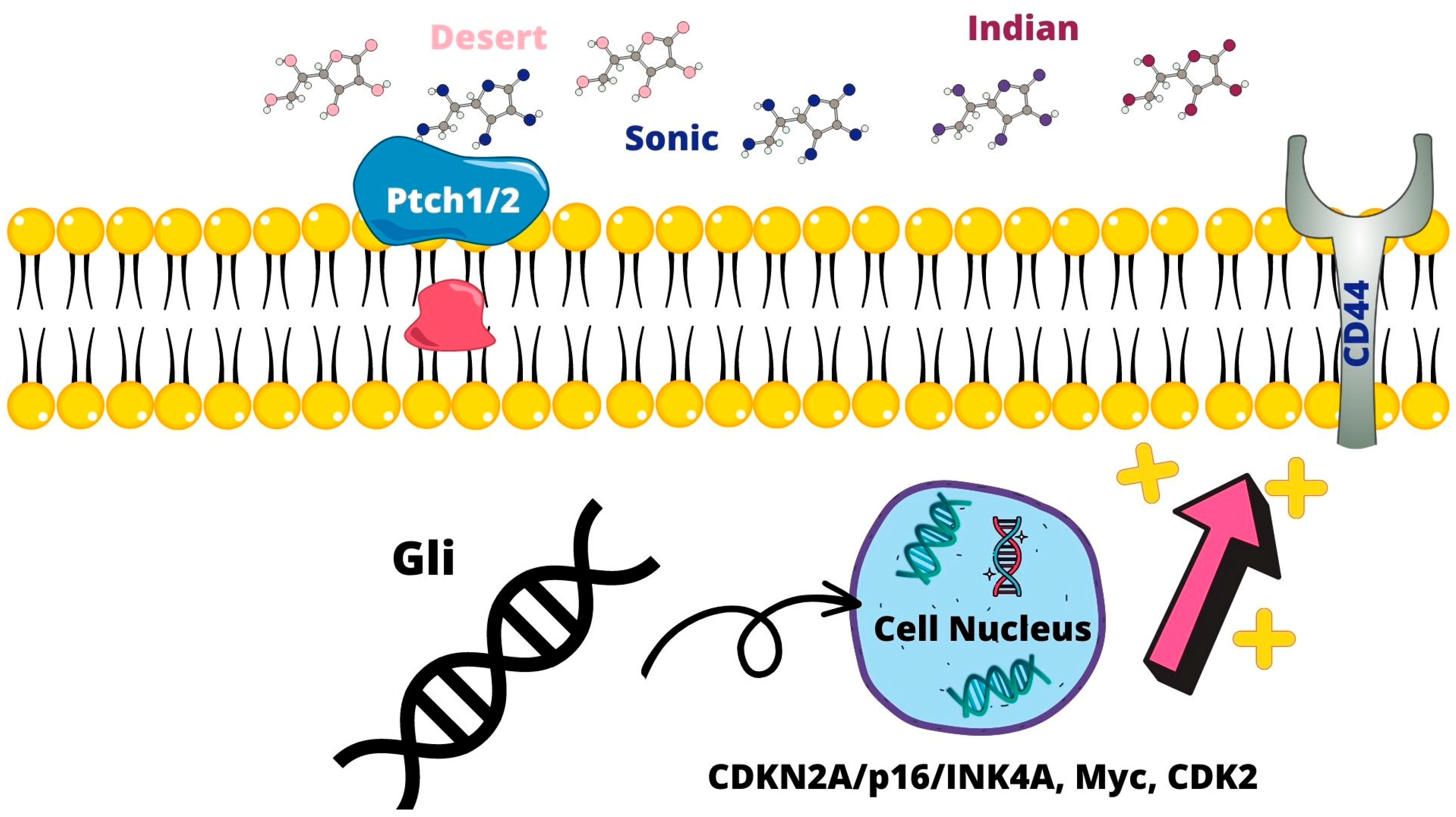
The Sonic Hedgehog signaling pathway controls cell renewal and survival. The ligands of this family are represented by Sonic, Desert, and Indian. These ligands, by binding to membrane receptors, Patched (Ptch1 and 2) and Smoothered, activate and mediate the nuclear translocation of glioma-associated oncogene homolog (Gli), expressing target genes that regulate cell survival. An abnormal Hedgehog signaling pathway is present in PCa and in CSC proliferation [38,39]. Gli-related genes, including CDKN2A/p16/INK4A, Myc, and CDK2, by promoting androgen-independent tumor cell growth, lead to biochemical and tumor recurrence and lead to tumor recurrence [40]. Furthermore, cell models have shown that FOXA1 and BCL2 have a role both in CRPCa and therapeutic resistance [41]. Hedgehog signaling was also found to be increased. Hedgehog signaling was found with PCSC CD44+, CD24− [21] (Figure 2).

2.2.3. Notch Signaling Pathway

The Notch signaling pathway is mediated by the Notch1-4 receptors and from ligands, such as DLL 1, DLL 3, DLL4, Jagged 1, and Jagged 2. In PCa Notch, the AR pathway and the PI3K/Akt pathway interact. Importantly, Notch interacts with the AR pathway and the phosphoinositide 3-kinase (PI3k)/Akt pathway, which are the two main signaling pathways that regulate development and carcinogenesis [42,43]. Increased levels of Jagged 1-Notch is associated with PCa progression, metastasis, and EMT, and increased Notch3 expression is CRPCA [44]. PC tumor development in the beginning stages is dependent on androgen signaling. Initially, most patients respond to ADT, but almost the totality of them develop CRPC, hence the shift to androgen-independent tumors [45]. It has been found that AR-variants (AR-V) present a truncation in the COOH terminal, thus lacking the ligand-binding domain but maintaining transcriptional capability. These AR-Vs are associated with PCa growth and progression. In particular, AR-V7 has been found in CTCs and in PC resistant to new antiandrogenic drugs such as enzalutamide and abiraterone [46] (Figure 3).

2.2.4. Metabolism of PCSC

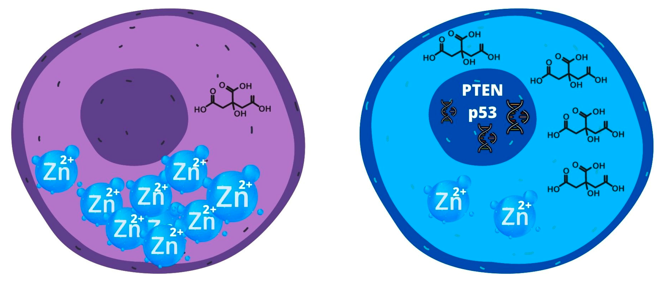
The normal luminal cell compartment of the prostate gland produces and secretes a large amount of citrate into the prostatic fluid to nourish and preserve sperm vitality. Normally citric acid represents an important substrate in the tricarboxylic acid cycle, being transported from the mitochondria, where it is produced, to the cell via transmembrane transporters [47]. In luminal cells, mitochondrial aconitase has low activity because of the expression of zinc transporter proteins, which keep elevated zinc levels, impeding citrate oxidation. High production of citrate in the prostate gland is a consequence of the low activity of mitochondrial aconitase (m-ACNT) and subsequent inhibition of citrate oxidation [47].
On the other hand, PCa cells present low levels of zinc in the mitochondria, inactivating the inhibitory effect on m-ACNT, thus enabling the use of citrate as a metabolic substrate [48], oxidizing citrate, and permitting oxidative phosphorylation. Further evidence has shown that the shift from oxidative phosphorylation to glycolysis is caused by mutations in mtDNA of the oncogenic suppressors PTEN and p53 [49]. This shift is needed due to the intrinsic properties of tumor cells, being fast-growing and metabolically active. This phenomenon, also known as the Warburg effect, is especially evident in metastasized CRPCA, where ROS produced and released by PCa cells cause oxidative stress in CAF, consequently shifting to glycolysis and producing high levels of lactate, which fuels ATP production [50] (Figure 4).
It is well known that tumor cells, particularly fast-growing PCa cells, utilize glutamine as one of the main metabolic substrates. Tissues with enhanced expression of MYC oncogene, such as PCa, are particularly dependent on glutamine metabolism to sustain their viability [51]. MYC is involved in glucose metabolism, regulating GLUT1, HK2, PFK1, enolase 1, and LDHA [16]. In addition to its role in glucose metabolism, MYC regulates the expression of GLS1, and genes such as SLC1A4 and SLC1A5 [52]. MYC involvement is well established in PCa: for example, the upregulation of MYC mRNA is present in most PCa; furthermore, its overexpression is present in about 30% of CRPC [53].

2.3. Epithelial–Mesenchymal Transition and CSC Correlation

Epithelial–mesenchymal transition (EMT) is a cellular process in which normal epithelial cells lose their epithelial features, transitioning in pseudo-mesenchymal cells [54]. EMT normally occurs in the embryonic phase of development, from before implantation to organogenesis. Tumor cells activate these processes once more, leading the cells to acquire motility and invasiveness potential. During prostate cancer progression, the epithelial cells can undergo EMT, characterized by morphological changes in their phenotype from cuboidal to spindle-shaped. Malignant cells lose E-cadherine (E-cad), syndecans, and tight-junction molecules while under the influence of SNAL1, SNAI2/Slug, and TWIST. On the other side, the expression of mesenchymal cell markers Vimentine, N-cadherin, and metalloproteinases are upregulated in malignant cells. Furthermore, the main causal factor for PCa progression is the loss of E-cadherin. This process results in the formation of a cell phenotype with increased migration and invasion potential as well as metastatization [55].

2.3.1. Loss of E-Cadherin Expression

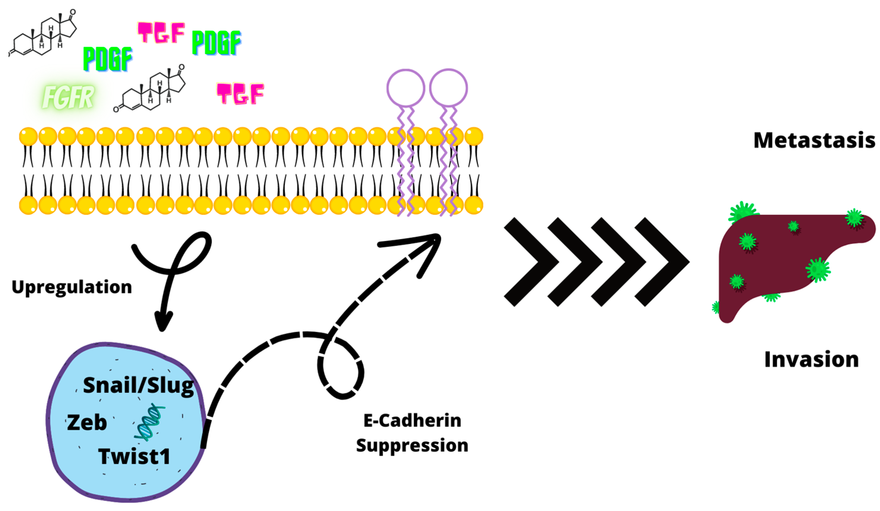
Overall, the EMT process is a complex genetic program, involving numerous interactions among several different EMT-transcription factors (EMT-TFs). Molecules related to these pathways include TGFβ, FGFRs, and PDGF. The primary EMT-TF families that downregulate E-cad expression are Zeb (Zeb1/Zeb2); Snail (Snail/Slug), and Twist1 [56]. Androgens are also involved in EMT. Mesenchymal phenotypes can be induced since androgens have the ability to suppress E-cadherin in normal prostatic cells. Zhu et al. have proven that elevated levels of DHT can facilitate EMT. DHT alone and in combination with TGFβ induces the expression of Snail leading to the reduction in E-cadherin and β-cathenin levels. In addition, Zhu et al. also evaluated AR levels, finding that AR levels influence EMT. In the presence of a high concentration of AR, DHT exposure did not cause the reduction in epithelial markers, while in cells with low AR concentration levels, the opposite effect was found, not only DHT downregulated E-cadherin, but also increased the cells’ invasion potential. AR loss is normally caused by androgen deprivation. To this date, ADT is the main medical treatment for PCa, but it has been speculated that the treatment might fuel EMT [57] (Figure 5).

2.3.2. Role of TGF-β

In normal circumstances, TGFβ’s role is that of tumor suppressor promoting apoptosis. Cancer cells, either inactivating TGFβ’s receptors or repressing its downstream effectors, are able to bypass its primary activity. As well, mutations in TGFβ receptors (TβRI and TβRII) may lead to the EMT process. On the other hand, loss of TβRI and TβRII is associated with poor prognosis and poor survival.
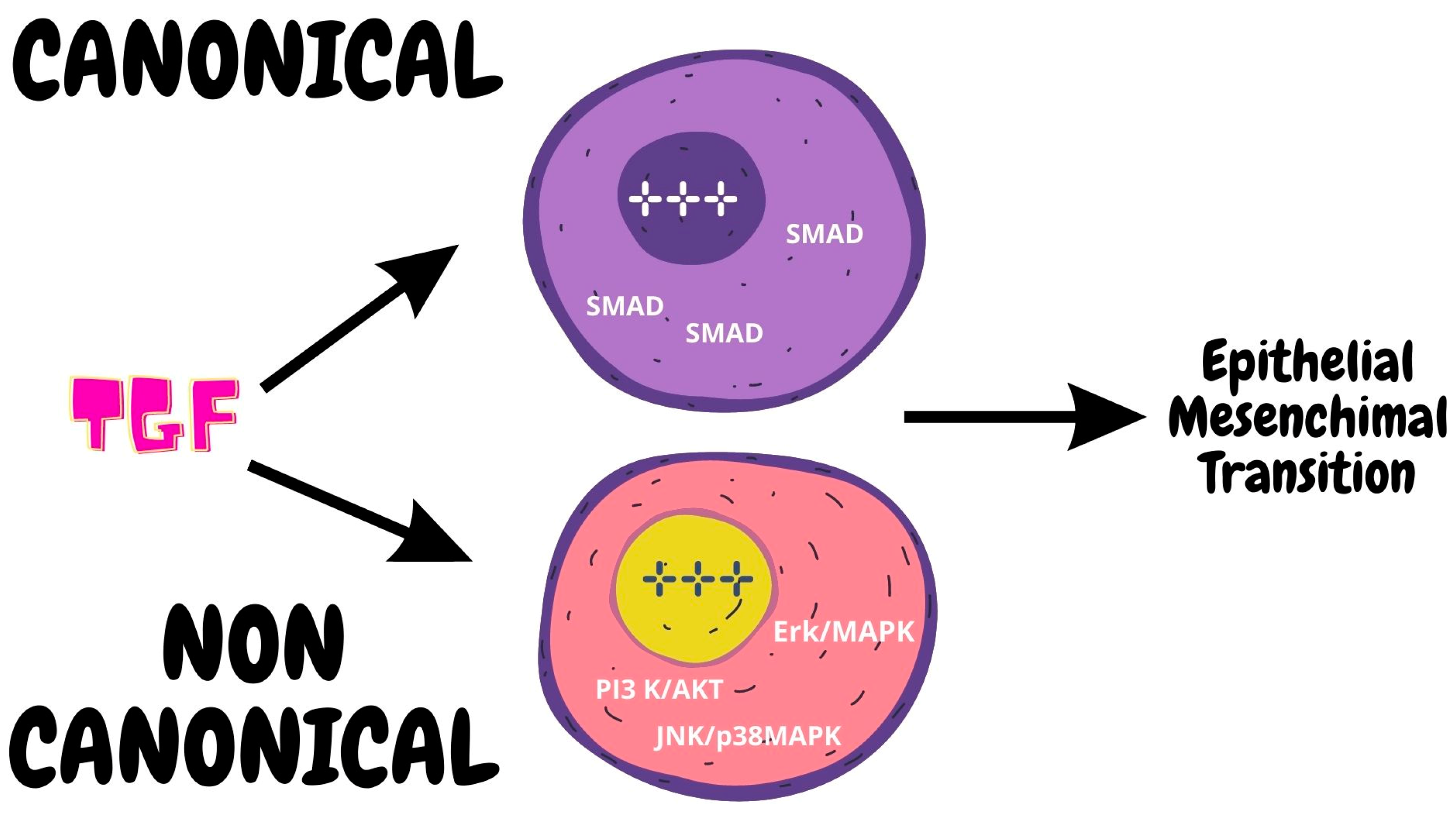
TGFβ’s signaling can follow either canonical or noncanonical pathways. The canonical signaling pathway proceeds after TGFβ binds with its receptors that activate the SMAD proteins that form a complex in the cell nucleus, acting as a facilitator with DNA-binding cofactors, thus activating the transcription process [58]. On the other hand, the noncanonical pathways utilize several different effectors such as Erk/MAPK, JNK/p38MAPK, and PI3 K/Akt. Interestingly every one of these pathways has a role in EMT [59]. For example, Erk regulates genes involved in cell motility, while overactivation of PI3K leads to cell junction disturbance. Furthermore, loss of TβRI and TβRII are also associated with poor prognosis and poor survival [56] (Figure 6).

2.3.3. Role of CD44

Cancerous cells that undergo EMT present stem-like properties such as the elevated expression of CD44 and their physical transformation in spheres [60]. Epigenetic mutations might be the primer for CSC development [61]. In favor of this argument, we find YAP1 to be an EMT facilitator. YAP1 was found overexpressed in cells that underwent EMT in normal differentiated cells that transitioned into CSCs [62]. This is just one example of many that suggests that the expression of gene-related EMT is a facilitator for CSCs. Furthermore, the TME in which CSCs live pullulates of cytokines and growth factors, favoring their stemness [63]. Furthermore, the hypoxic niche favors the expression of markers such as Nestin. The TME hypoxic state favors the upregulation of Nestin through the activation of the TGFβ-SMAD4 pathway [64].

2.4. MicroRNA and CSC

In the past few years, a new and interesting research field has been pursued: the study of microRNAs (miRNA) and their role in PCSC regulation. MicroRNAs seem to play a primary role in the regulation of PCSCs; they seem implicated in regulating the promotion or repression of metastasis. Evidence shows how miR-34a, let-7b, miR-106a, and miR-200 are found in PCa’s progenitor stem cell population [65]. miRNAs are small noncoding RNA molecules that play a role not only in tumorigenesis, but also in cell proliferation, differentiation, and apoptosis [66].
Hypermethylations of miR-34 have been observed in many malignancies with the p53 mutation. Inactivation of both p53 and miR-34a in mouse prostate epithelium causes the PSC compartment, aiding the development of early invasive adenocarcinoma and accelerating EMT-dependent growth, enhancing the self-renewal capability for EMT-dependent growth [67]. In addition, miR-34a is a negative regulator of CD44. CSCs derived from multiple malignant tumors have shown high expression of CD44. Other miRNAs downregulation has been described to have a role in the progression of PCA such as miR-320 and miR-7. The downregulation of the former is associated with increased β-catenin, favoring tumor sphere formation and chemoresistance [37]. Reduced levels of miR-7 correlate with CD133+/CD44+ PCa, and present CSC-like features.
MicroRNAs regulate both CSCs and normal stem cells, but miRNAs dysregulate the process of tumorigenesis. MiR-34a (a p53 target) acts as a key negative regulator of CD44+ in prostate cancer cells and establishes a strong therapeutic agent against prostate CSCs. Furthermore, miR143 and miR-145 suppressed colony formation of PC-3 cells from prostate cancer bone metastasis by inhibiting CSCs properties of PC-3 cancer.

2.5. MicroRNA and PCa

The direct role of microRNA in PCa has been widely investigated.
Walter et al. [68] and. Feng et al. [69] observed downregulation of both miR-148 and miR-152 in high-grade PCa. More specifically, miR-148-3p was found to inhibit PCa cell growth in vitro and in vivo, suggesting its possible role as a tumor suppressor. On the other hand, miR-148b-3p was found to be correlated with PSA and PCA3 in PCa tissue samples, suggesting a potential role as a PCa biomarker [70]. Another interesting study by Ostadrahimi et al. observed a downregulation of miR-185 expression in PCa tissues and cell lines with an upregulation of the antiapoptotic genes BCL2 and BCL2L1 [71].
Kristensen et al. evaluated the possible role of miRNAs as diagnostic and prognostic tools in PCa. In their analysis, they developed a prognostic classifier for biochemical recurrence after radical prostatectomy based on mi-R-185-5p, miR-221-3p, and miR-326 [72]. As well, Gurbuz et al. observed increased miR-185-5p expression in patients with elevated PSA levels, especially in patients with PSA > 10 ng/mL. Their study further suggests the potential of miRNAs as prognosis biomarkers.
Overall, microRNA represents an interesting field of research to improve PCa diagnosis, particularly to identify patients at high risk of metastasis and castration-resistant phenotypes [73].
MiRNAs involved with PCSCS and EMT are described in Table 2.

2.6. CSC Treatment

PCSC treatments are mainly based on the abovementioned pathways by targeting the microenvironment and immunotherapies.
Specific inhibitors against the Hh pathway (Sonidegib, GANT-61, and GDC-0449), the Wnt pathway (3289-8625, LGK974, Foxy-5, and OMP-54F28), the Notch pathway (RO4929097), and the NFkB pathway (bortezomib, PS1145, BMS345541, Aspyrin, 17-(allylamino)-17-demethoxygeldanamycin, and BKM120) are currently being tested in preclinical and clinical trials [50]. As well, Liu et al. observed that the inhibition of NOTCH1 with shRNA leads to reduction in ABCC1 expression resulting in restored chemosensitivity. Another transporter ABCG2 is also directly involved in the resistance to androgens, and when blocked PCa cells differentiate into androgen-sensitive phenotypes [74].
The PI3K/AKT/mTOR pathway is associated with PCA progression and ADT resistance [75]. Chang and colleagues by the use of the dual PI3K/mTOR inhibitor BEZ235 were able to restore radiosensitivity and induce apoptosis in radioresistant PCSCs [76] PCa.
Some studies suggest the monoclonal antibody Bevacizumab may play a role in modifying TME by reducing tumor neovasculature and disruption of CSC niches. Development of Bevacizumab resistance may be avoided by targeting Rac1 inhibition or P-Rex1 downregulation. The chemosensitivity of PCa cells may also be restored by targeting the CXCR4 receptor, which inhibits sphere formation. On the other side, the development of a castration-resistant phenotype may be triggered by ABC transporters. The idea of targeted differentiation by acting on TME is of great interest and should be confirmed in future experimental studies [77,78]. Different pathways are listed in Table 3.

3. Conclusions

The present review highlights and summarizes the available evidence on prostate cancer and stem cells. CSCs originate from luminal cells with epigenetic modifications mainly driven by the TMPRSS2:ERG complex. CSC development is mainly driven by three signaling pathways: the Wnt, Sonic Hedgehog, and Notch pathways. Overall CSC metabolism is characterized by high levels of oxidated citrate and low levels of Zn. In the development of CSCs, a pivotal role is played by the epithelial mesenchymal transition process which can be activated in a canonical and noncanonical way. As well, several miRNA are involved in this complex process. As previously highlighted, these different pathways and molecules can act as targets for new drugs, opening new strategies in the management and prevention of prostate cancer genesis and progression. CSCs represent a fertile and growing area of research, and ongoing and future studies in the coming years will help the understanding of PCa development and progression.

Author Contributions

Y.A.S.: conceptualizzation, investigation and methodology; M.B.S.: writing, editing and supervision; F.M.V.: writing, editing and supervision; A.F.: writing, and editing; A.M.: methodology and investigation; P.P.S.: supervision and project development; A.C. (Antonio Carbone): review and editing; G.T.: editing and supervision; R.L.: conceptualization and writing; A.C. (Antonio Cicione): editing and review; A.L.P.: conceptualizzation and reviewing; C.D.N.: project administration, reviewing and editing. All authors have read and agreed to the published version of the manuscript.

Funding

This research received no external funding.

Institutional Review Board Statement

Not applicable.

Informed Consent Statement

Not applicable.

Data Availability Statement

No new data were created.

Conflicts of Interest

The authors declare no conflict of interest.

References

  1. Gandaglia, G.; Leni, R.; Bray, F.; Fleshner, N.; Freedland, S.J.; Kibel, A.; Stattin, P.; Van Poppel, H.; La Vecchia, C. Epidemiology and Prevention of Prostate Cancer. Eur. Urol. Oncol. 2021, 4, 877–892. [Google Scholar] [CrossRef] [PubMed]
  2. Culp, M.B.B.; Soerjomataram, I.; Efstathiou, J.A.; Bray, F.; Jemal, A. Recent Global Patterns in Prostate Cancer Incidence and Mortality Rates. Eur. Urol. 2020, 77, 38–52. [Google Scholar] [CrossRef] [PubMed]
  3. Mottet, N.; Bellmunt, J.; Bolla, M.; Briers, E.; Cumberbatch, M.G.; De Santis, M.; Fossati, N.; Gross, T.; Henry, A.M.; Joniau, S.; et al. EAU–ESTRO–SIOG Guidelines on Prostate Cancer. Part 1: Screening, Diagnosis, and Local Treatment with Curative Intent. Natl. Cent. Biotechnol. Inf. 2016, 71, 618–629. [Google Scholar] [CrossRef]
  4. De Nunzio, C.; Lombardo, R.; Tema, G.; Cancrini, F.; Russo, G.I.; Chacon, R.; Garcia-Cruz, E.; Ribal, M.J.; Morgia, G.; Alcaraz, A.; et al. Mobile Phone Apps for the Prediction of Prostate Cancer: External Validation of the Coral and Rotterdam Apps. Eur. J. Surg. Oncol. 2019, 45, 471–476. [Google Scholar] [CrossRef]
  5. De Nunzio, C.; Lombardo, R.; Tema, G.; Alkhatatbeh, H.; Gandaglia, G.; Briganti, A.; Tubaro, A. External Validation of Chun, PCPT, ERSPC, Kawakami, and Karakiewicz Nomograms in the Prediction of Prostate Cancer: A Single Center Cohort-Study. Urol. Oncol. Semin. Orig. Investig. 2018, 36, 364.e1–364.e7. [Google Scholar] [CrossRef]
  6. Chan, K.M.; Gleadle, J.M.; O’Callaghan, M.; Vasilev, K.; MacGregor, M. Prostate Cancer Detection: A Systematic Review of Urinary Biosensors. Prostate Cancer Prostatic Dis. 2022, 25, 39–46. [Google Scholar] [CrossRef] [PubMed]
  7. Stultz, J.; Fong, L. How to Turn up the Heat on the Cold Immune Microenvironment of Metastatic Prostate Cancer. Prostate Cancer Prostatic Dis. 2021, 24, 697–717. [Google Scholar] [CrossRef]
  8. Zorko, N.A.; Ryan, C.J. Novel Immune Engagers and Cellular Therapies for Metastatic Castration-Resistant Prostate Cancer: Do We Take a BiTe or Ride BiKEs, TriKEs, and CARs? Prostate Cancer Prostatic Dis. 2021, 24, 986–996. [Google Scholar] [CrossRef] [PubMed]
  9. Wang, Z.; Yan, X.; Tang, P.; Tang, T.; Wang, Y.; Peng, S.; Wang, S.; Lan, W.; Wang, L.; Zhang, Y.; et al. Genetic Profiling of Hormone-Sensitive and Castration-Resistant Prostate Cancers and Identification of Genetic Mutations Prone to Castration-Resistant Prostate Cancer. Prostate Cancer Prostatic Dis. 2022, 26, 180–187. [Google Scholar] [CrossRef]
  10. Rahimi Tesiye, M.; Abrishami Kia, Z.; Rajabi-Maham, H. Mesenchymal Stem Cells and Prostate Cancer: A Concise Review of Therapeutic Potentials and Biological Aspects. Stem Cell Res. 2022, 63, 102864. [Google Scholar] [CrossRef]
  11. Collins, A.T.; Berry, P.A.; Hyde, C.; Stower, M.J.; Maitland, N.J. Prospective Identification of Tumorigenic Prostate Cancer Stem Cells. Cancer Res. 2005, 65, 10946–10951. [Google Scholar] [CrossRef]
  12. Harris, K.S.; Kerr, B.A. Prostate Cancer Stem Cell Markers Drive Progression, Therapeutic Resistance, and Bone Metastasis. Stem Cells Int. 2017, 2017, 8629234. [Google Scholar] [CrossRef] [PubMed]
  13. Cable, J.; Pei, D.; Reid, L.M.; Wang, X.W.; Bhatia, S.; Karras, P.; Melenhorst, J.J.; Grompe, M.; Lathia, J.D.; Song, E.; et al. Cancer Stem Cells: Advances in Biology and Clinical Translation-a Keystone Symposia Report. Ann. N. Y. Acad. Sci. 2021, 1506, 142–163. [Google Scholar] [CrossRef] [PubMed]
  14. Wolf, I.; Gratzke, C.; Wolf, P. Prostate Cancer Stem Cells: Clinical Aspects and Targeted Therapies. Front. Oncol. 2022, 12, 935715. [Google Scholar] [CrossRef]
  15. Berchuck, J.E.; Viscuse, P.V.; Beltran, H.; Aparicio, A. Clinical Considerations for the Management of Androgen Indifferent Prostate Cancer. Prostate Cancer Prostatic Dis. 2021, 24, 623–637. [Google Scholar] [CrossRef]
  16. Denunzio, C.; Presicce, F.; Giacinti, S.; Bassanelli, M.; Tubaro, A. Castration-Resistance Prostate Cancer: What Is in the Pipeline? Minerva Urol. Nefrol. 2018, 70, 22–41. [Google Scholar] [CrossRef]
  17. De Nunzio, C.; Lombardo, R.; Tema, G.; Voglino, O.; Sica, A.; Baldassarri, V.; Nacchia, A.; Iacovelli, R.; Bracarda, S.; Tubaro, A. Adverse Events Related to Abiraterone and Enzalutamide Treatment: Analysis of the EudraVigilance Database and Meta-Analysis of Registrational Phase III Studies. Prostate Cancer Prostatic Dis. 2020, 23, 199–206. [Google Scholar] [CrossRef]
  18. Skvortsov, S.; Skvortsova, I.I.; Tang, D.G.; Dubrovska, A. Concise Review: Prostate Cancer Stem Cells: Current Understanding. Stem Cells 2018, 36, 1457–1474. [Google Scholar] [CrossRef]
  19. Qin, J.; Liu, X.; Laffin, B.; Chen, X.; Choy, G.; Jeter, C.R.; Calhoun-Davis, T.; Li, H.; Palapattu, G.S.; Pang, S.; et al. The PSA(-/Lo) Prostate Cancer Cell Population Harbors Self-Renewing Long-Term Tumor-Propagating Cells That Resist Castration. Cell Stem. Cell 2012, 10, 556–569. [Google Scholar] [CrossRef]
  20. Zhang, Y.; Huang, Y.; Jin, Z.; Li, X.; Li, B.; Xu, P.; Huang, P.; Liu, C. A Convenient and Effective Strategy for the Enrichment of Tumor-Initiating Cell Properties in Prostate Cancer Cells. Tumour Biol. 2016, 37, 11973–11981. [Google Scholar] [CrossRef]
  21. Hurt, E.M.; Kawasaki, B.T.; Klarmann, G.J.; Thomas, S.B.; Farrar, W.L. CD44+ CD24 (−) Prostate Cells Are Early Cancer Progenitor/Stem Cells That Provide a Model for Patients with Poor Prognosis. Br. J. Cancer 2008, 98, 756–765. [Google Scholar] [CrossRef] [PubMed]
  22. Rajasekhar, V.K.; Studer, L.; Gerald, W.; Socci, N.D.; Scher, H.I. Tumour-Initiating Stem-like Cells in Human Prostate Cancer Exhibit Increased NF-ΚB Signalling. Nat. Commun. 2011, 2, 162. [Google Scholar] [CrossRef] [PubMed]
  23. Sampayo, R.G.; Bissell, M.J. Cancer Stem Cells in Breast and Prostate: Fact or Fiction? Adv. Cancer Res. 2019, 144, 315–341. [Google Scholar] [CrossRef]
  24. Takebe, N.; Harris, P.J.; Warren, R.Q.; Ivy, S.P. Targeting Cancer Stem Cells by Inhibiting Wnt, Notch, and Hedgehog Pathways. Nat. Rev. Clin. Oncol. 2011, 8, 97–106. [Google Scholar] [CrossRef] [PubMed]
  25. Goto, K.; Salm, S.N.; Coetzee, S.; Xiong, X.; Burger, P.E.; Shapiro, E.; Lepor, H.; Moscatelli, D.; Wilson, E.L. Proximal Prostatic Stem Cells Are Programmed to Regenerate a Proximal-Distal Ductal Axis. Stem Cells 2006, 24, 1859–1868. [Google Scholar] [CrossRef]
  26. Moad, M.; Hannezo, E.; Buczacki, S.J.; Wilson, L.; El-Sherif, A.; Sims, D.; Pickard, R.; Wright, N.A.; Williamson, S.C.; Turnbull, D.M.; et al. Multipotent Basal Stem Cells, Maintained in Localized Proximal Niches, Support Directed Long-Ranging Epithelial Flows in Human Prostates. Cell Rep. 2017, 20, 1609–1622. [Google Scholar] [CrossRef]
  27. Maitland, N.J.; Frame, F.M.; Polson, E.S.; Lewis, J.L.; Collins, A.T. Prostate Cancer Stem Cells: Do They Have a Basal or Luminal Phenotype? Horm. Cancer 2011, 2, 47–61. [Google Scholar] [CrossRef]
  28. Li, J.J.; Shen, M.M. Prostate Stem Cells and Cancer Stem Cells. Cold Spring Harb. Perspect. Med. 2019, 9, 368. [Google Scholar] [CrossRef]
  29. Mateo, J.; Boysen, G.; Barbieri, C.E.; Bryant, H.E.; Castro, E.; Nelson, P.S.; Olmos, D.; Pritchard, C.C.; Rubin, M.A.; de Bono, J.S. DNA Repair in Prostate Cancer: Biology and Clinical Implications. Eur. Urol. 2017, 71, 417–425. [Google Scholar] [CrossRef]
  30. Taylor, R.A.; Fraser, M.; Livingstone, J.; Espiritu, S.M.G.; Thorne, H.; Huang, V.; Lo, W.; Shiah, Y.J.; Yamaguchi, T.N.; Sliwinski, A.; et al. Germline BRCA2 Mutations Drive Prostate Cancers with Distinct Evolutionary Trajectories. Nat. Commun. 2017, 8, 13671. [Google Scholar] [CrossRef]
  31. Polson, E.S.; Lewis, J.L.; Celik, H.; Mann, V.M.; Stower, M.J.; Simms, M.S.; Rodrigues, G.; Collins, A.T.; Maitland, N.J. Monoallelic Expression of TMPRSS2/ERG in Prostate Cancer Stem Cells. Nat. Commun. 2013, 4, 1623. [Google Scholar] [CrossRef] [PubMed]
  32. Archer, L.K.; Frame, F.M.; Maitland, N.J. Stem Cells and the Role of ETS Transcription Factors in the Differentiation Hierarchy of Normal and Malignant Prostate Epithelium. J. Steroid Biochem. Mol. Biol. 2017, 166, 68–83. [Google Scholar] [CrossRef] [PubMed]
  33. Chen, H.; Nandi, A.K.; Li, X.; Bieberich, C.J. NKX-3.1 Interacts with Prostate-Derived Ets Factor and Regulates the Activity of the PSA Promoter. Cancer Res. 2002, 62, 338–340. [Google Scholar] [PubMed]
  34. Lin, C.J.; Lo, U.G.; Hsieh, J.T. The Regulatory Pathways Leading to Stem-like Cells Underlie Prostate Cancer Progression. Asian J. Androl. 2019, 21, 233–240. [Google Scholar] [CrossRef]
  35. Zhang, K.; Guo, Y.; Wang, X.; Zhao, H.; Ji, Z.; Cheng, C.; Li, L.; Fang, Y.; Xu, D.; Zhu, H.H.; et al. WNT/β-Catenin Directs Self-Renewal Symmetric Cell Division of HTERThigh Prostate Cancer Stem Cells. Cancer Res. 2017, 77, 2534–2547. [Google Scholar] [CrossRef]
  36. Bisson, I.; Prowse, D.M. WNT Signaling Regulates Self-Renewal and Differentiation of Prostate Cancer Cells with Stem Cell Characteristics. Cell Res. 2009, 19, 683–697. [Google Scholar] [CrossRef]
  37. Hsieh, I.S.; Chang, K.C.; Tsai, Y.T.; Ke, J.Y.; Lu, P.J.; Lee, K.H.; Yeh, S.D.; Hong, T.M.; Chen, Y.L. MicroRNA-320 Suppresses the Stem Cell-like Characteristics of Prostate Cancer Cells by Downregulating the Wnt/Beta-Catenin Signaling Pathway. Carcinogenesis 2013, 34, 530–538. [Google Scholar] [CrossRef]
  38. Zunich, S.M.; Valdovinos, M.; Douglas, T.; Walterhouse, D.; Iannaccone, P.; Lamm, M.L.G. Osteoblast-Secreted Collagen Upregulates Paracrine Sonic Hedgehog Signaling by Prostate Cancer Cells and Enhances Osteoblast Differentiation. Mol. Cancer 2012, 11, 30. [Google Scholar] [CrossRef]
  39. Chang, H.H.; Chen, B.Y.; Wu, C.Y.; Tsao, Z.J.; Chen, Y.Y.; Chang, C.P.; Yang, C.R.; Lin, D.P.C. Hedgehog Overexpression Leads to the Formation of Prostate Cancer Stem Cells with Metastatic Property Irrespective of Androgen Receptor Expression in the Mouse Model. J. Biomed. Sci. 2011, 18, 6. [Google Scholar] [CrossRef]
  40. Mohler, J.L. Castration-Recurrent Prostate Cancer Is Not Androgen-Independent. Adv. Exp. Med. Biol. 2008, 617, 223–234. [Google Scholar] [CrossRef]
  41. Sahu, B.; Laakso, M.; Ovaska, K.; Mirtti, T.; Lundin, J.; Rannikko, A.; Sankila, A.; Turunen, J.P.; Lundin, M.; Konsti, J.; et al. Dual Role of FoxA1 in Androgen Receptor Binding to Chromatin, Androgen Signalling and Prostate Cancer. EMBO J. 2011, 30, 3962–3976. [Google Scholar] [CrossRef] [PubMed]
  42. Shen, M.M.; Abate-Shen, C. Molecular Genetics of Prostate Cancer: New Prospects for Old Challenges. Genes Dev. 2010, 24, 1967–2000. [Google Scholar] [CrossRef] [PubMed]
  43. Chang, L.; Graham, P.H.; Ni, J.; Hao, J.; Bucci, J.; Cozzi, P.J.; Li, Y. Targeting PI3K/Akt/MTOR Signaling Pathway in the Treatment of Prostate Cancer Radioresistance. Crit. Rev. Oncol. Hematol. 2015, 96, 507–517. [Google Scholar] [CrossRef] [PubMed]
  44. Domingo-Domenech, J.; Vidal, S.J.; Rodriguez-Bravo, V.; Castillo-Martin, M.; Quinn, S.A.; Rodriguez-Barrueco, R.; Bonal, D.M.; Charytonowicz, E.; Gladoun, N.; de la Iglesia-Vicente, J.; et al. Suppression of Acquired Docetaxel Resistance in Prostate Cancer through Depletion of Notch- and Hedgehog-Dependent Tumor-Initiating Cells. Cancer Cell 2012, 22, 373–388. [Google Scholar] [CrossRef]
  45. Karantanos, T.; Corn, P.G.; Thompson, T.C. Prostate Cancer Progression after Androgen Deprivation Therapy: Mechanisms of Castrate Resistance and Novel Therapeutic Approaches. Oncogene 2013, 32, 5501–5511. [Google Scholar] [CrossRef]
  46. Guo, Z.; Yang, X.; Sun, F.; Jiang, R.; Linn, D.E.; Chen, H.; Chen, H.; Kong, X.; Melamed, J.; Tepper, C.G.; et al. A Novel Androgen Receptor Splice Variant Is Up-Regulated during Prostate Cancer Progression and Promotes Androgen Depletion-Resistant Growth. Cancer Res. 2009, 69, 2305–2313. [Google Scholar] [CrossRef]
  47. Mycielska, M.E.; Patel, A.; Rizaner, N.; Mazurek, M.P.; Keun, H.; Patel, A.; Ganapathy, V.; Djamgoz, M.B.A. Citrate Transport and Metabolism in Mammalian Cells: Prostate Epithelial Cells and Prostate Cancer. Bioessays 2009, 31, 10–20. [Google Scholar] [CrossRef]
  48. Franklin, R.B.; Feng, P.; Milon, B.; Desouki, M.M.; Singh, K.K.; Kajdacsy-Balla, A.; Bagasra, O.; Costello, L.C. HZIP1 Zinc Uptake Transporter down Regulation and Zinc Depletion in Prostate Cancer. Mol. Cancer 2005, 4, 32. [Google Scholar] [CrossRef]
  49. Wang, L.; Xiong, H.; Wu, F.; Zhang, Y.; Wang, J.; Zhao, L.; Guo, X.; Chang, L.J.; Zhang, Y.; You, M.J.; et al. Hexokinase 2-Mediated Warburg Effect Is Required for PTEN- and P53-Deficiency-Driven Prostate Cancer Growth. Cell Rep. 2014, 8, 1461–1474. [Google Scholar] [CrossRef]
  50. Pértega-Gomes, N.; Vizcaíno, J.R.; Attig, J.; Jurmeister, S.; Lopes, C.; Baltazar, F. A Lactate Shuttle System between Tumour and Stromal Cells Is Associated with Poor Prognosis in Prostate Cancer. BMC Cancer 2014, 14, 352. [Google Scholar] [CrossRef]
  51. Dang, C.V.; Le, A.; Gao, P. MYC-Induced Cancer Cell Energy Metabolism and Therapeutic Opportunities. Clin. Cancer Res. 2009, 15, 6479–6483. [Google Scholar] [CrossRef] [PubMed]
  52. White, M.A.; Lin, C.; Rajapakshe, K.; Dong, J.; Shi, Y.; Tsouko, E.; Mukhopadhyay, R.; Jasso, D.; Dawood, W.; Coarfa, C.; et al. Glutamine Transporters Are Targets of Multiple Oncogenic Signaling Pathways in Prostate Cancer. Mol. Cancer Res. 2017, 15, 1017–1028. [Google Scholar] [CrossRef]
  53. Nupponen, N.N.; Kakkola, L.; Koivisto, P.; Visakorpi, T. Genetic Alterations in Hormone-Refractory Recurrent Prostate Carcinomas. Am. J. Pathol. 1998, 153, 141–148. [Google Scholar] [CrossRef] [PubMed]
  54. Thiery, J.P.; Acloque, H.; Huang, R.Y.J.; Nieto, M.A. Epithelial-Mesenchymal Transitions in Development and Disease. Cell 2009, 139, 871–890. [Google Scholar] [CrossRef]
  55. Osorio, L.A.; Farfán, N.M.; Castellón, E.A.; Contreras, H.R. SNAIL Transcription Factor Increases the Motility and Invasive Capacity of Prostate Cancer Cells. Mol. Med. Rep. 2016, 13, 778–786. [Google Scholar] [CrossRef]
  56. Lamouille, S.; Subramanyam, D.; Blelloch, R.; Derynck, R. Regulation of Epithelial-Mesenchymal and Mesenchymal-Epithelial Transitions by MicroRNAs. Curr. Opin. Cell Biol. 2013, 25, 200–207. [Google Scholar] [CrossRef] [PubMed]
  57. Zhu, M.; Kyprianou, N. Role of Androgens and the Androgen Receptor in Epithelial-Mesenchymal Transition and Invasion of Prostate Cancer Cells. FASEB J. 2010, 24, 769–777. [Google Scholar] [CrossRef]
  58. Nakazawa, M.; Kyprianou, N. Epithelial-Mesenchymal-Transition Regulators in Prostate Cancer: Androgens and Beyond. J. Steroid Biochem. Mol. Biol. 2017, 166, 84–90. [Google Scholar] [CrossRef]
  59. Zavadil, J.; Bitzer, M.; Liang, D.; Yang, Y.C.; Massimi, A.; Kneitz, S.; Piek, E.; Böttinger, E.P. Genetic Programs of Epithelial Cell Plasticity Directed by Transforming Growth Factor-Beta. Proc. Natl. Acad Sci. USA 2001, 98, 6686–6691. [Google Scholar] [CrossRef]
  60. Mani, S.A.; Guo, W.; Liao, M.J.; Eaton, E.N.; Ayyanan, A.; Zhou, A.Y.; Brooks, M.; Reinhard, F.; Zhang, C.C.; Shipitsin, M.; et al. The Epithelial-Mesenchymal Transition Generates Cells with Properties of Stem Cells. Cell 2008, 133, 704–715. [Google Scholar] [CrossRef]
  61. Lathia, J.D.; Liu, H. Overview of Cancer Stem Cells and Stemness for Community Oncologists. Target. Oncol. 2017, 12, 387–399. [Google Scholar] [CrossRef] [PubMed]
  62. Zhu, P.; Wang, Y.; Wu, J.; Huang, G.; Liu, B.; Ye, B.; Du, Y.; Gao, G.; Tian, Y.; He, L.; et al. LncBRM Initiates YAP1 Signalling Activation to Drive Self-Renewal of Liver Cancer Stem Cells. Nat. Commun. 2016, 7, 13608. [Google Scholar] [CrossRef]
  63. French, R.; Pauklin, S. Epigenetic Regulation of Cancer Stem Cell Formation and Maintenance. Int. J. Cancer 2021, 148, 2884–2897. [Google Scholar] [CrossRef] [PubMed]
  64. Su, H.T.; Weng, C.C.; Hsiao, P.J.; Chen, L.H.; Kuo, T.L.; Chen, Y.W.; Kuo, K.K.; Cheng, K.H. Stem Cell Marker Nestin Is Critical for TGF-Β1-Mediated Tumor Progression in Pancreatic Cancer. Mol. Cancer Res. 2013, 11, 768–779. [Google Scholar] [CrossRef] [PubMed]
  65. Liu, C.; Kelnar, K.; Liu, B.; Chen, X.; Calhoun-Davis, T.; Li, H.; Patrawala, L.; Yan, H.; Jeter, C.; Honorio, S.; et al. The MicroRNA MiR-34a Inhibits Prostate Cancer Stem Cells and Metastasis by Directly Repressing CD44. Nat. Med. 2011, 17, 211–216. [Google Scholar] [CrossRef] [PubMed]
  66. Coradduzza, D.; Cruciani, S.; Arru, C.; Garroni, G.; Pashchenko, A.; Jedea, M.; Zappavigna, S.; Caraglia, M.; Amler, E.; Carru, C.; et al. Role of MiRNA-145, 148, and 185 and Stem Cells in Prostate Cancer. Int. J. Mol. Sci. 2022, 23, 1626. [Google Scholar] [CrossRef]
  67. Cheng, C.Y.; Hwang, C.I.; Corney, D.C.; Flesken-Nikitin, A.; Jiang, L.; Öner, G.M.; Munroe, R.J.; Schimenti, J.C.; Hermeking, H.; Nikitin, A.Y. MiR-34 Cooperates with P53 in Suppression of Prostate Cancer by Joint Regulation of Stem Cell Compartment. Cell Rep. 2014, 6, 1000–1007. [Google Scholar] [CrossRef]
  68. Walter, B.A.; Valera, V.A.; Pinto, P.A.; Merino, M.J. Comprehensive MicroRNA Profiling of Prostate Cancer. J. Cancer 2013, 4, 350–357. [Google Scholar] [CrossRef]
  69. Feng, F.; Liu, H.; Chen, A.; Xia, Q.; Zhao, Y.; Jin, X.; Huang, J. MiR-148-3p and MiR-152-3p Synergistically Regulate Prostate Cancer Progression via Repressing KLF4. J. Cell Biochem. 2019, 120, 17228–17239. [Google Scholar] [CrossRef]
  70. Arámbula-Meraz, E.; Bergez-Hernández, F.; Leal-León, E.; Romo-Martínez, E.; Picos-Cárdenas, V.; Luque-Ortega, F.; Romero-Quintana, J.; Alvarez-Arrazola, M.; García-Magallanes, N. Expression of MiR-148b-3p Is Correlated with Overexpression of Biomarkers in Prostate Cancer. Genet. Mol. Biol. 2020, 43. [Google Scholar] [CrossRef]
  71. Ostadrahimi, S.; Fayaz, S.; Parvizhamidi, M.; Abedi-Valugerdi, M.; Hassan, M.; Kadivar, M.; Teimoori-Toolabi, L.; Asgari, M.; Shahrokh, H.; Abolhasani, M.; et al. Downregulation of MiR-1266-5P, MiR-185-5P and MiR-30c-2 in Prostatic Cancer Tissue and Cell Lines. Oncol. Lett. 2018, 15, 8157–8164. [Google Scholar] [CrossRef] [PubMed]
  72. Kristensen, H.; Thomsen, A.R.; Haldrup, C.; Dyrskjøt, L.; Høyer, S.; Borre, M.; Mouritzen, P.; Ørntoft, T.F.; Sørensen, K.D. Novel Diagnostic and Prognostic Classifiers for Prostate Cancer Identified by Genome-Wide MicroRNA Profiling. Oncotarget 2016, 7, 30760–30771. [Google Scholar] [CrossRef] [PubMed]
  73. Esquela-Kerscher, A.; Slack, F.J. Oncomirs—MicroRNAs with a Role in Cancer. Nat. Rev. Cancer 2006, 6, 259–269. [Google Scholar] [CrossRef] [PubMed]
  74. Sabnis, N.G.; Miller, A.; Titus, M.A.; Huss, W.J. The Efflux Transporter ABCG2 Maintains Prostate Stem Cells. Mol. Cancer Res. 2017, 15, 128–140. [Google Scholar] [CrossRef] [PubMed]
  75. Edlind, M.P.; Hsieh, A.C. PI3K-AKT-MTOR Signaling in Prostate Cancer Progression and Androgen Deprivation Therapy Resistance. Asian J. Androl. 2014, 16, 378–386. [Google Scholar] [CrossRef] [PubMed]
  76. Chang, L.; Graham, P.H.; Hao, J.; Ni, J.; Bucci, J.; Cozzi, P.J.; Kearsley, J.H.; Li, Y. Acquisition of Epithelial-Mesenchymal Transition and Cancer Stem Cell Phenotypes Is Associated with Activation of the PI3K/Akt/MTOR Pathway in Prostate Cancer Radioresistance. Cell Death Dis. 2013, 4, e875. [Google Scholar] [CrossRef]
  77. Yang, L.; Shi, P.; Zhao, G.; Xu, J.; Peng, W.; Zhang, J.; Zhang, G.; Wang, X.; Dong, Z.; Chen, F.; et al. Targeting Cancer Stem Cell Pathways for Cancer Therapy. Signal Transduct. Target. Ther. 2020, 5, 8. [Google Scholar] [CrossRef]
  78. Corn, P.G. The Tumor Microenvironment in Prostate Cancer: Elucidating Molecular Pathways for Therapy Development. Cancer Manag. Res. 2012, 4, 183–193. [Google Scholar] [CrossRef]
Figure 1. Wnt ligands bind to LRP5/6 receptors accumulating beta-catenin and stimulating its translocation to the nucleus promoting cell survival. This also results in the expression of CD44 and CD133.
Ijms 24 07746 g001
Figure 2. The stimulation of Ptch1/2 receptors by Sonic, Indian, and Desert ligands result in the translocation of Gli into the nucleus, promoting CDKN2A/p16/INK4A, Myc, CDK2, and CD44 expression. This results in an androgen-independent tumor cell growth leading to biochemical and tumor recurrence.
Ijms 24 07746 g002
Figure 3. The Notch1/4 receptor is stimulated by DLL1, DLL3, DLL4, Jagged 1, and Jagged 2 ligands, resulting in the activation of the PI3k/AKT pathway and the androgen receptor pathway that promotes survival and progression of PCa cells.
Ijms 24 07746 g003
Figure 4. Differences between tumor (blue) and normal cells (purple) are in the Zn2+ and citrate concentrations. The high concentrations of citrate and low concentration of Zn2+ in tumor cells results in the upregulation of PTEN and p53 genes.
Ijms 24 07746 g004
Figure 5. TGF, PDGF, FGFR, and androgens upregulate genes such as Snail/Slug, Zeb, and Twist 1, resulting in E-cadherin suppression. These processes result in an increase in the invasion and metastatic potential of PCa cells through the EMT process.
Ijms 24 07746 g005
Figure 6. TGF signaling may follow the canonical or the noncanonical pathway. The canonical pathway includes the activation of SMAD molecules activating the transcription process. The noncanonical pathway involves different effectors such as Erk/MAPK, JNK/p38MAPK, and PI3 K/Ak. Both pathways lead to the EMT process.
Ijms 24 07746 g006
Table 1. Current known PCSC antigens.
AntigenFunctionRole
ABCB1TransporterChemoresistance
ABCg2TransporterChemoresistance, CSC maintenance
ALDHAldheyde dehydrogenaseRadioresistance, self-renewal, tumorigenicity
AR-7Transcriprion factorEMT, stemness
CD117Signaling, proliferation, apoptosis, differentiation, migrationCSC maintenance, sphere formation, proliferation, migration, invasion
CD133Currently unknownThepetic resistance, self-renewal, tumorigenicity
CD44Signaling, AdhesionSelf-renewal, invasion
E-CadherinAdhesion, epithelial morphogenesisSphere formation
EpCAMAdhesion, epithelial morphogenesisCSC maintenance
TG2TransferaseEMT, chemoresistance
Table 2. MiRNAs involved with PCSCS and EMT.
MicroRNAFunction on PCSCFunction on EMTTargeting EMT-TFTargeting EMT PathwayTargeting EMT-CSC
miR-1 / Inhibition Slug, Twist1 //
miR-18aProgression / / //
miR-21 Invasiveness, CRPC Promotion / BTG2, TGFβ /
miR-23a-3p / Promotion E-cadherin / /
miR-23b / Inhibition / Src kinase, Akt /
miR-25 Progression / / / /
miR-29b / Inhibition / MMP2 /
miR-30 / Inhibition / ERG /
miR-32 Anti-apoptotic Promotion / BTG2 /
miR-34a / Inhibition / LEF1 /
miR-34b / Inhibition / Akt /
miR-100 / Inhibition / Aug-02 /
miR-106Progression / / / /
miR-124 / Inhibition Slug //
miR-125bProliferation////
miR-141CRPC////
miR-145 / Inhibition ZEB2, HEF1 / Zeb2
miR-154 / Inhibition / HMGA2, SMAD7 /
miR-186 / Inhibition Twist1 /
miR-195 / Inhibition / FGF2, HMGA1, RPS6KB /
miR-200 / Inhibition Zeb1, Zeb2, Slug / Notch1
miR-203 / Inhibition Zeb2, Bmi1, Survivin CKAP2, LASP1, WASF1, ASAP1 mRNAs //
miR-205 / Inhibition Zeb2, Protein Kinase Cε / Zeb2, Protein Kinase Cε
miR-221 Proliferation, invasion / / / /
miR-222 Proliferation, invasion / / / /
miR-223 / Inhibition / ITGA3, ITGB1 /
miR-301a / Promotion / p63 /
miR-331-3p / Promotion / NRP2, NACC1 /
miR-375Diagnosis/ / / /
miR-379 / Promotion / FOXF2 /
miR-573 / Inhibition / FGFR1 /
miR-4534Tumorigenesis / / / /
Table 3. Drugs and pathways targeting CSCs.
PathwayDrugs
Hh pathway
  • Sonidegib,
  • GANT-61
  • GDC-0449
Wnt pathway
  • 3289–8625,
  • LGK974,
  • Foxy-5,
  • OMP-54F28
Notch pathway
  • RO4929097
  • shRNA
NFkB pathway
  • bortezomib,
  • PS1145,
  • BMS345541,
  • Aspyrin,
  • 17-A-17-DMOD
  • BKM120
PI3K/mTOR
  • BEZ235
EMT
transformation
  • Bevaciumab
  • ABC
transporter
  • Anti CXCR4
Disclaimer/Publisher’s Note: The statements, opinions and data contained in all publications are solely those of the individual author(s) and contributor(s) and not of MDPI and/or the editor(s). MDPI and/or the editor(s) disclaim responsibility for any injury to people or property resulting from any ideas, methods, instructions or products referred to in the content.