Article

The Effect of Silencing the Genes Responsible for the Level of Sphingosine-1-phosphate on the Apoptosis of Colon Cancer Cells

by
Adam R. Markowski
1,*,
Arkadiusz Żbikowski
2,
Piotr Zabielski
2,
Urszula Chlabicz
3,
Patrycja Sadowska
3,
Karolina Pogodzińska
3 and
Agnieszka U. Błachnio-Zabielska
3
1
Department of Internal Medicine and Gastroenterology, Polish Red Cross Memorial Municipal Hospital, 79 Henryk Sienkiewicz Street, 15-003 Bialystok, Poland
2
Department of Medical Biology, Medical University of Bialystok, 2C Adam Mickiewicz Street, 15-222 Bialystok, Poland
3
Department of Hygiene, Epidemiology and Metabolic Disorders, Medical University of Bialystok, 2C Adam Mickiewicz Street, 15-222 Bialystok, Poland
*
Author to whom correspondence should be addressed.
Int. J. Mol. Sci. 2023, 24(8), 7197; https://doi.org/10.3390/ijms24087197
Submission received: 29 January 2023 / Revised: 10 April 2023 / Accepted: 10 April 2023 / Published: 13 April 2023
(This article belongs to the Special Issue Future Challenges of Targeted Therapy of Cancers)

Abstract

:
Sphingosine-1-phosphate (S1P) and ceramides (Cer) are engaged in key events of signal transduction, but their involvement in the pathogenesis of colorectal cancer is not conclusive. The aim of our study was to investigate how the modulation of sphingolipid metabolism through the silencing of the genes involved in the formation (SPHK1) and degradation (SGPL1) of sphingosine-1-phosphate would affect the sphingolipid profile and apoptosis of HCT-116 human colorectal cancer cells. Silencing of SPHK1 expression decreased S1P content in HCT-116 cells, which was accompanied by an elevation in sphingosine, C18:0-Cer, and C18:1-Cer, increase in the expression and activation of Caspase-3 and -9, and augmentation of apoptosis. Interestingly, silencing of SGLP1 expression increased cellular content of both the S1P and Cer (C16:0-; C18:0-; C18:1-; C20:0-; and C22:0-Cer), yet inhibited activation of Caspase-3 and upregulated protein expression of Cathepsin-D. The above findings suggest that modulation of the S1P level and S1P/Cer ratio regulates both cellular apoptosis and CRC metastasis through Cathepsin-D modulation. The cellular ratio of S1P/Cer seems to be a crucial component of the above mechanism.

1. Introduction

Colorectal cancer (CRC) is one of the most commonly diagnosed cancers worldwide and therefore is of interest to many research teams [1]. Most cases of colorectal cancer are thought to develop through the accumulation of molecular and genetic alterations [2], including activation of specific oncogenes, inactivation of tumor suppressors, and additional epigenetic changes [3]. The CRC incidence trends between age groups are different, and a particularly worrying change is observed among younger people. Recently, it was established that the incidence rate of CRC for individuals aged 50 to 64 years increases by 1% per year, whereas for those under 50 years old it increases by 2% per year [4]. In order to find the cause of this phenomenon, it is necessary to recognize and understand as many aspects of the metabolism of cancer cells as possible.
Sphingolipids are a bioactive lipid group that participates in many crucial cellular functions (Figure 1). These compounds are involved in key events of signal transduction and cell regulation [5], such as cell adhesion and migration, aging, inflammatory response, and also in processes of proliferation, differentiation, growth arrest, and apoptosis. The literature data indicate that the cellular levels of sphingosine-1-phosphate (S1P) and ceramides (Cer) play a significant role in the pathogenesis of CRC, and the S1P/Cer ratio (described as sphingolipid rheostat) decides whether the cells will be directed towards proliferation or cellular senescence and apoptosis [6,7]. It should be emphasized that Cer and S1P play opposite roles in oncogenesis; ceramide is a pro-apoptotic compound, while S1P is a pro-proliferative compound. Hence, deregulation of sphingolipid metabolism in colorectal cancer cells, resulting in an increase in S1P levels, may contribute to their excessive proliferation and malignant transformation [8]. Moreover, extracellular S1P affects cellular motility and was shown to stimulate migration, sphere-forming ability, and stemness of CRC cells, leading to metastasis [9]. The results of lipidomic studies confirmed that, during the experimentally induced apoptosis in CRC cell lines, the greatest quantitative changes in the lipid profile concerned sphingolipids [10]. However, there is little data on the real role of ceramides in the pathogenesis of CRC, and existing data on this topic are not conclusive. Some studies have shown that the content of ceramide in human colorectal cancer samples was significantly lower compared to the normal colon mucosa [11]. On the other hand, there are data indicating that the total content of ceramides in tumor tissue was higher than in adjacent non-tumor tissue [1].
Studies performed on animal models and in vitro on human colon cancer cells have confirmed that many enzymes involved in sphingolipid metabolism are dysregulated in cancer cells. Due to the pro-proliferative properties of S1P, sphingosine kinases 1 and 2 (SPHK1 and SPHK2) received significant attention. SPHK1 is mainly localized in cytoplasm and plasma membrane and releases S1P both intra- and extracellularly, whereas SPHK2 localizes in the nucleus, mitochondria, and endoplasmic reticulum and produces intracellular S1P [12]. Although both isoforms seems to play a role in the regulation of cell cycle, the data show that SPHK2 can both stimulate or suppress cellular proliferation depending on cell type or expression level. In several studies, SPHK2 displayed pro-apoptotic properties inducing cell death in various cell lines [13,14,15]. In some cellular models, SPHK2 expression was crucial for the apoptotic signaling [16]. Involvement of SPHK2 in inhibition of cellular death seems to be connected with nuclear translocation and generation of S1P at the level of nuclei. Nevertheless, SPHK2 was shown to promote CRC proliferation [17], confer the resistance to 5-fluorouracil [18], and increase survivability of CRC cells in serum-deprived conditions [19]. Also, targeting of SPHK2 through shRNA or isoform-specific inhibitors was shown to inhibit cellular proliferation [20], and increase susceptibility of CRC to all-trans retinoic acid [21] or oxaliplatin treatment [22]. SPHK1 involvement in the stimulation of cellular proliferation is better defined [23]. Its expression has been shown to be significantly higher in human colon adenocarcinomas than in the normal surrounding epithelium, and simultaneously higher in metastatic CRC tissue than in non-metastatic CRC [24,25]. Thus, suppression of S1P synthesis trough SPHK1 targeting seems to be a more attractive approach compared to SPHK2 modulation in CRC cells. Other studies in CRC demonstrated decreased activity of enzymes responsible for cellular reduction of S1P levels through its dephosphorylation (by S1P phosphatase—S1PP) or irreversible degradation (by S1P lyase—SGPL) [26]. Although the activity of both enzymes decreases cellular S1P content, their effect on sphingolipid metabolism differs significantly. S1P dephosphorylation through SPP1 increases cellular sphingosine, and subsequently, ceramide levels in a similar fashion to SPHK inhibition, and stimulates cellular apoptosis [27], whereas inhibition of SGPL1 activity blocks the pathway of permanent removal of bioactive sphingoid backbones, which can trigger apoptosis [28,29]. In addition, it has been observed that the activity of alkaline sphingomyelinase, the enzyme responsible for the hydrolysis of sphingomyelin to ceramide, is significantly decreased in human colorectal carcinoma [30]. Furthermore, it has been found that inhibition of neutral ceramidase in colon cancer cells increases ceramide levels, and this is accompanied by a decrease in proliferation and cell survival and an increase in apoptosis [31]. The above data reinforce the statement that changes in sphingolipid metabolism may play an important role in the development and progression of colorectal cancer. Cancer arises as a consequence of an imbalance between cell proliferation and cell death; malfunctioning signaling pathways allow healthy cells to transform into cancer cells. Apoptosis is a naturally occurring, tightly regulated process through which damaged or unwanted cells are removed from an organism to make space for new tissues and maintain cellular homeostasis [32]. The literature data have shown that sphingolipids trigger apoptotic or proliferative signals in cancer cells through several possible mechanisms, including activation of RAS/MEK/ERK and PI3K/AKT pro-survival pathways (S1P, through G-protein-coupled S1PR receptors), stimulation of TNFα signaling (aSMase-mediated Cer release), or induction of mitochondrial depolarization and cytochrome-C release (SPH and/or Cer through direct effects) [12,33]. One of the proposed mechanisms is the activation and inhibition of caspases [34,35,36,37]. Caspases are synthesized as inactive forms, and under external or internal stimuli they activate each other in a reaction cascade [38]. When activated, initiator caspases (Caspase-2, -8, -9, -10) launch executioner caspases (Caspase-3, -6, -7), which lead to cleaving key structural and regulatory proteins to dismantle the cell from within [39]. However, cancer cells often avoid apoptosis, being able to promote their own growth and prevent removal, by blocking dimerization of initiator caspases, producing caspase inhibitors, and receiving mitogenic signals for the proliferation from neighboring cells; deregulation of caspase expression and activation contributes to cancer development [40,41]. However, tumor formation requires not only cell proliferation but also extracellular matrix remodeling. Cathepsins are well-known pro-invasive enzymes that, by degradation of proteins of the extracellular matrix as well as by processing of various growth factors or cytokines, remodel the tumor microenvironment and contribute to their growth, invasion, and metastasis; Cathepsin-D (CTSD) is the major lysosomal protease in mammals [42]. However, the role of Cathepsin-D in cell death appears to be the multifaceted, depending on its localization, condition of cells, and context in cancer cells [42]. This enzyme is mainly found in lysosomes, but under certain conditions, it can also be localized in the cytosol and extracellular space, which may explain its different abilities to modulate cell death. Cathepsin-D activity increases uncontrolled in cancer cells and its high extracellular level has been detected also in colorectal cancer [43]. This enzyme can break intercellular junctions, causing cancer cells to detach from one another and allow them to migrate through the body, leading to metastasis. Sphingolipids were shown to act as both the activators and inhibitors of Cathepsin-D, with ceramide-1-phosphate being able to activate, and sphingosine and short-chain ceramide inhibit, its activity by direct in situ molecular binding [44].
Also, S1P was shown to regulate proliferation through the impact on caspases and cathepsins, including Cathepsin-D [45,46]. Moreover, in addition to direct effects, sphingolipids are able to indirectly affect lysosomal/endosomal release and cellular localization of Cathepsin-D. The literature data indicate that aSMase-mediated endosomal ceramide accumulation is crucial for the endosomal release of Cathepsin-D, which subsequently triggers Caspase-3 and -9 activation and apoptosis in TNFα-treated cells [47]. Similarly, sphingosine accumulation is crucial for lysosomal permeabilization and subsequent release and activation of cathepsins [48,49]. Both processes release cathepsins intracellularly, aiding in caspase-mediated cell death. Much less is known about the effects of S1P on the activity/expression of Cathepsin-D, yet the S1P-mediated upregulation of extracellular Cathepsins-D could be one of the factors in CRC metastasis.
S1P possesses strong pro-proliferative properties, but data on the role of S1P in carcinogenesis are not conclusive. Therefore, the aim of the study was to investigate how the modification of sphingolipid metabolism through silencing of genes involved in the formation (SPHK1) and degradation (SGPL1) of sphingosine-1-phosphate change the S1P/Cer rheostat and affect the proliferation and apoptosis of CRC cells. To answer this question, we measured the cellular content of both ceramides and S1P, along with the expression of proteins involved in the proliferation/apoptosis pathways, and assessed the apoptosis rate of colon cancer cells in which the genes encoding SPHK1 and SGPL1 had been silenced.

2. Results

2.1. Gene Silencing

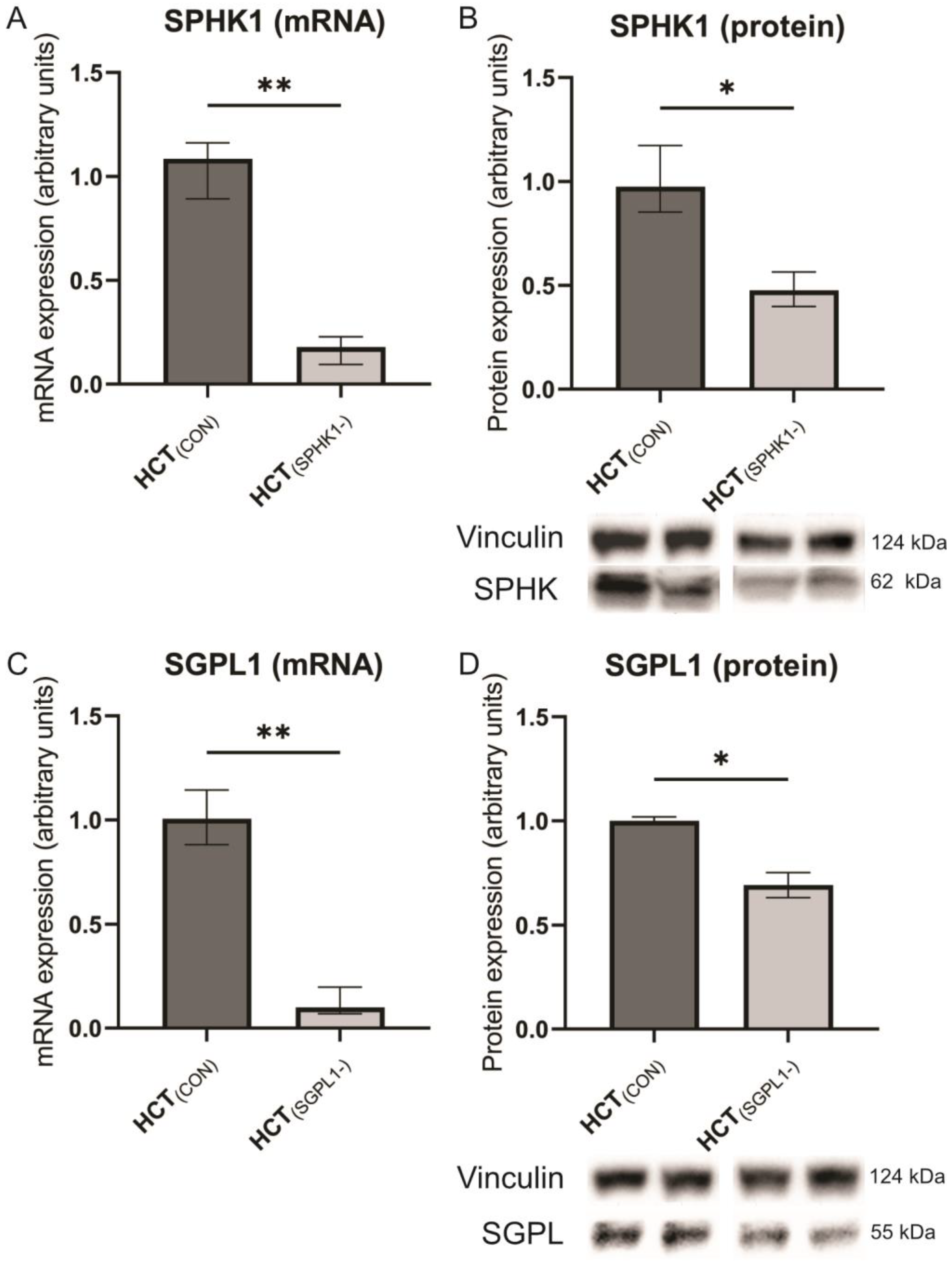
Both the RT-PCR assay and WB confirmed a significant reduction of SPHK1 mRNA and protein expression in silenced cells as compared to non-silenced control cells (p = 0.0022 and p = 0.0286, respectively) (Figure 2A,B). A similar profile of changes was observed in SGPL1-silenced cells at the level of mRNA and protein (p = 0.0022 and p = 0.0286, respectively) (Figure 2C,D).

2.2. Sphingolipids Content

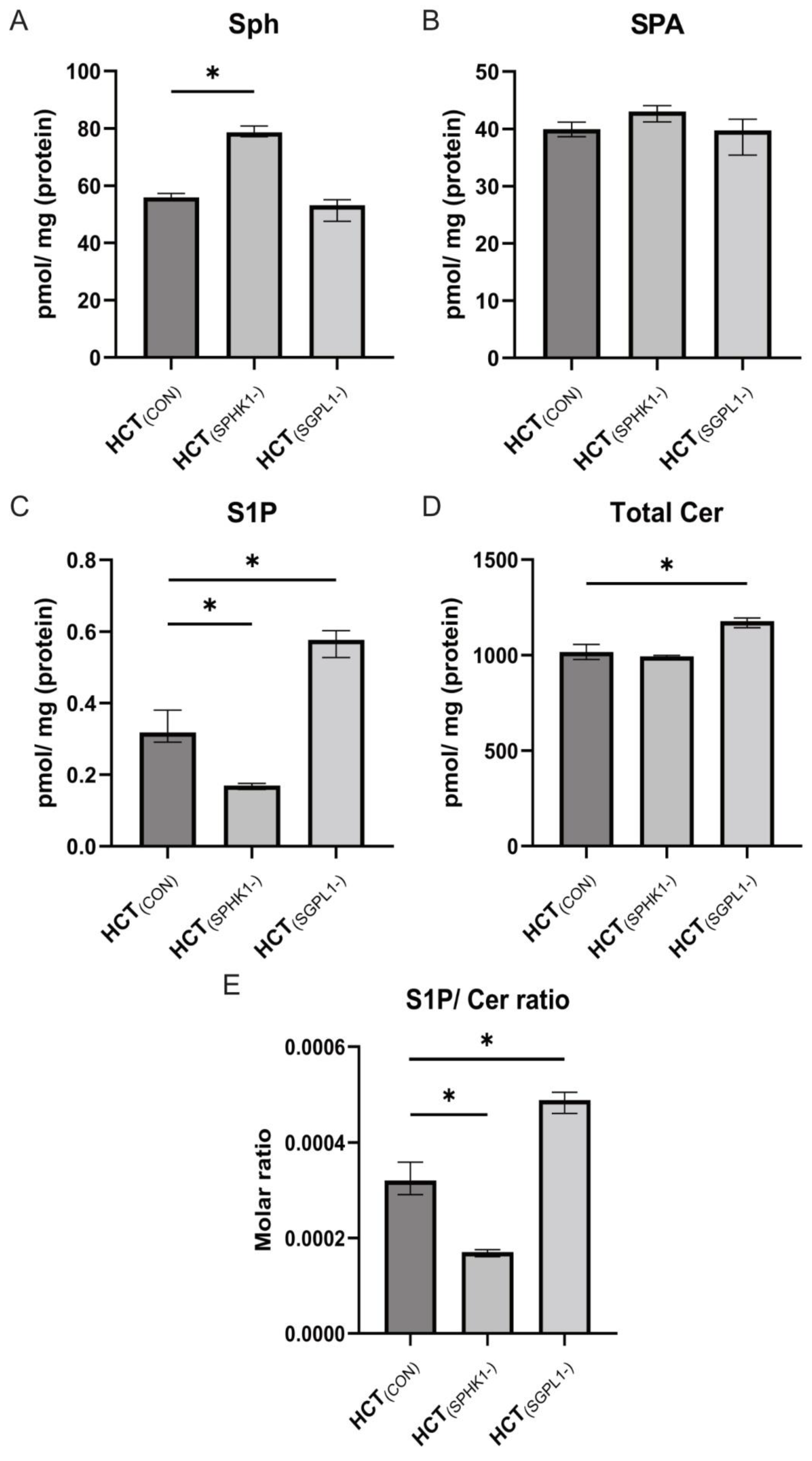
The silencing of genes encoding enzymes directly responsible for the level of S1P caused significant changes in the content of not only S1P, but also other sphingolipids. In cells in which the expression of SPHK1 was silenced, a significant increase in the content of sphingosine (Sph) (p = 0.0286) and a decrease in the content of S1P (p = 0.0286) were observed compared to control cells (Figure 3A,C). Apart from that, an increased content of C18:0-Cer and C18:1-Cer was observed in relation to the non-silenced control cells (p = 0.0286) (Table 1). A significant reduction in the S1P/Cer ratio (p = 0.0286) was also observed (Figure 3E).
In the case of silencing the SGPL1 gene, significant changes in the content of sphingolipids were also found compared to the control, non-silenced cells. Although the Sph and SPA levels did not change, there was an increase in the content of S1P, 16:0-Cer, C18:0-Cer, C18:1-Cer, C20:0-Cer, C22:0-Cer, and the total content of ceramides (p = 0.0286) (Table 1). Moreover, an increase in the S1P/Cer ratio was observed (p = 0.0286) (Figure 3E).

2.3. Caspases and Cathepsin-D

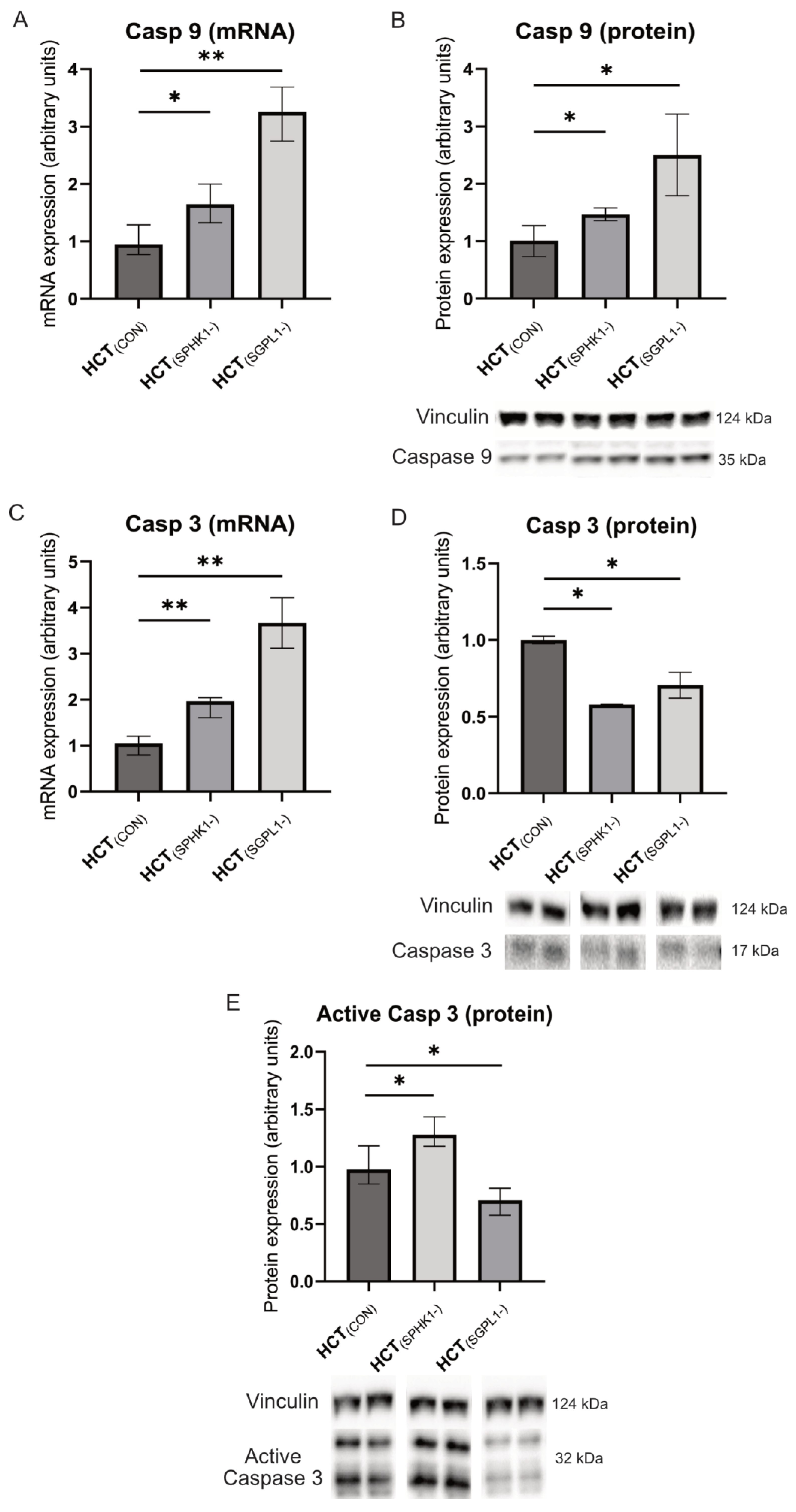
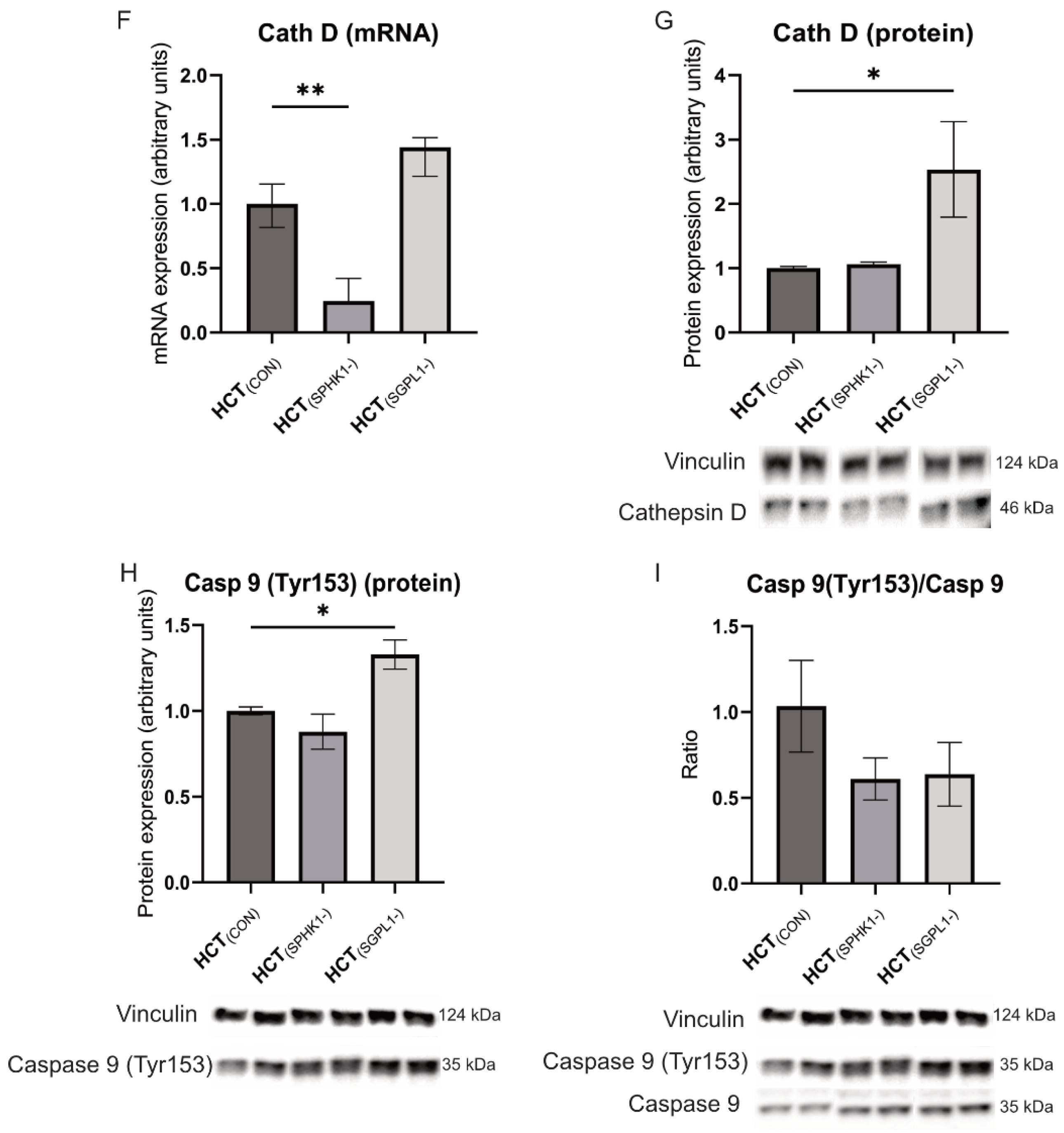
The RT-PCR technique revealed that silencing the SPHK1 gene in HCT-116 cells resulted in an increase in the expression level of caspases (Caspase-9 and Caspase-3) (p = 0.0152 and p = 0.0087, respectively) (Figure 4A,C) and a decrease in Cathepsin-D expression at the mRNA level (p = 0.0022) (Figure 4F). Western blot analysis showed an increased level and activation of Caspase-3 (Figure 4D,E), and an increased level of Caspase-9 compared to control, non-silenced cells (p = 0.0286) (Figure 4B). In addition, we noticed a negative trend in phosphorylation of the Tyr153 tyrosine residue in the Caspase-9 protein compared to non-silenced control cells (Figure 4H).
Silencing of the SGPL1 gene in HCT-116 cells upregulated Caspase-9, Caspase-3, and Cathepsin-D at the mRNA level as compared to non-silenced cells (Figure 4A,C,F). At the protein level, we observed a decreased Caspase-3 content and activation state (p = 0.0286) (Figure 4D,E) and an increased Caspase-9 and Cathepsin-D content as compared to non-silenced control cells (p = 0.0286) (Figure 4B,G). Moreover, despite the high content of Caspase-9 and its Tyr153-phosphorylated form, which suggest its activation, we observed a negative trend in the phosphorylation state of this protein in SGPL1-silenced cells as compared to non-silenced control cells (Figure 4I).

2.4. Cellular Apoptosis

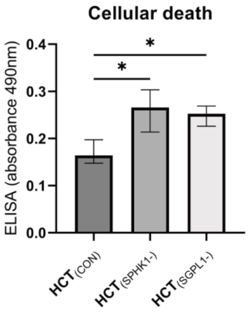
The apoptosis test (ELISA PLUS) confirmed the increase in the death rate of both the SPHK1 (p = 0.0286)- and SGPL1 (p = 0.0286)-silenced cells (Figure 5).

3. Discussion

Sphingosine kinase performs an essential role in the regulation of the metabolic balance of sphingolipids such as ceramides and S1P. Ceramides inhibit cell proliferation and stimulate apoptosis, whereas S1P promotes cellular proliferation, motility, and survival. The content of these compounds in a cell is dynamically maintained, and the balance between them seems to act as a switch that determines the fate of the cell, choosing between cellular proliferation and death [50,51].
SPHK1 is located predominantly in the cytosol, but after exposure to appropriate stimuli, is activated by phosphorylation and translocated to the cell membrane. Active, membrane-bound sphingosine kinase mediates the conversion of sphingosine to sphingosine-1-phosphate (S1P). S1P acts as a secondary signal transmitter, taking part in apoptosis, proliferation, migration, and cancer invasion.
In our study, silencing the gene encoding SPHK1, the enzyme responsible for the formation of S1P, led to the expected decrease in S1P levels and caused a reduction in the ratio of sphingosine-1-phosphate to total ceramide content. The decrease in the level of S1P and the ratio of S1P to ceramide at the same time resulted in a significant increase in apoptosis in these cells. Our findings have clinical relevance, as they strongly confirm the cause and effect relationship between a low level of S1P and intensification of apoptosis in CRC cells.
Our conclusions are confirmed by the existing literature data. It has been shown that SPHK1 is upregulated in multiple types of human cancers [52], and the expression of SPHK1 in CRC tissue is significantly increased compared to normal colorectal tissue [53]. Elevated SPHK1 augments colon cancer cell proliferation [54], and SPHK1 overexpression in intestinal epithelium significantly increases tumor multiplicity [55]. On the other hand, the intestinal epithelial deletion of SPHK1 prevents colitis-associated cancer development in mice [56], SPHK inhibitor treatment resulted in a dose-dependent decrease in colitis-driven colon cancer in mice [57], and SPHK1 knockout mice demonstrated a lower incidence of colon cancer development in murine model [24].
It seems that SPHK1 regulates tumorigenesis and tumor growth in early colon cancer [55]. Moreover, SPHK1 induces the first step of a metastatic cascade, which is the epithelial-to-mesenchymal transition [58]. The positive SPHK1 expression in advanced colorectal cancer (stages III and IV) was higher than in less advanced tumors [58]. The expression density of SPHK1 in primary colorectal cancer tissues was higher compared with normal colonic mucosa tissues, whereas the expression of E-cadherin was lower [58]. Moreover, patients with SPHK1-positive colorectal cancer cells had a significantly lower survival rate compared with patients with SPHK1-negative cancer [58]. Additionally, the recent meta-analysis involving 32 cohorts with 5965 patients confirmed that high SPHK1 expression was significantly associated with poor overall survival and worse disease-free survival [59]. S1P is catabolized by SGPL1 and the S1P phosphatases. Since only SGPL1 irreversibly degrades S1P, it has a particularly strong influence on intracellular and tissue levels of S1P. S1P stimulates cell proliferation and promotes invasion in colon cancer cell lines [60], and neutralizing S1P with highly specific anti-S1P monoclonal antibodies blocks S1P’s pro-survival actions (by increasing activation of Caspase-3), which was manifested by the inhibition of tumor progression and reduction of the tumor volume in human cancer models [61]. In turn, S1P treatment decreased Caspase-3 protein levels and activity in nontumorigenic intestinal epithelial cells [62].
Caspases (a family of protease enzymes) are important mediators of apoptosis. There are two main signaling pathways involved in apoptosis: the extrinsic (death receptor) pathway and the intrinsic (mitochondrial) pathway. Both require Caspase-8 and Caspase-9 activation, respectively, and both activate Caspase-3, leading to cellular death. Caspase-3 translocates to the nucleus during apoptosis to cleave targets such as poly-ADP ribose polymerase [63]. In colon cancer cells with deficiency in CerS6 expression, Caspase-3 may, despite its activation, not be able to translocate to the nucleus, altering late-stage apoptotic signaling [64]. Caspase-3 regulates the migration, invasion, and metastasis of colon cancer cells. It has been proven that Caspase-3 knockout HCT-116 cells show altered cellular morphology and reduced facility to epithelial-to-mesenchymal transition [65]. It was recently shown that mRNA expressions of Caspase-9 and Caspase-3 (unlike Caspase-8) were downregulated in human colorectal cancer tissues in comparison to marginal tissues [66]. The same study also identified a correlation between the caspases expression and the severity of tumor stage, distant metastases, and differentiation stage, while no significant association was seen with age, sex, and smoking. Another study revealed a positive association of Caspase-3 expression with a patient’s age and tumor histological type, and a negative correlation with depth of tumor invasion, tumor grade, tumor stage (II to IV), and the presence of lymph node metastases [67]. On the other hand, no relationship was demonstrated between Caspase-3 expression and patients’ sex, tumor location (right colon vs. left colon), tumor size, and presence of lymphovascular invasion or perineural invasion [67]. The above findings underline the diversity of the Caspase function in CRC, which can arise from diversity of molecular pathways responsible for carcinogenesis.
In our study, we have noticed an increase in the expression of Caspase-9 and Caspase-3 at the mRNA level after SPHK1 gene silencing. However, at the protein level, we observed only an increase in the Caspase-9 protein expression, while the Caspase-3 protein was decreased. Despite this, Caspase-3 active form (cut into low molecular mass subunits) displayed upregulation in SPHK1-silenced cells as compared to both the control and SGPL1-silenced cells, which coincided with the lowest S1P/Cer ratio and downregulation of Cathepsin-D mRNA expression. Interestingly, SGPL1 silencing led to downregulation of the Caspase-3 protein and its active form, despite increased Caspase-9 Tyr153 phosphorylation. SGPL1-silenced cells also displayed the highest S1P/Cer ratio and high protein expression of Cathepsin D, yet significantly higher cellular death than controls.
A high level of mRNA expression and protein level of Cathepsin-D were observed in colorectal cancer [43]. Up to 78% of CRC cases strongly express Cathepsin-D, while no significant staining is observed in the normal mucosa [68]. Additionally, it was found that Cathepsin-D expression is greatest in invasive areas, suggesting its involvement in tumor progression [68]. Inhibition of Cathepsin-D markedly enhances anticancer drug-induced apoptosis in a few human cancer cells [42]. Cathepsin-D, the most abundant lysosomal protease, appears to have diverse properties, from pro-apoptotic to pro-invasive, and pro-metastatic. This underlines that its effect on cell death is controversial and depends on cellular type, stimulators, and cell context [42,43,69]. Besides, Cathepsin-D mainly exists in lysosomes, but under specific conditions also localizes in the cytosol and extracellular space, displaying involvement in apoptosis and metastasis, respectively. Therefore, the ability to modulate cell death and proliferation may differ also based on Cathepsin-D localization [42].
The direct effects of sphingolipids on Cathepsin-D activity can yield inhibition or activation, which depends on the type and structure of the sphingolipid species. Sphingosine and short-chain ceramide inhibits Cathepsin-D activity, whereas sphingolipid phosphates were shown to activate this enzyme at low micromolar range (in both cases in a pH-dependent manner) [44]. Ceramide-1-phospahte displayed the strongest activatory properties, with S1P being somehow weaker in this aspect. At the gene expression level, modulation of the content of both the S1P and CER affects cathepsins expression. In prostate cancer cells, degradation of ceramide through the action of lysosomal acid ceramidase upregulates Cathepsin-B (CTSB) expression in a S1P-dependent manner [70]. Contrary to the above findings, ablation of the SGLP1 gene in mouse embryonic fibroblasts inhibits maturation of Cathepsin-D and decreases overall lysosomal activity [71]. Regarding ceramide, its accumulation is crucial in the activation of CTSB in the cellular model of Parkinson’s disease [72]. In light of the above data, we can hypothesize that expression of Cathepsin-D in CRC cells follows overall S1P content and the S1P/Cer ratio, with the lowest mRNA expression in this enzyme concomitant with a low S1P/Cer ratio and S1P content in SPHK1-silenced cells and the highest protein expression under a high S1P/Cer ratio and S1P accumulation in SGLP1-silenced cells.
In our study, silencing the SPHK1 gene in HCT-116 cells resulted in changes not only restricted to the expected reduction in S1P content, but also caused an increase in the level of sphingosine and some ceramides (C18:0-Cer and C18:1-Cer). As a result, we observed a significantly reduced S1P/Ceramide ratio, which is critical to cell death and survival. Accordingly, we observed activation of Caspase-3 and an increase in the apoptosis-mediated cell death. Moreover, SPHK1-silenced cells displayed an increase in the expression of Cas-9 and Cas-3, and a decrease in expression of Cathepsin-D at the mRNA level.
Sphingosine-1-phosphate lyase located in the endoplasmic reticulum is a membrane-bound important enzyme, which irreversibly hydrolyzes S1P, and acts as the metabolic sink for the highly bioactive sphingoid backbones. In HCT-116 cells with the SGPL1-silenced gene, we noted the expected elevation in the content of S1P, but also an increase in the total ceramide content. We also observed that, although the S1P-to-ceramide ratio increased in cells with the silenced SGPL1 gene, it did not prevent apoptosis. However, in these cells, the levels of ceramide also increased, and it seems that the observed intensification in apoptosis is precisely due to the increase in ceramide levels. The puzzling upregulation of ceramide content in SGPL1-silenced cells could be explained by the unique role of the SGLP1 enzyme in sphingolipid metabolism. Contrary to the salvage pathway (sphingosine re-acylation towards complex sphingolipids), SGLP1 ensures permanent degradation of sphingoid backbones of S1P and Spa1P towards phosphoethanolamine and hexadecenal via irreversible reaction. SGLP1 silencing creates a bottleneck in degradation of S1P and sphingoid backbones located upstream of this enzyme. Contrary to S1P, sphingosine can directly enter the ceramide synthesis pathway, which, in the case of the inhibition of sphingoid backbone degradation via SGLP1 silencing, can increase ceramide accumulation. This effect was likely the reason for the increase of CER content in the SGLP1-silenced cells.
Previous reports showed that SGPL1 was downregulated in colon cancer tissue, leading to S1P accumulation in neoplastic intestinal tissues [28]. A significant reduction in SGPL1 expression and activity was also found in adenomas and colitis-associated carcinoma in the mouse model compared to the control [28,73]. However, in contrast to these findings, the increased SGPL (and SPHK) mRNA levels were recently reported in human colon cancer tissues compared with the adjacent nontumorous tissues [74]. Moreover, loss of SGPL1 promotes oncogenesis [75], and reduction of SGPL levels in HCT 116 cells led to reduced cell proliferation and invasion, but unaltered migration [74]. It seems that the expression of SGPL in colorectal cancer tissue may change depending on the tumor stage, hence the different results of the SGPL role in different patient populations. The variable expression in cancer tissue of enzymes (or genes) involved in the synthesis and degradation of ceramides could also explain the varying levels of ceramides in cancer tissue observed in our previous study [1]. RT-PCR analysis of the surgically resected samples showed that SGPL1 mRNA tended to be highly expressed in human colorectal cancer tissues compared with normal mucosa tissues [76]. SGPL1 mRNA expression was by far the highest among all S1P metabolizing enzymes in two human colorectal cancer cell lines; selective reduction of SGPL1 mRNA expression (SPHK1, SPHK2, SGPP1, and SGPP2 remained unchanged) and complementary reduction of the SGPL1 enzyme level increased intracellular S1P levels but did not affect cell proliferation or metabolic activity of the cells [77]. It has been shown that diminished SGPL1 expression induced a partial redifferentiation of human colorectal cancer cell line towards normal colon epithelial cells, as a result of E-cadherin upregulation, an increase in intercellular adhesion, and inhibited cell migration. It seems that a change in SGPL1 expression is associated with the malignancy of already established colon cancer cells [77]. However, other studies showed a different mechanism of SGPL1 involvement in carcinogenesis [73,76] and revealed that SGPL1 knockout also promoted the transformation of normal mouse colon epithelium into cancer cells and provoked the immediate appearance of neoplastic tumors [78].

4. Materials and Methods

4.1. Cell Culture Experiments

The human colorectal carcinoma cell line (HCT-116) was purchased from American Type Cell Culture Collection (ATCC). HCT-116 were cultured in dedicated media (McCoy 5A, Gibco, New York, NY, USA) containing 10% fetal bovine serum (FBS, Gibco), streptomycin (50 µg/mL) and penicillin (50 units/mL; Gibco). Additionally, media were supplemented with albumin-conjugated free fatty acids (150 µM), namely, oleic and palmitic in a ratio 2:1 (mol/mol). Cell lines were cultured in humanified conditions (37 °C, 5% CO2). Cells from the third to sixth passage were acquired for experiments.
To silence the expression of SPHK1 and SGPL1 genes, we employed a siRNA-mediated RNA interference approach [79]. A total of 250 × 103 HCT-116 cells were seeded on a 6-well plate and incubated with esiRNA (10 nM/L) and 10 µL of transfectant per well (both supplied by Sigma Aldrich, Saint Louis, MO, USA). SPHK1 (HCT(SPHK1-)) and SGPL1 (HCT(SGPL1-)) genes were silenced, while human eGFP was used as control (HCT(CON)). After 72 h from transfection, cells were collected for Western blot, qPCR assay, and lipidomics. All the experiments were repeated four times (n = 4), except qPCR assays (n = 6).

4.2. Evaluation of Cellular Apoptosis

Cell culture was established as described above. Supernatants were collected for ELISA detection of histone release in apoptotic bodies. The ELISA procedure (Cell Death Detection ELISA Plus, Roche, Indianapolis, IN, USA) was performed according to standard manufacturer protocol. Results were acquired using the VarioscanLux instrument (ThermoFisher, Waltham, MA, USA) at 490 nm.

4.3. Sphingolipid Measurements

The level of sphingolipids was measured by the UHPLC/MS/MS method, according to Blachnio-Zabielska et al. [80], with minor modifications. Briefly, cells were homogenized in a buffer consisting of 0.25 M sucrose, 25 mM KCl, 50 mM Tris, and 0.5 mM EDTA, pH 7.4. The protein concentration in each homogenate was determined using a Pierce660nm protein assay kit (Thermo Fisher Scientific). Immediately afterwards, a mixture of internal standards (Sph-d7, SPA-d7, S1P-d7, C15:0-d7-Cer, C16:0-d7-Cer, C18:1-d7-Cer, C18:0-d7-Cer, 17C/20:0-Cer, C24:1-d7-Cer, C24-d7-Cer Avanti Polar Lipids, Alabaster, AL, USA) was added to each sample and the samples were vortexed. After that, an extraction mixture (isopropanol:water:ethyl acetate, 30:10:60; v:v:v) was added to each sample, followed by sonication and centrifugation at 4000 rpm for 10 min at 4 °C. After extraction, the samples were dried under a stream of nitrogen and suspended in LC Solvent B (2 mM ammonium formate, 0.1% formic acid in methanol) for UHPLC/MS/MS analysis. The chromatographic separation was performed on a reversed-phase column—Zorbax SB-C8 2.1 × 150 mm, 1.8 μm (Agilent Technologies, Santa Clara, CA, USA) in a binary gradient, using 1 mM ammonium formate, 0.1% formic acid in water as solvent A, and 2 mM ammonium formate, 0.1% formic acid in methanol as solvent B, at the flow rate of 0.4 mL/min. Sphingolipids were analyzed with the use of a Sciex QTRAP 6500 + triple quadrupole mass spectrometer (AB Sciex Germany GmbH, Darmstadt, Germany) with multiple reaction monitoring (MRM) against standard curves constructed for each compound.

4.4. Western Blotting

Proteins were isolated from cells in RIPA buffer (Sigma-Aldrich) containing 0.5 mM TCEP (Sigma-Aldrich) and protease and phosphatase inhibitors (cOmplete ULTRA mini and PhosSTOP tablets, Roche). The content of protein in homogenates was measured using a Pierce 660nm protein assay kit (Thermo Fisher Scientific). Fatty-acid-free bovine serum albumin was used as a standard. After denaturation in Laemmli buffer, proteins (30 µg) were separated by SDS-PAGE (Criterion Cell electrophoresis cell and Criterion TGX midi Any kD gel) and transferred to a PVDF membrane (BioRad Trans Blot SD semi-dry transfer cell with a discontinuous Tris/CAPS buffer system; 15% methanol for the anode and Tris/CAPS/0.1% SDS for the cathode). Membranes were incubated with the appropriate primary antibody. The following target proteins were quantified using primary antibodies: sphingosine kinase 1 (SphK1), sphingosine-1-phosphate lyase 1 (SGPL1), Caspase-3, active Caspase-3 (cleaved), Caspase-9, Caspase-9 (Tyr-153), Cathepsin-D, and vinculin (vendor Cell Signaling Technology, Danvers, MA, USA). To detect primary antibody binding to protein, the membrane was incubated with an HRP-conjugated secondary antibody, followed by incubation in a Clarity ™ Western ECL chemiluminescent substrate (Bio-Rad) and visualization using a Bio-Rad ChemiDoc XRS + imaging system. Band intensities were quantified with the Bio-Rad Image Lab software package. The obtained values were normalized to the vinculin protein expression, as measured from parallel runs and expressed as fold changes over control group value. Unless otherwise stated, all chemicals and equipment used for immunoblotting were purchased from Bio-Rad (Hercules, CA, USA). Unless stated otherwise, all antibodies were purchased from Thermo Fisher Scientific (Waltham, MA, USA).

4.5. Real-Time PCR

Total RNA was isolated from cells with the use of a mirVana Isolation Kit (ThermoScientific, USA) according to the manufacturer’s instructions. The RNA was reverse-transcribed into cDNA using a Transcriptor First Strand cDNA Synthesis Kit (Roche). Real-time PCR was performed with the use of the following primers: sphingosine kinase 1 (SphK1), forward, 5′-TTCCTTGAACCATTATGCTG-3′, reverse, 5′-GATACTTCTCACTCTCTAGGTC-3′; Sphingosine-1-phosphate lyase 1 (SGPL1), forward, 5′-AATGAGAAGAGCTATCTCCAG-3′, reverse, 5′-TTTTGTATTTGACAGCCAGC-3′; Caspase-3, forward, 5′-TTTCGTGAGTGCTCGCAGC-3′, reverse, 5′-CCTTTATTAACGAAAACCAGAGCG-3′; Caspase-9, forward, 5′-TTGGTTCTGGAGGATTTGGT-3′, reverse, 5′-TGCTCAGGATGTAAGCCAAA-3′; Cathepsin-D, forward, 5′-GCTGATTCAGGGCGAGTACA-3′, reverse, 5′-TCCCAGCTTCAGTGTGATCG-3′ and glyceraldehyde 3-phosphate dehydrogenase (GAPDH), forward, 5′-TCGGAGTCAACGGATTTG-3′, reverse, 5′-CAACAATATCCACTTTACCAGAG-3′. GAPDH was used as a housekeeping gene. PCR reaction was performed using a LightCycler480 system (Roche Mannheim, Germany). The results were normalized to GAPDH expression measured in each sample and the relative expression levels were evaluated using the ΔΔCt method.

4.6. Statistical Analyses

GraphPad Prism 9 was used to perform all statistical analyses. The results were expressed as median from 4 independent experiments (n = 4; except qPCR n = 6) with an interquartile range. Significant differences were identified by the nonparametric Mann–Whitney test. The significance threshold was set at p < 0.05.

5. Conclusions

Our findings confirmed that the modification of sphingolipid metabolism at the level of sphingosine-1-phosphate and the S1P/Cer ratio through silencing of SPHK1 or SGPL1 affects the apoptosis of CRC cells. The decrease in S1P levels and the S1P/Cer ratio at the same time results in a significant increase in CRC cell apoptosis, which was accompanied by upregulation in the active form of executioner Caspase-3. Conversely, an increase in S1P levels and the S1P/Cer ratio in SGPL1-silenced cells led to downregulation of Caspase-3 active form and a subsequent increase in Cathepsin-D protein expression in CRC cells. The downregulation of active Caspase-3 and upregulation of Cathepsin-D suggest increased pro-metastatic capacity of those cells.

Author Contributions

Conceptualization, A.R.M. and A.U.B.-Z.; methodology, A.U.B.-Z., P.Z. and A.Ż.; formal analysis, A.U.B.-Z., A.Ż., U.C., P.S. and K.P.; writing—original draft preparation, A.R.M.; writing—review and editing, A.U.B.-Z. and P.Z.; visualization, A.Ż. and A.U.B.-Z. All authors have read and agreed to the published version of the manuscript.

Funding

This research was supported by grants SUB/1/DN/21/001/1117 and N/ST/ZB/18/002/1117 from the Medical University of Bialystok.

Institutional Review Board Statement

Not applicable.

Informed Consent Statement

Not applicable.

Data Availability Statement

Not applicable.

Conflicts of Interest

The authors declare no conflict of interest.

References

  1. Markowski, A.R.; Blachnio-Zabielska, A.U.; Guzińska-Ustymowicz, K.; Markowska, A.; Pogodzińska, K.; Roszczyc, K.; Zińczuk, J.; Zabielski, P. Ceramides Profile Identifies Patients with More Advanced Stages of Colorectal Cancer. Biomolecules 2020, 10, 632. [Google Scholar] [CrossRef] [Green Version]
  2. Vogelstein, B.; Fearon, E.R.; Hamilton, S.R.; Kern, S.E.; Preisinger, A.C.; Leppert, M.; Nakamura, Y.; White, R.; Smits, A.M.; Bos, J.L. Genetic alterations during colorectal-tumor development. N. Engl. J. Med. 1988, 319, 525–532. [Google Scholar] [CrossRef] [PubMed] [Green Version]
  3. Arnold, C.N.; Goel, A.; Blum, H.E.; Boland, C.R. Molecular pathogenesis of colorectal cancer: Implications for molecular diagnosis. Cancer 2005, 104, 2035–2047. [Google Scholar] [CrossRef]
  4. Siegel, R.L.; Miller, K.D.; Goding Sauer, A.; Fedewa, S.A.; Butterly, L.F.; Anderson, J.C.; Cercek, A.; Smith, R.A.; Jemal, A. Colorectal cancer statistics, 2020. CA Cancer J. Clin. 2020, 70, 145–164. [Google Scholar] [CrossRef] [PubMed] [Green Version]
  5. Hannun, Y.A. Functions of ceramide in coordinating cellular responses to stress. Science 1996, 274, 1855–1859. [Google Scholar] [CrossRef]
  6. Speirs, M.M.P.; Swensen, A.C.; Chan, T.Y.; Jones, P.M.; Holman, J.C.; Harris, M.c.C.B.; Maschek, J.A.; Cox, J.E.; Carson, R.H.; Hill, J.T.; et al. Imbalanced sphingolipid signaling is maintained as a core proponent of a cancerous phenotype in spite of metabolic pressure and epigenetic drift. Oncotarget 2019, 10, 449–479. [Google Scholar] [CrossRef] [PubMed] [Green Version]
  7. Newton, J.; Lima, S.; Maceyka, M.; Spiegel, S. Revisiting the sphingolipid rheostat: Evolving concepts in cancer therapy. Exp. Cell Res. 2015, 333, 195–200. [Google Scholar] [CrossRef] [Green Version]
  8. Liu, X.; Cheng, J.C.; Turner, L.S.; Elojeimy, S.; Beckham, T.H.; Bielawska, A.; Keane, T.E.; Hannun, Y.A.; Norris, J.S. Acid ceramidase upregulation in prostate cancer: Role in tumor development and implications for therapy. Expert Opin. Ther. Targets 2009, 13, 1449–1458. [Google Scholar] [CrossRef] [Green Version]
  9. Zeng, S.; Liang, Y.; Hu, H.; Wang, F.; Liang, L. Endothelial cell-derived S1P promotes migration and stemness by binding with GPR63 in colorectal cancer. Pathol. Res. Pract. 2022, 240, 154197. [Google Scholar] [CrossRef]
  10. del Solar, V.; Lizardo, D.Y.; Li, N.; Hurst, J.J.; Brais, C.J.; Atilla-Gokcumen, G.E. Differential Regulation of Specific Sphingolipids in Colon Cancer Cells during Staurosporine-Induced Apoptosis. Chem. Biol. 2015, 22, 1662–1670. [Google Scholar] [CrossRef]
  11. Kawamori, T.; Osta, W.; Johnson, K.R.; Pettus, B.J.; Bielawski, J.; Tanaka, T.; Wargovich, M.J.; Reddy, B.S.; Hannun, Y.A.; Obeid, L.M.; et al. Sphingosine kinase 1 is up-regulated in colon carcinogenesis. FASEB J. 2006, 20, 386–388. [Google Scholar] [CrossRef] [Green Version]
  12. Grbčić, P.; Sedić, M. Sphingosine 1-Phosphate Signaling and Metabolism in Chemoprevention and Chemoresistance in Colon Cancer. Molecules 2020, 25, 2436. [Google Scholar] [CrossRef]
  13. Maceyka, M.; Sankala, H.; Hait, N.C.; Stunff, H.L.; Liu, H.; Toman, R.; Collier, C.; Zhang, M.; Satin, L.S.; Merrill, A.H., Jr.; et al. SphK1 and SphK2, sphingosine kinase isoenzymes with opposing functions in sphingolipid metabolism. J. Biol. Chem. 2005, 280, 37118–37129. [Google Scholar] [CrossRef] [PubMed] [Green Version]
  14. Okada, T.; Ding, G.; Sonoda, H.; Kajimoto, T.; Haga, Y.; Khosrowbeygi, A.; Gao, S.; Miwa, N.; Jahangeer, S.; Nakamura, S.I. Involvement of N-terminal-extended form of sphingosine kinase 2 in serum-dependent regulation of cell proliferation and apoptosis. J. Biol. Chem. 2005, 280, 36318–36325. [Google Scholar] [CrossRef] [Green Version]
  15. Hait, N.C.; Allegood, J.; Maceyka, M.; Strub, G.M.; Harikumar, K.B.; Singh, S.K.; Luo, C.; Marmorstein, R.; Kordula, T.; Milstien, S.; et al. Regulation of histone acetylation in the nucleus by sphingosine-1-phosphate. Science 2009, 325, 1254–1257. [Google Scholar] [CrossRef] [PubMed] [Green Version]
  16. Don, A.S.; Martinez-Lamenca, C.; Webb, W.R.; Proia, R.L.; Roberts, E.; Rosen, H. Essential requirement for sphingosine kinase 2 in a sphingolipid apoptosis pathway activated by FTY720 analogues. J. Biol. Chem. 2007, 282, 15833–15842. [Google Scholar] [CrossRef] [Green Version]
  17. Zhang, L.; Liu, X.; Zuo, Z.; Hao, C.; Ma, Y. Sphingosine kinase 2 promotes colorectal cancer cell proliferation and invasion by enhancing MYC expression. Tumour Biol. 2016, 37, 8455–8460. [Google Scholar] [CrossRef]
  18. Zhang, Y.H.; Shi, W.N.; Wu, S.H.; Miao, R.R.; Sun, S.Y.; Luo, D.D.; Wan, S.B.; Guo, Z.K.; Wang, W.Y.; Yu, X.F.; et al. SphK2 confers 5-fluorouracil resistance to colorectal cancer via upregulating H3K56ac-mediated DPD expression. Oncogene 2020, 39, 5214–5227. [Google Scholar] [CrossRef]
  19. Mizutani, N.; Omori, Y.; Tanaka, K.; Ito, H.; Takagi, A.; Kojima, T.; Nakatochi, M.; Ogiso, H.; Kawamoto, Y.; Nakamura, M.; et al. Increased SPHK2 Transcription of Human Colon Cancer Cells in Serum-Depleted Culture: The Involvement of CREB Transcription Factor. J. Cell. Biochem. 2015, 116, 2227–2238. [Google Scholar] [CrossRef] [PubMed]
  20. Xun, C.; Chen, M.B.; Qi, L.; Zhang, T.-N.; Peng, X.; Ning, L.; Chen, Z.-X.; Wang, L.-W. Targeting sphingosine kinase 2 (SphK2) by ABC294640 inhibits colorectal cancer cell growth in vitro and in vivo. J. Exp. Clin. Cancer Res. 2015, 34, 94. [Google Scholar] [CrossRef] [PubMed] [Green Version]
  21. Chu, J.H.; Gao, Z.H.; Qu, X.J. Down-regulation of sphingosine kinase 2 (SphK2) increases the effects of all-trans-retinoic acid (ATRA) on colon cancer cells. Biomed. Pharmacother. 2014, 68, 1089–1097. [Google Scholar] [CrossRef]
  22. Nemoto, S.; Nakamura, M.; Osawa, Y.; Kono, S.; Itoh, Y.; Okano, Y.; Murate, T.; Hara, A.; Ueda, H.; Nozawa, Y.; et al. Sphingosine kinase isoforms regulate oxaliplatin sensitivity of human colon cancer cells through ceramide accumulation and Akt activation. J. Biol. Chem. 2009, 284, 10422–10432. [Google Scholar] [CrossRef] [PubMed] [Green Version]
  23. Zhang, Y.; Wang, Y.; Wan, Z.; Liu, S.; Cao, Y.; Zeng, Z. Sphingosine kinase 1 and cancer: A systematic review and meta-analysis. PLoS ONE 2014, 9, e90362. [Google Scholar] [CrossRef]
  24. Kawamori, T.; Kaneshiro, T.; Okumura, M.; Maalouf, S.; Uflacker, A.; Bielawski, J.; Hannun, Y.A.; Obeid, L.M. Role for sphingosine kinase 1 in colon carcinogenesis. FASEB J. 2009, 23, 405–414. [Google Scholar] [CrossRef] [PubMed] [Green Version]
  25. Tan, S.S.; Khin, L.W.; Wong, L.; Yan, B.; Ong, C.W.; Datta, A.; Salto-Tellez, M.; Lam, Y.; Yap, C.T. Sphingosine kinase 1 promotes malignant progression in colon cancer and independently predicts survival of patients with colon cancer by competing risk approach in South asian population. Clin. Transl. Gastroenterol. 2014, 5, e51. [Google Scholar] [CrossRef]
  26. Oskouian, B.; Saba, J. Sphingosine-1-phosphate metabolism and intestinal tumorigenesis: Lipid signaling strikes again. Cell Cycle 2007, 6, 522–527. [Google Scholar] [CrossRef]
  27. Le Stunff, H.; Galve-Roperh, I.; Peterson, C.; Milstien, S.; Spiegel, S. Sphingosine-1-phosphate phosphohydrolase in regulation of sphingolipid metabolism and apoptosis. J. Cell Biol. 2002, 158, 1039–1049. [Google Scholar] [CrossRef]
  28. Oskouian, B.; Sooriyakumaran, P.; Borowsky, A.D.; Crans, A.; Dillard-Telm, L.; Tam, Y.Y.; Bandhuvula, P.; Saba, J.D. Sphingosine-1-phosphate lyase potentiates apoptosis via p53- and p38-dependent pathways and is down-regulated in colon cancer. Proc. Natl. Acad. Sci. USA 2006, 103, 17384–17389. [Google Scholar] [CrossRef] [Green Version]
  29. Saba, J.D. Fifty years of lyase and a moment of truth: Sphingosine phosphate lyase from discovery to disease. J. Lipid Res. 2019, 60, 456–463. [Google Scholar] [CrossRef] [PubMed] [Green Version]
  30. Hertervig, E.; Nilsson, A.; Nyberg, L.; Duan, R.D. Alkaline sphingomyelinase activity is decreased in human colorectal carcinoma. Cancer 1997, 79, 448–453. [Google Scholar] [CrossRef]
  31. García-Barros, M.; Coant, N.; Kawamori, T.; Wada, M.; Snider, A.J.; Truman, J.P.; Wu, B.X.; Furuya, H.; Clarke, C.J.; Bialkowska, A.B.; et al. Role of neutral ceramidase in colon cancer. FASEB J. 2016, 30, 4159–4171. [Google Scholar] [CrossRef] [Green Version]
  32. Poreba, M.; Groborz, K.; Navarro, M.; Snipas, S.J.; Drag, M.; Salvesen, G.S. Caspase selective reagents for diagnosing apoptotic mechanisms. Cell Death Differ. 2019, 26, 229–244. [Google Scholar] [CrossRef] [PubMed]
  33. García-Barros, M.; Coant, N.; Truman, J.P.; Snider, A.J.; Hannun, Y.A. Sphingolipids in colon cancer. Biochim. Biophys. Acta 2014, 1841, 773–782. [Google Scholar] [CrossRef] [Green Version]
  34. Davaille, J.; Li, L.; Mallat, A.; Lotersztajn, S. Sphingosine 1-phosphate triggers both apoptotic and survival signals for human hepatic myofibroblasts. J. Biol. Chem. 2002, 277, 37323–37330. [Google Scholar] [CrossRef] [Green Version]
  35. Panneer Selvam, S.; Roth, B.M.; Nganga, R.; Kim, J.; Cooley, M.A.; Helke, K.; Smith, C.D.; Ogretmen, B. Balance between senescence and apoptosis is regulated by telomere damage-induced association between p16 and caspase-3. J. Biol. Chem. 2018, 293, 9784–9800. [Google Scholar] [CrossRef] [Green Version]
  36. Shin, J.H.; Choi, G.S.; Kang, W.H.; Myung, K.B. Sphingosine 1-phosphate triggers apoptotic signal for B16 melanoma cells via ERK and caspase activation. J. Korean Med. Sci. 2007, 22, 298–304. [Google Scholar] [CrossRef] [Green Version]
  37. Weigert, A.; Cremer, S.; Schmidt, M.V.; von Knethen, A.; Angioni, C.; Geisslinger, G.; Brüne, B. Cleavage of sphingosine kinase 2 by caspase-1 provokes its release from apoptotic cells. Blood 2010, 115, 3531–3540. [Google Scholar] [CrossRef] [Green Version]
  38. Trejo-Solis, C.; Escamilla-Ramirez, A.; Jimenez-Farfan, D.; Castillo-Rodriguez, R.A.; Flores-Najera, A.; Cruz-Salgado, A. Crosstalk of the Wnt/β-Catenin Signaling Pathway in the Induction of Apoptosis on Cancer Cells. Pharmaceuticals 2021, 14, 871. [Google Scholar] [CrossRef]
  39. Boice, A.; Bouchier-Hayes, L. Targeting apoptotic caspases in cancer. Biochim. Biophys. Acta 2020, 1867, 118688. [Google Scholar] [CrossRef]
  40. Hounsell, C.; Fan, Y. The Duality of Caspases in Cancer, as Told through the Fly. Int. J. Mol. Sci. 2021, 22, 8927. [Google Scholar] [CrossRef]
  41. Pérez, E.; Lindblad, J.L.; Bergmann, A. Tumor-promoting function of apoptotic caspases by an amplification loop involving ROS, macrophages and JNK in Drosophila. eLife 2017, 6, e26747. [Google Scholar] [CrossRef] [PubMed]
  42. Seo, S.U.; Woo, S.M.; Im, S.S.; Jang, Y.; Han, E.; Kim, S.H.; Lee, H.; Lee, H.S.; Nam, J.O.; Gabrielson, E.; et al. Cathepsin D as a potential therapeutic target to enhance anticancer drug-induced apoptosis via RNF183-mediated destabilization of Bcl-xL in cancer cells. Cell Death Dis. 2022, 13, 115. [Google Scholar] [CrossRef] [PubMed]
  43. Oliveira, C.S.F.; Pereira, H.; Alves, S.; Castro, L.; Baltazar, F.; Chaves, S.R.; Preto, A.; Côrte-Real, M. Cathepsin D protects colorectal cancer cells from acetate-induced apoptosis through autophagy independent degradation of damaged mitochondria. Cell Death Dis. 2015, 6, e1788. [Google Scholar] [CrossRef] [PubMed] [Green Version]
  44. Zebrakovská, I.; Máša, M.; Srp, J.; Horn, M.; Vávrová, K.; Mareš, M. Complex modulation of peptidolytic activity of cathepsin D by sphingolipids. Biochim. Biophys. Acta 2011, 1811, 1097–1104. [Google Scholar] [CrossRef]
  45. Cuvillier, O.; Rosenthal, D.S.; Smulson, M.E.; Spiegel, S. Sphingosine 1-phosphate inhibits activation of caspases that cleave poly(ADP-ribose) polymerase and lamins during Fas- and ceramide-mediated apoptosis in Jurkat T lymphocytes. J. Biol. Chem. 1998, 273, 2910–2916. [Google Scholar] [CrossRef] [Green Version]
  46. Cuvillier, O.; Levade, T. Sphingosine 1-phosphate antagonizes apoptosis of human leukemia cells by inhibiting release of cytochrome c and Smac/DIABLO from mitochondria. Blood 2001, 98, 2828–2836. [Google Scholar] [CrossRef] [Green Version]
  47. Heinrich, M.; Neumeyer, J.; Jakob, M.; Hallas, C.; Tchikov, V.; Winoto-Morbach, S.; Wickel, M.; Schneider-Brachert, W.; Trauzold, A.; Hethke, A.; et al. Cathepsin D links TNF-induced acid sphingomyelinase to Bid-mediated caspase-9 and -3 activation. Cell Death Differ. 2004, 11, 550–563. [Google Scholar] [CrossRef] [Green Version]
  48. Ullio, C.; Casas, J.; Brunk, U.T.; Sala, G.; Fabriàs, G.; Ghidoni, R.; Bonelli, G.; Baccino, F.M.; Autelli, R. Sphingosine mediates TNFα-induced lysosomal membrane permeabilization and ensuing programmed cell death in hepatoma cells. J. Lipid Res. 2012, 53, 1134–1143. [Google Scholar] [CrossRef] [Green Version]
  49. Kågedal, K.; Zhao, M.; Svensson, I.; Brunk, U.T. Sphingosine-induced apoptosis is dependent on lysosomal proteases. Biochem. J. 2001, 359, 335–343. [Google Scholar] [CrossRef] [PubMed]
  50. Baran, Y.; Salas, A.; Senkal, C.E.; Gunduz, U.; Bielawski, J.; Obeid, L.M.; Ogretmen, B. Alterations of ceramide/sphingosine 1-phosphate rheostat involved in the regulation of resistance to imatinib-induced apoptosis in K562 human chronic myeloid leukemia cells. J. Biol. Chem. 2007, 282, 10922–10934. [Google Scholar] [CrossRef] [Green Version]
  51. Duan, R.D.; Nilsson, A. Metabolism of sphingolipids in the gut and its relation to inflammation and cancer development. Prog. Lipid Res. 2009, 48, 62–72. [Google Scholar] [CrossRef] [PubMed] [Green Version]
  52. Li, J.; Zhang, B.; Bai, Y.; Liu, Y.; Zhang, B.; Jin, J. Upregulation of sphingosine kinase 1 is associated with recurrence and poor prognosis in papillary thyroid carcinoma. Oncol. Lett. 2019, 18, 5374–5382. [Google Scholar] [CrossRef] [Green Version]
  53. Su, Y.J.; Huang, J.A.; Liu, S.Q.; Huang, J.X.; Zhong, Y.Y.; Tang, G.D.; Jiang, H.X. The expression and clinical significance of SphK1 and nuclear factor-KB p65 in human colon carcinoma. Zhonghua Nei Ke Za Zhi 2012, 51, 220–224. [Google Scholar]
  54. Zheng, X.; Li, W.; Ren, L.; Liu, J.; Pang, X.; Chen, X.; Kang, D.; Wang, J.; Du, G. The sphingosine kinase-1/ sphingosine-1-phosphate axis in cancer: Potential target for anti-cancer therapy. Pharmacol. Ther. 2018, 195, 85–99. [Google Scholar] [CrossRef]
  55. Furuya, H.; Shimizu, Y.; Tamashiro, P.M.; Iino, K.; Bielawski, J.; Chan, O.T.M.; Pagano, I.; Kawamori, T. Sphingosine kinase 1 expression enhances colon tumor growth. J. Transl. Med. 2017, 15, 120. [Google Scholar] [CrossRef] [Green Version]
  56. Park, S.B.; Choi, B.I.; Lee, B.J.; Kim, N.J.; Jeong, Y.A.; Joo, M.K.; Kim, H.J.; Park, J.J.; Kim, J.S.; Noh, Y.S.; et al. Intestinal epithelial deletion of sphk1 prevents colitis-associated cancer development by inhibition of epithelial stat3 activation. Dig. Dis. Sci. 2020, 65, 2284–2293. [Google Scholar] [CrossRef]
  57. Chumanevich, A.A.; Poudyal, D.; Cui, X.; Davis, T.; Wood, P.A.; Smith, C.D.; Hofseth, L.J. Suppression of colitis-driven colon cancer in mice by a novel small molecule inhibitor of sphingosine kinase. Carcinogenesis 2010, 31, 1787–1793. [Google Scholar] [CrossRef] [Green Version]
  58. Liu, S.Q.; Xu, C.Y.; Wu, W.H.; Fu, Z.H.; He, S.W.; Qin, M.B.; Huang, J.A. Sphingosine kinase 1 promotes the metastasis of colorectal cancer by inducing the epithelial-mesenchymal transition mediated by the FAK/AKT/MMPs axis. Int. J. Oncol. 2019, 54, 41–52. [Google Scholar] [CrossRef] [Green Version]
  59. Zhang, C.; Zhou, C.; Xu, J.; Xue, S. Increased Sphingosine Kinase 1 Expression Is Associated with Poor Prognosis in Human Solid Tumors: A Meta-Analysis. Dis. Markers 2022, 2022, 8443932. [Google Scholar] [CrossRef] [PubMed]
  60. Thamilselvan, V.; Li, W.; Sumpio, B.E.; Basson, M.D. Sphingosine-1-phosphate stimulates human Caco-2 intestinal epithelial proliferation via p38 activation and activates ERK by an independent mechanism. In Vitro Cell Dev. Biol. Anim. 2002, 38, 246–253. [Google Scholar] [CrossRef] [PubMed]
  61. Visentin, B.; Vekich, J.A.; Sibbald, B.J.; Cavalli, A.L.; Moreno, K.M.; Matteo, R.G.; Garland, W.A.; Lu, Y.; Yu, S.; Hall, H.S.; et al. Validation of an anti-sphingosine-1-phosphate antibody as a potential therapeutic in reducing growth, invasion, and angiogenesis in multiple tumor lineages. Cancer Cell 2006, 9, 225–238. [Google Scholar] [CrossRef] [Green Version]
  62. Greenspon, J.; Li, R.; Xiao, L.; Rao, J.N.; Marasa, B.S.; Strauch, E.D.; Wang, J.Y.; Turner, D.J. Sphingosine-1-Phosphate Protects Intestinal Epithelial Cells from Apoptosis Through the Akt Signaling Pathway. Dig. Dis. Sci. 2009, 54, 499–510. [Google Scholar] [CrossRef] [Green Version]
  63. Widlak, P.; Garrard, W.T. Discovery, regulation, and action of the major apoptotic nucleases DFF40/CAD and endonuclease G. J. Cell Biochem. 2005, 94, 1078–1087. [Google Scholar] [CrossRef] [PubMed]
  64. White-Gilbertson, S.; Mullen, T.; Senkal, C.; Lu, P.; Ogretmen, B.; Obeid, L.; Voelkel-Johnson, C. Ceramide synthase 6 modulates TRAIL sensitivity and nuclear translocation of active caspase 3 in colon cancer cells. Oncogene 2009, 28, 1132–1141. [Google Scholar] [CrossRef] [Green Version]
  65. Zhou, M.; Liu, X.; Li, Z.; Huang, Q.; Li, F.; Li, C.Y. Caspase-3 regulates the migration, invasion, and metastasis of colon cancer cells. Int. J. Cancer 2018, 143, 921–930. [Google Scholar] [CrossRef] [PubMed] [Green Version]
  66. Asadi, M.; Shanehbandi, D.; Kermani, T.A.; Sanaat, Z.; Zafari, V.; Hashemzadeh, S. Expression Level of Caspase Genes in Colorectal Cancer. Asian Pac. J. Cancer Prev. 2018, 19, 1277–1280. [Google Scholar]
  67. Ahmed, S.F.M.; Shaheed, C.N.B.; Muhammad, E.M.S. Immunohistochemical Expression of Caspase-3 in Colorectal Carcinoma. SVU-IJMS 2022, 5, 240–251. [Google Scholar] [CrossRef]
  68. Basu, S.; Cheriyamundath, S.; Gavert, N.; Brabletz, T.; Haase, G.; Ben-Ze’ev, A. Increased expression of cathepsin D is required for L1-mediated colon cancer progression. Oncotarget 2019, 10, 5217–5228. [Google Scholar] [CrossRef] [PubMed] [Green Version]
  69. Benes, P.; Vetvicka, V.; Fusek, M. Cathepsin D—Many functions of one aspartic protease. Crit. Rev. Oncol. Hematol. 2008, 68, 12–28. [Google Scholar] [CrossRef] [Green Version]
  70. Beckham, T.H.; Lu, P.; Cheng, J.C.; Zhao, D.; Turner, L.S.; Zhang, X.; Hoffman, S.; Armeson, K.E.; Liu, A.; Marrison, T.; et al. Acid ceramidase-mediated production of sphingosine 1-phosphate promotes prostate cancer invasion through upregulation of cathepsin B. Int. J. Cancer 2012, 131, 2034–2043. [Google Scholar] [CrossRef] [Green Version]
  71. Karaca, I.; Tamboli, I.Y.; Glebov, K.; Richter, J.; Fell, L.H.; Grimm, M.O.; Haupenthal, V.J.; Hartmann, T.; Gräler, M.H.; van Echten-Deckert, G.; et al. Deficiency of sphingosine-1-phosphate lyase impairs lysosomal metabolism of the amyloid precursor protein. J. Biol. Chem. 2014, 289, 16761–16772. [Google Scholar] [CrossRef] [PubMed] [Green Version]
  72. Kim, M.J.; Jeong, H.; Krainc, D. Lysosomal ceramides regulate cathepsin B-mediated processing of saposin C and glucocerebrosidase activity. Hum. Mol. Genet. 2022, 31, 2424–2437. [Google Scholar] [CrossRef]
  73. Degagné, E.; Pandurangan, A.; Bandhuvula, P.; Kumar, A.; Eltanawy, A.; Zhang, M.; Yoshinaga, Y.; Nefedov, M.; de Jong, P.J.; Fong, L.G.; et al. Sphingosine-1-phosphate lyase downregulation promotes colon carcinogenesis through STAT3-activated microRNAs. J. Clin. Investig. 2014, 124, 5368–5384. [Google Scholar] [CrossRef] [PubMed] [Green Version]
  74. Uranbileg, B.; Nishikawa, T.; Ikeda, H.; Kurano, M.; Sato, M.; Saigusa, D.; Aoki, J.; Watanabe, T.; Yatomi, Y. Evidence Suggests Sphingosine 1-Phosphate Might Be Actively Generated, Degraded, and Transported to Extracellular Spaces with Increased S1P2 and S1P3 Expression in Colon Cancer. Clin. Color. Cancer 2018, 17, e171–e182. [Google Scholar] [CrossRef] [PubMed]
  75. Colié, S.; Van Veldhoven, P.P.; Kedjouar, B.; Bedia, C.; Albinet, V.; Sorli, S.C.; Garcia, V.; Djavaheri-Mergny, M.; Bauvy, C.; Codogno, P.; et al. Disruption of sphingosine 1-phosphate lyase confers resistance to chemotherapy and promotes oncogenesis through Bcl-2/Bcl-xL upregulation. Cancer Res. 2009, 69, 9346–9353. [Google Scholar] [CrossRef] [PubMed] [Green Version]
  76. Takahashi, K.; Fujiya, M.; Konishi, H.; Murakami, Y.; Iwama, T.; Sasaki, T.; Kunogi, T.; Sakatani, A.; Ando, K.; Ueno, N.; et al. Heterogenous nuclear ribonucleoprotein H1 promotes colorectal cancer progression through the stabilization of mRNA of sphingosine-1-phosphate lyase 1. Int. J. Mol. Sci. 2020, 21, 4515. [Google Scholar] [CrossRef]
  77. Faqar-Uz-Zaman, W.F.; Schmidt, K.G.; Thomas, D.; Pfeilschifter, J.M.; Radeke, H.H.; Schwiebs, A. S1P lyase siRNA dampens malignancy of DLD-1 colorectal cancer cells. Lipids 2021, 56, 155–166. [Google Scholar] [CrossRef] [PubMed]
  78. Schwiebs, A.; Herrero San Juan, M.; Schmidt, K.G.; Wiercinska, E.; Anlauf, M.; Ottenlinger, F.; Thomas, D.; Elwakeel, E.; Weigert, A.; Farin, H.F.; et al. Cancer-induced inflammation and inflammation-induced cancer in colon: A role for S1P lyase. Oncogene 2019, 38, 4788–4803. [Google Scholar] [CrossRef]
  79. Scherr, M.; Morgan, M.A.; Eder, M. Gene silencing mediated by small interfering RNAs in mammalian cells. Curr. Med. Chem. 2003, 10, 245–256. [Google Scholar] [CrossRef]
  80. Blachnio-Zabielska, A.U.; Persson, X.M.; Koutsari, C.; Zabielski, P.; Jensen, M.D. A liquid chromatography/tandem mass spectrometry method for measuring the in vivo incorporation of plasma free fatty acids into intramyocellular ceramides in humans. Rapid Commun. Mass Spectrom. 2012, 26, 1134–1140. [Google Scholar] [CrossRef] [Green Version]
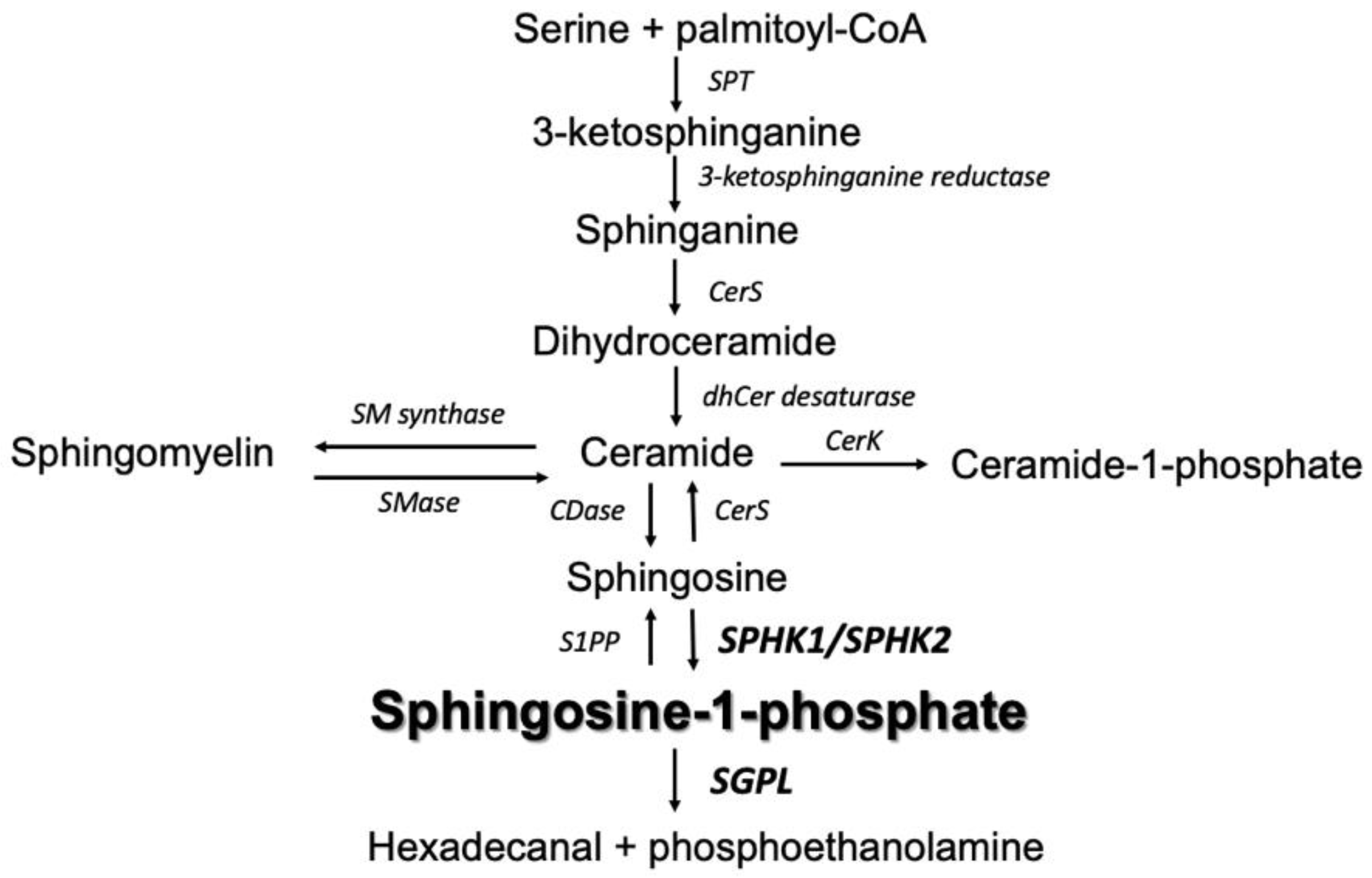
Figure 1. Overview of sphingolipid metabolism. SPT serine palmitoyltransferase. CerS ceramide synthase. CerK ceramide kinase. dhCer desaturase dihydroceramide desaturase. CDase ceramidase. SMase sphingomyelinase. SM synthase sphingomyelinase synthase. S1PP sphingosine-1-phosphate phosphatase. SPHK1/SPHK2 sphingosine kinase 1 and 2, respectively. SGPL sphingosine 1-phosphate lyase.
Ijms 24 07197 g001
Figure 2. The impact of particular gene silencing on the expression of SPHK1 (panels A and B) and SGPL1 (panels C and D) in human colorectal carcinoma HCT-116 cell line. Values are control-normalized medians +/− interquartile ranges (n = 6 qPCR; n = 4 WB). Bands from two independent representative WBs are presented below protein expression graphs; * p < 0.05; ** p < 0.01 vs. control (HCT(CON)).
Ijms 24 07197 g002
Figure 3. The effect of SPHK1 and SGPL1 silencing on the content of free sphingoid bases (Sph, SPA; panels A and B), sphingosine-1-phosphate (S1P, panel C), total ceramide (panel D) and S1P/Cer ratio (panel E) in human colorectal carcinoma HCT-116 cells. The S1P/Cer ratio was normalized to control. Values are medians +/− interquartile ranges (n = 4); * p < 0.05; vs. control (HCT(CON)).
Ijms 24 07197 g003
Figure 4. The impact of SPHK1 and SGPL1 gene silencing in human colorectal tumor HCT-116 cell line on the expression and phosphorylation state of initiator Caspase-9 (panels A,B,H,I), executioner Caspase-3 (panels CE) and Cathepsin-D (panels F,G). Two lower bands in Panel 4E show active (cleaved) Caspase-3. Protein bands from two independent representative WBs are presented below expression protein graphs. Values are control-normalized medians +/− interquartile ranges (n = 6 qPCR; n = 4 WB). * p < 0.05; ** p < 0.01 vs. control (HCT(CON)).
Ijms 24 07197 g004aIjms 24 07197 g004b
Figure 5. Cellular death by apoptosis (as measured by ELISA apoptosis assay) in SPHK1- and SGPL1-silenced human colon tumor cells. Values are medians +/− interquartile ranges (n = 4); * p < 0.05 vs. control (HCT(CON)).
Ijms 24 07197 g005
Table 1. Composition of individual Cer molecular species in SPHK1- and SGPL1-gene-silenced human colorectal tumor HCT-116 cells.
HCT(CON)HCT(SPHK1-)HCT(SGPL1-)
CeramideQ1MedianQ3Q1MedianQ3Q1MedianQ3
C14:022.2523.5624.922.4624.5525.1520.7121.4522.92
C16:0419.2462.6489.7393.3426.6436.5547.8566.8 *594.2
C18:110.5110.7811.0811.912.75 *13.0313.2113.99 *14.29
C18:08.599.0910.5611.7512.29 *14.214.7616.14 *16.55
C20:03.643.834.463.573.684.144.935.25 *5.43
C22:019.9923.1624.1424.3826.3827.2930.0831.05 *33.51
C24:1402.2409.5412.5391.5407.9439.3409.9421.4450.9
C24:076.8981.0886.4378.9882.7687.9984.0187.8789.05
Values are expressed as median with an interquartile range (Q1; Q3) (n = 4). * p < 0.05 vs. control (HCT(CON)).
Disclaimer/Publisher’s Note: The statements, opinions and data contained in all publications are solely those of the individual author(s) and contributor(s) and not of MDPI and/or the editor(s). MDPI and/or the editor(s) disclaim responsibility for any injury to people or property resulting from any ideas, methods, instructions or products referred to in the content.