Article

Syndecan-3 Coregulates Milk Fat Metabolism and Inflammatory Reactions in Bovine Mammary Epithelial Cells through AMPK/SIRT1 Signaling Pathway

1
College of Coastal Agricultural Sciences, Guangdong Ocean University, Zhanjiang 524088, China
2
The Key Laboratory of Animal Resources and Breed Innovation in Western Guangdong Province, Zhanjiang 524088, China
3
College of Animal Science, Jilin University, Changchun 130062, China
*
Authors to whom correspondence should be addressed.
These authors contributed equally to this work.
Int. J. Mol. Sci. 2023, 24(7), 6657; https://doi.org/10.3390/ijms24076657
Submission received: 25 January 2023 / Revised: 24 March 2023 / Accepted: 27 March 2023 / Published: 3 April 2023
(This article belongs to the Section Molecular Genetics and Genomics)

Abstract

:
Transcriptome sequencing showed that syndecan-3 (SDC3) was differentially expressed in high-fat and low-fat mammary epithelial cells of Chinese Holstein cows. Previous studies found that SDC3 plays an important role in inflammatory diseases and virus infection. However, those studies did not confirm whether or not the functional gene SDC3, which plays an important role in regulating milk fat metabolism, has an effect on susceptibility to breast tissue diseases. Therefore, we studied the effects of SDC3 on milk lipid metabolism and inflammation in bovine mammary epithelial cells (BMECs) and further explored the common regulatory pathway of SDC3 in both. The overexpression of SDC3 increased the contents of triglycerides and cholesterol, reduced the content of non-esterified fatty acids, inhibited the expression of inflammatory factors (IL-6, IL-1β, TNF-α and COX-2), and reduced the production of ROS in BMECs. However, silenced SDC3 had the opposite effect. Further exploring the mechanisms of SDC3, we found that SDC3 upregulated the expression of peroxisome proliferator-activated receptor gamma (PPARG) through the AMPK/SIRT1 signal pathway to promote milk fat synthesis. It also regulated the activation of the NF-κB pathway through the AMPK/SIRT1 signal pathway, reducing the expression of inflammatory factors and ROS production, thus inhibiting the inflammatory response of BMECs. Nuclear factor kappa B subunit 1 (NF-κB p50) was an important target of SDC3 in this process. To sum up, our results showed that SDC3 coregulated milk fat metabolism and inflammation through the AMPK/SIRT1 signaling pathway. This study laid a foundation for the comprehensive evaluation of breeding value based on multi-effect functional genes in dairy cow molecular breeding.

1. Introduction

Milk fat is one of the main substances that make up milk nutrition. However, mastitis, a very common disease in dairy cows, can lead to a decline in breast lactation function and reduce milk quality. Some studies found that the contents of long-chain fat (C16-C18), total fatty acids [1], polyunsaturated fatty acids [2], and milk fat in milk from recessive mastitis cows were lower than in milk from healthy cows, indicating a close relationship between dairy quality traits and breast inflammation. Our team used high-fat and low-fat BMEC lines for transcriptome analysis, without the interference of immune cells. In addition to obtaining a large number of functional milk-fat-metabolism-related genes, it was determined that some immune-related genes are differentially expressed in high-fat and low-fat bovine mammary epithelial cells (BMECs) [3]. Therefore, we speculated that there could be a coordinate regulation network between fat metabolism and immune traits that could jointly regulate milk quality traits and mastitis susceptibility. At the same time, this phenomenon also suggested that some genes with complex biological functions could coordinate the regulation on milk fat metabolism and immune traits. However, whether the complexity of the biological functions of these genes affects breeding value evaluations and their application in the breeding process remains unknown. Thus, determining whether the comprehensive effects of functional genes need to be considered in the future molecular breeding of functional genes (which play an important role in regulating milk fat metabolism) could be worthwhile. What is the coordinate regulation of fat metabolism-related genes in BMECs and immunomodulatory factors? Do functional genes that play an important role in regulating milk fat metabolism affect the susceptibility to breast tissue diseases? These questions need to be answered through the functional verification of candidate genes.
The heparan sulfate proteoglycan (syndecan, SDC) gene family is a type 1 transmembrane protein. It is composed of core proteoglycan and its covalent glycosaminoglycan polypeptide chain. Its core protein is composed of a variable length amino terminal, a transmembrane region, and a short conserved cytoplasmic region. The SDC family consists of four cell surface molecules (SDC1 to SDC4) with different biological functions, of which syndecan-3 (SDC3) is the largest. It was first cloned in the rat from a Schwann cell and a rat brain library [4]. The full-length 38589 bp of bovine SDC3 gene, located on chromosome 2, consists of 5 exons and 4 introns, encoding a total of 346 amino acids. As a new regulator of feeding behavior and body weight, SDC3 is highly expressed in the hypothalamus, which controls energy balance [5], and participates in the regulation of melanocortin system activity [6], effectively regulating energy intake, energy consumption, and peripheral glucose metabolism. In addition to participating in energy metabolism as a weight regulator, SDC3 is also directly related to fat metabolism. It was found that mice with SDC3 gene deletion (induced by high-fat diet) showed resistance to obesity, significantly reduced fat accumulation, and good glucose tolerance [7]. Two single nucleotide polymorphisms in exon 3 of SDC3 were significantly associated with obesity [8]. Based on the regulatory role of SDC3 in fat metabolism, it was speculated that the gene could be involved in milk fat metabolism. The transcriptome research results of our group showed that SDC3 showed differential expression in the transcriptome data analysis of high-fat and low-fat mammary gland epithelial cells of Chinese Holstein cows [3]. This suggested that our SDC3 gene could be involved in—and regulate—milk fat metabolism. Furthermore, SDC3 plays a dual role in inflammation. SDC3 showed pro-inflammatory effects in joint tissue, and leukocyte recruitment and cartilage damage in antigen-induced arthritis mice were significantly lower than those in wild-type mice due to SDC3 gene deletion. However, SDC3 had an anti-inflammatory effect in skin and secretions, and leukocyte rolling and adhesion increased in skin and genital inflammation models, indicating that SDC3 had anti-inflammatory activity [9]. For cells, SDC3 is expressed in many cell types, participates in the regulation of growth factor signal [10], cell adhesion and migration [11,12], and proliferation and differentiation [13], and also plays an important role in inflammation and virus infection [14]. These results suggest that the SDC3 gene not only participates in milk fat metabolism, but also plays an important role in immune regulation. The effects of the SDC3 gene on milk fat metabolism and inflammation, and how the gene plays a coregulatory role in milk fat traits and immune characteristics, are worthy of further study. Analysis of the function of SDC3 gene could contribute to the comprehensive selection of related traits in the breeding of milk fat and immune traits in dairy cows in the future.
AMPK is a serine/threonine protein kinase that plays an important role in regulating energy homeostasis [15], cell growth [16], and lipid metabolism [17]. It was further found that AMPK was a key factor for trans-10, cis-12 conjugated linoleic acid (t10c12-CLA) to regulate milk fat synthesis in goat mammary epithelial cells [18]. AMPK protein kinase is a heterotrimer consisting of one catalytic subunit (α) and two regulatory subunits (β and γ) [19]. Protein kinase AMP-activated catalytic subunit alpha 1 (AMPKα1) is mainly expressed in breast tissue [20], and the phosphorylation of α-subunit Thr172 site is a necessary condition for the full activation of AMPK. In addition, AMPK can activate sirtuin 1 (SIRT1) [21], which regulates glucose and lipid metabolism and tumorigenesis, whereas SIRT1 can regulate the core factor of fat synthesis, peroxisome proliferator activated receptor gamma (PPARG) [22], and the key regulator of the inflammatory network, NF-κB [23]. NF-κB is the aggregation point of many signal transduction pathways and plays an important role in regulating inflammation, immune response, and stress response [24]. The phosphorylation of its inhibitory protein, IκB, is a necessary condition for its release and entry into the nucleus before it can exert its biological function [25].
Based on previous studies and the results of our transcriptome, we knew that SDC3 participated in and regulated milk fat metabolism and inflammation, but the effects of SDC3 on milk fat metabolism and inflammation and its molecular mechanism in BMECs had not yet been reported. Furthermore, according to the above description, it was interesting that the AMPK signal pathway regulated milk fat synthesis, whereas NF-κB was a key regulator in the inflammatory network, involved in the regulation of inflammatory response. We wanted to know whether SDC3 coregulated milk lipid metabolism and inflammation of BMECs through these two signal pathways. Therefore, in this study, we aimed to explore the functions of SDC3 in milk fat metabolism and inflammation, and to further analyze the coregulation pathway of SDC3 on milk lipid metabolism and immune traits in BMECs.

2. Results

2.1. Identification of Transfection Efficiency of Plasmid and siRNAs in BMECs

To ensure the effectiveness of the SDC3 overexpression vector and inhibitor siRNA in BMECs, after the vectors and siRNAs were transfected into cells, green fluorescence was examined using fluorescence microscopy. RT-qPCR and Western blot were also used. After 24 h, green fluorescence was observed under a fluorescence microscope. Findings suggested that the vectors (pBI-CMV3-SDC3 and pBI-CMV3) and siRNAs were successfully transfected into the BMECs (Figure 1A,E). Compared with the control, the overexpression of SDC3 significantly increased the levels of SDC3 transcription (p < 0.01) (Figure 1B) and protein (p < 0.01) (Figure 1C,D). The transcriptional levels of SDC3 in BMECs transfected with siRNA-1218, siRNA-791 and siRNA-979 decreased by 70%, 57%, and 6%, respectively (Figure 1F), and siRNA-1218 significantly decreased the levels of SDC3 protein (p < 0.05) (Figure 1G,H). siRNA-1218 had the highest efficiency of SDC3 knockdown and was used as an inhibitor of SDC3 in subsequent experiments.

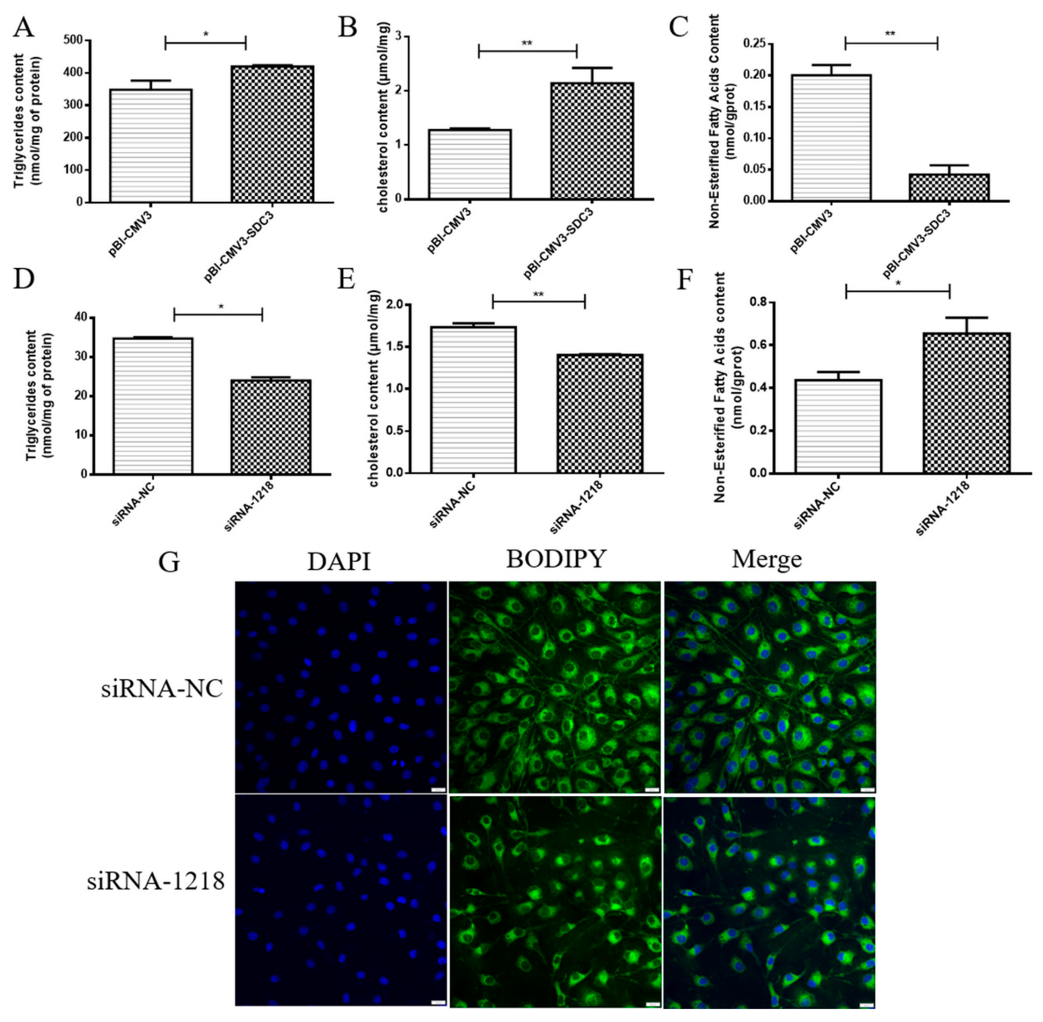
2.2. SDC3 Promotes the Synthesis of Milk Fat in BMECs

To study the effects of SDC3 on milk lipid metabolism in BMECs, the BMECs were seeded on 6-well plates and harvested after 24 h, post-transfection with vectors or siRNAs. The contents of triglycerides, cholesterol, NEFA, and the distribution of lipid droplets in BMECs were used to evaluate the effects of SDC3 on milk fat metabolism in BMECs. The results showed that, compared with the control group, the overexpression of SDC3 significantly increased the content of triglycerides (p < 0.05) (Figure 2A) and cholesterol (p < 0.01) (Figure 2B) but significantly decreased the NEFA content (p < 0.01) (Figure 2C). On the contrary, compared with the negative control group, the siRNA-1218 group obviously decreased the contents of triglycerides (p < 0.05) (Figure 2D) and cholesterol (p < 0.01) (Figure 2E). They also decreased the accumulation of lipid droplets (Figure 2G) but obviously increased NEFA content (p < 0.05) (Figure 2F). To sum up, the overexpression of SDC3 was able to significantly promote the synthesis of triglycerides and cholesterol, which are the main components of milk fat in BMECs. It also played a positive role in regulating milk fat synthesis while promoting the metabolism of NEFA. SDC3 knockout had the opposite effect.

2.3. SDC3 Inhibits the Inflammatory Response of BMECs by Inhibiting the Expression of Inflammatory Factors and the Production of ROS

In order to investigate whether SDC3 was able to regulate the inflammatory response in BMECs, we tested the effects of overexpression of SDC3 and silencing SDC3 interventions on the levels of inflammatory markers (IL-6, IL-1β, TNF-α and COX-2) in normal growth BMECs at both the transcriptional and translational level. This was carried out using RT-qPCR and ELISA assays, whereas intracellular ROS generation detection was accomplished using flow cytometry. The results of our RT-qPCR assay were consistent with the changes observed in the ELISA assays, which showed that the expression levels of interleukin 6 (IL-6), interleukin 1 beta (IL-1β), and tumor necrosis factor (TNF-α) in cells were significantly downregulated in the overexpression of SDC3 group versus the control group (p < 0.05) (Figure 3A–F). On the other hand, the expression levels of these inflammatory factors in cells were significantly upregulated in the silencing SDC3 group versus the control group (p < 0.05) (Figure 3G–L). The results of the flow cytometry showed that the overexpression of SDC3 could significantly inhibit (p < 0.01) intracellular ROS generation, whereas knockout of SDC3 could increase (p < 0.01) the levels of intracellular ROS (Figure 3O–R). At the same time, the transcriptional levels of cytochrome c oxidase subunit II (COX-2), which plays a central role in ROS generation [26], decreased with the overexpression of SDC3 and increased when SDC3 was silenced (p < 0.05) (Figure 3D,N). In short, the overexpression of SDC3 without inflammatory stimulation decreased the expression of inflammatory factors and ROS generation in BMECs and inhibited the occurrence of inflammatory responses to a certain extent. Thus, SDC3 demonstrated anti-inflammatory effects and could potentially regulate intracellular inflammatory responses.

2.4. SDC3 Exerts a Cooperatively Regulatory Effect on Milk Lipid Metabolism and Inflammatory Reactions in BMECs through the AMPK/SIRT1 Signal Pathway

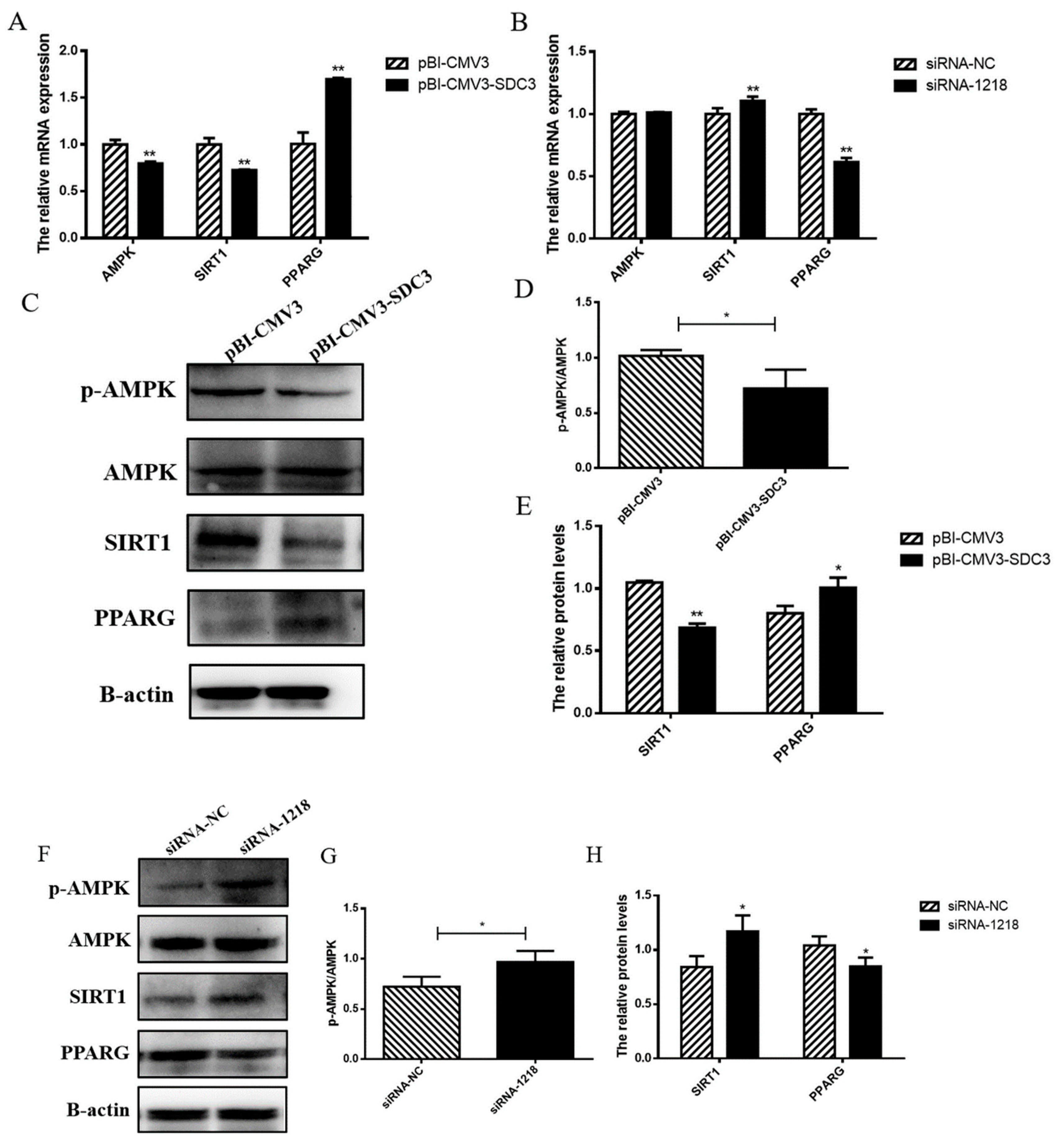
2.4.1. SDC3 Promotes Milk Fat Synthesis through AMPK/SIRT1/PPARG Signal Pathway

Previous studies have shown that the AMPK signaling pathway plays an important role in fat synthesis [17]. SIRT1, known as NAD-dependent deacetylase, can be activated by AMPK to inhibit lipid accumulation [21]. It can also inhibit the expression of PPARG, the core regulator of fat synthesis, to reduce fat synthesis [22]. Therefore, we transfected the vector (pBI-CMV3-SDC3 and pBI-CMV3) and siRNAs (siRNA-1218 and siRNA-NC) into BMECs. We then detected the effects of SDC3 on AMPK and its downstream genes using RT-qPCR and Western blot methods in order to explore the mechanism by which SDC3 promoted milk fat synthesis. The results showed that with pBI-CMV3 as the negative control, the overexpression of SDC3 inhibited activation of the AMPK signal pathway by significantly reducing the protein expression of p-AMPK (p < 0.05) (Figure 4C,D) and the transcriptional level of AMPK (p < 0.01) (Figure 4A). It also decreased the expression of its downstream gene SIRT1 (p < 0.01) (Figure 4A,C,E) and increased the expression of PPARG (p < 0.05) (Figure 4A,C,E). On the contrary, silencing SDC3 significantly promoted the protein expression of p-AMPK (p < 0.05) (Figure 4F,G), had no effect on transcriptional levels of AMPK (Figure 4B), and increased the expression of SIRT1 (p < 0.05) (Figure 4B,F,H). It also activated the AMPK/SIRT1 signal pathway, thus inhibiting the expression of PPARG (p < 0.05) (Figure 4B,F,H). These results suggested that SDC3 upregulated the expression of PPARG, the core regulator of fat synthesis, by inhibiting the AMPK/SIRT1 signal pathway—which led to the increase in fat synthesis and accumulation, promoting milk fat synthesis.

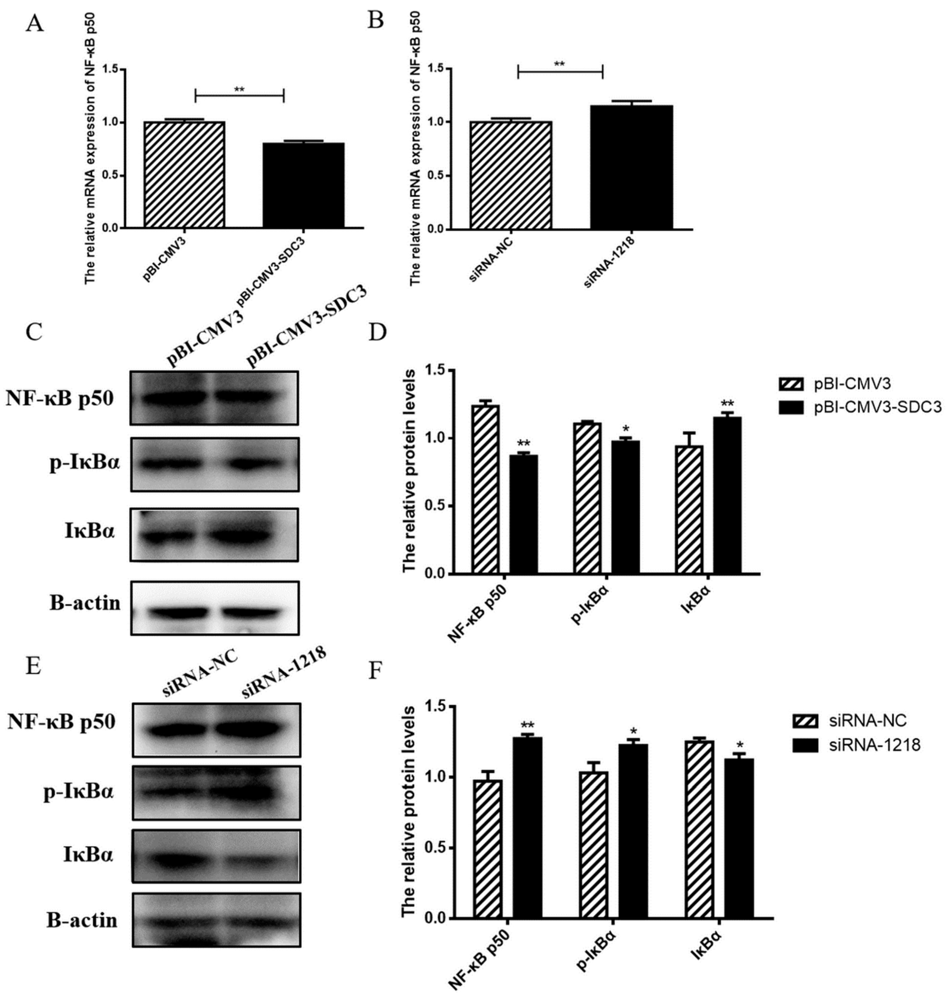
2.4.2. SDC3 Inhibits Inflammatory Reaction through the AMPK/SIRT1/NF-κB P50 Signaling Pathway

The above results showed that SDC3 significantly inhibited the expression of inflammatory factors and ROS generation, and had a good anti-inflammatory effect, as well. Thus, it was necessary for us to further explore the anti-inflammatory mechanism of SDC3. Because NF-κB can activate the transcription of inflammatory cytokines IL-6, IL-1β, TNF-α, and COX-2 [27], and AMPK and SIRT1 participate in the regulation of inflammatory network as upstream regulators of NF-κB [23], we studied the effects of SDC3 on NF-κB, a key factor in the inflammatory network. The overexpression of SDC3 significantly inhibited the expression of NF-κB p50 (p < 0.01) (Figure 5A,C,D), decreased the protein expression of P-IκBα (p < 0.05) (Figure 5C,D), and increased the expression of inhibitory protein IκBα (p < 0.01) (Figure 5C,D). This inhibited the migration of NF-κB p50 to the nucleus and the release of downstream inflammation-related transcription factors (IL-6, IL-1β, TNF-α and COX-2), thereby also inhibiting the inflammatory responses of BMECs. However, in the absence of inflammatory stimulation, with siRNA-NC as the negative control, silencing SDC3 significantly increased the expression of NF-κB p50 (p < 0.01) (Figure 5B,E,F) and upregulated the protein expression of P-IκBα (p < 0.05) (Figure 5E,F). Thus, the inhibitory protein IκBα was ubiquitinated and degraded (p < 0.05) (Figure 5E,F), and NF-κB p50 entered the nucleus to activate downstream transcription factors, promoting inflammatory responses, increasing intracellular ROS generation, and resulting in cell damage. The results suggested that NF-κB p50 could be an important target for SDC3 to regulate NF-κB pathways, as SDC3 activated or inhibited the NF-κB signal pathway by regulating its expression, and then regulated the inflammatory response of BMECs. In summary, SDC3 regulated the expression of inflammatory cytokines IL-6, IL-1β, TNF-α, and COX-2 and the levels of intracellular ROS through the AMPK/SIRT1/NF-κB p50 signal pathway in order to regulate the inflammatory responses of BMECs (Figure 6).

3. Discussion

BMECs are the basic unit of synthesizing and secreting milk and are the target cells for studying the mechanism of lactation in breast tissue in vitro. In this study, BMECs were used as the research object to explore the effects of SDC3 on milk fat synthesis by transfection of siRNAs and plasmids in vitro. The main components of milk fat include 97–98% triglycerides, 0.2–0.4% cholesterol, and 0.1% fatty acids [28]. The overexpression of SDC3 led to increases in triglycerides and cholesterol content and decreases of NEFA content in BMECs. On the contrary, silencing the SDC3 gene decreased triglyceride content, cholesterol content, and lipid droplet accumulation in BMECs, and increased NEFA content. It was suggested that SDC3 could regulate milk fat metabolism in BMECs. AMPK is a recognized protein that participates in various life activities by regulating energy balance [15]; it has been significantly associated with obesity and lipid metabolism disorders. Therefore, we detected the expression levels of p-AMPK after overexpression and silencing of SDC3 to explore whether SDC3 regulated milk fat metabolism by activating the AMPK signal pathway. We found that the overexpression of SDC3 decreased the phosphorylation level of AMPK and significantly inhibited the activation of AMPK. Silencing SDC3 significantly increased the phosphorylation levels of AMPK and activated the AMPK signal pathway, suggesting that SDC3 was able to regulate milk fat metabolism through that pathway. In addition, the activation of AMPK increased the intracellular NAD+ concentration, and then activated SIRT1, which jointly regulated energy consumption [21,29]. It has been observed that LB100 attenuates FFA-induced lipid accumulation in normal human hepatic cell lines through the AMPK/SIRT1 signal pathway [30]. SIRT1 is a NAD+-dependent protein deacetylase that regulates the activity of downstream signal proteins by deacetylating these proteins. It is widely involved in cell senescence, glucose and lipid metabolism [31], and other life processes. Crucially, SIRT1 inhibits fat production and promotes fat mobilization by inhibiting PPARG [22], which plays a central role in adipocyte differentiation [32] and fat deposition [33]. We found that the overexpression of SDC3 could significantly reduce the expression of SIRT1 and increase the expression of PPARG. However, the opposite was observed after silencing SDC3, which indicated that SDC3 was involved in milk fat synthesis as a positive regulator. Previous studies have found that both p-AMPK and SIRT1 cooperatively inhibited the expression of PPARG, resulting in the inhibition of lipid synthesis [34]. Gynostemma Pentaphyllum Extract (GPE) decreased the levels of serum triglycerides and total cholesterol in mice fed a high-fat diet and inhibited increased body weight, fat mass, and adipocyte hypertrophy through the AMPK/SIRT1/PPARG signaling pathways [35]. In summation, consistent with previous studies, SDC3 was shown to regulate milk fat synthesis in BMECs through the AMPK/SIRT1/PPARG signal pathways.
BMECs have an important physiological function: synthesizing and secreting milk. However, they also play an important role in the process of antimicrobial invasion of mammary glands. When external pathogenic microorganisms invade breast tissue, BMECs make first contact with pathogenic microorganisms. They can identify pathogens and play a sentinel role [36], which could be of great significance to the development and prognosis of dairy cow mastitis. Studies showed that when breast tissue was infected, BMECs were able to synthesize and secrete a variety of cytokines, such as TNF-a, IL-lβ, and IL-6 [37]. E. coli produced a strong, rapid inflammatory response, inducing BMECs to produce TNF-α and IL-1β. The inflammatory response of BMECs induced by S. aureus mainly depended on the production of IL-6 [38]. In addition, it is well known that excessive production of ROS can cause inflammation and cell damage, or even cell death, by inducing apoptosis. The results of RT-qPCR, ELISA, and flow cytometry showed that the overexpression of SDC3 could notably downregulate expression of IL-6, IL-1β, TNF-α, and COX-2 and reduce ROS generation in BMECs without inflammatory stimulation. On the other hand, silencing SDC3 led to the high expression of IL-6, IL-1β, TNF-α, and COX-2 and increased the level of intracellular ROS. This indicated that SDC3 was able to regulate the inflammatory response of BMECs and exert certain anti-inflammatory effects. This result was consistent with the findings of Kehoe et al., who found that, in the model of skin inflammation and secretory inflammation, the absence of SDC3 enhanced recruitment and the interaction between leukocytes and endothelial cells [39]. The rolling and adhesion of leukocytes in SDC3-deficient mice increased, suggesting that SDC3 played an important role in anti-inflammation, as well [40]. In addition, because both the transmembrane and cytoplasmic regions of the SDC family core proteins have binding sites for protein kinase phosphorylation and multiple signal molecules [41], SDC can be used as receptors for cytokines, chemokine, and growth factors to regulate different signal pathways [42,43]. We found that inflammatory markers (IL-6, IL-1β, TNF-α, and COX-2) regulated by SDC3 were downstream genes of the key NF-κB signaling pathway in the inflammatory response [44]. Therefore, we speculated that SDC3 could regulate the inflammatory response of BMECs through the NF-κB signal pathway.
Transcription factor kappa B was first discovered in 1986. It was so named because NF-κB binds to the enhancer element of the immunoglobulin κ light chain and then activates B cells [45]. In resting cells, NF-κB forms a complex with its inhibitory protein IκB, masking the nuclear localization signal of NF-κB, which results in it remaining in the cytoplasm as an inactive complex. When stimulated, IκBα is phosphorylated by its kinase IKK and degraded, and the activated NF-κB is transferred to the nucleus to bind to the KB site of the target gene promoter, thus playing the role of transcriptional regulation [25]. Our study found that the overexpression of SDC3 significantly decreased the phosphorylation levels of IκBα, increased the expression levels of NF-κB inhibitor protein IκBα, and then inhibited the activation of NF-κB. On the contrary, silencing SDC3 significantly increased the phosphorylation level of IκBα, degraded IκBα, and released NF-κB into the nucleus to activate downstream transcription factors, thereby increasing the expression of inflammatory factors and intracellular ROS levels. These results demonstrated that SDC3 regulated the inflammatory response of BMECs through the NF-κB signaling pathway. It is well known that NF-κB mainly exists as a heterodimer, composed of NF-κB p65, and NF-κB p50 subunits. Therein, NF-κB p65 shows transactivation activity through the transactivation domain at the C-terminal, whereas NF-κB p50 directly binds to a specific DNA sequence (the NF-κB binding element). These two subunits jointly target gene promoters in various inflammatory responses. Previous studies showed that SIRT1 deacetylated NF-κB p65 at lysine 310, thereby reducing the transcriptional activation of NF-κB and inhibiting the inflammatory response mediated by NF-κB [27]. However, our results showed that silencing SDC3 could increase the phosphorylation of AMPK and upregulate the expression of downstream gene SIRT1. The upregulation of SIRT1 expression did not inhibit the activation of NF-κB, however. On the contrary, NF-κB was activated. Therefore, we explored the effects of NF-κB p50. The results showed that, after silencing SDC3, the expression of NF-κB p50 increased and then activated the NF-κB pathway, whereas the overexpression group of SDC3 reacted to the contrary. Consistent with our studies, Eriocalyxin B (ERIB) interfered with the binding of NF-κB and response elements by targeting NF-κB p50, thus exerting its anti-autoimmune inflammatory activity [46]. In Mesangial cells (MCs), LincRNA-Gm4419 can activate the NF-κB pathway through interaction with the NF-κB subunit p50 [47]. All in all, from our results, we were able to conclude that SDC3 regulated the activation of the NF-κB signaling pathway through NF-κB p50, thus exerting its anti-inflammatory activity.

4. Materials and Methods

4.1. Reagents and Antibodies

DMEM and DPBS were purchased from HyClone (UT, USA). Fetal bovine serum (FBS) was purchased from ZATA Life (CA, USA). The triglycerides and cholesterol detection kit were purchased from Pulilai (Beijing, China). The non-esterified fatty acids assay kit was purchased from Nanjing Jiancheng Bioengineering Institute (Nanjing, China). The lipid dye BODIPY505/515 was purchased from Invitrogen (Waltham, MA, USA), and three ELISA kits (IL-6, IL-1β and TNF-α) were purchased from MEIMIAN (Wuxi, China). The primary antibodies against AMPKα1, Phosphorylated AMP-activated protein kinase (p-AMPK), SDC3, PPARG, and Phospho-IκB Alpha (p-IκBα) were purchased from Bioss (Beijing, China). Antibodies against nuclear factor kappa B subunit 1 (NF-κB p50), IκBα, and SIRT1 were purchased from Proteintech Group (Wuhan, China). The antibodies against β-actin and anti-rabbit secondary antibodies were acquired from Bioworld Technology (St. Louis Park, MN, USA).

4.2. Cell Culture

The BMECs used in this study were derived from the mammary gland tissue of Chinese Holstein cows and were provided by the Laboratory of Molecular Genetics, College of Animal Science, Jilin University [48,49]. Throughout the experiment, BMECs were cultured in DMEM-F12 with 10% fetal bovine serum at 37 °C, in humidified air with 5% CO2. When the fusion degree of BMECs in the six-well plates (Nest Biotechnology, Shanghai, China) reached 80%, the plasmid or small interfering RNA (siRNA) was transfected into the cells. After 24 h, the overexpression and inhibition efficiency of cells were detected by observation of green fluorescence under microscope, RT-qPCR, and Western blotting methods.

4.3. Transfection

Plasmids with pBI-CMV3-SDC3 vectors containing SDC3 overexpression sequences were given by Jilin University. Plasmids with pBI-CMV3 vectors containing any non-target sequences were used as negative controls. The SDC3 siRNA pool and its negative control, siRNA-NC, were designed and manufactured by GenePharma Company (GenePharma, Shanghai, China). The SDC3 siRNA pool contained three siRNAs targeting different portions of the SDC3 mRNA sequence. They were designed to have no homology with any bovine gene. The sequences were: siRNA-1218, 5′-GCUCAUCUACCGCAUGAAGTT-3′; siRNA-791, 5′-CCACCCAGAAGCCUGAUAUTT-3′; siRNA-979, 5′-CCAGACACAGCCAAUGAAGTT-3′; and siRNA-NC, 5′-UUCUCCGAACGUGUCACGUTT-3′. In short, 1 × 10⁶ cells were inoculated into a 6-well plate until the fusion reached about 80%. Then, 3μg of vectors were transfected into the cells using 6 μL of ViaFect (Promega, WI, USA) and 9.4μL of siRNAs were transfected into the cells using 3.75 μL of lipo3000 (Invitrogen, MA, USA) in a serum-free medium for 6 h. Then, the medium was supplemented with serum and maintained in culture for 24 h. After that, the transfection efficiency was observed using fluorescence microscopy (ESCO, Singapore), and the cells were collected for subsequent experiments.

4.4. Measurement of Triglycerides and Cholesterol

After the 24 h transfection, BMECs were lysed and collected, and the concentrations of standards, triglycerides, and cholesterol were measured using a triglyceride kit and a cholesterol kit. In addition, a BCA protein assay kit (TaKaRa, Beijing, China) was used to measure the protein concentration in the cells. A standard curve was plotted to calculate triglyceride and cholesterol concentrations. The triglyceride/cholesterol to protein concentration ratio represented the final utilized data.

4.5. Non-Esterified Fatty Acids (NEFA) Measurement

A full 24 h after the plasmid or siRNAs were transferred into the BMECs, the content of NEFAs in the cell was detected using a non-esterified fatty acid kit. Briefly, after the cells were lysed with lysate, the cells were collected and bathed in water at 70 °C for 10 min and then centrifuged. A total of 4 µL of supernatant was placed in 96-well plates and mixed with 200 µL of a working solution, then cultured at 37 °C in the dark for 5 min. A microplate reader measured the absorbance as A1. Then, 50 µL of B working solution was added and incubated under the same conditions and the absorbance was measured to be A2. Protein concentration was detected using a BCA protein assay kit. Both absorbance values were measured at 546 nm, and the following formula calculated the content of NEFA in the cells:
  Intracellular   NEFA   concentration ( mmol / gprot )   = { ( A 1     A 2 )   sample     ( A 1     A 2 ) blank } / { ( A 1     A 2 )   standard     ( A 1     A 2 ) blank }   Standard   concentration   ( mmol / L ) / Intracellular   protein   concentration   ( gprot / L )

4.6. BODIPY Staining

siRNA-1218 and siRNA-NC were transfected into the cells by lipo3000 for 24 h. Then, they were washed with precooled PBS three times. Then, lipid dye BODIPY505/515 was added, diluted by DMEM, and stained in the dark for 30 min, before an additional washing with PBS (three times). Then, the cells were fixed with a fixation solution (Biosharp, Anhui, China) for 5 min and washed an additional three times with PBS for 5 min each. DAPI (Beyotime, Shanghai, China), diluted by PBS, was added and stained in the dark for 10 min. The cells were washed with PBS three times for 5 min, and the distribution of lipid droplets was observed under a laser confocal microscope (Olympus, Tokyo, Japan).

4.7. Protein Expression Analysis

The protein was extracted from the transfected cells and the protein concentration was measured by BCA protein assay kit (TaKaRa, Beijing, China). The total protein concentration of the control and treatment groups was standardized by adjusting the volume of the protein lysate. Sample diluent (40 μL), protein of cells (10 μL), and standard sample (50 mL) were added to appropriate wells. In addition to the blank control wells, enzyme-labeled reagents (100 μL) were added to the other wells and incubated at 37 ℃ for 1 h. The plate was washed with wash buffer five times. After drying, Substrate Solution A and Substrate Solution B (50 μL) were added to each well for 15 min of incubation. Finally, Stop Solution (50 μL) was added into each well and they were placed into a microplate reader (BioTek, VT, USA) to detect the absorbance value under 450 nm.

4.8. Cell ROS Analysis

After treatment according to the requirements of the transfection test, intracellular reactive oxygen species (ROS) production was detected using a 2′,7′-dichlorofluorescin diacetate (DCFH-DA, Beyotime, Shanghai, China) probe. Then, the treated cells were collected, after pancreatin digestion, and co-incubated in 37 °C, in darkness, with serum-free culture medium containing 5 μM DCFH-DA. After 20 min, they were washed three times with serum-free medium. Finally, the cells were centrifuged and resuspended in 500 μL PBS. A flow cytometer (Beckman Coulter, Brea, CA, USA) was used to analyze ROS levels in BMECs.

4.9. Gene Expression Analysis

According to the steps of the RNA extraction kit (TaKaRa, Beijing, China), the cells were lysed in 6-well plates with buffer RL lysate containing 2% DTT in order to extract total RNA. The OD value and RNA concentration were evaluated using a Nanodrop Lite (Thermo Scientific, Waltham, MA, USA) spectrophotometer. A reverse transcription kit (TaKaRa, Beijing, China) was applied to reverse-transcribe the 1μg RNA into cDNA. RT-qPCR primers (Table 1) were designed by primer5 software and analyzed by BLAST to confirm the specificity of each pair of primers to its target gene. Then, the cDNA was mixed with the primers of the target gene (Table 1) and the Chamq Universal SYBR qPCR master mix (Vazyme, Nanjing, China) was used for amplification reaction in a real-time PCR detection system (BIO-RAD, Hercules, CA, USA). β-actin was used as the internal reference gene. Each experiment was run in triplicate. The 2 −∆∆CT method was used to calculate the relative mRNA expression levels [50].

4.10. Western Blot

After transfection, the culture medium was removed and the cells were washed twice with PBS. The cells were lysed with RIPA lysis buffer (Meilunbio, Dalian, China) containing PMSF protease inhibitors (Beyotime, Shanghai, China) on ice for 30 min, then centrifuged for 30 min. The cells were collected and bathed in ice for 30 min, then centrifuged for 30 min. Then, 10 μL of supernatant was extracted and diluted 10 times before being used to detect protein concentration using a BCA protein detection kit. The protein was mixed with Tris-Tricinesds-SDS-PAGE loading buffer (Solarbio, Beijing, China) and RAPI lysate, then denatured at 98 °C for 10 min. Protein samples were separated by SDS-PAGE, then transferred to the activated PVDF membranes (Merck Millipore, Darmstadt, Germany) by wet electrophoretic transfer. Subsequently, samples were blocked using 5% skim milk (Coolaber, Beijing, China) dissolved in TBST (Sangon biotech, Shanghai, China) for 2 h and subjected to TBST cleaning membranes. Then, PVDF membranes were incubated with primary antibody at 4 °C overnight. Membranes were washed with TBST and then incubated with a rabbit secondary antibody for 2 h at room temperature. Then, protein signals were visualized using the ECL reagent (Beyotime, Shanghai, China) and analyzed using ImageLab 6.1 software. The gray value of the target protein was divided by the gray value of β-actin, and this was used as the final statistical value [51].

4.11. Statistical analysis

All data are presented as means ± standard error from three independent experiments. The data were analyzed using Student’s t-test and SPSS 21.0 software (IBM, Armonk, New York, NY, USA). A value of p < 0.05 was declared statistically significant.

5. Conclusions

Our results revealed that SDC3 upregulated the expression of PPARG through the AMPK/SIRT1 signaling pathway to promote milk fat synthesis. It also regulated the inflammatory response of BMECs by regulating the activation of the NF-κB pathway—through the AMPK/SIRT1 signaling pathway—decreasing the expression levels of inflammatory cytokines (IL-6, IL-1β, TNF-α and COX-2) and reducing ROS generation. NF-κB p50 is an important target of SDC3 in this process. Collectively, this study determined that SDC3, as a functional gene with complex biological effects, played a coregulatory role in milk lipid metabolism and inflammatory responses in BMECs. This study, therefore, could provide a theoretical foundation for future comprehensive breeding value evaluations of multi-gene effect functional genes in dairy cow molecular breeding.

Author Contributions

Conceptualization, P.J. and H.Y.; methodology, J.F.; investigation, H.W., F.M. and X.F.; data curation, X.C., X.J. and J.L.; writing—original draft preparation, J.F. and Z.Z.; supervision, P.J. and H.Y.; funding acquisition, Z.Z., H.Y. and P.J. All authors have read and agreed to the published version of the manuscript.

Funding

This work was supported by the National Natural Science Foundation of China (Nos. 32202632, 32072717, 32002165); the Natural Science Foundation of Guangdong Province (No. 2021A1515010867); the Key Laboratory of Animal Resources and Breed Innovation in West Guangdong (No. 2021A05024); and the Young innovative talents of Innovative Strong School Engineering by the Department of Education of Guangdong Province, the Project of (2019KQNCX042).

Institutional Review Board Statement

All experimental procedures were performed according to the Guide for Guangdong Ocean University Ethics Committee. The experimental protocol was approved by the Departmental Animal Ethics Committee of Guangdong Ocean University (protocol code University Science and Technology (2019) No.1 and 20 June 2020). As suggested by the animal welfare protocol, all efforts were made to reduce animal suffering and to use only the number of animals required to produce dependable scientific data.

Informed Consent Statement

Not applicable.

Data Availability Statement

All data generated or analyzed during this study are available from the corresponding author by request.

Conflicts of Interest

The authors declare no conflict of interest.

References

  1. Randolph, H.E.; Erwin, R.E. Influence of mastitis on properties of milk. X. Fatty acid composition. J. Dairy Sci. 1974, 57, 865–868. [Google Scholar] [CrossRef]
  2. Pan, Z.; Zhou, Q.; Ma, H.; Gong, Q.; Wang, S.; Yao, H.; Ma, J.; Wang, K. Identification of a novel bacterial taxon associated with bovine mastitis showing a close evolutionary relationship with Elizabethkingia sp. Microbiol. Res. 2020, 236, 126443. [Google Scholar] [CrossRef] [PubMed]
  3. Xia, L.; Zhao, Z.; Yu, X.; Lu, C.; Jiang, P.; Yu, H.; Li, X.; Yu, X.; Liu, J.; Fang, X.; et al. Integrative analysis of miRNAs and mRNAs revealed regulation of lipid metabolism in dairy cattle. Funct. Integr. Genom. 2021, 21, 393–404. [Google Scholar] [CrossRef] [PubMed]
  4. Carey, D.J.; Conner, K.; Asundi, V.K.; O’Mahony, D.J.; Stahl, R.C.; Showalter, L.; Cizmeci-Smith, G.; Hartman, J.; Rothblum, L.I. cDNA cloning, genomic organization, and in vivo expression of rat N-syndecan. J. Biol. Chem. 1997, 272, 2873–2879. [Google Scholar] [CrossRef] [PubMed] [Green Version]
  5. Huang, Y.Z.; Wang, Q.; Zhang, C.L.; Fang, X.T.; Song, E.L.; Chen, H. Genetic Variants in SDC3 Gene are Significantly Associated with Growth Traits in Two Chinese Beef Cattle Breeds. Anim. Biotechnol. 2016, 27, 190–198. [Google Scholar] [CrossRef]
  6. Reizes, O.; Lincecum, J.; Wang, Z.; Goldberger, O.; Huang, L.; Kaksonen, M.; Ahima, R.; Hinkes, M.T.; Barsh, G.S.; Rauvala, H.; et al. Transgenic expression of syndecan-1 uncovers a physiological control of feeding behavior by syndecan-3. Cell 2001, 106, 105–116. [Google Scholar] [CrossRef] [Green Version]
  7. Strader, A.D.; Reizes, O.; Woods, S.C.; Benoit, S.C.; Seeley, R.J. Mice lacking the syndecan-3 gene are resistant to diet-induced obesity. J. Clin. Investig. 2004, 114, 1354–1360. [Google Scholar] [CrossRef] [Green Version]
  8. Roh, J.J.; Kim, M.J.; Oh, D.J.; Noh, T.H.; Won, S.G.; Yang, K.Y.; Jung, D.H.; Jo, C.L.; Chung, J.H.; Kim, J.W. [P11-21] Positive association of obesity with single nucleotide polymorphisms of syndecan 3 (SDC3) in Korean population. J. Clin. Endocrinol. Metab. 2006, 91, 5095–5099. [Google Scholar]
  9. Kehoe, O.; Kalia, N.; King, S.; Eustace, A.; Boyes, C.; Reizes, O.; Williams, A.; Patterson, A.; Middleton, J. Syndecan-3 is selectively pro-inflammatory in the joint and contributes to antigen-induced arthritis in mice. Arthritis Res. Ther. 2014, 16, R148. [Google Scholar] [CrossRef] [Green Version]
  10. Cornelison, D.D.; Wilcox-Adelman, S.A.; Goetinck, P.F.; Rauvala, H.; Rapraeger, A.C.; Olwin, B.B. Essential and separable roles for Syndecan-3 and Syndecan-4 in skeletal muscle development and regeneration. Genes Dev. 2004, 18, 2231–2236. [Google Scholar] [CrossRef] [Green Version]
  11. Arokiasamy, S.; Balderstone, M.J.M.; De Rossi, G.; Whiteford, J.R. Syndecan-3 in Inflammation and Angiogenesis. Front. Immunol. 2019, 10, 3031. [Google Scholar] [CrossRef] [Green Version]
  12. Kempf, A.; Boda, E.; Kwok, J.C.F.; Fritz, R.; Grande, V.; Kaelin, A.M.; Ristic, Z.; Schmandke, A.; Schmandke, A.; Tews, B.; et al. Control of Cell Shape, Neurite Outgrowth, and Migration by a Nogo-A/HSPG Interaction. Dev. Cell 2017, 43, 24–34.e25. [Google Scholar] [CrossRef] [Green Version]
  13. Pisconti, A.; Cornelison, D.D.; Olguín, H.C.; Antwine, T.L.; Olwin, B.B. Syndecan-3 and Notch cooperate in regulating adult myogenesis. J. Cell Biol. 2010, 190, 427–441. [Google Scholar] [CrossRef] [Green Version]
  14. Yamada, Y.; Arai, T.; Kojima, S.; Sugawara, S.; Kato, M.; Okato, A.; Yamazaki, K.; Naya, Y.; Ichikawa, T.; Seki, N. Regulation of antitumor miR-144-5p targets oncogenes: Direct regulation of syndecan-3 and its clinical significance. Cancer Sci. 2018, 109, 2919–2936. [Google Scholar] [CrossRef] [PubMed] [Green Version]
  15. Hardie, D.G.; Ross, F.A.; Hawley, S.A. AMPK: A nutrient and energy sensor that maintains energy homeostasis. Nat. Rev. Mol. Cell Biol. 2012, 13, 251–262. [Google Scholar] [CrossRef] [PubMed] [Green Version]
  16. Mihaylova, M.M.; Shaw, R.J. The AMPK signalling pathway coordinates cell growth, autophagy and metabolism. Nat. Cell Biol. 2011, 13, 1016–1023. [Google Scholar] [CrossRef] [PubMed]
  17. Cheng, J.; Zhang, Y.; Ge, Y.; Li, W.; Cao, Y.; Qu, Y.; Liu, S.; Guo, Y.; Fu, S.; Liu, J. Sodium butyrate promotes milk fat synthesis in bovine mammary epithelial cells via GPR41 and its downstream signalling pathways. Life Sci 2020, 259, 118375. [Google Scholar] [CrossRef]
  18. Zhang, T.Y.; Huang, J.T.; Tian, H.B.; Ma, Y.; Chen, Z.; Wang, J.J.; Shi, H.P.; Luo, J. trans-10,cis-12 conjugated linoleic acid alters lipid metabolism of goat mammary epithelial cells by regulation of de novo synthesis and the AMPK signaling pathway. J. Dairy Sci. 2018, 101, 5571–5581. [Google Scholar] [CrossRef] [Green Version]
  19. Hardie, D.G.; Schaffer, B.E.; Brunet, A. AMPK: An Energy-Sensing Pathway with Multiple Inputs and Outputs. Trends Cell Biol. 2016, 26, 190–201. [Google Scholar] [CrossRef] [Green Version]
  20. Quentin, T.; Kitz, J.; Steinmetz, M.; Poppe, A.; Bär, K.; Krätzner, R. Different expression of the catalytic alpha subunits of the AMP activated protein kinase--an immunohistochemical study in human tissue. Histol. Histopathol. 2011, 26, 589–596. [Google Scholar] [CrossRef]
  21. Ji, S.; Zheng, Z.; Liu, S.; Ren, G.; Gao, J.; Zhang, Y.; Li, G. Resveratrol promotes oxidative stress to drive DLC1 mediated cellular senescence in cancer cells. Exp. Cell Res. 2018, 370, 292–302. [Google Scholar] [CrossRef]
  22. Picard, F.; Kurtev, M.; Chung, N.; Topark-Ngarm, A.; Senawong, T.; Machado De Oliveira, R.; Leid, M.; McBurney, M.W.; Guarente, L. Sirt1 promotes fat mobilization in white adipocytes by repressing PPAR-gamma. Nature 2004, 429, 771–776. [Google Scholar] [CrossRef] [Green Version]
  23. Zheng, Z.; Bian, Y.; Zhang, Y.; Ren, G.; Li, G. Metformin activates AMPK/SIRT1/NF-κB pathway and induces mitochondrial dysfunction to drive caspase3/GSDME-mediated cancer cell pyroptosis. Cell Cycle 2020, 19, 1089–1104. [Google Scholar] [CrossRef]
  24. Ansari, N.; Khodagholi, F.; Amini, M.; Shaerzadeh, F. Attenuation of LPS-induced apoptosis in NGF-differentiated PC12 cells via NF-κB pathway and regulation of cellular redox status by an oxazine derivative. Biochimie 2011, 93, 899–908. [Google Scholar] [CrossRef]
  25. Hayden, M.S.; Ghosh, S. Shared principles in NF-kappaB signaling. Cell 2008, 132, 344–362. [Google Scholar] [CrossRef] [Green Version]
  26. Rawat, C.; Kukal, S.; Dahiya, U.R.; Kukreti, R. Cyclooxygenase-2 (COX-2) inhibitors: Future therapeutic strategies for epilepsy management. J. Neuroinflammation 2019, 16, 197. [Google Scholar] [CrossRef] [Green Version]
  27. Bagul, P.K.; Deepthi, N.; Sultana, R.; Banerjee, S.K. Resveratrol ameliorates cardiac oxidative stress in diabetes through deacetylation of NFkB-p65 and histone 3. J. Nutr. Biochem. 2015, 26, 1298–1307. [Google Scholar] [CrossRef]
  28. Lubary, M.; Hofland, G.W.; ter Horst, J.H. The potential of milk fat for the synthesis of valuable derivatives. Eur. Food Res. Technol. 2011, 232, 1–8. [Google Scholar] [CrossRef] [Green Version]
  29. Cantó, C.; Gerhart-Hines, Z.; Feige, J.N.; Lagouge, M.; Noriega, L.; Milne, J.C.; Elliott, P.J.; Puigserver, P.; Auwerx, J. AMPK regulates energy expenditure by modulating NAD+ metabolism and SIRT1 activity. Nature 2009, 458, 1056–1060. [Google Scholar] [CrossRef] [Green Version]
  30. Chen, X.Y.; Cai, C.Z.; Yu, M.L.; Feng, Z.M.; Zhang, Y.W.; Liu, P.H.; Zeng, H.; Yu, C.H. LB100 ameliorates nonalcoholic fatty liver disease via the AMPK/Sirt1 pathway. World J. Gastroenterol. 2019, 25, 6607–6618. [Google Scholar] [CrossRef] [PubMed]
  31. Banerjee, J.; Bruckbauer, A.; Zemel, M.B. Activation of the AMPK/Sirt1 pathway by a leucine-metformin combination increases insulin sensitivity in skeletal muscle, and stimulates glucose and lipid metabolism and increases life span in Caenorhabditis elegans. Metab. Clin. Exp. 2016, 65, 1679–1691. [Google Scholar] [CrossRef] [PubMed]
  32. Itoh, T.; Fairall, L.; Amin, K.; Inaba, Y.; Szanto, A.; Balint, B.L.; Nagy, L.; Yamamoto, K.; Schwabe, J.W. Structural basis for the activation of PPARgamma by oxidized fatty acids. Nat. Struct. Mol. Biol. 2008, 15, 924–931. [Google Scholar] [CrossRef] [PubMed]
  33. Rosen, E.D.; Sarraf, P.; Troy, A.E.; Bradwin, G.; Moore, K.; Milstone, D.S.; Spiegelman, B.M.; Mortensen, R.M. PPAR gamma is required for the differentiation of adipose tissue in vivo and in vitro. Mol. Cell 1999, 4, 611–617. [Google Scholar] [CrossRef]
  34. Matsusue, K.; Aibara, D.; Hayafuchi, R.; Matsuo, K.; Takiguchi, S.; Gonzalez, F.J.; Yamano, S. Hepatic PPARγ and LXRα independently regulate lipid accumulation in the livers of genetically obese mice. FEBS Lett. 2014, 588, 2277–2281. [Google Scholar] [CrossRef] [Green Version]
  35. Lee, H.S.; Lim, S.M.; Jung, J.I.; Kim, S.M.; Lee, J.K.; Kim, Y.H.; Cha, K.M.; Oh, T.K.; Moon, J.M.; Kim, T.Y.; et al. Gynostemma Pentaphyllum Extract Ameliorates High-Fat Diet-Induced Obesity in C57BL/6N Mice by Upregulating SIRT1. Nutrients 2019, 11, 2475. [Google Scholar] [CrossRef] [Green Version]
  36. Guo, W.; Liu, B.; Hu, G.; Kan, X.; Li, Y.; Gong, Q.; Xu, D.; Ma, H.; Cao, Y.; Huang, B.; et al. Vanillin protects the blood-milk barrier and inhibits the inflammatory response in LPS-induced mastitis in mice. Toxicol. Appl. Pharmacol. 2019, 365, 9–18. [Google Scholar] [CrossRef]
  37. Alluwaimi, A.M. The cytokines of bovine mammary gland: Prospects for diagnosis and therapy. Res. Vet. Sci. 2004, 77, 211–222. [Google Scholar] [CrossRef]
  38. Günther, J.; Liu, S.; Esch, K.; Schuberth, H.J.; Seyfert, H.M. Stimulated expression of TNF-alpha and IL-8, but not of lingual antimicrobial peptide reflects the concentration of pathogens contacting bovine mammary epithelial cells. Vet. Immunol. Immunopathol. 2010, 135, 152–157. [Google Scholar] [CrossRef]
  39. Kehoe, O.; Cartwright, A.; Askari, A.; El Haj, A.J.; Middleton, J. Intra-articular injection of mesenchymal stem cells leads to reduced inflammation and cartilage damage in murine antigen-induced arthritis. J. Transl. Med. 2014, 12, 157. [Google Scholar] [CrossRef] [Green Version]
  40. Eustace, A.D.; McNaughton, E.F.; King, S.; Kehoe, O.; Kungl, A.; Mattey, D.; Nobbs, A.H.; Williams, N.; Middleton, J. Soluble syndecan-3 binds chemokines, reduces leukocyte migration in vitro and ameliorates disease severity in models of rheumatoid arthritis. Arthritis Res. Ther. 2019, 21, 172. [Google Scholar] [CrossRef] [Green Version]
  41. Reizes, O.; Clegg, D.J.; Strader, A.D.; Benoit, S.C. A role for syndecan-3 in the melanocortin regulation of energy balance. Peptides 2006, 27, 274–280. [Google Scholar] [CrossRef] [PubMed]
  42. Kirkpatrick, C.A.; Selleck, S.B. Heparan sulfate proteoglycans at a glance. J. Cell Sci. 2007, 120, 1829–1832. [Google Scholar] [CrossRef] [PubMed] [Green Version]
  43. Sarrazin, S.; Lamanna, W.C.; Esko, J.D. Heparan sulfate proteoglycans. Cold Spring Harb. Perspect. Biol. 2011, 3, a004952. [Google Scholar] [CrossRef] [PubMed] [Green Version]
  44. Karin, M.; Lin, A. NF-kappaB at the crossroads of life and death. Nat. Immunol. 2002, 3, 221–227. [Google Scholar] [CrossRef] [PubMed]
  45. Sen, R.; Baltimore, D. Multiple nuclear factors interact with the immunoglobulin enhancer sequences. Cell 1986, 46, 705–716. [Google Scholar] [CrossRef]
  46. Kong, L.M.; Deng, X.; Zuo, Z.L.; Sun, H.D.; Zhao, Q.S.; Li, Y. Identification and validation of p50 as the cellular target of eriocalyxin B. Oncotarget 2014, 5, 11354–11364. [Google Scholar] [CrossRef] [Green Version]
  47. Yi, H.; Peng, R.; Zhang, L.Y.; Sun, Y.; Peng, H.M.; Liu, H.D.; Yu, L.J.; Li, A.L.; Zhang, Y.J.; Jiang, W.H.; et al. LincRNA-Gm4419 knockdown ameliorates NF-κB/NLRP3 inflammasome-mediated inflammation in diabetic nephropathy. Cell Death Dis. 2017, 8, e2583. [Google Scholar] [CrossRef] [Green Version]
  48. Jiang, P.; Xia, L.; Jin, Z.; Ali, S.; Wang, M.; Li, X.; Yang, R.; Fang, X.; Zhao, Z. New function of the CD44 gene: Lipid metabolism regulation in bovine mammary epithelial cells. J. Dairy Sci. 2020, 103, 6661–6671. [Google Scholar] [CrossRef]
  49. Jiang, P.; Zhao, Z.; Li, X.; Wang, M.; Xia, L.; Cao, Y.; Yang, R.; Fang, X. RNA Interference Mediated Knockdown of ATP Binding Cassette Subfamily A Member 1 Decreases the Triglyceride Content of Bovine Mammary Epithelial Cells. Pak. J. Zool 2020, 52, 239–245. [Google Scholar] [CrossRef]
  50. Livak, K.J.; Schmittgen, T.D. Analysis of relative gene expression data using real-time quantitative PCR and the 2-DDCt method. Methods 2001, 25, 402–408. [Google Scholar] [CrossRef]
  51. Jiang, P.; Iqbal, A.; Wang, M.; Li, X.; Fang, X.; Yu, H.; Zhao, Z. Transcriptomic Analysis of Short/Branched-Chain Acyl-Coenzyme a Dehydrogenase Knocked Out BMECs Revealed Its Reg-ulatory Effect on Lipid Metabolism. Front. Vet. Sci. 2021, 8, 744287. [Google Scholar] [CrossRef]
Figure 1. Identification of transfection efficiency of SDC3 overexpression and SDC3 knockout in bovine mammary epithelial cells (BMECs). The empty vector (pBI-CMV3) (A1), overexpression vector (pBI-CMV3-SDC3) (A2), small interference RNA (siRNA-1218) (E1), and its negative control (siRNA-NC) (E2) were transfected into BMECs (Magnification 40×). (A,E) Then, after 24 h, green fluorescence was observed under a fluorescence microscope (Magnification 40×). The expression of SDC3 in BMECs was detected by RT-qPCR and Western blot. The overexpression of SDC3 increases the transcription (B) and translation (C,D) of SDC3 in BMECs, whereas the knockout of SDC3 decreases the transcription (F) and translation (G,H) of SDC3 in BMECs. Data presented as means ± SD (n = 3). Statistically significant differences are indicated: * p < 0.05, ** p < 0.01. pBI-CMV3, siRNA-NC = nonspecific scrambled control.
Ijms 24 06657 g001
Figure 2. Effects of SDC3 on milk fat synthesis. The vectors (pBI-CMV3, pBI-CMV3-SDC3) and siRNAs (siRNA-NC, siRNA-1218) were transfected into BMECs for 24 h. Triglyceride (A,D), cholesterol (B,E) and non-esterified fatty acid (C,F) kits were used to measure milk fat synthesis in BMECs, whereas BODIPY staining (G) was used to observe milk fat in BMECs. Data presented as means ± SD (n = 3). Statistically significant differences are indicated: * p < 0.05, ** p < 0.01. pBI-CMV3, siRNA-NC = nonspecific scrambled control. Scar bar = 20 μm.
Ijms 24 06657 g002
Figure 3. Effects of SDC3 on the expression of inflammatory markers in BMECs. After BMECs were treated with vectors (pBI-CMV3, pBI-CMV3-SDC3) and siRNAs (siRNA-NC, siRNA-1218) for 24 h, the transcriptional levels of IL-6, IL-1β, TNF-α and COX-2 in BMECs were detected by RT-qPCR (AC,GI,M,N). An ELISA kit was used to measure the protein expression levels of IL-6, IL-1β, and TNF-α in BMECs (DF,JL). The intracellular ROS level was measured using flow cytometry (O,Q), and ROS levels were analyzed (P,R). Data are presented as means ± SD (n = 3). Statistically significant differences are indicated: * p < 0.05, ** p < 0.01. pBI-CMV3, siRNA-NC = nonspecific scrambled control.
Ijms 24 06657 g003aIjms 24 06657 g003b
Figure 4. SDC3 regulates milk fat synthesis through the AMPK pathway. pBI-CMV3-SDC3 vector or siRNA-1218 transfected into BMECs. (A,B) The relative mRNA expression of AMPK, SIRT1 and PPARG in BMECs, after over-expression and knockout of SDC3, as analyzed by RT-qPCR. (C,F) Western blotting, used to analyze the protein expression levels of Phosphorylated AMP-activated protein kinase (P-AMPK), AMPK, SIRT1, and PPARG in BMECs after overexpression and knockout of SDC3. (D,G) The ratios of phosphorylated-to-total AMPK, quantified by gray scale scan. (E,H) Relative folds of SIRT1 and PPARG protein levels (protein/β-actin) from Western blots, quantified by gray scale scan. Data presented as means ± SD (n = 3). Statistically significant differences are indicated: * p < 0.05, ** p < 0.01. pBI-CMV3, siRNA-NC = nonspecific scrambled control.
Ijms 24 06657 g004
Figure 5. SDC3 is a negative regulator of the NF-κB signaling pathway. pBI-CMV3-SDC3 vector or siRNA-1218, was transfected into BMECs. (A,B) RT-qPCR was used to detect the transcriptional levels of NF-κB p50. (C,E) The expression levels of NF-κB p50, Phospho-IκB Alpha (P-IκBα), and IκBα in all groups of BMECs were examined using Western blot. (D,F) Relative folds of NF-κB p50, P-IκBα, and IκBα protein levels (protein/β-actin) from the Western blots, quantified by gray scale scan. Data are presented as means ± SD (n = 3). Statistically significant differences are indicated: * p < 0.05, ** p < 0.01. pBI-CMV3, siRNA-NC = nonspecific scrambled control.
Ijms 24 06657 g005
Figure 6. Summary of the mechanism by which SDC3 cooperates with the regulation of milk fat synthesis and inflammatory response. The overexpression of SDC3 can promote the synthesis of milk fat in BMECs by inhibiting the activation of AMPK, downregulating the expression of downstream gene SIRT1, and upregulating the expression of PPARG. In addition, the overexpression of SDC3 inhibits the expression of NF-κB p50 and P-IκBα, increases the expression of IκBα, and inhibits the NF-κB pathway, reducing the expression of downstream inflammatory markers (IL-6, IL-1β, TNF-α, and COX-2) and ROS generation—and, finally, inhibiting the inflammatory response of BMECs.
Ijms 24 06657 g006
Table 1. Primer sequences used for quantitative real-time PCR 1.
GeneSequence NumberPrimer Sequence (5′ to 3′)Product Size (bp)
AMPKα1XM_005221557.3F: CCCGTATTATTTGCGTGTTCG160
R: CTGTGGCGTAGCAGTCCCT
SIRT1NM_001192980.3F: TGAAGAATGCTGTCTCCAAT271
R: GCGTTTACTAATCTGCTCCT
PPARGNM_181024.2F: TGACCCGATGGTTGCAGATTAT100
R: ATGAGGGAGTTGGAAGGCTCT
NF-κB p50NM_001076409.1F: AACATCCACCTGCATGCACAC155
R: GGCATCTGTCATTCGTGCTTC
IL-6NM_173923.2F: GATGCTTCCAATCTGGGTTCA115
R: TCCTGATTTCCCTCATACTCG
IL-1βNM_174093.1F: GATGGCTTACTACAGTGACGA254
R: AGATGAATGAAAGGATGCTC
TNF-αXM_005223596.4F: ACGGGCTTTACCTCATCTACTC132
R: TGGCAGACAGGATGTTGACC
COX-2YP_209208.1F: CCAGAGCTCTTCCTCCTGTG213
R: AAGCTGGTCCTCGTTCAAAA
SDC3NM_001206531.2F: CGCCCTCTTTGCTGCCTT103
R: TGTGACGCTCGCCTGCTT
ACTBNM_173979.3F:AGAGCAAGAGAGGCATCC133
R:TCGTTGTAGAAGGTGTGGT
1 F, forward, R, reverse; AMPKα1, protein kinase AMP-activated catalytic subunit alpha 1; SIRT1, sirtuin 1; PPARG, peroxisome proliferator activated receptor gamma; NF-κB p50, nuclear factor kappa B subunit 1; IL-6, interleukin 6; IL-1β, interleukin 1 beta; TNF-α, tumor necrosis factor; COX-2, cytochrome c oxidase subunit II; SDC3, Syndecan-3; and ACTB, actin beta.
Disclaimer/Publisher’s Note: The statements, opinions and data contained in all publications are solely those of the individual author(s) and contributor(s) and not of MDPI and/or the editor(s). MDPI and/or the editor(s) disclaim responsibility for any injury to people or property resulting from any ideas, methods, instructions or products referred to in the content.

