Article

Investigating the Effects of Atrial Natriuretic Peptide on the Maternal Endothelium to Determine Potential Implications for Preeclampsia

by
Natalie K. Binder
1,2,3,
Sally Beard
1,2,3,
Natasha de Alwis
1,2,3,
Bianca R. Fato
1,2,3,
Tuong-Vi Nguyen
2,3,4,
Tu’uhevaha J. Kaitu’u-Lino
2,3,4 and
Natalie J. Hannan
1,2,3,*
1
Therapeutics Discovery and Vascular Function in Pregnancy Laboratory, Heidelberg, VIC 3084, Australia
2
Department of Obstetrics and Gynaecology, University of Melbourne, Heidelberg, VIC 3084, Australia
3
Mercy Perinatal, Mercy Hospital for Women, Heidelberg, VIC 3084, Australia
4
Diagnostics Discovery and Reverse Translation Laboratory, Heidelberg, VIC 3084, Australia
*
Author to whom correspondence should be addressed.
Int. J. Mol. Sci. 2023, 24(7), 6182; https://doi.org/10.3390/ijms24076182
Submission received: 7 February 2023 / Revised: 15 March 2023 / Accepted: 20 March 2023 / Published: 24 March 2023
(This article belongs to the Special Issue Recent Molecular Research on Preeclampsia)

Abstract

:
Preeclampsia is associated with an increased lifelong risk of cardiovascular disease (CVD). It is not clear whether this is induced by persistent systemic organ and vascular damage following preeclampsia or due to a predisposition to both conditions that share cardiovascular pathophysiology. Common to both CVD and preeclampsia is the dysregulation of corin and its proteolytic product, atrial natriuretic peptide (ANP). ANP, a hypotensive hormone converted from pro-ANP by corin, is involved in blood pressure homeostasis. While corin is predominantly a cardiac enzyme, both corin and pro-ANP are significantly upregulated in the gravid uterus and dysregulated in preeclampsia. Relatively little is known about ANP function in the endothelium during a pregnancy complicated by preeclampsia. Here, we investigated the effect of ANP on endothelial cell proliferation and migration, markers of endothelial dysfunction, and receptor expression in omental arteries exposed to circulating preeclamptic toxins. ANP receptor expression is significantly upregulated in preeclamptic vasculature but not because of exposure to preeclampsia toxins tumour necrosis factor α or soluble fms-like tyrosine kinase-1. The supplementation of endothelial cells with ANP did not promote proliferation or migration, nor did ANP improve markers of endothelial dysfunction. The role of ANP in preeclampsia is unlikely to be via endothelial pathways.

1. Introduction

Preeclampsia is a disease of pregnancy characterised by new-onset hypertension and multiorgan injury after 20 weeks gestation. Affecting 3–8% of pregnancies around the world [1,2], preeclampsia is a major cause of both fetal and maternal morbidity and mortality [3,4]. While the aetiology of preeclampsia has not been conclusively defined, placental dysfunction leading to placental ischemic injury and hypoxia is believed to have a key role in disease initiation. The hypoxic placenta releases excess levels of anti-angiogenic and pro-inflammatory factors, resulting in widespread maternal endothelial dysfunction and systemic vasoconstriction [5,6]. Women who have experienced a pregnancy complicated by preeclampsia are at an increased long-term risk of developing cardiovascular disease (CVD) [7,8,9,10,11,12]. It is not clear whether the association between preeclampsia and CVD can be ascribed to systemic injury or toxin exposure during the preeclamptic pregnancy or whether women inherently at high risk for CVD are also at high risk for preeclampsia due to shared pathophysiology [13,14].
Common to both preeclampsia and CVD is the dysregulation of corin [15,16,17,18,19,20]. Corin is a cardiac transmembrane serine protease that converts pro-atrial natriuretic peptide (ANP) to the hypotensive hormone ANP [21,22,23]. ANP is essential for blood pressure and electrolyte homeostasis [24], and circulating levels are a prognostic marker of heart disease and hypertension [25]. Changes in circulating corin levels have been linked to CVD, and corin has potential as a biomarker in predicting the severity and outcome of certain cardiac events [19]. Similarly, mutations in the corin gene affecting its proteolytic activity have been identified in women with preeclampsia and are prevalent amongst ethnicities with a high incidence of both preeclampsia and CVD [26]. The evaluation of samples collected from cases of preeclampsia has shown increased corin and pro-ANP in the maternal circulation [27,28,29,30], reduced corin and increased pro-ANP in the uterus [27], and inconsistencies regarding corin levels in the placenta; one study showed increased placental expression and the secretion of corin protein [15], while another found no significant differences at the messenger (m)RNA level [31].
A healthy pregnancy typically coincides with a significant increase in corin and pro-ANP in the gravid uterus, which is dysregulated in cases of preeclampsia [27]. This is particularly poignant given that corin-mediated ANP production promotes trophoblast invasion and spiral artery remodelling in mice and in human cell line experiments [27,32]. Spiral artery remodelling is a crucial step in early placentation and is likely aberrant in preeclampsia [33]. Additionally, pregnant mice deficient in corin or ANP have delayed trophoblast invasion, increased blood pressure, and proteinuria, characteristic of preeclampsia [27], as well as cardiac phenotypes [34]. Rats with pregnancy-induced hypertension have reduced placental corin, coinciding with fetal growth restriction [35]. This preeclampsia-like phenotype persists in uterine-specific corin knockout mice that retain cardiac corin expression.
While there is mounting evidence regarding the importance of corin and ANP for spiral artery remodelling in early placentation, corin and ANP are also important regulators of blood pressure homeostasis; ANP regulates blood volume and vascular tone via its actions on the endothelium as a vasodilator [36]. However, far less is known about the role of corin and ANP in endothelial function during a preeclamptic pregnancy [37]. Here, we investigated whether its action occurs through the endothelium, assessing the effect of ANP on endothelial cell proliferation and migration, markers of endothelial dysfunction, and receptor expression in omental arteries exposed to aberrantly produced circulating factors. We have focused on preterm preeclampsia, as this early-onset form of the disease has the greatest association with cardiovascular disease and is thus more likely to involve corin/ANP [38].

2. Results

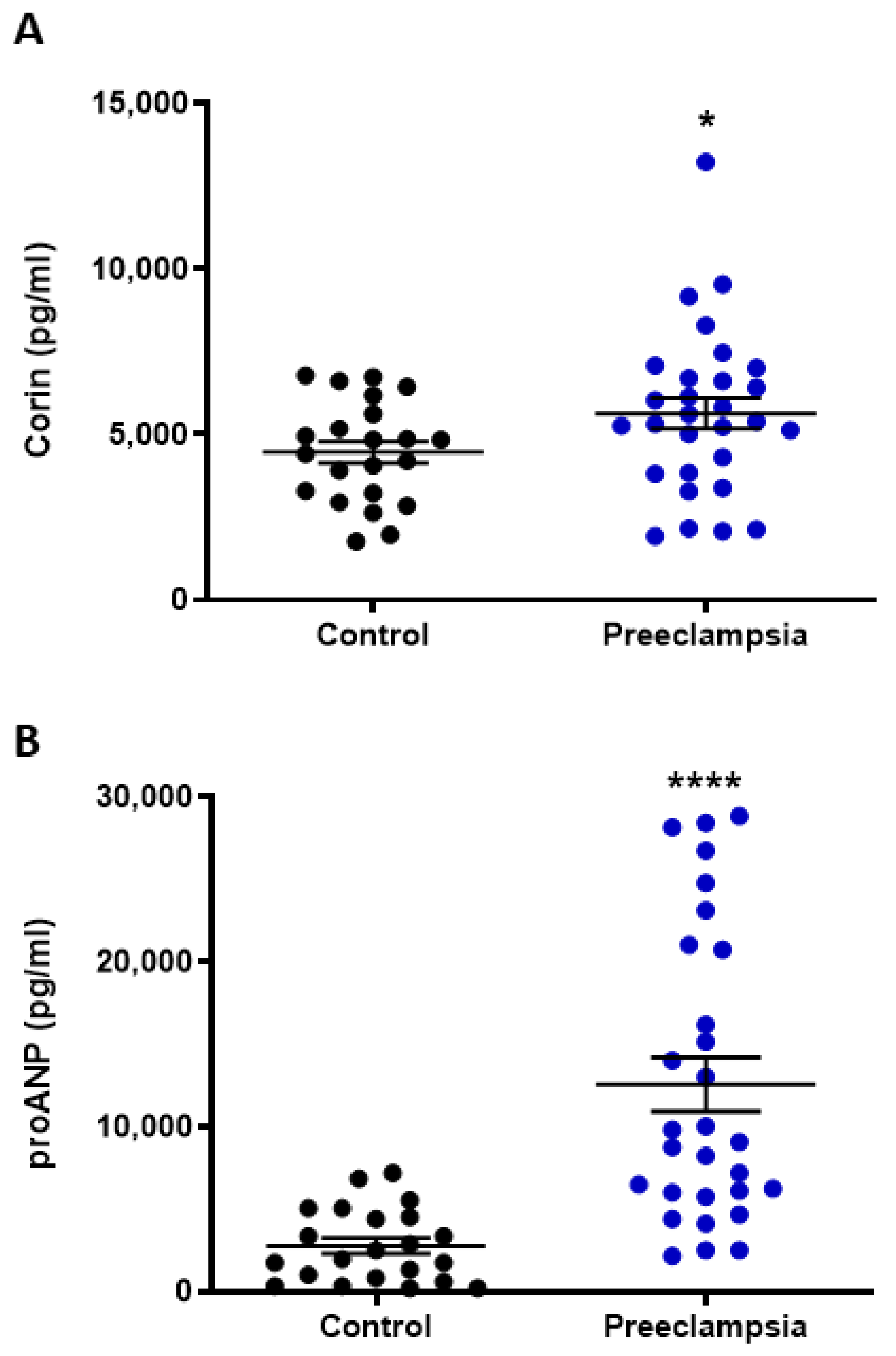
2.1. Circulating Levels of Corin and NT-proANP Increased with Preeclampsia

Compared to gestation-matched normotensive controls, the circulating plasma levels of both corin (p < 0.05, Figure 1A) and N-terminal (NT)-proANP (p < 0.0001, Figure 1B) were significantly elevated in pregnancies complicated by preterm preeclampsia requiring delivery before 34 weeks of gestation.
Within the placenta, the mRNA expression of corin and the ANP-encoding gene, natriuretic peptide precursor A (NPPA), did not significantly differ between pregnancies complicated by preterm preeclampsia or gestation-matched normotensive controls (Figure 2A,B).

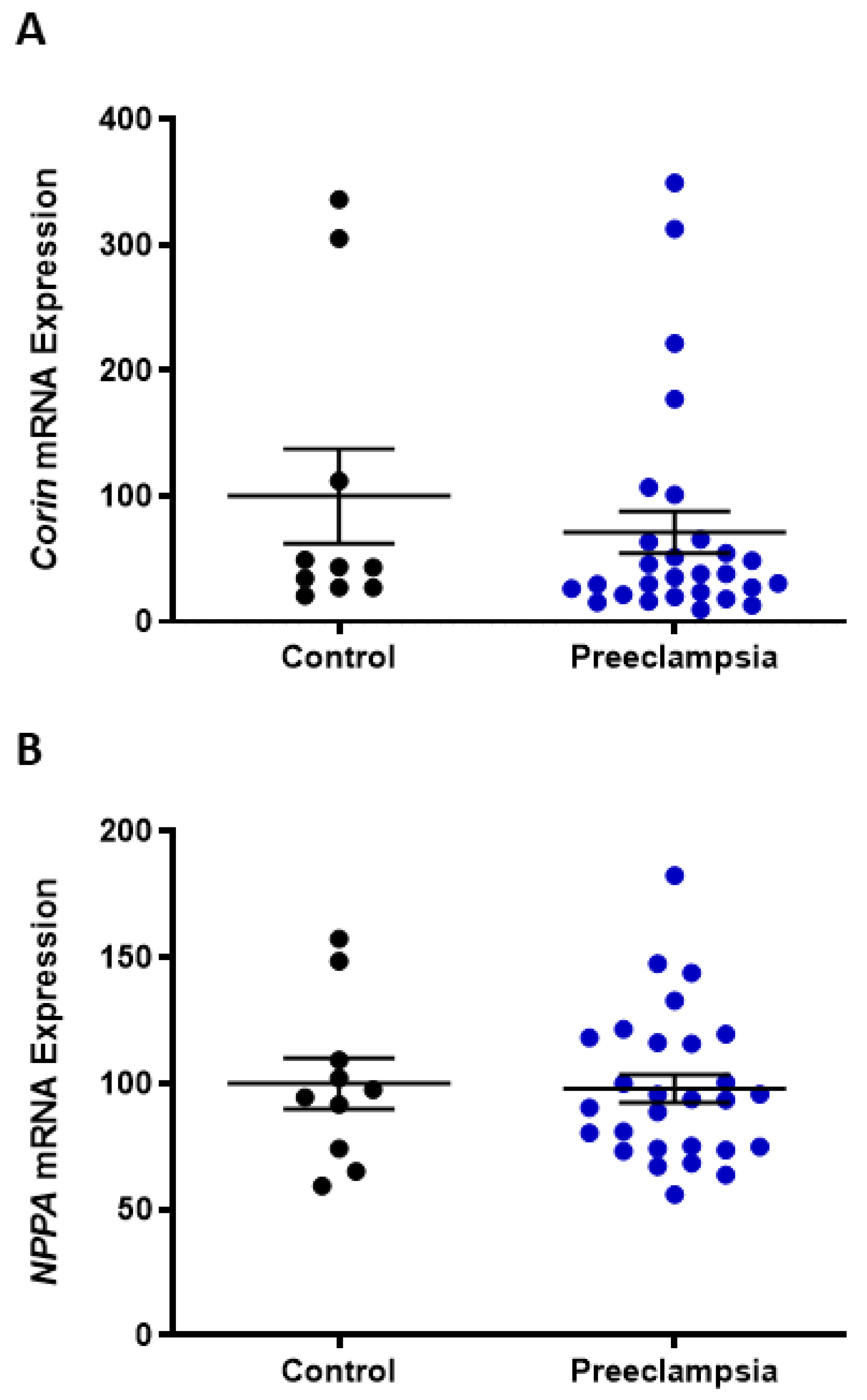
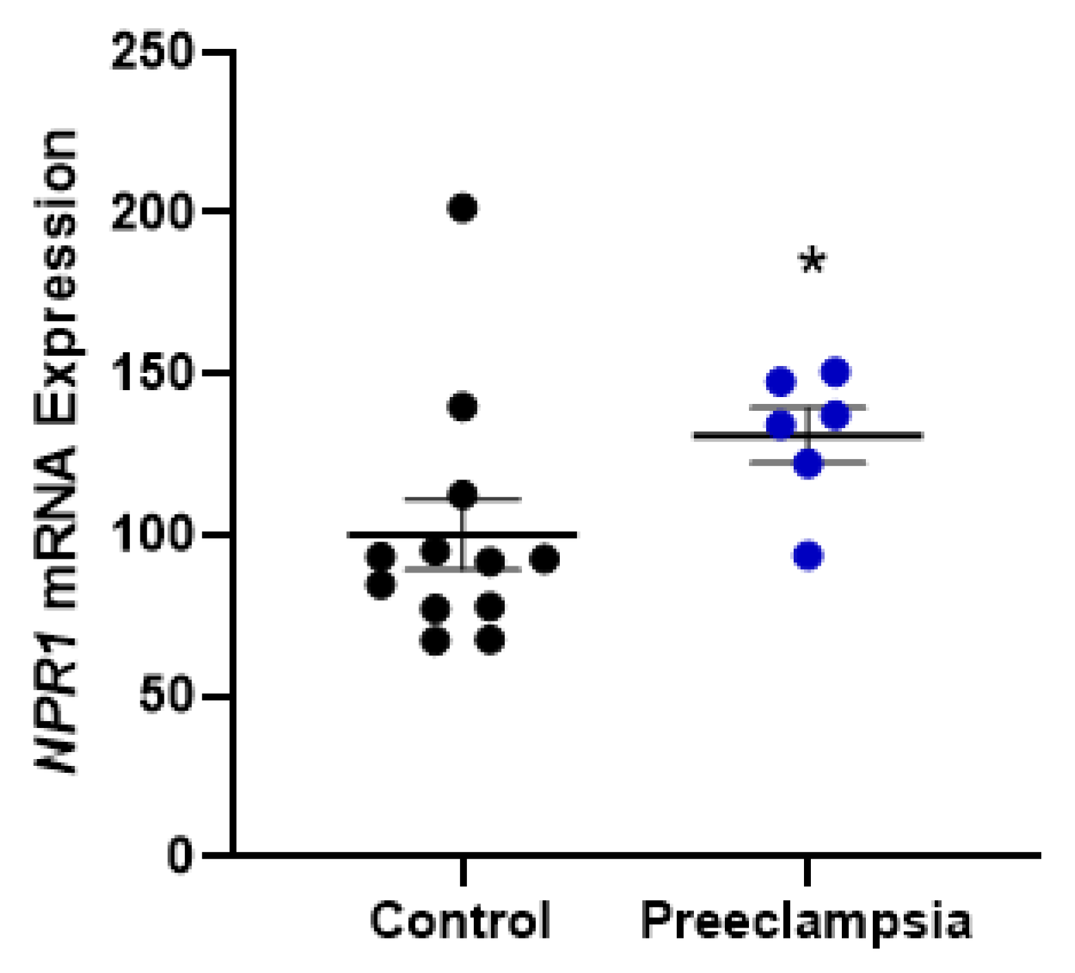
2.2. Omental Artery Expression of ANP Receptor, Natriuretic Peptide Receptor A (NPR1), Significantly Increased with Preeclampsia

The mRNA expression of NPR1 was significantly higher in omental arteries from pregnancies complicated by preterm preeclampsia compared to normotensive controls delivering an appropriately grown baby at term (p > 0.05 Figure 3).
Given the observation that soluble fms-like tyrosine kinase (sFlt)-1 and tumour necrosis factor (TNF)α are implicated in the endothelial dysfunction associated with preeclampsia [39,40,41], we investigated their effect on cultured normotensive term omental arteries. Neither NRP1 mRNA expression (Figure 4A) nor protein levels (Figure 4B) were regulated by TNFα or sFlt-1, alone or in combination.

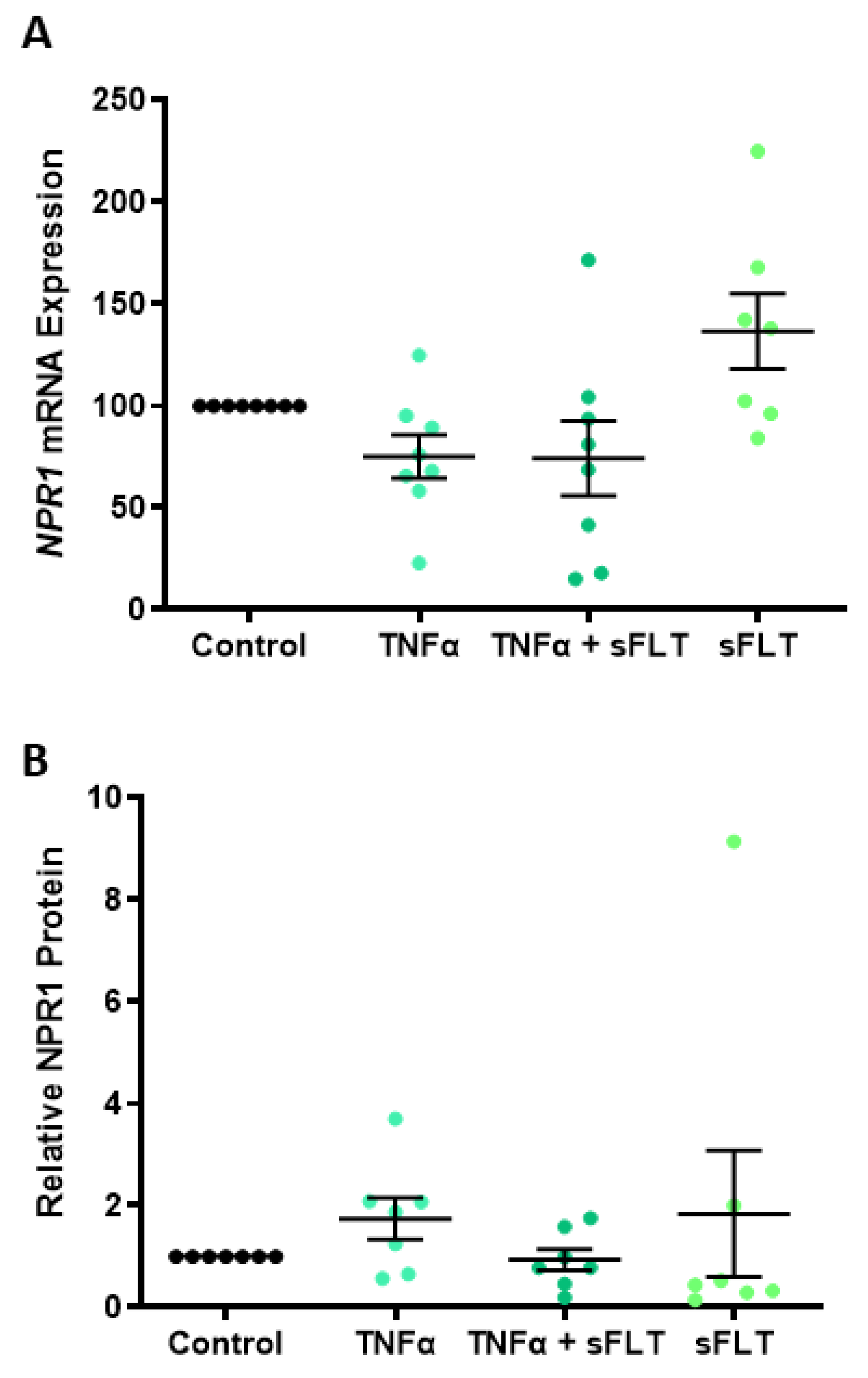
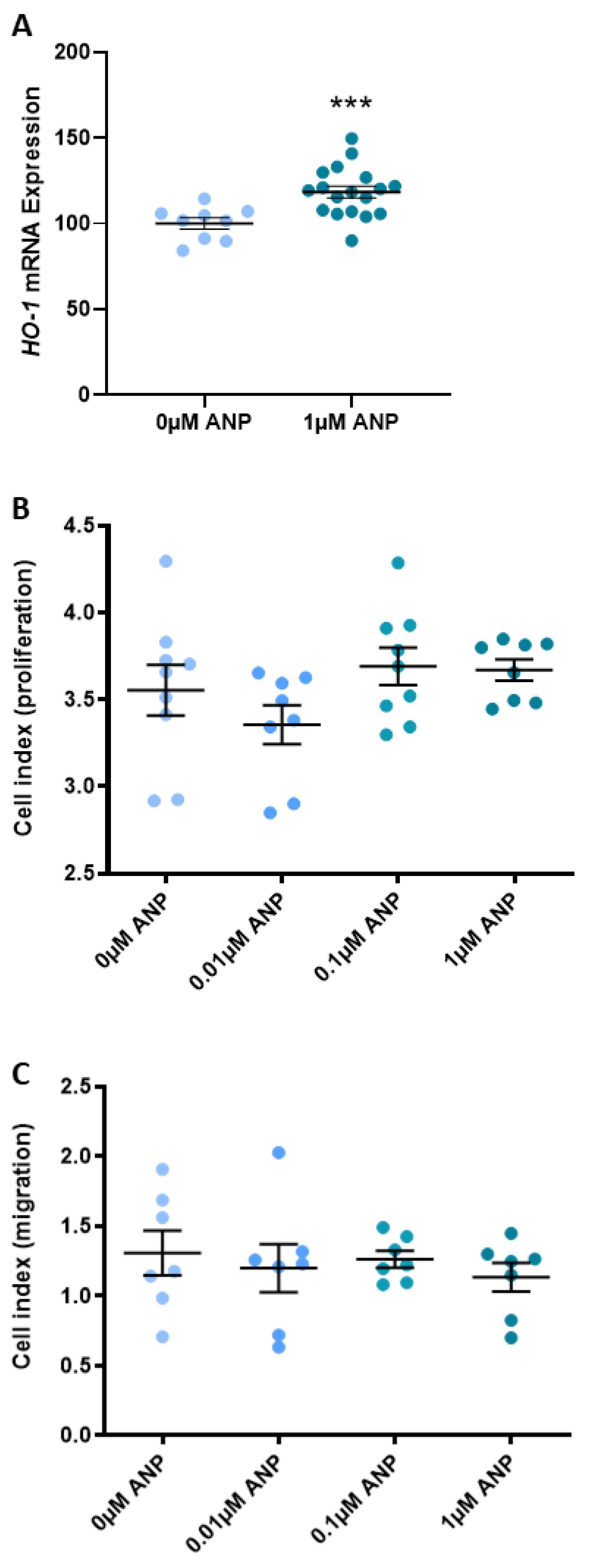
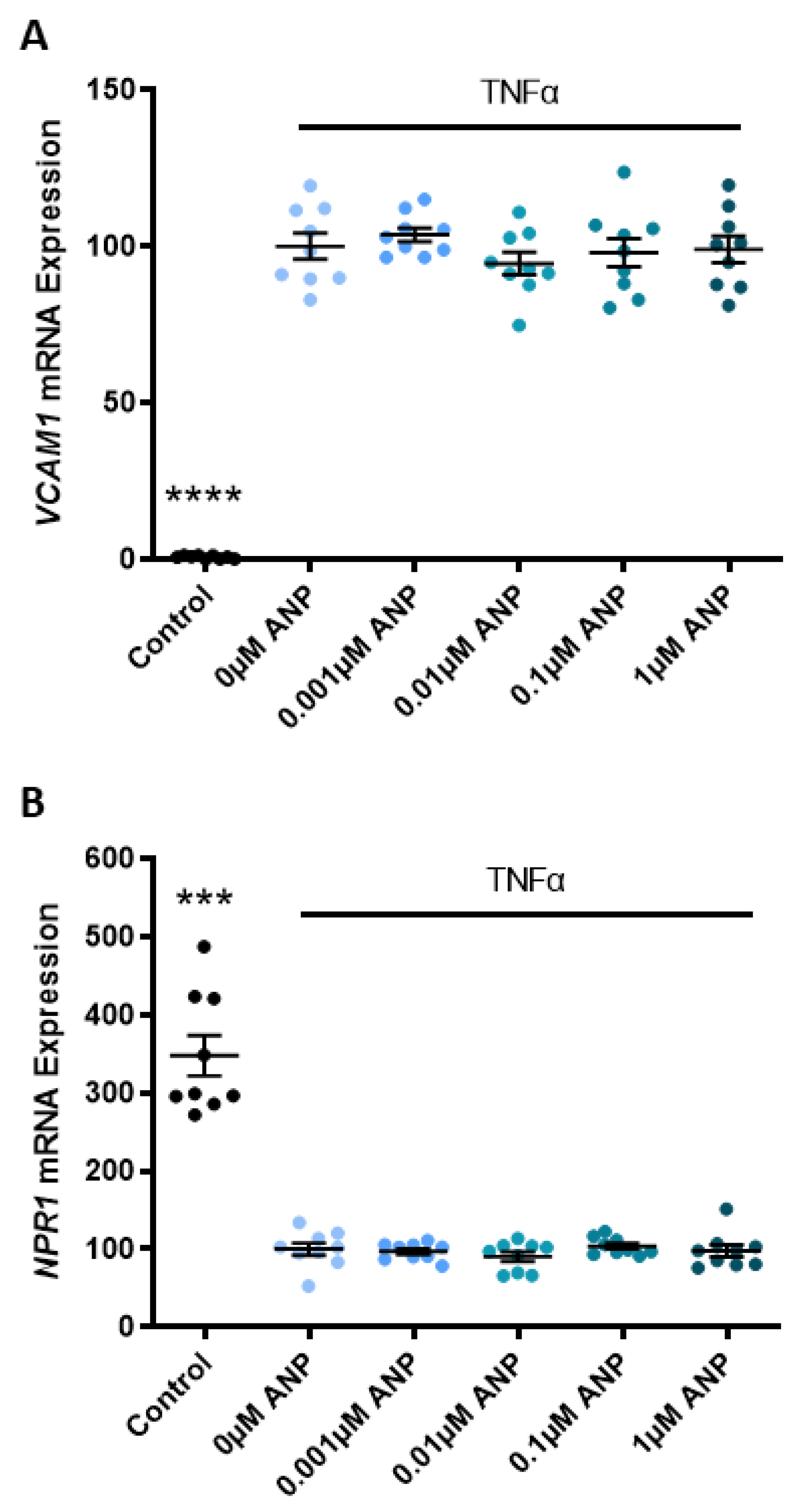
2.3. ANP Increases Heme Oxygenase (HO)-1 Expression but Does Not Affect Human Umbilical Vein Endothelial Cell (HUVEC) Proliferation or Migration

Known for its critical role in the prevention of vascular inflammation, HO-1 mRNA expression in isolated primary HUVEC is significantly increased with ANP treatments compared to the vehicle control (0 µM ANP, p < 0.001; Figure 5A). However, incrementally increasing doses of ANP did not affect HUVEC proliferation (Figure 5B) or migration (Figure 5C) compared to the vehicle control.
In a model of endothelial dysfunction, TNFα significantly increased the expression of vascular cell adhesion molecule (VCAM)1 mRNA (p < 0.0001; Figure 6A) and significantly decreased the expression of NPR1 mRNA (p < 0.001; Figure 6B) in primary HUVEC. Incrementally increasing doses of ANP did not mitigate the TNFα-induced change in VCAM1 or NPR1 mRNA expression (Figure 6A,B, respectively).

3. Discussion

We have previously identified corin expression in endometrium receptive to embryo implantation and first-trimester implantation sites, specifically localised to the maternal decidual cells surrounding the spiral arteries [31]. These findings support reports from murine studies suggesting a critical role for corin and its proteolytic product, ANP, in trophoblast migration and spiral artery remodelling [27,42]. Defective spiral artery remodelling resulting in placental ischemic injury and hypoxia is believed to be key to the pathogenesis of preeclampsia. Relatively little, however, is known about the role of ANP in endothelial function during preeclampsia.
A major focus of this study was to investigate if the hypotensive hormone ANP has a direct effect on the endothelium in response to preeclampsia-induced endothelial dysfunction, particularly with respect to endothelial cell proliferation and migration, markers of endothelial dysfunction, and receptor expression in omental arteries exposed to circulating preeclamptic toxins. Here, we demonstrate that the treatment of HUVECs with ANP did not promote proliferation or migration, nor did ANP treatment reduce markers of endothelial dysfunction in in vitro models of dysfunction. ANP receptor expression is significantly upregulated in preeclamptic vasculature, but not because of exposure to preeclampsia toxins TNFα or sFlt-1.

3.1. Circulating Corin and Pro-ANP Are Elevated in Preeclamptic Pregnancies

Several reports have identified elevated levels of corin and/or pro-ANP in the serum or plasma of women with preeclampsia at approximately 36 weeks gestation [27,30,43] or earlier as a predictive biomarker in high-risk women [44]. Here, we identified significantly elevated plasma corin and pro-ANP in a carefully collected cohort of cases of preterm preeclampsia (<34 weeks gestation) compared to gestation-matched controls. A study by Degrelle and colleagues has suggested that this increased circulating corin in women with preeclampsia is of placental origin [15]. However, this study was significantly underpowered and failed to distinguish between preterm and term preeclampsia for this analysis. Here, we investigated the expression of corin and the gene encoding pro-ANP, NPPA, in human placenta collected from cases of preterm preeclampsia (requiring delivery before 34 weeks completed gestation) and normotensive gestation-matched controls. Neither corin nor NPPA expression was overtly altered in placentas complicated with preeclampsia, which is consistent with our previous report [31].
The increased circulating pro-ANP with preeclampsia is likely a response to increasing blood pressure [45]. However, a corresponding decrease in blood pressure is not observed, suggesting there may be an impairment in the proteolytic action of corin converting pro-ANP to the hypotensive hormone, ANP. There have been identifications of several corin mutations amongst women with preeclampsia that may result in decreased enzymatic activity [27,46]. It is possible that decreased corin activity with preeclampsia causes a bottleneck in the pathway of pro-ANP conversion to ANP, resulting in a build-up of pro-ANP due to a lack of conversion and, hence, the absence of a blood-pressure-lowering effect with respect to ANP. Several reports have found circulating levels of ANP to be correspondingly elevated with preeclampsia [47,48], although there is the suggestion that ANP may have altered functions under the preeclamptic pathophysiological environment [49,50].

3.2. Omental Artery Expression of the ANP Receptor, NPR1, Is Elevated in Preeclamptic Pregnancies

Similarly, changes in the expression of ANP receptors with preeclampsia could also contribute to changes in the way the corin/ANP system works to regulate blood pressure. Here, the mRNA expression of NPR1, one of three natriuretic receptors and the principal receptor of ANP, was significantly upregulated in omental arteries from women with preterm preeclampsia compared to normotensive controls. As omental arteries contribute to systemic vascular resistance, a change in receptor expression could indicate a mechanism for blood pressure dysregulation. Unfortunately, these samples were not matched for gestation. This is a limitation of this finding and should be interpreted with caution, as this may, in part, be a gestation-related expression pattern. It is not clear from the literature whether pro-ANP/ANP, potentially including its receptors, fluctuates across gestation [51,52,53]; hence, further investigation is needed. However, there have been other reports of the dysregulation of vascular ANP receptors with preeclampsia, but again, these samples were not gestationally matched [37]. The importance of these endothelial receptor changes is yet to be elucidated.
Given the significant damage caused to the endothelium by anti-angiogenic and pro-inflammatory mediators (in a preeclamptic pregnancy), we wanted to test if two key factors, TNFα and sFlt-1, regulate the expression of NPR1 in omental arteries. In culture, neither TNFα or sFlt-1, alone or in combination, affected NPR1 mRNA expression or protein levels. Interestingly, when we induced dysfunction in HUVECs with TNFα, NPR1 expression significantly decreased. Treating with increasing concentrations of ANP did not restore NPR1 expression in the presence of TNFα. Of note, NPR1 knockout mice experience dysregulated cardiac ANP expression [54], suggesting a direct relationship between ligand and receptor expression [55]. The immunoreactivity of NPR1 should also be considered in the context of preeclampsia and warrants further investigation.

3.3. ANP Enhances Endothelial Production of HO-1 but Does Not Affect Proliferation, Migration, or Endothelial Dysfunction

Given the importance of angiogenic balance in preeclampsia and the critical role of corin and ANP in trophoblast migration and spiral artery remodelling, we wanted to assess whether ANP promotes endothelial cell migration and proliferation. Despite ANP significantly increasing endothelial HO-1 expression, which some studies have demonstrated plays a role in regulating both proliferation and angiogenesis [56,57,58], we show that ANP did not promote HUVEC proliferation or migration. When we induced dysfunction in these endothelial cells with TNFα, vascular cell adhesion molecule 1 (VCAM1) expression significantly increased. This was not mitigated by treatment with ANP. Endothelial dysfunction is synonymous with both preeclampsia and CVD, and we hypothesised that this may have been a unifying factor highlighting the importance of corin and ANP between the two pathologies. However, our data do not support this.

4. Materials and Methods

4.1. Tissue Collection

Ethical approval for this study was obtained from the Mercy Health Human Research Ethics Committee (R11/34 and R14/11). All methods were performed in accordance with the National Health and Medical Research Council’s ethical guidelines. Women presenting to the Mercy Hospital for Women, Heidelberg, Australia, gave informed written consent for tissue collection. Maternal venous blood was obtained prior to 34 weeks completed gestation from pregnancies complicated by preterm preeclampsia (requiring delivery <34 weeks gestation) and gestation-matched healthy controls who later went on to deliver an appropriately grown baby at term (patient characteristics Table 1). Placentas were obtained exclusively at caesarean section from pregnancies complicated by preterm preeclampsia (<34 weeks gestation) and gestation-matched preterm normotensive pregnancies with an appropriately grown for gestational age baby (<34 weeks gestation; patient characteristics in Table 2). Omental fatty tissue was obtained at caesarean section from pregnancies complicated by preterm preeclampsia (<34 weeks gestation) and normotensive term pregnancies that delivered an appropriately grown baby (≥37 weeks gestation up to 41 weeks gestation). Preeclampsia was diagnosed in accordance with the guidelines set by the American College of Obstetrics and Gynecology [59]. Umbilical cords were collected from normal-term pregnancies (≥37 weeks gestation up to 41 weeks gestation) at caesarean section. Tissue samples were collected within 30 min of delivery.

4.2. Blood and Placenta Processing

Maternal venous blood was collected into plasma EDTA tubes (BD Biosciences, Franklin Lakes, NJ, USA), inverted gently, and then centrifuged at 1000× g for 5 min. Plasma fractions were collected and frozen at −80 °C until assessment by an enzyme-linked immunosorbent assay (ELISA).
Four sites were sampled from individual placentas according to CoLab recommendations [60]. Following the removal of the basal plate and chorionic plate surfaces, samples were washed in sterile phosphate-buffered saline (PBS) and stabilised in RNAlater (Thermo Fisher Scientific, Waltham, MA, USA) before being snap-frozen with liquid nitrogen and stored at −80 °C until processing for RNA extraction.

4.3. Isolation and Treatment of Omental Arteries

Omental arteries were selected as they are accessible vasculature from pregnant women, and in the case of preeclampsia, they have been exposed to the circulating toxins responsible for endothelial dysfunction and end-organ injury. Omental arteries were dissected from omental fat biopsies and placed in RNAlater for a minimum of 48 h before being snap-frozen with liquid nitrogen and stored at −80 °C until processing for RNA extraction.
A subset of omental arteries obtained from term normotensive pregnancies was cut into 3 mm lengths and cultured overnight in DMEM high Glutamax (Life Technologies, Carlsbad, CA, USA) containing 10% fetal calf serum and 1% antibiotic-antimycotic (Life Technologies) at 37 °C in 20% O2 and 5% CO2. Artery segments were treated with 10 ng/mL TNFα (Sigma-Aldrich, St Louis, MO, USA), 250 ng/mL recombinant human sFlt-1 (Sigma-Aldrich), or TNFα and sFlt-1 in combination. Following the overnight culture, artery segments were snap-frozen with liquid nitrogen and stored at −80 °C until processing for protein and RNA extraction.

4.4. Isolation and Treatment of Primary HUVECs

The umbilical cord vein was infused with 10 mL (1 mg/mL) of collagenase (Worthington, Lakewood, NJ, USA), and cells were isolated as previously described [61]. HUVECs were cultured in M199 media (Life Technologies) containing 20% fetal calf serum, 1% antibiotic-antimycotic, 1% heparin, and 1% endothelial cell growth factor (Sigma-Aldrich) at 37 °C in 20% O2 and 5% CO2 and used between passage 2 and 4.
Endothelial dysfunction experiments were undertaken using primary HUVECs isolated from six normotensive term pregnancies. HUVECs were pretreated with 10 ng/mL TNF-α for 2 h before ANP was added at the following concentrations of 0 µM (vehicle control), 0.001, 0.01, 0.1, or 1 µM in the presence of TNF-α for a further 24 h. This concentration range has been shown to induce antioxidant defences (HO-1 expression) in HUVEC [62] and does not affect cell viability. At the cessation of the experiment, cells were permeabilised with lysis buffer and stored at −80 °C until processing for RNA extraction.

4.5. ELISA Analysis

Concentrations of corin and proANP were measured in frozen plasma samples using the DuoSet Human Corin and Human NT-ProANP kits, respectively (R&D systems by Bioscience, Waterloo, Australia), as per the manufacturer’s instructions.

4.6. Quantitative RT-PCR Analysis

RNA was extracted from primary HUVECs, placenta, and omental arteries using the RNeasy mini kit (Qiagen, Valencia, CA, USA) and quantified using the Nanodrop ND 1000 spectrophotometer (NanoDrop Technologies Inc., Wilmington, DE, USA). We converted 0.2 μg of RNA to cDNA using the Applied Biosystems High-Capacity cDNA Reverse Transcriptase Kit (Life Technologies) as per the manufacturer’s guidelines.
The gene expression of VCAM1 (Hs01003372_m1), HO-1 (Hs01110250_m1), corin (Hs00198141_m1), NPPA (Hs00383230_g1), NPR1 (Hs00181445_m1), HUVEC housekeeper YWHAZ (Hs01122454_m1), placenta housekeepers TOP1 (Hs00243257_m1), and CYC1 (Hs00357717_m1) and omental artery housekeepers B2M (Hs00187842_m1) and Actin (Hs99999903_m1) (Life Technologies) were quantified by real-time PCR (RT-PCR) on the CFX 384 (Bio-Rad, Hercules, CA, USA) using FAM-labeled Taqman universal PCR mastermix and its specific primer/probe set (Life Technologies) with the following run conditions: 50 °C for 2 min; 95 °C for 10 min; 95 °C for 15 s; and 60 °C for 1 min (40 cycles).

4.7. Western Blot Analysis

Following overnight culture, protein was extracted from omental arteries with RIPA lysis buffer and quantitated with a BCA assay. Omental artery protein lysates (13 µg) were separated on 10% polyacrylamide gels with wet transfer to PVDF membranes (Millipore, Billerica, MA, USA). Membranes were blocked in 5% skim milk prior to overnight incubation with the primary antibody NPR1 1:5000 (Anti-NPR-A, ab14356, Abcam, Cambridge, UK) in Can Get Signal (Toyobo, Osaka, Japan). Following incubation in secondary anti-rabbit antibody 1:2500 (Anti-Rabbit IgG, W401, Promega, Madison, WI, USA) in Can Get Signal, the bands were visualised using a chemiluminescence detection system (GE Healthcare Life Sciences, Piscataway, NJ, USA) and ChemiDoc XRS (BioRad). Relative densitometry was determined using QuantityOne software (BioRad). Β-actin loading controls were used for densitometric analysis.

4.8. xCELLigence

The xCELLigence Real-Time Cell Analyzer Dual Purpose instrument (Roche Diagnostic, Basel, Switzerland) was used to measure cellular proliferation and the migration of HUVECs following treatments with increasing concentrations of recombinant human ANP. For assessments via a proliferation assay, 5000 cells/well were plated in an E-Plate in media containing 0 µM (vehicle control), 0.01, 0.1, or 1 µM ANP in acetic acid. For assessment via a migration assay, 40,000 cells/well were plated in the top chamber of a CIM-Plate, with media in the wells below containing 0 µM (vehicle control), 0.01, 0.1, or 1 µM ANP in acetic acid as a stimulant. Assays were maintained at 37 °C in 20% O2 and 5% CO2, and electrical impedance was used to determine proliferation after 48 h culture and migration after 24 h culture.

4.9. Statistical Analysis

Experimental assays were run in technical duplicates, and the mean of the raw data was used for statistical analysis. qPCR data are represented as a fold change from the control (normalised to 100) and calculated using the 2^-deltadeltaCT method. Data were tested for normal distribution. Student’s t-test with Welch’s correction and a 1-way ANOVA (for parametric data) or Kruskal–Wallis test (for non-parametric data) were used as appropriate. Post hoc analysis was carried out using either Tukey’s (parametric) or Dunn’s test (non-parametric). All data are expressed as the mean ± standard error of the mean (SEM). p values < 0.05 were considered significant. Statistical analysis was performed using GraphPad Prism 9 software (GraphPad Software, La Jolla, CA, USA).

5. Conclusions

These studies using primary human endothelial cells, placenta, plasma, and omental arteries, both normal and pathological tissue, were unable to demonstrate a significant role for ANP in the regulation of endothelial function and/or dysfunction in preeclampsia.

Author Contributions

N.J.H., S.B. and N.K.B. designed the experiments. N.K.B. and N.J.H. wrote the main manuscript. T.J.K.-L., N.J.H. and S.B. provided intellectual input and assisted with manuscript editing. T.J.K.-L. and N.J.H. obtained funding. N.K.B., S.B., N.d.A., B.R.F. and T.-V.N. were involved in data generation. All authors reviewed and edited the manuscript drafts. All authors have read and agreed to the published version of the manuscript.

Funding

This study was funded by The National Health Medical Research Council via a Project Grant (#1062016) and Career Development Fellowship salary support to T.J.K.-L. (#1159261) and N.J.H. (#1146128). The funders had no role in the study’s design, data collection, analysis, or decision to publish.

Institutional Review Board Statement

This study was conducted in accordance with the Declaration of Helsinki and approved by the Human Research Ethics Committee of Mercy Health (R11/34 approved 26 September 2011 and R14/11 approved 21 August 2014).

Informed Consent Statement

Informed consent was obtained from all subjects involved in the study.

Data Availability Statement

Reasonable requests for access to data generated in this study should be directed to the corresponding author, N.J.H.

Acknowledgments

We would like to thank the research midwives, Gabrielle Pell, Genevieve Christophers, and Rachel Murdoch, Mercy Hospital for Women Obstetrics and Midwifery staff and patients, for participating in this research.

Conflicts of Interest

The authors report no conflict of interest.

References

  1. Wallis, A.B.; Saftlas, A.F.; Hsia, J.; Atrash, H.K. Secular trends in the rates of preeclampsia, eclampsia, and gestational hypertension, United States, 1987–2004. Am. J. Hypertens. 2008, 21, 521–526. [Google Scholar] [CrossRef] [PubMed] [Green Version]
  2. Duley, L. The global impact of pre-eclampsia and eclampsia. Semin. Perinatol. 2009, 33, 130–137. [Google Scholar] [CrossRef] [PubMed]
  3. Porreco, R.P.; Barkey, R. Peripartum intensive care. J. Matern. Fetal. Neonatal. Med. 2010, 23, 1136–1138. [Google Scholar] [CrossRef]
  4. Goldenberg, R.L.; Rouse, D.J. Prevention of premature birth. N. Engl. J. Med. 1998, 339, 313–320. [Google Scholar] [CrossRef] [PubMed]
  5. Sircar, M.; Thadhani, R.; Karumanchi, S.A. Pathogenesis of preeclampsia. Curr. Opin. Nephrol. Hypertens. 2015, 24, 131–138. [Google Scholar] [CrossRef]
  6. El-Sayed, A.A.F. Preeclampsia: A review of the pathogenesis and possible management strategies based on its pathophysiological derangements. Taiwan. J. Obstet. Gynecol. 2017, 56, 593–598. [Google Scholar] [CrossRef]
  7. Irgens, H.U.; Reisaeter, L.; Irgens, L.M.; Lie, R.T. Long term mortality of mothers and fathers after pre-eclampsia: Population based cohort study. BMJ 2001, 323, 1213–1217. [Google Scholar] [CrossRef] [Green Version]
  8. Smith, G.C.; Pell, J.P.; Walsh, D. Pregnancy complications and maternal risk of ischaemic heart disease: A retrospective cohort study of 129,290 births. Lancet 2001, 357, 2002–2006. [Google Scholar] [CrossRef]
  9. Ray, J.G.; Vermeulen, M.J.; Schull, M.J.; Redelmeier, D.A. Cardiovascular health after maternal placental syndromes (CHAMPS): Population-based retrospective cohort study. Lancet 2005, 366, 1797–1803. [Google Scholar] [CrossRef]
  10. Wilson, B.J.; Watson, M.S.; Prescott, G.J.; Sunderland, S.; Campbell, D.M.; Hannaford, P.; Smith, W.C. Hypertensive diseases of pregnancy and risk of hypertension and stroke in later life: Results from cohort study. BMJ 2003, 326, 845. [Google Scholar] [CrossRef] [Green Version]
  11. Wikström, A.K.; Haglund, B.; Olovsson, M.; Lindeberg, S.N. The risk of maternal ischaemic heart disease after gestational hypertensive disease. BJOG Int. J. Obstet. Gynaecol. 2005, 112, 1486–1491. [Google Scholar] [CrossRef] [PubMed]
  12. Bellamy, L.; Casas, J.P.; Hingorani, A.D.; Williams, D.J. Pre-eclampsia and risk of cardiovascular disease and cancer in later life: Systematic review and meta-analysis. BMJ 2007, 335, 974. [Google Scholar] [CrossRef] [PubMed] [Green Version]
  13. Hermes, W.; Van Kesteren, F.; De Groot, C.J. Preeclampsia and cardiovascular risk. Minerva Ginecol. 2012, 64, 281–292. [Google Scholar] [PubMed]
  14. Harskamp, R.E.; Zeeman, G.G. Preeclampsia: At risk for remote cardiovascular disease. Am. J. Med. Sci. 2007, 334, 291–295. [Google Scholar] [CrossRef]
  15. Degrelle, S.A.; Chissey, A.; Stepanian, A.; Fournier, T.; Guibourdenche, J.; Mandelbrot, L.; Tsatsaris, V. Placental Overexpression of Soluble CORIN in Preeclampsia. Am. J. Pathol. 2020, 190, 970–976. [Google Scholar] [CrossRef]
  16. Zhou, Y.; Wu, Q. Role of corin and atrial natriuretic peptide in preeclampsia. Placenta 2013, 34, 89–94. [Google Scholar] [CrossRef] [Green Version]
  17. Dong, N.; Niu, Y.; Chen, Y.; Sun, S.; Wu, Q. Function and regulation of corin in physiology and disease. Biochem. Soc. Trans. 2020, 48, 1905–1916. [Google Scholar] [CrossRef]
  18. Wu, Q. The serine protease corin in cardiovascular biology and disease. Front. Biosci. J. Virtual Libr. 2007, 12, 4179–4190. [Google Scholar] [CrossRef] [Green Version]
  19. Yu, R.; Han, X.; Zhang, X.; Wang, Y.; Wang, T. Circulating soluble corin as a potential biomarker for cardiovascular diseases: A translational review. Clin. Chim. Acta Int. J. Clin. Chem. 2018, 485, 106–112. [Google Scholar] [CrossRef]
  20. Wu, P.; Haththotuwa, R.; Kwok, C.S.; Babu, A.; Kotronias, R.A.; Rushton, C.; Zaman, A.; Fryer, A.A.; Kadam, U.; Chew-Graham, C.A.; et al. Preeclampsia and Future Cardiovascular Health: A Systematic Review and Meta-Analysis. Circ. Cardiovasc. Qual. Outcomes 2017, 10, e003497. [Google Scholar] [CrossRef]
  21. Yan, W.; Sheng, N.; Seto, M.; Morser, J.; Wu, Q. Corin, a mosaic transmembrane serine protease encoded by a novel cDNA from human heart. J. Biol. Chem. 1999, 274, 14926–14935. [Google Scholar] [CrossRef] [PubMed] [Green Version]
  22. Potter, L.R.; Yoder, A.R.; Flora, D.R.; Antos, L.K.; Dickey, D.M. Natriuretic peptides: Their structures, receptors, physiologic functions and therapeutic applications. Handb. Exp. Pharmacol. 2009, 191, 341–366. [Google Scholar] [CrossRef] [Green Version]
  23. Armaly, Z.; Assady, S.; Abassi, Z. Corin: A new player in the regulation of salt-water balance and blood pressure. Curr. Opin. Nephrol. Hypertens. 2013, 22, 713–722. [Google Scholar] [CrossRef] [PubMed]
  24. Kenyon, C.J.; Jardine, A.G. Atrial natriuretic peptide: Water and electrolyte homeostasis. Bailliere’s Clin. Endocrinol. Metab. 1989, 3, 431–450. [Google Scholar] [CrossRef] [PubMed]
  25. Goetze, J.P.; Bruneau, B.G.; Ramos, H.R.; Ogawa, T.; de Bold, M.K.; de Bold, A.J. Cardiac natriuretic peptides. Nat. Rev. Cardiol. 2020, 17, 698–717. [Google Scholar] [CrossRef]
  26. Dong, N.; Zhou, T.; Zhang, Y.; Liu, M.; Li, H.; Huang, X.; Liu, Z.; Wu, Y.; Fukuda, K.; Qin, J.; et al. Corin mutations K317E and S472G from preeclamptic patients alter zymogen activation and cell surface targeting. J. Biol. Chem. 2014, 289, 17909–17916. [Google Scholar] [CrossRef] [Green Version]
  27. Cui, Y.; Wang, W.; Dong, N.; Lou, J.; Srinivasan, D.K.; Cheng, W.; Huang, X.; Liu, M.; Fang, C.; Peng, J.; et al. Role of corin in trophoblast invasion and uterine spiral artery remodelling in pregnancy. Nature 2012, 484, 246–250. [Google Scholar] [CrossRef] [Green Version]
  28. Liu, Y.; Hu, J.; Yu, Q.; Zhang, P.; Han, X.; Peng, H. Increased serum soluble corin in mid pregnancy is associated with hypertensive disorders of pregnancy. J. Women’s Health 2015, 24, 572–577. [Google Scholar] [CrossRef]
  29. Zaki, M.A.; El-Banawy Sel, D.; El-Gammal, H.H. Plasma soluble corin and N-terminal pro-atrial natriuretic peptide levels in pregnancy induced hypertension. Pregnancy Hypertens. 2012, 2, 48–52. [Google Scholar] [CrossRef]
  30. Miyazaki, J.; Nishizawa, H.; Kambayashi, A.; Ito, M.; Noda, Y.; Terasawa, S.; Kato, T.; Miyamura, H.; Shiogama, K.; Sekiya, T.; et al. Increased levels of soluble corin in pre-eclampsia and fetal growth restriction. Placenta 2016, 48, 20–25. [Google Scholar] [CrossRef]
  31. Kaitu’u-Lino, T.J.; Ye, L.; Tuohey, L.; Dimitriadis, E.; Bulmer, J.; Rogers, P.; Menkhorst, E.; Van Sinderen, M.; Girling, J.E.; Hannan, N.; et al. Corin, an enzyme with a putative role in spiral artery remodeling, is up-regulated in late secretory endometrium and first trimester decidua. Hum. Reprod. 2013, 28, 1172–1180. [Google Scholar] [CrossRef] [PubMed] [Green Version]
  32. Zhang, W.; Li, S.; Lou, J.; Li, H.; Liu, M.; Dong, N.; Wu, Q. Atrial natriuretic peptide promotes uterine decidualization and a TRAIL-dependent mechanism in spiral artery remodeling. J. Clin. Investig. 2021, 131, e151053. [Google Scholar] [CrossRef] [PubMed]
  33. Staff, A.C.; Fjeldstad, H.E.; Fosheim, I.K.; Moe, K.; Turowski, G.; Johnsen, G.M.; Alnaes-Katjavivi, P.; Sugulle, M. Failure of physiological transformation and spiral artery atherosis: Their roles in preeclampsia. Am. J. Obstet. Gynecol. 2022, 226, S895–S906. [Google Scholar] [CrossRef]
  34. Baird, R.C.; Li, S.; Wang, H.; Naga Prasad, S.V.; Majdalany, D.; Perni, U.; Wu, Q. Pregnancy-Associated Cardiac Hypertrophy in Corin-Deficient Mice: Observations in a Transgenic Model of Preeclampsia. Can. J. Cardiol. 2019, 35, 68–76. [Google Scholar] [CrossRef] [PubMed]
  35. Abassi, Z.; Kinaneh, S.; Skarzinski, G.; Cinnamon, E.; Smith, Y.; Bursztyn, M.; Ariel, I. Aberrant corin and PCSK6 in placentas of the maternal hyperinsulinemia IUGR rat model. Pregnancy Hypertens. 2020, 21, 70–76. [Google Scholar] [CrossRef] [PubMed]
  36. Zhang, X.; Gu, X.; Zhang, Y.; Dong, N.; Wu, Q. Corin: A Key Mediator in Sodium Homeostasis, Vascular Remodeling, and Heart Failure. Biology 2022, 11, 717. [Google Scholar] [CrossRef]
  37. Gu, Y.; Thompson, D.; Xu, J.; Lewis, D.F.; Morgan, J.A.; Cooper, D.B.; McCathran, C.E.; Wang, Y. Aberrant pro-atrial natriuretic peptide/corin/natriuretic peptide receptor signaling is present in maternal vascular endothelium in preeclampsia. Pregnancy Hypertens. 2018, 11, 1–6. [Google Scholar] [CrossRef]
  38. Veerbeek, J.H.; Hermes, W.; Breimer, A.Y.; van Rijn, B.B.; Koenen, S.V.; Mol, B.W.; Franx, A.; de Groot, C.J.; Koster, M.P. Cardiovascular disease risk factors after early-onset preeclampsia, late-onset preeclampsia, and pregnancy-induced hypertension. Hypertension 2015, 65, 600–606. [Google Scholar] [CrossRef] [Green Version]
  39. Fraser, R.; Whitley, G.S.; Johnstone, A.P.; Host, A.J.; Sebire, N.J.; Thilaganathan, B.; Cartwright, J.E. Impaired decidual natural killer cell regulation of vascular remodelling in early human pregnancies with high uterine artery resistance. J. Pathol. 2012, 228, 322–332. [Google Scholar] [CrossRef] [Green Version]
  40. Sunderland, N.S.; Thomson, S.E.; Heffernan, S.J.; Lim, S.; Thompson, J.; Ogle, R.; McKenzie, P.; Kirwan, P.J.; Makris, A.; Hennessy, A. Tumor necrosis factor alpha induces a model of preeclampsia in pregnant baboons (Papio hamadryas). Cytokine 2011, 56, 192–199. [Google Scholar] [CrossRef]
  41. Cindrova-Davies, T.; Sanders, D.A.; Burton, G.J.; Charnock-Jones, D.S. Soluble FLT1 sensitizes endothelial cells to inflammatory cytokines by antagonizing VEGF receptor-mediated signalling. Cardiovasc. Res. 2011, 89, 671–679. [Google Scholar] [CrossRef] [PubMed] [Green Version]
  42. Wang, C.; Wang, Z.; He, M.; Zhou, T.; Niu, Y.; Sun, S.; Li, H.; Zhang, C.; Zhang, S.; Liu, M.; et al. Krüppel-like factor 17 upregulates uterine corin expression and promotes spiral artery remodeling in pregnancy. Proc. Natl. Acad. Sci. USA 2020, 117, 19425–19434. [Google Scholar] [CrossRef]
  43. Pouta, A.M.; Vuolteenaho, O.J.; Laatikainen, T.J. An increase of the plasma N-terminal peptide of proatrial natriuretic peptide in preeclampsia. Obstet. Gynecol. 1997, 89, 747–753. [Google Scholar] [CrossRef] [PubMed]
  44. Boroń, D.; Kornacki, J.; Gutaj, P.; Mantaj, U.; Wirstlein, P.; Wender-Ozegowska, E. Corin-The Early Marker of Preeclampsia in Pregestational Diabetes Mellitus. J. Clin. Med. 2022, 12, 61. [Google Scholar] [CrossRef] [PubMed]
  45. Wang, T.J.; Larson, M.G.; Keyes, M.J.; Levy, D.; Benjamin, E.J.; Vasan, R.S. Association of plasma natriuretic peptide levels with metabolic risk factors in ambulatory individuals. Circulation 2007, 115, 1345–1353. [Google Scholar] [CrossRef] [Green Version]
  46. Knappe, S.; Wu, F.; Madlansacay, M.R.; Wu, Q. Identification of domain structures in the propeptide of corin essential for the processing of proatrial natriuretic peptide. J. Biol. Chem. 2004, 279, 34464–34471. [Google Scholar] [CrossRef] [Green Version]
  47. Senöz, S.; Sahin, N.; Ozcan, T.; Direm, B.; Gökmen, O. The concentration of plasma atrial natriuretic peptide in normotensive and preeclamptic pregnancies. Eur. J. Obstet. Gynecol. Reprod. Biol. 1995, 62, 173–177. [Google Scholar] [CrossRef]
  48. Adam, B.; Malatyalioğlu, E.; Alvur, M.; Kökçü, A.; Bedir, A. Plasma Atrial Natriuretic Peptide Levels in Preeclampsia and Eclampsia. J. Matern.-Fetal Investig. 1998, 8, 85–88. [Google Scholar]
  49. Irons, D.W.; Baylis, P.H.; Butler, T.J.; Davison, J.M. Atrial natriuretic peptide in preeclampsia: Metabolic clearance, sodium excretion and renal hemodynamics. Am. J. Physiol. 1997, 273, F483–F487. [Google Scholar] [CrossRef]
  50. Hatjis, C.G.; Grogan, D.M. Changes in placental atrial natriuretic peptide receptors associated with severe toxemia of pregnancy. Placenta 1989, 10, 153–159. [Google Scholar] [CrossRef]
  51. Minegishi, T.; Nakamura, M.; Abe, K.; Tano, M.; Andoh, A.; Yoshida, M.; Takagi, T.; Nishikimi, T.; Kojima, M.; Kangawa, K. Adrenomedullin and atrial natriuretic peptide concentrations in normal pregnancy and pre-eclampsia. Mol. Hum. Reprod. 1999, 5, 767–770. [Google Scholar] [CrossRef] [PubMed] [Green Version]
  52. Sala, C.; Campise, M.; Ambroso, G.; Motta, T.; Zanchetti, A.; Morganti, A. Atrial natriuretic peptide and hemodynamic changes during normal human pregnancy. Hypertension 1995, 25, 631–636. [Google Scholar] [CrossRef] [PubMed]
  53. Thomsen, J.K.; Fogh-Andersen, N.; Jaszczak, P.; Giese, J. Atrial natriuretic peptide (ANP) decrease during normal pregnancy as related to hemodynamic changes and volume regulation. Acta Obstet. Gynecol. Scand. 1993, 72, 103–110. [Google Scholar] [CrossRef] [PubMed]
  54. Ellmers, L.J.; Knowles, J.W.; Kim, H.S.; Smithies, O.; Maeda, N.; Cameron, V.A. Ventricular expression of natriuretic peptides in Npr1(-/-) mice with cardiac hypertrophy and fibrosis. Am. J. Physiol. Heart Circ. Physiol. 2002, 283, H707–H714. [Google Scholar] [CrossRef] [Green Version]
  55. Khurana, M.L.; Mani, I.; Kumar, P.; Ramasamy, C.; Pandey, K.N. Ligand-Dependent Downregulation of Guanylyl Cyclase/Natriuretic Peptide Receptor-A: Role of miR-128 and miR-195. Int. J. Mol. Sci. 2022, 23, 13381. [Google Scholar] [CrossRef] [PubMed]
  56. Sunamura, M.; Duda, D.G.; Ghattas, M.H.; Lozonschi, L.; Motoi, F.; Yamauchi, J.; Matsuno, S.; Shibahara, S.; Abraham, N.G. Heme oxygenase-1 accelerates tumor angiogenesis of human pancreatic cancer. Angiogenesis 2003, 6, 15–24. [Google Scholar] [CrossRef]
  57. Was, H.; Cichon, T.; Smolarczyk, R.; Rudnicka, D.; Stopa, M.; Chevalier, C.; Leger, J.J.; Lackowska, B.; Grochot, A.; Bojkowska, K.; et al. Overexpression of heme oxygenase-1 in murine melanoma: Increased proliferation and viability of tumor cells, decreased survival of mice. Am. J. Pathol. 2006, 169, 2181–2198. [Google Scholar] [CrossRef] [Green Version]
  58. Evazi Bakhshi, S.; Mohammadi Roushandeh, A.; Habibi Roudkenar, M.; Shekarchi, S.; Bahadori, M.H. CRISPR/Cas9-mediated knockout of HO-1 decreased the proliferation and migration of T47D cells and increased cisplatin-induced apoptosis: An In Vitro study. Med. Oncol. 2022, 39, 175. [Google Scholar] [CrossRef]
  59. Gestational Hypertension and Preeclampsia: ACOG Practice Bulletin, Number 222. Obstet. Gynecol. 2020, 135, e237–e260. [CrossRef]
  60. Burton, G.J.; Sebire, N.J.; Myatt, L.; Tannetta, D.; Wang, Y.L.; Sadovsky, Y.; Staff, A.C.; Redman, C.W. Optimising sample collection for placental research. Placenta 2014, 35, 9–22. [Google Scholar] [CrossRef]
  61. Brownfoot, F.C.; Hannan, N.; Onda, K.; Tong, S.; Kaitu’u-Lino, T. Soluble endoglin production is upregulated by oxysterols but not quenched by pravastatin in primary placental and endothelial cells. Placenta 2014, 35, 724–731. [Google Scholar] [CrossRef] [PubMed]
  62. Kiemer, A.K.; Bildner, N.; Weber, N.C.; Vollmar, A.M. Characterization of heme oxygenase 1 (heat shock protein 32) induction by atrial natriuretic peptide in human endothelial cells. Endocrinology 2003, 144, 802–812. [Google Scholar] [CrossRef] [PubMed] [Green Version]
Figure 1. Circulating levels of corin and NT-proANP increased with preeclampsia. In pregnancies complicated by preterm preeclampsia (delivery <34 weeks gestation), there was a significant increase in plasma concentrations of corin (A) and N-terminal (NT)-proANP (B) compared to gestation-matched (normotensive) controls. Data are mean ± SEM, n = 22–29, * p < 0.05, **** p < 0.0001.
Ijms 24 06182 g001
Figure 2. Placental corin and NPPA (pro-ANP-encoding gene) expression are not altered with preeclampsia. In preterm pregnancies (<34 weeks gestation), placental corin (A) and NPPA (B) mRNA expression was not significantly altered between pregnancies complicated by preeclampsia and normotensive controls. Data are mean ± SEM, n = 10–28.
Ijms 24 06182 g002
Figure 3. Omental artery expression of ANP receptor NPR1 significantly increased with preeclampsia. Omental artery expression of NPR1 is significantly increased in cases of preterm preeclampsia (<34 weeks gestation) compared to normotensive controls. Data are mean ± SEM, n = 6–12, * p < 0.05.
Ijms 24 06182 g003
Figure 4. NPR1 expression and protein in cultured omental arteries are not affected by TNFα or sFlt-1. In cultured omental arteries, NRP1 mRNA expression (A) and protein levels (B) were unaltered by treatment with 10 ng/mL TNFα or 250 ng/mL recombinant human sFlt-1 alone or in combination. Data are mean ± SEM, n = 7–8.
Ijms 24 06182 g004
Figure 5. ANP increases HO-1 expression but does not affect HUVEC proliferation or migration. In isolated primary HUVEC, 1 µM ANP significantly increases HO-1 mRNA expression compared to 0 µM ANP control (A). Incrementally increasing doses of ANP did not affect HUVEC proliferation (B) or migration (C) compared to 0 µM ANP control. Data are mean ± SEM, n = 7–9, *** p < 0.001.
Ijms 24 06182 g005
Figure 6. ANP does not reduce the expression of endothelial dysfunction marker VCAM1 in a model of endothelial dysfunction. In a model of endothelial dysfunction, TNFα significantly increases the expression of VCAM1 mRNA (A) and significantly decreases the expression of NPR1 mRNA (B) in HUVECs. Incrementally increasing doses of ANP did not mitigate the TNFα-induced change in VCAM1 (A) or NPR1 (B) mRNA expression. Data are mean ± SEM, n = 9, *** p < 0.001, **** p < 0.0001.
Ijms 24 06182 g006
Table 1. Patient characteristics for collected plasma samples.
Control
(n = 22)
Preeclampsia
(n = 29)
Maternal Age
Median (IQR)
31 (28–33)31 (28–35)
Gestation at Sampling
Median (IQR)
28.5 (27.4–30.3)29.6 (28.4–31.4)
Gestation at Delivery
Median (IQR)
39.6 (38.9–40.7)30.6 (28.3–31.7) ****
BMI (kg/m2)
Median (IQR)
23.5 (21.8–29.0)32.0 (25.0–37.3) ***
Parity no. (%)
010 (45)22 (76)
111 (50)4 (14)
≥21 (5)3 (10)
Highest SBP prior to delivery (mmHg)
Median (IQR)
130 (125–131)174 (170–180) ****
Highest DBP prior to delivery (mmHg)
Median (IQR)
76 (70–81.3)103 (100–110) ****
Birth weight (g)
Median (IQR)
3550 (3158–3700)1305 (918–1497) ****
Body mass index (BMI) data unavailable for n = 4 preeclampsia. *** p < 0.001, **** p < 0.0001.
Table 2. Patient characteristics for collected placental tissue samples.
Control
(n = 10)
Preeclampsia
(n = 28)
Maternal Age
Median (IQR)
29 (24–36)31 (27–34)
Gestation at Delivery
Median (IQR)
29.9 (27.4–32)30.4 (27.6–31.8)
BMI (kg/m2)
Median (IQR)
28.4 (24.0–40.9)27.0 (24.5–37.0)
Parity no. (%)
02 (20)18 (64)
16 (60)7 (25)
≥22 (20)3 (11)
Highest SBP prior to delivery (mmHg)
Median (IQR)
125 (110–130)175 (160–180) ****
Highest DBP prior to delivery (mmHg)
Median (IQR)
70 (64–76)100 (90–110) ****
Birth weight (g)
Median (IQR)
1454 (941–2011)1314 (811–1424)
BMI data unavailable for n = 3 control; n = 7 preeclampsia. **** p < 0.0001.
Disclaimer/Publisher’s Note: The statements, opinions and data contained in all publications are solely those of the individual author(s) and contributor(s) and not of MDPI and/or the editor(s). MDPI and/or the editor(s) disclaim responsibility for any injury to people or property resulting from any ideas, methods, instructions or products referred to in the content.

