Communication

Autoimmune Regulator Gene Polymorphisms and the Risk of Primary Immune Thrombocytopenic Purpura: A Case-Control Study

by
Muhammad T. Abdel Ghafar
1,*,
Ola A. Elshora
1,
Alzahraa A. Allam
2,
Raghda Gabr Mashaal
2,
Shereen Awny Abdelsalam Hamous
2,
Sarah Ragab Abd El-Khalik
3,
Rania Nagi Abd-Ellatif
3,
Reham A. Mariah
3,
Radwa Eissa
4,
Mai Mwafy
4,
Rasha E. Shalaby
4,5,
Elham Nasif
6 and
Rasha A. Elkholy
1
1
Department of Clinical Pathology, Faculty of Medicine, Tanta University, Tanta 31511, Egypt
2
Department of Internal Medicine, Faculty of Medicine, Tanta University, Tanta 31511, Egypt
3
Department of Medical Biochemistry, Faculty of Medicine, Tanta University, Tanta 31511, Egypt
4
Department of Microbiology and Immunology, Faculty of Medicine, Tanta University, Tanta 31511, Egypt
5
Department of Basic Medical Sciences, Faculty of Medicine, Galala University, Galala 43511, Egypt
6
Department of Physiology, Faculty of Medicine, Tanta University, Tanta 31511, Egypt
*
Author to whom correspondence should be addressed.
Int. J. Mol. Sci. 2023, 24(5), 5007; https://doi.org/10.3390/ijms24055007
Submission received: 25 December 2022 / Revised: 31 January 2023 / Accepted: 6 February 2023 / Published: 5 March 2023
(This article belongs to the Section Molecular Immunology)

Abstract

:
This study aimed to assess the possible association between two single nucleotide polymorphisms (SNPs) of the autoimmune regulator (AIRE) gene (rs2075876 G/A and rs760426 A/G) with the risk of primary immune thrombocytopenia (ITP), as well as AIRE serum levels, in the Egyptian population. In this case-control study, 96 cases with primary ITP and 100 healthy subjects were included. Two SNPs of the AIRE gene (rs2075876 G/A and rs760426 A/G) were genotyped via Taqman allele discrimination real-time polymerase chain reaction (PCR). Additionally, serum AIRE levels were measured using the enzyme-linked immunosorbent assay (ELISA) technique. After adjusting for age, gender, and family history of ITP, the AIRE rs2075876 AA genotype and A allele were associated with increased ITP risk (adjusted odds ratio (aOR): 4.299, p = 0.008; aOR: 1.847, p = 0.004, respectively). Furthermore, there was no significant association between AIRE rs760426 A/G different genetic models and ITP risk. A linkage disequilibrium revealed that A-A haplotypes were associated with an increased ITP risk (aOR: 1.821, p = 0.020). Serum AIRE levels were found to be significantly lower in the ITP group, positively correlated with platelet counts, and were even lower in the AIRE rs2075876 AA genotype and A allele, as well as A-G and A-A haplotype carriers (all p < 0.001). The AIRE rs2075876 genetic variants (AA genotype and A allele) and A-A haplotype are associated with an increased ITP risk in the Egyptian population and lower serum AIRE levels, whereas the SNP rs760426 A/G is not.

1. Introduction

Immune thrombocytopenia (ITP) is an autoimmune bleeding disorder caused by excessive immune-mediated platelet destruction and inadequate bone marrow production [1]. However, the pathogenesis of ITP is relatively complicated, and the precise etiology and pathogenesis remain unknown. Several environmental factors, including viral infection, autoimmune disorders, and medications, have been linked to ITP pathogenesis. In addition, genetic factors have been identified as a risk factor for the development of ITP [2].
Recent evidence suggests that several mechanisms involving regulatory B- and T-cells, as well as natural killer cells, myeloid-derived suppressor cells, and dendritic cells [3,4,5,6] are all responsible for thrombocytopenia that develops in patients with ITP [7]. However, the mechanism underlying autoreactive T- and B-cell generation in autoimmune diseases remains unclear.
Autoimmune Regulator (AIRE) gene is one of the key genes that regulate immune tolerance, making it a candidate for better understanding the pathogenesis of autoimmune disorders [8]. It is located on chromosome 21q22.3 region and has an 11.9 kb genomic region with 14 exons. It encodes a 545 amino acid transcriptional regulator protein of 58 kD. This protein promotes the negative selection of thymic autoreactive T-cells by controlling the expression of a diverse set of self-antigens known as tissue-restricted antigens. The AIRE protein could promote the proteasome pathway, enhancing cell trafficking and DNA repair, as well as self-tolerance via its E3 ubiquitin ligase activity [9,10]. In addition to medullary thymic epithelial cells, evidence suggests that AIRE is also expressed in peripheral lymphoid organs [11]. Extra-thymic AIRE-expressing cells were discovered as a distinct “bone-marrow-derived antigen-presenting CD45low” population that functionally inhibits effector (CD4+) T-cells, preventing co-stimulation and inducing tolerance [12].
Several single nucleotide polymorphisms (SNPs) in the AIRE gene have been identified and reported to be associated with several autoimmune disorders, such as rheumatoid arthritis (RA) [13,14,15], systemic lupus erythematosus (SLE) [16,17], autoimmune thyroiditis, and systemic sclerosis [18], as well as myasthenia gravis [19], vitiligo [20], type 1 diabetes mellitus [21], and autoimmune hepatitis [22]. However, only two SNPs (rs2075876 and rs760426) have received attention due to their significant association with autoimmune disorders, particularly RA and SLE [14], as well as their impact on AIRE expression, particularly the A allele of rs2075876.
Experimental studies have shown that AIRE SNPs alter AIRE gene transcription, altering peripheral tissue antigen expression that controls peripheral antigen presentation, providing less efficient negative selection, promoting autoimmune T-cell survival, and increasing susceptibility to autoimmune diseases [23]. Furthermore, depending on the strain’s genetic background, AIRE-deficient mice can exhibit a wide range of autoimmune phenomena [24], including multi-organ lymphocytic infiltration and circulatory antibodies [25], implying a critical role in setting a self-tolerance threshold in immune regulation [26].
On the basis of these data, the AIRE gene may be a candidate gene for several autoimmune disorders. However, the association between AIRE gene polymorphisms and ITP risk has yet to be investigated. Therefore, our study aimed to assess the possible association between two SNPs (rs2075876 G/A and rs760426 A/G) of the AIRE gene with the risk of primary ITP, as well as AIRE serum levels, in the Egyptian population.

2. Results

2.1. Basic Characteristics of the Studied Cohorts

In this study, 96 patients with primary ITP and 100 healthy subjects were included. They did not differ significantly in terms of age or gender. However, the ITP group had a significantly higher proportion of positive family history and a lower platelet count than the healthy control group (Table 1).

2.2. AIRE SNPs and ITP Risk

The Hardy–Weinberg equilibrium (HWE) of different genotypes of the two AIRE SNPs (rs2075876 G/A and rs760426 A/G) in the control group was confirmed, indicating that our sample size was sufficient to adequately represent our population. The AIRE rs2075876 AA genotype and A allele were more frequent in the ITP group than in the control group (p = 0.004 and 0.005, respectively; Table 2). After adjusting for age, gender, and family history of ITP, the ITP risk was found to be higher in the AIRE rs2075876 AA genotype (adjusted odds ratio (aOR): 4.299, 95% confidence interval (CI): 1.650–11.202, p = 0.008) and A allele carriers (aOR: 1.847, 95% CI: 1.209–2.822, p = 0.004) than in the GG genotype and G allele carriers. Additionally, an increased ITP risk was observed under the recessive genetic model (aOR: 3.257, 95% CI: 1.406–7.545, p = 0.005) after adjusting to the confounders, whereas the dominant model revealed no association (Table 3). Furthermore, there was no significant difference in the distribution of AIRE rs760426 A/G different genetic models between ITP and healthy control groups.

2.3. AIRE SNP Haplotypes and ITP Risk

A linkage disequilibrium was detected between the two AIRE SNPs studied (D′ = 0.89), revealing four main haplotypes: G-A; G-G; A-G; and A-A. After adjusting for the confounders, A-A haplotypes were more frequent in the ITP group than in the control group and were associated with an increased ITP risk (aOR: 1.821, 95% CI: 1.099–3.017, p = 0.020) (Table 4).

2.4. AIRE SNPs and Haplotypes and Serum AIRE Levels

Serum AIRE levels were found to be significantly lower in the ITP group than in the control group (median, 3.1 ng/mL (IQR, 3.2) vs. median, 5.5 ng/mL (IQR, 3.0), respectively, p < 0.001; Figure 1A). Furthermore, a positive correlation was found between serum AIRE levels and platelet counts (r = 0.535, p < 0.001; Figure 1B). Serum AIRE levels differed significantly among different AIRE rs2075876 genotype and allele carriers (all p < 0.001), with the lowest levels detected in the AA genotype (Figure 1C) and A allele (Figure 1D) carriers (all p < 0.001). In contrast, AIRE serum levels did not differ between different AIRE rs760426 genotypes (p = 0.438; Figure 1E) and alleles (p = 0.748; Figure 1F). Furthermore, serum AIRE levels were significantly lower in the A-G and A-A haplotypes than in the G-A and G-G haplotypes (Figure 1G).

3. Discussion

The hallmark of the normal adaptive immune system is the induction of self-tolerance. For instance, the CD40 gene SNP (rs1883832 C>T) is associated with an increased risk of ITP, particularly when combined with CD40 rs4810485 G>T in the Egyptian population [27,28]. In addition, the histone deacetylase 3 gene SNP (rs2530223 C>T) was found to be associated with increased susceptibility to ITP in all genetic models, with TC/TT genotypes associated with severe thrombocytopenia in the Han population [29].
In this regard, AIRE is regarded as a key player in self-tolerance machinery. Variants in the AIRE gene have been shown to suppress its transcription and protein levels, reducing negative selection and enhancing autoimmune T-cell survival. Autoimmune T-cells play a central role in promoting the development of a variety of immunological disorders via producing a wide range of autoantibodies [23]. Therefore, it is not surprising that the existence of AIRE genetic variations has been linked to a variety of autoimmune disorders [30].
Notably, only two of the eleven SNPs identified in the AIRE gene (rs2075876 G/A and rs760426 A/G) have received attention and have been found to carry a risk for several autoimmune disorders, particularly RA and SLE [17,31]. Although rs2075876 G/A and rs760426 A/G exist in non-coding intronic regions, they have been implicated in inhibiting AIRE gene expression, thereby impairing thymic negative selection and increasing the risk for autoimmune disorders [32,33,34]. Although the mechanisms underlying RA and SLE are unknown, evidence suggests that the loss of self-tolerance with the activation of autoreactive T- and B-cells plays a role [35,36]. In the same vein, ITP is characterized by the excessive activation and proliferation of platelet autoantigen reactive cytotoxic T lymphocytes [37], abnormal T helper cells [38], and B-cells [39], indicating a shared pathogenicity with RA and SLE in terms of altered immune tolerance. Therefore, we conducted this study to assess, for the first time, the association between these two AIRE SNPs and ITP risk in the Egyptian population.
Our study revealed that AIRE rs2075876 G/A was associated with increased ITP risk under the additive, recessive, and multiplicative genetic models, with the AA genotype and A allele conferring the higher ITP risk. Other relevant studies revealed that AIRE rs2075876 plays a crucial role in determining the risk for various autoimmune disorders, such as RA and SLE, which is consistent with our findings. In the Chinese population, for instance, the AIRE rs2075876 A allele has been reported to increase RA risk under recessive [31,40] as well as dominant and co-dominant genetic models [13,40]. Furthermore, A. Moneim et al. reported that the AIRE rs2075876 A allele was more frequent in patients with RA than in the healthy subjects under both co-dominant and over-dominant models [15]. Recently, Salesi et al. reported that AIRE rs2075876 homozygous AA and heterozygous AG genotypes increased RA risk when compared to the GG genotype in the Iranian population [41]. In addition, Attia et al. reported an association between this polymorphism and SLE risk in the Egyptian population [17]. On the contrary, Alghamdi et al. found that the AIRE rs2075876 A allele was associated with a low risk of SLE in the Egyptian population [16]. However, no association between this polymorphism and the SLE risk has been reported in the Mexican population [42].
On the other hand, our study findings did not find any significant association between the different genetic models of AIRE rs760426 and ITP risk in our population. In contrast, Shao et al. and Li et al. found that the AIRE rs760426 G allele was more frequent in patients with RA than in healthy controls and was associated with higher RA risk under the recessive model in the Chinese population [31,40]. However, a borderline association between rs760426 and RA risk was detected by Feng et al. [13]. Moreover, A. Moneim et al. found that the AIRE rs760426 AG genotype and G allele were more frequent in patients with RA than in healthy controls in both co-dominant and over-dominant genetic models [15]. Furthermore, Attia et al. reported that the AIRE rs760426 homozygous genotype was more frequent in patients with SLE than in healthy controls, indicating a stronger association with SLE risk in the Egyptian population [17].
Since the association between the two studied polymorphisms and autoimmune disorders has been reported, such as in a previous meta-analysis that revealed an association between these two SNPs and high RA risk under all genetic models [23], it is reasonable to assume that linkage disequilibrium may exist between these polymorphisms and may confer a risk for autoimmune disorders. In this study, such linkage disequilibrium was observed, with the A-A haplotype being associated with a 1.821-fold increased ITP risk. In contrast, A. Moneim et al. revealed no evidence of linkage disequilibrium between the two SNPs in RA [15]. In terms of other AIRE SNPs, several SNPs were examined in combination and revealed that AIRE haplotype CCTGCC (AIRE C-103T, C4144G, T5238C, G6528A, T7215C, and T11787C) showed a three-fold increased risk for vitiligo [20]. Furthermore, the AIRE CCTGCT and CGTGCC haplotypes (C-103T, C4144G, T5238C, G6528A, T7215C, and T11787C) showed a 9.47- and 3.51-fold increased risk for alopecia areata, respectively [43]. Furthermore, the haplotype of five AIRE SNPs (rs2075876, rs2075877, rs933150, rs1003854, and rs1078480) and the two SNPs (rs2256817 and rs760426) revealed a 1.852- and 1.950-fold increased risk for RA, respectively [13]. On the other hand, the variant effect prediction for the two studied SNPs in the study revealed that 36% of them are upstream gene variants, and 36% are intronic variants. In contrast, other AIRE SNPs (rs2075876, rs2075877, rs933150, rs1003854, and rs1078480) were 43% intronic variants and 25% noncoding transcript variants.
In this study, we estimated serum AIRE levels in the studied groups to investigate the potential impact of AIRE genetic variants (rs2075876 G/A and rs760426 A/G) on its expression levels. Our findings revealed that the AIRE rs2075876 AA genotype and A allele had a strong impact on AIRE expression, lowering its serum levels. This is the first study to investigate the relationship between AIRE SNPs and their protein expression levels in autoimmune disorders; no previous studies have investigated this issue so far. However, this effect was demonstrated in silico using data from the Gene Expression Omnibus (GEO) database for 210 lymphoblastoid cells, revealing that the AIRE rs2075876 A allele was significantly correlated with decreased AIRE transcription and, thus, decreased protein levels [14]. However, GEO revealed no association between the AIRE rs760426 G allele and AIRE expression [44].
Although this study was the first to investigate the association between AIRE rs2075876 G/A and rs760426 A/G genetic variants and haplotypes with ITP risk, as well as serum AIRE levels, it does have some limitations that should be considered. First, this is a single-center study. Second, because this study was conducted on a single population, our findings cannot be generalized to other populations. Third, no functional analysis was conducted to assess the causal relationship between AIRE SNPs on one side and AIRE expression and ITP pathogenesis on the other. Fourth, because the samples were collected prior to any treatment, the impact of treatment, particularly corticosteroids, on serum AIRE levels could not be investigated. Finally, serum AIRE levels were not correlated with the duration of ITP. Further studies on other populations and AIRE SNPs, as well as the impact of ITP treatment on AIRE levels and the relationship between AIRE levels and ITP duration, are warranted to support our findings.

4. Materials and Methods

4.1. Study Cohort

In this case-control study, 96 patients with primary ITP at their initial presentation were recruited from the hematology unit of the Internal Medicine Department, Tanta University Hospitals, as a case group. Their inclusion criteria were all cases diagnosed with ITP based on the standard criteria of the International Working Group of isolated thrombocytopenia (platelet counts of less than 100 × 103/µL in the absence of other causes of thrombocytopenia) [45]. Patients with secondary causes of ITP such as viral infections, drugs, and autoimmune disorders, such as RA, SLE, and Chron’s disease, were excluded from the study.
In addition, 100 healthy subjects with normal platelet counts were recruited from those attending the out-patient clinics at Tanta University Hospitals as a control group. The exclusion criteria of the case group were also applied to the control group. This study was conducted in accordance with the Helsinki Declaration and was approved by the local ethical committee of the Faculty of Medicine, Tanta University (approval code no. 35353/3/22). Informed written consent was obtained from all the participants.

4.2. Clinical and Laboratory Assessment

All participants were asked about their personal and family history of ITP, and patients with ITP were examined clinically and radiologically via ultrasonography to detect splenomegaly. Data on complete blood count (CBC), bone marrow aspiration cytology, antinuclear antibodies (ANAs), and direct anti-globulin test (if Evan syndrome was suspected) used to diagnose ITP, as well as data on viral screening for hepatitis C, cytomegalovirus, human immunodeficiency virus, and helicobacter pylori antigen in stool to exclude secondary ITP, were obtained from the patients’ medical records.

4.3. Sampling

Blood samples were collected prior to receiving any treatment. Five milliliters of venous blood was collected via standard venipuncture, and two milliliters was delivered into a K2 EDTA vacutainer tube for genotyping. The remainder was delivered into a gel tube at room temperature, centrifuged at 3000 rpm for 5 min, and the serum was then separated and immediately frozen in −20 °C for the assay of AIRE serum levels.

4.4. AIRE SNP Genotyping

The genomic DNA was extracted from blood samples using the GeneJet genomic DNA purification kit (Thermo-Fisher Scientific, Waltham, MA, USA), according to the manufacturer’s instructions and kept at −80 °C for AIRE genotyping. Two AIRE SNPs (rs2075876 G/A and rs760426 A/G) were genotyped using real-time polymerase chain reaction (PCR) by allelic discrimination assay with Taqman SNP genotyping assay kit (Thermo-Fisher Scientific, MA, USA). In a total volume of 25 µL, the extracted DNA (25 ng/3 µL) was mixed with 2xTaqman genotyping master mix (12.5 µL), 20xTaqman SNP genotyping assay (1.25 µL), and nuclease-free water. The reaction mixture was then run on an Applied Biosystem StepOne real-time PCR instrument (Foster City, CA, USA) under the following thermal cycling conditions: pre-denaturation at 95 °C for 5 min, followed by 40 cycles of denaturation at 95 °C for 15 s and annealing/extension at 60 °C for 60 s. The genotypes were determined with cycle thresholds on the multicomponent plot.

4.5. Serum AIRE Assay

Serum AIRE levels were measured using a commercially available enzyme-linked immunosorbent assay (ELISA) kit (Human AIRE, Biovision, Milpitas, CA, USA, catalog no. E4971-100), according to the manufacturer’s instructions, and colorimetrically detected at 450 nm on a Tecan Spectra II Microplate Reader (canton of Zürich, Switzerland). The sample concentration was calculated using a logit–log standard curve. The assay sensitivity was 0.094 ng/mL, with intra-assay and inter-assay coefficients of variation of <8% and <10%, respectively.

4.6. Statistical Analysis

All datasets were analyzed using the Statistical Package for the Social Sciences (SSPS) software Version 22 (IBM Corp, Armonk, NY, USA). The data were tested for normality using the Kolmogorov–Smirnov test. The normally distributed numerical variables were presented as mean and standard deviation and were compared using Student’s t-test, while the non-normally distributed variables were presented as median and interquartile range (IQR) and were compared using the Mann–Whitney U test. The categorical variables were presented as numbers and percentages and were compared using the Chi-square χ2 test. The significant comparisons were further corrected (Pcorr) using Bonferroni’s correction for multiple testing. The HWE of AIRE SNPs (rs2075876 and rs760426) genotypes was determined in the control group using the χ2 test. The web-based SHEsisPlus platform (http://shesisplus.bio-x.cn/SHEsis.html (accessed on 8 August 2022)) was used for haplotype analysis. The ITP risk was calculated as a crude OR with a 95% CI for both AIRE SNP genetic models and haplotypes and then adjusted for confounders such as age, gender, and family history of ITP using multiple logistic regression analysis. A two-sided p-value of less than 0.05 was considered statistically significant.

5. Conclusions

Our findings reveal that the AIRE rs2075876 genetic variants (AA genotype and A allele) and A-A haplotype are associated with increased ITP risk in the Egyptian population and lower serum AIRE levels, whereas the rs760426 A/G SNP is not.

Author Contributions

Conceptualization, M.T.A.G. and R.A.E.; methodology, M.T.A.G. and R.A.E.; software, M.T.A.G.; validation, S.R.A.E.-K., R.N.A.-E. and R.A.M.; formal analysis, M.T.A.G.; investigation, M.T.A.G., R.A.E., O.A.E., S.R.A.E.-K., R.N.A.-E., R.A.M., R.E., M.M. and R.E.S.; resources, A.A.A., R.G.M. and S.A.A.H.; data curation, A.A.A., R.G.M., E.N. and S.A.A.H.; writing—original draft preparation, M.T.A.G. and R.A.E.; writing—review and editing, M.T.A.G. and R.A.E.; supervision, M.T.A.G. and R.A.E. All authors have read and agreed to the published version of the manuscript.

Funding

This research received no external funding.

Institutional Review Board Statement

The study was conducted in accordance with the Declaration of Helsinki and approved by the Ethics Committee of the Faculty of Medicine, Tanta University (approval code no. 35353/3/22).

Informed Consent Statement

Informed consent was obtained from all subjects involved in the study.

Data Availability Statement

The data used in this study are available from the corresponding author upon request.

Conflicts of Interest

The authors declare no conflict of interest.

References

  1. Gernsheimer, T. Chronic idiopathic thrombocytopenic purpura: Mechanisms of pathogenesis. Oncologist 2009, 14, 12–21. [Google Scholar] [CrossRef] [PubMed] [Green Version]
  2. Despotovic, J.M.; Polfus, L.M.; Flanagan, J.M.; Bennett, C.M.; Lambert, M.P.; Neunert, C.; Kumar, M.; Klaassen, R.J.; Thornburg, C.; Jeng, M.; et al. Genes Influencing the Development and Severity of Chronic ITP Identified through Whole Exome Sequencing. Blood 2015, 126, 73. [Google Scholar] [CrossRef]
  3. Audia, S.; Mahévas, M.; Samson, M.; Godeau, B.; Bonnotte, B. Pathogenesis of immune thrombocytopenia. Autoimmun. Rev. 2017, 16, 620–632. [Google Scholar] [CrossRef] [PubMed]
  4. McKenzie, C.G.; Guo, L.; Freedman, J.; Semple, J.W. Cellular immune dysfunction in immune thrombocytopenia (ITP). Br. J. Haematol. 2013, 163, 10–23. [Google Scholar] [CrossRef] [PubMed]
  5. Hou, Y.; Feng, Q.; Xu, M.; Li, G.S.; Liu, X.N.; Sheng, Z.; Zhou, H.; Ma, J.; Wei, Y.; Sun, Y.X.; et al. High-dose dexamethasone corrects impaired myeloid-derived suppressor cell function via Ets1 in immune thrombocytopenia. Blood 2016, 127, 1587–1597. [Google Scholar] [CrossRef] [Green Version]
  6. Ebbo, M.; Audonnet, S.; Grados, A.; Benarous, L.; Mahevas, M.; Godeau, B.; Viallard, J.F.; Piperoglou, C.; Cognet, C.; Farnarier, C.; et al. NK cell compartment in the peripheral blood and spleen in adult patients with primary immune thrombocytopenia. Clin. Immunol. 2017, 177, 18–28. [Google Scholar] [CrossRef]
  7. Grodzielski, M.; Goette, N.P.; Glembotsky, A.C.; Constanza Baroni Pietto, M.; Méndez-Huergo, S.P.; Pierdominici, M.S.; Montero, V.S.; Rabinovich, G.A.; Molinas, F.C.; Heller, P.G.; et al. Multiple concomitant mechanisms contribute to low platelet count in patients with immune thrombocytopenia. Sci. Rep. 2019, 9, 2208. [Google Scholar] [CrossRef] [Green Version]
  8. Rizzi, M.; Ferrera, F.; Filaci, G.; Indiveri, F. Disruption of immunological tolerance: Role of AIRE gene in autoimmunity. Autoimmun. Rev. 2006, 5, 145–147. [Google Scholar] [CrossRef]
  9. Nagamine, K.; Peterson, P.; Scott, H.S.; Kudoh, J.; Minoshima, S.; Heino, M.; Krohn, K.J.; Lalioti, M.D.; Mullis, P.E.; Antonarakis, S.E.; et al. Positional cloning of the APECED gene. Nat. Genet. 1997, 17, 393–398. [Google Scholar] [CrossRef]
  10. Abramson, J.; Giraud, M.; Benoist, C.; Mathis, D. Aire’s partners in the molecular control of immunological tolerance. Cell 2010, 140, 123–135. [Google Scholar] [CrossRef] [Green Version]
  11. Poliani, P.L.; Kisand, K.; Marrella, V.; Ravanini, M.; Notarangelo, L.D.; Villa, A.; Peterson, P.; Facchetti, F. Human peripheral lymphoid tissues contain autoimmune regulator-expressing dendritic cells. Am. J. Pathol. 2010, 176, 1104–1112. [Google Scholar] [CrossRef] [PubMed]
  12. Gardner, J.M.; Metzger, T.C.; McMahon, E.J.; Au-Yeung, B.B.; Krawisz, A.K.; Lu, W.; Price, J.D.; Johannes, K.P.; Satpathy, A.T.; Murphy, K.M.; et al. Extrathymic Aire-expressing cells are a distinct bone marrow-derived population that induce functional inactivation of CD4⁺ T cells. Immunity 2013, 39, 560–572. [Google Scholar] [CrossRef] [PubMed] [Green Version]
  13. Feng, Z.J.; Zhang, S.L.; Wen, H.F.; Liang, Y. Association of rs2075876 polymorphism of AIRE gene with rheumatoid arthritis risk. Hum. Immunol. 2015, 76, 281–285. [Google Scholar] [CrossRef]
  14. Terao, C.; Yamada, R.; Ohmura, K.; Takahashi, M.; Kawaguchi, T.; Kochi, Y.; Okada, Y.; Nakamura, Y.; Yamamoto, K.; Melchers, I.; et al. The human AIRE gene at chromosome 21q22 is a genetic determinant for the predisposition to rheumatoid arthritis in Japanese population. Hum. Mol. Genet. 2011, 20, 2680–2685. [Google Scholar] [CrossRef] [PubMed] [Green Version]
  15. NH, A.M.; Mansour, M.F.; Omar, H.H.; Fouad, M.M.; Metwally, L.; El-Abaseri, T.B.; Abdelnaby, M.M. Association of autoimmune regulator gene polymorphism with susceptibility to rheumatoid arthritis in Egyptian population. Immunol. Res. 2020, 68, 90–96. [Google Scholar]
  16. Alghamdi, S.A.; Kattan, S.W.; Toraih, E.A.; Alrowaili, M.G.; Fawzy, M.S.; Elshazli, R.M. Association of AIRE (rs2075876), but not CTLA4 (rs231775) polymorphisms with systemic lupus erythematosus. Gene 2021, 768, 145270. [Google Scholar] [CrossRef] [PubMed]
  17. Attia, D.H.; Dorgham, D.A.; El Maghraby, A.A.; Alkaffas, M.; Abdel Kawy, M.A.; Sherif, M.M.; Abdel Halim, R.M. Autoimmune Regulator Gene Polymorphisms in Egyptian Systemic Lupus Erythematosus Patients: Preliminary Results. Int. J. Rheumatol. 2021, 2021, 5546639. [Google Scholar] [CrossRef]
  18. Ferrera, F.; Rizzi, M.; Sprecacenere, B.; Balestra, P.; Sessarego, M.; Di Carlo, A.; Filaci, G.; Gabrielli, A.; Ravazzolo, R.; Indiveri, F. AIRE gene polymorphisms in systemic sclerosis associated with autoimmune thyroiditis. Clin. Immunol. 2007, 122, 13–17. [Google Scholar] [CrossRef]
  19. Ströbel, P.; Chuang, W.Y.; Chuvpilo, S.; Zettl, A.; Katzenberger, T.; Kalbacher, H.; Rieckmann, P.; Nix, W.; Schalke, B.; Gold, R.; et al. Common cellular and diverse genetic basis of thymoma-associated myasthenia gravis: Role of MHC class II and AIRE genes and genetic polymorphisms. Ann. N. Y. Acad. Sci. 2008, 1132, 143–156. [Google Scholar] [CrossRef]
  20. Tazi-Ahnini, R.; McDonagh, A.J.; Wengraf, D.A.; Lovewell, T.R.; Vasilopoulos, Y.; Messenger, A.G.; Cork, M.J.; Gawkrodger, D.J. The autoimmune regulator gene (AIRE) is strongly associated with vitiligo. Br. J. Dermatol. 2008, 159, 591–596. [Google Scholar] [CrossRef]
  21. Bonner, S.M.; Pietropaolo, S.L.; Fan, Y.; Chang, Y.; Sethupathy, P.; Morran, M.P.; Beems, M.; Giannoukakis, N.; Trucco, G.; Palumbo, M.O.; et al. Sequence variation in promoter of Ica1 gene, which encodes protein implicated in type 1 diabetes, causes transcription factor autoimmune regulator (AIRE) to increase its binding and down-regulate expression. J. Biol. Chem. 2012, 287, 17882–17893. [Google Scholar] [CrossRef] [PubMed] [Green Version]
  22. Lankisch, T.O.; Mourier, O.; Sokal, E.M.; Habes, D.; Lacaille, F.; Bridoux-Henno, L.; Hermeziu, B.; Lenaerts, C.; Strassburg, C.P.; Jacquemin, E. AIRE gene analysis in children with autoimmune hepatitis type I or II. J. Pediatr. Gastroenterol. Nutr. 2009, 48, 498–500. [Google Scholar] [CrossRef] [PubMed]
  23. Bérczi, B.; Gerencsér, G.; Farkas, N.; Hegyi, P.; Veres, G.; Bajor, J.; Czopf, L.; Alizadeh, H.; Rakonczay, Z.; Vigh, É.; et al. Association between AIRE gene polymorphism and rheumatoid arthritis: A systematic review and meta-analysis of case-control studies. Sci. Rep. 2017, 7, 14096. [Google Scholar] [CrossRef] [PubMed] [Green Version]
  24. Kuroda, N.; Mitani, T.; Takeda, N.; Ishimaru, N.; Arakaki, R.; Hayashi, Y.; Bando, Y.; Izumi, K.; Takahashi, T.; Nomura, T.; et al. Development of autoimmunity against transcriptionally unrepressed target antigen in the thymus of Aire-deficient mice. J. Immunol. 2005, 174, 1862–1870. [Google Scholar] [CrossRef] [PubMed] [Green Version]
  25. Eldershaw, S.A.; Sansom, D.M.; Narendran, P. Expression and function of the autoimmune regulator (Aire) gene in non-thymic tissue. Clin. Exp. Immunol. 2011, 163, 296–308. [Google Scholar] [CrossRef]
  26. Proekt, I.; Miller, C.N.; Lionakis, M.S.; Anderson, M.S. Insights into immune tolerance from AIRE deficiency. Curr. Opin. Immunol. 2017, 49, 71–78. [Google Scholar] [CrossRef]
  27. AbdelGhafar, M.T.; El-Kholy, R.A.; Elbedewy, T.A.; Allam, A.A.; Eissa, R.A.E.; Samy, S.M.; El-Khalik, S.R.A.; Rabah, H. Impact of CD40 gene polymorphisms on the risk of immune thrombocytopenic purpura. Gene 2020, 736, 144419. [Google Scholar] [CrossRef]
  28. Ellithy, H.N.; Yousry, S.M.; Abdel-Aal, A.; Tawadros, L.; Momen, N. Association of CD40 gene polymorphisms and immune thrombocytopenic purpura in the adult Egyptian population. Blood Res. 2022, 57, 229–234. [Google Scholar] [CrossRef]
  29. Liu, Y.; Wang, Y.; Zhang, C.; Feng, Q.; Hou, M.; Peng, J.; Hu, X.; Wang, S. HDAC3 single-nucleotide polymorphism rs2530223 is associated with increased susceptibility and severity of primary immune thrombocytopenia. Int. J. Lab. Hematol. 2022, 44, 875–882. [Google Scholar] [CrossRef]
  30. Bruserud, Ø.; Oftedal, B.E.; Wolff, A.B.; Husebye, E.S. AIRE-mutations and autoimmune disease. Curr. Opin. Immunol. 2016, 43, 8–15. [Google Scholar] [CrossRef]
  31. Shao, S.; Li, X.R.; Cen, H.; Yin, Z.S. Association of AIRE polymorphisms with genetic susceptibility to rheumatoid arthritis in a Chinese population. Inflammation 2014, 37, 495–499. [Google Scholar] [CrossRef] [PubMed]
  32. Lovewell, T.R.; McDonagh, A.J.; Messenger, A.G.; Azzouz, M.; Tazi-Ahnini, R. The AIRE -230Y Polymorphism Affects AIRE Transcriptional Activity: Potential Influence on AIRE Function in the Thymus. PLoS ONE 2015, 10, e0127476. [Google Scholar] [CrossRef] [PubMed]
  33. Rose, A.B. Intron-mediated regulation of gene expression. Curr. Top Microbiol. Immunol. 2008, 326, 277–290. [Google Scholar] [PubMed]
  34. Yanagihara, T.; Sanematsu, F.; Sato, T.; Uruno, T.; Duan, X.; Tomino, T.; Harada, Y.; Watanabe, M.; Wang, Y.; Tanaka, Y.; et al. Intronic regulation of Aire expression by Jmjd6 for self-tolerance induction in the thymus. Nat. Commun. 2015, 6, 8820. [Google Scholar] [CrossRef] [PubMed] [Green Version]
  35. Mellado, M.; Martínez-Muñoz, L.; Cascio, G.; Lucas, P.; Pablos, J.L.; Rodríguez-Frade, J.M. T Cell Migration in Rheumatoid Arthritis. Front. Immunol. 2015, 6, 384. [Google Scholar] [CrossRef] [PubMed] [Green Version]
  36. D’Cruz, D.P.; Khamashta, M.A.; Hughes, G.R. Systemic lupus erythematosus. Lancet 2007, 369, 587–596. [Google Scholar] [CrossRef]
  37. Zhang, X.L.; Peng, J.; Sun, J.Z.; Guo, C.S.; Yu, Y.; Wang, Z.G.; Chu, X.X.; Hou, M. Modulation of immune response with cytotoxic T-lymphocyte-associated antigen 4 immunoglobulin-induced anergic T cells in chronic idiopathic thrombocytopenic purpura. J. Thromb. Haemost. 2008, 6, 158–165. [Google Scholar] [CrossRef]
  38. Shan, N.N.; Ji, X.B.; Wang, X.; Li, Y.; Liu, X.; Zhu, X.J.; Hou, M. In vitro recovery of Th1/Th2 balance in PBMCs from patients with immune thrombocytopenia through the actions of IL-18BPa/Fc. Thromb. Res. 2011, 128, e119–e124. [Google Scholar] [CrossRef]
  39. Kuwana, M.; Okazaki, Y.; Ikeda, Y. Detection of circulating B cells producing anti-GPIb autoantibodies in patients with immune thrombocytopenia. PLoS ONE 2014, 9, e86943. [Google Scholar] [CrossRef] [Green Version]
  40. Li, X.; Li, T.; Chen, M.; Chai, Y. Association of AIRE gene polymorphisms with susceptibility to rheumatoid arthritis among ethnic Han Chinese from Shaanxi. Zhonghua Yi Xue Yi Chuan Xue Za Zhi = Zhonghua Yixue Yichuanxue Zazhi = Chin. J. Med. Genet. 2016, 33, 373–377. [Google Scholar]
  41. Salesi, M.; Noormohamadi, E.; Pakzad, B.; Yousefisadr, F.; Salehi, R. Single Nucleotide Polymorphism rs2075876 in AIRE Gene Is a Strong Rheumatoid Arthritis Determinant. Acta Med. Iran. 2021, 59, 259–264. [Google Scholar] [CrossRef]
  42. Montufar-Robles, I.; Robles-Garnica, J.C.; Cadena-Sandoval, D.; Barbosa-Cobos, R.E.; González-Castillo, D.D.; Romero-Diaz, J.; Sánchez-Muñoz, F.; Saavedra, M.A.; Olivares-Martínez, E.; Miranda-Hernández, D.; et al. The AIRE Ser196Ser synonymous variant is a risk factor for systemic lupus erythematosus. Cell. Immunol. 2019, 346, 103986. [Google Scholar] [CrossRef] [PubMed]
  43. Wengraf, D.A.; McDonagh, A.J.; Lovewell, T.R.; Vasilopoulos, Y.; Macdonald-Hull, S.P.; Cork, M.J.; Messenger, A.G.; Tazi-Ahnini, R. Genetic analysis of autoimmune regulator haplotypes in alopecia areata. Tissue Antigens 2008, 71, 206–212. [Google Scholar] [CrossRef] [PubMed]
  44. Stranger, B.E.; Forrest, M.S.; Dunning, M.; Ingle, C.E.; Beazley, C.; Thorne, N.; Redon, R.; Bird, C.P.; de Grassi, A.; Lee, C.; et al. Relative impact of nucleotide and copy number variation on gene expression phenotypes. Science 2007, 315, 848–853. [Google Scholar] [CrossRef] [PubMed] [Green Version]
  45. Neunert, C.; Lim, W.; Crowther, M.; Cohen, A.; Solberg, L., Jr.; Crowther, M.A.; American Society of Hematology. The American Society of Hematology 2011 evidence-based practice guideline for immune thrombocytopenia. Blood 2011, 117, 4190–4207. [Google Scholar] [CrossRef] [PubMed] [Green Version]
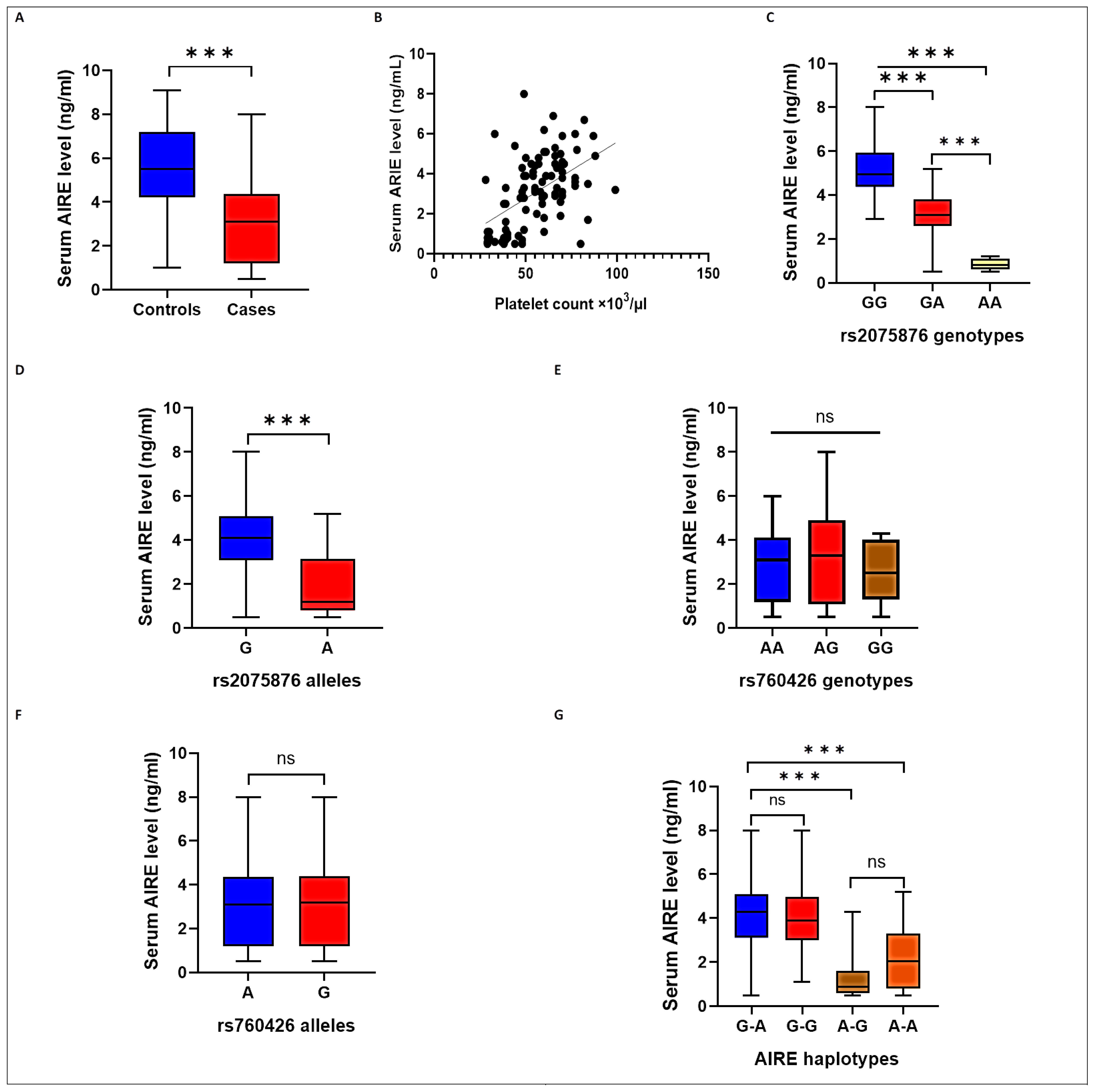
Figure 1. Serum AIRE levels in the studied groups: (A) in cases and controls; (B) correlated with platelet counts in the ITP group; (C) among different AIRE rs2075876 genotypes; (D) among different AIRE rs2075876 alleles; (E) among different AIRE rs760426 genotypes; (F) among different AIRE rs760426 alleles; and (G) among different AIRE haplotypes. *** p <0.001, ns, non-significant.
Ijms 24 05007 g001
Table 1. Basic characteristics of the studied groups.
CharacteristicsCase Group
(n = 96)
Control Group
(n = 100)
p-Value
Age, years, mean ± SD44.6 ± 12.642.3 ± 13.00.201
Gender
Male, n (%)
Female, n (%)

31 (32.3)
65 (67.7)

40 (40.0)
60 (60.0)
0.262
Positive family history, n (%)23 (24.0)6 (6.0%)<0.001 *
Platelets count (×103/μL), median (IQR)55.5 (23)210.5 (112)<0.001 *
n—number; SD—standard deviation; and IQR—interquartile range. * p < 0.05 vs. control group is significant.
Table 2. Distribution of the different genetic models of AIRE SNPs (rs2075876 G/A and rs760426 A/G) between the studied groups.
SNPsGenetic ModelCase Group, n (%)
(n = 96)
Control Group, n (%)
(n = 100)
χ2p
AIRE
(rs2075876 G/A)
AdditiveGG26 (0.27)41 (41.0)----
GA47 (0.49)49 (49.0)1.6450.200
AA23 (0.24)10 (10.0)8.4430.004 *
HWE p--0.399
DominantGG26 (0.27)41 (0.41)2.2160.040 *
GA/AA70 (0.73)59 (0.59)
RecessiveGG/GA73 (0.76)90 (0.90)6.8150.009 *
AA23 (0.24)10 (0.10)
Multiplicative
(Allele)
G99 (0.52)131 (0.655)7.8480.005 *
A93 (0.48)69 (0.345)
AIRE
(rs760426 A/G)
Co-dominantAA43 (0.45)46 (0.46)----
AG47 (0.49)39 (0.39)0.7030.402
GG6 (0.06)15 (0.15)2.6810.102
HWE p 0.170
DominantAA43 (0.45)46 (0.46)0.0290.865
AG/GG53 (0.55)54 (0.54)
RecessiveAA/AG90 (0.94)85 (0.85)3.9200.054
GG6 (0.06)15 (0.15)
Multiplicative (Allele)A133 (0.69)131 (0.655)0.6330.426
G59 (0.31)69 (0.345)
n—number; SNP—single nucleotide polymorphism. * p < 0.05 is significant.
Table 3. Different genetic models of AIRE SNPs (rs2075876 G/A and rs760426 A/G) and ITP risk.
SNPsModelGenotypeCrude OR (95% CI)pAdjusted OR (95% CI)pp-corr
AIRE
(rs2075876 G/A)
AdditiveGGRef--Ref--
GA1.513 (0.803–2.851)0.2011.460 (0.754–2.826)0.262
AA3.627 (1.489–8.835)0.005 *4.299 (1.650–11.202)0.003 *0.008 *
DominantGGRef--Ref--
GA/AA1.871 (1.026–3.413)0.041 *1.859 (0.994–3.477)0.052
RecessiveGG/GARef--Ref--
AA2.836 (1.269–6.336)0.011 *3.257 (1.406–7.545)0.006 *0.005 *
Multiplicative
(Allele)
GRef--Ref--
A1.783 (1.188–2.678)0.005 *1.847 (1.209–2.822)0.005 *0.004 *
AIRE
(rs760426 A/G)
Co-dominantAARef--Ref--
AG1.289 (0.712–2.336)0.4021.248 (0.675–2.309)0.480
GG0.428 (0.152–1.203)0.1080.421 (0.144–1.233)0.115
DominantAARef--Ref--
AG/GG0.952 (0.543–1.672)0.8650.985 (0.549–1.767)0.960
RecessiveAA/AGRef--Ref--
GG0.378 (0.140–1.019)0.0540.380 (0.135–1.064)0.065
Multiplicative
(Allele)
ARef--Ref--
G0.842 (0.552–1.286)0.4260.828 (0.533–1.286)0.401
n—number; SNP—single nucleotide polymorphism; OR—odds ratio; and CI—confidence interval. * p < 0.05 is significant.
Table 4. Distribution of different haplotypes of AIRE SNPs between ITP cases and controls.
AIRE SNP HaplotypesCase Group
(n = 192)
Control Group
(n = 200)
Crude OR (95% CI)pAdjusted OR (95% CI)p
G-An (%)59 (0.307)79 (0.395)------------
G-Gn (%)40 (0.208)52 (0.26)0.971 (0.570–1.654)0.9131.089 (0.623–1.902)0.765
A–Gn (%)19 (0.099)17 (0.085)1.497 (0.717–3.124)0.2831.823 (0.836–3.973)0.131
A–An (%)74 (0.385)52 (0.23)1.905 (1.168–3.109)0.010*1.821 (1.099–3.017)0.020 *
n—number; SNP—single nucleotide polymorphism; and OR—odds ratio; CI—confidence interval. * p < 0.05 is significant.
Disclaimer/Publisher’s Note: The statements, opinions and data contained in all publications are solely those of the individual author(s) and contributor(s) and not of MDPI and/or the editor(s). MDPI and/or the editor(s) disclaim responsibility for any injury to people or property resulting from any ideas, methods, instructions or products referred to in the content.