Article

Rational Design, Synthesis and Binding Affinity Studies of Anthraquinone Derivatives Conjugated to Gonadotropin-Releasing Hormone (GnRH) Analogues towards Selective Immunosuppression of Hormone-Dependent Cancer

1
Department of Chemistry, University of Patras, 26504 Rion, Greece
2
Department of Pharmacology, School of Medicine, University of Crete, 71003 Heraklion, Greece
3
Institute of Nuclear & Radiological Sciences & Technology, Energy & Safety, National Centre for Scientific Research “Demokritos”, 15341 Athens, Greece
4
Department of Clinical Chemistry, School of Medicine, University of Crete, 71003 Heraklion, Greece
*
Authors to whom correspondence should be addressed.
These authors contributed equally to this work.
Int. J. Mol. Sci. 2023, 24(20), 15232; https://doi.org/10.3390/ijms242015232
Submission received: 25 August 2023 / Revised: 6 October 2023 / Accepted: 12 October 2023 / Published: 16 October 2023

Abstract

:
Gonadotropin-releasing hormone (GnRH) is pivotal in regulating human reproduction and fertility through its specific receptors. Among these, gonadotropin-releasing hormone receptor type I (GnRHR I), which is a member of the G-protein-coupled receptor family, is expressed on the surface of both healthy and malignant cells. Its presence in cancer cells has positioned this receptor as a primary target for the development of novel anti-cancer agents. Moreover, the extensive regulatory functions of GnRH have underscored decapeptide as a prominent vehicle for targeted drug delivery, which is accomplished through the design of appropriate conjugates. On this basis, a rationally designed series of anthraquinone/mitoxantrone–GnRH conjugates (con1con8) has been synthesized herein. Their in vitro binding affinities range from 0.06 to 3.42 nM, with six of them (con2con7) demonstrating higher affinities for GnRH than the established drug leuprolide (0.64 nM). Among the mitoxantrone based GnRH conjugates, con3 and con7 show the highest affinities at 0.07 and 0.06 nM, respectively, while the disulfide bond present in the conjugates is found to be readily reduced by the thioredoxin (Trx) system. These findings are promising for further pharmacological evaluation of the synthesized conjugates with the prospect of performing future clinical studies.

1. Introduction

Gonadotropin-releasing hormone (GnRH) or luteinizing hormone-releasing hormone (LHRH), has a fundamental role in the reproductive function in mammalian cells, constituting the initial step of hypothalamic–pituitary–gonadal axis regulation. GnRH was isolated and identified by A.V. Schally and R.C.L. Guillemin in 1971 [1] as a decapeptide (pGlu1-His2-Trp3-Ser4-Tyr5-Gly6-Leu7-Arg8-Pro9-Gly10NH2). It is responsible for the regulation and release of gonadotropins, namely follicle-stimulating hormone (FSH), and luteinizing hormone (LH) from the anterior pituitary [1,2,3]. In particular, GnRH binds to and activates GnRH receptors (GnRHRs) on the surface of the pituitary gonadotrope cells, thus initiating the synthesis and secretion of LH and FSH. In turn, LH and FSH act on the gonads and regulate steroidogenesis and gametogenesis [2,4]. LH induces testosterone production and supports the proliferation of the Leydig cells in males, also inducing the synthesis of androgens and hormonal precursors for estradiol production in the theca cells in the ovaries. FSH, on the other hand, acts upon the Sertoli cells of the testes, supporting the maturation of gametes and enhancing spermatogenesis, while it also stimulates the maturation of the ovarian follicles affecting granulosa cells [5,6,7,8,9].
The pulsatile secretion of GnRH (with frequencies from ~30 min to 120 min) from the hypothalamus is critical for the regulation of the reproductive cycle [10,11,12,13]. In addition to the hypothalamus, GnRHR I expression has been reported from cancer cells located in breast tissues, ovaries, endometrium, and prostate. Studies have shown that GnRHR I is expressed also in extrapituitary tissues unrelated to the reproductive system, including the lungs and the pancreas [14,15,16,17,18]. The GnRH-GnRHR I complex constitutes an important target for infertility treatments and cancer therapy [19,20,21]. Although there are crystallographic data for GnRHR I [22], many research groups focus on further structural conformation of GnRHR I in order to gain further insight into GnRH-GnRHR I dynamics and provide valuable information that can be applied to targeted treatments [23,24,25].
Several GnRH analogues, used as therapeutic agents, have been developed as agonists and antagonists [26]. Since the GnRH half-life is very short (2–4 min) [27], GnRH analogues with altered sequences have been developed. Solid-phase peptide synthesis has contributed to the rapid production of such analogues [28], introducing, namely, modifications at the C-terminal of GnRH. Analogues with the substitution of glycine amide (Gly-NH2) and/or Gly in position 6 with an ethyl amide and a D-amino acid, respectively, have demonstrated greater potency and enhanced properties against proteolysis [29,30]. Leuprolide [31,32,33], buserelin [34,35], triptorelin [36], nafarelin [37,38,39] and goserelin [40] are some of the approved GnRH agonists for the treatment of prostate and breast cancer. GnRH agonists stimulate LH and FSH expression continuously, leading to the “flare” effect and subsequently to the desensitization and downregulation of the GnRHR. After the administration of GnRH agonists, a transient deterioration of the existing symptoms is observed, which can cause discomfort in patients [41,42,43,44,45].
An encouraging concept, proposed in this paper, is the design and development of delivery systems based on GnRH analogues, conjugated with anti-cancer drugs, and targeting the GnRHR I expressed on cancer cells [26,46]. A strategy based on the selective recognition of cancer cells using a carrier molecule linked to anti-neoplastic agents is a promising approach since it limits the side-effects of chemotherapy [47]. According to the literature, conjugates with similar structures to GnRH demonstrate significantly higher affinity for GnRHR compared to the free drug. In particular, coupling an anti-cancer agent to a peptide analogue enhanced its antiproliferative efficacy compared to the administration of a standalone anti-cancer agent [48,49]. Furthermore, the toxicity levels associated with GnRH conjugates were lower in comparison to their anti-neoplastic counterparts owing to the enhanced stability of the conjugates in serum [48,50,51]. Along these lines, the GnRH agonist DLys6-GnRH, conjugated via a covalent bond with doxorubicin (Zoptarelin doxorubicin or AEZS-108 or AN-152), was proven to exhibit high binding affinity when tested in human breast, ovarian and endometrial cancer cells [52,53,54,55]. When tested in GnRHR(+) experimental cancers in mice, it demonstrated lower toxicity levels and improved action on growth inhibition compared to the use of the cytotoxic doxorubicin alone [56,57]. Although AEZS-108 did not meet the relevant endpoints in a phase III clinical trial, it was used to establish a new approach based on GnRH-hybrid molecules with enhanced physicochemical properties [58,59].
In this study, we present a novel approach for the treatment of hormone-related cancer based on chemical modifications of the AEZS-108 conjugate. Such modifications aim to improve drug potency via increased binding efficiency. Our strategy focuses on three key points: (i) the implementation of a GnRH peptide analogue based on the structure of leuprolide and a D-amino acid substitution at position 6; (ii) the use of a mitoxantrone analogue as a cytotoxic agent; and (iii) the covalent connection of the two aforementioned parts through a disulfide bridge, which lacks the disadvantage of the hydrolysis of previous GnRH conjugates via serum carboxylesterase enzymes [60].

2. Results and Discussion

2.1. Rational Design of Mitoxantrone–GnRH Analogues

Our aim was to design and synthesize a series of GnRH analogues, conjugated to a cytotoxic agent via a disulfide to specifically target the overexpressed GnRH receptors on the surface of cancer cells [16,61]. The implemented cytotoxic agents comprised the anthraquinone derivative mitoxantrone (Novantrone®) [62,63], which is a well-established anti-neoplastic agent also used for the treatment of multiple sclerosis (MS) [64,65], non-Hodgkin’s lymphoma [66,67], and breast and prostate cancer [68,69]. Mitoxantrone was chosen due to its improved pharmacological profile relative to doxorubicin and its fewer side-effects, particularly in patients with a high risk of cardiovascular complications [70,71,72,73]. Instead of using an ester bond, as in AEZS-108, we opted for a disulfide linkage in anticipation of its potential reduction via the thioredoxin (Trx) system of cellular surfaces [74]. Such a reduction would subsequently trigger the release of the cytotoxic agent selectively near the cancer cells [75]. Previously conducted studies have already shown the successful reduction of the disulfide bond via the Trx system, leading to the release of the anthraquinone moiety and its entrance into the cell [76]. The reduction of the disulfide bond could be also attained via the cytoplasmic thioredoxin system upon the binding of the GnRH conjugate to GnRHR I and the subsequent internalization of the ligand–receptor complex.
Given the high expression of GnRHR in hormone-related cancers and considering the key points outlined above, our primary objective was to develop altered peptide conjugates with selective binding to GnRHR and with the capacity to facilitate the subsequent release of the incorporated cytotoxic compound (mitoxantrone) (Scheme 1). Throughout the synthetic process, the ethyl amide counterpart, found in leuprolide (see Section S1.5, Figures S9 and S10), was retained in the structure as it increases binding affinity for GnRHR. Furthermore, DLeu6 was replaced by DLys6/DCys6 residues, which possess similarly sized charged side chains. In the case of the DCys6 conjugate, the disulfide was directly bound to mitoxantrone by modifying a terminal hydroxyl group in the drug using a thiol group. The amide moiety in the DLys6 conjugate allowed for the incorporation of a linker via the formation of a peptide bond. This linker was designed to feature a thiol group for the subsequent formation of the disulfide bridge with the anthraquinone analogue. To this end, an aminohexanoic acid coupled with SPDP [succinimidyl 3-(2-pyridyldithio) propionate] as a thiol precursor donor acted as such a linker, providing flexibility to the resulting molecule.
Quinizarin conjugates (con1, con2, con46 and con8) were initially synthesized to establish reactions procedures and assess their binding affinities to GnRHR. After the synthesis and evaluation of these conjugates, mitoxantrone was used for the synthesis of the potent conjugates con3 and con7. Both mitoxantrone and the quinizarin analogues share a di-substituted amino alkyl-amino moiety and an anthraquinone ring. These structural features have been widely investigated for their anti-neoplastic properties, which are mainly attributed to the interference of the anthraquinone ring amongst DNA nucleobase pairs and the interaction between the nitrogen groups in the alkyl chains and the negatively charged phosphorous groups of DNA [77,78,79]. Additionally, a thiol group was incorporated into the alkyl chain of the synthesized analogues in order to facilitate the formation of a disulfide bridge between the anthraquinone and the GnRH analogues.

2.2. Synthesis of Anthraquinone and Mitoxantrone-GnRH Analogues

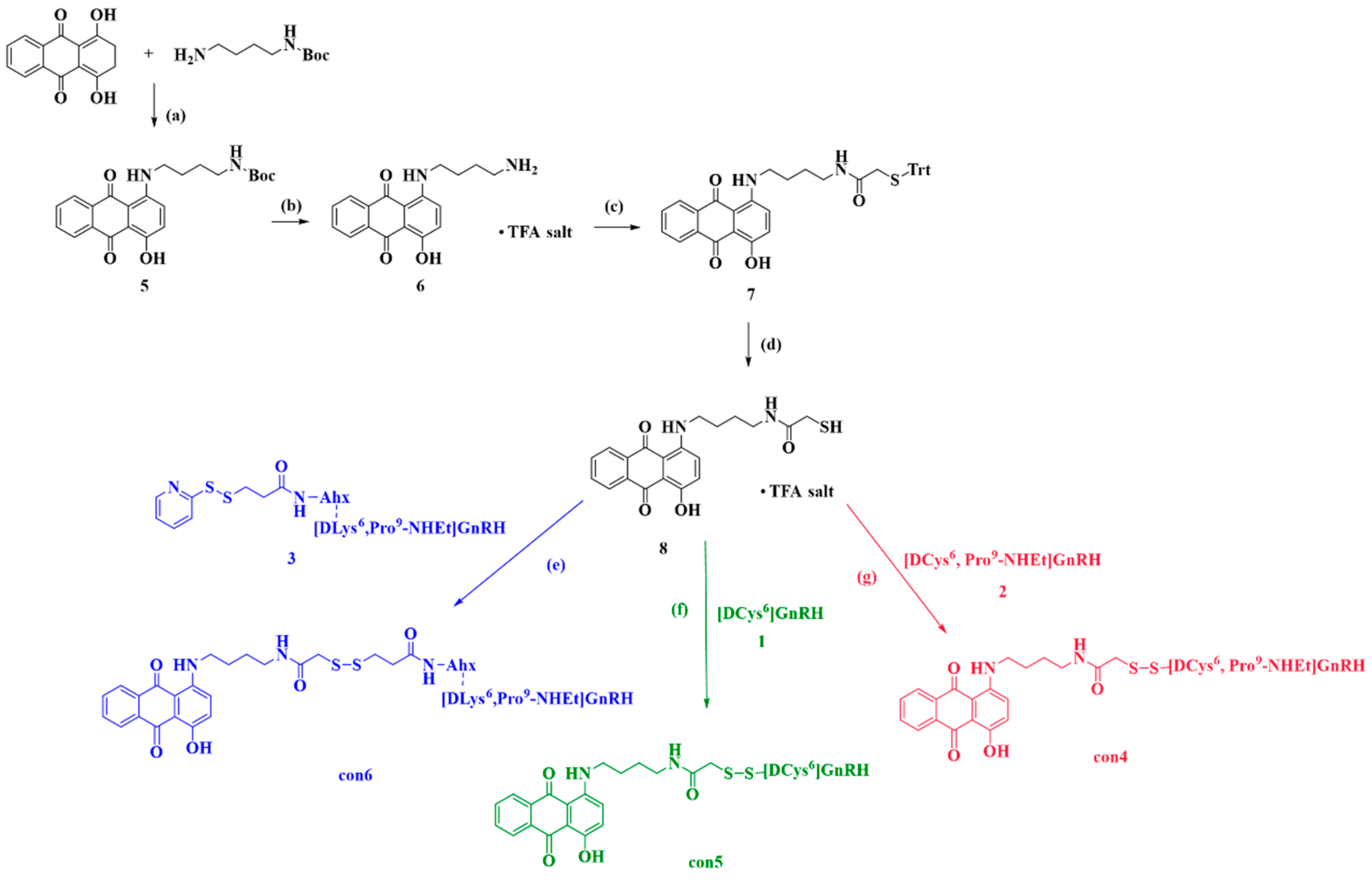
2.2.1. Synthesis of Anthraquinone—GnRH Conjugates con4; con5; con6

Conjugates con4, con5 and con6 were prepared according to the synthetic protocol outlined in Scheme 2. Con4 and con5 were synthesized via the oxidation of thiols groups; however, con6 was prepared via a thiol–disulfide exchange reaction. The same starting material, analogue 8 (see Section S2.1.4), reacted with synthesized [DLys(Ahx-PDP)6, Pro9-NHEt]GnRH (analogue 3; see Section S1.3, Figures S5 and S6), [DCys6]GnRH peptide (analogue 1; see Section S1.1, Figures S1 and S2) and [DCys6, Pro9-NHEt]GnRH peptide (analogue 2; see Section S1.2, Figures S3 and S4) for the synthesis of con6, con5 and con4, respectively.

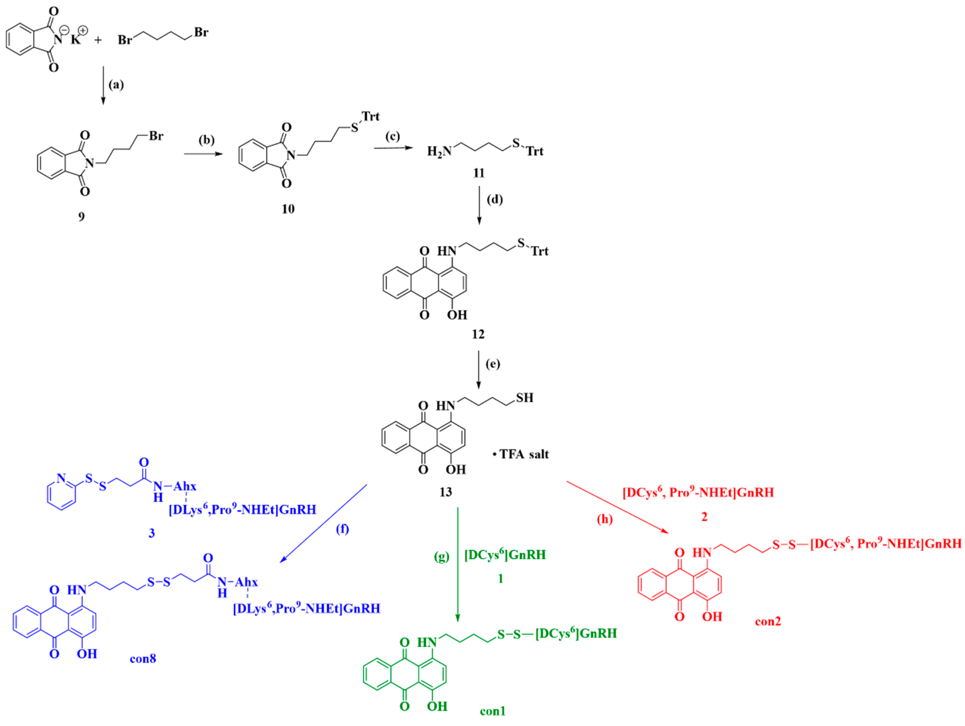
2.2.2. Synthesis of Anthraquinone—GnRH Conjugates con1; con2; con8

Conjugates con1, con2 and con3 were prepared according to the synthetic protocol outlined in Scheme 3. Con1 and con2 were synthesized analogously to con4 and con5 and con8 to con6. Again, the same starting substrate was used for all reactions. Thus, analogue 13 (see Section S2.2.5) reacted with analogue 3 (see Section S1.3, Figures S5 and S6), analogue 1 (see Section S1.1, Figures S1 and S2) and analogue 2 (see Section S1.2, Figures S3 and S4), to give the desired final compounds con1, con2 and con8, respectively.

2.2.3. Synthesis of Anthraquinone—GnRH Conjugates con7; con3

The synthetic protocols for conjugates con7 and con3 are described in Scheme 4. Analogue 16 (see Section S2.3.1) reacted with synthesized analogue 3 (see Section S1.3, Figures S5 and S6), while analogue 18 (see Section S2.3.2) reacted with analogue 2 (see Section S1.2, Figures S3 and S4), to give the desired final compounds con7 and con3, respectively.

2.3. Conformational Studies of con7 and con3

MD simulations provide the tools needed to analyze the conformations of analogues in aqueous solution. The analysis of RMSD values for the backbone atoms of (DLys6, Pro9-NHEt)GnRH peptide (analogue 4, see Section S1.4, Figures S7 and S8), included in con7, shows that it underwent small changes from the initial peptide conformation over the MD simulation time (Figure S66A). Similarly, RMSD values for the backbone atoms in (DCys6, Pro9-NHEt)GnRH peptide (analogue 2), included in con3, do not vary greatly in the first 100 ns and show a greater variability over the last 50 ns of simulation (Figure S66B). At the respective conjugates, con7 presents small conformational changes at the start of the simulation; conversely, at 60 ns, the conjugate presents a change in its conformation that is present until the end of the simulation (Figure S66C). On the other hand, the RMSD values of con3 are smaller than those of the respective peptide, but a steep leap is observed around 35 ns and a differentiation also appears between 55 and 70 ns (Figure S66D). The small conformational changes in analogue 4 and con7 are mirrored in the atomic fluctuations in which the mean atomic fluctuations for each residue are similar for these two analogues (Figure S67A). The only difference is observed in the values for DLys6 residue in the con7 (Figure S67A). These changes can be attributed to the modified side chain of DLys6 and the attachment of the mitoxantrone analogue (con7). In the same manner, the atomic fluctuations of analogue 2 mirror the small conformational changes of residues 1–7 and 9, while residues 8 and 10 have higher fluctuation values (Figure S67B). These changes in the residues could be attributed to the small carbon side chain of DCys6, which possibly provides more flexibility to the peptide compared to the analogue 4. The atomic fluctuations of con3 are higher than those of analogue 2. An exception is the case of Ser4, which has the smallest value due to hydrogen bond formation between Ser4 and DCys6 (Figure S67B).
The clustering analysis revealed the presence of two clusters for analogue 4 and analogue 2, as well as three clusters for conjugates con7 and con3. The dominant cluster 1 of analogue 4 was present for 71% of the time (Figure 1A). Similarly, the dominant cluster 1 of analogue 2 was present for 58% of the time (Figure 1B). On the other hand, the dominant cluster 1 of con7 was present for 45% of the time (Figure 1A) and the dominant cluster 1 of con3 was present for 50% of the time (Figure 1C). The dominant clusters for both peptide conjugates revealed the differentiations that occur from changes in position 6. The substitution with DLys allowed the peptide to adopt a U-conformation (Figure 1A beige), which is considered favorable for binding to GnRHR, while substitution with DCys led to a helical conformation (Figure 1B green). These conformations were changed in the conjugates. In the case of con7 (Figure 1A cyan), the U-formation was now more extended. Conversely, con3 (Figure 1B purple), the helical conformation, changed into a closed U-structure. In both conjugates (Figure 1C), the most important characteristic was the retention of a bent conformation in position 6, while the disulfide bridge in both cases was exposed to the solvent. The exposed positioning of the disulfide bond in both cases can potentially allow for the faster reduction of the bond via the thioredoxin system and lead to the rapid release of mitoxantrone analogue.
Further analysis of the conformational characteristics revealed the secondary structural elements of the synthesized analogues. The analogue 4 presented beta-turn features (Figure S68A) between Trp3 and DLys6 due to the presence of a hydrogen bond between Trp3 (i residue) and DLys6 (i + 3 residue), while the distance between the residues was <7 Å (5.33 Å). Additionally, con7 also showed beta-turn (Figure S68C) features between Trp3 and Tyr5. The analogue 2 also presented beta-turn features (Figure S68B) between Trp3 and Leu7. However, as mentioned above, we found that there were α-helical characteristics shared between residues Trp3 and DCys6 (Figure 1B (green) and Figure S68B), with a hydrogen bond between Trp3 (i residue) and DCys6 (i+4 residue). On the other hand, the addition of mitoxantrone to create con3 altered the conformation from helical to bent (Figure S68D).
The most important aspect observed during the conformational analysis was the bent conformation adopted by residues in position 6 (Figure 1C). This conformation allowed the side chains of residues in position 6 to be exposed to the solvent. Specifically, con7 had a peptide skeleton that was more extended from the respective peptide (analogue 4), meaning that the residues were more available to interact with the solvent (H2O) or other residues (e.g., GnRH receptor, proteins). On the contrary, con3 appeared to adopt a tighter U-shape (Figure 1B), but still the residues were exposed to the solvent and thus retained the ability to readily interact with residues on GnRHR. The secondary structural characteristics of both conjugates revealed that the positioning of important residues (His2, Trp3 and Arg8) involved in GnRHR binding was exposed to the solvent and thus allowed the conjugates to bind strongly to the receptor (Figure S69 and Table S1) [80]. Moreover, as mentioned above, the conformation of the conjugates exposed the disulfide bond to the action of the thioredoxin system.

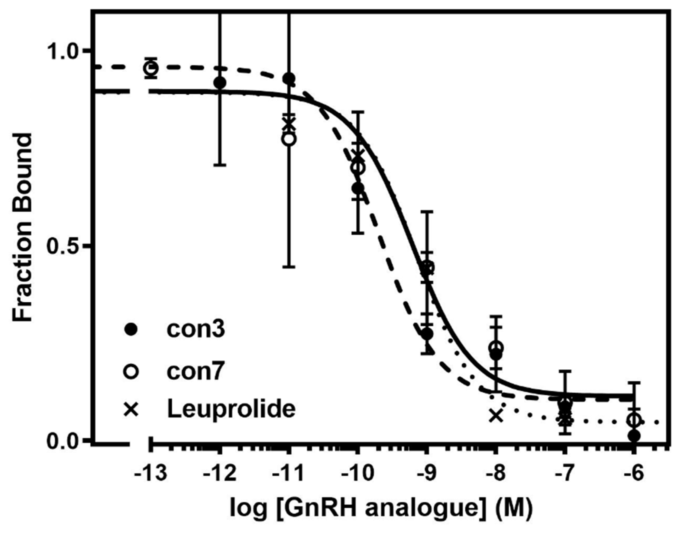
2.4. Binding Affinity Studies of Synthesized Conjugates

The apparent binding affinities (IC50) of the novel GnRH analogues, conjugated with anthraquinone or mitoxantrone (con1con8) for human GnRHR I, were determined using competitive radioligand binding assays. In these experiments, membranes from HEK 293 cells, stably expressing GnRHR I, were incubated with 125I-Tyr6, His5-GnRH in the presence and absence of increasing concentrations of GnRH analogues or leuprolide (positive control). All GnRH analogues reduced the specific binding of 125I-DTyr6-His5-GnRH in a dose-dependent manner, with affinities (0.06–3.42 nM) that were higher than or similar to those of leuprolide (0.64 nM) (Figure 2 and Figure 3). The compounds with the highest binding affinities were con3 (0.07 nM), con6 (0.07 nM) and con7 (0.06 nM) (Figure 2 and Figure 3). In marked contrast to GnRH conjugates, mitoxantrone did not bind to GnRHR (see Section S5, Figure S70). Analogues con3 and con7 were further evaluated based on the presence of the anti-cancer drug, mitoxantrone, in their structure. Moreover, analogues 4 and 2 conjugated with mitoxantrone gave con7 and con3, respectively, whose binding affinities were similar to those of their parental compounds (see Section S5, Figure S71).

2.5. Disulfides Reduction of con7 and Insulin by EcoTrx1

The thioredoxin system can successfully reduce the disulfide bond of similar conjugates and release the anthraquinone moiety, as has been described in our previous studies [76]. For that reason, con7 (one of the most promising candidates) was evaluated for the release of mitoxantrone analogue via the thioredoxin system. The reduction of con7 was compared to the reduction of insulin containing three disulfide bonds. The reduction of con7 and insulin from the thioredoxin system was successful, as assessed by the decrease of NADPH absorbance over time in samples containing TrxR (see Section S4). The absorbance values of con7 were higher than those of insulin because of the presence of the anthraquinone aromatic system in the conjugate. Moreover, the reduction of con7 was more rapid than that of insulin (Figure 4). This was most likely due to the exposure of con7′s disulfide bond to the solution and ability to react with the thioredoxin system, in contrast to the less exposed disulfides of insulin. In the samples that did not contain TrxR, the absorbance values saw a very slight decrease due to the oxidation of NADPH from atmospheric air (Figure 4).

3. Materials and Methods

All commercially available solvents and reagents were used without further purification and purchased from Merck (Darmstadt, Germany), Sigma Aldrich (St. Louis, MO, USA), Fluka (Buchs, Switzerland), Acros (Geel, Belgium), Fischer Chemical (Zurich, Switzerland), ChemBiotin (Voula, Greece), and Thermo Scientific (Waltham, MA, USA). Fmoc-protected amino acids, 2-chlorotrityl chloride (CLTR-Cl) resin (1% DVB, 200–400 mesh) and piperidine were purchased from Chemical and Biopharmaceutical Laboratories of Patras (Patras, Greece). Ethyl indole AM [3-([Ethyl-Fmoc-amino]-methyl)-1-indol-1-yl]-acetyl AM resin was purchased from Novabiochem and mitoxantrone dihydrochloride (CAS: 70476-82-3) was purchased from Fluorochem. Na125I was purchased from Perkin Elmer (Waltham, MA, USA); we acquired Triptorelin from Bachem (Bubendorf, Switzerland). HPLC-grade water was prepared using a Millipore Simplicity system (Burlington, MA, USA).
The purity of the synthesized analogues was determined via analytical reverse-phase high-performance liquid chromatography (RP-HPLC) (1260 Infinity, Quaternary Pump VL Agilent, Waldbronn, Germany) performed using an Agilent ZORBAX Eclipse Plus C18 column (3.5 μm, 100 × 4.6 mm, at 214 and 254 nm). The purification of products was achieved via flash column chromatography (silica gel, 200–400 mesh) or was carried out on a semi-preparative RP-HPLC system (Waters 600 solvent delivery system, combined with a Waters 996 photodiode array detector) using a Nucleosil RP-C18 column (7 μm, 250 × 10 mm, Merck, Darmstadt, Germany) and on a preparative RP-HPLC system (Waters Delta Prep, Empower Pro system, combined with a Waters 2996 photodiode array detector) using a Delta Pak RP-C18 column (15 μm, 300 × 19 mm, flow rate: 12 mL/min). Mobile phases were 0.08% TFA in H2O and 0.08% TFA in ACN. The radiolabeled peptide was purified via high-performance liquid chromatography (HPLC) with gradient elution (Waters 600 chromatography system, USA, coupled to a UV detector and NaI(Tl) detector in-line) using an analytical Macherey−Nagel Nucleosil RP C-18 column (250 × 4.6 mm, 5 μm, flow rate: 1 mL/min). Mobile phases were 0.05% TFA in H2O and 0.05% TFA in ACN.
Electrospray spray ionization mass spectrometry (ESI-MS) experiments were performed with a Waters Micromass ZQ Electrospray Platform coupled to a MassLynx2.3 data system and with a Bruker amaZon SL Ion Trap MS coupled to a TrapControl data system. Nuclear magnetic resonance spectra were obtained on 600 MHz spectrometer Bruker Avance 400 DPX, using tetramethylsilane (TMS) as reference standard.

3.1. Synthesis of Anthraquinone and Mitoxantrone-GnRH Analogues

3.1.1. Synthesis of Anthraquinone—GnRH Conjugate con4

Analogue 2 (12.5 mg, 0.010 mmol, 1.0 eq) and analogue 8 (5.4 mg, 0.014 mmol, 1.4 eq) were dissolved in DMSO (0.4 mL) and DIPEA (0.015 mL). The reaction mixture was stirred in RT for 24 h and monitored via analytical RP-HPLC. Purification via semi-preparative HPLC (gradient separation 20 to 80% ACN in 40 min, flow rate 3 mL/min) provided conjugate con4 as a purple solid (2.3 mg, 0.0015 mmol, 14.54%). Mtheoritical: 1581.83; (Μtheoritical + H)+: 1582.83; found 1581.86; (Μtheoritical + 2H+)/2: 791.92; found 791.77; (Μtheoritical + 3H+)/3: 528.28; found 528.55; (Figures S22 and S23).

3.1.2. Synthesis of Anthraquinone—GnRH Conjugate con5

Analogue 1 (19.8 mg, 0.016 mmol, 1.0 eq) and analogue 8 (8.6 mg, 0.022 mmol, 1.4 eq) were dissolved in DMSO (0.4 mL) and DIPEA (0.015 mL). The reaction mixture was stirred in RT for 52 h and monitored via analytical RP-HPLC. Purification performed using semi-preparative HPLC (gradient separation 20 to 80% ACN in 40 min, flow rate 3 mL/min) provided conjugate con5 as a purple solid (2.7 mg, 0.0017 mmol, 10.48%). Mtheoritical: 1610.83; (Μtheoritical + H)+: 1611.83; found 1611.66; (Μtheoritical + 2H+)/2: 806.42; found 806.31; (Μtheoritical + 3H+)/3: 537.94; found 538.05; (Figures S24 and S25).

3.1.3. Synthesis of Anthraquinone—GnRH Conjugate con6

Analogue 3 (15.7 mg, 0.010 mmol, 1.0 eq) and analogue 8 (5.4 mg, 0.014 mmol, 1.4 eq) were dissolved in DMSO (0.4 mL) and DIPEA (0.015 mL). The reaction mixture was stirred in RT for 4.5 h and monitored via analytical RP-HPLC. Purification using semi-preparative HPLC (gradient separation 20 to 80% ACN in 40 min, flow rate 3 mL/min) provided conjugate con6 as a purple solid (6.3 mg, 0.0035 mmol, 34.84%). Mtheoritical: 1808.15; (Μtheoritical + H)+: 1809.15; found 1808.66; (Μtheoritical + 2H+)/2: 905.08; found 904.99; (Μtheoritical + 3H+)/3: 603.72; found 603.69; (Figures S26 and S27).

3.1.4. Synthesis of Anthraquinone—GnRH Conjugate con1

Analogue 1 (16.6 mg, 0.014 mmol, 1.0 eq) and analogue 13 (6.5 mg, 0.020 mmol, 1.4 eq) were dissolved in DMSO (0.4 mL) and DIPEA (0.015 mL). The reaction mixture was stirred at RT for 48 h and monitored via analytical RP-HPLC. Purification using semi-preparative HPLC (gradient separation 20 to 80% ACN in 40 min, flow rate 3 mL/min) provided conjugate con1 as a purple solid (3.7 mg, 0.0024 mmol, 17.02%). Mtheoritical: 1553.64; (Μtheoritical + H)+: 1554.64; found 1554.08; (Μtheoritical + 2H+)/2: 777.32; found 777.81; (Figures S42 and S43).

3.1.5. Synthesis of Anthraquinone—GnRH Conjugate con2

Analogue 2 (9.5 mg, 0.008 mmol, 1.0 eq) and analogue 13 (3.6 mg, 0.011 mmol, 1.4 eq) were dissolved in DMSO (0.4 mL) and DIPEA (0.015 mL). The reaction mixture was stirred at RT for 44 h and monitored via analytical RP-HPLC. Purification using semi-preparative HPLC (gradient separation 20 to 80% ACN in 40 min, flow rate 3 mL/min) provided conjugate con2 as a purple solid (2.1 mg, 0.0014 mmol, 17.21%). Mtheoritical: 1524.78; (Μtheoritical + H)+: 1525.78; found 1524.86; (Μtheoritical + 2H+)/2: 763.39; found 763.27; (Figures S44 and S45).

3.1.6. Synthesis of Anthraquinone—GnRH Conjugate con8

Analogue 3 (16.4 mg, 0.011 mmol, 1.0 eq) and analogue 13 (4.9 mg, 0.015 mmol, 1.4 eq) were dissolved in DMSO (0.4 mL) and DIPEA (0.015 mL). The reaction mixture was stirred at RT for 4 h and monitored via analytical RP-HPLC. Purification using semi-preparative HPLC (gradient separation 20 to 80% ACN in 40 min, flow rate 3 mL/min) provided conjugate con8 as a purple solid (7.5 mg, 0.0043 mmol, 38.96%). Mtheoritical: 1749.82; (Μtheoritical + H)+: 1750.82; found 1750.80; (Μtheoritical + 2H+)/2: 875.91; found 876.31; (Μtheoritical + 3H+)/3: 584.27; found 585.02; (Figures S46 and S47).

3.1.7. Synthesis of Mitoxantrone—GnRH Conjugate con7

Analogue 3 (16.1 mg, 0.011 mmol, 1.0 eq) and analogue 16 (6.0 mg, 0.012 mmol, 1.1 eq) were dissolved in a high dilution of MeOH (6 mL). The reaction mixture was stirred at RT for 6 h and monitored via analytical RP-HPLC. Purification using semi-preparative HPLC (gradient separation 30 to 60% ACN in 40 min, flow rate 3 mL/min) provided conjugate con7 as a blue solid (17.5 mg, 0.009 mmol, 81.89%). Mtheoritical: 1942.29; (Μtheoritical + H)+: 1943.29; found 1942.52 (Μtheoritical + 2H+)/2: 972.15; found 971.93 (Μtheoritical + 3H+)/3: 648.43; found 648.56; (Μtheoritical + 7H+)/7: 278.47; found 279.67; (Figures S57 and S58).

3.1.8. Synthesis of Mitoxantrone—GnRH Conjugate con3

Analogue 2 (23.6 mg, 0.02 mmol, 1.0 eq) and analogue 18 (13.9 mg, 0.021 mmol, 1.1 eq) were dissolved in a high dilution of MeOH (7 mL). The reaction mixture was stirred at RT for 24 h and monitored via analytical RP-HPLC. Purification using preparative HPLC (gradient separation 30 to 40% ACN in 43 min, flow rate 12 mL/min) provided conjugate con3 as a blue solid (11.7 mg, 0.0068 mmol, 33.82%). Mtheoritical: 1730.00; (Μtheoritical + H)+: 1731.00; found 1729.86; (Μtheoritical + 2H+)/2: 866.00; found 865.91; (Μtheoritical + 3H+)/3: 577.67; found 577.79; (Figures S64 and S65).

3.2. Conformational Studies of con7 and con3

The peptides and the conjugates were designed using the USF Chimera 1.16 software [81]. The template for the GnRH backbone of the conjugates was derived from a previously reported [82] GnRH MD simulation, with the relevant changes implemented at position 6. Molecular dynamics calculations were performed for the peptide conjugates con7 and con3 and for the peptides which are present in these conjugates (analogue 4 and analogue 2, respectively) using AMBER14 software [83]. All structures were optimized with the GAMESS R1 software [84,85] using the density-functional theory (DFT) [86,87] and the Hartree–Fock (HF) approximation. The B3LYP/6-311G atomic basis set was applied, while the convergence criterion was set at 0.0001 [87,88]. Detailed information on the construction of the parameters is provided in Supplementary Materials (see Sections S3 and S6).

3.3. GnRH Receptor Binding Assay of Synthesized Analogues

Iodination of the 125I-DTyr6-His5-GnRH: The radioiodination of GnRH peptide was performed following the chloramine-T method. Specifically, 5 μL of a 1 mg/mL DTyr6-His5-GnRH solution was diluted with 5 μL of iodination buffer (0.25 M phosphate, pH 7.4) and then mixed with 10 μL of 0.5 mg/mL chloramine-T solution in iodination buffer and 2.2 μL of Na125I solution corresponding to 1 mCi. After incubation for 1 min under continuous shaking, 20 μL of 1 mg/mL sodium metabisulfite solution in iodination buffer was added to stop the reaction. The radiolabeled peptide was purified via HPLC (gradient elution 3% to 50% ACN in 15 min, flow rate 1 mL/min).
Cell culture and membrane preparation: We prepared HEK 293 cells stably expressing the GnRH-R [89] grown in DMEM/F12 (1:1) containing 3.15 g/L glucose and 10% bovine calf serum at 37 ºC and 5% CO2. The cells were grown in 100 mm dishes to achieve 90–100% confluency on the day of the experiment. The cells were washed using phosphate-buffered saline (PBS) (4.3 mM Na2HPO4.7H20, 1.4 mM KH2PO4, 137 mM NaCl, 2.7 mM KCl, pH 7.2–7.3 at RT), briefly treated with PBS containing 2 mM EDTA (PBS/EDTA), and then dissociated in PBS/EDTA. A cell suspension was centrifuged at 1000× g for 5 min at room temperature, and the pellet was homogenized in 1 mL of buffer O (50 mM Tris—HCl containing 0.5 mM EDTA, 10% sucrose, 10 mM MgCl2, pH 7.4 at 4 °C) using a Janke & Kunkel IKA Ultra Turrax T25 homogenizer (Staufen im Breisgau, Germany), at setting ~20, for 10–15 s, at 4 °C. The homogenate was centrifuged at 250× g for 5 min at room temperature. The pellet was discarded, and the supernatant was centrifuged at 16,000× g, for 10 min, at 4 °C. The membrane pellet was resuspended in buffer B (25 mM HEPES containing 1 mM CaCl2, 10 mM MgCl2, 0.5% BSA, pH 7.4 at 4 °C) (1 mL/100 mm dish) and used for radioligand binding studies.
125I-DTyr6-His5-GnRH binding: Aliquots of diluted membrane suspension (50 μL) were added into low-retention tubes containing buffer B and 120,000–240,000 cpm 125I-DTyr6-His5-GnRH, with or without increasing concentrations of GnRH analogues, in a final volume of 0.5 mL. The mixtures were incubated at 4 °C for 16–19 h and then filtered using a Brandel cell harvester through Whatman GF/C glass fiber filters and then presoaked for 1–2 h in 0.5% polyethylenimine at 4 °C. The filters were washed 4 times with 2 mL of ice-cold 50 mM Tris—HCl, pH 7.4 at 4 °C. Filters were assessed for radioactivity in a gamma counter (LKB Wallac 1275 minigamma, 80% efficiency). The amount of membrane used was adjusted to ensure that the specific binding was always equal to or less than 10% of the total concentration of the added radioligand. Specific 125I-Tyr6-His5-GnRH binding was defined as total binding less nonspecific binding in the presence of 1000 nM triptorelin. Data for competition binding were analyzed via nonlinear regression analysis using GraphPad Prism 4.0 (GraphPad Software, San Diego, CA, USA). IC50 values were obtained by fitting the data from competition studies into a one-site competition model.

3.4. Disulfide Bond Reduction Assays by EcoTrx1

The assay employed was based on changes in the absorbance at 340 nm from the oxidation of NADPH using a modified version of the protocol laid out in [90]. Briefly, the decrease in A340 was measured in 96-well plates containing 100 μL of 200 mM potassium phosphate pH 7.4, 1 mM EDTA, 0.1 mg/mL BSA, 200 μM NADPH, 2 μM E. coli Trx1 and 10 nM E. coli TrxR for 5 min at 25 °C with insulin at a final concentration of 0.067 mM (0.2 mM disulfides) (see Table S2). The reduction of con7 was measured at a final concentration of 0.2 mM (see Table S3). Blanks were samples without EcoTrxR.

4. Conclusions

The design and synthesis of the GnRH conjugates presented in this study was based on chemical modifications to the AEZS-108 conjugate. All developed conjugates featured an anthraquinone-type cytotoxic compound tethered to a leuprolide analogue for enhanced binding efficiency. AEZS-108 initially held great promise as an anti-cancer agent; however, the premature release of doxorubicin in plasma [60], along with the resultant toxicity, led to the termination of clinical trials [91,92]. For that reason, the conjugates reported in this study were meticulously designed to ensure the selective release of their cytotoxic agent only upon binding to GnRHR I to cancer cells via the thioredoxin system. The linkage between the cytotoxic agent and the GnRH peptides was achieved through either a direct disulfide bond or the addition of a linker, as exemplified in con3 and con7, respectively. Mitoxantrone analogues were selected as the cytotoxic compounds due to their favorable pharmacological profile in comparison to doxorubicin.
The evaluation of the synthesized conjugates (con1con8) for their interaction with GnRHR I showed significant enhanced binding affinities compared to leuprolide. Notably, the highest binding affinities were predominantly observed with con3 and con7 (Figure 3), while the release of the cytotoxic agent, facilitated by the thioredoxin system after the reduction of the disulfide bond, was estimated in con7 (Figure 4). Molecular dynamic (MD) simulations conducted for both conjugates con7 and con3 revealed that the conformation of these analogues exposed the disulfide bond to the surrounding solvent environment, rendering it susceptible to the thioredoxin system within cancer cells. Additionally, the simulations proved that these conjugates adopted U-shape/bent conformations, a characteristic feature closely associated with GnRH peptides and agonistic activity, which allowed their binding to GnRHR I [42].
The findings derived from our in vitro evaluation indicated that con3 and con7, both mitoxantrone analogues, exhibited robust binding affinities for GnRHR I. These results suggested that these compounds could be considered promising candidates for further comprehensive pharmacological investigations, both in vitro and in vivo, in the ongoing battle against cancer. In conclusion, the outcomes presented in this paper underscore the potential of these novel GnRH conjugates as candidates for the development of new innovative drug carriers designed to selectively target cancer cells. More specifically, novel GnRH-mitoxantrone conjugates could be used in the treatment of hormone-dependent cancers that overexpress the GnRH receptors, including ovarian and endometrial disease. Before entering clinical studies, these conjugates should be studied in vivo for their ability to inhibit the proliferation and migration of cancer cells in combination with stability, toxicology and extensive pharmacological evaluation.

Supplementary Materials

The following supporting information can be downloaded at: https://www.mdpi.com/article/10.3390/ijms242015232/s1.

Author Contributions

Conceptualization, G.L. and T.T.; Investigation G.L. and T.T.; Methodology, G.B., C.M., A.N., H.T., C.S., A.V.-G., V.K., I.P., P.P., M.V., G.L. and T.T.; Supervision, H.T., A.V.-G., M.V., G.L. and T.T.; Validation, H.T., C.S., A.V.-G., I.P., P.P., M.V., G.L. and T.T.; Writing—original draft, G.B., H.T., G.L. and T.T.; Writing—review and editing, G.B., C.M., H.T., C.S., A.V.-G., V.K., M.V., G.L. and T.T. All authors have read and agreed to the published version of the manuscript.

Funding

This research was co-financed by the European Union and the Greek National Funds through the Operational Program Competitiveness, Entrepreneurship and Innovation, under the call Research—Create—Innovate (project code: Τ2ΕΔΚ 02056).

Data Availability Statement

Data is contained within the article or Supplementary Materials.

Conflicts of Interest

The authors declare no conflict of interest.

References

  1. Schally, A.V.; Arimura, A.; Kastin, A.J.; Matsuo, H.; Baba, Y.; Redding, T.W.; Nair, R.M.; Debeljuk, L.; White, W.F. Gonadotropin-Releasing Hormone: One Polypeptide Regulates of Luteinizing and Follicle-Stimulating Hormones. Science 1979 1971, 173, 1036–1038. [Google Scholar] [CrossRef] [PubMed]
  2. McCann, S.M.; Dhariwal, A.P.S. Hypothalamic Factors Which Influence Gonadotrophin Secretion. Trans. N. Y. Acad. Sci. 1964, 27, 39–44. [Google Scholar] [CrossRef] [PubMed]
  3. Clarke, I.J.; Pompolo, S. Synthesis and Secretion of GnRH. Anim. Reprod. Sci. 2005, 88, 29–55. [Google Scholar] [CrossRef]
  4. Schally, A.V.; Arimura, A.; Baba, Y.; Nair, R.M.; Matsuo, H.; Redding, T.W.; Debeljuk, L. Isolation and Properties of the FSH and LH-Releasing Hormone. Biochem. Biophys. Res. Commun. 1971, 43, 393–399. [Google Scholar] [CrossRef]
  5. Simoni, M.; Weinbauer, G.F.; Gromoll, J.; Nieschlag, E. Role of FSH in Male Gonadal Function. Ann. Endocrinol. 1999, 60, 102–106. [Google Scholar]
  6. Simoni, M.; Gromoll, J.; Nieschlag, E. The Follicle-Stimulating Hormone Receptor: Biochemistry, Molecular Biology, Physiology, and Pathophysiology. Endocr. Rev. 1997, 18, 739–773. [Google Scholar]
  7. Richards, J.S. Maturation of Ovarian Follicles: Actions and Interactions of pituitary and Ovarian Hormones on Follicular Cell Differentiation. Physiol. Rev. 1980, 60, 51–89. [Google Scholar] [CrossRef]
  8. Howles, C.M. Role of LH and FSH in Ovarian Function. Mol. Cell. Endocrinol. 2000, 161, 25–30. [Google Scholar] [CrossRef]
  9. Richards, J.S. Hormonal Control of Gene Expression in the Ovary. Endocr. Rev. 1994, 15, 725–751. [Google Scholar] [CrossRef]
  10. Wildt, L.; Häusler, A.; Marshall, G.; Hutchison, J.S.; Plant, T.M.; Belchetz, P.E.; Knobil, E. Frequency and Amplitude of Gonadotropin-Releasing Hormone and Gonadotropin Secretion in the Rhesus Monkey. Endocrinology 1981, 109, 376–385. [Google Scholar] [CrossRef]
  11. Nett, T.M.; Turzillo, A.M.; Baratta, M.; Rispoli, L.A. Pituitary Effects of Steroid Hormones on Secretion Of-Stimulating Hormone and Luteinizing Hormone. Domest. Anim. Endocrinol. 2002, 23, 33–42. [Google Scholar] [CrossRef]
  12. Ferris, H.A.; Shupnik, M.A. Mechanisms for Pulsatile Regulation of the Gonadotropin Subunit by GNRH1. Biol. Reprod. 2006, 74, 993–998. [Google Scholar] [CrossRef] [PubMed]
  13. Savoy-Moore, R.T.; Swartz, K.H. Several GnRH Stimulation Frequencies Differentially Release FSH and LH from Isolated, Perfused Rat Anterior Pituitary Cells. In Regulation of Ovarian and Testicular Function; Mahesh Virendra, B., Dhindsa, D.S., Anderson, E., Kalra, S.P., Eds.; Springer: Boston, MA, USA, 1987; pp. 641–645. ISBN 978-1-4684-5395-9. [Google Scholar]
  14. Schwanzel-Fukuda, M.; Bick, D.; Pfaff, D.W. Luteinizing Hormone-Releasing Hormone (LHRH)-Expressing Cells Do Not Migrate Normally in an Inherited Hypogonadal (Kallmann) Syndrome. Mol. Brain Res. 1989, 6, 311–326. [Google Scholar] [CrossRef] [PubMed]
  15. Gibbons, J.M.; Mitnick, M.; Chieffo, V. In Vitro Biosynthesis of TSH- and LH-Releasing Factors by the Human Placenta. Am. J. Obstet. Gynecol. 1975, 121, 127–131. [Google Scholar] [CrossRef] [PubMed]
  16. Schally, A.V.; Nagy, A. Chemotherapy Targeted to Cancers through Tumoral Hormone. Trends Endocrinol. Metab. 2004, 15, 300–310. [Google Scholar] [CrossRef]
  17. Brantley, K.D.; Hankinson, S.E.; Heather Eliassen, A. Hormones and Cancer. In Encyclopedia of Cancer, 3rd ed.; Boffetta, P., Hainaut, P., Eds.; Academic Press: Oxford, UK, 2019; pp. 237–253. ISBN 978-0-12-812485-7. [Google Scholar]
  18. Cheung, L.W.T.; Wong, A.S.T. Gonadotropin-Releasing Hormone: GnRH Receptor Signaling in extrapituitary Tissues. FEBS J. 2008, 275, 5479–5495. [Google Scholar] [CrossRef]
  19. Naor, Z. Signaling by G-Protein-Coupled Receptor (GPCR): Studies on the GnRH Receptor. Front. Neuroendocrinol. 2009, 30, 10–29. [Google Scholar] [CrossRef]
  20. Liu, S.V.; Schally, A.V.; Hawes, D.; Xiong, S.; Fazli, L.; Gleave, M.; Cai, J.; Groshen, S.; Brands, F.; Engel, J.; et al. Expression of Receptors for Luteinizing Hormone-Releasing (LH-RH) in Prostate Cancers Following Therapy with LH-RH Agonists. Clin. Cancer Res. 2010, 16, 4675–4680. [Google Scholar] [CrossRef]
  21. Yano, T.; Pinski, J.; Radulovic, S.; Schally, A. V Inhibition of Human Epithelial Ovarian Cancer Cell Growth in vitro by Agonistic and Antagonistic Analogues of Luteinizing-Releasing Hormone. Proc. Natl. Acad. Sci. USA 1994, 91, 1701–1705. [Google Scholar] [CrossRef]
  22. Yan, W.; Cheng, L.; Wang, W.; Wu, C.; Yang, X.; Du, X.; Ma, L.; Qi, S.; Wei, Y.; Lu, Z.; et al. Structure of the Human Gonadotropin-Releasing Hormone Receptor GnRH1R Reveals an Unusual Ligand Binding Mode. Nat. Commun. 2020, 11, 5287. [Google Scholar] [CrossRef]
  23. Flanagan, C.A.; Zhou, W.; Chi, L.; Yuen, T.; Rodic V and Robertson, D.; Johnson, M.; Holland, P.; Millar R P and Weinstein, H.; Mitchell, R.; Sealfon, S.C. The Functional Microdomain in Transmembrane Helices 2 and 7 Expression, Activation, and Coupling Pathways of the gonadotropin-Releasing Hormone Receptor. J. Biol. Chem. 1999, 274, 28880–28886. [Google Scholar] [CrossRef] [PubMed]
  24. Tzoupis, H.; Nteli, A.; Platts, J.; Mantzourani, E.; Tselios, T. Refinement of the Gonadotropin Releasing Hormone Receptor I Homology Model by Applying Molecular Dynamics. J. Mol. Graph. Model. 2019, 89, 147–155. [Google Scholar] [CrossRef]
  25. Limonta, P.; Manea, M. Gonadotropin-Releasing Hormone Receptors as Molecular Targets in Prostate Cancer: Current Options and emerging Strategies. Cancer Treat. Rev. 2013, 39, 647–663. [Google Scholar] [CrossRef]
  26. Emons, G.; Schally, A.V. The Use of Luteinizing Hormone Releasing Hormone Agonists and antagonists in Gynaecological Cancers. Hum. Reprod. 1994, 9, 1364–1379. [Google Scholar] [CrossRef]
  27. Redding, T.W.; Kastin, A.J.; Gonzales-Barcena, D.; Coy, D.H.; Coy, E.J.; Schalch, D.S.; Schally, A.V. The Half-Life, Metabolism and Excretion of Tritiated Luteinizing-Releasing Hormone (LH-RH) in Man. J. Clin. Endocrinol. Metab. 1973, 37, 626–631. [Google Scholar] [CrossRef] [PubMed]
  28. Karten, M.J.; Rivier, J.E. Gonadotropin-Releasing Hormone Analog Design. Structure-Function Studies Toward the Development of Agonists and Antagonists: Rationale and Perspective. Endocr. Rev. 1986, 7, 44–66. [Google Scholar] [CrossRef] [PubMed]
  29. Fujino, M.; Kobayashi, S.; Obayashi, M.; Fukuda, T.; Shinagawa, S.; Yamazaki, I.; Nakayama, R.; White, W.F.; Rippel, R.H. Syntheses and Biological Activities of Analogs of Luteinizing Hormone Releasing Hormone (LH-RH). Biochem. Biophys. Res. Commun. 1972, 49, 698–705. [Google Scholar] [CrossRef]
  30. Monahan, M.W.; Amoss, M.S.; Anderson, H.A.; Vale, W. Synthetic Analogs of the Hypothalamic Luteinizing Hormone Releasing Factor with Increased Agonist or Antatonist Properties. Biochemistry 1973, 12, 4616–4620. [Google Scholar] [CrossRef]
  31. Meyer, J.D.; Manning, M.C.; Vander Velde, D.G. Characterization of the Solution Conformations of Leuprolide. J. Pept. Res. 2002, 60, 159–168. [Google Scholar] [CrossRef]
  32. Wilson, A.C.; Meethal, S.V.; Bowen, R.L.; Atwood, C.S. Leuprolide Acetate: A Drug of Diverse Clinical Applications. Expert Opin. Investig. Drugs 2007, 16, 1851–1863. [Google Scholar] [CrossRef]
  33. Fujino, M.; Fukuda, T.; Shinagawa, S.; Kobayashi, S.; Yamazaki, I.; Nakayama, R.; Seely, J.H.; White, W.F.; Rippel, R.H. Synthetic Analogs of Luteinizing Hormone Releasing Hormone (LH-RH) Substituted in Position 6 and 10. Biochem. Biophys. Res. Commun. 1974, 60, 406–413. [Google Scholar] [CrossRef] [PubMed]
  34. Kong, J.; Su, F.; Liu, Y.; Yang, Y.; Cao, Y.; Qiu, J.; Wang, Y.; Zhang, L.; Wang, J.; Cao, X. The Pharmacokinetics of Buserelin after Intramuscular Administration in Pigs and Cows. BMC Vet. Res. 2022, 18, 136. [Google Scholar] [CrossRef] [PubMed]
  35. Bélanger, A.; Labrie, F.; Lemay, A.; Caron, S.; Raynaud, J.P. Inhibitory Effects of a Single Intranasal Administration of [d-Ser(TBU)6, Des-Gly-NH210]LHRK Ethylamide, a Potent LHRK Agonist, on Serum Steroid Levels in Normal Adult Men. J. Steroid Biochem. 1980, 13, 123–126. [Google Scholar] [CrossRef] [PubMed]
  36. Emons, G.; Muller, V.; Ortmann, O.; Grossmann, G.; Trautner, U.; Stuckrad, B.; Schulz, K.; Schally, A. Luteinizing Hormone-Releasing Hormone Agonist Triptorelin Signal Transduction and Mitogenic Activity of epidermal Growth Factor in Human Ovarian and Endometrial Cancer Lines. Int. J. Oncol. 1996, 9, 1129–1137. [Google Scholar] [CrossRef] [PubMed]
  37. Letassy, N.A.; Thompson, D.F.; Britton, M.L.; Suda Sr, R.R. Nafarelin Acetate: A Gonadotropin-Releasing Hormone Agonist for the Treatment of Endometriosis. DICP 1990, 24, 1204–1209. [Google Scholar] [CrossRef]
  38. Chrisp, P.; Goa, K.L. Nafarelin. Drugs 1990, 39, 523–551. [Google Scholar] [CrossRef]
  39. Mizutani, T.; Sakata, M.; Terakawa, N. Effect of Gonadotropin-Releasing Hormone Agonists, Nafarelin, Buserelin, and Leuprolide, on Experimentally Induced in the Rat. Int. J. Fertil. Menopausal Stud. 1995, 40, 106–111. [Google Scholar]
  40. Jonat, W.; Kaufmann, M.; Sauerbrei, W.; Blamey, R.; Cuzick, J.; Namer, M.; Fogelman, I.; de Haes, J.C.; de Matteis, A.; Stewart, A.; et al. Goserelin versus Cyclophosphamide, Methotrexate, and fluorouracil as Adjuvant Therapy in Premenopausal Patients with node-Positive Breast Cancer: The Zoladex Early Breast Cancer Association Study. J. Clin. Oncol. 2002, 20, 4628–4635. [Google Scholar] [CrossRef]
  41. Ferguson, S.S. Evolving Concepts in G Protein-Coupled Receptor Endocytosis: The Role in Receptor Desensitization and Signaling. Pharmacol. Rev. 2001, 53, 1–24. [Google Scholar]
  42. Söderhäll, J.A.; Polymeropoulos, E.E.; Paulini, K.; Günther, E.; Kühne, R. Antagonist and Agonist Binding Models of the Human-Releasing Hormone Receptor. Biochem. Biophys. Res. Commun. 2005, 333, 568–582. [Google Scholar] [CrossRef]
  43. Conn, P.M.; Crowley, W.F., Jr. Gonadotropin-Releasing Hormone and Its Analogs. Annu. Rev. Med. 1994, 45, 391–405. [Google Scholar] [CrossRef] [PubMed]
  44. Finch, A.R.; Caunt, C.J.; Armstrong Stephen P and McArdle, C.A. Agonist-Induced Internalization and Downregulation of gonadotropin-Releasing Hormone Receptors. Am. J. Physiol. Cell Physiol. 2009, 297, C591–C600. [Google Scholar] [CrossRef] [PubMed]
  45. Knobil, E. The Neuroendocrine Control of the Menstrual Cycle. Recent Prog. Horm. Res. 1980, 36, 53–88. [Google Scholar]
  46. Janáky, T.; Juhász, A.; Bajusz, S.; Csernus, V.; Srkalovic, G.; Bokser, L.; Milovanovic, S.; Redding T W and Rékási, Z.; Nagy, A. Analogues of Luteinizing Hormone-Releasing Hormone Containing Groups. Proc. Natl. Acad. Sci. USA 1992, 89, 972–976. [Google Scholar] [CrossRef] [PubMed]
  47. Colombo, P.; Gunnarsson, K.; Iatropoulos, M.; Brughera, M. Toxicological Testing of Cytotoxic Drugs (Review). Int. J. Oncol. 2001, 19, 1021–1028. [Google Scholar] [CrossRef]
  48. Karampelas, T.; Argyros, O.; Sayyad, N.; Spyridaki, K.; Pappas, C.; Morgan, K.; Kolios, G.; Millar, R.P.; Liapakis, G.; Tzakos, A.G.; et al. GnRH-Gemcitabine Conjugates for the Treatment of Androgen-Independent Prostate Cancer: Pharmacokinetic Enhancements Combined with Targeted Drug Delivery. Bioconjug. Chem. 2014, 25, 813–823. [Google Scholar] [CrossRef]
  49. Schally, A.V.; Nagy, A. Cancer Chemotherapy Based on Targeting of Cytotoxic Peptide Conjugates to Their Receptors on Tumors. Eur. J. Endocrinol. 1999, 141, 1–14. [Google Scholar] [CrossRef]
  50. Li, S.; Zhao, H.; Chang, X.; Wang, J.; Zhao, E.; Yin, Z.; Mao, X.; Deng, S.; Hao, T.; Wang, H.; et al. Synthesis, in Vitro Stability, and Antiproliferative Effect of d-Cysteine Modified GnRH-Doxorubicin Conjugates. J. Pept. Sci. 2019, 25, e3135. [Google Scholar] [CrossRef]
  51. Vrettos, E.I.; Mező, G.; Tzakos, A.G. On the Design Principles of Peptide–Drug Conjugates for Targeted Drug Delivery to the Malignant Tumor Site. Beilstein J. Org. Chem. 2018, 14, 930–954. [Google Scholar] [CrossRef]
  52. Szepeshazi, K.; Schally, A.V.; Keller, G.; Block, N.L.; Benten, D.; Halmos, G.; Szalontay, L.; Vidaurre, I.; Jaszberenyi, M.; Rick, F.G. Receptor-Targeted Therapy of Human Experimental Urinary Bladder with Cytotoxic LH-RH Analog AN-152 [AEZS- 108]. Oncotarget 2012, 3, 686–699. [Google Scholar] [CrossRef]
  53. Bajo, A.M.; Schally, A.V.; Halmos, G.; Nagy, A. Targeted Doxorubicin-Containing Luteinizing Hormone-Releasing Analogue AN-152 Inhibits the Growth of doxorubicin-Resistant MX-1 Human Breast Cancers. Clin. Cancer Res. 2003, 9, 3742–3748. [Google Scholar] [PubMed]
  54. Engel, J.B.; Keller, G.; Schally, A.V.; Nagy, A.; Chism, D.D.; Halmos, G. Effective Treatment of Experimental Human Endometrial cancers with Targeted Cytotoxic Luteinizing Hormone-Releasing Hormone AN-152 and AN-207. Fertil. Steril. 2005, 83 (Suppl. S1), 1125–1133. [Google Scholar] [CrossRef] [PubMed]
  55. Emons, G.; Kaufmann, M.; Gorchev, G.; Tsekova, V.; Gründker, C.; Günthert, A.R.; Hanker, L.C.; Velikova, M.; Sindermann, H.; Engel, J.; et al. Dose Escalation and Pharmacokinetic Study of AEZS-108(AN-152), an LHRH Agonist Linked to Doxorubicin, in women with LHRH Receptor-Positive Tumors. Gynecol. Oncol. 2010, 119, 457–461. [Google Scholar] [CrossRef] [PubMed]
  56. Gründker, C.; Völker, P.; Griesinger, F.; Ramaswamy, A.; Nagy, A.; Schally, A.V.; Emons, G. Antitumor Effects of the Cytotoxic Luteinizing Hormone-Releasing Analog AN-152 on Human Endometrial and Ovarian Cancers into Nude Mice. Am. J. Obstet. Gynecol. 2002, 187, 528–537. [Google Scholar] [CrossRef]
  57. Günthert, A.R.; Gründker, C.; Bongertz, T.; Nagy, A.; Schally, A.V.; Emons, G. Induction of Apoptosis by AN-152, a Cytotoxic Analog of luteinizing Hormone-Releasing Hormone (LHRH), in LHRH-R Human Breast Cancer Cells Is Independent of Multidrug-1 (MDR-1) System. Breast Cancer Res. Treat. 2004, 87, 255–264. [Google Scholar] [CrossRef] [PubMed]
  58. Emons, G.; Gorchev, G.; Sehouli, J.; Wimberger, P.; Stähle, A.; Hanker, L.; Hilpert, F.; Sindermann, H.; Gründker Carsten and Harter, P. Efficacy and Safety of AEZS-108 (INN: Zoptarelin Doxorubicin) an LHRH Agonist Linked to Doxorubicin in Women with platinum Refractory or Resistant Ovarian Cancer Expressing Receptors: A Multicenter Phase II Trial of the ago-Study Group (AGO GYN 5). Gynecol. Oncol. 2014, 133, 427–432. [Google Scholar] [CrossRef]
  59. Miller, D.S.; Scambia, G.; Bondarenko, I.; Westermann, A.M.; Oaknin, A.; Oza, A.M.; Lisyanskaya, A.S.; Vergote, I.; Wenham, R.M.; Temkin, S.M.; et al. ZoptEC: Phase III Randomized Controlled Study Comparing Zoptarelin with Doxorubicin as Second Line Therapy for Locally Advanced, Recurrent, or Metastatic Endometrial Cancer (NCT01767155). J. Clin. Oncol. 2018, 36, 5503. [Google Scholar] [CrossRef]
  60. Nagy, A.; Plonowski, A.; Schally, A.V. Stability of Cytotoxic Luteinizing Hormone-Releasing Hormone Conjugate (AN-152) Containing Doxorubicin 14-O-Hemiglutarate in Mouse and Human Serum in Vitro: Implications for the Design of Preclinical Studies. Proc. Natl. Acad. Sci. USA 2000, 97, 829–834. [Google Scholar] [CrossRef]
  61. Limonta, P.; Montagnani Marelli, M.; Mai, S.; Motta, M.; Martini, L.; Moretti, R.M. GnRH Receptors in Cancer: From Cell Biology to Novel Targeted Strategies. Endocr. Rev. 2012, 33, 784–811. [Google Scholar] [CrossRef]
  62. Murdock, K.C.; Child, R.G.; Fabio, P.F.; Angier, R.B.; Wallace, R.E.; Durr, F.E.; Citarella, R. V Antitumor Agents. 1. 1,4-Bis[(Aminoalkyl)Amino]-9,10-Anthracenediones. J. Med. Chem. 1979, 22, 1024–1030. [Google Scholar] [CrossRef]
  63. Kapuscinski, J.; Darzynkiewicz, Z. Relationship between the Pharmacological Activity of Antitumor Drugs Ametantrone and Mitoxantrone (Novatrone) and Their Ability to Condense Nucleic Acids. Proc. Natl. Acad. Sci. USA 1986, 83, 6302–6306. [Google Scholar] [CrossRef]
  64. Fox, E.J. Mechanism of Action of Mitoxantrone. Neurology 2004, 63, S15–S18. [Google Scholar] [CrossRef] [PubMed]
  65. White, R.J.; Durr, F.E. Development of Mitoxantrone. Investig. New Drugs 1985, 3, 85–93. [Google Scholar] [CrossRef] [PubMed]
  66. Armitage, J.O. The Role of Mitoxantrone in Non-Hodgkin’s Lymphoma. Oncology 2002, 16, 490–502, 507–508, 511–512, 517. [Google Scholar]
  67. Cheng, C.C.; Zee-Cheng, R.K. The Design, Synthesis and Development of a New Class of Potent Anthraquinones. Prog. Med. Chem. 1983, 20, 83–118. [Google Scholar]
  68. Landys, K.; Borgstrom, S.; Andersson, T.; Noppa, H. Mitoxantrone as a First-Line Treatment of Advanced Breast Cancer. Investig. New Drugs 1985, 3, 133–137. [Google Scholar] [CrossRef]
  69. van Dodewaard-de Jong, J.M.; Santegoets, S.J.; de Ven, P.M.; Versluis, J.; Verheul Henk M W and de Gruijl, T.D.; Gerritsen, W.R.; van den Eertwegh, A.J.M. Improved Efficacy of Mitoxantrone in Patients With-Resistant Prostate Cancer after Vaccination with GM-CSF-Transduced Allogeneic Prostate Cancer Cells. Oncoimmunology 2016, 5, e1105431. [Google Scholar] [CrossRef] [PubMed]
  70. Conner, C.S. Mitoxantrone: A Replacement for Doxorubicin? Drug Intell. Clin. Pharm. 1984, 18, 479–480. [Google Scholar] [CrossRef]
  71. Neidhart, J.A.; Gochnour, D.; Roach, R.; Hoth, D.; Young, D. A Comparison of Mitoxantrone and Doxorubicin in Breast Cancer. J. Clin. Oncol. 1986, 4, 672–677. [Google Scholar] [CrossRef] [PubMed]
  72. Henderson, I.C.; Allegra, J.C.; Woodcock, T.; Wolff, S.; Bryan, S.; Cartwright, K.; Dukart, G.; Henry, D. Randomized Clinical Trial Comparing Mitoxantrone with Doxorubicin in Previously Treated Patients with Metastatic Breast Cancer. J. Clin. Oncol. 1989, 7, 560–571. [Google Scholar] [CrossRef]
  73. Damiani, R.M.; Moura, D.J.; Viau, C.M.; Caceres, R.A.; Henriques, J.A.P.; Saffi, J. Pathways of Cardiac Toxicity: Comparison between Chemotherapeutic Doxorubicin and Mitoxantrone. Arch. Toxicol. 2016, 90, 2063–2076. [Google Scholar] [CrossRef] [PubMed]
  74. Cebula, M.; Moolla, N.; Capovilla, A.; Arnér, E.S.J. The Rare TXNRD1_v3 (“v3”) Splice Variant of Human Reductase 1 Protein Is Targeted to Membrane Rafts by N-Acylation and Induces Filopodia Independently of Its Redox Site Integrity. J. Biol. Chem. 2013, 288, 10002–10011. [Google Scholar] [CrossRef] [PubMed]
  75. Powis, G.; Mustacich, D.; Coon, A. The Role of the Redox Protein Thioredoxin in Cell Growth and Cancer. Free Radic. Biol. Med. 2000, 29, 312–322. [Google Scholar] [CrossRef]
  76. Tapeinou, A.; Giannopoulou, E.; Simal, C.; Hansen, B.E.; Kalofonos, H.; Apostolopoulos, V.; Vlamis-Gardikas, A.; Tselios, T. Design, Synthesis and Evaluation of an Anthraquinone Derivative to Myelin Basic Protein Immunodominant (MBP85-99) Epitope: Towards Selective Immunosuppression. Eur. J. Med. Chem. 2018, 143, 621–631. [Google Scholar] [CrossRef] [PubMed]
  77. Lown, J.W. Anthracycline and Anthraquinone Anticancer Agents: Current and Recent Developments. Pharmacol. Ther. 1993, 60, 185–214. [Google Scholar] [CrossRef]
  78. Krishnamoorthy, C.R.; Yen, S.F.; Smith, J.C.; Lown, J.W.; Wilson, W.D. Stopped-Flow Kinetic Analysis of the Interaction of Anthraquinone Drugs with Calf Thymus DNA, Poly[d(G-C)].Poly[d(G-C)], and Poly[d(A-T)].Poly[d(A-T)]. Biochemistry 1986, 25, 5933–5940. [Google Scholar] [CrossRef]
  79. Nishio, A.; Uyeki, E.M. Cellular Uptake and Inhibition of DNA Synthesis by dihydroxyanthraquinone and Two Analogues. Cancer Res. 1983, 43, 1951–1956. [Google Scholar]
  80. Laimou, D.K.; Katsara, M.; Matsoukas, M.T.I.; Apostolopoulos, V.; Troganis, A.N.; Tselios, T.V. Structural Elucidation of Leuprolide and Its Analogues in Solution: Insight into Their Bioactive Conformation. Amino Acids 2010, 39, 1147–1160. [Google Scholar] [CrossRef]
  81. Pettersen, E.F.; Goddard, T.D.; Huang, C.C.; Couch, G.S.; Greenblatt, D.M.; Meng, E.C.; Ferrin, T.E. UCSF Chimera–a Visualization System for Exploratory research and Analysis. J. Comput. Chem. 2004, 25, 1605–1612. [Google Scholar] [CrossRef]
  82. Tzoupis, H.; Nteli, A.; Androutsou, M.-E.; Tselios, T. Molecular Dynamics Simulations Studies of Gonadotropin Releasing Hormone (GnRH) and GnRH Receptor. J. Peptide Sci. 2018, 24, S129–S130. [Google Scholar]
  83. Case, D.; Babin, V.; Berryman, J.; Betz, R.; Cai, Q.; Cerutti, D.; Cheatham, T.; Darden, T.; Duke, R.; Gohlke, H.; et al. Amber 14; University of California: San Fransisco, CA, USA, 2014. [Google Scholar]
  84. Schmidt, M.W.; Baldridge, K.K.; Boatz, J.A.; Elbert, S.T.; Gordon, M.S.; Jensen, J.H.; Koseki, S.; Matsunaga, N.; Nguyen, K.A.; Su, S.; et al. General Atomic and Molecular Electronic Structure System. J. Comput. Chem. 1993, 14, 1347–1363. [Google Scholar] [CrossRef]
  85. Gordon, M.S.; Schmidt, M.W. Chapter 41—Advances in Electronic Structure Theory: GAMESS a Decade Later. In Theory and Applications of Computational Chemistry; Dykstra, C.E., Frenking, G., Kim, K.S., Scuseria, G.E., Eds.; Elsevier: Amsterdam, The Netherlands, 2005; pp. 1167–1189. ISBN 978-0-444-51719-7. [Google Scholar]
  86. Becke, A.D. Density-functional Thermochemistry. III. The Role of Exact Exchange. J. Chem. Phys. 1993, 98, 5648–5652. [Google Scholar] [CrossRef]
  87. Pople, J.A.; Gill, P.M.W.; Johnson, B.G. Kohn—Sham Density-Functional Theory within a Finite Basis Set. Chem. Phys. Lett. 1992, 199, 557–560. [Google Scholar] [CrossRef]
  88. Hertwig, R.H.; Koch, W. On the Parameterization of the Local Correlation Functional. What Is Becke-3-LYP? Chem. Phys. Lett. 1997, 268, 345–351. [Google Scholar] [CrossRef]
  89. Laimou, D.; Katsila, T.; Matsoukas, J.; Schally, A.; Gkountelias, K.; Liapakis, G.; Tamvakopoulos, C.; Tselios, T. Rationally Designed Cyclic Analogues of Luteinizing Hormone-Releasing Hormone: Enhanced Enzymatic Stability and Biological Properties. Eur. J. Med. Chem. 2012, 58, 237–247. [Google Scholar] [CrossRef]
  90. Luthman, M.; Holmgren, A. Rat Liver Thioredoxin and Thioredoxin Reductase: Purification Characterization. Biochemistry 1982, 21, 6628–6633. [Google Scholar] [CrossRef]
  91. Sflakidou, E.; Leonidis, G.; Foroglou, E.; Siokatas, C.; Sarli, V. Recent Advances in Natural Product-Based Hybrids as Anti-Cancer Agents. Molecules 2022, 27, 6632. [Google Scholar] [CrossRef]
  92. Engel, J.; Emons, G.; Pinski, J.; Schally, A.V. AEZS-108: A Targeted Cytotoxic Analog of LHRH for the Treatment of Cancers Positive for LHRH Receptors. Expert Opin. Investig. Drugs 2012, 21, 891–899. [Google Scholar] [CrossRef]
Scheme 1. Representative structure of study mitoxantrone conjugates.
Ijms 24 15232 sch001
Scheme 2. Synthetic pathway for the synthesis of anthraquinone–GnRH conjugates con4, con5 and con6. Reagents and conditions: (a) (i) ethanol, Ar, from reflux for 3 h to 50 °C for 16 h; (ii) [O], room temperature (RT), 3 h (oxidation in flowing air) (Figures S11–S13); (b) TFA, CH2Cl2, RT, 2 h (Figures S14–S16); (c) TrtSCH2CO2H, DCC, DMAP, CH2Cl2, RT, 5 h (Figures S17–S19); (d) TFA, TES, CH2Cl2, RT, 3 h (Figures S20 and S21); (e) 3, DIPEA, DMSO, RT, 4.5 h (Figures S26 and S27); (f) 1, DIPEA, DMSO, RT, 52 h (Figures S24 and S25); (g) 2, DIPEA, DMSO, RT, 24 h (Figures S22 and S23). The synthetic procedures for analogues 18 are described thoroughly in Supplementary Materials (see Section S2.1).
Ijms 24 15232 sch002
Scheme 3. Synthetic pathway for the synthesis of anthraquinone–GnRH conjugates con1, con2 and con8. Reagents and conditions: (a) DMF anhydrous, Ar, from 0 °C to RT for 16 h (Figures S28–S30); (b) (Ph)3CSH, NaH, DMF anhydrous, Ar, from 0 °C to RT for 16 h, (Figures S31 and S32); (c) N2H4, n-butanol/ethanol, reflux, 24 h (Figures S33–S35); (d) (i) leucoquinizarin, ethanol, from reflux for 3 h to 50 °C for 18 h; (ii) [O], RT, 3 h (oxidation in flowing air) (Figures S36–S38); (e) TFA, TES, CH2Cl2, RT, 5 h (Figures S39–S41); (f) 3, DIPEA, DMSO, RT, 4 h; (g) 1, DIPEA, DMSO, RT, 48 h; (h) 2, DIPEA, DMSO, RT, 44 h. The synthetic procedures for analogues 913 are described thoroughly in Supplementary Materials (see Section S2.2).
Ijms 24 15232 sch003
Scheme 4. Synthetic pathway for the synthesis of mitoxantrone–GnRH conjugates con7 and con3. Reagents and conditions: (a) Boc2O, anhydrous MeOH/THF, TEA, from 0 °C for 1 h to RT for 2 h (Figures S48–S50); (b) TrtSCH2CO2H, DMAP, HOBt, DIC, CH2Cl2, 0 °C, 24 h (Figures S51–S53); (c) TFA, TES, CH2Cl2, RT, 1.5 h (Figures S54–S56); (d) 3, MeOH, RT, 6 h (Figures S57 and S58); (e) 3-(2-pyridyldithio)propanoic acid, DMAP, HOBt, DIC, CH2Cl2, 0 °C, 24 h (Figures S59 and S60); (f) TFA, TES, CH2Cl2, 1.5 h (Figures S61–S63); (g) 2, MeOH, RT, 24 h (Figures S64 and S65). The synthetic procedures for analogues 1418 are described thoroughly in Supplementary Materials (see Section S2.3).
Ijms 24 15232 sch004
Figure 1. Comparison of the dominant conformations for con7, con3 and their respective peptides. (A) Comparison of con7 (cyan) and analogue 4 (beige); (B) Comparison of con3 (purple) and analogue 2 (green); and (C) Comparison of con7 (cyan) and con3 (purple). Atoms in yellow, red and dark blue colours are sulfur (S), oxygen (O) and nitrogen (N), respectively.
Ijms 24 15232 g001
Figure 2. Competition binding isotherms of GnRH analogues to human GnRHR. We compared 125I-Tyr6, His5-GnRH-specific binding by increasing concentrations of GnRH analogues, con1, con 2, con5, con6, con8, or leuprolide, as described in Section 3.4, present on membranes from HEK 293 cells stably expressing human GnRHR I. The means and S.E. are shown from a representative experiment performed 3–6 times with similar results. The data were fitted to a one-site competition model via nonlinear regression and the IC50 values were determined as described in Section 3.4. Con4 is not included due to solubility issues and the fact that binding affinity could not be accurately estimated.
Ijms 24 15232 g002
Figure 3. Competition binding isotherms of con3 and con7 to human GnRHR. Competition of 125I-Tyr6, His5-GnRH-specific binding by increasing concentrations of GnRH analogues, con3, con7 and leuprolide was performed, as described in Section 3.4, on membranes from HEK 293 cells stably expressing human GnRHR I. The means and S.E. are shown from a representative experiment performed 3–6 times with similar results. The data were fitted to a one-site competition model via nonlinear regression and the IC50 values were determined as described in Section 3.4.
Ijms 24 15232 g003
Figure 4. Reduction of (A) con7 and (B) insulin by EcoTrx1. The assay mixture contains 200 mM potassium phosphate pH 7.4, 1 mM EDTA, 0.1 mg/mL BSA, 200 μM NADPH, 2 μM Escherichia coli Trx1, 10 nM E. coli TrxR, (A) 0.2 mM con7 or (B) 0.067 mM (0.2 mM disulfides) insulin. The reaction was carried out at 25 °C by monitoring the decrease of NADPH absorbance at 340 nm for (A) con7 in the absence (Ijms 24 15232 i001) or presence of TrxR (Ijms 24 15232 i002) and (B) insulin in the absence (Ijms 24 15232 i003) or presence of TrxR (Ijms 24 15232 i004).
Ijms 24 15232 g004aIjms 24 15232 g004b
Disclaimer/Publisher’s Note: The statements, opinions and data contained in all publications are solely those of the individual author(s) and contributor(s) and not of MDPI and/or the editor(s). MDPI and/or the editor(s) disclaim responsibility for any injury to people or property resulting from any ideas, methods, instructions or products referred to in the content.

