

Systemic Effects of Homoarginine Supplementation on Arginine Metabolizing Enzymes in Rats with Heart Failure with Preserved Ejection Fraction
 , , ,
, , ,  and
and
Abstract
:1. Introduction
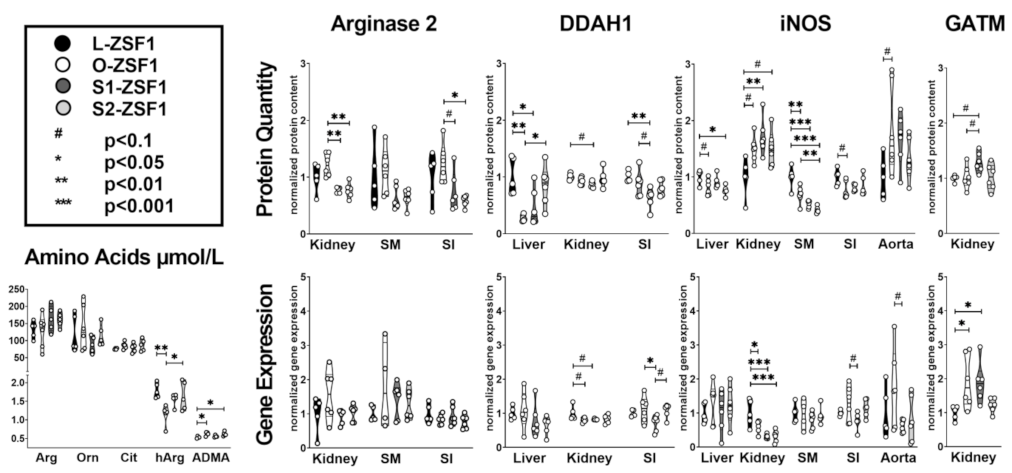
2. Results
3. Discussion
4. Materials and Methods
5. Conclusions
6. Limitations
Author Contributions
Funding
Institutional Review Board Statement
Informed Consent Statement
Data Availability Statement
Acknowledgments
Conflicts of Interest
References
- Durante, W.; Johnson, F.K.; Johnson, R.A. Arginase: A critical regulator of nitric oxide synthesis and vascular function. Clin. Exp. Pharmacol. Physiol. 2007, 34, 906–911. [Google Scholar] [CrossRef] [PubMed]
- Atzler, D.; Schönhoff, M.; Cordts, K.; Ortland, I.; Hoppe, J.; Hummel, F.C.; Gerloff, C.; Jaehde, U.; Jagodzinski, A.; Böger, R.H.; et al. Oral supplementation with L-homoarginine in young volunteers. Br. J. Clin. Pharmacol. 2016, 82, 1477–1485. [Google Scholar] [CrossRef] [PubMed]
- Morris, S.M. Arginine Metabolism Revisited. J. Nutr. 2016, 146, 2579S–2586S. [Google Scholar] [CrossRef]
- Nayor, M.; Houstis, N.E.; Namasivayam, M.; Rouvina, J.; Hardin, C.; Shah, R.V.; Ho, J.E.; Malhotra, R.; Lewis, G.D. Impaired Exercise Tolerance in Heart Failure With Preserved Ejection Fraction: Quantification of Multiorgan System Reserve Capacity. JACC Heart Fail. 2020, 8, 605–617. [Google Scholar] [CrossRef] [PubMed]
- Penicka, M.; Bartunek, J.; Trakalova, H.; Hrabakova, H.; Maruskova, M.; Karasek, J.; Kocka, V. Heart failure with preserved ejection fraction in outpatients with unexplained dyspnea: A pressure-volume loop analysis. J. Am. Coll. Cardiol. 2010, 55, 1701–1710. [Google Scholar] [CrossRef]
- Baldassarri, F.; Schwedhelm, E.; Atzler, D.; Böger, R.H.; Cordts, K.; Haller, B.; Pressler, A.; Müller, S.; Suchy, C.; Wachter, R.; et al. Relationship between exercise intervention and NO pathway in patients with heart failure with preserved ejection fraction. Biomarkers 2018, 23, 540–550. [Google Scholar] [CrossRef]
- Bilan, V.P.; Salah, E.M.; Bastacky, S.; Jones, H.B.; Mayers, R.M.; Zinker, B.; Poucher, S.M.; Tofovic, S.P. Diabetic nephropathy and long-term treatment effects of rosiglitazone and enalapril in obese ZSF1 rats. J. Endocrinol. 2011, 210, 293–308. [Google Scholar] [CrossRef]
- Conceição, G.; Heinonen, I.; Lourenço, A.P.; Duncker, D.J.; Falcão-Pires, I. Animal models of heart failure with preserved ejection fraction. Neth. Heart J. 2016, 24, 275–286. [Google Scholar] [CrossRef]
- Büttner, P.; Werner, S.; Baskal, S.; Tsikas, D.; Adams, V.; Lurz, P.; Besler, C.; Knauth, S.; Bahls, M.; Schwedhelm, E.; et al. Arginine metabolism and nitric oxide turnover in the ZSF1 animal model for heart failure with preserved ejection fraction. Sci. Rep. 2021, 11, 20684. [Google Scholar] [CrossRef]
- Büttner, P.; Adams, V.; Werner, S.; Ossmann, S.; Besler, C.; Schwedhelm, E.; Thiele, H. Effects of homoarginine supplementation on heart and skeletal muscle of rats with heart failure with preserved ejection fraction. ESC Heart Fail. 2022, 9, 4348–4351. [Google Scholar] [CrossRef]
- Triposkiadis, F.; Butler, J.; Abboud, F.M.; Armstrong, P.W.; Adamopoulos, S.; Atherton, J.J.; Backs, J.; Bauersachs, J.; Burkhoff, D.; Bonow, R.O.; et al. The continuous heart failure spectrum: Moving beyond an ejection fraction classification. Eur. Heart J. 2019, 40, 2155–2163. [Google Scholar] [CrossRef]
- Mishra, S.; Kass, D.A. Cellular and molecular pathobiology of heart failure with preserved ejection fraction. Nat. Rev. Cardiol. 2021, 18, 400–423. [Google Scholar] [CrossRef] [PubMed]
- Crisci, G.; de Luca, M.; D’Assante, R.; Ranieri, B.; D’Agostino, A.; Valente, V.; Giardino, F.; Capone, V.; Chianese, S.; Rega, S.; et al. Effects of Exercise on Heart Failure with Preserved Ejection Fraction: An Updated Review of Literature. J. Cardiovasc. Dev. Dis. 2022, 9, 241. [Google Scholar] [CrossRef]
- Drechsler, C.; Meinitzer, A.; Pilz, S.; Krane, V.; Tomaschitz, A.; Ritz, E.; März, W.; Wanner, C. Homoarginine, heart failure, and sudden cardiac death in haemodialysis patients. Eur. J. Heart Fail. 2011, 13, 852–859. [Google Scholar] [CrossRef]
- Atzler, D.; Rosenberg, M.; Anderssohn, M.; Choe, C.; Lutz, M.; Zugck, C.; Böger, R.H.; Frey, N.; Schwedhelm, E. Homoarginine an independent marker of mortality in heart failure. Int. J. Cardiol. 2013, 168, 4907–4909. [Google Scholar] [CrossRef] [PubMed]
- März, W.; Meinitzer, A.; Drechsler, C.; Pilz, S.; Krane, V.; Kleber, M.E.; Fischer, J.; Winkelmann, B.R.; Böhm, B.O.; Ritz, E. Homoarginine, cardiovascular risk, and mortality. Circulation 2010, 122, 967–975. [Google Scholar] [CrossRef]
- Pilz, S.; Meinitzer, A.; Tomaschitz, A.; Drechsler, C.; Ritz, E.; Krane, V.; Wanner, C.; Boehm, B.O.; März, W. Low homoarginine concentration is a novel risk factor for heart disease. Heart. 2011, 97, 1222–1227. [Google Scholar] [CrossRef]
- Atzler, D.; McAndrew, D.J.; Cordts, K.; Schneider, J.E.; Zervou, S.; Schwedhelm, E.; Neubauer, S.; Lygate, C.A. Dietary Supplementation with Homoarginine Preserves Cardiac Function in a Murine Model of Post-Myocardial Infarction Heart Failure. Circulation 2017, 135, 400–402. [Google Scholar] [CrossRef]
- Faller, K.M.E.; Atzler, D.; McAndrew, D.J.; Zervou, S.; Whittington, H.J.; Simon, J.N.; Aksentijevic, D.; Hove, M.; Choe, C.; Isbrandt, D.; et al. Impaired cardiac contractile function in arginine:glycine amidinotransferase knockout mice devoid of creatine is rescued by homoarginine but not creatine. Cardiovasc. Res. 2018, 114, 417–430. [Google Scholar] [CrossRef] [PubMed]
- Cullen, M.E.; Yuen, A.H.Y.; Felkin, L.E.; Smolenski, R.T.; Hall, J.L.; Grindle, S.; Miller, L.W.; Birks, E.J.; Yacoub, M.H.; Barton, P.J.R. Myocardial expression of the arginine: Glycine amidinotransferase gene is elevated in heart failure and normalized after recovery: Potential implications for local creatine synthesis. Circulation 2006, 114, I16–I20. [Google Scholar] [CrossRef]
- Lygate, C.A.; Lake, H.A.; McAndrew, D.J.; Neubauer, S.; Zervou, S. Influence of homoarginine on creatine accumulation and biosynthesis in the mouse. Front. Nutr. 2022, 9, 969702. [Google Scholar] [CrossRef] [PubMed]
- Hara, M.; Torisu, K.; Tomita, K.; Kawai, Y.; Tsuruya, K.; Nakano, T.; Kitazono, T. Arginase 2 is a mediator of ischemia-reperfusion injury in the kidney through regulation of nitrosative stress. Kidney Int. 2020, 98, 673–685. [Google Scholar] [CrossRef] [PubMed]
- Saavedra-Alvarez, A.; Pereyra, K.V.; Toledo, C.; Iturriaga, R.; Del Rio, R. Vascular dysfunction in HFpEF: Potential role in the development, maintenance, and progression of the disease. Front. Cardiovasc. Med. 2022, 9, 1070935. [Google Scholar] [CrossRef]
- Eghbalzadeh, K.; Brixius, K.; Bloch, W.; Brinkmann, C. Skeletal muscle nitric oxide (NO) synthases and NO-signaling in “diabesity”—What about the relevance of exercise training interventions? Nitric Oxide 2014, 37, 28–40. [Google Scholar] [CrossRef]
- Gambardella, J.; Khondkar, W.; Morelli, M.B.; Wang, X.; Santulli, G.; Trimarco, V. Arginine and Endothelial Function. Biomedicines 2020, 8, 277. [Google Scholar] [CrossRef] [PubMed]
- Brandt, A.; Baumann, A.; Hernández-Arriaga, A.; Jung, F.; Nier, A.; Staltner, R.; Rajcic, D.; Schmeer, C.; Witte, O.W.; Wessner, B.; et al. Impairments of intestinal arginine and NO metabolisms trigger aging-associated intestinal barrier dysfunction and ‘inflammaging’. Redox Biol. 2022, 58, 102528. [Google Scholar] [CrossRef] [PubMed]
- Dong, Z.; Zheng, S.; Shen, Z.; Luo, Y.; Hai, X. Trimethylamine N-Oxide is Associated with Heart Failure Risk in Patients with Preserved Ejection Fraction. Lab. Med. 2021, 52, 346–351. [Google Scholar] [CrossRef]
- Kinugasa, Y.; Nakamura, K.; Kamitani, H.; Hirai, M.; Yanagihara, K.; Kato, M.; Yamamoto, K. Trimethylamine N-oxide and outcomes in patients hospitalized with acute heart failure and preserved ejection fraction. ESC Heart Fail. 2021, 8, 2103–2110. [Google Scholar] [CrossRef]
- Kim, K. Interaction between HSP 70 and iNOS in skeletal muscle injury and repair. J. Exerc. Rehabil. 2015, 11, 240–243. [Google Scholar] [CrossRef]
- Taylor, B.S.; Alarcon, L.H.; Billiar, T.R. Inducible nitric oxide synthase in the liver: Regulation and function. Biochemistry 1998, 63, 766–781. [Google Scholar]
- Perl, K.; Ushakov, K.; Pozniak, Y.; Yizhar-Barnea, O.; Bhonker, Y.; Shivatzki, S.; Geiger, T.; Avraham, K.B.; Shamir, R. Reduced changes in protein compared to mRNA levels across non-proliferating tissues. BMC Genomics 2017, 18, 305. [Google Scholar] [CrossRef] [PubMed]
- Pautz, A.; Art, J.; Hahn, S.; Nowag, S.; Voss, C.; Kleinert, H. Regulation of the expression of inducible nitric oxide synthase. Nitric Oxide 2010, 15, 75–93. [Google Scholar] [CrossRef] [PubMed]
- Schauer, A.; Draskowski, R.; Jannasch, A.; Kirchhoff, V.; Goto, K.; Männel, A.; Barthel, P.; Augstein, A.; Winzer, E.; Tugtekin, M.; et al. ZSF1 rat as animal model for HFpEF: Development of reduced diastolic function and skeletal muscle dysfunction. ESC Heart Fail. 2020, 7, 2123–2134. [Google Scholar] [CrossRef]
- Kim, J.H.; Choi, I. Choosing the Level of Significance: A Decision-theoretic Approach. Abacus 2021, 57, 27–71. [Google Scholar] [CrossRef]

| L-ZSF1 | O-ZSF1 | S1-ZSF1 | S2-ZSF1 | L-ZSF1 vs. O-ZSF1 | L-ZSF1 vs. S1-ZSF1 | L-ZSF1 vs. S2-ZSF1 | O-ZSF1 vs. S1-ZSF1 | O-ZSF1 vs. S2-ZSF1 | ||
|---|---|---|---|---|---|---|---|---|---|---|
| Weight | Body weight (g) | 283 ± 9 | 529 ± 23 | 533 ± 24 | 513 ± 21 | <0.001 | <0.001 | <0.001 | 0.760 | 0.236 |
| Heart (g) | 1.16 ± 0.12 | 1.56 ± 0.15 | 1.71 ± 0.10 | 1.58 ± 0.09 | 0.002 | <0.001 | <0.001 | 0.098 | 0.854 | |
| Liver (g) | 9.49 ± 0.30 | 23.71 ± 3.76 | 26.05 ± 1.28 | 23.39 ± 1.92 | <0.001 | <0.001 | <0.001 | 0.197 | 0.859 | |
| Kidney (g) | 1.10 ± 0.04 | 1.69 ± 0.16 | 1.78 ± 0.21 | 1.68 ± 0.11 | <0.001 | <0.001 | <0.001 | 0.429 | 0.902 | |
| Serum | Arginine (umol/L) | 132 ± 25 | 127 ± 48 | 164 ± 37 | 161 ± 21 | 0.995 | 0.507 | 0.586 | 0.347 | 0.418 |
| Citrulline (umol/L) | 76 ± 2 | 86 ± 11 | 79 ± 13 | 87 ± 17 | 0.655 | 0.984 | 0.570 | 0.825 | 0.999 | |
| Homoarginine (umol/L) | 1.79 ± 0.2 | 1.15 ± 0.27 | 1.50 ± 0.22 | 1.64 ± 0.39 | 0.008 | 0.527 | 0.812 | 0.113 | 0.045 | |
| Ornithine (umol/L) | 119 ± 55 | 139 ± 65 | 89 ± 24 | 114 ± 31 | 0.910 | 0.753 | 0.998 | 0.352 | 0.838 | |
| ADMA (umol/L) | 0.53 ± 0.03 | 0.63 ± 0.05 | 0.56 ± 0.03 | 0.62 ± 0.06 | 0.025 | 0.818 | 0.041 | 0.131 | 0.992 | |
| Western Blot results | Arginase 1 liver | 1.00 ± 0.19 | 0.81 ± 0.52 | 1.01 ± 0.44 | 1.27 ± 0.23 | 0.842 | 1.000 | 0.653 | 0.795 | 0.191 |
| Arginase 2 kidney | 1.00 ± 0.25 | 1.22 ± 0.20 | 0.78 ± 0.05 | 0.77 ± 0.13 | 0.172 | 0.167 | 0.141 | 0.001 | 0.001 | |
| Arginase 2 SM | 1.00 ± 0.56 | 1.11 ± 0.40 | 0.63 ± 0.20 | 0.64 ± 0.16 | 0.955 | 0.345 | 0.357 | 0.126 | 0.131 | |
| Arginase 2 SI | 1.00 ± 0.42 | 1.24 ± 0.35 | 0.74 ± 0.35 | 0.58 ± 0.10 | 0.610 | 0.559 | 0.172 | 0.064 | 0.012 | |
| AGXT2 liver | 1.00 ± 0.39 | 0.90 ± 0.25 | 0.93 ± 0.16 | 0.82 ± 0.17 | 0.907 | 0.966 | 0.615 | 0.996 | 0.935 | |
| AGXT2 kidney | 1.00 ± 0.14 | 1.02 ± 0.25 | 1.12 ± 0.10 | 1.05 ± 0.18 | 0.999 | 0.684 | 0.966 | 0.747 | 0.988 | |
| DDAH1 liver | 1.00 ± 0.33 | 0.28 ± 0.05 | 0.46 ± 0.32 | 0.85 ± 0.35 | 0.003 | 0.026 | 0.809 | 0.697 | 0.014 | |
| DDAH1 kidney | 1.00 ± 0.05 | 0.93 ± 0.06 | 0.87 ± 0.05 | 0.97 ± 0.15 | 0.532 | 0.089 | 0.942 | 0.633 | 0.832 | |
| DDAH1 SI | 1.00 ± 0.09 | 0.89 ± 0.22 | 0.63 ± 0.17 | 0.80 ± 0.13 | 0.680 | 0.008 | 0.230 | 0.064 | 0.806 | |
| eNOS aorta | 1.00 ± 0.16 | 1.43 ± 0.37 | 1.53 ± 0.45 | 1.43 ± 0.49 | 0.303 | 0.164 | 0.302 | 0.978 | 1.000 | |
| GATM kidney | 1.00 ± 0.06 | 1.02 ± 0.21 | 1.29 ± 0.18 | 1.02 ± 0.23 | 0.997 | 0.083 | 0.997 | 0.098 | 1.000 | |
| iNOS liver | 1.00 ± 0.13 | 0.82 ± 0.14 | 0.91 ± 0.11 | 0.76 ± 0.08 | 0.070 | 0.568 | 0.013 | 0.518 | 0.148 | |
| iNOS kidney | 1.00 ± 0.34 | 1.50 ± 0.23 | 1.72 ± 0.32 | 1.50 ± 0.32 | 0.061 | 0.005 | 0.060 | 0.600 | 0.606 | |
| iNOS SM | 1.00 ± 0.19 | 0.69 ± 0.15 | 0.53 ± 0.06 | 0.41 ± 0.06 | 0.003 | <0.001 | <0.001 | 0.155 | 0.006 | |
| iNOS SI | 1.00 ± 0.14 | 0.77 ± 0.15 | 0.81 ± 0.06 | 0.83 ± 0.17 | 0.054 | 0.144 | 0.209 | 0.948 | 0.868 | |
| iNOS aorta | 1.00 ± 0.37 | 1.85 ± 0.81 | 1.67 ± 0.46 | 1.32 ± 0.38 | 0.077 | 0.211 | 0.770 | 0.935 | 0.348 | |
| Gene Expression | Arginase 1 liver | 1.00 ± 0.50 | 1.78 ± 1.34 | 0.77 ± 0.44 | 0.88 ± 0.46 | 0.388 | 0.961 | 0.994 | 0.155 | 0.233 |
| Arginase 2 kidney | 1.00 ± 0.52 | 1.55 ± 0.74 | 0.92 ± 0.21 | 1.03 ± 0.24 | 0.254 | 0.993 | 1.000 | 0.136 | 0.260 | |
| Arginase 2 SM | 1.00 ± 0.21 | 1.80 ± 1.27 | 1.41 ± 0.51 | 1.30 ± 0.45 | 0.300 | 0.737 | 0.878 | 0.792 | 0.642 | |
| Arginase 2 SI | 1.00 ± 0.28 | 0.83 ± 0.14 | 0.94 ± 0.28 | 0.77 ± 0.20 | 0.624 | 0.973 | 0.353 | 0.841 | 0.957 | |
| AGXT2 liver | 1.00 ± 0.44 | 1.71 ± 0.79 | 1.22 ± 0.55 | 1.26 ± 0.58 | 0.251 | 0.932 | 0.892 | 0.518 | 0.588 | |
| AGXT2 kidney | 1.00 ± 0.55 | 2.15 ± 0.98 | 1.95 ± 1.38 | 1.58 ± 0.67 | 0.235 | 0.389 | 0.761 | 0.984 | 0.734 | |
| DDAH1 liver | 1.00 ± 0.19 | 1.09 ± 0.54 | 0.78 ± 0.48 | 0.66 ± 0.26 | 0.982 | 0.799 | 0.523 | 0.547 | 0.285 | |
| DDAH1 kidney | 1.00 ± 0.20 | 0.81 ± 0.07 | 0.82 ± 0.02 | 0.83 ± 0.12 | 0.058 | 0.092 | 0.101 | 0.995 | 0.990 | |
| DDAH1 SI | 1.00 ± 0.09 | 1.12 ± 0.38 | 0.71 ± 0.23 | 1.09 ± 0.20 | 0.855 | 0.259 | 0.933 | 0.048 | 0.996 | |
| eNOS aorta | 1.00 ± 0.81 | 0.86 ± 0.65 | 1.26 ± 1.04 | 1.68 ± 1.60 | 0.997 | 0.978 | 0.743 | 0.921 | 0.587 | |
| GATM kidney | 1.00 ± 0.21 | 1.92 ± 0.78 | 1.89 ± 0.59 | 1.22 ± 0.21 | 0.042 | 0.050 | 0.900 | 1.000 | 0.131 | |
| iNOS liver | 1.00 ± 0.29 | 1.32 ± 0.59 | 1.06 ± 0.60 | 1.25 ± 0.56 | 0.761 | 0.998 | 0.873 | 0.833 | 0.995 | |
| iNOS kidney | 1.00 ± 0.38 | 0.61 ± 0.18 | 0.31 ± 0.06 | 0.32 ± 0.17 | 0.042 | <0.001 | <0.001 | 0.116 | 0.138 | |
| iNOS SM | 1.00 ± 0.27 | 0.96 ± 0.44 | 0.79 ± 0.24 | 0.92 ± 0.23 | 0.997 | 0.649 | 0.966 | 0.811 | 0.996 | |
| iNOS SI | 1.00 ± 0.08 | 1.27 ± 0.53 | 0.77 ± 0.23 | 1.15 ± 0.25 | 0.532 | 0.641 | 0.867 | 0.065 | 0.921 | |
| iNOS aorta | 1.00 ± 0.73 | 1.73 ± 1.16 | 0.62 ± 0.19 | 0.80 ± 0.67 | 0.417 | 0.844 | 0.974 | 0.090 | 0.190 |
Disclaimer/Publisher’s Note: The statements, opinions and data contained in all publications are solely those of the individual author(s) and contributor(s) and not of MDPI and/or the editor(s). MDPI and/or the editor(s) disclaim responsibility for any injury to people or property resulting from any ideas, methods, instructions or products referred to in the content. |
© 2023 by the authors. Licensee MDPI, Basel, Switzerland. This article is an open access article distributed under the terms and conditions of the Creative Commons Attribution (CC BY) license (https://creativecommons.org/licenses/by/4.0/).
Share and Cite
Büttner, P.; Werner, S.; Böttner, J.; Ossmann, S.; Schwedhelm, E.; Thiele, H. Systemic Effects of Homoarginine Supplementation on Arginine Metabolizing Enzymes in Rats with Heart Failure with Preserved Ejection Fraction. Int. J. Mol. Sci. 2023, 24, 14782. https://doi.org/10.3390/ijms241914782
Büttner P, Werner S, Böttner J, Ossmann S, Schwedhelm E, Thiele H. Systemic Effects of Homoarginine Supplementation on Arginine Metabolizing Enzymes in Rats with Heart Failure with Preserved Ejection Fraction. International Journal of Molecular Sciences. 2023; 24(19):14782. https://doi.org/10.3390/ijms241914782
Chicago/Turabian StyleBüttner, Petra, Sarah Werner, Julia Böttner, Susann Ossmann, Edzard Schwedhelm, and Holger Thiele. 2023. "Systemic Effects of Homoarginine Supplementation on Arginine Metabolizing Enzymes in Rats with Heart Failure with Preserved Ejection Fraction" International Journal of Molecular Sciences 24, no. 19: 14782. https://doi.org/10.3390/ijms241914782
APA StyleBüttner, P., Werner, S., Böttner, J., Ossmann, S., Schwedhelm, E., & Thiele, H. (2023). Systemic Effects of Homoarginine Supplementation on Arginine Metabolizing Enzymes in Rats with Heart Failure with Preserved Ejection Fraction. International Journal of Molecular Sciences, 24(19), 14782. https://doi.org/10.3390/ijms241914782