Review

Current Progress of Platelet-Rich Derivatives in Cartilage and Joint Repairs

1
Graduate Institute of Biomedical Engineering, National Taiwan University of Science and Technology, Taipei 10607, Taiwan
2
Adjunct Appointment to the Department of Biomedical Engineering, National Defense Medical Center, Taipei 11490, Taiwan
3
International Ph.D. Program in Medicine, College of Medicine, Taipei Medical University, Taipei 11031, Taiwan
4
Department of Radiology, Thai Nguyen National Hospital, Thai Nguyen 24000, Vietnam
5
Department of Pharmacy Practice, Tri-Service General Hospital, Taipei 11490, Taiwan
6
Department of Orthopedics and Trauma, Cho Ray Hospital, Ho Chi Minh 70000, Vietnam
7
Department of Radiology, School of Medicine, College of Medicine, Taipei Medical University, Taipei 11031, Taiwan
8
Department of Orthopedics, Shuang Ho Hospital, Taipei Medical University, New Taipei City 23561, Taiwan
9
Department of Orthopedics, School of Medicine, College of Medicine, Taipei Medical University, Taipei 11011, Taiwan
10
International Ph.D. Program for Cell Therapy and Regenerative Medicine, College of Medicine, Taipei Medical University, Taipei 11011, Taiwan
11
Department of Radiology, Wan Fang Hospital, Taipei Medical University, Taipei 116081, Taiwan
*
Author to whom correspondence should be addressed.
These authors contributed equally to this work.
Int. J. Mol. Sci. 2023, 24(16), 12608; https://doi.org/10.3390/ijms241612608
Submission received: 13 June 2023 / Revised: 4 August 2023 / Accepted: 5 August 2023 / Published: 9 August 2023
(This article belongs to the Special Issue The Role of the Platelet in Human Diseases)

Abstract

:
In recent years, several types of platelet concentrates have been investigated and applied in many fields, particularly in the musculoskeletal system. Platelet-rich fibrin (PRF) is an autologous biomaterial, a second-generation platelet concentrate containing platelets and growth factors in the form of fibrin membranes prepared from the blood of patients without additives. During tissue regeneration, platelet concentrates contain a higher percentage of leukocytes and a flexible fibrin net as a scaffold to improve cell migration in angiogenic, osteogenic, and antibacterial capacities during tissue regeneration. PRF enables the release of molecules over a longer period, which promotes tissue healing and regeneration. The potential of PRF to simulate the physiology and immunology of wound healing is also due to the high concentrations of released growth factors and anti-inflammatory cytokines that stimulate vessel formation, cell proliferation, and differentiation. These products have been used safely in clinical applications because of their autologous origin and minimally invasive nature. We focused on a narrative review of PRF therapy and its effects on musculoskeletal, oral, and maxillofacial surgeries and dermatology. We explored the components leading to the biological activity and the published preclinical and clinical research that supports its application in musculoskeletal therapy. The research generally supports the use of PRF as an adjuvant for various chronic muscle, cartilage, and tendon injuries. Further clinical trials are needed to prove the benefits of utilizing the potential of PRF.

1. Introduction

Platelet concentrates, also known as platelet-rich plasma (PRP) or platelet-rich concentrates (PRCs), have a history of several decades. Platelet concentrates were first produced in the early 1970s [1]. Since then, they have been used in various medicinal applications. They contain platelets, fibrin, growth factors, and mitogenic factors that can release cytokines, chemokines, and other factors. Hence, they can stimulate vessel formation, cell proliferation, and differentiation; improve angiogenesis; and decrease the occurrence of inflammation [2,3,4]. In the early 2000s, an evolution of PRP called platelet-rich fibrin (PRF) was introduced by Choukroun et al. [5], who prepared PRF by centrifuging blood without anticoagulants, which resulted in a fibrin matrix enriched with platelets and other blood components. PRF has gained popularity owing to its simplified preparation process and absence of biochemical modifications. PRF also contains many types of growth factors such as platelet-derived growth factor (PDGF- a/b and c), vascular endothelial growth factor (VEGF), hepatocyte growth factor (HGF), epidermal growth factor (EGF), connective tissue growth factor (CTGF), fibroblast growth factor (FGF), insulin-like growth factor (IGF), and transforming growth factor β1 (TGF-β1) [6,7,8]. Growth factors are transformed into active stages after platelet activation. They are mainly present in the α-granules of platelets [9] and have many functions, such as mediating cell proliferation and growth, angiogenesis, wound healing, and bone metabolism. Many PRF formulations of PRF been investigated, including leukocyte platelet-rich fibrin (L-PRF) [10,11], advanced platelet-rich fibrin (A-PRF) [10,12], titanium-prepared platelet-rich fibrin (T-PRF) [13,14], and injectable platelet-rich fibrin (i-PRF) [15,16]. The therapeutic potential of PRFs in regenerative medicine, especially in musculoskeletal regeneration [17], means they have promising clinical applications. In addition, many studies have reported that PRF has a positive effect not only on musculoskeletal injury but also in other fields, such as diabetic ulcers [18], cosmetic surgery [19], maxillofacial surgery [20,21], and cardiac surgery [22].
Despite the wide range of PRF applications, there is some variation in the outcomes when using PRF treatments. The present article aims to summarize the current relevant application of PRFs in the musculoskeletal system and provide the essential roles of components of PRFs in the musculoskeletal system and maxillofacial surgery.

2. Evolution of Platelet-Rich Derivatives

2.1. Fibrin Glue

Fibrin glue, also known as fibrin sealant, fibrin adhesive, or fibrin sutures, is the oldest and has been the most widely used fibrin clot for the past 40 years [1]. Fibrin glue has been used to mimic the blood clot mechanism. It was originally constituted by polymerizing fibrinogen, which is obtained from human plasma, along with thrombin and calcium. However, the fibrinogen concentration in plasma is very low (0.2% volume of whole blood) [23], so it is very difficult to obtain with autologous techniques and always results in unstable quality of the fibrin glue [24]. In addition, the non-industrial preparation of fibrin glue requires massive amounts of blood, time, and preparation to collect and obtain fibrinogen [25]. Therefore, autologous protocols are unsuitable for patients who require immediate surgery.
Despite the advantages of autologous products, commercial fibrin glue has become the most popular fibrin glue since it became available on the market in 1998. Pharmaceutical companies such as Tisseel from Baxter Healthcare produce commercial fibrin glue as a surgical additive [26]. It is produced and supplied in separate vials with a dual syringe delivery system, in which human-plasma-derived thrombin containing ionic calcium and a fibrinogen-containing anticoagulant are first separated into two vials. Before use, these two components are immediately mixed using a syringe, and it is then ready for treatment. Although it has been claimed to be virus-inactivated, it still poses an infinitely low risk of viral contamination. Furthermore, because commercial fibrin glue is not an autologous blood-derived product, the risks of immune rejection, disease transmission, and manufacturing costs are also the main concerns for the market [27,28].
In a recent meta-analyses, fibrin glue was found to be effectively combined with polyglycolic acid sheets to reduce the risk of bleeding after endoscopic submucosal dissection for gastric cancer [29] and glue-based mesh fixation to reduce the incidence of chronic postoperative inguinal pain and hematoma after laparoscopic inguinal hernia repair [30]. However, the fibrin patch applied to the pancreatic stump did not reduce the incidence of postoperative pancreatic fistulas after distal pancreatectomy [31].
Fibrin glue embedded with drugs, antibiotics, cytostatic agents, or stem cells may increase the effectiveness of therapy and facilitate the targeted delivery of active substances for localized drug release. The use of fibrin glue alone resulted in suture repair for nerve regeneration [32]. There is clear evidence that fibrin glue combined with mesenchymal stem cells (MSCs) can regenerate nervous system lesions [33]. Articular cartilage repair using fibrin glue alone has not been reported.

2.2. Platelet-Rich Plasma (PRP)

With the development of technology, scientists have been able to produce autologous fibrin glue (PPP) using a more simplified method [24,34], since some scientists have identified the important role of growth factors in cellular process regulation [35], while the focus of autologous fibrin glue research has been on new biomaterials named platelet concentrates [36]. Scientists have developed a first-generation platelet concentrate, PRP, as an autologous modification of fibrin glue [2,34]. PRP, a true concentrate of platelets, is a fibrin clot rich in platelets (containing 95% platelets, 4% red blood cells, and 1% white blood cells) that is obtained from plasma [36]. PRP is a safe and autologous product that is readily available at the point of care and minimizes the risks of immune rejection and disease transmission [37]. The PRP protocols have varied among researchers, although the main concept of the protocols remains consistent. They all include the concept of blood cell separation, regardless of the use of a typical centrifugation method or a commercial PRP kit called a cell separator [38]. They require whole blood to be separated by centrifugation into different layers of blood cells, red blood cells (RBC), buffy coat (leukocytes), and plasma. The basic rules of PRP protocols include three steps. First, an anticoagulant is added to whole blood before blood processing to stop blood coagulation. Second, the whole blood is processed using a two-step discontinuous centrifugation process or one-step centrifugation with a cell separator kit to obtain the preliminary PRP product. Third, after liquid PRP is obtained using various blood separation methods, with the addition of thrombin to activate fibrinogen and calcium chloride to neutralize the effect of anticoagulants, the fibrinogen in liquid PRP can finally be cross-linked and polymerized to form a gel-like fibrin clot.
Based on the biochemical compositions of PRP types resulting from different protocols, PRPs can be categorized into two groups: leukocyte-rich (L-PRP) and leukocyte-poor (P-PRP) [28]. Despite the differences between the compositions and protocols of all PRP types, they share common weaknesses. These include long processing times, high costs, poor mechanical properties [39], unstable product volumes, low fibrin densities, and weak fibrin polymerization [28], as well as the potential risk of allergic reactions and cross-contamination with bovine thrombin. PRP has limited potential to stimulate bone regeneration, as it releases growth factors quickly just before cell outgrowth from the surrounding tissue [40]. Therefore, in 2000, French scientists developed a new generation of platelet concentrate, PRF, to overcome the existing risks of PRP [5].

2.3. Platelet-Rich Fibrin (PRF)

For the purposes of this review, the PRF mentioned in this article is defined as a second-generation platelet concentrate, which was first developed by Choukroun et al. [5] and is also referred to as Choukroun’s platelet-rich fibrin [41] or leukocyte- and platelet-rich fibrin (L-PRF) [28] to avoid confusion with other PRF products that contain biochemical additions [42,43]. PRF was first developed in France for specific use in oral and maxillofacial surgery to avoid legal restrictions on the reimplantation of blood-derived products [24]. Unlike other platelet concentrates, fibrin glue, and PRP, PRF is not a blood-derived product; it is simply centrifuged without any biochemical blood handling.

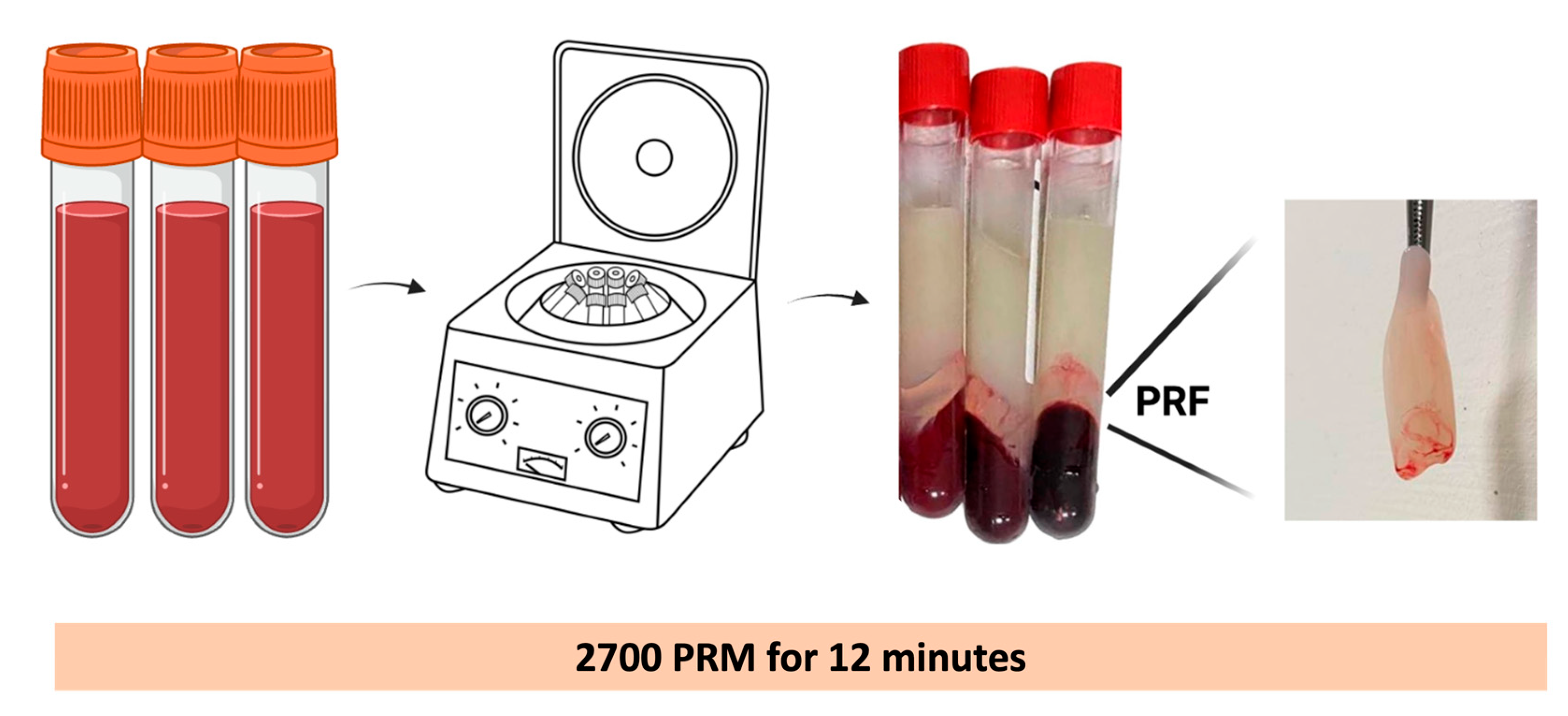
2.3.1. PRF Preparation

PRF is an autologous platelet concentrate that is produced via one simple, short, and soft centrifugation cycle of 100% natural and non-additive whole blood. The first protocol for PRF production involved collecting 10 mL of a blood sample without anticoagulant, which is immediately centrifuged at 2700 rpm (approximately 400× g) for 12 min (Figure 1). In the absence of an anticoagulant, most platelets in a blood sample become activated within a few minutes of contact with the walls of the collection tube. This activation triggers the release of coagulation cascades, leading to blood clot formation. Initially, the fibrinogen is concentrated in the upper section of the tube. Subsequently, circulating thrombin interacts with fibrinogen, resulting in its conversion to fibrin. As a result, a fibrin clot is generated in the middle of the tube, positioned between the settled red blood cells at the bottom and acellular plasma at the top. The slow polymerization of fibrin during PRF preparation generates a fibrin clot with a fibrin structure that is very similar to that of natural fibrin. Comparing the fibrin structures of PRP and PRF, PRF seems to be a more friendly environment for cell migration and proliferation, and consequently cicatrization. The speed and method of the polymerization affect the 3D structure of the fibrin network. Fibrin glue and PRP favor the formation of bilateral fibrin glue owing to the high thrombin concentration and rapid polymerization. In contrast, PRF has a low thrombin concentration and slow polymerization rate, implying a more equilateral junction. Compared to the bilateral junction, the equilateral junction is a finer and more flexible fibrin structure with greater elasticity that provides better support for cytokine enmeshment and cellular migration [7,24].
Unlike fibrin glue and PRP, PRF is an autologous fibrin clot that requires only a small amount of money and time for production. PRF preparation is a simpler and more cost-effective process than PRP preparation [8,27,40,44] (Table 1). As PRF is an autologous platelet concentrate without any biochemical addition, it can avoid the risks of cross-infection, contamination, and product safety, which are the biggest concerns for commercial fibrin glues. PRF has several advantages over autologous fibrin glue. This method does not require a massive amount of blood for production and requires only 4 mL of fresh blood to produce 1 mL of PRF [45].
In contrast to PRP, PRF is an economical biomaterial and safe bio-scaffold. It is produced without the addition of anticoagulants or bovine thrombin, making it free from the risks of cross-contamination and immune rejection caused by biochemical adjuvants. The characteristics of fibrin glue, PRP, and PRF are summarized in Table 2.

2.3.2. Components of PRF

PRF is distinguished by its fibrin matrix, which contains a concentrated number of platelets, growth factors, cytokines, and other bioactive molecules (Figure 2). Platelets, the major components of PRF, are the primary cells responsible for the biological activity of PRF. Platelets are multifunctional cells that not only contribute to clotting but also have a significant impact on immune responses, wound healing, and tissue regeneration. These are stored by three types of particles (alpha, delta, and lambda) located inside platelets. Alpha granules are the most abundant and contain fibrinogen, immunoglobulins, and growth factors [54]. These granules contain various growth factors that are released through exocytosis when platelets are activated and are responsible for regenerating both soft and hard tissues after injury [55]. Delta granules contain adenosine diphosphate, tissue plasminogen activator, serotonin, and fewer lambda granules containing lysosomal enzymes [56,57]. PRF contains some of the most important growth factors, including platelets, cytokines, and granulocytes, as shown in Figure 2.
PDGF is the initial growth factor present at the site of the injury. Platelet-released PDGF promotes the movement, multiplication, and survival of mesenchymal cells [7]. PDGF possesses chemotactic properties that attract macrophages to the site of injury. The combination of PDGF, TGF-β, and IGF produces a synergistic effect that stimulates blood vessel growth, facilitates cell division, promotes skin and bone matrix formation, and enhances collagen synthesis. With approximately 1200 PDGF molecules per platelet, the high concentration of PDGF in PRF may have a significant impact on wound healing and bone regeneration [8]. TGF-β is recognized as a factor that attracts macrophages, stimulates endogenous cells to secrete cytokines, and enhances the synthesis of the extracellular matrix, especially collagen I. Activated platelets secrete the active form of TGF-β1, which plays a significant role in stimulating fibroblast chemotaxis and promoting fibronectin and collagen production. It also acts as a protective factor by preventing collagen breakdown. Furthermore, TGF-β1 induces the formation of new blood vessels and attracts immune cells through chemotaxis [58]. Additionally, TGF-β1 enhances the proliferation and deposition of osteoblasts and inhibits the formation of osteoclasts and bone degeneration [8]. IGF exerts multiple effects on mesenchymal cells. It promotes differentiation and mitogenesis and stimulates growth and development [59]. Moreover, IGF-1 provides survival signals that protect cells from various triggers that induce apoptosis [7]. Additionally, IGF-1 stimulates chemotaxis, attracting and activating osteoblasts, which ultimately leads to increased bone formation [60]. IGF-1 also cooperates with growth factor TGF-β and bone morphogenetic proteins (BMPs) to participate in the synthesis of the basic substance of articular cartilage. EGF is an epidermal growth factor that promotes cell growth, differentiation, angiogenesis, and collagen formation [60]. Similarly, EGF promotes the secretion of cytokines by epithelial and mesenchymal cells [61]. EGF enhances the production and release of cytokines, which are important signaling molecules involved in various cellular activities, including immune responses, inflammation, and tissue repair.
In addition, PRF includes immune cytokines (Figure 2), such as interleukin (IL)-1β, IL-6, IL-4, and tumor necrosis factor (TNF)-α [24]. IL-1 is produced by most nucleated cells, such as monocytes, macrophages, B cells, NK cells, astrocytes, fibroblasts, endothelial cells, and neutrophils [62]. IL-1 has two subtypes, IL-1α and IL-1β. This particular cytokine enhances the production of adhesive molecules on endothelial cells and promotes the movement of phagocytes and lymphocytes towards the injury site [63]. In addition, they stimulate the helper T cells. IL-1β, in conjunction with TNF-α, triggers osteoclast activation and inhibits bone formation [64]. IL-6 is produced by T cells and macrophages to stimulate the immune response [65]. IL-6 is also produced by the muscle tissue and increases in response to muscle contraction. In contrast, IL-6 derived from osteoblasts stimulates osteoclasts, which in turn stimulates bone marrow cell destruction. This promotes the differentiation of B cells (white blood cells that produce antibodies), promotes growth in some cells, and inhibits growth in others. Moreover, IL-6 is an essential cytokine required to induce the transformation of naive T cells into cytotoxic T lymphocytes [64]. It is extensively produced during processes such as inflammation and tissue remodeling [66]. IL-4 is produced by macrophages and Th2 cells. It stimulates the proliferation of Th2 cells and promotes their differentiation into Th2 cells, which induces antibody-producing responses. It also stimulates the B cell class to convert IgE [67]. IL-4 can stimulate the activation of macrophages into M2 macrophages. The induced production of M2 macrophages leads to increased secretion of IL-10 and TGF-β, which reduces the severity of pathological inflammation. The increased secretion of M2 macrophages is closely associated with wound healing and fibrosis development [68]. TNF-α is a cytokine produced by various immune cells, primarily by macrophages. It plays a crucial role in inflammation, the immune response, and the regulation of cell death (apoptosis). TNF-α is involved in the recruitment and activation of immune cells such as neutrophils and monocytes to sites of infection or injury [69]. It induces the expression of adhesion molecules in endothelial cells, thereby facilitating leukocyte migration. TNF-α also stimulates the production of other inflammatory cytokines and chemokines, thereby amplifying the immune response [64]. Bai et al. [70] reported that the microstructure and proportion of PRF in a rabbit model were positively correlated with cytokine concentrations. PRF gel exhibited a quasi-graded distribution of PDGF-BB and TGF-β1. The concentrations of these cytokines in the PRF gel were significantly higher than those in the plasma because of the combination of two factors: (1) an extrinsic factor attributed to the fibrin gel structure; (2) the molecular characteristics of the different cytokines, which serve as intrinsic factors. Although cytokines are typically soluble and are expected to concentrate in the plasma after centrifugation, the highest concentrations of these cytokines were found at the red blood cell end of the gel. This suggests that the cytokines were stoichiometrically trapped in the PRF gel. They also analyzed the histology of PRF sections obtained from young and middle-aged men and women and showed a gradual increase in average porosity over time [71]. Furthermore, a decline in compactness was observed along the longitudinal axis of the PRF gel. They concluded that the section of the PRF gel nearest to the red blood cell layer is considered the core of the PRF clot and that both sex and age in humans influence their platelet generation capacity.
Nevertheless, in recent years, there have been various alterations to the PRF protocol, resulting in the emergence of distinct products with diverse capabilities and potential uses in biology. Many formulations of PRF been investigated, including leukocyte platelet-rich fibrin (L-PRF), advanced platelet-rich fibrin (A-PRF), titanium-prepared platelet-rich fibrin (T-PRF), and injectable platelet-rich fibrin (i-PRF) (Figure 3).
In our previous studies, we investigated the characteristics of PRF and its clinical applications. Some novel observations from our series were reported for the first time, as follows: (1) the RBC portion of a PRF gel is characterized by the highest concentration of platelets and cytokines, making it an essential component known as platelet-rich fibrin essence (ePRF) [70]; (2) the reconstruction of the PRF microstructure in three dimensions from a series of two-dimensional SEM images revealing a dense fibrin matrix with a gauze-like surface morphology [70]; (3) the utilization of PRF and cartilage granules in the absence of bovine thrombin offers the possibility for a one-step cartilage repair surgery, which may yield favorable outcomes [45]. In rabbit models, we investigated the effectiveness of PRF in promoting meniscal tissue healing [73]. PRF stimulates cellular migration and enhances proliferation and ECM synthesis in cultured meniscocytes. Additionally, PRF contributed to increases in the formation and deposition of the cartilaginous matrix produced by cultured meniscocytes. We also developed a feasible one-step procedure to combine PRF and autologous cartilage grafts for articular chondral defects [74]. PRF has beneficial effects on the viability, differentiation, and migration of chondrocytes, and is a promising approach for cartilage repair [75]. Together, these data demonstrate the effectiveness of a single-stage, culture-free procedure combining the PRF and cartilage repair via cartilage autografts. In addition, we explored whether the ability to produce platelets from ePRF was influenced by human sex and age [71].

3. Clinical Application

3.1. Oral and Maxillofacial Surgery

Ghoneim et al. [76] evaluated the effectiveness of injecting injectable platelet-rich fibrin (i-PRF) into the joint space after arthrocentesis compared with arthrocentesis alone for treating patients with temporomandibular joint (TMJ) disc displacement with reduction. The findings revealed statistically significant decreases in pain intensity and clicking sounds, as well as increases in mouth opening and lateral movement, in the group of patients treated with i-PRF compared with the group treated with arthrocentesis alone. The TMJ has a distinct structure characterized by articular surfaces that are covered by nearly acellular fibrocartilage consisting of a minimal number of chondrocyte-like and fibroblast-like cells. Additionally, the predominant collagen type present in the TMJ is type I collagen, which is primarily synthesized by fibroblast-like cells. Kütük et al. [77] applied PRP to the right joints of rabbits and physiological saline to the left joints. The study demonstrated a significant increase in new bone regeneration in the PRP group compared with that in the saline group. A scanning electron microscopy analysis showed an improved ultrastructural architecture of the collagen fibrils, specifically in the PRP group. Giacomello et al. [78] assessed the efficacy of platelet-rich growth factor–Endoret® injections for the treatment of TMJ osteoarthritis in 52 patients who were followed for 1 year. Their findings showed the effectiveness of platelet-rich in growth factor–Endoret injections in decreasing osteoarthritis symptoms and improving them over time.

3.2. Musculoskeletal Disorders

3.2.1. Repair and Regeneration of Cartilage

In the last decade, platelet concentrates (such as PRP and PRF) have been widely investigated as useful therapeutic agents for the treatment of musculoskeletal disorders. PRF has been investigated as a potential treatment option for cartilage damage. The specialized connective tissue of diarthrodial joints is known as the articular cartilage. It is deficient in blood vessels, lymphatics, and nerves, leading to a limited natural ability to heal and repair itself. Owing to the absence of blood vessels in articular cartilage, it cannot trigger the same healing process as other tissues that have robust regenerative potential. However, the introduction of a PRF scaffold may simulate the initial stages of wound healing and tissue repair. The growth factors and cytokines present in PRF have been shown to exert chondrogenic and anti-inflammatory effects by enhancing the viability, differentiation, and migration of chondrocytes, which can support cartilage healing [75]. Since cartilage repair and regeneration are always limited by vascular inadequacy, it is important to have adequate nutrition in the development of cell-based therapies. PRF has been effective in providing nutritional support and increasing the number of cultured chondrocytes, comparable with other PRP-related in vitro studies [75]. In addition, PRF has been proven to provide an appropriate environment for the proliferation and maturation of chondrocytes; therefore, it can be used as a potential bioactive scaffold for cartilage regeneration [79]. Wong et al. [75] developed a one-stage method to combine PRF and autologous cartilage autografts for porcine articular cartilage repair. They reported that the regenerated cartilage surfaces in the treatment groups were smooth and continuous, suggesting that the cartilage repair was relatively complete. Chien et al. [80] demonstrated that human platelet-rich fibrin exudates with high platelet cytokine and growth factor levels can be integrated into biodegradable fibrin scaffolds for use as a regeneration matrix to stimulate chondrocyte proliferation and redifferentiation. In an injured cartilage rabbit model, Kuo et al. [45] reported the regenerative potential of cartilage with higher T2 values via MRI in the PRF-treated group compared to the control group, showing a reduction in proteoglycans and a progressive increase in collagen content. In a cartilage defect rabbit model, Taufik et al. [81] developed a treatment method involving the integration of microfractures, synovial grafts, and a PRF membrane. This combined method effectively promoted the regeneration of cartilage defects. The PRF membrane contributed essential growth factors, whereas the synovium supplied stem cells. The researchers observed significantly increased levels of aggrecan and type 2 collagen expression in the healing tissue of cartilage treated with microfracture and synovium–PRF transplantation. Several studies [82,83,84,85,86] have focused on the ability of chondrocytes to proliferate and differentiate in response to PRP and i-PRF. Appropriate cell types and chondrocytes are required for the first stage of cartilage tissue healing. Mustafa et al. [84] demonstrated that compared to PRP, the injection of i-PRF using the concept of low-speed centrifugation led to significantly increased chondrocyte activity and enhanced cartilage regeneration. Wang et al. [87] investigated the effects of arthroscopic surgery combined with PRP and PRF gels in 28 patients with defective knee cartilages. They showed that this combination therapy could repair knee cartilage defects, improve patient function, and relieve symptoms. Knee osteoarthritis (OA) is a chronic joint disease that mainly results from wear and tear and a progressive loss of articular cartilage. Many previous studies [88,89,90,91] have proven the positive effects of PRP or its combination with hyaluronic acid in the treatment of knee osteoarthritis, although few studies have investigated the benefits of PRF. Cheeva-Akrapan et al. [92] conducted a 36-month survival analysis of treatment with PRP enhanced with injectable PRF in osteoarthritis knee patients, resulting in an 80.18% survival rate in patients who did not require surgical intervention during the follow-up period. PRP releases growth factors shortly after injection, whereas PRF acts as a natural mesh for PRP and releases growth factors slowly.

3.2.2. Meniscal Repair

Many meniscal injuries to the knee are caused by trauma, which results in instability and loss of joint function. They can lead to pain or disability, degenerative joint changes, and symptomatic osteoarthritis [93,94]. When the meniscus is damaged, especially because of the complete or partial cutting of the meniscus, it changes the distribution of forces on the meniscus, accelerating the process of knee osteoarthritis. Unlike PRP, which requires an additional scaffold for in situ tissue transplantation, PRF is a strictly autogenous fibrin-based biomaterial that encourages microvascularization and enables the local and progressive delivery of growth factors, which can be used to enhance bone and tissue regeneration [7,64]. Wong et al. [75] reported the positive stimulatory effects of PRF on meniscocyte migration, proliferation, and extracellular matrix synthesis in a rabbit model. Furthermore, they observed that PRF supplementation resulted in the increased formation and deposition of the cartilaginous matrix produced by cultured meniscocytes. Through morphological and histological evaluations, this study demonstrated that PRF facilitates meniscal repair in rabbits. Researchers have highlighted the potential benefits of using PRF to enhance the healing process of meniscal injuries. Narita et al. [95] employed biodegradable gelatin hydrogel as a carrier for PRP application in horizontal meniscal tears. They discovered that the combination of fibroblast growth factor-2 (FGF-2) with a gelatin hydrogel significantly promoted the proliferation of meniscal cells. Moreover, this combination effectively inhibited meniscal cell death for up to four weeks, leading to increased meniscal cell density and enhanced meniscal repair in a rabbit model. In contrast, PRP did not positively contribute to the healing process in Shin’s study [96]. They evaluated the effect of PRP on horizontal meniscal tears using an experimental rabbit model with a single injection. Griffin et al. [97] indicated that there was no significant difference in the clinical and functional scores between a group treated with PRP and a control group. Indeed, future prospective randomized studies with adequate sample sizes are needed to clarify the use of PRP and PRF in meniscal healing following meniscal repair. By conducting robust investigations, we can enhance our understanding of the role of platelets in this context and make informed decisions regarding their clinical application.

3.2.3. Repair and Regeneration of Tendons

In addition, PRF has been reported to enhance tendon healing and improve clinical symptoms, particularly chronic pain. Owing to insufficient tissue vascularization, tendinopathies have a limited ability to repair and cause irreversible lesion symptoms. During tendon damage, PRF improves cellular and biomechanical responses and enhances the quality of repair. Alviti et al. [94] investigated the effectiveness of PRF in enhancing Achilles tendon healing and restoring tendon elongation through a gait analysis evaluation in 20 males during 6 months of follow-up compared with surgical repair alone. Controversially, Zumstein et al. [95] conducted a prospective randomized, controlled study of thirty-five patients randomized to receive arthroscopic rotator cuff repair with or without L-PRF at the repair site. Their findings revealed no significant improvement in structural integrity or tissue quality. In addition, the overall rates of non-healing were not significantly different between the two groups. Two systematic reviews and meta-analyses examining treatment with PRP reported that lateral epicondylitis, commonly known as tennis elbow, improves pain and function more effectively than corticosteroid injections in the intermediate term (12–26 weeks) [96] and for long-term follow-up (24 weeks post-treatment) [97]. Another systematic review demonstrated that PRP injection did not significantly reduce the pain intensity in chronic greater trochanteric pain syndrome compared with placebo injection (saline) [98], and a randomized, double-blind, controlled trial showed that PRP could achieve greater clinical improvements at 12 weeks than corticosteroid injection [99]. A systematic review of 34 randomized trials revealed that the use of PRP injections has a low risk of harm and is beneficial for long-term outcomes (≥12 months). Other systematic reviews of musculoskeletal disorders, such as carpal tunnel syndrome, patellar tendinopathy, and plantar fasciopathy, have described the promising efficacy of PRP treatment [100,101,102].

3.2.4. Repair Ligament

The use of PRF for acute ligament injuries has also grown in popularity, despite the limited evidence. Matsunaga et al. [98] found that PRF scaffolds promote medial collateral ligament repair in rabbit models. In a rabbit model, Weng et al. [99] demonstrated the effectiveness of L-PRF in enhancing the biological healing of anterior cruciate ligament (ACL) mid-substance tears. When cultured in a three-dimensional environment, viable cells exhibited a high-density arrangement, forming layers on the surface of the L-PRF scaffold. This culture condition demonstrated significant cell ingrowth and the deposition of an abundant collagenous matrix. A comparative MRI study involved 44 patients with ACL injury who underwent arthroscopic ACL reconstruction with a semitendinosus tendon graft and intervention-sprayed PRF to the surface of the graft [100]. The results showed lower MRI signal intensities and less fluid in the tunnel in the PRF-treated group than in the control group. A study [101] evaluated the use of a platelet-rich plasma preparation rich in growth factors (PRGF) during ACL surgery, leading to more remodeling compared with the untreated graft group, with excellent ratings of 57.1% and 33.3%, respectively. Here, 77% of PRGF-treated grafts had histologically identifiable newly generated connective tissue enclosing them compared to 40% of the controls. Eggli et al. [102] reported that incorporating dynamic intraligamentary stabilization microfracturing and L-PRF led to stable clinical and radiological healing of torn ACL patients after one year. Given the existing in vitro and clinical evidence, further investigation is warranted to explore the role of PRF in tendon augmentation and repair. Specifically, a controlled randomized trial is necessary to assess its potential as a therapeutic modality for clinical use.
However, the outcomes of PRF administration in the treatment of musculoskeletal disorders are variable. For instance, two systematic reviews on the use of PRP injections in the treatment of rotator cuff tears reported that the constant shoulder scores, simple shoulder test scores, UCLA scores, and visual analog scale scores improved with PRP compared to the control group [103,104]. In contrast, PRF does not improve the tendon healing rates or functional outcomes [103]. The current evidence for the clinical application of PRP or PRF is summarized in Table 3.

4. Conclusions and Future Perspectives

PRP and PRF continue to evolve as investigational treatments in maxillofacial surgery, musculoskeletal disorders, dermatology, and other fields. The inconsistencies in the clinical results were due to the large heterogeneity of the preparation protocols. Large variations in the centrifugal force and total centrifugation time significantly affected the platelet concentrations and growth factor release from the final products. The concentrations of PDGF-AB, TGF-β1, and VEGF in PRP were 3133–293,500, 20–153,863, and 0–44,000 pg/mL, respectively, according to the different centrifugation protocols [127]. Multiple modifications of basic protocols have led to the development of many techniques for obtaining PRP using either commercial centrifugation kits or manual or homemade procedures. Different protocols obtain different platelet concentrations; leukocyte concentrations; and growth factor (PDGF) concentrations (PDGF-AB, TGF-β1, and VEGF) according to Mariani et al. [127]. Similarly, the accumulated growth factor concentration ranges in PRF are 593-774 (VEGF), 23-36 (TGF-β1), and 859-1147 (EGF) pg/mL [128]. Furthermore, individual characteristics, especially age and sex, also influence the growth factor levels in PRF [71]. Therefore, a standard preparation protocol for PRP and PRF is necessary [129].
Our study obtained several results from the systematic reviews (Table 3). However, most systematic reviews concluded a lack of high-quality studies because the primary clinical studies included a small number of participants, were unblinded and unrandomized, lacked a proper control treatment, and lacked consistent treatment procedures for PRP or PRF. Although the standard treatment protocol for PRP and PRF is still being investigated, there is increasing evidence that PRP can provide positive outcomes in disease treatment. We suggest that more high-quality clinical trials are needed to determine the efficacy of PRF treatment and the most suitable treatment for PRP or PRF.

Author Contributions

Conception of the work, M.-Y.B., C.-C.W. and W.P.C.; acquisition and analysis of data for the work, V.P.T.V., S.-L.T., T.N.K.H., C.-W.W. and J.-Y.L.; writing—original draft preparation, M.-Y.B. and V.P.T.V.; writing—review and editing, all authors. All authors have read and agreed to the published version of the manuscript.

Funding

This research was funded by the Taipei Medical University IIT, Investigator-Initiated Trials (grant no. IIT-1072-1).

Institutional Review Board Statement

Not applicable.

Informed Consent Statement

Not applicable.

Data Availability Statement

Data are contained within the manuscript.

Conflicts of Interest

The authors declare no conflict of interest.

References

  1. Matras, H. Effect of various fibrin preparations on reimplantations in the rat skin. Osterr. Z. Stomatol. 1970, 67, 338–359. [Google Scholar] [PubMed]
  2. Marx, R.E.; Carlson, E.R.; Eichstaedt, R.M.; Schimmele, S.R.; Strauss, J.E.; Georgeff, K.R. Platelet-rich plasma: Growth factor enhancement for bone grafts. Oral Surg. Oral Med. Oral Pathol. Oral Radiol. Endodontol. 1998, 85, 638–646. [Google Scholar] [CrossRef] [PubMed]
  3. Deppermann, C.; Kubes, P. Start a fire, kill the bug: The role of platelets in inflammation and infection. Innate Immun. 2018, 24, 335–348. [Google Scholar] [CrossRef] [Green Version]
  4. Boswell, S.G.; Cole, B.J.; Sundman, E.A.; Karas, V.; Fortier, L.A. Platelet-rich plasma: A milieu of bioactive factors. Arthrosc. J. Arthrosc. Relat. Surg. 2012, 28, 429–439. [Google Scholar] [CrossRef]
  5. Choukroun, J.; Adda, F.; Schoeffler, C.; Vervelle, A. Une opportunité en paro-implantologie: Le PRF. Implantodontie 2001, 42, e62. [Google Scholar]
  6. Pavlovic, V.; Ciric, M.; Jovanovic, V.; Trandafilovic, M.; Stojanovic, P. Platelet-rich fibrin: Basics of biological actions and protocol modifications. Open Med. Wars 2021, 16, 446–454. [Google Scholar] [CrossRef] [PubMed]
  7. Dohan, D.M.; Choukroun, J.; Diss, A.; Dohan, S.L.; Dohan, A.J.; Mouhyi, J.; Gogly, B. Platelet-rich fibrin (PRF): A second-generation platelet concentrate. Part II: Platelet-related biologic features. Oral Surg. Oral Med. Oral Pathol. Oral Radiol. Endodontol. 2006, 101, e45–e50. [Google Scholar] [CrossRef]
  8. Vinaya Kumar, R.; Shubhashini, N. Platelet rich fibrin: A new paradigm in periodontal regeneration. Cell Tissue Bank. 2013, 14, 453–463. [Google Scholar] [CrossRef]
  9. Masuki, H.; Okudera, T.; Watanebe, T.; Suzuki, M.; Nishiyama, K.; Okudera, H.; Nakata, K.; Uematsu, K.; Su, C.Y.; Kawase, T. Growth factor and pro-inflammatory cytokine contents in platelet-rich plasma (PRP), plasma rich in growth factors (PRGF), advanced platelet-rich fibrin (A-PRF), and concentrated growth factors (CGF). Int. J. Implant. Dent. 2016, 2, 19. [Google Scholar] [CrossRef] [PubMed] [Green Version]
  10. Esfahrood, Z.R.; Ardakani, M.T.; Shokri, M.; Shokri, M. Effects of leukocyte-platelet-rich fibrin and advanced platelet-rich fibrin on the viability and migration of human gingival fibroblasts. J. Indian Soc. Periodontol. 2020, 24, 15–19. [Google Scholar] [CrossRef]
  11. Bi, J.; Intriago, M.F.B.; Koivisto, L.; Jiang, G.; Häkkinen, L.; Larjava, H. Leucocyte- and platelet-rich fibrin regulates expression of genes related to early wound healing in human gingival fibroblasts. J. Clin. Periodontol. 2020, 47, 851–862. [Google Scholar] [CrossRef]
  12. Ghanaati, S.; Booms, P.; Orlowska, A.; Kubesch, A.; Lorenz, J.; Rutkowski, J.; Landes, C.; Sader, R.; Kirkpatrick, C.; Choukroun, J. Advanced platelet-rich fibrin: A new concept for cell-based tissue engineering by means of inflammatory cells. J. Oral Implant. 2014, 40, 679–689. [Google Scholar] [CrossRef]
  13. Tunalı, M.; Özdemir, H.; Küçükodacı, Z.; Akman, S.; Fıratlı, E. In vivo evaluation of titanium-prepared platelet-rich fibrin (T-PRF): A new platelet concentrate. Br. J. Oral Maxillofac. Surg. 2013, 51, 438–443. [Google Scholar] [CrossRef] [PubMed]
  14. Tunalı, M.; Özdemir, H.; Küçükodacı, Z.; Akman, S.; Yaprak, E.; Toker, H.; Fıratlı, E. A novel platelet concentrate: Titanium-prepared platelet-rich fibrin. Biomed. Res. Int. 2014, 2014, 209548. [Google Scholar] [CrossRef] [PubMed] [Green Version]
  15. Shashank, B.; Bhushan, M. Injectable Platelet-Rich Fibrin (PRF): The newest biomaterial and its use in various dermatological conditions in our practice: A case series. J. Cosmet. Dermatol. 2021, 20, 1421–1426. [Google Scholar] [CrossRef] [PubMed]
  16. Hassan, H.; Quinlan, D.J.; Ghanem, A. Injectable platelet-rich fibrin for facial rejuvenation: A prospective, single-center study. J. Cosmet. Dermatol. 2020, 19, 3213–3221. [Google Scholar] [CrossRef]
  17. Grecu, A.F.; Reclaru, L.; Ardelean, L.C.; Nica, O.; Ciucă, E.M.; Ciurea, M.E. Platelet-Rich Fibrin and its Emerging Therapeutic Benefits for Musculoskeletal Injury Treatment. Medicina 2019, 55, 141. [Google Scholar] [CrossRef] [Green Version]
  18. Kartika, R.W.; Alwi, I.; Suyatna, F.D.; Yunir, E.; Waspadji, S.; Immanuel, S.; Bardosono, S.; Sungkar, S.; Rachmat, J.; Hediyati, M.; et al. Wound Healing in Diabetic Foot Ulcer Patients Using Combined Use of Platelet Rich Fibrin and Hyaluronic Acid, Platelet Rich Fibrin and Placebo: An Open Label, Randomized Controlled Trial. Acta Med. Indones 2021, 53, 268–275. [Google Scholar]
  19. Cieslik-Bielecka, A.; Choukroun, J.; Odin, G.; Dohan Ehrenfest, D.M. L-PRP/L-PRF in esthetic plastic surgery, regenerative medicine of the skin and chronic wounds. Curr. Pharm. Biotechnol. 2012, 13, 1266–1277. [Google Scholar] [CrossRef]
  20. Kumar, K.R.; Genmorgan, K.; Abdul Rahman, S.M.; Rajan, M.A.; Kumar, T.A.; Prasad, V.S. Role of plasma-rich fibrin in oral surgery. J. Pharm. Bioallied Sci. 2016, 8 (Suppl. S1), S36–S38. [Google Scholar] [CrossRef]
  21. Fan, Y.; Perez, K.; Dym, H. Clinical Uses of Platelet-Rich Fibrin in Oral and Maxillofacial Surgery. Dent. Clin. N. Am. 2020, 64, 291–303. [Google Scholar] [CrossRef]
  22. Tashnizi, M.A.; Alamdari, D.H.; Khayami, M.E.; Rahimi, H.R.; Moeinipour, A.; Amouzeshi, A.; Seifalian, A.M. Treatment of non-healing sternum wound after open-heart surgery with allogenic platelet-rich plasma and fibrin glue-preliminary outcomes. Indian J. Plast. Surg. 2013, 46, 538–542. [Google Scholar] [CrossRef]
  23. Silver, F.H.; Wang, M.-C.; Pins, G.D. Preparation and use of fibrin glue in surgery. Biomaterials 1995, 16, 891–903. [Google Scholar] [CrossRef]
  24. Dohan, D.M.; Choukroun, J.; Diss, A.; Dohan, S.L.; Dohan, A.J.; Mouhyi, J.; Gogly, B. Platelet-rich fibrin (PRF): A second-generation platelet concentrate. Part I: Technological concepts and evolution. Oral Surg. Oral Med. Oral Pathol. Oral Radiol. Endodontol. 2006, 101, e37–e44. [Google Scholar] [CrossRef] [PubMed]
  25. Tayapongsak, P.; O’Brien, D.A.; Monteiro, C.B.; Arceo-Diaz, L.Y. Autologous fibrin adhesive in mandibular reconstruction with particulate cancellous bone and marrow. J. Oral Maxillofac. Surg. 1994, 52, 161–165. [Google Scholar] [CrossRef] [PubMed]
  26. Goczyńska, P.; Lasocka, J.; Lachert, E. Fibrin glues—The current state of knowledge. J. Transfus. Med. 2021, 14, 214–224. [Google Scholar] [CrossRef]
  27. Raja, V.S.; Naidu, E.M. Platelet-rich fibrin: Evolution of a second-generation platelet concentrate. Indian J. Dent. Res. 2008, 19, 42. [Google Scholar] [CrossRef]
  28. Ehrenfest, D.M.D.; Rasmusson, L.; Albrektsson, T. Classification of platelet concentrates: From pure platelet-rich plasma (P-PRP) to leucocyte-and platelet-rich fibrin (L-PRF). Trends Biotechnol. 2009, 27, 158–167. [Google Scholar] [CrossRef]
  29. Jia, Y.; Zhai, G.; Wang, E.; Li, P. Efficacy of local hemostatic agents after endoscopic submucosal dissection: A meta-analysis. Minim. Invasive Ther. Allied Technol. 2022, 31, 1017–1025. [Google Scholar] [CrossRef]
  30. Habib Bedwani, N.A.R.; Kelada, M.; Smart, N.; Szydlo, R.; Patten, D.K.; Bhargava, A. Glue versus mechanical mesh fixation in laparoscopic inguinal hernia repair: Meta-analysis and trial sequential analysis of randomized clinical trials. Br. J. Surg. 2021, 108, 14–23. [Google Scholar] [CrossRef]
  31. Mungroop, T.H.; van der Heijde, N.; Busch, O.R.; de Hingh, I.H.; Scheepers, J.J.; Dijkgraaf, M.G.; Groot Koerkamp, B.; Besselink, M.G.; van Eijck, C.H.; on behalf of the Dutch Pancreatic Cancer Group. Randomized clinical trial and meta-analysis of the impact of a fibrin sealant patch on pancreatic fistula after distal pancreatectomy: CPR trial. BJS Open 2021, 5, zrab001. [Google Scholar] [CrossRef]
  32. Koopman, J.E.; Duraku, L.S.; de Jong, T.; de Vries, R.B.M.; Michiel Zuidam, J.; Hundepool, C.A. A systematic review and meta-analysis on the use of fibrin glue in peripheral nerve repair: Can we just glue it? J. Plast. Reconstr. Aesthetic Surg. 2022, 75, 1018–1033. [Google Scholar] [CrossRef] [PubMed]
  33. Ortiz, A.C.; Fideles, S.O.M.; Pomini, K.T.; Bellini, M.Z.; Pereira, E.; Reis, C.H.B.; Pilon, J.P.G.; de Marchi, M.; Trazzi, B.F.M.; da Silva, W.S.; et al. Potential of Fibrin Glue and Mesenchymal Stem Cells (MSCs) to Regenerate Nerve Injuries: A Systematic Review. Cells 2022, 11, 221. [Google Scholar] [CrossRef] [PubMed]
  34. Whitman, D.H.; Berry, R.L.; Green, D.M. Platelet gel: An autologous alternative to fibrin glue with applications in oral and maxillofacial surgery. J. Oral Maxillofac. Surg. 1997, 55, 1294–1299. [Google Scholar] [CrossRef] [PubMed]
  35. Giannobile, W. Periodontal tissue engineering by growth factors. Bone 1996, 19, S23–S37. [Google Scholar] [CrossRef] [PubMed]
  36. Marx, R.E. Platelet-rich plasma: Evidence to support its use. J. Oral Maxillofac. Surg. 2004, 62, 489–496. [Google Scholar] [CrossRef] [PubMed]
  37. Jameson, C.A. Autologous platelet concentrate for the production of platelet gel. Lab. Med. 2007, 38, 39–42. [Google Scholar] [CrossRef]
  38. Weibrich, G.; Kleis, W.K.; Hafner, G.; Hitzler, W.; Wagner, W. Comparison of platelet, leukocyte, and growth factor levels in point-of-care platelet-enriched plasma, prepared using a modified Curasan kit, with preparations received from a local blood bank. Clin. Oral Implant. Res. 2003, 14, 357–362. [Google Scholar] [CrossRef]
  39. Lucarelli, E.; Beretta, R.; Dozza, B.; Tazzari, P.; O’Connel, S.; Ricci, F.; Pierini, M.; Squarzoni, S.; Pagliaro, P.; Oprita, E. A recently developed bifacial platelet-rich fibrin matrix. Eur. Cell Mater. 2010, 20, 13–23. [Google Scholar] [CrossRef]
  40. Saluja, H.; Dehane, V.; Mahindra, U. Platelet-Rich fibrin: A second generation platelet concentrate and a new friend of oral and maxillofacial surgeons. Ann. Maxillofac. Surg. 2011, 1, 53. [Google Scholar] [CrossRef] [Green Version]
  41. Charrier, J.; Monteil, J.; Albert, S.; Collon, S.; Bobin, S.; Dohan, D.E. Relevance of Choukroun’s Platelet-Rich Fibrin (PRF) and SMAS flap in primary reconstruction after superficial or subtotal parotidectomy in patients with focal pleiomorphic adenoma: A new technique. Rev. Laryngol.-Otol.-Rhinol. 2008, 129, 313–318. [Google Scholar]
  42. Bergeson, A.G.; Tashjian, R.Z.; Greis, P.E.; Crim, J.; Stoddard, G.J.; Burks, R.T. Effects of platelet-rich fibrin matrix on repair integrity of at-risk rotator cuff tears. Am. J. Sports Med. 2012, 40, 286–293. [Google Scholar] [CrossRef] [PubMed]
  43. Sclafani, A.P. Safety, efficacy, and utility of platelet-rich fibrin matrix in facial plastic surgery. Arch. Facial Plast. Surg. 2011, 13, 247–251. [Google Scholar] [CrossRef] [PubMed] [Green Version]
  44. He, L.; Lin, Y.; Hu, X.; Zhang, Y.; Wu, H. A comparative study of platelet-rich fibrin (PRF) and platelet-rich plasma (PRP) on the effect of proliferation and differentiation of rat osteoblasts in vitro. Oral Surg. Oral Med. Oral Pathol. Oral Radiol. Endodontol. 2009, 108, 707–713. [Google Scholar] [CrossRef]
  45. Kuo, T.-F.; Lin, M.-F.; Lin, Y.-H.; Lin, Y.-C.; Su, R.-J.; Lin, H.-W.; Chan, W.P. Implantation of platelet-rich fibrin and cartilage granules facilitates cartilage repair in the injured rabbit knee: Preliminary report. Clinics 2011, 66, 1835–1838. [Google Scholar] [CrossRef] [Green Version]
  46. Simonpieri, A.; Del Corso, M.; Vervelle, A.; Jimbo, R.; Inchingolo, F.; Sammartino, G.; M Dohan Ehrenfest, D. Current knowledge and perspectives for the use of platelet-rich plasma (PRP) and platelet-rich fibrin (PRF) in oral and maxillofacial surgery part 2: Bone graft, implant and reconstructive surgery. Curr. Pharm. Biotechnol. 2012, 13, 1231–1256. [Google Scholar] [CrossRef] [PubMed]
  47. Kanayama, T.; Sigetomi, T.; Sato, H.; Yokoi, M. Crestal approach sinus floor elevation in atrophic posterior maxilla using only platelet rich fibrin as grafting material: A computed tomography evaluation of 2 cases. J. Oral Maxillofac. Surg. Med. Pathol. 2014, 26, 519–525. [Google Scholar] [CrossRef]
  48. Del Corso, M.; Dohan Ehrenfest, D. Immediate implantation and peri-implant Natural Bone Regeneration (NBR) in the severely resorbed posterior mandible using Leukocyte-and Platelet-Rich Fibrin (L-PRF): A 4-year follow-up. Poseido 2013, 1, 109–116. [Google Scholar]
  49. Kang, Y.-H.; Jeon, S.H.; Park, J.-Y.; Chung, J.-H.; Choung, Y.-H.; Choung, H.-W.; Kim, E.-S.; Choung, P.-H. Platelet-rich fibrin is a Bioscaffold and reservoir of growth factors for tissue regeneration. Tissue Eng. Part A 2011, 17, 349–359. [Google Scholar] [CrossRef]
  50. Wu, C.L.; Lee, S.S.; Tsai, C.H.; Lu, K.H.; Zhao, J.H.; Chang, Y.C. Platelet-rich fibrin increases cell attachment, proliferation and collagen-related protein expression of human osteoblasts. Aust. Dent. J. 2012, 57, 207–212. [Google Scholar] [CrossRef]
  51. Choukroun, J.; Diss, A.; Simonpieri, A.; Girard, M.-O.; Schoeffler, C.; Dohan, S.L.; Dohan, A.J.; Mouhyi, J.; Dohan, D.M. Platelet-rich fibrin (PRF): A second-generation platelet concentrate. Part V: Histologic evaluations of PRF effects on bone allograft maturation in sinus lift. Oral Surg. Oral Med. Oral Pathol. Oral Radiol. Endodontol. 2006, 101, 299–303. [Google Scholar] [CrossRef] [PubMed]
  52. Hauser, F.; Gaydarov, N.; Badoud, I.; Vazquez, L.; Bernard, J.-P.; Ammann, P. Clinical and histological evaluation of postextraction platelet-rich fibrin socket filling: A prospective randomized controlled study. Implant Dent. 2013, 22, 295–303. [Google Scholar] [CrossRef]
  53. Choukroun, J.; Diss, A.; Simonpieri, A.; Girard, M.-O.; Schoeffler, C.; Dohan, S.L.; Dohan, A.J.; Mouhyi, J.; Dohan, D.M. Platelet-rich fibrin (PRF): A second-generation platelet concentrate. Part IV: Clinical effects on tissue healing. Oral Surg. Oral Med. Oral Pathol. Oral Radiol. Endodontol. 2006, 101, e56–e60. [Google Scholar] [CrossRef] [PubMed]
  54. Whiteheart, S.W. Platelet granules: Surprise packages. Blood 2011, 118, 1190–1191. [Google Scholar] [CrossRef]
  55. Mihaylova, Z.; Mitev, V.; Stanimirov, P.; Isaeva, A.; Gateva, N.; Ishkitiev, N. Use of platelet concentrates in oral and maxillofacial surgery: An overview. Acta Odontol. Scand. 2017, 75, 1–11. [Google Scholar] [CrossRef]
  56. Gunning, W.T. The Platelet Delta Granule and Its Role in Hemostasis. Microsc. Microanal. 2003, 9, 1362–1363. [Google Scholar] [CrossRef] [Green Version]
  57. Ciferri, S.; Emiliani, C.; Guglielmini, G.; Orlacchio, A.; Nenci, G.G.; Gresele, P. Platelets release their lysosomal content in vivo in humans upon activation. Thromb. Haemost. 2000, 83, 157–164. [Google Scholar] [CrossRef]
  58. Gurtner, G.C.; Werner, S.; Barrandon, Y.; Longaker, M.T. Wound repair and regeneration. Nature 2008, 453, 314–321. [Google Scholar] [CrossRef]
  59. Marques, L.F.; Stessuk, T.; Camargo, I.C.C.; Sabeh Junior, N.; Santos, L.D.; Ribeiro-Paes, J.T. Platelet-rich plasma (PRP): Methodological aspects and clinical applications. Platelets 2015, 26, 101–113. [Google Scholar] [CrossRef]
  60. Pavlovic, V.; Ciric, M.; Jovanovic, V.; Stojanovic, P. Platelet rich plasma: A short overview of certain bioactive components. Open Med. 2016, 11, 242–247. [Google Scholar] [CrossRef]
  61. Knezevic, N.N.; Candido, K.D.; Desai, R.; Kaye, A.D. Is platelet-rich plasma a future therapy in pain management? Med. Clin. 2016, 100, 199–217. [Google Scholar] [CrossRef]
  62. Dinarello, C.A. Interleukin-1 in the pathogenesis and treatment of inflammatory diseases. Blood J. Am. Soc. Hematol. 2011, 117, 3720–3732. [Google Scholar] [CrossRef] [PubMed] [Green Version]
  63. Artlett, C.M. The IL-1 family of cytokines. Do they have a role in scleroderma fibrosis? Immunol. Lett. 2018, 195, 30–37. [Google Scholar] [CrossRef] [PubMed]
  64. Dohan, D.M.; Choukroun, J.; Diss, A.; Dohan, S.L.; Dohan, A.J.; Mouhyi, J.; Gogly, B. Platelet-rich fibrin (PRF): A second-generation platelet concentrate. Part III: Leucocyte activation: A new feature for platelet concentrates? Oral Surg. Oral Med. Oral Pathol. Oral Radiol. Endodontol. 2006, 101, e51–e55. [Google Scholar] [CrossRef]
  65. Karin, M.; Clevers, H. Reparative inflammation takes charge of tissue regeneration. Nature 2016, 529, 307–315. [Google Scholar] [CrossRef] [PubMed] [Green Version]
  66. Xue, X.; Falcon, D.M. The role of immune cells and cytokines in intestinal wound healing. Int. J. Mol. Sci. 2019, 20, 6097. [Google Scholar] [CrossRef] [Green Version]
  67. Sokol, C.L.; Barton, G.M.; Farr, A.G.; Medzhitov, R. A mechanism for the initiation of allergen-induced T helper type 2 responses. Nat. Immunol. 2008, 9, 310–318. [Google Scholar] [CrossRef] [Green Version]
  68. Salmon-Ehr, V.; Ramont, L.; Godeau, G.; Birembaut, P.; Guenounou, M.; Bernard, P.; Maquart, F.-X. Implication of interleukin-4 in wound healing. Lab. Investig. 2000, 80, 1337–1343. [Google Scholar] [CrossRef] [Green Version]
  69. Brockmann, L.; Giannou, A.D.; Gagliani, N.; Huber, S. Regulation of TH17 cells and associated cytokines in wound healing, tissue regeneration, and carcinogenesis. Int. J. Mol. Sci. 2017, 18, 1033. [Google Scholar] [CrossRef] [Green Version]
  70. Bai, M.Y.; Wang, C.W.; Wang, J.Y.; Lin, M.F.; Chan, W.P. Three-dimensional structure and cytokine distribution of platelet-rich fibrin. Clinics 2017, 72, 116–124. [Google Scholar] [CrossRef]
  71. Bai, M.-Y.; Chuang, M.-H.; Lin, M.-F.; Tang, S.-L.; Wong, C.-C.; Chan, W.P. Relationships of Age and Sex with Cytokine Content and Distribution in Human Platelet Fibrin Gels. Sci. Rep. 2018, 8, 10642. [Google Scholar] [CrossRef] [Green Version]
  72. Ravi, S.; Santhanakrishnan, M. Mechanical, chemical, structural analysis and comparative release of PDGF-AA from L-PRF, A-PRF and T-PRF—An in vitro study. Biomater. Res. 2020, 24, 16. [Google Scholar] [CrossRef] [PubMed]
  73. Wong, C.C.; Kuo, T.F.; Yang, T.L.; Tsuang, Y.H.; Lin, M.F.; Chang, C.H.; Lin, Y.H.; Chan, W.P. Platelet-Rich Fibrin Facilitates Rabbit Meniscal Repair by Promoting Meniscocytes Proliferation, Migration, and Extracellular Matrix Synthesis. Int. J. Mol. Sci. 2017, 18, 1722. [Google Scholar] [CrossRef] [PubMed] [Green Version]
  74. Wong, C.C.; Chen, C.H.; Chan, W.P.; Chiu, L.H.; Ho, W.P.; Hsieh, F.J.; Chen, Y.T.; Yang, T.L. Single-Stage Cartilage Repair Using Platelet-Rich Fibrin Scaffolds With Autologous Cartilaginous Grafts. Am. J. Sports Med. 2017, 45, 3128–3142. [Google Scholar] [CrossRef] [PubMed]
  75. Wong, C.C.; Ou, K.L.; Lin, Y.H.; Lin, M.F.; Yang, T.L.; Chen, C.H.; Chan, W.P. Platelet-Rich Fibrin Facilitates One-Stage Cartilage Repair by Promoting Chondrocytes Viability, Migration, and Matrix Synthesis. Int. J. Mol. Sci. 2020, 21, 577. [Google Scholar] [CrossRef] [Green Version]
  76. Ghoneim, N.I.; Mansour, N.A.; Elmaghraby, S.A.; Abdelsameaa, S.E. Treatment of temporomandibular joint disc displacement using arthrocentesis combined with injectable platelet rich fibrin versus arthrocentesis alone. J. Dent. Sci. 2022, 17, 468–475. [Google Scholar] [CrossRef]
  77. Kütük, N.; Baş, B.; Soylu, E.; Gönen, Z.B.; Yilmaz, C.; Balcioğlu, E.; Özdamar, S.; Alkan, A. Effect of Platelet-Rich Plasma on Fibrocartilage, Cartilage, and Bone Repair in Temporomandibular Joint. J. Oral Maxillofac. Surg. 2014, 72, 277–284. [Google Scholar] [CrossRef] [PubMed]
  78. Giacomello, M.; Mortellaro, C.; Viganoni, C.; Crimella, A.; Fossati, J.; Lauritano, D. PRGF® endoret injections for temporomandibular joint osteoarthritis treatment: A one-year follow-up. J. Biol. Regul. Homeost. Agents 2019, 33 (Suppl. S2), 215–222. [Google Scholar]
  79. Xie, A.; Nie, L.; Shen, G.; Cui, Z.; Xu, P.; Ge, H.; Tan, Q. The application of autologous platelet-rich plasma gel in cartilage regeneration. Mol. Med. Rep. 2014, 10, 1642–1648. [Google Scholar] [CrossRef] [PubMed] [Green Version]
  80. Chien, C.-S.; Ho, H.-O.; Liang, Y.-C.; Ko, P.-H.; Sheu, M.-T.; Chen, C.-H. Incorporation of exudates of human platelet-rich fibrin gel in biodegradable fibrin scaffolds for tissue engineering of cartilage. J. Biomed. Mater. Res. Part B Appl. Biomater. 2012, 100B, 948–955. [Google Scholar] [CrossRef] [PubMed]
  81. Taufik, S.A.; Dirja, B.T.; Utomo, D.N.; Usman, M.A.; Sakti, M.; Saleh, M.R.; Hatta, M.; Budu. Double membrane platelet-rich fibrin (PRF)—Synovium succeeds in regenerating cartilage defect at the knee: An experimental study on rabbit. Heliyon 2023, 9, e13139. [Google Scholar] [CrossRef] [PubMed]
  82. Smyth, N.A.; Murawski, C.D.; Fortier, L.A.; Cole, B.J.; Kennedy, J.G. Platelet-rich plasma in the pathologic processes of cartilage: Review of basic science evidence. Arthroscopy 2013, 29, 1399–1409. [Google Scholar] [CrossRef] [PubMed]
  83. Zhu, Y.; Yuan, M.; Meng, H.Y.; Wang, A.Y.; Guo, Q.Y.; Wang, Y.; Peng, J. Basic science and clinical application of platelet-rich plasma for cartilage defects and osteoarthritis: A review. Osteoarthr. Cartil. 2013, 21, 1627–1637. [Google Scholar] [CrossRef] [PubMed] [Green Version]
  84. Mustafa Abd El, R.; Xuzhu, W.; Si, M.; Jihua, C.; Abdel Basit Mohamed, A.-A.; Mekkawy, M.N.H.; Shahram, G.; Joseph, C.; Elisa, C.; Yufeng, Z.; et al. Injectable-platelet rich fibrin using the low speed centrifugation concept improves cartilage regeneration when compared to platelet-rich plasma. Platelets 2019, 30, 213–221. [Google Scholar] [CrossRef] [PubMed]
  85. Cavallo, C.; Filardo, G.; Mariani, E.; Kon, E.; Marcacci, M.; Pereira Ruiz, M.T.; Facchini, A.; Grigolo, B. Comparison of Platelet-Rich Plasma Formulations for Cartilage Healing: An in Vitro Study. JBJS 2014, 96, 423–429. [Google Scholar] [CrossRef]
  86. Spreafico, A.; Chellini, F.; Frediani, B.; Bernardini, G.; Niccolini, S.; Serchi, T.; Collodel, G.; Paffetti, A.; Fossombroni, V.; Galeazzi, M.; et al. Biochemical investigation of the effects of human platelet releasates on human articular chondrocytes. J. Cell. Biochem. 2009, 108, 1153–1165. [Google Scholar] [CrossRef]
  87. Wang, M.; Gao, W. Fixation of platelet-rich plasma and fibrin gels on knee cartilage defects after microfracture with arthroscopy. Int. Orthop. 2022, 46, 1761–1766. [Google Scholar] [CrossRef]
  88. Raeissadat, S.A.; Rayegani, S.M.; Ahangar, A.G.; Abadi, P.H.; Mojgani, P.; Ahangar, O.G. Efficacy of intra-articular injection of a newly developed plasma rich in growth factor (PRGF) versus hyaluronic acid on pain and function of patients with knee osteoarthritis: A single-blinded randomized clinical trial. Clin. Med. Insights Arthritis Musculoskelet. Disord. 2017, 10, 1179544117733452. [Google Scholar] [CrossRef]
  89. Bottegoni, C.; Dei Giudici, L.; Salvemini, S.; Chiurazzi, E.; Bencivenga, R.; Gigante, A. Homologous platelet-rich plasma for the treatment of knee osteoarthritis in selected elderly patients: An open-label, uncontrolled, pilot study. Ther. Adv. Musculoskelet. Dis. 2016, 8, 35–41. [Google Scholar] [CrossRef] [Green Version]
  90. Gobbi, A.; Lad, D.; Karnatzikos, G. The effects of repeated intra-articular PRP injections on clinical outcomes of early osteoarthritis of the knee. Knee Surg. Sports Traumatol. Arthrosc. 2015, 23, 2170–2177. [Google Scholar] [CrossRef]
  91. Vaquerizo, V.; Plasencia, M.Á.; Arribas, I.; Seijas, R.; Padilla, S.; Orive, G.; Anitua, E. Comparison of intra-articular injections of plasma rich in growth factors (PRGF-Endoret) versus Durolane hyaluronic acid in the treatment of patients with symptomatic osteoarthritis: A randomized controlled trial. Arthrosc. J. Arthrosc. Relat. Surg. 2013, 29, 1635–1643. [Google Scholar] [CrossRef]
  92. Cheeva-Akrapan, V.; Turajane, T. The 36-Month Survival Analysis of Conservative Treatment Using Platelet-Rich Plasma Enhanced With Injectable Platelet-Rich Fibrin in Patients With Knee Osteoarthritis. Cureus 2023, 15, e35632. [Google Scholar] [CrossRef] [PubMed]
  93. Andersson-Molina, H.; Karlsson, H.; Rockborn, P. Arthroscopic partial and total meniscectomy: A long-term follow-up study with matched controls. Arthroscopy 2002, 18, 183–189. [Google Scholar] [CrossRef] [PubMed]
  94. Lindhorst, E.; Vail, T.P.; Guilak, F.; Wang, H.; Setton, L.A.; Vilim, V.; Kraus, V.B. Longitudinal characterization of synovial fluid biomarkers in the canine meniscectomy model of osteoarthritis. J. Orthop. Res. 2000, 18, 269–280. [Google Scholar] [CrossRef]
  95. Narita, A.; Takahara, M.; Sato, D.; Ogino, T.; Fukushima, S.; Kimura, Y.; Tabata, Y. Biodegradable gelatin hydrogels incorporating fibroblast growth factor 2 promote healing of horizontal tears in rabbit meniscus. Arthrosc. J. Arthrosc. Relat. Surg. 2012, 28, 255–263. [Google Scholar] [CrossRef]
  96. Shin, K.H.; Lee, H.; Kang, S.; Ko, Y.-J.; Lee, S.-Y.; Park, J.-H.; Bae, J.-H. Effect of Leukocyte-Rich and Platelet-Rich Plasma on Healing of a Horizontal Medial Meniscus Tear in a Rabbit Model. BioMed Res. Int. 2015, 2015, 179756. [Google Scholar] [CrossRef] [PubMed] [Green Version]
  97. Griffin, J.W.; Hadeed, M.M.; Werner, B.C.; Diduch, D.R.; Carson, E.W.; Miller, M.D. Platelet-rich Plasma in Meniscal Repair: Does Augmentation Improve Surgical Outcomes? Clin. Orthop. Relat. Res. 2015, 473, 1665–1672. [Google Scholar] [CrossRef] [PubMed] [Green Version]
  98. Matsunaga, D.; Akizuki, S.; Takizawa, T.; Omae, S.; Kato, H. Compact platelet-rich fibrin scaffold to improve healing of patellar tendon defects and for medial collateral ligament reconstruction. Knee 2013, 20, 545–550. [Google Scholar] [CrossRef]
  99. Weng, P.W.; Chen, C.H.; Lin, Y.C.; Chen, K.H.; Yeh, Y.Y.; Lai, J.M.; Chiang, C.J.; Wong, C.C. Platelet-Rich Fibrin-Augmented Gap-Bridging Strategy in Rabbit Anterior Cruciate Ligament Repair. Am. J. Sports Med. 2023, 51, 642–655. [Google Scholar] [CrossRef]
  100. Beyzadeoglu, T.; Pehlivanoglu, T.; Yildirim, K.; Buldu, H.; Tandogan, R.; Tuzun, U. Does the Application of Platelet-Rich Fibrin in Anterior Cruciate Ligament Reconstruction Enhance Graft Healing and Maturation? A Comparative MRI Study of 44 Cases. Orthop. J. Sports Med. 2020, 8, 2325967120902013. [Google Scholar] [CrossRef] [Green Version]
  101. Sánchez, M.; Anitua, E.; Azofra, J.; Prado, R.; Muruzabal, F.; Andia, I. Ligamentization of tendon grafts treated with an endogenous preparation rich in growth factors: Gross morphology and histology. Arthroscopy 2010, 26, 470–480. [Google Scholar] [CrossRef]
  102. Eggli, S.; Kohlhof, H.; Zumstein, M.; Henle, P.; Hartel, M.; Evangelopoulos, D.S.; Bonel, H.; Kohl, S. Dynamic intraligamentary stabilization: Novel technique for preserving the ruptured ACL. Knee Surg. Sports Traumatol. Arthrosc. 2015, 23, 1215–1221. [Google Scholar] [CrossRef] [Green Version]
  103. Hurley, E.T.; Lim Fat, D.; Moran, C.J.; Mullett, H. The efficacy of platelet-rich plasma and platelet-rich fibrin in arthroscopic rotator cuff repair: A meta-analysis of randomized controlled trials. Am. J. Sports Med. 2019, 47, 753–761. [Google Scholar] [CrossRef]
  104. Han, C.; Na, Y.; Zhu, Y.; Kong, L.; Eerdun, T.; Yang, X.; Ren, Y. Is platelet-rich plasma an ideal biomaterial for arthroscopic rotator cuff repair? A systematic review and meta-analysis of randomized controlled trials. J. Orthop. Surg. Res. 2019, 14, 183. [Google Scholar] [CrossRef]
  105. Liu, C.-J.; Yu, K.-L.; Bai, J.-B.; Tian, D.-H.; Liu, G.-L. Platelet-rich plasma injection for the treatment of chronic Achilles tendinopathy: A meta-analysis. Medicine 2019, 98, e15278. [Google Scholar] [CrossRef] [PubMed]
  106. Grassi, A.; Napoli, F.; Romandini, I.; Samuelsson, K.; Zaffagnini, S.; Candrian, C.; Filardo, G. Is platelet-rich plasma (PRP) effective in the treatment of acute muscle injuries? A systematic review and meta-analysis. Sports Med. 2018, 48, 971–989. [Google Scholar] [CrossRef] [PubMed]
  107. Maisel-Campbell, A.L.; Ismail, A.; Reynolds, K.A.; Poon, E.; Serrano, L.; Grushchak, S.; Farid, C.; West, D.P.; Alam, M. A systematic review of the safety and effectiveness of platelet-rich plasma (PRP) for skin aging. Arch. Dermatol. Res. 2019, 312, 301–315. [Google Scholar] [CrossRef] [PubMed]
  108. Marchitto, M.C.; Qureshi, A.; Marks, D.; Awosika, O.; Rengifo-Pardo, M.; Ehrlich, A. Emerging Nonsteroid-Based Procedural Therapies for Alopecia Areata: A Systematic Review. Dermatol. Surg. 2019, 45, 1484–1506. [Google Scholar] [CrossRef]
  109. Schiavone, G.; Paradisi, A.; Ricci, F.; Abeni, D. Injectable platelet-, leukocyte-, and fibrin-rich plasma (iL-PRF) in the management of androgenetic alopecia. Dermatol. Surg. 2018, 44, 1183–1190. [Google Scholar] [CrossRef]
  110. Mao, G.; Zhang, G.; Fan, W. Platelet-Rich Plasma for Treating Androgenic Alopecia: A Systematic Review. Aesthetic Plast. Surg. 2019, 43, 1326–1336. [Google Scholar] [CrossRef]
  111. Al-Moraissi, E.A.; Wolford, L.M.; Ellis III, E.; Neff, A. The Hierarchy of Different Treatments for Arthrogenous Temporomandibular Disorders: A Network Meta-Analysis of Randomized Clinical Trials. J. Cranio-Maxillofac. Surg. 2019, 48, 9–23. [Google Scholar] [CrossRef]
  112. Catapano, M.; Catapano, J.; Borschel, G.; Alavania, S.M.; Robinson, L.R.; Mittal, N. Effectiveness of platelet rich plasma injections for non-surgical management of carpal tunnel syndrome: A systematic review and meta-analysis of randomized controlled trials. Arch. Phys. Med. Rehabil. 2019, 101, 897–906. [Google Scholar] [CrossRef]
  113. Hu, Z.; Qu, S.; Zhang, J.; Cao, X.; Wang, P.; Huang, S.; Shi, F.; Dong, Y.; Wu, J.; Tang, B. Efficacy and Safety of Platelet-Rich Plasma for Patients with Diabetic Ulcers: A Systematic Review and Meta-analysis. Adv. Wound Care 2019, 8, 298–308. [Google Scholar] [CrossRef]
  114. Li, A.; Wang, H.; Yu, Z.; Zhang, G.; Feng, S.; Liu, L.; Gao, Y. Platelet-rich plasma vs corticosteroids for elbow epicondylitis: A systematic review and meta-analysis. Medicine 2019, 98, e18358. [Google Scholar] [CrossRef] [PubMed]
  115. Houck, D.A.; Kraeutler, M.J.; Thornton, L.B.; McCarty, E.C.; Bravman, J.T. Treatment of lateral epicondylitis with autologous blood, platelet-rich plasma, or corticosteroid injections: A systematic review of overlapping meta-analyses. Orthop. J. Sports Med. 2019, 7, 2325967119831052. [Google Scholar] [CrossRef] [Green Version]
  116. Thompson, G.; Pearson, J.F. No attributable effects of PRP on greater trochanteric pain syndrome. N. Z. Med. J. 2019, 54, 22–32. [Google Scholar]
  117. Fitzpatrick, J.; Bulsara, M.K.; O’Donnell, J.; McCrory, P.R.; Zheng, M.H. The effectiveness of platelet-rich plasma injections in gluteal tendinopathy: A randomized, double-blind controlled trial comparing a single platelet-rich plasma injection with a single corticosteroid injection. Am. J. Sports Med. 2018, 46, 933–939. [Google Scholar] [CrossRef] [PubMed]
  118. Charlesworth, J.; Fitzpatrick, J.; Perera, N.K.P.; Orchard, J. Osteoarthritis-a systematic review of long-term safety implications for osteoarthritis of the knee. BMC Musculoskelet. Disord. 2019, 20, 151. [Google Scholar] [CrossRef]
  119. Canellas, J.d.S.; Ritto, F.; Medeiros, P. Evaluation of postoperative complications after mandibular third molar surgery with the use of platelet-rich fibrin: A systematic review and meta-analysis. Int. J. Oral Maxillofac. Surg. 2017, 46, 1138–1146. [Google Scholar] [CrossRef]
  120. Ortega-Mejia, H.; Estrugo-Devesa, A.; Saka-Herrán, C.; Ayuso-Montero, R.; López-López, J.; Velasco-Ortega, E. Platelet-Rich Plasma in Maxillary Sinus Augmentation: Systematic Review. Materials 2020, 13, 622. [Google Scholar] [CrossRef] [Green Version]
  121. Fortunato, L.; Bennardo, F.; Buffone, C.; Giudice, A. Is the application of platelet concentrates effective in the prevention and treatment of Medication-Related Osteonecrosis of the Jaw? A systematic review. J. Cranio-Maxillofac. Surg. 2020, 48, 268–285. [Google Scholar] [CrossRef] [PubMed]
  122. Andriolo, L.; Altamura, S.A.; Reale, D.; Candrian, C.; Zaffagnini, S.; Filardo, G. Nonsurgical treatments of patellar tendinopathy: Multiple injections of platelet-rich plasma are a suitable option: A systematic review and meta-analysis. Am. J. Sports Med. 2019, 47, 1001–1018. [Google Scholar] [CrossRef]
  123. Vander Doelen, T.; Jelley, W. Non-surgical treatment of patellar tendinopathy: A systematic review of randomized controlled trials. J. Sci. Med. Sport 2019, 23, 118–124. [Google Scholar] [CrossRef]
  124. Chen, Y.-J.; Wu, Y.-C.; Tu, Y.-K.; Cheng, J.-W.; Tsai, W.-C.; Yu, T.-Y. Autologous Blood-Derived Products Compared With Corticosteroids for Treatment of Plantar Fasciopathy: A Systematic Review and Meta-Analysis. Am. J. Phys. Med. Rehabil. 2019, 98, 343–352. [Google Scholar] [CrossRef]
  125. Balli, G.; Ioannou, A.; Powell, C.A.; Angelov, N.; Romanos, G.E.; Soldatos, N. Ridge Preservation Procedures after Tooth Extractions: A Systematic Review. Int. J. Dent. 2018, 2018, 1–7. [Google Scholar] [CrossRef] [PubMed]
  126. Zumstein, M.A.; Rumian, A.; Thélu, C.É.; Lesbats, V.; O’Shea, K.; Schaer, M.; Boileau, P. SECEC Research Grant 2008 II: Use of platelet-and leucocyte-rich fibrin (L-PRF) does not affect late rotator cuff tendon healing: A prospective randomized controlled study. J. Shoulder Elb. Surg. 2016, 25, 2–11. [Google Scholar] [CrossRef] [PubMed] [Green Version]
  127. Mariani, E.; Pulsatelli, L. Platelet concentrates in musculoskeletal medicine. Int. J. Mol. Sci. 2020, 21, 1328. [Google Scholar] [CrossRef] [Green Version]
  128. El Bagdadi, K.; Kubesch, A.; Yu, X.; Al-Maawi, S.; Orlowska, A.; Dias, A.; Booms, P.; Dohle, E.; Sader, R.; Kirkpatrick, C. Reduction of relative centrifugal forces increases growth factor release within solid platelet-rich-fibrin (PRF)-based matrices: A proof of concept of LSCC (low speed centrifugation concept). Eur. J. Trauma Emerg. Surg. 2017, 45, 467–479. [Google Scholar] [CrossRef] [PubMed] [Green Version]
  129. Fadadu, P.P.; Mazzola, A.J.; Hunter, C.W.; Davis, T.T. Review of concentration yields in commercially available platelet-rich plasma (PRP) systems: A call for PRP standardization. Reg. Anesth. Pain Med. 2019, 44, 652–659. [Google Scholar] [CrossRef] [PubMed]
Figure 1. Illustration demonstrating the centrifugation of whole blood to obtain PRF. Created with BioRender.com.
Ijms 24 12608 g001
Figure 2. Components of PRF. Created with BioRender.com.
Ijms 24 12608 g002
Figure 3. Various forms of PRF. A-PRF is synthesized from blood spun at 1500 rpm for 14 min leading leucocytes to shift to the bottom of the tube; i-PRF forms a platelet-rich yellow layer at the top and is easy to use in an injectable form; T-PRF is generated by using a sample of blood with titanium tube to centrifuge. T-PRF’s fibrin meshwork shows more firmness, thickness, and intricacy, thereby enhancing its overall consistency and integrity [14,72]. Created with Biorender.com.
Ijms 24 12608 g003
Table 1. The advantages and disadvantages of platelet-rich fibrin.
Advantages of Platelet-Rich FibrinReferences
Preparation and application are easy and efficient[46,47,48]
Natural biomaterial, obtained by autologous blood sample. No risk of infection, immune rejection, or a reaction[5,47]
Does not require bovine thrombin and anticoagulants[24,49]
Fibrin’s three-dimensional architecture with growth factors is better suited for tissue regeneration[50]
It can increase the healing rate[51,52]
Decreased patient bleeding, discomfort, and pain after surgery[47]
Disadvantages of Platelet-Rich Fibrin
Autologous blood means the final amount is limited[53]
Short handling time and fast degradation[40]
Table 2. Characteristics of fibrin glue, PRP, and PRF.
TypeProcessConcentrationFibrinSafety and Risk
NameBlood SourceTimeCostPlateletLeukocyteDensityCross-InfectionImmune Rejection
Fibrin GlueTisseelCommercialVery LongHighNoneNoneHighHighHigh
PPPAutologousVery LongVery HighNoneNoneLowLowLow
PRPL-PRPAutologousLongHighLowHighLowLowLow
P-PRPAutologousLongHighHighNoneLowLowLow
PRFL-PRFAutologousShortVery LowHighVery HighHighNoneNone
Table 3. Current evidence of clinical applications of PRP and PRF.
Indication or ProcedurePRPPRF
Achilles tendinopathyPRP is not superior to placebo treatment [105]
Acute muscle injuriesAbundance of high-quality evidence [106]
Aging skinTemporarily induce modest improvement in facial skin appearance, texture, and lines [107]
Alopecia areataAbundance of high-quality evidence [108]Leukocyte PRF is superior to control treatment [109]
Androgenic alopeciaPRP is likely to reduce hair loss, increase hair diameter and density [110]
Arthrogenous temporomandibular disordersPRP is more effective than conservative treatments [111]
Carpal tunnel syndromePRP represents a promising therapy for patients with mild to moderate CTS [112]
Diabetic ulcersPRP may improve ulcer healing [113]
Elbow tendinopathyPRP injections improved pain and function more effectively than corticosteroid injections at the long-term follow-up [114]
PRP significantly improved pain and elbow function in the intermediate term (12–26 weeks) [115]
Greater trochanteric pain syndromePRP is not superior to placebo treatment [116]
A single PRP injection is superior to a single corticosteroid injection [117]
Knee osteoarthritisPRP injections have a low risk of harm and beneficial ≥12 month outcomes [118]
Mandibular third molar surgery Decrease in prevalence of alveolar osteitis [119]
Maxillary sinus augmentation Improves the healing period and bone formation [120]
Medication-related osteonecrosis of the jawAbundance of high-quality evidence [121]Abundance of high-quality evidence [121]
Patellar tendinopathyMultiple injections of PRP obtained positive outcome [122]
Pain relieving and functional improvement [123]
Plantar fasciopathyPRP may provide a long-term effect in relieving pain [124]
Ridge preservation procedure L-PRF reduced the magnitude of vertical and horizontal bone resorption [125]
Rotator cuff tearsPRP treatment decreases the retear rate and improves the clinical outcomes [104]
PRP improves patient outcomes [103]
L-PRF yields no beneficial effect in clinical outcome [126]
PRF has no benefit in improving patient outcomes [103]
Disclaimer/Publisher’s Note: The statements, opinions and data contained in all publications are solely those of the individual author(s) and contributor(s) and not of MDPI and/or the editor(s). MDPI and/or the editor(s) disclaim responsibility for any injury to people or property resulting from any ideas, methods, instructions or products referred to in the content.

