
Biomedical Applications of the Biopolymer Poly(3-hydroxybutyrate-co-3-hydroxyvalerate) (PHBV): Drug Encapsulation and Scaffold Fabrication


Abstract
1. Introduction
1.1. Properties of Polyhydroxyalkanoates
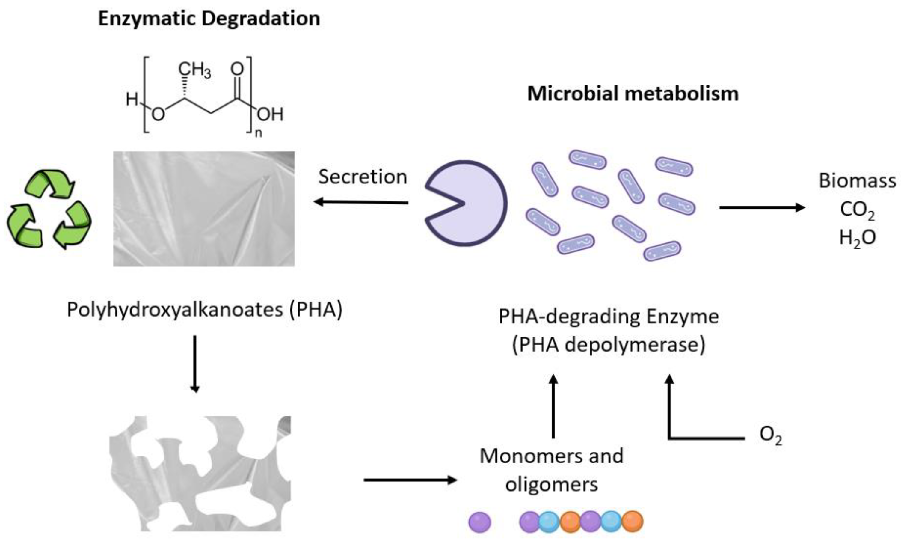
1.1.1. Biodegradability
1.1.2. Biocompatibility
1.1.3. Toxicity
1.2. Poly(3-hydroxybutyrate-co-3-hydroxyvalerate)
2. Applications
2.1. PHBV Composites for Drug Delivery Applications
2.2. PHBV Composites for Tissue Engineering Applications
3. Conclusions
4. Future Perspectives
Author Contributions
Funding
Conflicts of Interest
Abbreviations
| BC | Bacterial Cellulose |
| PHBV | Poly(3-hydroxybutyrate-co-3-hydroxyvalerate) |
| PHA | Polyhydroxyalkanoates |
| PLA | Polylactic acid |
| PCL | Polycaprolactone |
| 3HV | 3-Hydroxyvalerate |
| P(3HB) | Poly (3-Hydroxybutyrate) |
| HCC | Hepatocellular Carcinoma |
| PTX | Paclitaxel |
| PLGA | Poly (lactic-co-glycolic acid) |
| 5-FU | 5-Fluorouracil |
| SEM | Scanning Electron Microscopy |
| FTIR | Fourier Transformed Infra-Red spectroscopy |
| PS | Photosensitizer |
| PEG | Polyethylene glycol |
| SPION | Super Paramagnetic Iron Oxide Nanoparticles |
| SLS | Selective Laser Sintering |
| PBS | Phosphate-Buffered Saline |
| BSA | Bovine Serum Albumin |
| PAM | Polyacrylamide |
| BMSCs | Bone Mesenchymal Stem Cells |
| DEX | Dextran |
| nanoHA | Nanohydroxyapatite |
| SR-Nano-HA | Strontium-substituted nanoHA |
| ALP | Alkaline phosphatase |
| HA | Hydroxyapatite |
| PUL | Pullulan |
| DS | Diatom Shells |
| iPSCs | induced Pluripotent Stem Cells |
| OIM | Osteogenic Inducing Medium |
| MSCs | Mesenchymal Stem Cells |
| q-PCR | Real-Time Quantitative Polymerase Chain Reaction |
| SCI | Spinal Cord Injury |
| CPCs | Cartilage Progenitor cells |
| QUE | Quercetin |
| 1H-NMR | Proton Nuclear Magnetic Resonance |
| PDX | Polydioxanone |
| SF | Silk Fibroin |
References
- Samir, A.; Ashour, F.A.; Hakim, A.A.A.; Bassyouni, M. Recent advances in biodegradable polymers for sustainable applications. Npj Mater. Degrad. 2022, 6, 68. [Google Scholar] [CrossRef]
- Song, R.; Murphy, M.; Li, C.; Ting, K.; Soo, C.; Zheng, Z. Current development of biodegradable polymeric materials for biomedical applications. Drug Des. Devel. Ther. 2018, 12, 3117. [Google Scholar] [CrossRef] [PubMed]
- Biswas, M.C.; Jony, B.; Nandy, P.K.; Chowdhury, R.; Halder, S.; Kumar, D.; Ramakrishna, S.; Hassan, M.; Ahsan, A.; Hoque, E.; et al. Recent Advancement of Biopolymers and Their Potential Biomedical Applications. J. Polym. Environ. 2022, 30, 51–74. [Google Scholar] [CrossRef]
- Lemoigne, M. Products of dehydration and of polymerization of β-hydroxybutyric acid. Bull. Soc. Chem. Biol. 1926, 8, 770–782. [Google Scholar]
- Ansari, S.; Sami, N.; Yasin, D.; Ahmad, N.; Fatma, T. Biomedical applications of environmental friendly poly-hydroxyalkanoates. Int. J. Bio. Macromol. 2021, 183, 549–563. [Google Scholar] [CrossRef] [PubMed]
- Sehgal, R.; Gupta, R. Polyhydroxyalkanoate and its efficient production: An eco-friendly approach towards development. 3 Biotech 2020, 10, 549. [Google Scholar] [CrossRef]
- Tan, G.; Chen, C.; Li, L.; Ge, L.; Wang, L.; Razaad, I.M.N.; Li, Y.; Zhao, L.; Mo, Y.; Wang, J. Start a Research on Biopolymer Polyhydroxyalkanoate (PHA): A Review. Polymers 2014, 6, 706–754. [Google Scholar] [CrossRef]
- Amstutz, V.; Hanik, N.; Pott, J.; Utsunomia, C.; Zinn, M. Chapter Four-Tailored biosynthesis of polyhydroxyalkanoates in chemostat cultures. Meth. Enzym. 2019, 627, 99–123. [Google Scholar] [CrossRef]
- Eesaee, M.; Ghassemi, P.; Nguyen, D.D.; Thomas, S.; Elkoun, S.; Nguyen-Tri, P. Morphology and crystallization behaviour of polyhydroxyalkanoates-based blends and composites: A review. Biochem. Eng. J. 2022, 187, 108588. [Google Scholar] [CrossRef]
- Grigore, M.E.; Grigorescu, R.M.; Iancu, L.; Ion, R.M.; Zaharia, C.; Andrei, E.R. Methods of synthesis, properties and biomedical applications of polyhydroxyalkanoates: A review. J. Biomater. Sci. Polym. Ed. 2019, 30, 695. [Google Scholar] [CrossRef]
- Yañez, L.; Conejeros, R.; Vergara-Fernández, A.; Scott, F. Beyond Intracellular Accumulation of Polyhydroxyalkanoates: Chiral Hydroxyalkanoic Acids and Polymer Secretion. Front. Bioeng. Biotechnol. 2020, 8, 248. [Google Scholar] [CrossRef] [PubMed]
- Lagoa-Costa, B. Producción de polihidroxialcanoatos a partir de suero lácteo y otras fuentes de carbono usando cultivos microbianos mixtos B. Memoria presentada para optar al grado de Doctor Internacional. Ph.D. Thesis, Universidad de La Coruña, A Coruña, Spain, 2021. [Google Scholar]
- Palmeiro-Sánchez, T.; O’Flaherty, V.; Lens, P.N.L. Polyhydroxyalkanoate bio-production and its rise as bio-material of the future. J. Biotechnol. 2022, 348, 10–25. [Google Scholar] [CrossRef] [PubMed]
- Gonzalez-García, Y.; Meza-Contreras, J.C.; Gonzalez-Reynoso, O.; Córdova-López, J.A. Síntesis y biodegradación de Polihidroxialcanoa-tos: Plásticos de origen microbiano. Int. Contam. Ambie. 2013, 29, 77–115. [Google Scholar] [CrossRef]
- Numata, K.; Abe, H.; Iwata, T. Biodegradability of Poly(hydroxyalkanoate) Materials. Materials 2009, 2, 1104–1126. [Google Scholar] [CrossRef]
- Akaraonye, E.; Keshavarz, T.; Roy, I. Production of polyhydroxyalkanoates: The future green materials of choice. J. Chem. Technol. Biotechnol. 2010, 85, 732–743. [Google Scholar] [CrossRef]
- Numata, K.; Abe, H.; Doi, Y. Enzymatic processes for biodegradation of poly(hydroxyalkanoate)s crystals. Can. J. Chem. 2008, 86, 471–483. [Google Scholar] [CrossRef]
- Ang, S.L.; Sivashankari, R.; Shaharuddin, B.; Chuah, J.; Tsuge, T.; Abe, H.; Sudesh, K. Potential Applications of Polyhydroxyalkanoates as a Biomaterial for the Aging Population. Polym. Degrad. Stab. 2020, 181, 109371. [Google Scholar] [CrossRef]
- Koller, M. Biodegradable and Biocompatible Polyhydroxy-alkanoates (PHA): Auspicious Microbial Macromolecules for Pharmaceutical and Therapeutic Applications. Molecules 2018, 23, 362. [Google Scholar] [CrossRef]
- Anjum, A.; Zuber, M.; Zia, K.M.; Noreen, A.; Anjum, M.N.; Tabasum, S. Microbial production of polyhydroxyalkanoates (PHAs) and its copolymers: A review of recent advancements. Int. J. Biol. Macromol. 2016, 89, 161–174. [Google Scholar] [CrossRef]
- Papaneophytou, C.; Katsipis, G.; Halevas, E.; Pantazaki, A.A. Polyhydroxyalkanoates applications in drug carriers. In Biotechnological Applications of Polyhydroxyalkanoates; Springer: Singapore, 2019; pp. 77–124. [Google Scholar] [CrossRef]
- Rivera Briso, A.L.; Serrano Aroca, Á. Métodos de refuerzo mecánico del poli(3-hidroxibutirato-co-3-hidroxivalerato) para aplicaciones industriales avanzadas. Rev. Iberoam. Interdiscip. Métodos Model. Simul. 2018, 10, 79–94. [Google Scholar]
- Singh, S.; Mohanty, A.K. Wood fiber reinforced bacterial bioplastic composites: Fabrication and performance evaluation. Compos. Sci. Technol. 2007, 67, 1753–1763. [Google Scholar] [CrossRef]
- Silverman, T.; Naffakh, M.; Marco, C.; Ellis, G. Effect of WS2 Inorganic Nanotubes on Isothermal Crystallization Behavior and Kinetics of Poly(3-Hydroxybutyrate-co-3-hydroxyvalerate). Polymers 2018, 10, 166. [Google Scholar] [CrossRef] [PubMed]
- Yu, H.; Qin, Z.; Zhou, Z. Cellulose nanocrystals as green fillers to improve crystallization and hydrophilic property of poly(3-hydroxybutyrate-co-3-hydroxyvalerate). Prog. Nat. Sci. Mater. 2011, 21, 478. [Google Scholar] [CrossRef]
- Montanheiro, T.L.; Cristóvan, F.H.; Machado, J.P.; Tada, D.; Durán, N.; Lemes, A.P. Effect of MWCNT functionalization on thermal and electrical properties of PHBV/MWCNT nanocomposites. J. Mater. Res. 2015, 30, 55–65. [Google Scholar] [CrossRef]
- Vidhate, S.; Innocentini-Mei, L.; D’Souza, N.A. Mechanical and electrical multifunctional poly(3-hydroxybutyrate-co-3-hydroxyvalerate)-multiwall carbon nanotube nanocomposites. Polym. Eng. Sci. 2012, 52, 1367–1374. [Google Scholar] [CrossRef]
- Râpă, M.; Stefan, L.M.; Seciu-Grama, A.; Gaspar-Pintiliescu, A.; Matei, E.; Zaharia, C.; Stănescu, P.O.; Predescu, C. Poly(3-hydroxybutyrate-co-3-hydroxyvalerate) (P(3HB-co-3HV))/Bacterial Cellulose (BC) Biocomposites for Potential Use in Biomedical Applications. Polymers 2022, 14, 5544. [Google Scholar] [CrossRef] [PubMed]
- Gómez-Gaete, C. Nanopartículas poliméricas: Tecnología y aplicaciones farmacéuticas (Polymeric nanoparticles: Technologie and pharmaceutical applications). Rev. Farmacol. Chile 2014, 7, 7–16. [Google Scholar]
- Göz, E.; Karakeçili, A. Effect of emulsification-diffusion parameters on the formation of poly (3-hydroxybutyrate-co-3-hydroxyvalerate) particles. Artif. Cells Nanomed. Biotechnol. 2016, 44, 226–234. [Google Scholar] [CrossRef]
- Leimann, F.V.; Filho, L.C.; Sayer, C.; Araújo, P.H. Poly(3-hydroxybutyrate-co-3- hydroxyvalerate) nanoparticles prepared by a miniemulsion/solvent evaporation technique: Effect of phbv molar mass and concentration. Braz. J. Chem. Eng. 2013, 30, 369–377. [Google Scholar] [CrossRef]
- Farrag, Y.; Montero, B.; Rico, M.; Barral-Losada, L.F.; Bouza, R. Preparation and characterization of nano and micro particles of poly(3-hydroxybutyrate-co-3-hydroxyvalerate) (PHBV) via emulsification/solvent evaporation and nanoprecipitation techniques. J. Nanopart. Res. 2018, 20, 1–17. [Google Scholar] [CrossRef]
- Wu, M.; Zhong, C.; Zhang, Q.; Wang, L.; Wang, L.; Liu, Y.; Zhang, X.; Zhao, X. pH-responsive delivery vehicle based on RGD-modified polydopamine-paclitaxel-loaded poly (3-hydroxybutyrate-co-3-hydroxyvalerate) nanoparticles for targeted therapy in hepatocellular carcinoma. J. Nanobiotechnol. 2021, 19, 39. [Google Scholar] [CrossRef] [PubMed]
- Handali, S.; Moghimipour, E.; Rezaei, M.; Ramezani, Z.; Dorkoosh, F.A. PHBV/PLGA nanoparticles for enhanced delivery of 5-fluorouracil as promising treatment of colon cancer. Pharm. Dev. Technol. 2019, 25, 206. [Google Scholar] [CrossRef] [PubMed]
- Handali, S.; Moghimipour, E.; Rezaei, M.; Saremy, S.; Dorkoosh, F.A. Co-delivery of 5-fluorouracil and oxaliplatin in novel poly (3-hydroxybutyrate-co-3-hydroxyvalerate acid)/poly(lactic-co-glycolic acid) nanoparticles for colon cancer therapy. Int. J. Biol. Macromol. 2019, 124, 1299–1311. [Google Scholar] [CrossRef] [PubMed]
- Radu, I.C.; Hudita, A.; Zaharia, C.; Galateanu, B.; Iovu, H.; Tanasa, E.V.; Nitu, S.G.; Ginghina, O.; Negrei, C.; Tsatsakis, A.; et al. Poly(3-hydroxybutyrate-CO-3-hydroxyvalerate) PHBHV biocompatible nanocarriers for 5-FU delivery targeting colorectal cancer. Drug Deliv. 2019, 26, 318. [Google Scholar] [CrossRef] [PubMed]
- Radu, I.C.; Hudita, A.; Zaharia, C.; Stanescu, P.O.; Vasile, E.; Iovu, H.; Stan, M.; Ginghina, O.; Galateanu, B.; Costache, M.; et al. Poly (HydroxyButyrate-co-HydroxyValerate) (PHBHV) Nanocarriers for Silymarin Release as Adjuvant Therapy in Colo-rectal Cancer. Front. Pharmacol. 2017, 8, 508. [Google Scholar] [CrossRef] [PubMed]
- Vardhan, H.; Mittal, P.; Adena, S.K.; Upadhyay, M.; Yadav, S.K.; Mishra, B. Process optimization and in vivo performance of docetaxel loaded PHBV-TPGS therapeutic vesicles: A synergistic approach. Int. J. Biol. Macromol. 2018, 108, 729–743. [Google Scholar] [CrossRef]
- Vardhan, H.; Mittal, P.; Adena, S.K.; Upadhyay, M.; Mishra, B. Development of long-circulating docetaxel loaded poly (3-hydroxybutyrate-co-3-hydroxyvalerate) nanoparticles: Optimization, pharmacokinetic, cytotoxicity and in vivo assessments. Int. J. Biol. Macromol. 2017, 103, 791–801. [Google Scholar] [CrossRef]
- Peñaloza, J.; Márquez-Miranda, V.; Cabaña-Brunod, M.; Reyes-Ramírez, R.; Llancalahuen, F.M.; Vilos, C.; Maldonado-Biermann, F.; Velásquez, L.A.; Fuentes, J.A.; González-Nilo, F.D.; et al. Intracellular trafficking and cellular uptake mechanism of PHBV nanoparticles for targeted delivery in epithelial cell lines. J. Nanobiotechnol. 2017, 15, 1. [Google Scholar] [CrossRef]
- Vilos, C.; Morales, F.A.; Solar, P.A.; Herrera, N.S.; Gonzalez-Nilo, F.D.; Aguayo, D.A.; Mendoza, H.L.; Comer, J.; Bravo, M.L.; Gonzalez, P.A.; et al. Paclitaxel-PHBV nanoparticles and their toxicity to endometrial and primary ovarian cancer cells. Biomaterials 2013, 34, 4098–4108. [Google Scholar] [CrossRef]
- Masood, F.; Chen, P.; Yasin, T.; Fatima, N.; Hasan, F.; Hameed, A. Encapsulation of Ellipticine in poly-(3-hydroxybutyrate-co-3-hydroxyvalerate) based nanoparticles and its in vitro application. Mater. Sci. Eng. C 2013, 33, 1054–1060. [Google Scholar] [CrossRef]
- Masood, F.; Chen, P.; Yasin, T.; Hasan, F.; Ahmad, B.; Hameed, A. Synthesis of poly-(3-hydroxybutyrate-co-12 mol % 3-hydroxyvalerate) by Bacillus cereus FB11: Its characterization and application as a drug carrier. J. Mater. Sci. Mater. Med. 2013, 24, 1927–1937. [Google Scholar] [CrossRef] [PubMed]
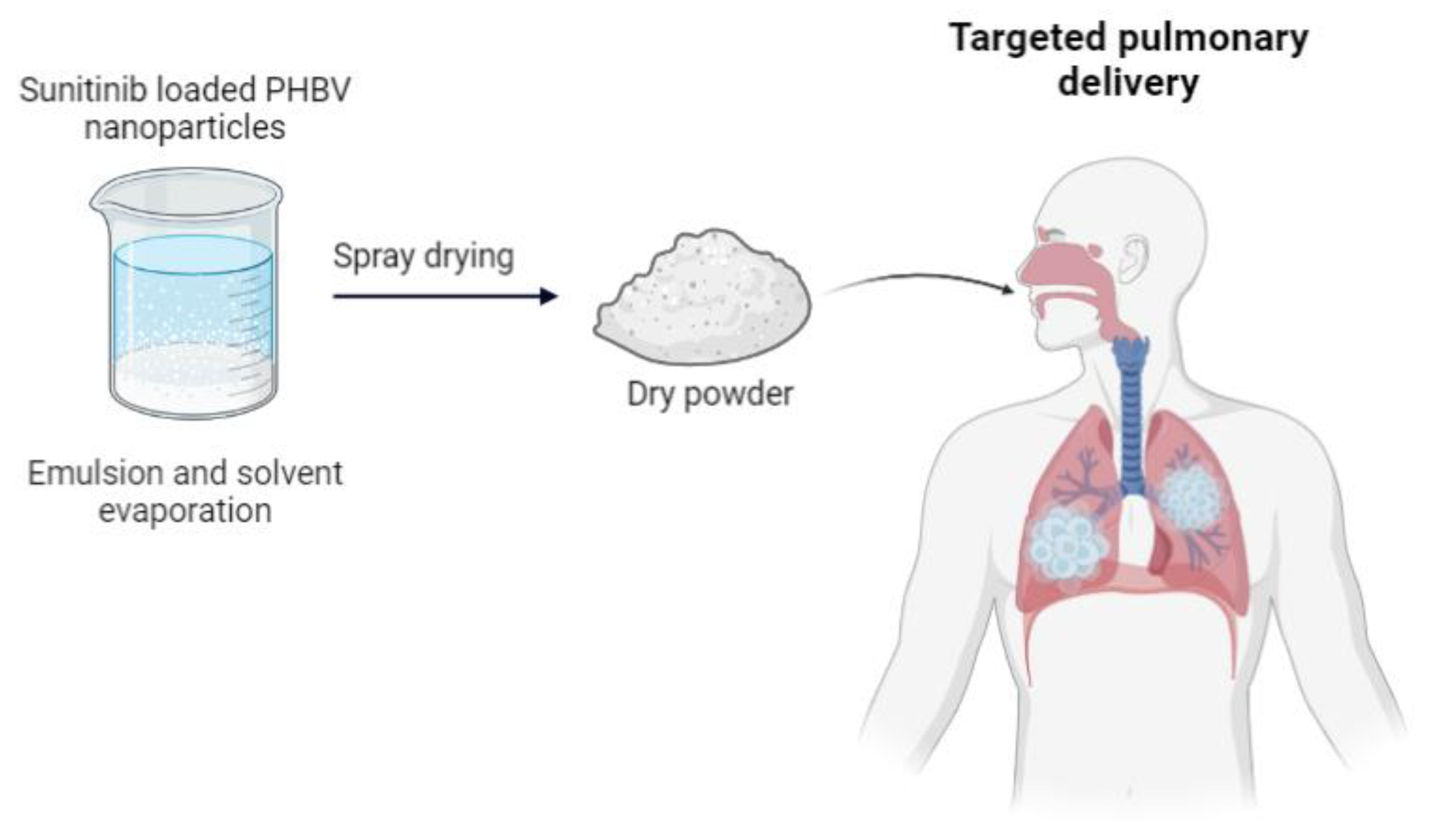
- Otroj, M.; Taymouri, S.; Varshosaz, J.; Mirian, M. Preparation and characterization of dry powder containing sunitinib loaded PHBV nanoparticles for enhanced pulmonary delivery. J. Drug Deliv. Sci. Technol. 2020, 56, 101570. [Google Scholar] [CrossRef]
- Alp, E.; Cirak, T.; Demirbilek, M.; Türk, M.; Güven, E. Targeted delivery of etoposide to osteosarcoma cells using poly (3-hydroxybutyrate-co-3-hydroxyvalerate) (PHBV) nanoparticles. Turk. J. Biol. 2017, 41, 719–733. [Google Scholar] [CrossRef]
- Pramual, S.; Lirdprapamongkol, K.; Svasti, J.; Bergkvist, M.; Jouan-Hureaux, V.; Arnoux, P.; Frochot, C.; Barberi-Heyob, M.; Niamsiri, N. Polymer-lipid-PEG hybrid nanoparticles as photosensitizer carrier for photodynamic therapy. J. Photochem. Photobiol. 2017, 173, 12–22. [Google Scholar] [CrossRef]
- Solar, P.; González, G.; Vilos, C.; Herrera, N.; Juica, N.; Moreno, M.; Simon, F.; Velásquez, L. Multifunctional polymeric nanoparticles doubly loaded with SPION and ceftiofur retain their physical and biological properties. J. Nanobiotechnol. 2015, 13, 14. [Google Scholar] [CrossRef] [PubMed]
- Vilos, C.; Gutierrez, M.; Escobar, J.; Morales, F.; Denardin, J.C.; Velásquez, L.; Altbir, D. Superparamagnetic Poly (3-hydroxybutyrate-co-3 hydroxyvalerate) (PHBV) nanoparticles for biomedical applications. Electron. J. Biotechnol. 2013, 16, 8. [Google Scholar] [CrossRef]
- Bahari-Javan, N.; Rezaie-Shirmard, L.; Jafary-Omid, N.; Akbari-Javar, H.; Rafiee-Tehrani, M.; Abedin-Dorkoosh, F. Preparation, statistical optimisation, and in vitro characterisation of poly (3-hydroxybutyrate-co-3-hydroxyvalerate)/poly (lactic-co-glycolic acid) blend nanoparticles for prolonged delivery of teriparatide. J. Microencapsul. 2016, 33, 460–474. [Google Scholar] [CrossRef]
- Bahari-Javan, N.; Montazeri, H.; Rezaie-Shirmard, L.; Jafary-Omid, N.; Barbari, G.; Amini, M.; Ghahremani, M.; Rafiee-Tehrani, M.; Abedin-Dorkoosh, F. Preparation, characterization and in vivo evaluation of a combination delivery system based on hyaluronic acid/jeffamine hydrogel loaded with PHBV/PLGA blend nanoparticles for prolonged delivery of Teriparatide. Eur. J. Pharm. Sci. 2017, 101, 167–181. [Google Scholar] [CrossRef]
- Perveen, K.; Masood, F.; Hameed, A. Preparation, characterization and evaluation of antibacterial properties of epirubicin loaded PHB and PHBV nanoparticles. Int. J. Biol. Macromol. 2020, 144, 259–266. [Google Scholar] [CrossRef]
- Masood, F.; Muhammad, S.; Bokhari, H.; Yasin, T.; Hameed, A. Characterisation and evaluation of antibacterial potential of guava extract loaded poly-3-hydroxybutyrate-co-3-hydroxyvalerate nanoparticles against multidrug-resistant bacteria. Micro Nano Syst. Lett. 2017, 12, 352. [Google Scholar] [CrossRef]
- Lightfoot-Vidal, S.; Rojas, C.; Bouza-Padín, R.; Pérez-Rivera, M.; Haensgen, A.; González, M.; Rodríguez-Llamazares, S. Synthesis and characterization of polyhydroxybutyrate-co-hydroxyvalerate nanoparticles for encapsulation of quercetin. J. Bioact. Compat. Polym. 2016, 31, 439–452. [Google Scholar] [CrossRef]
- Rezaie-Shirmard, L.; Bahari-Javan, N.; Khoshayand, M.; Kebriaeezadeh, A.; Dinarvand, R.; Dorkoosh, F. Nanoparticulate fingolimod delivery system based on biodegradable poly (3-hydroxybutyrate-co-3-hydroxyvalerate) (PHBV): Design, optimization, characterization and in-vitro evaluation. Pharm. Dev. Technol. 2015, 22, 860. [Google Scholar] [CrossRef] [PubMed]
- Bayrami, S.; Esmaili, Z.; Seyedalinaghi, S.; Moghadam, S.J.; Bayrami, S.; Akbari-Javar, H.; Rafiee Tehrani, M.; Dorkoosh, F. Fabrication of long-acting insulin formulation based on poly (3-hydroxybutyrate-co-3-hydroxyvalerate) (PHBV) nanoparticles: Preparation, optimization, characterization, and in vitro evaluation. Pharm. Dev. Technol. 2018, 24, 176. [Google Scholar] [CrossRef] [PubMed]
- Álvarez-Álvarez, L.; Barral, L.; Bouza, R.; Farrag, Y.; Otero-Espinar, F.; Feijóo-Bandín, S.; Lago, F. Hydrocortisone loaded poly-(3-hydroxybutyrate-co-3-hydroxyvalerate) nanoparticles for topical ophthalmic administration: Preparation, characterization and evaluation of ophthalmic toxicity. Int. J. Pharm. 2019, 568, 118519. [Google Scholar] [CrossRef]
- Bahari-Javan, N.; Jafary-Omid, N.; Moosavi-Hasab, N.; Rezaie-Shirmard, L.; Rafiee-Tehrani, M.; Dorkoosh, F. Preparation, statistical optimization and in vitro evaluation of pramipexole prolonged delivery system based on poly (3-hydroxybutyrate-co-3-hydroxyvalerate) nanoparticles. J. Drug Deliv. Sci. Technol. 2018, 44, 82–90. [Google Scholar] [CrossRef]
- Eke, G.; Goñi-de-Cerio, F.; Suarez-Merino, B.; Hasirci, N.; Hasirci, V.N. Biocompatibility of Dead Sea Water and retinyl palmitate carrying poly(3-hydroxybutyrate-co-3-hydroxyvalerate) micro/nanoparticles designed for transdermal skin therapy. J. Bioact. Compat. Polym. 2015, 30, 455–471. [Google Scholar] [CrossRef]
- Eke, G.; Anna, M.; Anastasia, V.; Ekaterina, I.; Hasirci, N.; Hasirci, V. Fate of Poly-3-Hydroxybutyrate-co3-Hydroxyvalerate on Skin. J. Sib. Fed. Univ. Biol. 2012, 5, 404–416. [Google Scholar] [CrossRef]
- Koller, M. Advances in polyhydroxyalkanoate (PHA) production. Bioengineering 2017, 4, 88. [Google Scholar] [CrossRef]
- Yilgor, P.; Hasirci, N.; Hasirci, V. Sequential BMP-2/BMP-7 delivery from polyester nanocapsules. J. Biomed. Mater. Res. A 2010, 93A, 528–536. [Google Scholar] [CrossRef]
- Elmowafy, E.; Abdal-Hay, A.; Skouras, A.; Tiboni, M.; Casettari, L.; Guarino, V. Polyhydroxyalkanoate (PHA): Applications in drug delivery and tissue engineering. Expert. Rev. Med. Devices 2019, 16, 467–482. [Google Scholar] [CrossRef]
- Rivera-Briso, A.L.; Serrano-Aroca, Á. Poly (3-Hydroxybutyrate-co-3-Hydroxyvalerate): Enhancement strategies for advanced applications. Polymers 2018, 10, 732. [Google Scholar] [CrossRef] [PubMed]
- Diermann, S.H.; Lu, M.; Zhao, Y.; Vandi, L.; Dargusch, M.; Huang, H. Synthesis, microstructure, and mechanical behaviour of a unique porous PHBV scaffold manufactured using selective laser sintering. J. Mech. Behav. Biomed. Mater. 2018, 84, 151–160. [Google Scholar] [CrossRef] [PubMed]
- Diermann, S.H.; Lu, M.; Edwards, G.; Dargusch, M.; Huang, H. In vitro degradation of a unique porous PHBV scaffold manufactured using selective laser sintering. J. Biomed. Mater. Res. 2019, 107, 154–162. [Google Scholar] [CrossRef]
- Duan, B.; Wang, M.; Zhou, W.Y.; Cheung, W.L.; Li, Z.Y.; Lu, W.W. Three-dimensional nanocomposite scaffolds fabricated via selective laser sintering for bone tissue engineering. Acta Biomater. 2010, 6, 4495–4505. [Google Scholar] [CrossRef]
- Duan, B.; Wang, M. Encapsulation and release of biomolecules from Ca–P/PHBV nanocomposite microspheres and three-dimensional scaffolds fabricated by selective laser sintering. Polym. Degrad. Stab. 2010, 95, 1655–1664. [Google Scholar] [CrossRef]
- Ke, Y.; Wang, Y.J.; Ren, L.; Zhao, Q.C.; Huang, W. Modified PHBV scaffolds by in situ UV polymerization: Structural characteristic, mechanical properties and bone mesenchymal stem cell compatibility. Acta Biomater. 2010, 6, 1329–1336. [Google Scholar] [CrossRef]
- Zou, P.; Liu, H.; Li, Y.; Huang, J.; Dai, Y. Surface dextran modified electrospun poly (3-hydroxybutyrate-co-3-hydroxyvalerate) (PHBV) fibrous scaffold promotes the proliferation of bone marrow-derived mesenchymal stem cells. Mater. Lett. 2016, 179, 109–113. [Google Scholar] [CrossRef]
- Kontogianni, G.; Bonatti, A.F.; De Maria, C.; Naseem, R.; Melo, P.; Coelho, C.; Vozzi, G.; Dalgarno, K.; Quadros, P.; Vitale-Brovarone, C.; et al. Promotion of In Vitro Osteogenic Activity by Melt Extrusion-Based PLLA/PCL/PHBV Scaffolds Enriched with Nano-Hydroxyapatite and Strontium Substituted Nano-Hydroxyapatite. Polymers 2023, 15, 1052. [Google Scholar] [CrossRef]
- Dhania, S.; Rani, R.; Kumar, R.; Thakur, R. Fabricated polyhydroxyalkanoates blend scaffolds enhance cell viability and cell proliferation. J. Biotechnol. 2023, 361, 30–40. [Google Scholar] [CrossRef]
- Pecorini, G.; Braccini, S.; Parrini, G.; Chiellini, F.; Puppi, D. Additive Manufacturing of Poly(3-hydroxybutyrate-co-3-hydroxyvalerate)/Poly(D,L-lactide-co-glycolide) Biphasic Scaffolds for Bone Tissue Regeneration. Int. J. Mol. Sci. 2022, 23, 3895. [Google Scholar] [CrossRef]
- Nahanmoghadam, A.; Asemani, M.; Goodarzi, V.; Ebrahimi-Barough, S. Design and fabrication of bone tissue scaffolds based on PCL/PHBV containing hydroxyapatite nanoparticles: Dual-leaching technique. J. Biomed. Mater. Res. 2021, 109, 981–993. [Google Scholar] [CrossRef] [PubMed]
- Karbowniczek, J.E.; Kaniuk, Ł.; Berniak, K.; Gruszczyński, A.; Stachewicz, U. Enhanced Cells Anchoring to Electrospun Hybrid Scaffolds with PHBV and HA Particles for Bone Tissue Regeneration. Front. Bioeng. Biotechnol. 2021, 9, 632029. [Google Scholar] [CrossRef] [PubMed]
- Kara, A.; Gunes, O.C.; Albayrak, A.Z.; Bilici, G.; Erbil, G.; Havitcioglu, H. Fish scale/poly(3-hydroxybutyrate-co-3-hydroxyvalerate) nanofibrous composite scaffolds for bone regeneration. J. Biomater. Appl. 2020, 34, 1201–1215. [Google Scholar] [CrossRef] [PubMed]
- Dalgic, A.D.; Atila, D.; Karatas, A.; Tezcaner, A.; Keskin, D. Diatom shell incorporated PHBV/PCL-pullulan co-electrospun scaffold for bone tissue engineering. Mater. Sci. Eng. C Mater. Biol. Appl. 2019, 100, 735–746. [Google Scholar] [CrossRef]
- Hosseini, F.S.; Soleimanifar, F.; Aidun, A.; Enderami, S.E.; Saburi, E.; Marzouni, H.Z.; Khani, M.M.; Khojasteh, A.; Ardeshirylajimi, A. Poly (3-hydroxybutyrate-co-3-hydroxyvalerate) improved osteogenic differentiation of the human induced pluripotent stem cells while considered as an artificial extracellular matrix. J. Cell. Physiol. 2018, 234, 11537. [Google Scholar] [CrossRef]
- Lyu, L.X.; Zhang, X.F.; Deegan, A.J.; Liang, G.F.; Yang, H.N.; Hu, S.Q.; Yan, X.L.; Huang, N.P.; Xu, T. Comparing hydroxyapatite with osteogenic medium for the osteogenic differentiation of mesenchymal stem cells on PHBV nanofibrous scaffolds. J. Biomater. Sci. Polym. Ed. 2018, 30, 150. [Google Scholar] [CrossRef]
- Gheibi, A.; Khoshnevisan, K.; Ketabchi, N.; Derakhshan, M.A.; Babadi, A. Application of Electrospun Nanofibrous PHBV Scaffold in Neural Graft and Regeneration: A Mini-Review. J. Nanomed. Res. 2016, 1, 107–111. [Google Scholar] [CrossRef]
- Biazar, E.; Heidari-Keshel, S. Development of chitosan-crosslinked nanofibrous PHBV guide for repair of nerve defects. Artif. Cells Nanomed. Biotechnol. 2014, 42, 385–391. [Google Scholar] [CrossRef]
- Zhao, T.; Jing, Y.; Zhou, X.; Wang, J.; Huang, X.; Gao, L.; Zhu, Y.; Wang, L.; Gou, Z.; Liang, C.; et al. PHBV/PLA/Col-Based Nanofibrous Scaffolds Promote Recovery of Locomotor Function by Decreasing Reactive Astrogliosis in a Hemisection Spinal Cord Injury Rat Model. J. Biomed. Nanotechnol. 2018, 14, 1921–1933. [Google Scholar] [CrossRef]
- Zhao, T.; Xu, K.; Wu, Q.; Wang, C.; Xiao, S.; Li, H.; He, T.; Wang, L.; Li, F.; Chen, Q. Duraplasty of PHBV/PLA/Col membranes promotes axonal regeneration by inhibiting NLRP3 complex and M1 macrophage polarization in rats with spinal cord injury. FASEB J. 2020, 34, 12147. [Google Scholar] [CrossRef]
- Xing, Z.C.; Chae, W.P.; Baek, J.Y.; Choi, M.J.; Jung, Y.; Kang, I.K. In Vitro Assessment of Antibacterial Activity and Cytocompatibility of Silver-Containing PHBV Nanofibrous Scaffolds for Tissue Engineering. Biomacromolecules 2010, 11, 1248–1253. [Google Scholar] [CrossRef] [PubMed]
- Khamplod, T.; Winterburn, J.B.; Cartmell, S.H. Electrospun poly(3-hydroxybutyrate-co-3-hydroxyvalerate) scaffolds–a step towards ligament repair applications. Sci. Technol. Adv. Mater. 2022, 23, 895. [Google Scholar] [CrossRef] [PubMed]
- Meng, W.; Kim, S.Y.; Yuan, J.; Kim, J.C.; Kwon, O.H.; Kawazoe, N.; Chen, G.; Ito, Y.; Kang, I.K. Electrospun PHBV/collagen composite nanofibrous scaffolds for tissue engineering. J. Biomater. Sci. Polym. Ed. 2007, 18, 81–94. [Google Scholar] [CrossRef] [PubMed]
- Xue, K.; Zhang, S.; Ge, J.; Wang, Q.; Qi, L.; Liu, K. Integration of Bioglass Into PHBV-Constructed Tissue-Engineered Cartilages to Improve Chondrogenic Properties of Cartilage Progenitor Cells. Front. Bioeng. Biotechnol. 2022, 10, 868719. [Google Scholar] [CrossRef] [PubMed]
- Chen, W.; Li, Y.; Huang, Y.; Dai, Y.; Xi, T.; Zhou, Z.; Liu, H. Quercetin modified electrospun PHBV fibrous scaffold enhances cartilage regeneration. J. Mater. Sci. Mater. Med. 2021, 32, 92. [Google Scholar] [CrossRef] [PubMed]
- Xue, K.; Zhang, X.; Gao, Z.; Xia, W.; Qi, L.; Liu, K. Cartilage progenitor cells combined with PHBV in cartilage tissue engineering. J. Transl. Med. 2019, 17, 104. [Google Scholar] [CrossRef] [PubMed]
- Jacob, J.; More, N.; Mounika, C.; Gondaliya, P.; Kalia, K.; Kapusetti, G. Smart Piezoelectric Nanohybrid of Poly(3-hydroxybutyrate-co-3-hydroxyvalerate) and Barium Titanate for Stimulated Cartilage Regeneration. ACS Appl. Bio Mater. 2019, 2, 4922–4931. [Google Scholar] [CrossRef]
- Dalgic, A.D.; Koman, E.; Karatas, A.; Tezcaner, A.; Keskin, D. Natural origin bilayer pullulan-PHBV scaffold for wound healing applications. Biomater. Adv. 2022, 134, 112554. [Google Scholar] [CrossRef]
- El-Newehy, M.H.; Kim, H.Y.; Khattab, T.A.; Abdulhameed, M.M.; El-Naggar, M.E. Fabrication, microstructure characterization, and degradation performance of electrospun mats based on poly (3-hydroxybutyrate-co-3 hydroxyvalerate)/polyethylene glycol blend for potential tissue engineering. Luminescence 2022, 37, 323–331. [Google Scholar] [CrossRef]
- Goonoo, N.; Gimié, F.; Ait-Arsa, I.; Cordonin, C.; Andries, J.; Jhurry, D.; Bhaw-Luximon, A. Piezoelectric core-shell PHBV/PDX blend scaffolds for reduced superficial wound contraction and scarless tissue regeneration. Biomater. Sci. 2021, 9, 5259–5274. [Google Scholar] [CrossRef]
- Gong, W.; Cheng, T.; Liu, Q.; Xiao, Q.; Li, J. Surgical repair of abdominal wall defect with biomimetic nano/microfibrous hybrid scaffold. Mater. Sci. Eng. C 2018, 93, 828–837. [Google Scholar] [CrossRef] [PubMed]


| Nanoparticle | Method of Fabrication | Applications | Encapsulated Drug | Studies Performed | Ref. |
|---|---|---|---|---|---|
| PHBV | Emulsion and solvent evaporation method | Liver cancer, Hepatocellular carcinoma | Paclitaxel | Size and morphology Entrapment and drug loading efficient Fourier-transform infra-red spectroscopy X-ray diffraction Differential scanning calorimetry Thermogravimetry analysis Hemolysis experiment In vitro paclitaxel release Cell culture and cellular viability Cell uptake Xenograft tumor model In vivo antitumor efficacy In vivo biodistribution study Statistical analysis | [33] |
| PHBV/PLGA | Double-emulsion method | Colon cancer | 5-Fluorouracil | Experimental design Determination of encapsulation efficiency Particle size and morphological studies Differential scanning calorimetry Thermogravimetry analysis Fourier-transform infra-red spectroscopy In vitro release study In vitro cellular uptake of nanoparticles Cytotoxicity assay Hemolysis assay In vivo anti-tumor activity Statistical analysis | [34] |
| PHBV/PLGA | Double-emulsion method | Colon cancer | 5-Fluorouracil and oxaliplatin | Determination of encapsulation efficiency Particle size and morphological studies Differential scanning calorimetry In vitro release study In vitro cellular uptake of nanoparticles Cell viability Reactive oxygen species detection Apoptosis study Hemolysis analysis In vivo anti-tumor activity Statistical analysis | [35] |
| PHBV | Emulsion-diffusion method | Colon cancer | 5-Fluorouracil | Determination of encapsulation efficiency Particle size and morphological studies In vitro release study Cytotoxicity analysis | [36] |
| PHBV | Nanoprecipitation | Colon cancer | Silymarin | Silymarin drug release Characterization of the nanocarriers by scanning electronic microscopy and atomic force microscopy In vitro cytotoxicity assessment of the Nanocarriers Statistical analysis | [37] |
| PHBV | Modified emulsion and solvent evaporation method | Breast cancer | Docetaxel | Colloidal morphology Particle size and surface charge Entrapment efficiency Analysis of drug by high-performance liquid chromatography In vitro drug release In vivo-in silico and in vitro–in vivo correlation studies Cytotoxicity Cell uptake Anticancer activity Drug–plasma interaction and hemolysis studies | [38] |
| PHBV | Emulsion and solvent evaporation method | Breast cancer | Docetaxel | Box–Behnken optimization studies Morphological studies Particle size and surface charge Entrapment efficiency In vitro drug release Pharmacokinetics study In vitro and in vivo correlation and in silico simulation Cell-line studies In vivo anticancer activity | [39] |
| PHBV | Modified double-emulsion and solvent evaporation method | Epithelial cell lines: -Ovarian cancer cells (SKOV-3) -Cervical cancer cells (HeLa) | NA | Dynamic light scattering Transmission electron microscopy Cell culture Flow cytometry Immunofluorescence Reducible biotin assay Western blot MTT assay | [40] |
| PHBV | Double-emulsion and solvent evaporation method | -Ovarian cancer cells -Endometrial cancer cells | Paclitaxel | Characterization of the nanoparticles Low-voltage electron microscopy Drug encapsulation efficiency Efficiency and release kinetics Ultra-performance liquid chromatography Fourier-transform infra-red spectroscopy All-atom molecular dynamics method Coarse-grain molecular dynamics methods Confocal laser scanning microscopy MTT assay and flow cytometry analysis | [41] |
| PHBV | Emulsion and solvent evaporation method | Lung carcinoma epithelial cells A549 | Ellipticine | Particle size and zeta potential Scanning electron microscopy Drug encapsulation efficiency In vitro experiment Statistical analysis | [42] |
| PHBV | Oil-in-water emulsion technique | Lung carcinoma epithelial cells A549 | Ellipticine | Characterization of the nanoparticles Drug loading efficiency In vitro cytotoxicity Statistical analysis | [43] |
| PHBV | Emulsion and solvent evaporation | Lung carcinoma epithelial cells A549 | Sunitinib | Particle size, polydispersity index, and zeta potential Encapsulation efficiency and drug loading Drug-release study Study kinetics and mechanism of drug release Fourier-transform infra-red spectroscopy In vitro cellular uptake study In vitro cytotoxicity Scanning electron microscopy | [44] |
| Folic acid-functionalized PHB | Emulsion and solvent evaporation method | Osteosarcoma | Etoposide | Characterization of PHBV nanoparticles Determination of entrapment efficiency In vitro etoposide release studies Cytotoxicity assay Apoptosis/necrosis assay | [45] |
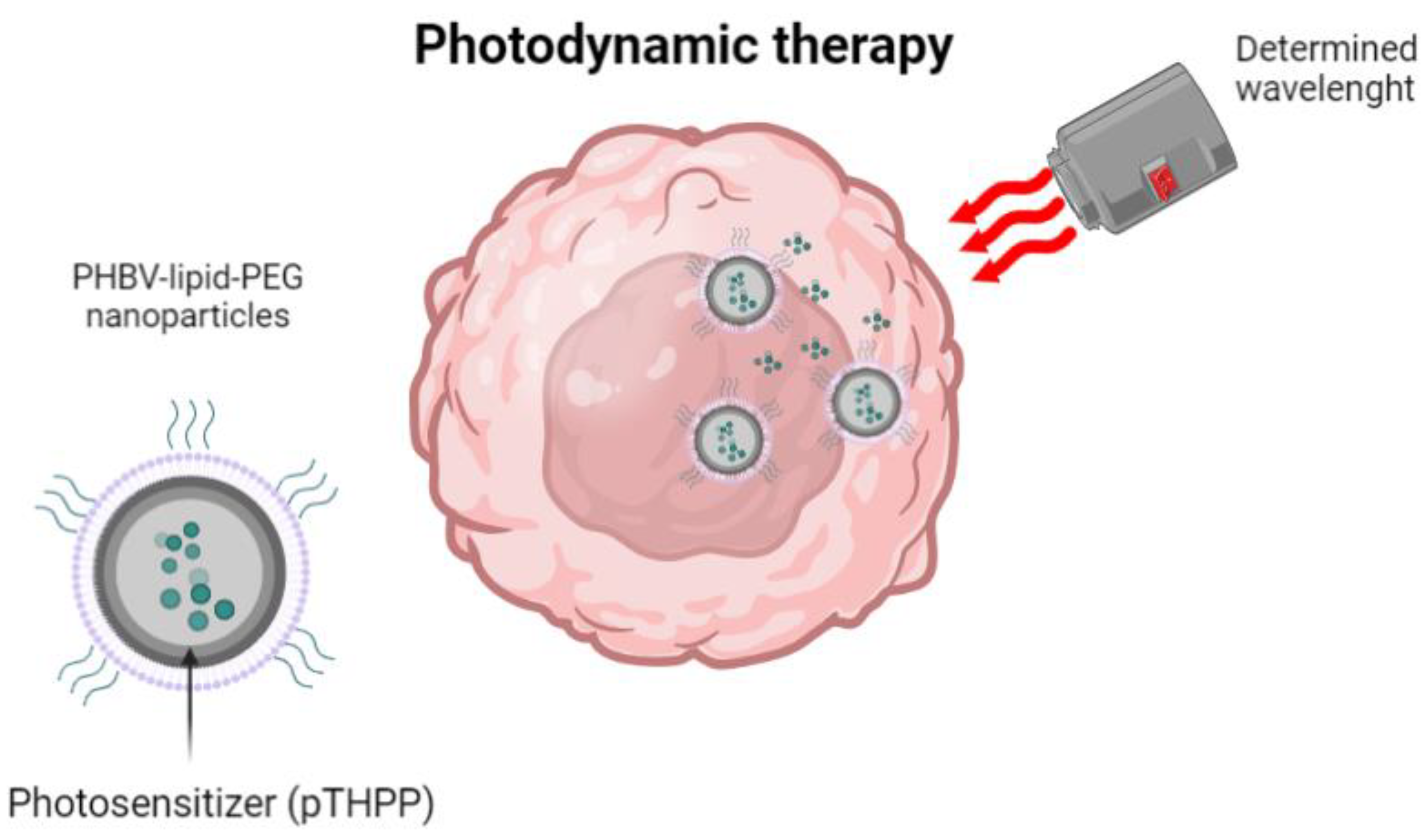
| PHBV-lipid-PEG | Nanoprecipitation technique combined with self-assembly | Photodynamic therapy | 5,10,15,20-Tetrakis (4-hydroxy-phenyl)-21H,23H-porphine | Particle size analysis and zeta potential Measurement Determination of drug loading and entrapment efficiency X-ray Diffraction analysis Photophysical properties Cell culture experiments Intracellular uptake Photodynamic therapy studies Statistical analysis | [46] |
| Super-paramagnetic iron oxide functionalized PHBV | Double-emulsion and solvent evaporation method | Contrast medium Hyperthermia applications Tissue-specific drug delivery | Ceftiofur | Transmission electron microscopy Dynamic light scattering Ultraviolet-visible spectrophotometry Fourier-transform infra-red spectroscopy Differential scanning calorimetry Ceftiofur entrapment efficiency Cell cultures Antibacterial activity Vibrating sample magnetometer Cytotoxicity assays for polymeric nanoparticles Statistical analysis | [47] |
| PHBV | Double-emulsion and solvent evaporation method | Contrast medium for magnetic resonance imaging agent for hyperthermia Nanocarriers for targeted drug delivery | Super-paramagnetic iron oxide | X-ray diffraction of magnetite Analysis by electronic transmission microscopy Size and zeta potential of magnetite-loaded nanoparticles Magnetic measurements Magnetic accumulation of the loaded nanoparticles in intestinal tissue | [48] |
| PHBV/PLGA | Double-emulsion and solvent evaporation method | Osteoporosis | Teriparatide | Experimental design Particle size and size distribution Morphological studies High-performance liquid chromatography analysis Entrapment and drug-loading efficiency Fourier-transform infra-red spectroscopy X-ray powder diffraction Differential scanning calorimetry Intrinsic fluorescence spectroscopy In vitro teriparatide release Teriparatide stability in release medium Mathematical modeling | [49] |
| PHBV/PLGA loaded in hyaluronic acid/jeffamine hydrogel | Crosslinking reaction | Osteoporosis | Teriparatide | Characterization of the hydrogel Degree of swelling ratio Measurement of crosslinking efficiency Rheological characterizations Morphological characterizations of Teriparatide loaded delivery system In vitro teriparatide release studies Mathematical modeling of release data Stability of teriparatide Cytotoxicity studies In vivo studies | [50] |
| PHBV | Nanoprecipitation | Antibacterial properties | Epirubicin | Scanning electron microscope Energy dispersive X-ray spectroscopy Particle size analysis X-ray diffraction analysis Fourier-transform infra-red spectroscopy Drug-loading and encapsulation efficiency In vitro release studies Antibacterial studies | [51] |
| PHBV | Nanoprecipitation | Antibacterial properties | Different plant leaves | Characterization of guava extract: Thin layer chromatography, Fourier -transform infra-red spectroscopy, wide-angle X-ray diffraction analysis Characterization of nanoparticles Scanning electron microscopy Antibacterial studies Statistical analysis | [52] |
| PHBV | Double-emulsion method with high-speed homogenization | Antimicrobial, antimicrobial, anti-inflammatory, and antiviral properties | Quercetin | Dynamic light scattering Scanning electron microscopy Transmission electron microscopy X-ray diffraction Differential scanning calorimetry Thermogravimetry analysis The release profile of quercetin-loaded nanoparticles Cell culture and cytotoxicity assay In vitro release studies Analysis of insulin release kinetic | [53] |
| PHBV | Single- and double-emulsification and solvent evaporation technique | Immuno-suppression | Fingolimod | Experimental design study Characterization of fingolimod nanoparticles Size measurement, determination of loading efficacy, and capacity of the nanoparticles Determination of the morphology of the nanoparticles Stability study In vitro drug release study Evaluating release kinetics and the mechanism using a mathematical model | [54] |
| PHBV | Double-emulsification and solvent evaporation method | Diabetes | Insulin | Experimental design study Particle size, polydispersity index, and zeta potential measurement Morphological studies Determination of insulin encapsulation efficiency Fourier-transform infra-red spectroscopy X-ray diffraction Differential scanning calorimetry study Circular dichroism spectrophotometry | [55] |
| PHBV | Emulsion and solvent evaporation technique | Topical ocular administration | Hydrocortisone | Characterization of nanoparticles Entrapment efficiency and drug loading Cumulative release assay Evaluation of the ophthalmic toxicity Confocal studies | [56] |
| PHBV | Double-emulsion and solvent evaporation method | Parkinson’s disease | Pramipexole | Experimental design Particle size and size distribution Residual solvent determinations Morphological study High-performance liquid chromatography analysis Linearity and range Detection and quantification limits Encapsulation and drug-loading efficiency calculations Fourier-transform infra-red spectroscopy Differential scanning calorimetry In vitro pramipexole release profile Mathematical modeling | [57] |
| PHBV | Oil-in-water technique | Treatment of skin conditions | Retinyl palmitate and Dead Sea Water or MgCl2 | Particle topography and size distribution analyses by scanning electron microscopy Retinyl palmitate loading and encapsulation efficiency In situ release of Dead Sea water and MgCl2 from micro/nanocapsules Determination of the hemolytic activity of the micro/nanoparticles Cell cultures Cell viability Determination of cytokines Production of reactive oxygen species Genotoxicity Detection of particle uptake by the cells Studies on penetration through human skin Statistical analysis | [58] |
| PHBV | Emulsion and solvent evaporation | Transdermal drug delivery systems | Fluorescent dye Nile Red | Nanoparticle topography with scanning electron microscopy Particle size distribution analysis In vivo studies Determination of the polymer of the nanocapsules in the skin with gas chromatography-mass spectrometry Histological preparation studies Statistical analysis | [59] |
| Scaffold | Method of Fabrication | Applications | Structural Modifications/Drug Loading | Studies Performed | Ref. |
|---|---|---|---|---|---|
| PHBV | Selective Laser Sintering | Bone-tissue engineering | NA | Microstructural characterization Relative density estimation Mechanical testing | [64] |
| PHBV | Selective Laser Sintering | Bone-tissue engineering | NA | In vitro degradation test Characterization | [65] |
| Ca-P/PHBV | Selective Laser Sintering | Bone-tissue engineering | NA | Characterization of microspheres and scaffolds Cell viability Cell proliferation Alkaline phosphatase activity Cell morphology Statistical analysis | [66] |
| Ca-P/PHBV | Selective Laser Sintering | Bone-tissue engineering | Bovine Serum Albumin | Characterization of bovine serum albumin-loaded microspheres and scaffolds In vitro release from microspheres and scaffolds Statistical analysis | [67] |
| PHBV and PHBV modify by the introduction of polyacrylamide | Particulate-leaching method modified on the inner surface of scaffolds using in situ Ultra-Violet polymerization | Tissue engineering: Sheep bone mesenchymal stem cells | NA | Chemical structure Porosity Pore size distribution Morphology Compressive properties Wettability Cell studies Statistical analysis | [68] |
| PHBV | Electrospun PHBV fibrous scaffolds that had been treated with methacrylic acid under ultra-violet light | Tissue engineering: Sheep bone mesenchymal stem cells | Dextran covalently attached to the surface of electrospun PHBV fibrous scaffolds | Characterization of tested samples Cell morphology investigation Statistical analysis | [69] |
| PLLA/PCL/PHB | Blend filaments with twin-screw extrusion Three-dimensional scaffolds were printed by fused deposition modelling | Bone-tissue engineering | Nano-hydroxyapatite and strontium-substituted nano-HA | Scaffold characterization In vitro biological evaluation of pre-osteoblasts in 3D composite scaffolds Adhesion and morphology of pre-osteoblasts within scaffolds Cell viability assessment within scaffolds Alkaline phosphatase activity measurement Calcium production Collagen production Statistical analysis | [70] |
| PHB/PHBV | Salt leaching technique | Tissue engineering applications | NA | Morphology and surface area analysis Fourier-transform infra-red spectrophotometer analysis Differential scanning calorimetry analysis Water contact angle Hemolysis assay In vitro cell viability assay In vitro cell attachment study Statistical analysis | [71] |
| PHBV/PLGA | Solution-extrusion additive manufacturing technique | Bone-tissue engineering | NA | Morphological characterization Acetone leaching and proton nuclear magnetic resonance analysis Contact angle measurements Thermal characterization Mechanical characterization Biological characterization | [72] |
| PCL/ PHBV | Dual-leaching technique | Bone-tissue engineering | Hydroxyapatite nanoparticles | Morphological observation of prepared scaffolds Porosity measurement Degradation rate Mechanical property Fourier-transform infra-red analysis Contact angle characterization In vitro biological study Cell viability assay Cell morphology analysis Data analysis | [73] |
| PHBV | Electrospinning | Bone-tissue engineering | Hydroxyapatite | Scaffold characterization Cell viability Cell imaging: confocal and scanning electron microscopy Statistical analysis | [74] |
| Fish scale/PHBV | Wet-electrospun and freeze-drying | Bone-tissue engineering | NA | Characterization of the fish scales Biocompatibility of the decellularized fish scales Characterization of fish scale/PHBV scaffolds Biomineralization studies In vitro studies of fish scale/PHBV scaffolds Histologic analysis Statistical analysis | [75] |
| Diatom shells and PHBV/PCL | Co-electrospinning system | Bone-tissue engineering | Cefuroxime axetil | Purification and characterization of diatomic shells Characterization of scaffolds Drug loading and release In vitro cell culture studies Statistical analysis | [76] |
| PHBV | Electrospun | Bone-tissue engineering | NA | Scanning electron microscopy MTT assay Alkaline phosphates activity Calcium content assay Real-time polymerase chain reaction Western blot Statistical analysis | [77] |
| PHBV | Electrospinning | Bone-tissue engineering | Hydroxyapatite | Fabrication and characterization of electrospun nanofibers Mesenchymal imaging Staining and semi-quantification of osteogenic markers Real-time quantitative polymerase chain reaction for gene and microRNA analyses Statistical analysis | [78] |
| PHBV | Electrospinning | Neural tissue regeneration | NA | LITERATURE REVIEW | [79] |
| PHBV | Electrospinning | Neural tissue regeneration | Cross-linked chitosan by chemical method | Structural characterization Cellular culture studies | [80] |
| PHBV/PLA/Collagen | Co-electrospinning | Neural tissue regeneration | NA | Characterization of nanofibrous scaffolds Cell cultures studies Scaffold biocompatibility and scanning electron microscopy evaluation Immunocytochemistry and Changes in the gene expressions of astrocytes In vivo experiments Hematoxylin and eosin and Immunofluorescence staining Western blotting analyses Behavioral testing Statistical analysis | [81] |
| PHBV/PLA/Collagen | Co-electrospinning | Neural tissue regeneration | NA | Characterization of nanofibrous substitutes Scaffold biocompatibility and Scanning electron microscopy In vivo experiments Hematoxylin and eosin and immunofluorescence staining Stereological assessment of spinal cord lesions Western blotting analyses Statistical analysis | [82] |
| PHBV | Electrospinning | In vitro antibacterial activity | Metallic silver particles | In vitro biodegradation Antibacterial assessment Silver release Cell adhesion Cell proliferation and viability Alkaline phosphatase activity | [83] |
| PHBV | Electrospinning | Anterior cruciate ligament | NA | Nuclear magnetic resonance Surface property characterization Characterization of thermal properties Wide-angle X-ray diffraction Mechanical testing Cytotoxicity testing Cell morphology observation Statistical analysis | [84] |
| PHBV/collagen | Electrospinning | Cartilage tissue engineering | NA | Surface characterization In vitro biodegradation Cell-counting assay | [85] |
| PHBV/ 10% Bioglass | Solvent casting/ particulate leaching method | Cartilage tissue engineering | NA | Properties of the PHBV and PHBV/10% Bioglass scaffolds Hydrophilicity, Water absorption, and cell-adhesion determination Cell proliferation Chondrogenic induction in vitro Characterization of in vivo tissue-engineered cartilages Quantitative real-time quantitative polymerase chain reaction Statistical Analysis | [86] |
| PHBV-g-QUE | Two-step surface modification method | Cartilage tissue engineering | NA | Characterization of PHBV fibrous scaffolds and PHBV-g-QUE fibrous scaffolds Cell culture studies with PHBV fibrous scaffolds and PHBV-g-QUE fibrous scaffolds Cartilage regeneration evaluation. In vivo Statistical analysis | [87] |
| PHBV | Solvent casting particulate | Cartilage tissue engineering | NA | Cell proliferation in vitro Wet weight and volume measurement Glycosaminoglycan and total collagen Histology and immunohistochemistry GAG, total collagen, and biomechanical analysis Real-time quantitative polymerase chain reaction Enzyme-linked immunosorbent assay Statistical analysis | [88] |
| PHBV and Barium Titanate (BaTiO3) | Electrospinning | Cartilage Regeneration | NA | Characterization of the nanofiber scaffolds Piezoelectric coefficient In vitro cell culture study Statistical Analysis | [89] |
| Pullulan-PHBV | Wet and dry electrospinning | Wound healing | NA | Hydrogen nuclear magnetic resonance analysis of the polymer Calculation of valerate mole percentage in the copolymer Determination of average molecular weight of PHBV by static light scattering analysis Fourier transform infra-red spectroscopy analysis of PHBV Differential scanning calorimetry analysis Fiber morphology analyses of the scaffold by scanning electron microscopy Enzymatic degradation and water retention study Tensile strength analyses Bacterial transmission, oxygen, and water vapor permeability analyses of scaffolds In vitro cell culture study Statistical analyses | [90] |
| PHBV/PEG | Electrospinning | Wound healing | NA | Morphology of electrospun mats Chemical analysis Water uptake Enzymatic degradation Cells and incubation conditions Statistical analysis | [91] |
| Piezoelectric core–shell PHBV/PDX | Electrospinning | Wound healing | NA | Characterization of fibrous PDX/PHBV mats Dehydration of scaffolds for Scanning electron microscopy MTT assay Analysis of the RAW 264.7 cell morphology Enzyme-linked immunosorbent assay TNF-α Fibroblast spheroid formation and cell migration assay Fibroblast-induced contraction of scaffolds In vivo biocompatibility tests Wound-healing studies Histological analysis Epithelial length from the wound edges Calculation of wound contraction indices Calculation of granulation tissue scoring Statistical analysis | [92] |
| Silk fibroin/PHBV | Electrospinning | Abdominal wall | NA | Characterizations of the electrospun nanofibers In vitro cytocompatibility In vivo biocompatibility Statistical analysis | [93] |
Disclaimer/Publisher’s Note: The statements, opinions and data contained in all publications are solely those of the individual author(s) and contributor(s) and not of MDPI and/or the editor(s). MDPI and/or the editor(s) disclaim responsibility for any injury to people or property resulting from any ideas, methods, instructions or products referred to in the content. |
© 2023 by the authors. Licensee MDPI, Basel, Switzerland. This article is an open access article distributed under the terms and conditions of the Creative Commons Attribution (CC BY) license (https://creativecommons.org/licenses/by/4.0/).
Share and Cite
Rodríguez-Cendal, A.I.; Gómez-Seoane, I.; de Toro-Santos, F.J.; Fuentes-Boquete, I.M.; Señarís-Rodríguez, J.; Díaz-Prado, S.M. Biomedical Applications of the Biopolymer Poly(3-hydroxybutyrate-co-3-hydroxyvalerate) (PHBV): Drug Encapsulation and Scaffold Fabrication. Int. J. Mol. Sci. 2023, 24, 11674. https://doi.org/10.3390/ijms241411674
Rodríguez-Cendal AI, Gómez-Seoane I, de Toro-Santos FJ, Fuentes-Boquete IM, Señarís-Rodríguez J, Díaz-Prado SM. Biomedical Applications of the Biopolymer Poly(3-hydroxybutyrate-co-3-hydroxyvalerate) (PHBV): Drug Encapsulation and Scaffold Fabrication. International Journal of Molecular Sciences. 2023; 24(14):11674. https://doi.org/10.3390/ijms241411674
Chicago/Turabian StyleRodríguez-Cendal, Ana Isabel, Iván Gómez-Seoane, Francisco Javier de Toro-Santos, Isaac Manuel Fuentes-Boquete, José Señarís-Rodríguez, and Silvia María Díaz-Prado. 2023. "Biomedical Applications of the Biopolymer Poly(3-hydroxybutyrate-co-3-hydroxyvalerate) (PHBV): Drug Encapsulation and Scaffold Fabrication" International Journal of Molecular Sciences 24, no. 14: 11674. https://doi.org/10.3390/ijms241411674
APA StyleRodríguez-Cendal, A. I., Gómez-Seoane, I., de Toro-Santos, F. J., Fuentes-Boquete, I. M., Señarís-Rodríguez, J., & Díaz-Prado, S. M. (2023). Biomedical Applications of the Biopolymer Poly(3-hydroxybutyrate-co-3-hydroxyvalerate) (PHBV): Drug Encapsulation and Scaffold Fabrication. International Journal of Molecular Sciences, 24(14), 11674. https://doi.org/10.3390/ijms241411674