Review

Systematic Review, Meta-Analysis and Radiomics Quality Score Assessment of CT Radiomics-Based Models Predicting Tumor EGFR Mutation Status in Patients with Non-Small-Cell Lung Cancer

1
Median Technologies, F-06560 Valbonne, France
2
Centre Antoine Lacassagne, F-06100 Nice, France
3
AP-HP, Service d’Imagerie Adulte, Hôpital Necker Enfants Malades, Université de Paris Cité, F-75015 Paris, France
4
CNRS UMR 7371, INSERM U 1146, Laboratoire d’Imagerie Biomédicale, Sorbonne Université, F-75006 Paris, France
*
Author to whom correspondence should be addressed.
Int. J. Mol. Sci. 2023, 24(14), 11433; https://doi.org/10.3390/ijms241411433
Submission received: 2 July 2023 / Revised: 11 July 2023 / Accepted: 12 July 2023 / Published: 14 July 2023
(This article belongs to the Special Issue Molecular Research on Lung Cancer: Translational Perspectives)

Abstract

:
Assessment of the quality and current performance of computed tomography (CT) radiomics-based models in predicting epidermal growth factor receptor (EGFR) mutation status in patients with non-small-cell lung carcinoma (NSCLC). Two medical literature databases were systematically searched, and articles presenting original studies on CT radiomics-based models for predicting EGFR mutation status were retrieved. Forest plots and related statistical tests were performed to summarize the model performance and inter-study heterogeneity. The methodological quality of the selected studies was assessed via the Radiomics Quality Score (RQS). The performance of the models was evaluated using the area under the curve (ROC AUC). The range of the Risk RQS across the selected articles varied from 11 to 24, indicating a notable heterogeneity in the quality and methodology of the included studies. The average score was 15.25, which accounted for 42.34% of the maximum possible score. The pooled Area Under the Curve (AUC) value was 0.801, indicating the accuracy of CT radiomics-based models in predicting the EGFR mutation status. CT radiomics-based models show promising results as non-invasive alternatives for predicting EGFR mutation status in NSCLC patients. However, the quality of the studies using CT radiomics-based models varies widely, and further harmonization and prospective validation are needed before the generalization of these models.

1. Introduction

Targeted therapy for non-small-cell lung carcinoma (NSCLC) is rapidly becoming a standard treatment approach in oncology [1]. The identification of molecular factors in lung cancers is crucial for selecting appropriate targeted therapies. Particular interest was shown towards mutations of the epidermal growth factor receptor (EGFR) due to their presence in important proportions of patients with lung adenocarcinomas (up to 50% in Asian populations and 10–35% in Western populations), as well as their ability to predict therapeutic response to EGFR-specific tyrosine kinase inhibitors (TKIs) [2,3,4]. Current methods for detecting EGFR mutations involve obtaining tissue specimens invasively through surgery or needle biopsy. These methods are costly and time-consuming and lack spatial resolution to describe intra-tumor heterogeneity and the complexity of the tumor microenvironment [5]. On the other hand, liquid biopsies are non-invasive alternatives to traditional tissue biopsies and can provide valuable information. However, their limitations include lower sensitivity due to low tumor-derived material, the possibility of detecting mutations not associated with the tumor, and the cost and availability of tests [6].
Radiomics-based models using computed tomography (CT) scans offer a promising non-invasive alternative for predicting EGFR mutation status in NSCLC patients [7]. By extracting quantifiable features from medical images obtained via radiomics methods and using them to train machine learning models, CT radiomics-based models can generate a signature for the molecular status of tumors, thus reflecting the abundant information and multi-dimensionality of the entire tumor and its surrounding tissue. Radiomics workflows involve several general steps, such as image acquisition, segmentation, feature extraction, and model building. These can be limited by factors such as image quality, data preprocessing, and model validation [7,8]. The number of new publications on radiomics has been increasing. However, the techniques and software used for feature extraction vary from one study to another, and most studies include small patient cohorts with a high number of extracted features, lacking validation [9,10,11,12]. Therefore, a systematic literature review was deemed necessary to evaluate the accurate and actual performance of CT radiomics-based models for predicting EGFR mutation status in NSCLC patients.
Overall, this systematic review aims to assess the quality and present effectiveness of CT radiomics-based models in predicting EGFR mutation status using a systematic approach.

2. Materials and Methods

This systematic review was conducted according to the Preferred Reporting Items for Systematic Reviews and Meta-analysis for Diagnostic Test Accuracy (PRISMA-DTA) statement [13]. This statement describes an evidence-based minimum set of items for reporting in systematic reviews and meta-analyses of diagnostic studies and is registered under the number CRD42023411128 (PROSPERO).

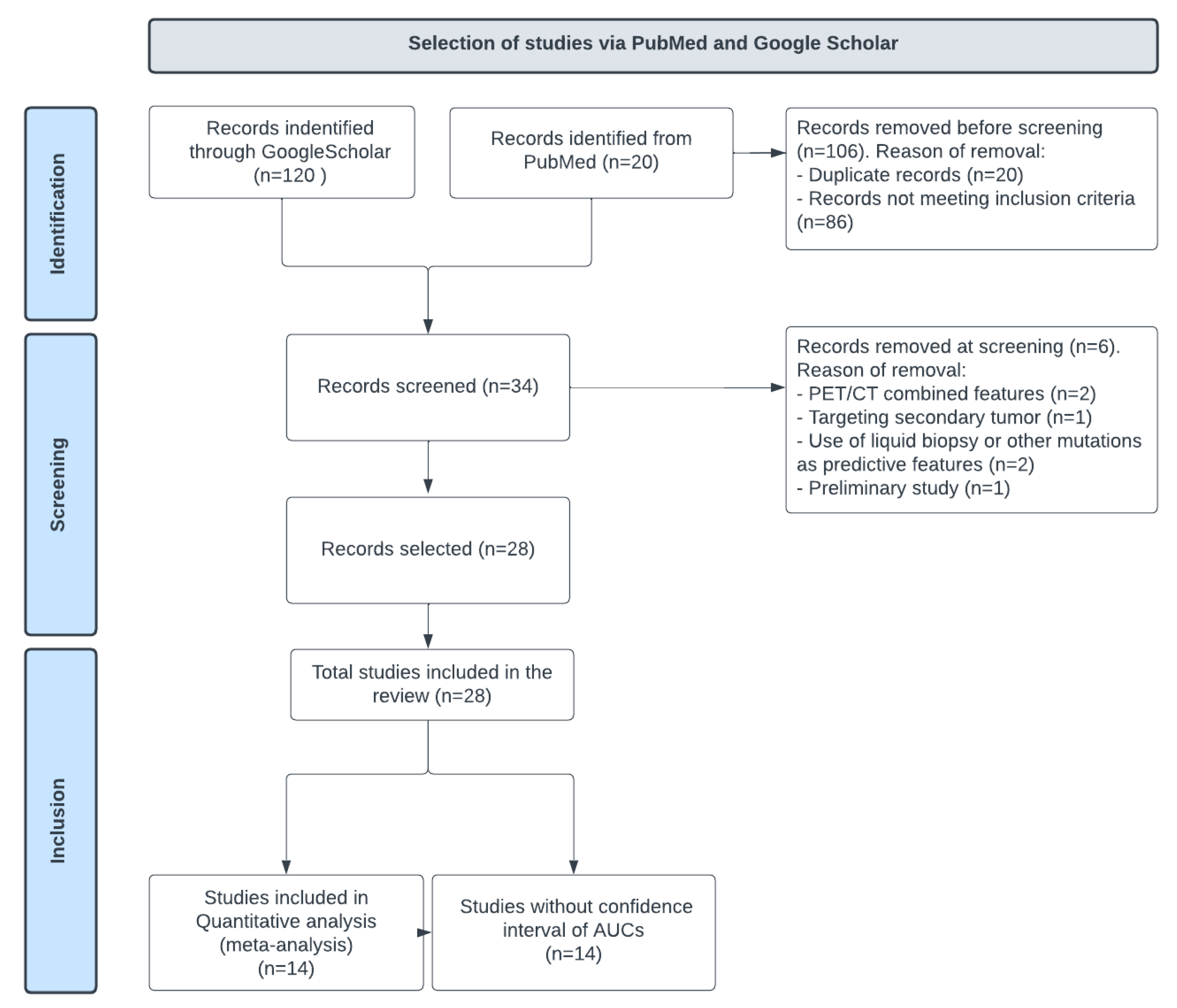
2.1. Search Strategy

A systematic search for eligible publications published up to 27 April 2023 was performed in the PubMed and Google Scholar databases using the keywords “Radiomics”, ”Model”, “CT scan”, “NSCLC”, “Lung”, and “EGFR”. Only original studies were selected.
The PubMed and Google Scholar databases were chosen for the references’ high quality and ease of accessibility. Two reviewers assessed the eligibility of the articles identified through the search: the first reviewer screened each record, each report, as well as the reference lists of the included studies; the second reviewer checked the results by following the same process.

2.2. Inclusion and Exclusion Criteria

Only publications on original research studies were considered for inclusion. Studies were deemed relevant for inclusion if they included cohorts of pathology-confirmed NSCLC patients who had CT scans before surgery or biopsy, did not receive anti-tumor treatment before the CT scan, and had gene testing for EGFR mutation. The analysis of the CT radiomics or CT radiomics-based model had to be conducted to predict the EGFR mutation status. Models trained on deep learning features were not considered to focus on radiomics features.
Excluded published articles included reviews, meta-analyses, case reports, conference abstracts, letters to the editor, comments, posters, technical reports, duplicate studies, publications not in English, and non-human studies. Moreover, original research articles were excluded if the model’s performance was not assessed via the area under the receiver operating characteristic curve (ROC AUC) or indirect/combined measures.
The process for identifying and selecting published articles is further detailed in Figure 1.

2.3. Data Extraction

The following data were extracted upon availability: name of the first author, year of publication, used standard reference, sample size, cohort origin, histological subtype, feature extraction, feature reduction and modeling methods, number of involved centers (either single or multicenter), study objectives, and prediction performance in terms of ROC AUC with its corresponding confidence interval (CI). Data extraction was performed independently by two reviewers. As a quality control measure, extracted data were compared, and consensus resolved disagreements.

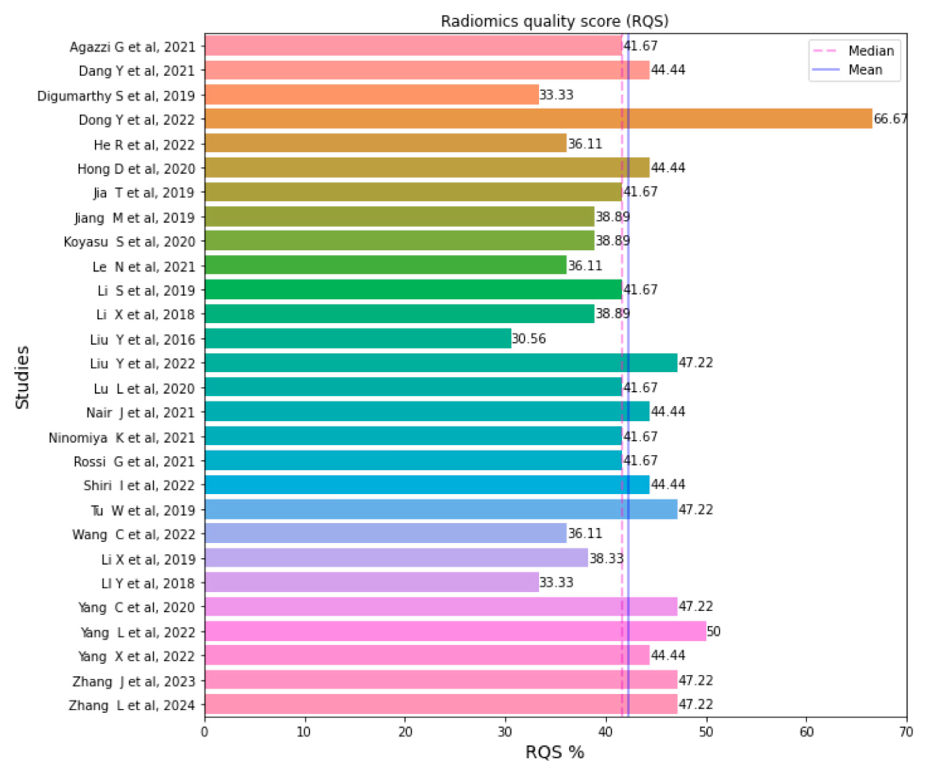
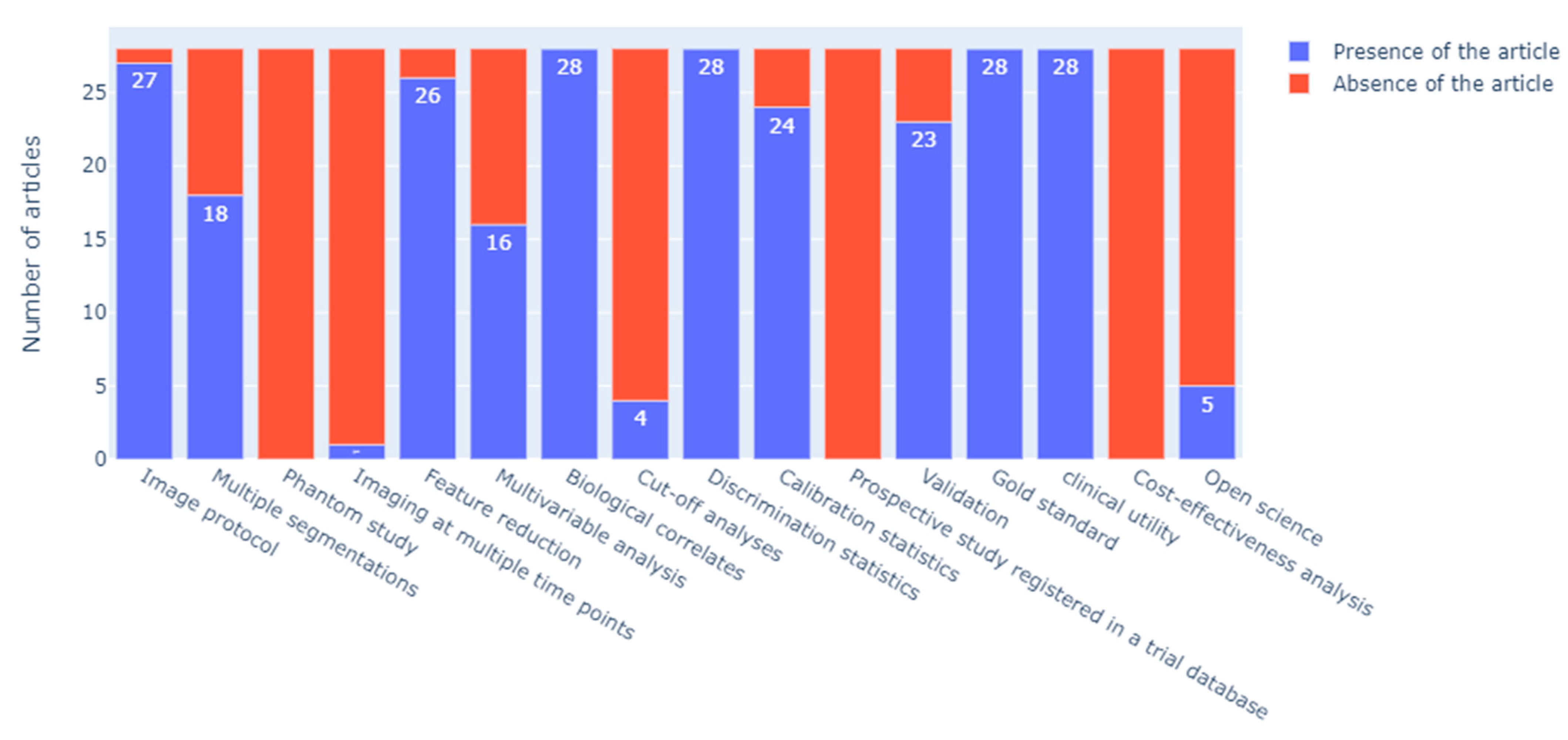
2.4. Quality Analysis

Radiomics is a multi-step process with methodological challenges to ensure the findings’ robustness, reproducibility, and generalizability. Therefore, studies reporting correlations between imaging biomarkers and patient outcomes need standardized acquisition and processing techniques to confirm causality. To improve the radiomics workflow, a set of recommendations was proposed in 2017 by Lambin et al. [7], including the Radiomics Quality Score (RQS). This tool has become the standard for systematic literature reviews on radiomics quality assessment [14].
The RQS includes 16 items that assess protocol quality, reproducibility, reporting, validation, biological/clinical validation and utility, performance index, demonstration of a higher evidence level, and open science. The total scale score ranges from −8 to +36; higher scores indicate better quality, and a percentage score of 100% corresponds to a score of +36 [7].

2.5. Statistical Analysis

Statistical analysis was conducted using the Python 3.7 software. Forest plots were generated using the MedCalc Statistical Software (version 19.2.6; MedCalc Software bv, Ostend, Belgium; https://www.medcalc.org (accessed on 25 May 2023)). The RQS scores and percentages were summed and calculated as described by Lambin et al. [7].
Various measures were employed to assess the risk of bias in the included studies. The z-score and heterogeneity test I2 value were used to evaluate the heterogeneity across the studies. Funnel plots were generated to assess publication bias, and Egger’s test and Kendall’s tau value were used to quantify funnel plot asymmetry.

3. Results

A total of 140 full-text manuscripts were identified using the pre-specified search terms. After excluding 106 publications (20 duplicated publications and 86 articles meeting at least one of the exclusion criteria), 28 were included [3,9,10,11,12,15,16,17,18,19,20,21,22,23,24,25,26,27,28,29,30,31,32,33,34,35,36,37]. The retained publications targeted the prediction of the EGFR mutation status using radiomics and/or an AI-based radiomics model, with ROC AUCs as a performance metric. Articles on studies evaluating the EGFR as secondary endpoints relative to survival or therapeutic response were excluded.
The quality of the 28 included publications was assessed using the RQS tool. Scores ranged from 11 (30.56%) to 24 (66.67%). The mean score obtained with the RQS was 15.25, accounting for 42.34% of the total score. The median score was 41.67% (interquartile range [IQR] = 15). The RQS of each publication included in this literature review is presented in Figure 2.
All studies used gold standards using radiologist segmentation for lesions and Polymerase Chain reaction (PCR) testing or sequencing for EGFR mutation status. The feature selection steps were performed in a total of 26 studies. However, none were prospective or reported phantom studies and cost-effectiveness analyses. Imaging feature extraction at multiple time points was performed in one study only [34].
Other items assessed by the RQS, including “Validation”, “Cut-off analyses”, “Calibration statistics”, “Multivariable analysis”, and “Open science and data” varied the most between studies. More specifically, resampling method techniques were widely applied (24/28 studies). However, calibration statistics and their statistical significance were performed in fewer studies (8/28 studies), and the determination of risk groups was performed in a total of 4/28 studies. An overview of the RQS item scores is provided in Figure 3, and the RQS of each included article (overall and item scores [7]) is provided in Table S1 (Supplementary Materials).
Regarding radiomics workflow, most studies used the Pyradiomics or LifeX feature extraction tools. However, one study (Lu et al., 2020) utilized the Imaging Biomarker Explorer (IBEX) tool [27], and others used an in-house tool. The extracted features varied between the studies, ranging from 11 to 4306 (median number: 580), with an average of 751 ± 866 features. The most commonly used machine learning models for classification were logistic regression, random forest, and support vector machine. All the studies were retrospective; five used data from The Cancer Imaging Archive (TCIA) open-source repository. For ground truth, delineation performed by radiologists was used for segmentation, and PCR or sequencing was used to determine the EGFR mutation status. Correlation coefficients and least absolute shrinkage and selection operator (LASSO) were the two most commonly used methods for feature reduction.
Based on the forest plot analysis, which included 14 studies from 28 that reported ROC AUC confidence intervals, the pooled ROC AUC value for the performance of CT radiomics-based models for the prediction of the EGFR mutation status in NSCLC patients was 0.8, with a 95% CI of [0.757–0.845] indicating moderate-to-high prediction accuracy (Figure 4).
The z-score was 35.459 with a p-value of less than 0.001, indicating that the pooled ROC AUC value was statistically significant (Table 1).
However, the studies had high heterogeneity, with an I-squared value of 82.78% and a p-value of less than 0.0001. This heterogeneity may be attributed to differences in the included patient populations, study designs, manufacturers, and protocols, as well as the used model and radiomics features.
Egger’s test resulted in an intercept of −0.013, indicating that there may have been a small degree of asymmetry in the funnel plot (Figure 5).
However, this asymmetry was not statistically significant, suggesting no evidence of publication bias in this meta-analysis. The Kendall’s tau value was 0.098, indicating a weak positive correlation between the effect size and its standard error. This could have been due to the publication bias, relative to studies with a small database and low performance not being published, or to other sources of heterogeneity related to variations in radiomics workflows.

4. Discussion

The quality of the studies included in this meta-analysis was assessed using the RQS tool. It should be noted that this tool did not provide a definitive assessment of the study quality but rather identified potential methodological weaknesses in radiomics studies. Also, the RQS rating was subject to subjective assessments and could vary between raters. Measurement of the inter-rater variability was not performed in this meta-analysis.
The RQS results obtained in this meta-analysis also revealed heterogeneity in the qualities and methodologies of the included studies, primarily due to the number of steps in the radiomics workflow. This indicated the need for improved methodological rigor in the conduct and reporting of future radiomics studies in order to increase the reliability and reproducibility of CT radiomics-based models for predicting EGFR mutation status.
This meta-analysis of studies investigating the performance of CT radiomics-based models for predicting EGFR mutation status in NSCLC patients yielded several significant findings. First, the analysis demonstrated that radiomics analysis and radiomics models could provide accurate predictions of the EGFR mutation status using CT imaging data. The analysis of the ROC AUC suggested that these models had a high degree of diagnostic accuracy with an overall ROC AUC of 0.8 (95% CI: 0.757–0.845), which agreed with previous publications [38]. Radiomics-based models performed significantly better than clinical features, as demonstrated by multivariable analyses with non-radiomics features [3,10,16,19,20,24,27,30,31,32,35,36,37].
Furthermore, a combined model with clinical and radiomics features seemed to have better results than only radiomics-based models alone [9,16,37].
The performed analyses also suggested that radiomics-based analyses combined with radiologist assessment could be helpful in clinical decision-making for NSCLC patients. By helping the accurate prediction of the EGFR mutation status, CT radiomics-based methods can help clinicians identify NSCLC patients who may benefit from targeted therapies, improving treatment outcomes and reducing morbidity and mortality.
Noninvasive CT radiomics-based models offer distinct advantages over traditional methods. These models utilize existing medical imaging data, eliminating the need for invasive procedures and reducing patient discomfort, risk, and potential complications [39]. Indeed, CT scans are a routine diagnostic procedure, providing readily available data for radiomics-based models without requiring additional invasive procedures or specialized testing. Unlike current methods that sample only a small portion of the tumor, CT radiomics-based models can analyze the entire tumor volume with tumor microenvironment, offering a more comprehensive evaluation of tumor characteristics [5,7,40]. These models employ advanced imaging analysis techniques to extract quantitative features, objectively assessing tumor characteristics such as shape, texture, and density. Quantifying these features could enhance the accuracy and reliability of predicting genetic mutations [8,41]. Moreover, serial CT scans enable longitudinal monitoring of tumor characteristics, facilitating assessment of treatment response, disease progression evaluation, and monitoring emerging genetic mutations over time. It is important to note that CT radiomics-based models should not replace confirmatory molecular testing methods but can serve as valuable adjunct tools to assist clinicians in decision-making, risk stratification, and treatment planning.
This meta-analysis also revealed limitations and gaps in the current literature. For example, while radiomics-based models have shown promising results in predicting EGFR mutation status, the studies included in the current meta-analysis were largely retrospective, and some were conducted on a small sample size (less than 60 cases) [11,28,33]. Also, most of the studies lacked external validation and multicentric data sources. Future studies should aim to prospectively validate these models on more extensive and more diverse patient populations, as well as investigate their performance in real-world clinical settings. Given the high degree of heterogeneity, harmonization and reproducibility remain the main obstacles to generalizing CT radiomics-based methods [42,43].
Overall, this meta-analysis suggests that CT radiomics-based models have the potential to be a valuable tool in predicting EGFR mutation status in NSCLC patients, but further research is needed to fully establish their clinical utility and address the limitations and gaps in the current literature.

5. Conclusions

In conclusion, despite the heterogeneity and methodological weaknesses observed in the methodology of studies, this meta-analysis highlights that CT radiomics-based models show promise in accurately predicting EGFR mutation status in NSCLC patients with high diagnostic accuracy. These noninvasive techniques offer advantages over invasive procedures, could comprehensively evaluate tumor characteristics, and might enable longitudinal monitoring. However, the current literature lacks external validation, multicentric data sources, and prospective studies on larger and more diverse patient populations. Harmonization and reproducibility remain crucial challenges.

Supplementary Materials

The supporting information can be downloaded at: https://www.mdpi.com/article/10.3390/ijms241411433/s1.

Author Contributions

Conceptualization, M.F., Y.L. and S.B.; methodology, M.F., Y.L. and S.B.; software, M.F., Y.L. and S.B.; validation, M.F., Y.L., S.B., F.Z., C.V., A.T., S.J. and A.I.; formal analysis, M.F.; investigation, M.F.; resources, M.F., Y.L. and S.B.; data curation, M.F., Y.L., S.B., F.Z., C.V., A.T., S.J. and A.I.; writing—original draft preparation, M.F., Y.L. and S.B.; writing—review and editing, M.F., Y.L. and S.B.; visualization, M.F., Y.L. and S.B.; supervision, S.B. and Y.L.; project administration, S.B. and Y.L. All authors have read and agreed to the published version of the manuscript.

Funding

This research received no external funding.

Institutional Review Board Statement

This study is registered under the number CRD42023411128 in PROSPERO.

Informed Consent Statement

Not applicable.

Data Availability Statement

Not applicable.

Conflicts of Interest

Yan Liu, Mehdi Felfli, Fadila Zarka, Charles Voyton, Alexandre Thinnes, Sebastien Jacques, and Antoine Iannessi are employees of Median Technology. The other authors declare no conflict of interest.

References

  1. Imyanitov, E.N.; Iyevleva, A.G.; Levchenko, E.V. Molecular testing and targeted therapy for non-small cell lung cancer: Current status and perspectives. Crit. Rev. Oncol. Hematol. 2021, 157, 103194. [Google Scholar] [CrossRef] [PubMed]
  2. Lindeman, N.I.; Cagle, P.T.; Aisner, D.L.; Arcila, M.E.; Beasley, M.B.; Bernicker, E.H.; Colasacco, C.; Dacic, S.; Hirsch, F.R.; Kerr, K.; et al. Updated Molecular Testing Guideline for the Selection of Lung Cancer Patients for Treatment With Targeted Tyrosine Kinase Inhibitors. J. Thorac. Oncol. 2018, 13, 323–358. [Google Scholar] [CrossRef] [Green Version]
  3. Dang, Y.; Wang, R.; Qian, K.; Lu, J.; Zhang, H.; Zhang, Y. Clinical and radiological predictors of epidermal growth factor receptor mutation in nonsmall cell lung cancer. J. Appl. Clin. Med. Phys. 2021, 22, 271–280. [Google Scholar] [CrossRef]
  4. Metro, G.; Finocchiaro, G.; Toschi, L.; Bartolini, S.; Magrini, E.; Cancellieri, A.; Trisolini, R.; Castaldini, L.; Tallini, G.; Crino, L. Epidermal Growth Factor Receptor (EGFR) Targeted Therapies in Non-Small Cell Lung Cancer (NSCLC). Rev. Recent. Clin. Trials 2006, 1, 1–13. [Google Scholar] [CrossRef] [PubMed]
  5. Zito Marino, F.; Bianco, R.; Accardo, M.; Ronchi, A.; Cozzolino, I.; Morgillo, F.; Rossi, G.; Franco, R. Molecular heterogeneity in lung cancer: From mechanisms of origin to clinical implications. Int. J. Med. Sci. 2019, 16, 981–989. [Google Scholar] [CrossRef] [PubMed] [Green Version]
  6. Di Capua, D.; Bracken-Clarke, D.; Ronan, K.; Baird, A.M.; Finn, S. The Liquid Biopsy for Lung Cancer: State of the Art, Limitations and Future Developments. Cancers 2021, 13, 3923. [Google Scholar] [CrossRef]
  7. Lambin, P.; Leijenaar, R.T.H.; Deist, T.M.; Peerlings, J.; de Jong, E.E.C.; van Timmeren, J.; Sanduleanu, S.; Larue, R.T.H.M.; Even, A.J.G.; Jochems, A.; et al. Radiomics: The bridge between medical imaging and personalized medicine. Nat. Rev. Clin. Oncol. 2017, 14, 749–762. [Google Scholar] [CrossRef] [Green Version]
  8. Lambin, P.; Rios-Velazquez, E.; Leijenaar, R.; Carvalho, S.; van Stiphout, R.G.; Granton, P.; Zegers, C.M.; Gillies, R.; Boellard, R.; Dekker, A.; et al. Radiomics: Extracting more information from medical images using advanced feature analysis. Eur. J. Cancer 2012, 48, 441–446. [Google Scholar] [CrossRef] [Green Version]
  9. Wang, C.; Ma, J.; Shao, J.; Zhang, S.; Liu, Z.; Yu, Y.; Li, W. Predicting EGFR and PD-L1 Status in NSCLC Patients Using Multitask AI System Based on CT Images. Front. Immunol. 2022, 13, 813072. [Google Scholar] [CrossRef]
  10. Yang, L.; Xu, P.; Li, M.; Wang, M.; Peng, M.; Zhang, Y.; Wu, T.; Chu, W.; Wang, K.; Meng, H.; et al. PET/CT Radiomic Features: A Potential Biomarker for EGFR Mutation Status and Survival Outcome Prediction in NSCLC Patients Treated With TKIs. Front. Oncol. 2022, 12, 894323. [Google Scholar] [CrossRef]
  11. Nair, J.K.R.; Saeed, U.A.; McDougall, C.C.; Sabri, A.; Kovacina, B.; Raidu, B.V.S.; Khokhar, R.A.; Probst, S.; Hirsh, V.; Chankowsky, J.; et al. Radiogenomic Models Using Machine Learning Techniques to Predict EGFR Mutations in Non-Small Cell Lung Cancer. Can. Assoc. Radiol. J. 2021, 72, 109–119. [Google Scholar] [CrossRef] [PubMed]
  12. Jiang, M.; Zhang, Y.; Xu, J.; Ji, M.; Guo, Y.; Guo, Y.; Xiao, J.; Yao, X.; Shi, H.; Zeng, M. Assessing EGFR gene mutation status in non-small cell lung cancer with imaging features from PET/CT. Nucl. Med. Commun. 2019, 40, 842–849. [Google Scholar] [CrossRef] [PubMed]
  13. McInnes, M.D.F.; Moher, D.; Thombs, B.D.; McGrath, T.A.; Bossuyt, P.M.; The PRISMA-DTA Group; Clifford, T.; Cohen, J.F.; Deeks, J.J.; Gatsonis, C.; et al. Preferred Reporting Items for a Systematic Review and Meta-analysis of Diagnostic Test Accuracy Studies. JAMA 2018, 319, 388. [Google Scholar] [CrossRef]
  14. Spadarella, G.; Stanzione, A.; Akinci D’Antonoli, T.; Andreychenko, A.; Fanni, S.C.; Ugga, L.; Kotter, E.; Cuocolo, R. Systematic review of the radiomics quality score applications: An EuSoMII Radiomics Auditing Group Initiative. Eur. Radiol. Mar. 2023, 33, 1884–1894. [Google Scholar] [CrossRef]
  15. Agazzi, G.M.; Ravanelli, M.; Roca, E.; Medicina, D.; Balzarini, P.; Pessina, C.; Vermi, W.; Berruti, A.; Maroldi, R.; Farina, D. CT texture analysis for prediction of EGFR mutational status and ALK rearrangement in patients with non-small cell lung cancer. Radiol. Med. 2021, 126, 786–794. [Google Scholar] [CrossRef]
  16. Digumarthy, S.R.; Padole, A.M.; Gullo, R.L.; Sequist, L.V.; Kalra, M.K. Can CT radiomic analysis in NSCLC predict histology and EGFR mutation status? Medicine 2019, 98, e13963. [Google Scholar] [CrossRef] [PubMed]
  17. Dong, Y.; Jiang, Z.; Li, C.; Dong, S.; Zhang, S.; Lv, Y.; Sun, F.; Liu, S. Development and validation of novel radiomics-based nomograms for the prediction of EGFR mutations and Ki-67 proliferation index in non-small cell lung cancer. Quant. Imaging Med. Surg. 2022, 12, 2658–2671. [Google Scholar] [CrossRef] [PubMed]
  18. He, R.; Yang, X.; Li, T.; He, Y.; Xie, X.; Chen, Q.; Zhang, Z.; Cheng, T. A Machine Learning-Based Predictive Model of Epidermal Growth Factor Mutations in Lung Adenocarcinomas. Cancers 2022, 14, 4664. [Google Scholar] [CrossRef]
  19. Hong, D.; Xu, K.; Zhang, L.; Wan, X.; Guo, Y. Radiomics Signature as a Predictive Factor for EGFR Mutations in Advanced Lung Adenocarcinoma. Front. Oncol. 2020, 10, 28. [Google Scholar] [CrossRef] [Green Version]
  20. Jia, T.Y.; Xiong, J.F.; Li, X.Y.; Yu, W.; Xu, Z.Y.; Cai, X.W.; Ma, J.C.; Ren, Y.C.; Larsson, R.; Zhang, J.; et al. Identifying EGFR mutations in lung adenocarcinoma by noninvasive imaging using radiomics features and random forest modeling. Eur. Radiol. 2019, 29, 4742–4750. [Google Scholar] [CrossRef] [PubMed]
  21. Koyasu, S.; Nishio, M.; Isoda, H.; Nakamoto, Y.; Togashi, K. Usefulness of gradient tree boosting for predicting histological subtype and EGFR mutation status of non-small cell lung cancer on 18F FDG-PET/CT. Ann. Nucl. Med. 2020, 34, 49–57. [Google Scholar] [CrossRef]
  22. Le, N.Q.K.; Kha, Q.H.; Nguyen, V.H.; Chen, Y.C.; Cheng, S.J.; Chen, C.Y. Machine Learning-Based Radiomics Signatures for EGFR and KRAS Mutations Prediction in Non-Small-Cell Lung Cancer. Int. J. Mol. Sci. 2021, 22, 9254. [Google Scholar] [CrossRef] [PubMed]
  23. Li, S.; Ding, C.; Zhang, H.; Song, J.; Wu, L. Radiomics for the prediction of EGFR mutation subtypes in non-small cell lung cancer. Med. Phys. 2019, 46, 4545–4552. [Google Scholar] [CrossRef] [PubMed]
  24. Li, X.Y.; Xiong, J.F.; Jia, T.Y.; Shen, T.L.; Hou, R.P.; Zhao, J.; Fu, X.L. Detection of epithelial growth factor receptor (EGFR) mutations on CT images of patients with lung adenocarcinoma using radiomics and/or multi-level residual convolutionary neural networks. J. Thorac. Dis. 2018, 10, 6624–6635. [Google Scholar] [CrossRef] [PubMed]
  25. Liu, Y.; Kim, J.; Balagurunathan, Y.; Li, Q.; Garcia, A.L.; Stringfield, O.; Ye, Z.; Gillies, R.J. Radiomic Features Are Associated with EGFR Mutation Status in Lung Adenocarcinomas. Clin. Lung Cancer 2016, 17, 441–448.e6. [Google Scholar] [CrossRef] [PubMed] [Green Version]
  26. Liu, Y.; Zhou, J.; Wu, J.; Wang, W.; Wang, X.; Guo, J.; Wang, Q.; Zhang, X.; Li, D.; Xie, J.; et al. Development and Validation of Machine Learning Models to Predict Epidermal Growth Factor Receptor Mutation in Non-Small Cell Lung Cancer: A Multi-Center Retrospective Radiomics Study. Cancer Control 2022, 29, 107327482210929. [Google Scholar] [CrossRef]
  27. Lu, L.; Sun, S.H.; Yang, H.; E, L.; Guo, P.; Schwartz, L.H.; Zhao, B. Radiomics Prediction of EGFR Status in Lung Cancer—Our Experience in Using Multiple Feature Extractors and The Cancer Imaging Archive Data. Tomography 2020, 6, 223–230. [Google Scholar] [CrossRef]
  28. Ninomiya, K.; Arimura, H.; Chan, W.Y.; Tanaka, K.; Mizuno, S.; Muhammad Gowdh, N.F.; Yaakup, N.A.; Liam, C.K.; Chai, C.S.; Ng, K.H. Robust radiogenomics approach to the identification of EGFR mutations among patients with NSCLC from three different countries using topologically invariant Betti numbers. PLoS ONE 2021, 16, e0244354. [Google Scholar] [CrossRef]
  29. Rossi, G.; Barabino, E.; Fedeli, A.; Ficarra, G.; Coco, S.; Russo, A.; Adamo, V.; Buemi, F.; Zullo, L.; Dono, M.; et al. Radiomic Detection of EGFR Mutations in NSCLC. Cancer Res. 2021, 81, 724–731. [Google Scholar] [CrossRef]
  30. Shiri, I.; Amini, M.; Nazari, M.; Hajianfar, G.; Haddadi Avval, A.; Abdollahi, H.; Oveisi, M.; Arabi, H.; Rahmim, A.; Zaidi, H. Impact of feature harmonization on radiogenomics analysis: Prediction of EGFR and KRAS mutations from non-small cell lung cancer PET/CT images. Comput. Biol. Med. 2022, 142, 105230. [Google Scholar] [CrossRef] [PubMed]
  31. Tu, W.; Sun, G.; Fan, L.; Wang, Y.; Xia, Y.; Guan, Y.; Li, Q.; Zhang, D.; Liu, S.; Li, Z. Radiomics signature: A potential and incremental predictor for EGFR mutation status in NSCLC patients, comparison with CT morphology. Lung Cancer 2019, 132, 28–35. [Google Scholar] [CrossRef] [PubMed]
  32. Li, X.; Yin, G.; Zhang, Y.; Dai, D.; Liu, J.; Chen, P.; Zhu, L.; Ma, W.; Xu, W. Predictive Power of a Radiomic Signature Based on 18F-FDG PET/CT Images for EGFR Mutational Status in NSCLC. Front. Oncol. 2019, 9, 1062. [Google Scholar] [CrossRef] [Green Version]
  33. Li, Y.; Lu, L.; Xiao, M.; Dercle, L.; Huang, Y.; Zhang, Z.; Schwartz, L.H.; Li, D.; Zhao, B. CT Slice Thickness and Convolution Kernel Affect Performance of a Radiomic Model for Predicting EGFR Status in Non-Small Cell Lung Cancer: A Preliminary Study. Sci. Rep. 2018, 8, 17913. [Google Scholar] [CrossRef] [Green Version]
  34. Yang, C.; Chen, W.; Gong, G.; Li, Z.; Qiu, Q.; Yin, Y. Application of CT radiomics features to predict the EGFR mutation status and therapeutic sensitivity to TKIs of advanced lung adenocarcinoma. Transl. Cancer Res. 2020, 9, 6683–6690. [Google Scholar] [CrossRef] [PubMed]
  35. Yang, X.; Liu, M.; Ren, Y.; Chen, H.; Yu, P.; Wang, S.; Zhang, R.; Dai, H.; Wang, C. Using contrast-enhanced CT and non-contrast-enhanced CT to predict EGFR mutation status in NSCLC patients—A radiomics nomogram analysis. Eur. Radiol. 2022, 32, 2693–2703. [Google Scholar] [CrossRef] [PubMed]
  36. Zhang, J.; Zhao, X.; Zhao, Y.; Zhang, J.; Zhang, Z.; Wang, J.; Wang, Y.; Dai, M.; Han, J. Value of pre-therapy 18F-FDG PET/CT radiomics in predicting EGFR mutation status in patients with non-small cell lung cancer. Eur. J. Nucl. Med. Mol. Imaging 2020, 47, 1137–1146. [Google Scholar] [CrossRef] [PubMed]
  37. Zhang, L.; Chen, B.; Liu, X.; Song, J.; Fang, M.; Hu, C.; Dong, D.; Li, W.; Tian, J. Quantitative Biomarkers for Prediction of Epidermal Growth Factor Receptor Mutation in Non-Small Cell Lung Cancer. Transl. Oncol. 2018, 11, 94–101. [Google Scholar] [CrossRef] [PubMed]
  38. Abdurixiti, M.; Nijiati, M.; Shen, R.; Ya, Q.; Abuduxiku, N.; Nijiati, M. Current progress and quality of radiomic studies for predicting EGFR mutation in patients with non-small cell lung cancer using PET/CT images: A systematic review. Br. J. Radiol. 2021, 94, 20201272. [Google Scholar] [CrossRef]
  39. Azour, L.; Liu, S.; Washer, S.L.; Moore, W.H. Percutaneous Transthoracic Lung Biopsy: Optimizing Yield and Mitigating Risk. J. Comput. Assist. Tomogr. 2021, 45, 765–775. [Google Scholar] [CrossRef] [PubMed]
  40. Wu, J.; Mayer, A.T.; Li, R. Integrated imaging and molecular analysis to decipher tumor microenvironment in the era of immunotherapy. Semin. Cancer Biol. 2022, 84, 310–328. [Google Scholar] [CrossRef]
  41. Binczyk, F.; Prazuch, W.; Bozek, P.; Polanska, J. Radiomics and artificial intelligence in lung cancer screening. Transl. Lung Cancer Res. 2021, 10, 1186–1199. [Google Scholar] [CrossRef] [PubMed]
  42. Mali, S.A.; Ibrahim, A.; Woodruff, H.C.; Andrearczyk, V.; Müller, H.; Primakov, S.; Salahuddin, Z.; Chatterjee, A.; Lambin, P. Making Radiomics More Reproducible across Scanner and Imaging Protocol Variations: A Review of Harmonization Methods. J. Pers. Med. 2021, 11, 842. [Google Scholar] [CrossRef] [PubMed]
  43. Horng, H.; Singh, A.; Yousefi, B.; Cohen, E.A.; Haghighi, B.; Katz, S.; Noël, P.B.; Shinohara, R.T.; Kontos, D. Generalized ComBat harmonization methods for radiomic features with multi-modal distributions and multiple batch effects. Sci. Rep. 2022, 12, 4493. [Google Scholar] [CrossRef] [PubMed]
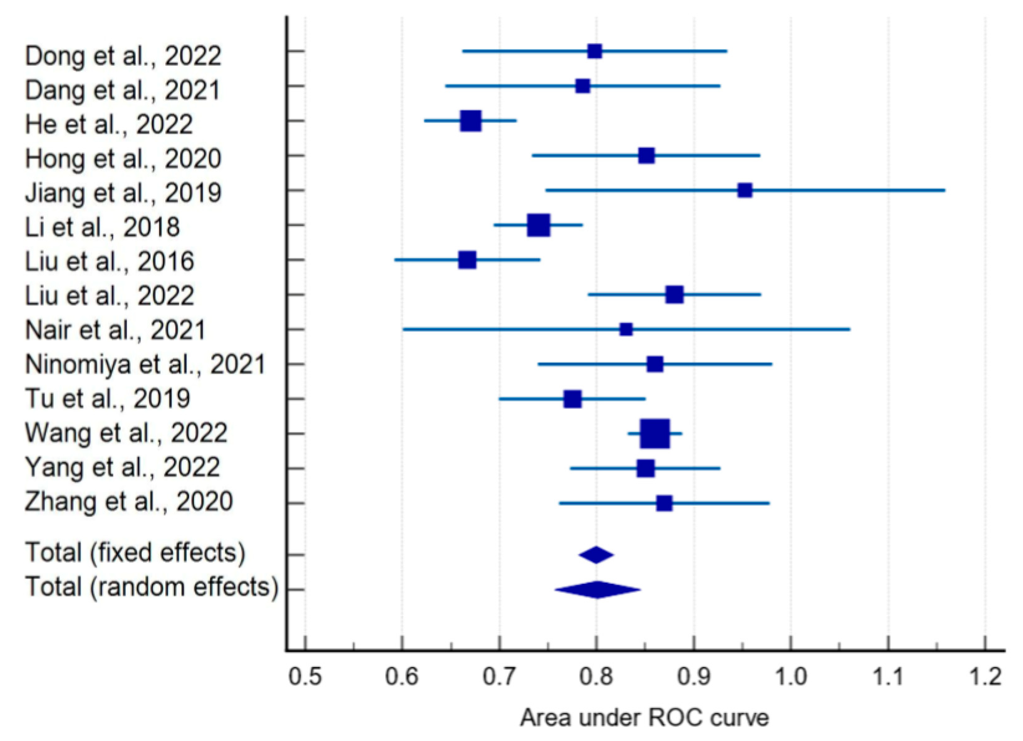
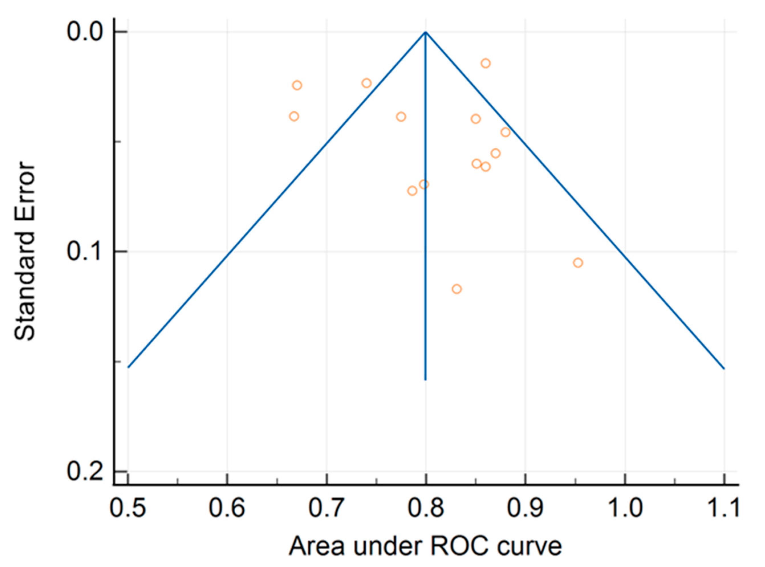
Figure 1. Selection process pipeline.
Ijms 24 11433 g001
Figure 2. Bar plot of attributed radiomics quality score (RQS) by study paper [3,9,10,11,12,15,16,17,18,19,20,21,22,23,24,25,26,27,28,29,30,31,32,33,34,35,36,37].
Ijms 24 11433 g002
Figure 3. RQS item scores of the studies included in this meta-analysis (the sum reported is the number of studies with at least one point in the RQS workflow step).
Ijms 24 11433 g003
Figure 4. Forest plot of ROC AUCs [3,9,10,11,12,17,18,19,24,25,26,28,31,36].
Ijms 24 11433 g004
Figure 5. Funnel plot of ROC AUCs with 95% confidence limits.
Ijms 24 11433 g005
Table 1. Forest plot statistics.
StudyROC Curve AreaStandard Error95% CIz-Scorep-ValueWeight(%)Sample SizeStudy Centers
FixedRandom
Dong et al., 2022 [17]0.7980.06940.662 to 0.934 1.685.47132Single center
Dang et al., 2021 [3]0.7860.07230.644 to 0.928 1.555.24118Multicenter
He et al., 2022 [18]0.6700.02430.622 to 0.718 13.679.99758Multicenter
Hong et al., 2020 [19]0.8510.06000.733 to 0.969 2.256.29449Single center
Jiang et al., 2019 [12]0.9530.1050.747 to 1.000 0.733.2880Single center
Li et al., 2018 [24]0.7400.02330.694 to 0.786 14.9110.09810Single center
Liu et al., 2016 [25]0.6670.03840.592 to 0.742 5.508.52397Single center
Liu et al., 2022 [26]0.8800.04560.791 to 0.969 3.907.74211Single center
Nair et al., 2021 [11]0.8310.1170.600 to 1.000 0.592.7980Single center
Ninomiya et al., 2021 [28]0.8600.06140.740 to 0.980 2.156.16194Multicenter
Tu et al., 2019 [31]0.7750.03860.699 to 0.851 5.458.50404Single center
Wang et al., 2022 [9]0.8600.01430.832 to 0.888 39.8210.813629Single center
Yang et al., 2022 [10]0.8500.03960.772 to 0.928 5.168.391028Single center
Zhang et al., 2020 [36]0.8700.05520.762 to 0.978 2.656.74728Single center
Total (fixed effects)0.8000.009000.782 to 0.81788.858<0.001100.00100.00
Total (random effects)0.8010.02260.757 to 0.84535.459<0.001100.00100.00
Disclaimer/Publisher’s Note: The statements, opinions and data contained in all publications are solely those of the individual author(s) and contributor(s) and not of MDPI and/or the editor(s). MDPI and/or the editor(s) disclaim responsibility for any injury to people or property resulting from any ideas, methods, instructions or products referred to in the content.

