Article

Inhibitory Effect of Adsorption of Streptococcus mutans onto Scallop-Derived Hydroxyapatite

by
Momoko Usuda
1,†,
Mariko Kametani
1,†,
Masakazu Hamada
2,
Yuto Suehiro
3,
Saaya Matayoshi
3,
Rena Okawa
3,4,
Shuhei Naka
5,
Michiyo Matsumoto-Nakano
5,
Tatsuya Akitomo
1,
Chieko Mitsuhata
1,
Kazuya Koumoto
6,
Keiko Kawauchi
6,
Takahito Nishikata
6,
Masatoshi Yagi
4,7,
Toshiro Mizoguchi
4,8,
Koki Fujikawa
4,8,
Taizo Taniguchi
4,7,
Kazuhiko Nakano
3,4 and
Ryota Nomura
1,3,4,*
1
Department of Pediatric Dentistry, Graduate School of Biomedical and Health Sciences, Hiroshima University, Hiroshima 734-8553, Japan
2
Department of Oral & Maxillofacial Oncology and Surgery, Osaka University Graduate School of Dentistry, Suita 565-0871, Japan
3
Department of Pediatric Dentistry, Osaka University Graduate School of Dentistry, Suita 565-0871, Japan
4
Joint Research Laboratory of Next-Generation Science for Oral Infection Control, Osaka University Graduate School of Dentistry, Suita 565-0871, Japan
5
Department of Pediatric Dentistry, Okayama University Graduate School of Medicine, Dentistry and Pharmaceutical Sciences, Okayama 700-8558, Japan
6
Faculty of Frontiers of Innovative Research in Science and Technology (FIRST), Konan University, Kobe 650-0047, Japan
7
Pharmacrea Kobe Co., Ltd., Kobe 651-0085, Japan
8
TSET Co., Ltd., Kariya 448-0022, Japan
*
Author to whom correspondence should be addressed.
These authors contributed equally to this work.
Int. J. Mol. Sci. 2023, 24(14), 11371; https://doi.org/10.3390/ijms241411371
Submission received: 30 May 2023 / Revised: 30 June 2023 / Accepted: 10 July 2023 / Published: 12 July 2023

Abstract

:
Hydroxyapatite adsorbs various substances, but little is known about the effects on oral bacteria of adsorption onto hydroxyapatite derived from scallop shells. In the present study, we analyzed the effects of adsorption of Streptococcus mutans onto scallop-derived hydroxyapatite. When scallop-derived hydroxyapatite was mixed with S. mutans, a high proportion of the bacterial cells adsorbed onto the hydroxyapatite in a time-dependent manner. An RNA sequencing analysis of S. mutans adsorbed onto hydroxyapatite showed that the upregulation of genes resulted in abnormalities in pathways involved in glycogen and histidine metabolism and biosynthesis compared with cells in the absence of hydroxyapatite. S. mutans adsorbed onto hydroxyapatite was not killed, but the growth of the bacteria was inhibited. Electron microscopy showed morphological changes in S. mutans cells adsorbed onto hydroxyapatite. Our results suggest that hydroxyapatite derived from scallop shells showed a high adsorption ability for S. mutans. This hydroxyapatite also caused changes in gene expression related to the metabolic and biosynthetic processes, including the glycogen and histidine of S. mutans, which may result in a morphological change in the surface layer and the inhibition of the growth of the bacteria.

1. Introduction

Dental caries is considered an endogenous infection caused by commensal oral microbiota [1]. When homeostasis in biofilms becomes disrupted during acidification, streptococci, the most widely distributed species in the oral cavity, can adapt to and survive in the acidic environment [2]. The acid-adapted bacteria gain a selective advantage over other species, resulting in the development of dental caries lesions [1]. S. mutans can increase in abundance in the acidic environment and promote the progression of dental caries through the loss of minerals on the tooth surface [3].
Antibiotics are used for the prevention of dental caries because of their bactericidal effects and inhibitory effects on biofilm formation [4]. However, antibiotics have drawbacks, such as the emergence of resistant bacteria and drug allergies [5]. Hydroxyapatite occurs in a variety of natural resources, including bones or teeth of vertebrates and minerals, and is a safe material with a good biocompatibility [6]. Hydroxyapatite is a potential adsorbent for the removal of various substances, including bacteria [7,8].
Hydroxyapatite adsorbs on commensal bacteria associated with systemic diseases, such as Staphylococcus aureus, Escherichia coli, Enterococcus faecalis, and oral bacteria, such as Aggregatibacter actinomycetemcomitans, Fusobacterium nucleatum, Candida albicans, and S. mutans [9,10]. Hydroxyapatite is also known to have antibacterial properties, and hydroxyapatite uses ions to exert its antibacterial effect, but the detailed mechanism is unknown [9]. Modified hydroxyapatite with a more effective antimicrobial effect has been synthesized by structurally causing ionic substitution in hydroxyapatite [9].
The importance of plaque control for maintaining oral health has led to a continuous search for innovative oral hygiene products, one of which is toothpaste containing hydroxyapatite [11,12]. In the dental field, hydroxyapatite is often focused on enamel remineralization rather than its effect on oral bacteria. However, recent in vitro studies have shown that toothpaste containing hydroxyapatite reduces saliva’s oral bacterial counts and biofilm levels [13,14].
Hydroxyapatite is derived from biogenic sources including animal bones, seashells, and eggshells, which have a high biocompatibility [15]. Hydroxyapatite derived from seashells and eggshells is considered at less of a risk of immunological rejection and infection than other hydroxyapatite [16]. Seashell-derived hydroxyapatite has been utilized in developing and clinically applying biomedical materials [17]. Seashell-derived hydroxyapatite is a bone substitute in bone reconstruction surgery, has a low toxicity to osteoblast precursor cells, and can effectively induce cell differentiation [18]. Hydroxyapatite derived from seashells can be applied to novel coatings for metallic dental implants due to its good cytocompatibility and antibacterial activity [10].
Research on hydroxyapatite’s effects on bacteria can focus on its properties, its impact on living organisms, and the changes that bacteria undergo. There is a steadily increasing number of reports on the properties of hydroxyapatite concerning the substitution of various ions [19]. In addition, research for bio-applications of hydroxyapatite in the fields of bone tissue engineering, remineralization of teeth, dental implants, and pharmacy has been progressing [20,21]. In bacteriology, hydroxyapatite or substituted ions inhibit bacterial activity by adsorbing various bacteria [9]. However, studies focusing on the details of the genetic level of the bacteria in the presence of hydroxyapatite are incredibly scarce. Therefore, in the present study, we decided to use bioinformatics to clarify the changes in gene expression of S. mutans in the presence of hydroxyapatite.

2. Results

2.1. Adsorption of S. mutans onto Scallop-Derived Hydroxyapatite

Scallop-derived hydroxyapatite powder of various concentrations (0%, 0.01%, 0.1%, 1%, and 10%) was added to 1.0 × 10⁹ colony-forming units (CFU)/mL of S. mutans MT8148 suspended in phosphate-buffered saline (PBS), and the bacteria were reacted with the hydroxyapatite by vortexing for 10 s. The number of S. mutans adsorbed onto the hydroxyapatite was determined. Immediately after the vortexing, the 0.01% and 0.1% hydroxyapatite-added groups hardly adsorbed S. mutans (Figure 1). In contrast, 8.5 × 10⁸ CFU/mL and 1.0 × 10⁹ CFU/mL of S. mutans were adsorbed onto hydroxyapatite in the 1% and 10% hydroxyapatite-added groups, respectively. Over time, the adsorption of S. mutans progressed even in the groups with lower concentrations of hydroxyapatite. After 24 h, the number of S. mutans in PBS without added hydroxyapatite was 6.8 × 10⁸ CFU/mL, and the number of S. mutans adsorbed onto hydroxyapatite ranged from 5.3 × 10⁸ CFU/mL to 6.8 × 10⁸ CFU/mL.

2.2. RNA Sequencing Analysis of S. mutans Treated with Scallop-Derived Hydroxyapatite

Using the method described above, S. mutans reacted with scallop-derived hydroxyapatite (0%, 0.1%, 1%, and 10%) for 10 s, and then stood at 37 °C for 24 h. RNA sequencing was performed to comprehensively analyze the gene expression changes that adsorption onto the hydroxyapatite causes in S. mutans. Figure 2 shows the schedule of this experiment as a schema. First, 1398 filter-passing genes were found from among 2042 detected genes. Then, we compared the data for different concentrations of hydroxyapatite: 0% vs. 0.1%, 0% vs. 1.0%, and 0% vs. 10%. In each condition, the top 5% upregulated and downregulated genes were identified. Among these, 6 upregulated genes and 15 downregulated genes were common to all three conditions. There were 156 upregulated genes and 142 downregulated genes detected in at least one of the three conditions. These data were used for a bioinformatic analysis, including protein–protein interaction (PPI) network and Gene Ontology (GO) enrichment analyses.

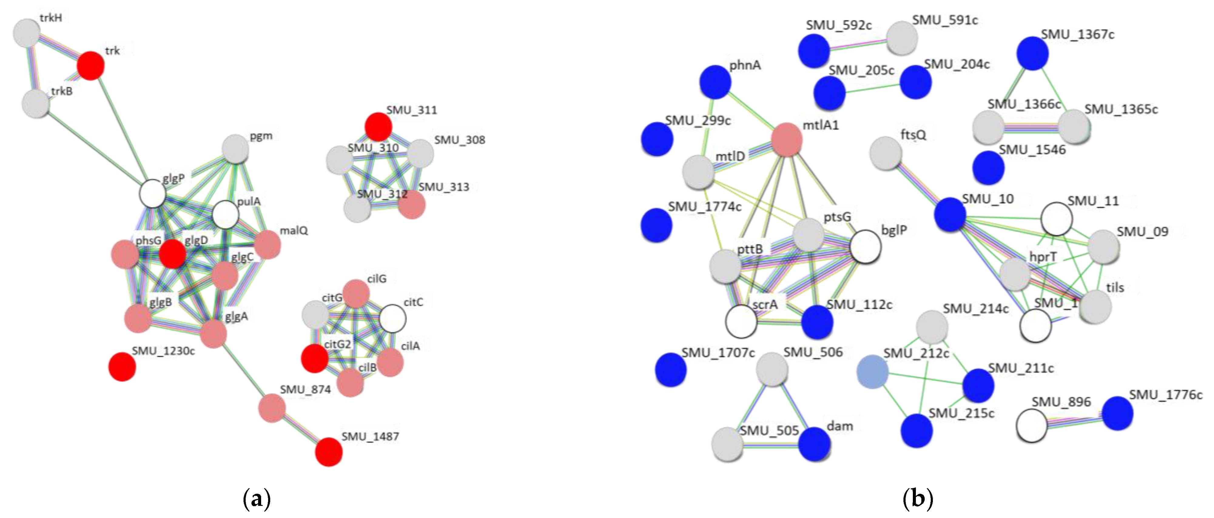
2.3. Protein–Protein Interaction Network Analysis of Upregulated and Downregulated Genes of S. mutans Treated with Scallop-Derived Hydroxyapatite

The PPI network analysis was performed on the 6 upregulated and 15 downregulated genes found in all three sets of conditions. Among the upregulated genes, 5 of the 6 (citG2, glgD, trk, SMU_311, and SMU_1487, but not SMU_1230c) were in a network of ≥5 genes, with the greatest interaction around glgD (Figure 3a). In contrast, only 3 of the 15 downregulated genes (phnA, SMU_10, and SMU_112c) were in a network of ≥5 genes (Figure 3b).

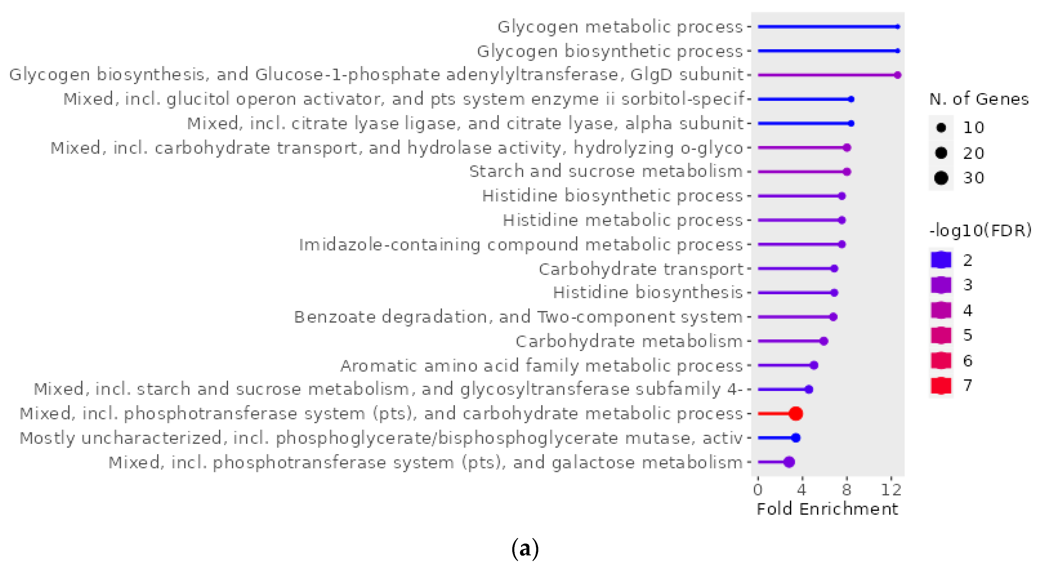
2.4. Gene Ontology Enrichment Analysis of Genes of S. mutans Upregulated on Treatment with Scallop-Derived Hydroxyapatite

The GO enrichment analysis was performed using ShinyGO for the 156 upregulated and 142 downregulated genes of S. mutans in the presence of hydroxyapatite (i.e., genes that were among the top 5% of up/downregulated genes in at least one of the 0% vs. 0.1%, 0% vs. 1%, and 0% vs. 10% hydroxyapatite conditions). The 156 upregulated genes were found to be related to pathways including the metabolism and biosynthesis of glycogen and histidine (Figure 4a,b). Interactive plots that show the relationships between enriched pathways highlighted interactions involving carbohydrate metabolism, including glycogen metabolism and biosynthetic processes; amino acid metabolism, including histidine metabolism and biosynthetic processes; and the phosphotransferase system (Figure 4c). In contrast, the analysis of the 142 downregulated genes had no pathway.

2.5. Principal Component Analysis and Heat Map Constructed Using Upregulated and Downregulated Genes of S. mutans Treated with Scallop-Derived Hydroxyapatite

A principal component analysis (PCA) using the 1398 filter-passing genes showed that PC1 (which accounted for 51.8% of the variance) had a peak change at 0.1% hydroxyapatite, with smaller changes at 1% and 10% (Figure 5a). PC2 (29.6%) showed a hydroxyapatite-concentration-dependent increase in the change. A heatmap representing the expression level of the 1398 filter-passing genes showed an accumulation of 109 upregulated genes in the presence of 10% hydroxyapatite (Figure 5b,c).

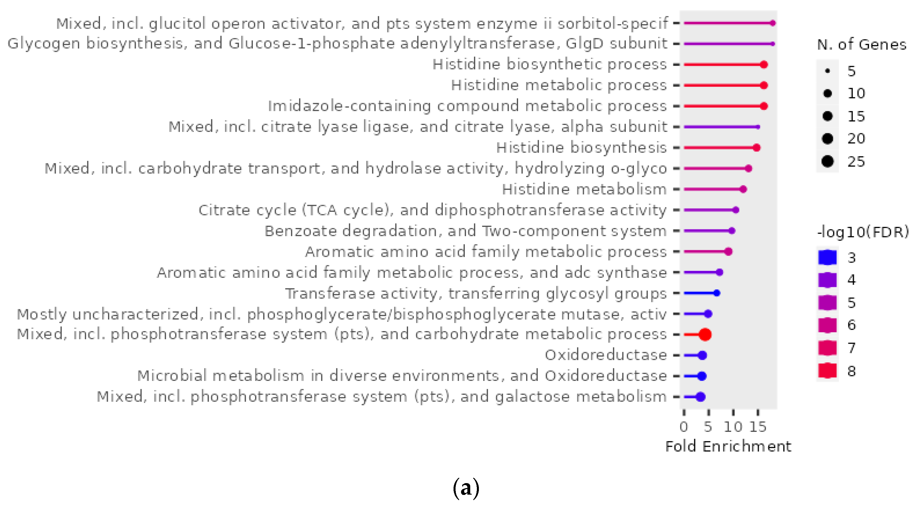
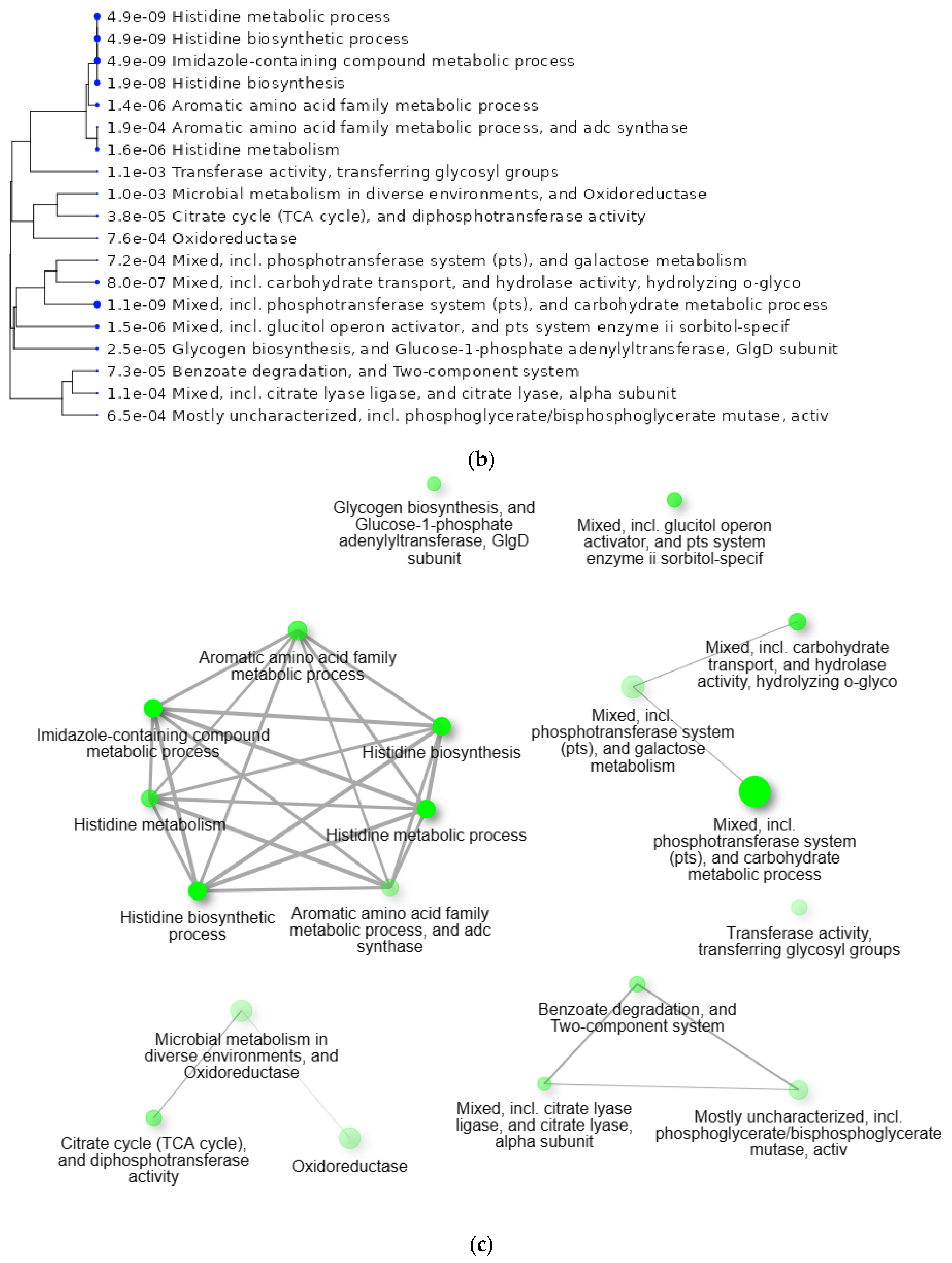
2.6. GO Enrichment Analysis of 109 Genes of S. mutans Upregulated in the Presence of 10% Scallop-Derived Hydroxyapatite

The GO enrichment analysis was performed using ShinyGO for the 109 upregulated genes of S. mutans that specifically accumulated in the presence of 10% scallop-derived hydroxyapatite (Figure 6a,b). The 109 upregulated genes were related to pathways, including histidine metabolic and biosynthetic processes, the aromatic amino acid family metabolic process, and the phosphotransferase system. Interactive plots show relationships between enriched pathways involving amino acid metabolism, including histidine metabolism and biosynthesis (Figure 6c).

2.7. Analysis of Bacterial Growth and Morphology of S. mutans in the Presence of Scallop-Derived Hydroxyapatite

Adsorption of S. mutans onto hydroxyapatite caused changes in gene expression that indicated abnormalities in metabolic and biosynthetic processes. We hypothesized that the presence of hydroxyapatite might kill S. mutans; however, the number of S. mutans adsorbed on hydroxyapatite was not significantly different from that of S. mutans in PBS for the same time period (Figure S1).
Next, we analyzed the bacterial growth and the morphology of S. mutans adsorbed on hydroxyapatite because abnormalities in metabolic or biosynthetic processes of carbohydrate and histidine can cause growth inhibition and morphological changes of bacteria [22,23,24]. When 1.0 × 10⁷ CFU/mL of S. mutans was reacted with hydroxyapatite for 10 s and was grown in a Brain Heart Infusion (BHI) broth at 37 °C, 0.01% and 0.1% hydroxyapatite had little effect on the bacterial growth (Figure 7a). The 1% hydroxyapatite reduced the number of S. mutans up to 6 h, but the bacteria grew after 6 h. Ten percent hydroxyapatite reduced the number of S. mutans bacteria at all time points. Scanning electron microscopy of S. mutans adsorbed onto hydroxyapatite in PBS showed that the outline of the bacteria was poorly marginated (Figure 7b).

3. Discussion

In the present in vitro study, we focused on treatment with hydroxyapatite derived from scallop shells as a novel method to inhibit S. mutans. The use of hydroxyapatite derived from scallop shells, which was originally an industrial waste product [25,26], is meaningful in terms of environmental protection. The scallop shells contain magnesium and other marine minerals, and they have a better biocompatibility than the widely known mineral-derived hydroxyapatite [27].
Many kinds of toothpaste containing hydroxyapatite have been developed [28], but most relevant studies have focused on how and whether they strengthen the dental structure, rather than the adsorption of oral bacteria as their effect. Our results show that hydroxyapatite derived from scallop shells has a high ability to adsorb S. mutans. At a high concentration (1% to 10%), this hydroxyapatite adsorbed S. mutans within a few minutes, and at a low concentration (0.01%, 0.1%), it showed an adsorption effect within a few hours. These results suggest that the application of scallop-derived hydroxyapatite in daily oral hygiene may be effective in lessening S. mutans in the oral cavity.
Hydroxyapatite adsorbs various bacteria [29]. However, no studies have analyzed the adsorption of S. mutans onto shell-derived hydroxyapatite and its effect on gene expression in S. mutans. Here, we identified changes in gene expression in S. mutans induced by adsorption onto scallop-derived hydroxyapatite. RNA sequencing results showed that multiple genes of S. mutans were up- or downregulated after adsorption onto hydroxyapatite. The GO enrichment analysis using these genes revealed pathways in which the upregulated genes were involved that may have significant physiological effects on S. mutans.
A PPI network analysis and GO enrichment analysis are widely used following gene expression analyses for clustering potentially up- or downregulated proteins and clarifying the biological significance of the obtained clusters, respectively [30,31]. Nevertheless, few studies have reported on the use of these methods for S. mutans [32,33]. In the present study, the PPI network analysis using genes whose expression increased in the presence of hydroxyapatite revealed that the glg operon, including glgA, glgB, glgC, and glgD, was part of the largest network. The glg operon is involved in the synthesis of glycogen via ADP–glucose from glucose-1-phosphate produced from glucose upstream of the Emden–Meyerhof pathway [34,35]. Therefore, S. mutans adsorbed onto hydroxyapatite may store excess glycogen. Glycogen metabolism and biosynthesis processes were also identified in the GO enrichment analysis and this may be the major effect of scallop-derived hydroxyapatite on S. mutans.
PCA is widely used as a method to overview gene expression changes [36]. Here, PC1 in the PCA showed a peak of change at a low concentration (0.1%) of hydroxyapatite, indicating a major difference in gene expression in S. mutans in the presence and absence of hydroxyapatite. PC2 showed a hydroxyapatite-concentration-dependent increase in the gene expression change. A heatmap indicated that 109 upregulated genes accumulated in the presence of a high concentration (10%) of hydroxyapatite compared with the absence of the hydroxyapatite. We performed the GO enrichment analysis using these 109 upregulated genes, which mainly highlighted an effect on histidine metabolic and biosynthetic processes; this result indicates that a high concentration of hydroxyapatite particularly affects histidine metabolism and biosynthesis.
Hydroxyapatite inhibited the growth of S. mutans and caused changes in the morphology of the bacteria. Studies have reported that in some bacteria, the abnormal expression of genes relating to carbohydrate metabolism results in the inhibition of bacterial growth and changes in the cell shape [22,23,37,38]. In addition, S. mutans showed growth inhibition and morphological changes when genes involved in histidine phosphorylation were mutated [24]. Furthermore, links between the inhibition of bacterial growth and cell shape have been reported [39,40]. Therefore, S. mutans adsorbed onto hydroxyapatite may undergo reciprocal processes of metabolic overexpression, growth inhibition, and morphological change. The oral microbiome has been the focus of much research in recent years [41]; a detailed analysis of the effects of hydroxyapatite on this microbiome is needed in the future.
Our study has certain limitations: the first concerns hydroxyapatite’s zeta potential. The zeta potential of ionic non-substituted hydroxyapatite is a negative charge [42,43], and the zeta potential of hydroxyapatite used in this study is unlikely to favor adhesion to S. mutans since the zeta potential of S. mutans is also a negative charge. According to recent reports, ion doping has changed the zeta potential of hydroxyapatite to a positive charge and acquired higher antibacterial properties [44,45]. Therefore, it is necessary to prepare hydroxyapatite with a positive zeta potential and analyze the changes in the gene expression of S. mutans in the presence of hydroxyapatite to achieve a higher inhibitory effect against S. mutans. The second limitation is regarding the ions of hydroxyapatite. Since ions released from antimicrobial substances can alter the adhesion capacity and gene expression of bacteria [46], ions released from hydroxyapatite can alter the gene expression of S. mutans. In this study, we used non-substituted hydroxyapatite with ions derived from shells. In the future, hydroxyapatite derived from various biogenic sources and hydroxyapatite substituted with ions should be analyzed for the relationship between the amount of ionic release and changes in the gene expression of S. mutans.

4. Materials and Methods

4.1. S. mutans Strain and Culture Conditions

The S. mutans strain MT8148 (serotype c) was cultured on Mitis Salivarius agar plates (Difco Laboratories) containing bacitracin (0.2 U/mL; Sigma-Aldrich, St. Louis, MO, USA) and 15% (w/v) sucrose (MSB-agar) at 37 °C for 48 h [47]. A single colony was inoculated into the BHI broth and cultured at 37 °C for 18 h and used in subsequent studies.

4.2. Scallop-Derived Hydroxyapatite

This study used a conventional wet method to prepare the hydroxyapatite derived from scallop shells [48]. The hydroxyapatite was ionically non-substituted and confirmed using an X-ray Fluorescence Analysis (XRF), Energy Dispersive X-ray Spectroscopy (EDX), and ion chromatography.

4.3. The Adsorption of S. mutans onto Scallop-Derived Hydroxyapatite

Hydroxyapatite powder was obtained from scallop shells collected in Hokkaido, Japan, and was provided by TSET Co. (Aichi, Japan). Cultured bacteria were collected using centrifugation at 1000× g at 4 °C for 10 min. The cultures were washed and resuspended in PBS to an OD550 value of 1.0, which corresponds to 1 × 10⁹ CFU/mL. Scallop-derived hydroxyapatite powder was then added to the bacterial suspensions at final concentrations of 0%, 0.01%, 0.1%, 1%, and 10%. The bacterial suspension was vortexed for 10 s to allow the bacteria to react with hydroxyapatite, and then stood at 37 °C for 5 min, 30 min, 3 h, or 24 h. Then, 100 µL of liquid containing S. mutans that had not adsorbed on the hydroxyapatite and precipitated at the bottom of the solution was collected from the top of the vessel. This potential bacterial suspension was cultured on MSB-agar at 37 °C for 48 h. The number of S. mutans adsorbed onto hydroxyapatite was calculated by subtracting the number of S. mutans not adsorbed on hydroxyapatite in the hydroxyapatite-added group at each time point from the number of S. mutans in the group without hydroxyapatite at each time point. In addition, the bacterial suspension of S. mutans that reacted with hydroxyapatite was sonicated and cultured on MSB-agar at 37 °C for 48 h to determine the total number of bacteria. All assays were carried out three times, and mean and standard deviation values were determined.

4.4. RNA Sequencing and FASTQ File Processing

S. mutans was treated with scallop-derived hydroxyapatite at various concentrations (0.1%, 1%, or 10%) for 24 h. Bacterial cells were lysed using Qiazol (Qiagen, Germantown, MD, USA) and the total RNA of S. mutans was isolated using an miRNeasy Micro Kit (Qiagen) according to the manufacturer’s instructions. Library preparation was performed using a GenNext RamDA-seq Single Cell Kit (Toyobo, Tokyo, Japan). Whole transcriptome sequencing was executed with an Illumina NovaSeq 6000 platform in the 100-base single-end mode. Sequenced reads were mapped to the reference genome sequence (S. mutans UA159; GenBank Accession: NC_004350.2) using HISAT2 ver. 2.1.0. Counts per gene were calculated with featureCounts v2.0.0.

4.5. Analysis of RNA Sequencing Count Data

The RNA sequencing counts were imported into Subio Platform v1.24.5853 (Subio Inc.; https://www.subioplatform.com, accessed on 8 March 2023) for the preprocessing, filtering, and extraction of differentially expressed genes. In the preprocessing, we turned the counts into log2 values and applied global normalization at the 90th percentile. Furthermore, we set the lower limit as 20 in the linear scale by replacing counts <20 with 20. Finally, we calculated the log2 ratio compared with the control sample. In the filtering, we excluded counts of tRNAs, rRNAs, and genes that were always <20 or had log2 ratios in the range −0.25 to 0.25 in all samples; 1398 genes remained after this filtering. We extracted the top 5% of up- or downregulated genes at each hydroxyapatite concentration from these filter-passed genes.

4.6. Bioinformatic Analysis

The GO enrichment analysis was performed using ShinyGO 0.77 online resources (http://bioinformatics.sdstate.edu/go/, accessed on 8 March 2023). A p-value cut-off of 0.05 for the false discovery rate was used to determine the genes used for the GO enrichment analysis. We used these genes to establish PPI networks based on the StringApp11.5 (Search Tool for the Retrieval of Interacting Genes/Proteins) online database (https://string-db.org/, accessed on 8 March 2023). Then, the most significant modules in the PPI networks were visualized. In addition, we performed PCA using the filtered genes.

4.7. Bacterial Growth Assay

A bacterial growth assay was performed in accordance with a previously described method, with some modifications [49,50]. Briefly, cultured bacteria were added to the BHI broth at a final concentration of 1.0 × 10⁷ CFU/mL in the presence of scallop-derived hydroxyapatite powder at a final concentration of 0%, 0.01%, 0.1%, 1%, and 10%. The bacterial suspensions were vortexed for 10 s to adsorb bacteria onto the hydroxyapatite. The bacterial mixtures were cultured at 37 °C for 3, 6, 12, and 24 h and then spread onto MSB-agar plates. The plates were incubated anaerobically at 37 °C for 48 h and the number of colonies was counted. All assays were carried out three times, and mean and standard deviation values were determined.

4.8. Electron Microscopy

An observation using electron microscopy was performed in accordance with a previously described method [51,52]. As a preparation for scanning electron microscopy (SEM) imaging, each bacterial sample was washed and fixed with 2% osmium tetroxide and 1% glutaraldehyde, dehydrated with ethanol, and then dried with t-butyl alcohol with the freeze-drying method. The dried samples were mounted on the stage and coated with osmium for conductive processing and then observed with SEM.

4.9. Statistical Analysis

GraphPad Prism 9 software (GraphPad Software Inc., La Jolla, CA, USA) was used for statistical analyses. Comparisons between two groups were performed using a Student’s t-test. Differences between multiple groups for each assay were determined using an analysis of variance. Bonferroni correction was used for a post hoc analysis. Results were considered significantly different at p < 0.05.

5. Conclusions

Hydroxyapatite derived from scallop shells showed a high adsorption capacity for S. mutans. A comprehensive genetic analysis using RNA sequencing revealed that in hydroxyapatite, changes in metabolic and biosynthetic processes, especially those related to glycogen and histidine, of S. mutans were promoted, as well as the inhibition of the bacterial growth and morphological changes. Based on the results of our in vitro study, this hydroxyapatite may influence the gene network of S. mutans and help maintain oral health by decreasing the risk of dental caries induction.

Supplementary Materials

The supporting information can be downloaded at: https://www.mdpi.com/article/10.3390/ijms241411371/s1.

Author Contributions

Conceptualization, K.F., T.T., K.N. and R.N.; methodology, M.H. and R.N.; software, M.H. and R.N.; validation, M.H., K.N. and R.N.; formal analysis, M.U., M.K., M.H., Y.S., S.M., R.O., S.N., M.M.-N., T.A., C.M., K.K. (Kazuya Koumoto), K.K. (Keiko Kawauchi), T.N., M.Y. and T.M.; investigation, M.U., M.K., M.H., Y.S., S.M., R.O., S.N., M.M.-N., T.A., C.M., K.K. (Kazuya Koumoto), K.K. (Keiko Kawauchi), T.N., M.Y. and T.M.; resources, T.M. and K.F.; writing—original draft preparation, M.H. and R.N.; writing—review and editing, M.H., K.N. and R.N.; supervision, K.N.; funding acquisition, K.F., T.T. and K.N. All authors have read and agreed to the published version of the manuscript.

Funding

This work was supported by the Fund for Scientific Promotion of TSET Co., Ltd. (Kariya, Japan) and Pharmacrea Kobe Co., Ltd. (Kobe, Japan).

Institutional Review Board Statement

Not applicable.

Informed Consent Statement

Not applicable.

Data Availability Statement

The data are available from the corresponding author upon reasonable request.

Acknowledgments

We thank the Genome Information Research Center, Research Institute for Microbial Diseases, Osaka University (Osaka, Japan) for supporting the RNA sequencing analysis.

Conflicts of Interest

The authors declare no conflict of interest.

References

  1. Marsh, P.D. Are dental diseases examples of ecological catastrophes? Microbiology 2003, 149 Pt 2, 279–294. [Google Scholar] [CrossRef]
  2. Marsh, P.D. Microbial ecology of dental plaque and its significance in health and disease. Adv. Dent. Res. 1994, 8, 263–271. [Google Scholar] [CrossRef] [PubMed]
  3. Lemos, J.A.; Palmer, S.R.; Zeng, L.; Wen, Z.T.; Kajfasz, J.K.; Freires, I.A.; Abranche, J.; Brady, L.J. The biology of Streptococcus mutans. Microbiol. Spectr. 2019, 7, 7. [Google Scholar] [CrossRef] [PubMed]
  4. Jiao, Y.; Tay, F.R.; Niu, L.N.; Chen, J.H. Advancing antimicrobial strategies for managing oral biofilm infections. Int. J. Oral Sci. 2019, 11, 28. [Google Scholar] [CrossRef] [PubMed] [Green Version]
  5. Segura-Egea, J.J.; Gould, K.; Sen, B.H.; Jonasson, P.; Cotti, E.; Mazzoni, A.; Sunay, H.; Tjäderhane, L.; Dummer, P.M.H. Antibiotics in endodontics: A review. Int. Endod. J. 2017, 50, 1169–1184. [Google Scholar] [CrossRef]
  6. Supová, M. Isolation and preparation of nanoscale bioapatites from natural sources: A review. J. Nanosci. Nanotechnol. 2014, 14, 546–563. [Google Scholar] [CrossRef]
  7. Wang, M.; Zhu, Y. Defect induced charge redistribution and enhanced adsorption of lysozyme on hydroxyapatite for efficient antibacterial activity. Langmuir 2021, 37, 10786–10796. [Google Scholar] [CrossRef]
  8. Shang, B.; Wang, S.; Lu, L.; Ma, H.; Liu, A.; Zupanic, A.; Jiang, L.; Elnawawy, A.S.; Yu, Y. Poultry eggshell-derived antimicrobial materials: Current status and future perspectives. J. Environ. Manag. 2022, 314, 115096. [Google Scholar] [CrossRef]
  9. Kolmas, J.; Groszyk, E.; Kwiatkowska-Różycka, D. Substituted hydroxyapatites with antibacterial properties. Biomed. Res. Int. 2014, 2014, 178123. [Google Scholar] [CrossRef] [Green Version]
  10. Dorcioman, G.; Grumezescu, V.; Stan, G.E.; Chifiriuc, M.C.; Gradisteanu, G.P.; Miculescu, F.; Matei, E.; Popescu-Pelin, G.; Zgura, I.; Craciun, V.; et al. Hydroxyapatite thin films of marine origin as sustainable candidates for dental implants. Pharmaceutics 2023, 15, 1294. [Google Scholar] [CrossRef]
  11. Devine, D.A.; Marsh, P.D.; Meade, J. Modulation of host responses by oral commensal bacteria. J. Oral Microbiol. 2015, 7, 26941. [Google Scholar] [CrossRef]
  12. Mira, A. Oral microbiome studies: Potential diagnostic and therapeutic implications. Adv. Dent. Res. 2018, 29, 71–77. [Google Scholar] [CrossRef] [PubMed]
  13. Meto, A.; Colombari, B.; Odorici, A.; Giva, L.B.; Pericolini, E.; Regina, A.L.; Blasi, E. Antibacterial effects of MicroRepair ®BIOMA-Based toothpaste and chewing gum on orthodontic elastics contaminated in vitro with saliva from healthy donors: A pilot study. Appl. Sci. 2020, 10, 6721. [Google Scholar] [CrossRef]
  14. Odorici, A.; Colombari, B.; Bellini, P.; Meto, A.; Venturelli, I.; Blasi, E. Novel options to counteract oral biofilm formation: In vitro evidence. Int. J. Environ. Res. Public Health 2022, 19, 8056. [Google Scholar] [CrossRef]
  15. Sadat-Shojai, M.; Khorasani, M.T.; Dinpanah-Khoshdargi, E.; Jamshidi, A. Synthesis methods for nanosized hydroxyapatite with diverse structures. Acta Biomater. 2013, 9, 7591–7621. [Google Scholar] [CrossRef] [PubMed]
  16. Cordonnier, T.; Sohier, J.; Rosset, P.; Layrolle, P. Biomimetic materials for bone tissue engineering—State of art and future trends. Adv. Eng. Mater. 2011, 13, 135–150. [Google Scholar] [CrossRef]
  17. Mucalo, M.R. Special issue: Novel advances and approaches in biomedical materials based on calcium phosphates. Materials 2019, 12, 405. [Google Scholar] [CrossRef] [PubMed] [Green Version]
  18. Mitran, V.; Ion, R.; Miculescu, F.; Necula, M.G.; Mocanu, A.-C.; Stan, G.E.; Antoniac, I.V.; Cimpean, A. Osteoblast cell response to naturally derived calcium phosphate-based materials. Materials 2018, 11, 1097. [Google Scholar] [CrossRef] [Green Version]
  19. Ressler, A.; Žužić, A.; Ivanišević, I.; Kamboj, N.; Ivanković, H. Ionic substituted hydroxyapatite for bone regeneration applications: A review. Open Ceram. 2021, 6, 100122. [Google Scholar] [CrossRef]
  20. DileepKumar, V.G.; Sridhar, M.S.; Aramwit, P.; Krut’ko, V.K.; Musskaya, O.N.; Glazov, I.E.; Reddy, N. A review on the synthesis and properties of hydroxyapatite for biomedical applications. J. Biomater. Sci. Polym. Ed. 2022, 33, 229–261. [Google Scholar] [CrossRef]
  21. Anil, A.; Ibraheem, W.I.; Meshni, A.A.; Preethanath, R.S.; Anil, S. Nano-hydroxyapatite (nHAp) in the remineralization of early dental caries: A scoping review. Int. J. Environ. Res. Public Health 2022, 19, 5629. [Google Scholar] [CrossRef] [PubMed]
  22. Marr, A.K.; Joseph, B.; Mertins, S.; Ecke, R.; Müller-Altrock, S.; Goebel, W. Overexpression of PrfA leads to growth inhibition of Listeria monocytogenes in glucose-containing culture media by interfering with glucose uptake. J. Bacteriol. 2006, 188, 3887–3901. [Google Scholar] [CrossRef] [Green Version]
  23. de Young, K.D.; Stankeviciute, G.; Klein, E.A. Sugar-phosphate metabolism regulates stationary-phase entry and stalk elongation in Caulobacter crescentus. J. Bacteriol. 2020, 202, e00468-19. [Google Scholar] [CrossRef]
  24. Wang, S.; Long, L.; Yang, X.; Qiu, Y.; Tao, T.; Peng, X.; Li, Y.; Han, A.; Senadheera, D.B.; Downey, J.S.; et al. Dissecting the role of VicK phosphatase in aggregation and biofilm formation of Streptococcus mutans. J. Dent. Res. 2021, 100, 631–638. [Google Scholar] [CrossRef]
  25. Gerhard, E.M.; Wang, W.; Li, C.; Guo, J.; Ozbolat, I.T.; Rahn, K.M.; Armstrong, A.D.; Xia, J.; Qian, G.; Yang, J. Design strategies and applications of nacre-based biomaterials. Acta Biomater. 2017, 54, 21–34. [Google Scholar] [CrossRef]
  26. Liu, C.; Chen, S.H.; Yang-Zhou, C.H.; Zhang, Q.G.; Michael, R.N. Application of nano-hydroxyapatite derived from oyster shell in fabricating superhydrophobic sponge for efficient oil/water separation. Molecules 2021, 26, 3703. [Google Scholar] [CrossRef]
  27. Zhou, Y.; Li, Z.; Zhang, H.; Hu, Q.; Zou, Y. Potential uses of scallop shell powder as a substrate for the cultivation of king oyster mushroom (Pleurotus eryngii). Horticulturae 2022, 8, 333. [Google Scholar] [CrossRef]
  28. Limeback, H.; Meyer, F.; Enax, J. Tooth whitening with hydroxyapatite: A systematic review. Dent. J. 2023, 11, 50. [Google Scholar] [CrossRef] [PubMed]
  29. Sato, Y.; Ishihara, M.; Nakamura, S.; Fukuda, K.; Takayama, T.; Hiruma, S.; Murakami, K.; Fujita, M.; Yokoe, H. Preparation and application of bioshell calcium oxide (BiSCaO) nanoparticle-dispersions with bactericidal activity. Molecules 2019, 24, 3415. [Google Scholar] [CrossRef] [PubMed] [Green Version]
  30. Athanasios, A.; Charalampos, V.; Vasileios, T.; Ashraf, G.M. Protein-protein interaction (PPI) network: Recent advances in drug discovery. Curr. Drug Metab. 2017, 18, 5–10. [Google Scholar] [CrossRef]
  31. Chen, L.; Zhang, Y.H.; Wang, S.; Zhang, Y.; Huang, T.; Cai, Y.D. Prediction and analysis of essential genes using the enrichments of gene ontology and KEGG pathways. PLoS ONE 2017, 12, e0184129. [Google Scholar] [CrossRef] [PubMed] [Green Version]
  32. Yost, S.; Duran-Pinedo, A.E.; Teles, R.; Krishnan, K.; Frias-Lopez, J. Functional signatures of oral dysbiosis during periodontitis progression revealed by microbial metatranscriptome analysis. Genome Med. 2015, 7, 27. [Google Scholar] [CrossRef] [PubMed] [Green Version]
  33. Qin, H.; Zou, Z.; Anderson, D.; Sang, Y.; Higashi, D.; Kreth, J.; Merritt, J. The transcription regulator BrsR serves as a network hub of natural competence protein-protein interactions in Streptococcus mutans. Proc. Natl. Acad. Sci. USA 2021, 118, e2106048118. [Google Scholar] [CrossRef] [PubMed]
  34. Carlsson, J.; Kujala, U.; Edlund, M.B. Pyruvate dehydrogenase activity in Streptococcus mutans. Infect. Immun. 1985, 49, 674–678. [Google Scholar] [CrossRef] [Green Version]
  35. Busuioc, M.; Mackiewicz, K.; Buttaro, B.A.; Piggot, P.J. Role of intracellular polysaccharide in persistence of Streptococcus mutans. J. Bacteriol. 2009, 191, 7315–7322. [Google Scholar] [CrossRef] [Green Version]
  36. David, C.C.; Jacobs, D.J. Principal component analysis: A method for determining the essential dynamics of proteins. Methods Mol. Biol. 2014, 1084, 193–226. [Google Scholar]
  37. Liao, J.C.; Chao, Y.P.; Patnaik, R. Alteration of the biochemical valves in the central metabolism of Escherichia coli. Ann. N. Y. Acad. Sci. 1994, 745, 21–34. [Google Scholar] [CrossRef]
  38. Shleeva, M.O.; Kaprelyants, A.S. Hypobiosis of mycobacteria: Biochemical aspects. Biochemistry 2023, 88, 52–74. [Google Scholar] [CrossRef]
  39. Lu, J.; Carter, D.A.; Turnbull, L.; Rosendale, D.; Hedderley, D.; Stephens, J.; Gannabathula, S.; Steinhorn, G.; Schlothauer, R.C.; Whitchurch, C.B. The effect of New Zealand kanuka, manuka and clover honeys on bacterial growth dynamics and cellular morphology varies according to the species. PLoS ONE 2013, 8, e55898. [Google Scholar] [CrossRef] [Green Version]
  40. Serbanescu, D.; Ojkic, N.; Banerjee, S. Cellular resource allocation strategies for cell size and shape control in bacteria. FEBS J. 2022, 289, 7891–7906. [Google Scholar] [CrossRef]
  41. Mitsuhata, C.; Kado, N.; Hamada, M.; Nomura, R.; Kozai, K. Characterization of the unique oral microbiome of children with Down syndrome. Sci. Rep. 2022, 12, 14150. [Google Scholar] [CrossRef] [PubMed]
  42. Reynolds, E.C.; Wong, A. Effect of adsorbed protein on hydroxyapatite zeta potential and Streptococcus mutans adherence. Infect. Immun. 1983, 39, 1285–1290. [Google Scholar] [CrossRef] [PubMed] [Green Version]
  43. Shimabayashi, S.; Hoshino, M.; Hino, T. Zeta potential of hydroxyapatite particles in the presence of polyethylene glycol mono-p-isooctylphenyl ether and sodium dodecylsulfate in an aqueous phase. Phosphorus Res. Bull. 2004, 17, 142–147. [Google Scholar] [CrossRef] [PubMed]
  44. Silingardi, F.; Bonvicini, F.; Cassani, M.C.; Mazzaro, R.; Rubini, K.; Gentilomi, G.A.; Bigi, A.; Boanini, E. Hydroxyapatite decorated with tungsten oxide nanoparticles: New composite materials against bacterial growth. J. Funct. Biomater. 2022, 3, 88. [Google Scholar] [CrossRef] [PubMed]
  45. Beiranvand, M.; Farhadi, S.; Mohammadi-Gholami, A. Ag NPs decorated on the magnetic rod-like hydroxyapatite/MIL-101(Fe) nanocomposite as an efficient catalyst for the reduction of some nitroaromatic compounds and as an effective antimicrobial agent. RSC Adv. 2023, 13, 13683–13697. [Google Scholar] [CrossRef]
  46. Nomura, R.; Morita, Y.; Matayoshi, S.; Nakano, K. Inhibitory effect of surface pre-reacted glass-ionomer (S-PRG) eluate against adhesion and colonization by Streptococcus mutans. Sci. Rep. 2018, 8, 5056. [Google Scholar] [CrossRef]
  47. Ooshima, T.; Izumitani, A.; Sobue, S.; Hamada, S. Cariostatic effect of palatinose on experimental dental caries in rats. Jpn. J. Med. Sci. Biol. 1983, 36, 219–223. [Google Scholar] [CrossRef] [Green Version]
  48. Kien, P.T.; Phu, H.D.; Linh, N.V.V.; Quyen, T.N.; Hoa, N.T. Recent trends in hydroxyapatite (HA) synthesis and the synthesis report of nanostructure HA by hydrothermal reaction. Adv. Exp. Med. Biol. 2018, 1077, 343–354. [Google Scholar]
  49. Nomura, R.; Ohata, J.; Otsugu, M.; Okawa, R.; Naka, S.; Matsumoto-Nakano, M.; Nakano, K. Inhibitory effects of flavedo, albedo, fruits, and leaves of Citrus unshiu extracts on Streptococcus mutans. Arch. Oral Biol. 2021, 124, 105056. [Google Scholar] [CrossRef]
  50. Nomura, R.; Kitamura, T.; Matayoshi, S.; Ohata, J.; Suehiro, Y.; Iwashita, N.; Okawa, R.; Nakano, K. Inhibitory effect of a gel paste containing surface pre-reacted glass-ionomer (S-PRG) filler on the cariogenicity of Streptococcus mutans. Sci. Rep. 2021, 11, 23495. [Google Scholar] [CrossRef] [PubMed]
  51. Nomura, R.; Otsugu, M.; Naka, S.; Teramoto, N.; Kojima, A.; Muranaka, Y.; Matsumoto-Nakano, M.; Ooshima, T.; Nakano, K. Contribution of Streptococcus mutans serotype k strains interaction with fibrinogen to the pathogenicity of infective endocarditis. Infect Immun. 2014, 82, 5223–5234. [Google Scholar] [CrossRef] [PubMed] [Green Version]
  52. Suehiro, Y.; Nomura, R.; Matayoshi, S.; Otsugu, M.; Iwashita, N.; Nakano, K. Evaluation of the collagen-binding properties and virulence of killed Streptococcus mutans in a silkworm model. Sci. Rep. 2022, 12, 2800. [Google Scholar] [CrossRef] [PubMed]
Figure 1. Adsorption of S. mutans onto scallop-derived hydroxyapatite. The number of S. mutans adsorbed onto scallop-derived hydroxyapatite after the reaction of the bacteria with the hydroxyapatite by vortexing for 10 s.
Ijms 24 11371 g001
Figure 2. Schema of analyses using data from RNA sequencing of S. mutans treated with scallop-derived hydroxyapatite.
Ijms 24 11371 g002
Figure 3. Protein–protein interaction (PPI) network analysis of (a) 6 upregulated and (b) 15 downregulated genes observed among the top 5% of up- or downregulated genes when S. mutans was treated with 0.1%, 1%, and 10% scallop-derived hydroxyapatite. The genes upregulated at all the concentrations of hydroxyapatite are shown in dark red, and the genes downregulated at all the concentrations are shown in dark blue. Genes upregulated at any of the concentrations are shown in pale red, and genes downregulated at any of the concentrations are shown in pale blue. Gray indicates genes whose expression did not change significantly in RNA sequencing of S. mutans treated with scallop-derived hydroxyapatite. White indicates that a gene was not detected.
Ijms 24 11371 g003
Figure 4. Gene Ontology (GO) enrichment analysis of 156 genes upregulated in S. mutans treated with scallop-derived hydroxyapatite (genes that were among the top 5% of upregulated genes in at least one of the 0% vs. 0.1%, 0% vs. 1%, and 0% vs. 10% hydroxyapatite conditions). (a) GO enrichment analysis was performed with ShinyGO and the pathways found are shown. (b) Hierarchical clustering tree summarizing correlations between pathways. Pathways with many shared genes are clustered together. Larger dots indicate more significant p-values. (c) Interactive plot showing the relationships between enriched pathways. Two pathways (nodes) are connected when they share ≥20% of genes. Darker nodes are more significantly enriched gene sets, and larger nodes represent larger gene sets. Thicker edges indicate more overlapping genes.
Ijms 24 11371 g004aIjms 24 11371 g004b
Figure 5. Principal component analysis (PCA) and heat map using 1398 genes up- or downregulated when S. mutans was treated with scallop-derived hydroxyapatite. (a) PCA of S. mutans in the presence of 0.1%, 1%, and 10% hydroxyapatite. (b) Heatmap and clustering of 1398 filter-passing upregulated or downregulated genes. (c) Detailed heatmap and clustering of 109 genes extracted from the part of (b) marked *.
Ijms 24 11371 g005aIjms 24 11371 g005b
Figure 6. GO enrichment analysis of 109 genes of S. mutans that are specifically upregulated in the presence of 10% scallop-derived hydroxyapatite compared with the absence of the hydroxyapatite. (a) GO enrichment analysis was performed on the 109 upregulated genes indicated in Figure 5c with ShinyGO and the pathways found are shown. (b) Hierarchical clustering tree summarizing correlations between pathways listed in the enrichment table pathways with many shared genes being clustered together. Larger dots indicate more significant p-values. (c) Interactive plot showing the relationships between enriched pathways. Two pathways (nodes) are connected when they share 20% or more of genes. Darker nodes are more significantly enriched gene sets, and larger nodes represent larger gene sets. Thicker edges indicate more overlapping genes.
Ijms 24 11371 g006aIjms 24 11371 g006b
Figure 7. Changes in bacterial growth and morphology of S. mutans adsorbed onto scallop-derived hydroxyapatite. (a) Growth of S. mutans treated with scallop-derived hydroxyapatite. (b) Representative scanning electron microscopy images of S. mutans adsorbed on scallop-derived hydroxyapatite. The lower panels show high-magnification images of the boxed regions in the upper images. White arrowheads indicate bacteria. Bars = 2 μm (upper images) and 500 nm (lower images).
Ijms 24 11371 g007aIjms 24 11371 g007b
Disclaimer/Publisher’s Note: The statements, opinions and data contained in all publications are solely those of the individual author(s) and contributor(s) and not of MDPI and/or the editor(s). MDPI and/or the editor(s) disclaim responsibility for any injury to people or property resulting from any ideas, methods, instructions or products referred to in the content.

