Article

Identification of N-Acyl Hydrazones as New Non-Zinc-Binding MMP-13 Inhibitors by Structure-Based Virtual Screening Studies and Chemical Optimization

1
Department of Pharmacy, University of Pisa, Via Bonanno 6, 56126 Pisa, Italy
2
Research Group in Cheminformatics & Nutrition, Departament de Bioquímica i Biotecnologia, Universitat Rovira i Virgili, Campus de Sescelades, 43007 Tarragona, Spain
*
Author to whom correspondence should be addressed.
These authors contributed equally to this work.
Int. J. Mol. Sci. 2023, 24(13), 11098; https://doi.org/10.3390/ijms241311098
Submission received: 18 May 2023 / Revised: 23 June 2023 / Accepted: 27 June 2023 / Published: 4 July 2023
(This article belongs to the Special Issue Recent Advances in Virtual Screening 3.0)

Abstract

:
Matrix metalloproteinase 13 plays a central role in osteoarthritis (OA), as its overexpression induces an excessive breakdown of collagen that results in an imbalance between collagen synthesis and degradation in the joint, leading to progressive articular cartilage degradation. Therefore, MMP-13 has been proposed as a key therapeutic target for OA. Here we have developed a virtual screening workflow aimed at identifying selective non-zinc-binding MMP-13 inhibitors by targeting the deep S1′ pocket of MMP-13. Three ligands were found to inhibit MMP-13 in the µM range, and one of these showed selectivity over other MMPs. A structure-based analysis guided the chemical optimization of the hit compound, leading to the obtaining of a new N-acyl hydrazone-based derivative with improved inhibitory activity and selectivity for the target enzyme.

1. Introduction

Osteoarthritis (OA) is the most common form of arthritis [1], affecting half of the elderly population (>65 years) [2]. It is characterized by the progressive degradation of articular collagen and can ultimately result in the prosthetic replacement of joints as they become completely dysfunctional. Matrix metalloproteinase 13 (MMP-13), also known as collagenase-3, plays a central role in the pathology as it is the main enzyme responsible for the cleavage of type II collagen in patients with OA [3]. MMP-13 is significantly overexpressed in the joints and articular cartilage in patients with OA and is not present in normal adult cartilage; therefore, it has been proposed as a key therapeutical target for the treatment of OA [4]. MMP-13 belongs to the MMP family, which consists of 23 zinc-dependent enzymes responsible for the degradation of different extracellular matrix (ECM) components [5]. In addition to tissue remodeling, MMPs are involved in the cleavage of many non-matrix targets, such as cell surface receptors, cytokines, chemokines, cell-cell adhesion molecules, clotting factors, and other proteinases [6]. Despite some broad-spectrum MMP inhibitors (MMPIs) having halted the destruction of cartilage in preclinical assays, they have failed clinical trials as patients developed musculoskeletal syndrome (MSS), possibly resulting from the alteration of the physiological functions of different members of the MMP family [7,8]. Therefore, selectivity is currently considered a priority in the development of MMP inhibitors, even if the high homology among MMP catalytic domains makes it a challenging task. So far, in MMPI design, the catalytic zinc ion chelation has been crucial, leading to inhibitors presenting mainly a hydroxamic acid or carboxylic acid as a zinc-binding group (ZBG) [9,10], with nanomolar activity but poor selectivity. In fact, the presence of a ZBG can negatively affect the selective targeting of a specific MMP, cross-interfering with other metalloproteases such as ADAMs (A Disintegrin and Metalloproteinase) [11,12] and ADAMTSs (A Disintegrin and Metalloproteinase with Thrombospondin Motifs) [13], but also with other zinc metalloenzymes such as carbonic anhydrases [14,15] or histone deacetylases [16,17]. In this regard, the characteristics of the catalytic binding site of MMP-13 are slightly different from those of other MMPs, thus providing an edge in the identification of selective inhibitors for this enzyme. More specifically, an adjacent region to the catalytic site, known as the S1′ pocket, is different in MMP-13 as the loop that delimits the pocket (Ω-loop) is longer and shows more flexibility in MMP-13 than in other MMPs [18]. In particular, the Ω-loop of MMP-13 encloses the so-called S1″ specificity pocket. This allows for an opportunity to identify inhibitors with a different binding mode that is not possible for other enzymes in the MMP family. Several non-zinc chelating inhibitors (Figure 1) exploited this difference in the MMP-13 binding site to achieve selectivity for this enzyme, as shown by their X-ray crystal structures [19,20,21,22]. In the present paper, we used these crystallographic data to design a virtual screening (VS) methodology able to identify MMP-13 inhibitors that can adopt a similar binding mode and therefore achieve selectivity towards MMP-13. Enzymatic assays of a limited selection of candidate compounds (20) allowed us to find three novel hits that were structurally unrelated to the known MMPIs. The most promising compound underwent a hit optimization study in order to improve its activity and selectivity profile, and a novel series of 12 derivatives was synthesized and tested in vitro.

2. Results and Discussion

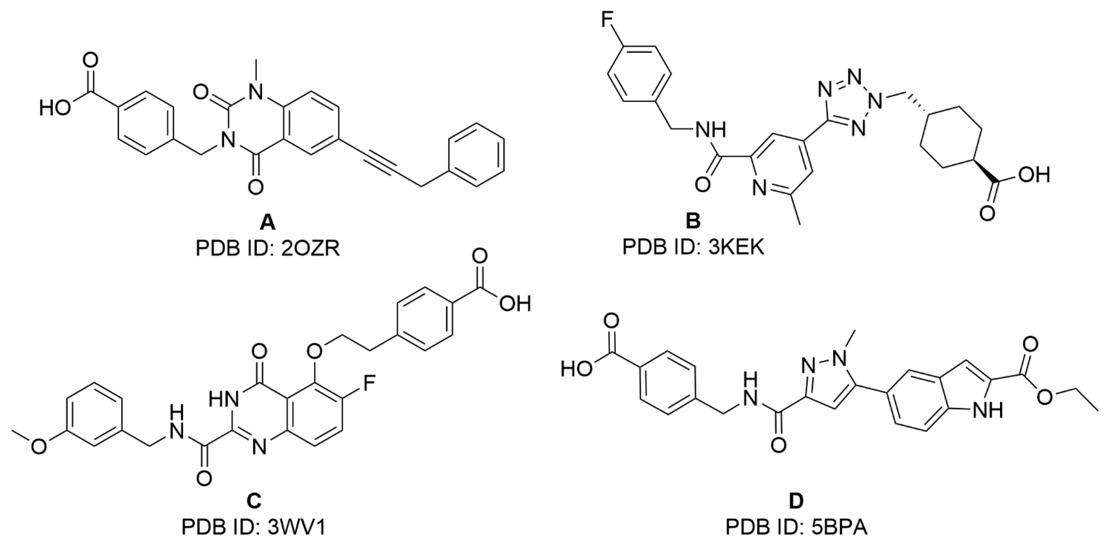
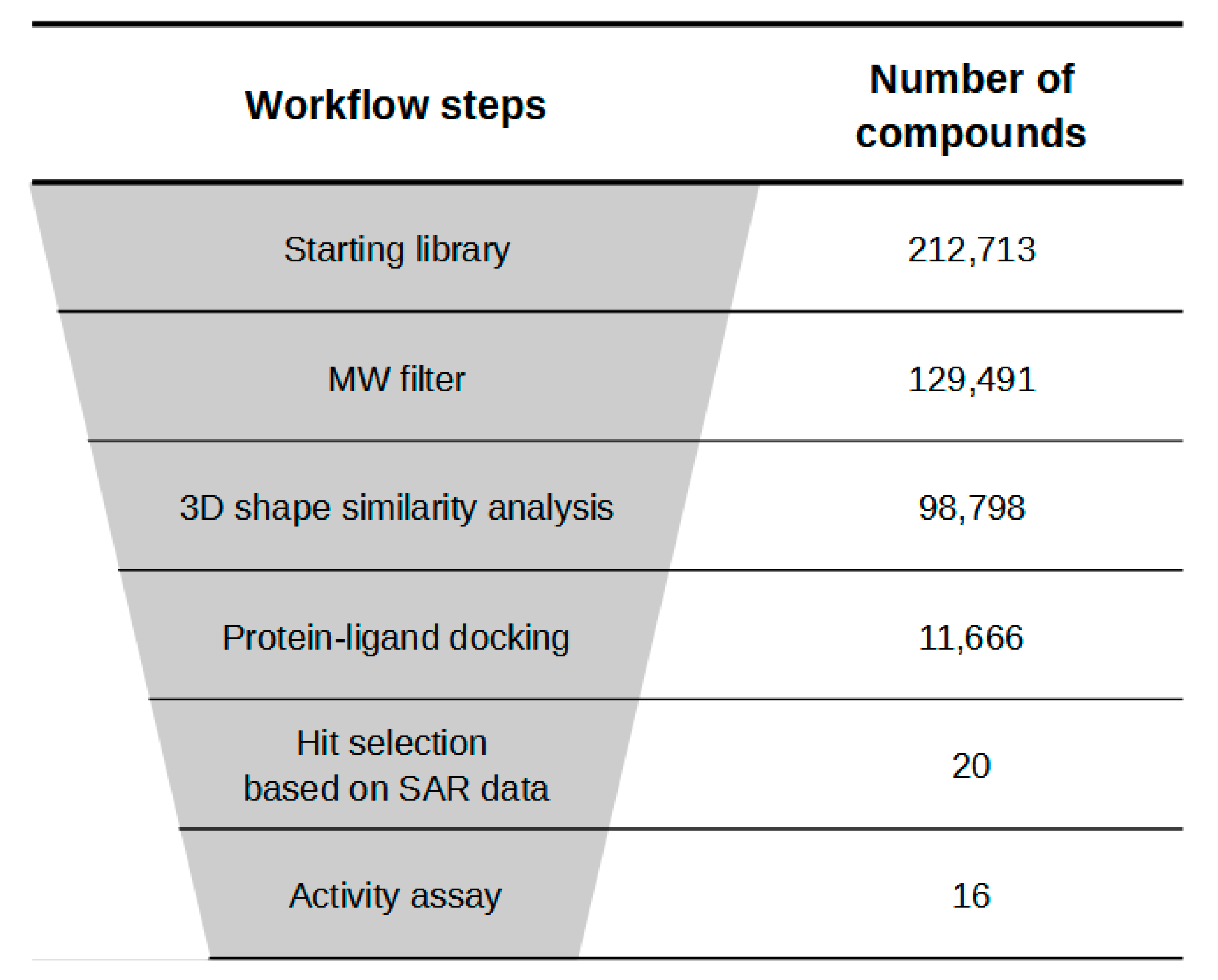
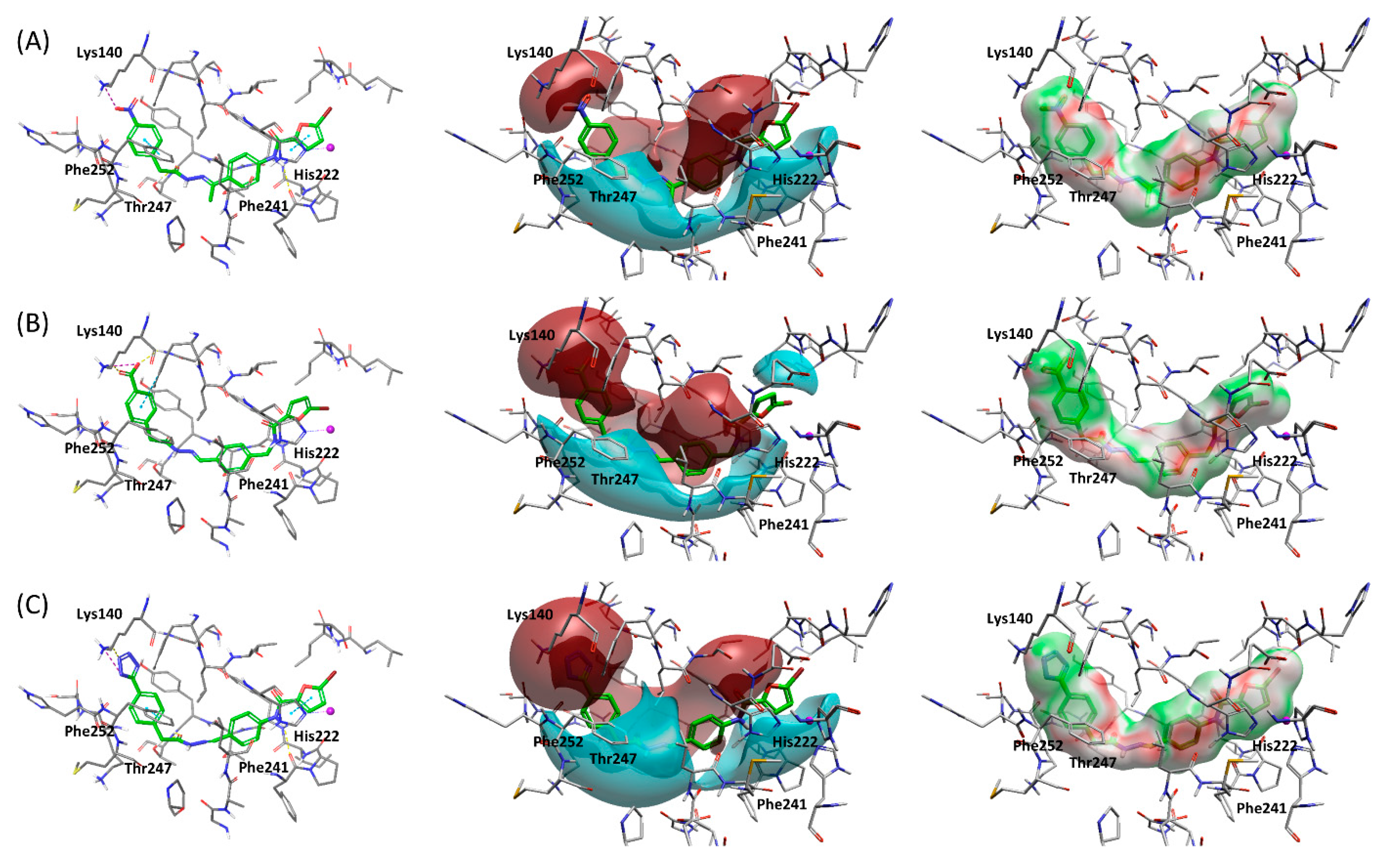
2.1. Virtual Screening Studies

Co-crystallized MMP-13 inhibitors that bind to the S1′ pocket without interacting with the catalytic zinc ion show a similar binding mode [19,20,21,22]. All of them present two common characteristics: (a) they contain two aromatic rings or ring systems at both ends of the molecule (with the exception of the co-crystallized inhibitor in the structure with PDB code: 3KEK [20], which instead presents a cyclohexyl group at one of its ends); and (b) they adopt a characteristic U shape, establishing hydrogen bond interactions between the core of the molecule and Thr245, Thr247, or both residues. We used these characteristics to design a VS workflow capable of identifying compounds that bind in a similar manner to the S1′ pocket of MMP-13. The VS workflow, which was applied to an initial library of 212,713 compounds obtained from Specs [23], consisted of four steps that are described in this section (Figure 2).
First, the compounds were filtered by molecular weight (MW); then, a shape-based alignment was performed to keep only the compounds that could adopt a conformation similar to that of the co-crystallized ligands; next, protein-ligand docking was performed on MMP-13; and, finally, compounds were selected based on the interactions with the binding site of MMP-13 that improved MMP-13 inhibitor activity in previously reported structure-activity relationship (SAR) studies. The compounds obtained from the Specs database were filtered by MW in order to discard compounds too small to fulfill the posterior protein-ligand docking constraints and compounds too large compared to the reference ligands used for the subsequent shape-based alignment, therefore reducing the computational time of the subsequent steps. The 300–700 Da range was selected as the filter, taking into account that the compounds used as references in the shape-based alignment step have MWs between 392 and 491 Da. As a result of this first step, 83,222 compounds were filtered out (Figure 2). Next, a maximum of 10 conformations were generated for each compound that survived the MW filter. These conformations were compared to those of selective MMP-13 inhibitors co-crystallized with MMP-13 that bind to the S1′ pocket and do not contain a ZBG. Only conformations similar to those of co-crystallized ligands were kept in order to reduce the computational cost of the VS, as compounds unable to adopt a similar shape to that of the co-crystallized ligands would likely not fit in the S1′ cavity during the protein-ligand docking step. As a result of this step, 30,693 compounds were filtered out (Figure 2). The ligands resulting from the shape similarity filter were docked onto MMP-13 using the crystal structure with PDB code 3WV1 [21], as it contains an inhibitor that binds to the S1′ pocket. In order to discard ligands unable to adopt a similar binding mode in the S1′ pocket to that of previously known selective MMP-13 inhibitors, two positional constraints were defined (one closer to the zinc-binding region and another one deep in the S1′ pocket) to be fulfilled by aromatic atoms. Moreover, it was required that the ligand perform a hydrogen bond interaction with Thr245 or Thr247, as all the co-crystallized inhibitors performed at least one of these interactions with the core of the molecule. In order to select the compounds that performed the appropriate interactions in the S1′ pocket of MMP-13, several SAR studies were analyzed to obtain information regarding which interactions are important to achieve high activity towards MMP-13. In particular, a potent MMP-13 inhibitor should: (1) have a negatively charged ring substituent that can establish a salt bridge interaction with Lys140, which is unique at the bottom of the S1″ side pocket of MMP-13 [21,22,24]; (2) make a π-π interaction with Tyr246 and Phe252 [21]; (3) have a hydrophobic moiety occupying the S1″ pocket [21,22]; (4) have an appropriate linker to join the S1′ pocket with the S1″ side pocket [24]; (5) have a hydrogen or halogen bond acceptor towards Met253 main chain nitrogen [21]; (6) make a hydrophobic interaction with Pro255 [20,24]; (7) have a hydrogen bond acceptor towards the side chain of Thr247 [21,22]; (8) establish hydrophobic contacts in the S1′ pocket [24]; (9) have a hydrogen bond acceptor towards Thr245 main chain nitrogen [24]; (10) have a hydrogen bond donor towards Ala238 main chain carbonyl oxygen [24]; and (11) have an appropriate ring substituent in the region close to the zinc-binding group [20,22].
After docking, the top 10 docked poses for each compound were selected based on their docking scores. The interactions they performed with the protein were carefully inspected in order to select the compounds that accomplished the criteria obtained through the analysis of the above-mentioned SAR data. Ideally, in this step, we would like to obtain a compound that meets the 11 criteria. However, this was not the case, as the compound that accomplished more criteria was compound 1 (Figure S1), with a total of eight. Therefore, this compound and compounds that fulfilled most of the criteria were selected, obtaining 20 compounds for in vitro tests (Figure S1). The structures of these 20 compounds were compared, using their molecular fingerprints, to those of previously reported MMP-13 inhibitors in the Reaxys [25] database. Except for compound 14, which showed a Tanimoto value of 0.71 with a described MMP-13 inhibitor, the Tanimoto similarity values of the selected compounds with any of the previously reported MMP-13 inhibitors analyzed were at most 0.6. Upon visual inspection, compound 14 was not considered structurally similar to the MMP-13 inhibitor obtained from Reaxys (see Figure S2). Moreover, hit compounds that were structurally similar to previously selected hit compounds were discarded to ensure that the final 20 compounds selected for in vitro tests were structurally different from each other (see Figure S3). Therefore, the hit compounds obtained by this virtual screening methodology proved not only to be different from previously reported MMP-13 inhibitors but also structurally diverse.
After the selection of the 20 hit compounds, they were purchased from Specs, and their activity for MMP-13 was analyzed in vitro. The activity data (% inhibition at 100 μM) were determined by a fluorometric assay on human recombinant enzyme, and four compounds were discarded due to very low solubility in DMSO or high fluorescence emission interfering with the assay. ARP100 [26], a hydroxamate-based MMP inhibitor previously developed by our research group, was used in the same assay conditions as the reference compound. To exclude any possible nonspecific inhibition of MMP-13 due to aggregate formation, we performed all the assays in the presence of 0.05% Brij-35, a nonionic detergent similar to Triton X-100, as indicated by Shoichet et al. [27]. Three ligands, out of the twenty tested, presented a % inhibition >40 at 100 μM and were further characterized. All other compounds were not further investigated (see Figure S1 for chemical structures). In Table 1, are displayed the structures, Specs codes, clogP, and MMP-13% inhibition of the selected ligands.

2.2. Enzymatic Assays

After the preliminary screening, compounds 11, 12, and 13 (Table 1) displayed the highest MMP-13 inhibitory activity, and their IC50 values were calculated (91 μM, 105 μM, and 14.6 μM, respectively). Next, the inhibitory activities of these three compounds towards MMP-1, MMP-2, MMP-9, and MMP-14 were determined (Table 2). The selectivity profile of the three ligands pointed out that compound 13, with an N-acyl hydrazone scaffold, presented low micromolar activity against MMP-13 and a promising selectivity over the other tested MMPs. For this reason, 13 was chosen as the hit compound and underwent an optimization study in order to improve its activity and selectivity profile. The N-acyl hydrazone group has been recently introduced in several approved drugs and molecules in clinical trials since it represents a peptide-mimetic subunit endowed with high metabolic stability [29,30].

2.3. Structure-Based Hit Optimization
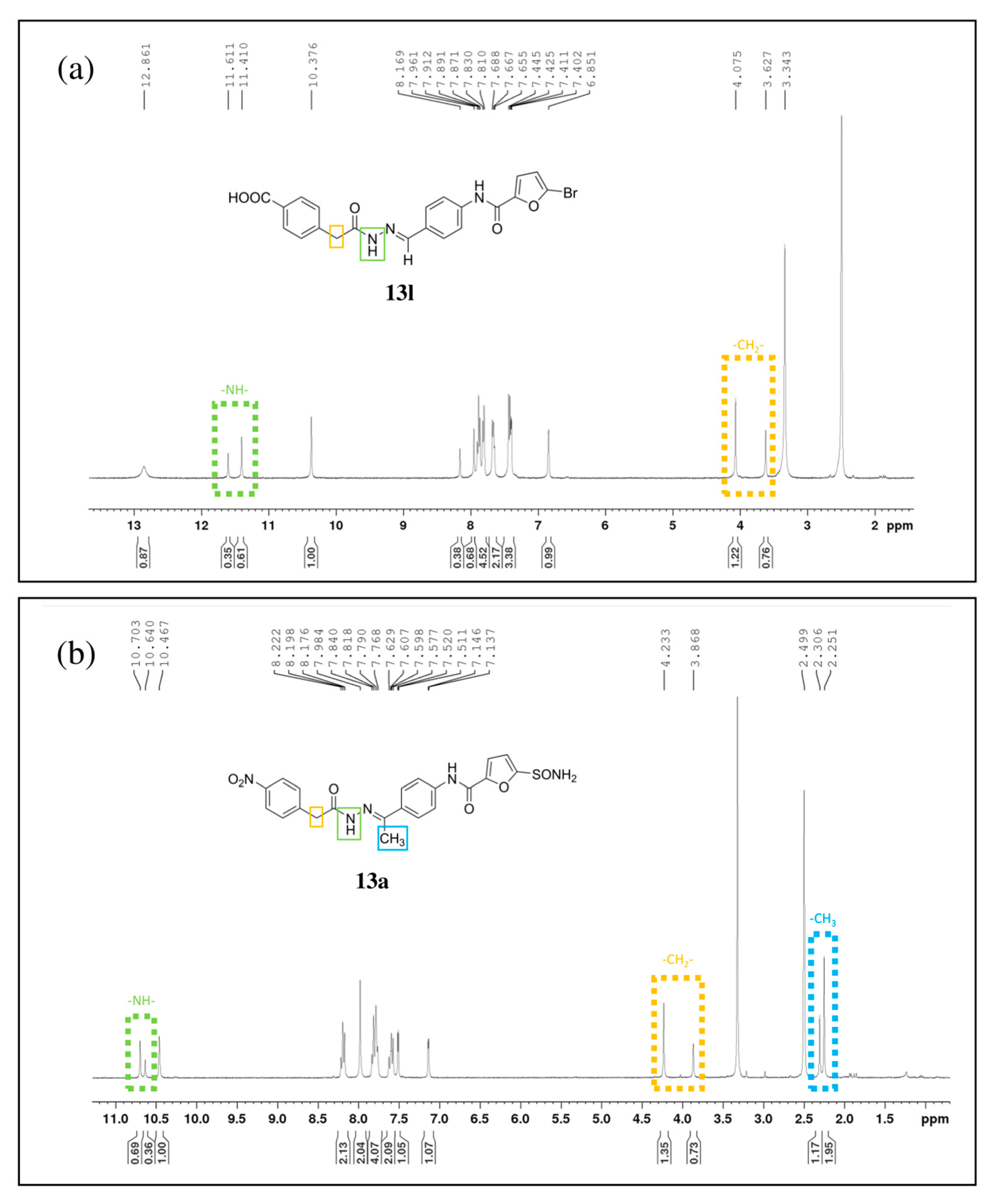
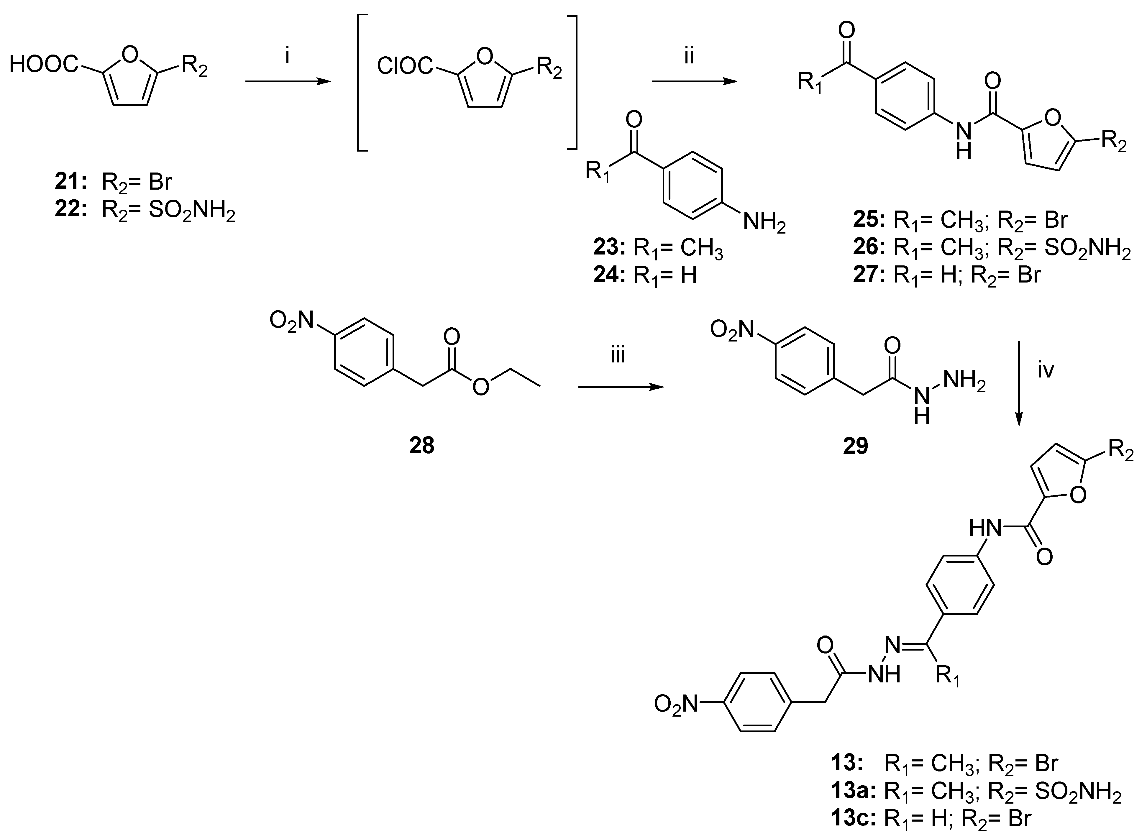
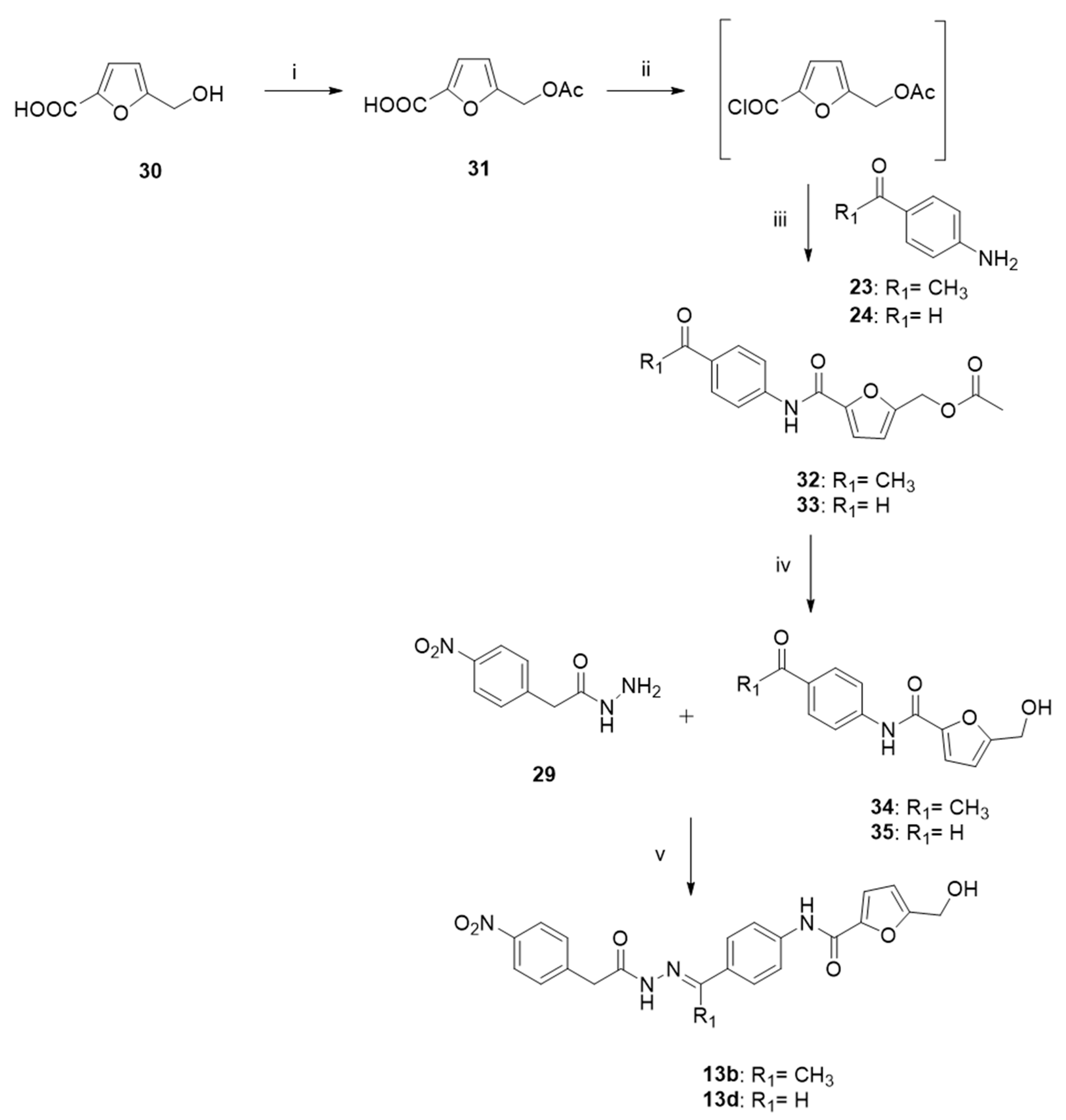
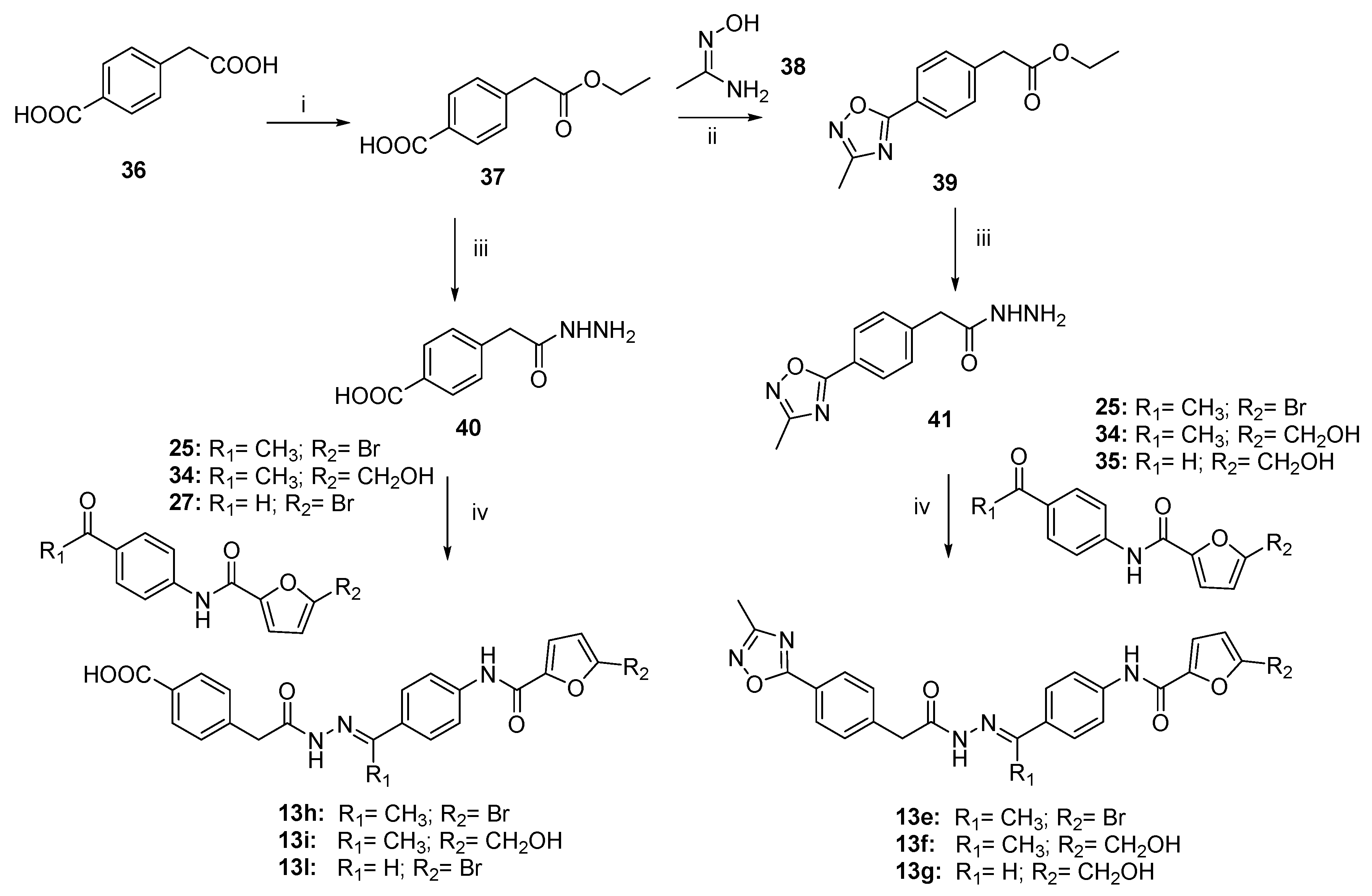
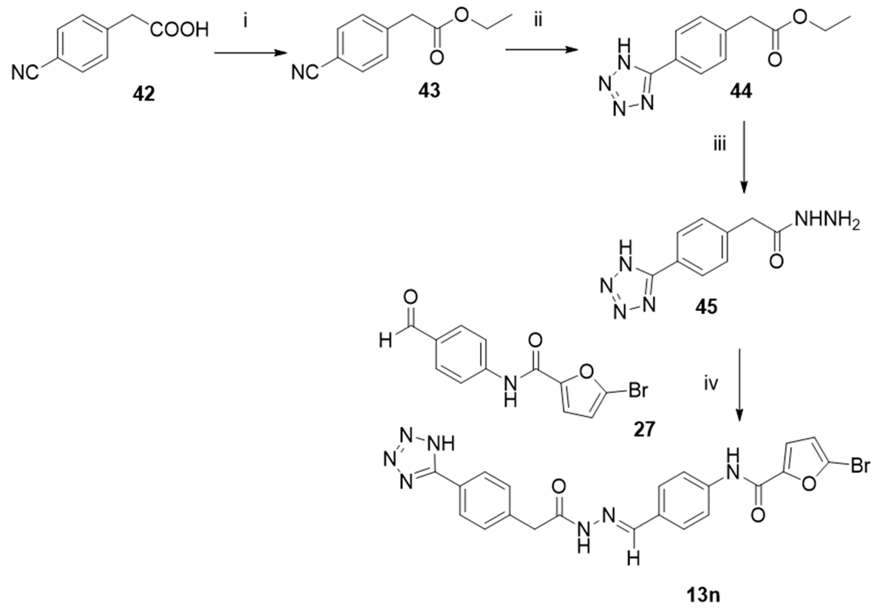
The docked position of compound 13 (Figure 3A and Figure S4A) in the MMP-13 binding site shows the characteristic U shape observed in MMP-13 inhibitors that bind to the S1′ pocket. The compound contains two amide bonds, one on each side, that establish hydrogen bond interactions with Phe241 and Thr247. These hydrogen bond interactions and the planar character of amide bonds were deemed important for the binding mode of the compound, so the amide moieties were not altered during the optimization. At the S1′ pocket, compound 13 establishes a π stacking interaction with Phe252 and a salt bridge interaction with Lys140. Given the strong nature of the electrostatic interaction with Lys140 and the fact that this residue is not present in other MMPs, this region of the molecule should be highly important for both activity and selectivity, and it was further explored in the optimization. At the zinc-binding region, compound 13 establishes a π stacking interaction with His222, but the bromine substituent may cause low solubility. Thus, to increase compound solubility, modifications were proposed in this region as well as to the methyl group present at the center of the molecule.
On the basis of these considerations, we planned to synthesize new derivatives of 13 in order to explore: (i) the p-substitution on the benzyl ring (R); (ii) the replacement of the methyl on the N-acyl hydrazone moiety with an hydrogen atom (R1); (iii) the elongation of the linker between the central phenyl ring and amido group; and (iv) the substitution on the furan nucleus (R2) (compounds 13ao, Table 3). The rigid parts of the molecule have been kept to maintain the degrees of freedom and the conformation of the compounds.

2.4. Chemistry

New compounds 13ao were obtained as described in Scheme 1, Scheme 2, Scheme 3, Scheme 4 and Scheme 5. Hit compound 13 was synthesized to be used as a positive control in the enzymatic assays.
The synthesis of N-acyl hydrazones 13, 13a, and 13c is outlined in Scheme 1. The condensation of carboxylic acids 21 and 22 with aromatic amines 23 or 24 was carried out through the intermediate formation of acyl chlorides by treatment with SOCl2 or (COCl)2, to give the corresponding amides 2527. Hydrazide 29 was obtained by direct treatment of ethyl ester 28 with hydrazine hydrate in equimolar proportion in EtOH. Finally, the target compounds 13, 13a, and 13c were obtained by conjugation of hydrazide 29 with the proper ketones 25 and 26 or aldehyde 27 in EtOH at reflux. The final compounds were easily isolated by crystallization in the reaction solvent.
The synthesis of N-acyl hydrazones 13b and 13d is reported in Scheme 2. The commercially available furoic alcohol 30 was acetylated by treatment with Ac2O/Et3N to yield the furoic acetate 31. Analogously to the synthesis described in Scheme 1, the furoic carboxylic acid 31 protected on the alcoholic function was condensed with aromatic amines 23 or 24 by treatment with thionyl chloride, affording amides 32 and 33. The latter were deprotected by treatment with a methanolic ammonia solution to give the corresponding alcohols. The final compounds 13b and 13d, obtained by conjugation of hydrazide 29 with the proper ketone 34 or aldehyde 35 in EtOH, were isolated as pure crystals from the reaction solvent.
The synthesis of N-acyl hydrazones 13el is reported in Scheme 3. Dicarboxylic acid 36 was selectively protected as an ethyl ester by controlled treatment with SOCl2 in EtOH, affording the monoester 37. 5-Methyl-1,2,4 oxadiazole derivative 39 was obtained by cyclodehydration reaction from the known amidoxime 38 [31] and ethyl ester 37. The resulting ethyl esters 37 and 39 were directly transformed into the corresponding hydrazides 40 and 41 by hydrazinolysis. Lastly, the condensation between the hydrazides 40 or 41 with the proper ketone (25 or 34) or aldehyde (27 or 35) in EtOH afforded the N-acyl hydrazones 13hl and 13eg purified by crystallization.
The N-acyl hydrazone 13n was prepared following the synthetic path described in Scheme 4. Ethyl ester 43 was obtained from carboxylic acid 42 by esterification with SOCl2. Subsequently, 1H-tetrazole derivative 44 was obtained by catalytic cyclization promoted by trimethylstannyl azide from aryl nitrile 43. The target compound 13n was afforded in high yield by condensation between hydrazide 45 and aldehyde 27.
The synthesis of the longest derivatives with n = 1, N-acyl hydrazones 13m and 13o, is shown in Scheme 5. The N-acyl hydrazones 47 and 48 were prepared by condensation between the commercially available aldehyde 46 and the proper hydrazides 40 or 45. The removal of Boc protection was conducted by acid hydrolysis using an excess of trifluoroacetic acid (TFA) to give the pure trifluoroacetate salts of benzylamines 49 and 50. The furoic carboxylic acid 21 was activated as an NHS-ester by reaction with N-hydroxysuccinimide and EDC as coupling agents. The resulting NHS-ester 51 was coupled with amines 49 or 50 to give the corresponding compounds 13m or 13o in high yields.
Usually, the N-acyl hydrazone structure (-C(O)-N-N=C<) is a hybrid between amide and imine functional groups, exhibiting both geometric and conformational stereoisomerism [32]. Rotation along C=N linkage results in the E/Z stereoisomers, where the E conformation is reported to be usually the preferred geometry in solution due to the unfavorable steric restriction of the Z form [33,34]. Moreover, the rotation across the amide C(O)-NH bond gives geometrical isomerism as synperiplanar (sp) and antiperiplanar (ap) conformers [35]. For these reasons, all the N-acyl hydrazones here reported (13 and 13ao) are described in their E form and as a mixture of synperiplanar (sp) and antiperiplanar (ap) conformers. As already reported by Munir et al. [35], in all the 1H NMR spectra of the synthesized N-acyl hydrazones (13 and 13ao; see Supporting Information and Experimental section), a particular pattern of two signal sets was detected, corresponding to the mixture of rotational syn- and antiperiplanar conformers. In fact, in the 1H NMR spectra resolved in DMSO-d6, duplicated signals were observed for -CONHN-, -CH2-, and -N=C(CH3) protons. In Figure 4, the 1H NMR spectra of two representative N-acyl hydrazones, 13l and 13a, are reported. In both spectra, the chemical shift of the -CONHN- proton (in green) resonated around 10.5–12 ppm, resulting in a double singlet, indicating the syn and the ap forms. Similar behavior is shown by benzylic -CH2- (in yellow) and methyl imine (-CH3) (in blue) protons, which resonate, respectively, at 4.2–3.8 ppm and 2.3–2.25 ppm. For -C(O)NHN- doublet-like peaks, the downfield signal was attributed to the antiperiplanar (ap) conformer, whereas the upfield signals were attributed to the synperiplanar (sp) conformer, as established from the literature [36,37,38]. On the contrary, regarding the methylene (-CH2) proton, the upfield signal is the anti-form and the downfield signal is the syn-isomer, as evidently demonstrated by the integration of the peaks. The ratio between two conformers was calculated considering the integral intensities of the paired peaks, revealing that a DMSO-d6 solution of these compounds contained E-synperiplanar and E-antiperiplanar conformers in an approximate 2:1 ratio with slight variation from compound to compound.

2.5. SAR Analysis

All new derivatives 13ao were tested by a fluorometric assay on recombinant MMP-13 in comparison with hit compound 13, and their inhibitory activity is reported in Table 3. The replacement of the bromine substituent on the furan ring (R2) with a sulfonamido or an alcohol group caused a drop in activity, with derivatives 13a and b displaying an IC50 > 100 µM.
In general, all derivatives bearing a hydrogen atom in R1 showed improved activity with respect to their N-methyl analogues, as can be seen by comparing compound 13d with compound 13b and compound 13g with compound 13f.
The introduction of a 1,2,4-oxadiazole ring in R (as in compounds 13e and f) caused a decrease in inhibitory activity for MMP-13 relative to 13. This heterocycle, known as an ester isostere, is present in many biologically active compounds [39] and was chosen in an attempt to ameliorate the solubility of our original N-acyl hydrazone scaffold. Actually, the simultaneous introduction of the oxadiazole ring in R with an alcohol group in R2 and a hydrogen in R1 caused an improvement in solubility, as can be seen from the logP value calculated for compound 13g with respect to 13 (Table 4), but did not increase the inhibitory activity.
On the contrary, the replacement of the nitro group in R with an acidic moiety, such as a carboxylic acid in 13l or a 1H-tetrazole in 13n, led to improved inhibitory activity relative to 13c. The incorporation of these acidic moieties introduced more negative electrostatic surfaces close to the residue Lys140, which resulted in better electrostatic complementarities between these compounds and the enzyme at the S1″ pocket, thus increasing their activities (Figure 3C and Figure S4C).
On the basis of these results and in order to increase its inhibitory potency, we decided to further modify 13l by introducing a methylene spacer between the central phenyl ring and amido group (n = 1). In fact, this modification could allow our hit compound to better interact with Lys140 in the S1″ pocket by establishing the salt bridge necessary for a proper fit with the MMP-13 binding site (Figure 3B and Figure S4B). As expected, compound 13m resulted in the best of the series, showing an 8-fold increase in inhibitory potency against MMP-13 (IC50 = 1.8 µM) relative to 13.
Of note, the same modification in the tetrazole analogue 13o caused a drop in activity, probably due to the excessive bulkiness of this substituent, negatively affecting the binding with the enzyme. 1H-tetrazole is an aromatic heterocyclic bioisoster of the carboxylic acid group, characterized by a higher lipophilicity and a similar acidity but a different volume [40].
In general, derivatives 13ao show good predicted ADMET properties (see Table 4). Thus, all of them fulfill the Lipinski rule of five: they have good intestinal absorption and show week/moderate cardiotoxicity. Only high hepatotoxicity is predicted for all of them, and this is necessary to be experimentally tested in future studies and before the next rounds of optimization of 13m (or other) derivatives.
Finally, the selectivity profile of 13m was evaluated to verify if the optimization process has led to a loss of selectivity for MMP-13. The results reported in Table 5 show that, with the exception of MMP-2, the selectivity over the other tested enzymes has been maintained or improved with respect to our hit compound 13. In particular, 13m displayed a 100-fold selectivity for MMP-13 over MMP-1 and MMP-14.

3. Materials and Methods

3.1. Chemistry

Melting points were determined on a Leica Galen III Microscope (Leica/Cambridge Instruments, Cambridge, UK) and are uncorrected. 1H and 13C NMR spectra were recorded on a Bruker Avance III HD 400 MHz spectrometer (Fällander, Switzerland). Chemical shifts (δ) are reported in parts per million, and coupling constants (J) are reported in hertz (Hz). 13C NMR spectra were fully decoupled. The following abbreviations were used to explain multiplicities: singlet (s), doublet (d), triplet (t), double doublet (dd), broad (br), and multiplet (m). Chromatographic separations were performed on silica gel columns by flash column chromatography (Kieselgel 40, 0.040−0.063 mm, Merck, Darmstad, Germany) or using ISOLUTE Flash Si II cartridges (Biotage) or using an Isolera automatic system (Biotage, Uppsala, Sweden) and SFÄR HC Duo silica cartridges (Biotage, Uppsala, Sweden). Reactions were followed by thin-layer chromatography (TLC) on Merck aluminum silica gel (60 F254) sheets that were visualized under a UV lamp. Evaporation was performed in vacuo (rotating evaporator). Sodium sulfate was always used as the drying agent. Commercially available chemicals were purchased from Merck (Darmstad, Germany). Elemental analysis was used to determine the purity of the target compounds (Table S1). Analytical results are within ±0.4% of the theoretical values. High-resolution ESI-MS spectra were recorded by direct injection at 5 (positive) and 7 (negative) μL min−1 flow rates in an Orbitrap high-resolution mass spectrometer (Thermo, San Jose, CA, USA), equipped with a HESI source.
  • 5-(acetoxymethyl)furan-2-carboxylic acid (31).
Triethylamine (2 mL, 14.07 mmol, 2 eq) was added to a white solution of commercial 5-(hydroxymethyl)furan-2-carboxylic acid (30) (1 g, 7.03 mmol, 1 eq) in Et2O (20.3 mL). Lastly, Ac2O (0.9 mL, 9.14 mmol, 1.3 eq) was added dropwise at 0 °C, and the reaction mixture was stirred at RT for 3.5 h. The crude was dissolved in HCl 1N (125 mL) and extracted (3 × 125 mL) with Et2O. The organic phases were combined, dried over Na2SO4, filtrated, and evaporated. The pure product 31 was obtained as a white solid (1165 mg). Yield: 90%. 1H NMR (400 MHz, DMSO-d6) δ: 13.15 (br s, 1H); 7.17 (d, J = 3.2 Hz, 1H); 6.67 (d, J = 3.2 Hz, 1H); 5.08 (s, 2H); and 2.06 (s, 3H).

3.1.1. General Procedure to Synthesize Conjugates 2527 and 3233

To a solution of furoic acids 21, 22, and 31 (1 eq) in dioxane (1 mL/mmol) or a mixture of DCM and a few drops of DMF, SOCl2 (4 eq) was added under a nitrogen atmosphere. The reaction mixture was stirred at 100 °C for 12 h and then evaporated under an inert atmosphere (N2). The crude product was dissolved in dioxane, or DCM (1.10 mL/mmol), and pyridine (1 eq) and the commercial aryl amines 2324 (1 eq) were added under a nitrogen atmosphere. The reaction was stirred at RT for 1 h and then diluted with EtOAc. The organic phase was washed with water, NaHCO3 saturated solution, and HCl 1N. The organic layer was dried over Na2SO4, filtered, and evaporated under reduced pressure. The pure product (2527 and 33) was afforded either without any further purification or by column chromatography (32).
  • N-(4-acetylphenyl)-5-bromofuran-2-carboxamide (25).
The title compound was synthesized as previously reported in the general procedure, starting from commercial furoic acid 21 (500 mg, 2.62 mmol) dissolved in dioxane and using commercial 4-aminoacetophenone 23 (354 mg). After workup, compound 25 was obtained as a yellow/orange solid (565 mg) without any further purification. Yield: 73%. 1H NMR (400 MHz, DMSO-d6) δ: 10.51 (s, 1H); 7.97–7.95 (m, 2H); 7.90–7.87 (m, 2H); 7.43 (d, J = 3.2 Hz, 1H); 6.86 (d, J = 3.2 Hz, 1H); and 2.54 (s, 3H).
  • N-(4-acetylphenyl)-5-sulfamoylfuran-2-carboxamide (26).
The title compound was synthesized as previously reported in the general procedure, starting from the commercial 5-(aminosulfonyl)-2-furoic acid 22 (100 mg, 0.523 mmol) dissolved in DCM/DMF and using 4-aminoacetophenone 23 (65mg). After workup, the crude was purified by flash chromatography using an ISOLUTE Si II 5 g cartridge (isocratic gradient: CHCl3/MeOH 30:1 v/v) to give the desired product 26 as a white solid (27 mg). Yield: 18%. 1H NMR (400 MHz, CDCl3) δ: 10.62 (s, 1H); 8.00–7.98 (m, 4H); 7.89–7.87 (m, 2H); 7.55 (d, J = 4 Hz, 1H); 7.15 (d, J = 3.6 Hz, 1H); and 2.55 (s, 3H).
  • 5-bromo-N-(4-formylphenyl) furan-2-carboxamide (27).
The title compound was synthesized as previously reported in the general procedure, starting from the commercial furoic acid 21 (500 mg, 2.62 mmol) dissolved in dioxane and using the commercially available 4-aminobenzaldehyde 24 (317 mg). After workup, compound 26 was obtained as a yellow solid (555 mg) without any further purification. Yield: 72.5%. 1H NMR (400 MHz, DMSO-d6) δ: 10.57 (s, 1H); 9.91 (s, 1H); 7.98–7.97 (m, 2H); 7.91–7.89 (m, 2H); 7.45 (d, J = 8.8 Hz, 1H); and 6.87 (d, J = 8.8 Hz, 1H).
  • (5-((4-acetylphenyl)carbamoyl)furan-2-yl)methyl acetate (32).
The title compound was synthesized as previously reported in the general procedure, starting from the derivative 31 (1.00 g, 5.43 mmol) dissolved in dioxane and 4-aminoacetophenone 23 (733 mg). After workup, compound 32 was purified by flash chromatography using an ISOLUTE Si II 5 g cartridge (n-hexane/EtOAc in a gradient from 40:1 to 1:1 v/v) to give the desired product 32 as a white solid (896 mg). Yield: 55%. 1H NMR (400 MHz, CDCl3) δ: 8.22 (br s, 1H); 8.00–7.97 (m, 2H); 7.79–7.76 (m, 2H); 7.24 (d, J = 3.6 Hz, 1H); 6.586 (d, J = 3.6 Hz, 1H); 5.12 (s, 2H); 2.59 (s, 3H); and 2.12 (s, 3H).
  • (5-((4-formylphenyl)carbamoyl)furan-2-yl)methyl acetate (33).
The title compound was synthesized as previously reported in the general procedure, starting from the derivative 31 (720 mg, 3.90 mmol) dissolved in dioxane and 4-aminoacetophenone 24 (474 mg). After workup, compound 33 was obtained as an orange solid (772 mg) without any further purification. Yield: 69%. 1H NMR (400 MHz, DMSO-d6) δ: 10.55 (s, 1H); 9.91 (s, 1H); 8.00–7.98 (m, 2H); 7.92–7.89 (m, 2H); 7.41 (d, J = 3.6 Hz, 1H); 6.75 (d, J = 3.6 Hz, 1H); 5.13 (s, 2H); and 2.08 (s, 3H).
  • N-(4-acetylphenyl)-5-(hydroxymethyl)furan-2-carboxamide (34).
To a solution of the ketone 32 (250 mg, 0.83 mmol, 1 eq) in MeOH (3 mL), NH3-MeOH 7N (6 mL) was added. The resulting mixture was stirred at RT for 1.5 h under a nitrogen atmosphere. Then, the reaction mixture was concentrated under vacuum conditions and purified by trituration with n-hexane to give pure compound 34 as a solid (215 mg). Yield: quantitative. 1H NMR (400 MHz, CDCl3) δ: 8.22 (br s, 1H); 7.99 (d, J = 8.8 Hz, 2H); 7.77 (d, J = 8.8 Hz, 2H); 7.24 (d, J = 3.6 Hz, 1H); 6.5 (d, J = 3.2 Hz, 1H); 4.72 (s, 2H); and 2.59 (s, 3H).
  • N-(4-formylphenyl)-5-(hydroxymethyl)furan-2-carboxamide (35).
To a solution of the aldehyde 33 (100 mg, 0.35 mmol, 1 eq) in MeOH (3.5 mL), NH3-MeOH 7N (14.50 mL) was added. The resulting mixture was stirred at RT for 1.5 h under a nitrogen atmosphere. Then, the reaction mixture was concentrated under vacuum conditions and purified by flash chromatography using an ISOLUTE Si II 5 g cartridge (Hexane/EtOAc gradient from 4:1 to 1:2 v/v) to give pure compound 35 as a white solid (51.9 mg). Yield: 61%. 1H NMR (400 MHz, DMSO-d6) δ: 10.47 (s, 1H); 9.90 (s, 1H), 8.00–7.98 (m, 2H); 7.91–7.89 (m, 2H); 7.38 (d, J = 3.6 Hz, 1H); 6.54 (d, J = 3.6 Hz, 1H); 5.47 (t, J = 5.6 Hz, 1H); and 4.51 (d, J = 5.6 Hz, 1H).
  • 4-(2-ethoxy-2-oxoethyl)benzoic acid (37).
To a solution of commercial 4-carboxyphenylacetic acid 36 (150 mg, 0.83 mmol, 1 eq) in toluene (1.85 mL) and EtOH (0.15 mL), SOCl2 (0.15 mL) was added dropwise under a nitrogen atmosphere. The reaction mixture was stirred at 80 °C overnight. The next day, the reaction mixture appeared as a suspension, and EtOH was added in 0.2 mL portions until the solution was clear. To obtain the pure product 37 (150.1 mg), the resulting crude was concentrated under vacuum conditions and purified by trituration with n-hexane. Yield: 87%. 1H NMR (400 MHz, DMSO-d6) δ: 12.92 (s, 1H), 7.90–7.88 (m, 2H), 7.40–7.38 (m, 2H), 4.08 (q, J = 7.2 Hz, 2H), 3.76 (s, 2H), and 1.18 (t, J = 7.2 Hz, 3H).
  • (E, Z)-N’-hydroxyacetimidamide (38).
The title compound was afforded as described in Fortuna et al. [31]. Yield: 57%. 1H NMR (400 MHz, DMSO-d6) δ: 8.60 (br s, 1H), 5.34 (br s, 2H), and 1.61 (s, 3H).
  • Ethyl 2-(4-(3-methyl-1,2,4-oxadiazol-5-yl)phenyl)acetate (39).
Compound 37 (429 mg, 2.06 mmol, 1 eq) was dissolved in DCM (2.75 mL) and added to SOCl2 (0.45 mL, 4.12 mmol, 3 eq) under a nitrogen atmosphere. The reaction mixture was stirred at RT for 4 h and concentrated under a nitrogen flux. Then the resulting crude was diluted with acetone and added dropwise to a solution of amidoxime 38 (167.7 mg, 2.27 mmol, 1.1 eq) and K2CO3 (569 mg, 4.12 mmol, 2 eq) in acetone (3 mL). The resulting mixture was stirred at RT overnight under a nitrogen atmosphere, and then the solvents were removed under vacuum conditions. In order to remove any remaining salt, the crude was treated with H2O, and the solid was filtrated under vacuum conditions and subsequentially warmed up to 130 °C solvent-free for 3 h. The crude was purified by flash chromatography using an ISOLUTE Si II 20 g cartridge (petroleum ether/EtOAc gradient from 30:1 to 15:1 v/v) to give pure compound 39 as a white solid (181 mg). Yield: 36%. 1H NMR (400 MHz, DMSO-d6) δ: 8.08–8.06 (m, 2H), 7.46–7.44 (m, 2H), 4.17 (q, J = 7.2 Hz, 2H), 3.69 (s, 2H), 2.47 (s, 3H), and 1.26 (t, J = 7.2 Hz, 3H).
  • Ethyl 2-(4-cyanophenyl)acetate (43).
Commercially available 2-(4-cyanophenyl)acetic acid (42) (100 mg, 0.62 mmol, 1 eq) was first dissolved in EtOH (0.6 mL) and then added to SOCl2 (0.06 mL, 0.81 mmol, 1.3 eq). The reaction was stirred at 65 °C for 2 h and subsequentially evaporated. Purification of the crude was achieved by trituration with n-hexane, obtaining pure 43 as a white solid (113.3 mg). Yield: 97%. 1H NMR (400 MHz, CDCl3) δ: 7.80–7.78 (m, 2H); 7.49–7.47 (m, 2H); 4.08 (q, J = 7.2 Hz, 2H); 3.80 (s, 2H); and 1.18 (t, J = 6.8 Hz, 3H).
  • Ethyl 2-(4-(1H-tetrazol-5-yl)phenyl)acetate (44).
To a solution of compound 43 (113 mg, 0.6 mmol, 1 eq) in THF dry (1 mL), azido trimethyltin (IV) (124.71 mg, 0.6 mmol, 1 eq) was added under a nitrogen atmosphere. The reaction mixture was maintained at 70 °C for 3 days. After that, the solution was diluted with HCl 1N (25 mL) and extracted with EtOAc (3 × 25 mL). The organic phases were then dried over Na2SO4, filtrated, and evaporated under vacuum conditions. The resulting crude was purified by flash chromatography using an ISOLUTE Si II 5 g cartridge (n-hexane/EtOAc in a gradient from 40:1 to 1:1 v/v) to give the desired product 44 as a white solid (96.1 mg). Yield: 69%. 1H NMR (400 MHz, DMSO-d6) δ: 7.99–7.97 (m, 2H); 7.51–7.49 (m, 2H); 4.08 (q, J = 7.2 Hz, 2H); 3.78 (s, 2H); and 1.19 (t, J = 7.2 Hz, 3H).

3.1.2. General Procedure to Synthesize Hydrazides 29, 4041, and 45

Hydrazine hydrate (10 eq) was added to a solution of ethyl esters 28, 37, 39, and 44 (1 eq) in EtOH (3 mL/mmol). The resulting mixture was stirred at 80 °C for 4–6 h under a nitrogen atmosphere and evaporated. The crude purification was conducted in different ways, depending on the substrate.
  • 2-(4-nitrophenyl) aceto-hydrazide (29).
The title compound was synthesized as described in the general procedure, starting with commercial ethyl 2-(4-nitrophenyl) acetate 28 (93.3 mg, 0.40 mmol) and stirring the solution for 4 h. The resulting mixture was purified by crystallization in MeOH at 0 °C. The crystals were dissolved in EtOAc and then washed with H2O (1 × 50 mL). The organic phases were combined, dried over Na2SO4, filtrated, and evaporated. The pure product 29 was achieved as a white solid (134.8 mg). Yield: 82%. 1H NMR (400 MHz, DMSO-d6) δ: 9.31 (s, 1H); 8.19–8.17 (m, 2H); 7.54–7.52 (m, 2H); 4.25 (d, J = 3.6 Hz, 2H); and 3.52 (s, 2H).
  • 4-(2-hydrazineyl-2-oxoethyl)benzoic acid (40).
The title compound was synthesized as reported in the general procedure, starting with commercial ethyl 2-(4-nitrophenyl) acetate 37 (250 mg, 1.20 mmol) and stirring the solution for 6 h. The resulting crude was purified by flash chromatography (Isolera Biotage automated chromatographer) using SFÄR HC Duo 10g (gradient CH2Cl2/MeOH 25:1 v/v), yielding pure compound 40 (148 mg). Yield: 64%. 1H NMR (400 MHz, DMSO-d6) δ: 9.25 (br s, 1H); 7.87–7.85 (m, 2H); 7.37–7.35 (m, 2H); and 3.42 (s, 2H).
  • 2-(4-(3-methyl-1,2,4-oxadiazol-5-yl)phenyl)acetohydrazide (41).
The title compound was synthesized as reported in the general procedure, starting with the commercial compound 39 (45 mg, 0.18 mmol) and stirring the solution for 3 h. After evaporation of the solvent, the pure product 41 was achieved as a yellow solid (33.4 mg) without any further purification. Yield: 89%. 1H NMR (400 MHz, DMSO-d6) δ: 9.30 (s, 1H); 8.03–8.01 (m, 2H); 7.51–7.49 (m, 2H); 4.27 (brs, 1H); 3.47 (s, 2H); and 2.41 (s, 3H).
  • 5-(4-(2-(Hydrazineyloxy)-2-oxoethyl)phenyl)-1H-tetrazole (45).
The title compound was synthesized as previously reported in the general procedure, starting with the derivative 44 (93.3 mg, 0.40 mmol) and stirring the solution for 4 h. The crude was purified by flash chromatography using an ISOLUTE C18 5 g cartridge (H2O/MeOH in a gradient of 8:1 to 1:10 v/v) to obtain pure compound 45 (26.3 mg) as a white solid. Yield: 30%. 1H NMR (400 MHz, DMSO-d6) δ: 9.27 (s, 1H); 7.95–7.93 (m, 2H); 7.43–7.41 (m, 2H); and 3.42 (s, 2H).

3.1.3. General Procedure to Synthesize N-Acyl Hydrazones 13, 13al, 13n, and 4748

Hydrazides 29, 4041, and 45 (1 eq) were dissolved in EtOH (7.75 mL/mmol), and carbonyl derivatives 2527, 3435, and 46 (1 eq) were added under a nitrogen atmosphere. The reaction was heated at 80 °C for 1–72 h, then cooled to room temperature and concentrated in vacuo. The crude was dissolved in EtOH, and the pure desired products 13, 13al, 13n, and 4748 were afforded as solids by crystallization in EtOH at 0 °C.
  • (E)-5-bromo-N-(4-(1-(2-(2-(4-nitrophenyl)acetyl)hydrazineylidene)ethyl)phenyl)furan-2-carboxamide (13).
The title compound was synthesized as reported in the general procedure, starting with hydrazide 29 (95 mg, 0.487 mmol) and ketone 25 (150 mg). The reaction was stirred overnight. Pure product 13 was collected by crystallization as an orange solid (203 mg). Yield: 86%. Mp: 206–209 °C. 1H NMR (400 MHz, DMSO-d6) δ: 10.71 sp, 10.64 ap (2s, 1H); 10.35 (sp+ap) (s, 1H); 8.22–8.18 (sp+ap) (m, 2H); 7.82–7.78 (sp+ap) (m, 4H); 7.63–7.57 (sp+ap) (m, 2H); 7.40 (sp+ap) (d, J = 3.6 Hz, 1H); 6.85 (sp+ap) (d, J = 3.6 H z, 1H); 4.23 sp, 3.87 ap (2s, 2H); 2.24 ap, 2.22 sp (2s, 3H). 13C NMR (100 MHz, DMSO-d6) δ: 172.6; 155.6; 149.5; 146.8; 145.0; 139.8; 133.8; 131.4; 130.9; 130.0; 127.3; 127.1; 125.1; 123.9; 123.7; 120.4; 118.9; 117.8; 114.8; 79.6; 41.5; 13.9. HRMS (ESI, m/z) calculated for C21H17BrN4O5 [M−H] 483.03096; found: 483.03085.
  • (E)-N-(4-(1-(2-(2-(4-nitrophenyl)acetyl)hydrazineylidene)ethyl)benzyl)-5-sulfamoylfuran-2-carboxamide (13a).
The title compound was synthesized as reported in the general procedure, starting with hydrazide 29 (19 mg, 0.17 mmol) and aldehyde 27 (28 mg). The reaction was stirred for 5 h. Pure product 13a was collected by crystallization as a slightly pink powder (21.2 mg). Yield: 45%. Mp: 255–258 °C. 1H NMR (400 MHz, DMSO-d6) δ: 10.70 sp, 10.64 ap (2s, 1H); 10.47 (sp+ap) (s, 1H); 8.22–8.18 (sp+ap) (m, 2H); 7.98 (sp+ap) (s, 2H); 7.84–7.77 (sp+ap) (m, 4H); 7.63–7.58 (sp+ap) (m, 2H); 7.51 (sp+ap) (d, J = 3.6 Hz, 1H); 7.14 (sp+ap) (d, J = 3.6 H z, 1H); 4.23 sp, 3.87 ap (2s, 2H); 2.31 ap, 2.25 sp (2s, 3H). 13C NMR (100 MHz, DMSO-d6) δ: 172.6; 166.4; 155.9; 154.2; 152.1; 148.8; 147.8; 146.8; 146.7; 144.8; 144.7; 139.6; 139.4; 134.2; 134.1; 131.4; 131.0; 127.4; 127.1; 123.9; 123.7; 120.5; 120.3; 115.3; 114.6; 41.0; 14.5; 14.0. HRMS (ESI, m/z) calculated for C21H19N5O7S [M−H]: 484.09324; found: 484.09366.
  • (E)-5-(hydroxymethyl)-N-(4-(1-(2-(2-(4nitrophenyl)acetyl)hydrazineylidene)ethyl) phenyl) furan-2-carboxamide (13b).
The title compound was synthesized as reported in the general procedure, starting with hydrazide 29 (33.2 mg, 0.17 mmol) and ketone 34 (44 mg). The reaction was stirred overnight. Pure product 13b was collected by crystallization as a light orange solid (57 mg). Yield: 77%. Mp: 238–241 °C. 1H NMR (400 MHz, DMSO-d6) δ: 10.67 sp, 10.61 ap (2s, 1H); 10.20 (sp+ap) (s, 1H); 8.21–8.16 (sp+ap) (m, 2H); 7.79–7.77 (sp+ap) (m, 4H); 7.62–7.56 (sp+ap) (m, 2H); 7.30 (sp+ap) (d, J = 3.6 Hz, 1H); 6.50 (sp+ap) (d, J = 3.6 H z, 1H); 5.43 (sp+ap) (t, J = 6 Hz, 1H); 4.48 (sp+ap) (d, J = 6 Hz, 2H); 4.22 sp, 3.85 ap (2s, 2H); 2.29 ap, 2.23 sp (2s, 3H). 13C NMR (100 MHz, DMSO-d6) δ: 172.2; 165.9; 158.7; 156.3; 151.9; 147.5; 146.4; 146.2; 144.4; 144.2; 139.7; 139.5; 133.2; 133.1; 131.0; 130.5; 129.3; 126.9; 126.6; 123.4; 123.2; 119.8; 119.7; 119.4; 115.7; 109.0; 55.9; 40.6; 14.1; 13.5. HRMS (ESI, m/z) calculated for C22H20N4O6 [M−H]: 435.13101; found: 435.13129.
  • (E)-5-bromo-N-(4-((2-(2-(4-nitrophenyl)acetyl)hydrazineylidene)methyl)phenyl)furan-2-carboxamide (13c).
The title compound was synthesized as reported in the general procedure, starting with hydrazide 29 (150 mg, 0.51 mmol) and aldehyde 26 (100 mg). The reaction was stirred for 5 h at 80 °C. Pure product 13c was collected by crystallization as a yellowish solid (237.1 mg). Yield: 98%. Mp: 277–279 °C. 1H NMR (400 MHz, DMSO-d6) δ: 11.64 ap, 11.46 sp (2s, 1H); 10.38 (sp+ap) (s, 1H); 8.22–8.18 (sp+ap) (m, 3H); 7.98 (sp+ap) (s, 1H); 7.83–7.81 (sp+ap) (m, 2H); 7.70–7.66 (sp+ap) (m, 2H); 7.61–7.59 (sp+ap) (m, 2H); 7.40 (sp+ap) (d, J = 3.6 Hz, 1H); 6.84 (sp+ap) (d, J = 3.6 Hz, 1H); 4.18 sp, 3.73 ap (2s, 2H). 13C NMR (400 MHz, DMSO-d6) δ: 171.2; 165.3; 155.1; 149.1; 149.0; 146.5; 146.4; 146.2; 144.1; 143.7; 142.9; 140.0; 139.8; 130.9; 130.5; 129.6; 127.7; 127.4; 125.6; 123.4; 123.2; 120.3; 117.5; 114.4; 40.8. HRMS (ESI, m/z) calculated for C20H15BrN4O5 [M−H]: 469.01475; found: 469.01541.
  • (E)-5-(hydroxymethyl)-N-(4-((2-(2-(4-nitrophenyl)acetyl)hydrazineylidene)methyl)phenyl) furan-2-carboxamide (13d).
The title compound was synthesized as previously reported in the general procedure, starting from hydrazide 29 (41.3 mg, 0.21 mmol) and aldehyde 35 (51.45 mg). The reaction was stirred overnight. Pure product 13d was collected by crystallization as a light-yellow solid (85.4 mg). Yield: 95%. Mp: 246–248 °C. 1H NMR (400 MHz, DMSO-d6) δ: 11.64 ap, 11.47 sp (2s, 1H); 10.26 (sp+ap) (s, 1H); 8.22–8.17 (sp+ap) (m, 3H); 8.18 (sp+ap) (s, 1H); 7.97 (sp+ap) (s, 1H); 7.85–7.83 (sp+ap) (m, 2H); 7.69–7.65 (sp+ap) (m, 2H); 7.61–7.59 (sp+ap) (m, 2H); 7.32 (sp+ap) (d, J = 3.2 Hz, 1H); 6.52 (sp+ap) (d, J = 3.2 Hz, 1H); 5.45 (sp+ap) (t, J = 6 Hz, 1H); 4.50 (sp+ap) (d, J = 6 Hz, 2H); 4.18 sp, 3.73 ap (2s, 2H). 13C NMR (100 MHz, DMSO-d6) δ: 171.6; 165.8; 159.2; 156.8; 147.1; 146.9; 146.7; 144.6; 144.2; 143.5; 140.8; 140.6; 131.4; 131.0; 130.8; 129.8; 128.1; 127.9; 123.9; 123.8; 123.7; 120.6; 116.2; 109.5; 56.3; 41.3. HRMS (ESI, m/z) calculated for C21H18N4O6 [M−H]: 421.11536; found: 421.11545.
  • (E)-5-bromo-N-(4-(1-(2-(2-(4-(3-methyl-1,2,4-oxadiazol-5-yl)phenyl)acetyl)hydrazineylidene)ethyl)phenyl)furan-2-carboxamide (13e).
The title compound was synthesized as reported in the general procedure, starting with hydrazide 41 (45 mg, 0.22 mmol) and ketone 25 (67 mg). The reaction mixture was stirred overnight. Pure product 13e (49.4 mg) was afforded as a white solid by crystallization in EtOH at 0 °C. Yield: 43%. Mp: 252–255 °C. 1H NMR (400 MHz, DMSO-d6) δ: 10.66 sp, 10.62 ap (2s, 1H); 10.34 (sp+ap) (s, 1H); 8.06–8.01 (sp+ap) (m, 2H); 7.78–7.72 (sp+ap) (m, 5H); 7.58–7.53 (sp+ap) (m, 2H); 7.39 (sp+ap) (d, J = 3.6 Hz, 1H); 6.84 (sp+ap) (d, J = 3.2 Hz, 1H); 4.17 sp, 3.81 ap (2s, 2H); 2.40 (sp+ap) (s, 3H); 2.29 ap, 2.23 sp (2s, 3H). 13C NMR (100 MHz, DMSO-d6) δ: 175.2; 173.2; 172.9; 168.1; 155.5; 152.1; 149.6; 147.7; 142.2; 139.6; 133.9; 131.2; 129.8; 128.1; 127.9; 126.0; 122.2; 120.4; 117.8; 114.8; 55.4; 41.3; 14.5; 13.9; 11.7. HRMS (ESI, m/z) calculated for C24H20BrN5O4 [M−H]: 520.06259; found: 520.06262.
  • (E)-5-(hydroxymethyl)-N-(4-(1-(2-(2-(4-(3-methyl-1,2,4-oxadiazol-5-yl)phenyl)acetyl)hydrazineylidene)ethyl)phenyl)furan-2-carboxamide (13f).
The title compound was synthesized as reported in the general procedure, starting with hydrazide 41 (40 mg, 0.196 mmol) and ketone 34 (50 mg). The reaction mixture was stirred for 3 days. Pure product 13f (73 mg) was afforded as a white solid by crystallization in EtOH at 0 °C. Yield: 80%. Mp: 243–246 °C. 1H NMR (400 MHz, DMSO-d6) δ: 10.64 sp, 10.61 ap (2s, 1H); 10.21 (sp+ap) (s, 1H); 8.07–8.02 (sp+ap) (m, 2H); 7.80–7.78 (sp+ap) (m, 5H); 7.60–7.54 (sp+ap) (m, 2H); 7.31 (sp+ap) (d, J = 3.2 Hz, 1H); 6.51 (sp+ap) (d, J = 3.2 Hz, 1H); 5.43 (sp+ap) (t, J = 6 Hz, 1H); 4.50 (sp+ap) (d, J = 6 Hz, 2H); 4.20 sp, 3.81 ap (2s, 2H); 2.41 (sp+ap) (s, 3H); 2.30 ap, 2.24 sp (2s, 3H). 13C NMR (100 MHz, DMSO-d6) δ: 174.7; 172.4; 167.6; 166.2; 158.6; 156.3; 147.3; 146.3; 141.7; 139.4; 133.2; 130.7; 130.3; 127.7; 126.8; 126.5; 121.7; 121.58; 119.8; 115.6; 109.0; 55.9; 40.8; 14.0; 13.5; 11.25. HRMS (ESI, m/z) calculated for C25H23N5O5 [M−H]: 472.16264; found: 472.16287.
  • (E)-5-(hydroxymethyl)-N-(4-((2-(2-(4-(3-methyl-1,2,4-oxadiazol-5-yl)phenyl)acetyl) hydrazono)methyl)phenyl)furan-2-carboxamide (13g).
The title compound was synthesized as reported in the general procedure, starting with hydrazide 41 (26.4 mg, 0.129 mmol) and aldehyde 35 (31.7 mg). The reaction mixture was stirred at 80 °C for 1.5 h. Pure product 13g (38.1 mg) was afforded as a yellow solid by crystallization in EtOH at 0 °C. Yield: 57%. Mp: 232–235 °C. 1H NMR (400 MHz, DMSO-d6) δ: 11.64 ap, 11.44 sp (2s, 1H), 10.26 (sp+ap) (s, 1H); 8.17 (sp+ap) (s, 1H); 8.07–8.03 (sp+ap) (m, 3H); 7.97 (sp+ap) (s, 1H); 7.85–7.83(sp+ap) (m, 2H); 7.69–7.65 (sp+ap) (m, 2H); 7.57–7.55 (sp+ap) (m, 2H); 7.32 (sp+ap) (d, J = 2.8 Hz, 1H); 6.52 (sp+ap) (d, J = 2.8 Hz, 1H);0 5.46 (sp+ap) (brs, 1H); 4.50 (sp+ap) (s, 2H); 4.13 sp, 3.67 ap (2s, 2H); 2.41 (sp+ap) (s, 3H). 13C NMR (100 MHz, DMSO-d6) δ: 175.2; 175.1; 171.9; 168.1; 166.1; 159.2; 156.8; 147.0; 146.7; 143.4; 142.0; 141.7; 140.8; 140.6; 131.1; 130.7; 129.8; 128.2, 128.1; 127.8; 122.3, 122.1; 120.7; 120.6; 116.2; 109.5; 56.3; 41.6; 11.7. HRMS (ESI, m/z) calculated for C24H21N5O5 [M−H]: 458.14699; found: 458.14728.
  • (E)-4-(2-(2-(1-(4-(5-bromofuran-2-carboxamido)phenyl)ethylidene)hydrazineyl)-2-oxoethyl)benzoic acid (13h).
The title compound was synthesized as reported in the general procedure, starting with hydrazide 40 (50 mg, 0.257 mmol) and ketone 25 (79.4 mg). The reaction mixture was stirred overnight. Pure product 13h (106 mg) was afforded as a yellowish solid by crystallization in EtOH at 0 °C. Yield: 85%. Mp: 292–295 °C. 1H NMR (400 MHz, DMSO-d6) δ: 12.82 (sp+ap) (brs, 1H); 10.62 sp, 10.59 ap (2s, 1H); 10.34 (sp+ap) (s, 1H); 7.96–7.87 (sp+ap) (m, 2H); 7.79–7.78 (sp+ap) (m, 3H); 7.46–7.39 (sp+ap) (m, 2H); 6.87–6.84 (sp+ap) (m, 1H); 4.13 sp, 3.79 ap (2s, 2H); 2.29 ap, 2.23 sp (2s, 3H). 13C NMR (100 MHz, DMSO-d6) δ: 172.6; 167.3; 166.4; 155.1; 151.6; 149.2; 147.2; 141.3; 139.3; 133.5; 129.7; 129.3; 128.9; 126.8; 126.6; 125.5; 119.9; 117.4; 114.4; 40.80; 14.0; 13.5. HRMS (ESI, m/z) calculated for C22H18BrN3O5 [M−H]: 482.03571; found: 482.03586.
  • (E)-4-(2-(2-(1-(4-(5-(hydroxymethyl)furan-2-carboxamido)phenyl)ethylidene)hydrazineyl)-2-oxoethyl)benzoic acid (13i).
The title compound was synthesized as reported in the general procedure, starting with hydrazide 40 (50 mg, 0.257 mmol) and ketone 34 (79.4 mg). The reaction mixture was stirred for 3 h. Pure product 13i was afforded as a solid by crystallization in EtOH at 0 °C. Yield: 62%. Mp: 232–235 °C. 1H NMR (400 MHz, DMSO-d6) δ: 10.56 (sp+ap) (s, 1H); 10.20 (sp+ap) (s, 1H); 7.94–7.76 (sp+ap) (m, 5H); 7.43–7.31(sp+ap) (m, 3H); 6.53–6.51 (sp+ap) (m, 1H); 5.43 (sp+ap) (brs. 1H); 4.50 (sp+ap) (s, 2H); 4.11 sp, 3.74 ap (2s, 2H); 2.29 ap, 2.23 sp (2s, 3H). 13C NMR: (100 MHz, DMSO-d6) δ: 173.0; 159.1; 157.9; 156.7; 147.7; 146.8; 141.7; 140.6; 140.1; 139.9; 133.7; 130.6; 130.1; 129.7; 127.5; 127.0; 126.9; 120.2; 120.1; 116.1; 109.5; 56.4; 14.5. HRMS (ESI, m/z) calculated for C23H21N3O6 [M−H]: 434.13576; found: 434.13580.
  • (E)-4-(2-(2-(4-(5-bromofuran-2-carboxamido)benzylidene)hydrazineyl)-2-oxoethyl)benzoic acid (13l).
The title compound was synthesized as reported in the general procedure, starting with hydrazide 40 (43.5 mg, 0.224 mmol) and aldehyde 27 (65.5 mg). The reaction mixture was stirred overnight. Pure product 13l (89.2 mg) was afforded as a yellowish solid by crystallization in EtOH at 0 °C. Yield: 85%. Mp: 307–310 °C. 1H NMR (400 MHz, DMSO-d6) δ: 12.86 (sp+ap) (brs, 1H); 11.61 ap, 11.41 sp (2s, 1H); 10.38 (sp+ap) (s, 1H); 8.17 ap, 7.96 sp (2s, 1H); 7.91–7.81 (sp+ap) (m, 5H); 7.69–7.65 (sp+ap) (m, 2H); 7.44–7.40 (sp+ap) (m, 3H); 6.85–6.84 (sp+ap) (m, 1H); 4.07 sp, 3.62 ap (2s, 2H).13C NMR (100 MHz, DMSO-d6) δ: 171.6; 167.2; 165.8; 155.1; 149.1; 141.1; 140.9; 140.0; 139.8; 129.7; 129.3; 128.9; 127.6; 127.4; 125.6; 120.3; 117.5; 114.4; 41.1. HRMS (ESI, m/z) calculated for C21H16BrN3O5 [M−H]: 468.02006; found: 468.02017.
  • (E)-N-(4-((2-(2-(4-(1H-tetrazol-5-yl)phenyl)acetyl)hydrazineylidene)methyl)phenyl)-5-bromofuran-2-carboxamide (13n).
The title compound was synthesized as reported in the general procedure, starting with hydrazide 45 (22.8 mg, 0.1045 mmol) and aldehyde 27 (30.7 mg). The reaction mixture was stirred overnight. Pure product 13n (37.4 mg) was afforded as a yellowish solid by crystallization in EtOH at 0 °C. Yield: 73%. Mp: 262–265 °C. 1H NMR (400 MHz, DMSO-d6) δ: 11.61 ap, 11.40 sp (2s, 1H); 10.37 (sp+ap) (s, 1H); 8.19 ap, 8.00 sp (2s, 1H); 8.00–7.96 (sp+ap) (m, 3H); 7.84–7.81 (sp+ap) (m, 2H); 7.70–7.66 (sp+ap) (m, 2H); 7.54–7.52 (sp+ap) (m, 2H); 7.40 (sp+ap) (d, J = 3.2 Hz, 1H); 6.85 40 (sp+ap) (d, J = 3.2 Hz, 1H); 4.09 sp, 3.64 ap (2s, 2H). 13C NMR (100 MHz, DMSO-d6) δ: 171.7; 165.9; 155.5; 155.1; 149.1; 146.3; 142.7; 139.9; 139.7; 138.9; 138.7; 130.4; 130.0; 129.7; 129.6; 127.6; 127.3; 127.1; 126.7; 125.5; 123.2; 122.8; 120.3; 119.9; 117.4; 114.3; 41.0. HRMS (ESI, m/z) calculated for C21H16N7O3Br [M−H]: 492.04252; found: 492.04276.
  • (E)-4-(2-(2-(4-(((tert-butoxycarbonyl)amino)methyl)benzylidene)hydrazineyl)-2-oxoethyl)benzoic acid (47).
The title compound was synthesized as reported in the general procedure, starting with hydrazide 40 (57.8 mg, 0.30 mmol) and aldehyde 46 (70 mg). The reaction was stirred overnight. Pure product 47 was collected by crystallization as a white solid (86.7 mg). Yield: 70%. 1H NMR (400 MHz, DMSO-d6) δ: 13.00 (sp+ap) (brs, 1H); 11.59 ap, 11.39 sp (2s, 1H); 8.19 sp, 7.98 ap (2s, 1H); 7.91–7.86 (sp+ap) (m, 2H); 7.64–7.62 (sp+ap) (m, 2H); 7.43–7.41 (sp+ap) (m, 3H); 7.30–7.28 (sp+ap) (m, 2H); 4.14 (sp+ap) (d, J = 5.6 Hz, 2H); 4.07 sp, 3.63 ap (2s, 2H); 1.39 (sp+ap) (s, 9H).
  • (tert-butyl (E)-(4-((2-(2-(4-(1H-tetrazol-5 yl)phenyl)acetyl)hydrazineylidene)methyl)benzyl) carbamate (48).
The title compound was synthesized as reported in the general procedure, starting with hydrazide 45 (80 mg, 0.37 mmol) and aldehyde 46 (86 mg). The reaction was stirred overnight. Since crystallization failed, the crude compound was purified by flash chromatography (gradient CHCl3/MeOH 10:1 to 8:1 v/v), yielding pure 48 (71 mg) as a yellow solid. Yield: 44%. 1H NMR (400 MHz, DMSO-d6) δ: 11.64 ap, 11.44 sp (2s, 1H); 8.32 sp, 8.19 ap (2s, 1H); 7.99–7.94 (sp+ap) (m, 2H); 7.66–7.62 (sp+ap) (m, 2H); 7.51–7.44 (sp+ap) (m, 3H); 7.30–7.28 (sp+ap) (m, 2H); 4.14 (sp+ap) (d, J = 5.6 Hz, 2H); 4.07 sp, 3.63 ap (2s, 2H); 1.39 (sp+ap) (s, 9H).
  • (E)-(4-((2-(2-(4-carboxyphenyl)acetyl)hydrazineylidene)methyl)phenyl)methanaminium 2,2,2-trifluoroacetate (49).
A suspension of 47 (139 mg, 0.338 mmol, 1 eq) in DCM dry (5.6 mL) was cooled at 0 °C and added to TFA (0.039 mL, 5.07 mmol, 15 eq). The reacting solution was stirred at RT for 2 h. Then, the reacting mixture was co-evaporated with toluene in vacuo to achieve pure compound 49 as a white solid without any further purification. Yield: quantitative. 1H NMR (400 MHz, DMSO-d6) δ: 12.90 (sp+ap) (brs, 1H); 11.71 ap, 11.49 sp (2s, 1H); 8.20 (sp+ap) (brs, 3H); 8.00 sp, 7.91 ap (2s, 1H); 7.89–7.86 (sp+ap) (m, 2H); 7.75–7.73 (sp+ap) (m, 2H); 7.52–7.50 (sp+ap) (m, 2H); 7.45–7.42 (sp+ap) (m, 2H); 4.09–4.08 (sp+ap) (m, 3H); 3.63 ap (s, 2H). 19F (376 MHz, DMSO-d6) δ: −73.46.
  • (E)-(4-((2-(2-(4-(1H-tetrazol-5-yl)phenyl)acetyl)hydrazono)methyl)phenyl) methanaminium 2,2,2-trifluoroacetate (50).
The title compound was synthesized as reported for compound 49, starting with derivative 48 (150 mg, 0.478 mmol). After workup, compound 50 was obtained as a white solid (71 mg) without any further purification. Yield: quantitative. 1H NMR (400 MHz, DMSO-d6) δ: 11.80 ap, 11.50 sp (2s, 1H); 8.23 sp (1s, 1H); 8.18 (sp+ap) (brs, 3H); 8.01–7.96 (sp+ap) (m, 2H); 7.77–7.71 (sp+ap) (m, 2H); 7.55–7.49 (sp+ap) (m, 3H); 4.11–4.07 (sp+ap) (m, 3H); 3.66 ap (s, 2H). 19F (376 MHz, DMSO-d6) δ: −73.46.
  • 2,5-dioxopyrrolidin-1-yl 5-bromofuran-2-carboxylate (51).
To a suspension of commercial compound 21 (300 mg, 1.57 mmol, 1 eq) in DCM (3 mL), N-hydroxysuccinimide (271 mg, 2.36 mmol, 1.5 eq) and EDC (451 mg, 2.36 mmol, 1.5 eq) were subsequently added. The reacting mixture was stirred at RT overnight, then diluted with CHCl3. The organic phase was washed with water (2 × 50 mL) and a NaHCO3-saturated solution (2 × 50 mL). The organic layer was dried over Na2SO4, filtered, and evaporated under reduced pressure. The obtained crude was purified by flash chromatography (isocratic gradient: CHCl3 100% v/v) using an ISOLUTE Flash Si II 10 g cartridge to give compound 51 (319 mg) as a white solid. Yield: 71%. 1H NMR (400 MHz, CDCl3) δ: 7.42 (d, J = 3.6 Hz, 1H); 6.57 (d, J = 3.6 Hz, 1H); 2.89 (s, 4H).

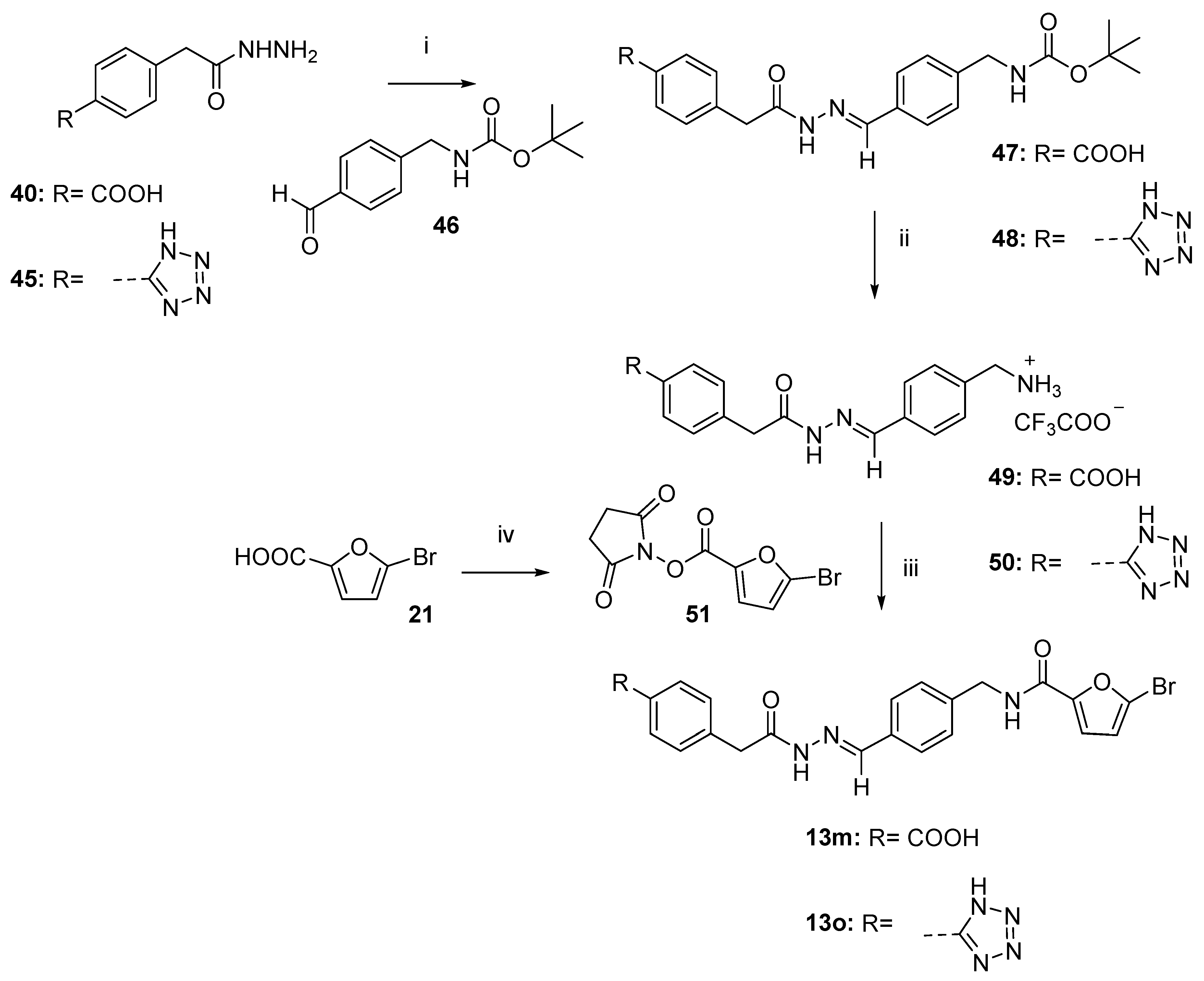
3.1.4. General Procedure to Synthesize Final Compounds 13m,o

To a solution of bromofuranosyl derivative 51 (1 eq) in DMF dry (4.2 mL/mmol), DIPEA (2 eq) was added. Lastly, a solution of trifluoroacetate salt 49 or 50 (1 eq) in DMF dry (8.4 mL/mmol) was added dropwise to the reacting mixture. The reaction was stirred for 2 h and then diluted with EtOAc.
  • (E)-4-(2-(2-(4-((5-bromofuran-2-carboxamido)methyl)benzylidene)hydrazineyl)-2-oxoethyl)benzoic acid (13m).
The title compound was synthesized as reported in the general procedure, starting with 51 (34 mg, 0.118 mmol) and trifluoroacetate 49 (50 mg, 0.118 mmol). After dilution with EtOAc (50 mL), the organic phase was washed with water (2 × 50 mL), and brine (1 × 50 mL). The water phase was acidified using HCl 37% until precipitation of a white solid, which was filtered under low pressure to yield pure compound 13m as a white powder (23 mg). Yield: 40%. Mp: 245–248 °C. 1H NMR (400 MHz, DMSO-d6) δ: 12.82 (sp+ap) (brs, 1H); 11.61 ap, 11.42 sp (2s, 1H); 9.02 (sp+ap) (t, J = 6 Hz, 1H); 8.19 ap, 7.97 sp (2s, 1H); 7.91–7.86 (sp+ap) (m, 2H); 7.66–7.63 (sp+ap) (m, 2H); 7.44–7.41 (sp+ap) (m, 2H); 7.36–7.34 (sp+ap) (m, 2H); 7.18–7.17 (sp+ap) (m, 1H); 6.77–6.76 (sp+ap) (m, 1H); 4.43 (sp+ap) (d, J = 6 Hz, 2H); 4.06 sp, 3.62 ap (2s, 2H). 13C NMR (100 MHz, DMSO-d6) δ: 171.6; 167.1; 165.8; 156.6; 149.4; 146.4; 142.8; 141.2; 141.0; 140.9; 140.7; 132.7; 129.5; 129.2; 129.1; 129.0; 128.8; 127.6; 127.0; 126.7; 124.4; 115.9; 113.9; 41.7; 41.0. HRMS (ESI, m/z) calculated for C22H18N3O5Br [M−H]: 482.03571, found: 482.03589.
  • (E)-N-(4-((2-(2-(4-(1H-tetrazol-5-yl)phenyl)acetyl)hydrazineylidene)methyl)benzyl)-5-bromofuran-2-carboxamide (13o).
The title compound was synthesized as reported in the general procedure, starting with 51 (48 mg, 0.167 mmol) and trifluoroacetate 50 (75 mg, 0.167 mmol). After dilution with EtOAc (50 mL), the organic phase was washed with water (2 × 50 mL). The water phase was acidified using HCl 37% and extracted with EtOAc (2 × 50 mL). The organic layers were dried over Na2SO4, filtered, and evaporated under vacuum conditions. The pure final compound 13o was obtained as a pinkish powder (35 mg). Yield: 41%. Mp: 245–248 °C. 1H NMR (400 MHz, DMSO-d6) δ: 11.65 ap, 11.45 sp (2s, 1H); 10.56 (sp+ap) (t, J = 5.6 Hz, 1H); 8.19 ap (s, 1H); 8.00–7.95 (sp+ap) (m, 3H); 7.68–7.63 (sp+ap) (m, 2H); 7.53–7.51 (sp+ap) (m, 2H); 7.37–7.35 (sp+ap) (m, 2H); 7.18 (sp+ap) (d, J = 3.2 Hz, 1H); 6.77 (sp+ap) (d, J = 3.2 Hz, 1H); 4.43 (sp+ap) (d, J = 5.6 Hz, 2H); 4.08 sp, 3.64 ap (2s, 2H). 13C NMR (100 MHz, DMSO-d6) δ: 172.8; 171.8; 166.1; 156.7; 149.6; 146.5; 142.9; 141.4; 141.1; 138.8; 138.6; 132.9; 130.4; 130.1; 127.8; 127.7; 127.1; 126.8; 124.6; 116.1; 114.0; 41.8; 41.0. HRMS (ESI, m/z) calculated for C22H17N7O3Br [M−H]: 506.05817, found: 506.05801.
The images of the 1H, 13C NMR, and HRMS spectra are represented in Supplementary Materials.

3.2. Biological Assays

Recombinant human MMP-14 catalytic domain was a kind gift from Prof. Gillian Murphy (Department of Oncology, University of Cambridge, UK). Pro-MMP-1, pro-MMP-2, pro-MMP-9, and pro-MMP-13 were purchased from Merck Millipore (Burlington, MA, USA). p-Aminophenylmercuric acetate (APMA) was from Sigma-Aldrich (St. Louis, MO, USA).

3.2.1. Enzyme Activation

Proenzymes were activated immediately prior to use (APMA 2 mM for 1 h at 37 °C for MMP-2, APMA 2 mM for 2 h at 37 °C for MMP-1, APMA 1 mM for 30 min at 37 °C for MMP-13, and APMA 1 mM for 1 h at 37 °C for MMP-9).

3.2.2. Enzyme Inhibition Assays

For assay measurements, the purchased compound stock solutions (10 mM in DMSO) were further diluted for each MMP in the fluorometric assay buffer (FAB: Tris 50 mM, pH = 7.5, NaCl 150 mM, CaCl2 10 mM, Brij 35 0.05%, and DMSO 1%), following the protocol already reported [41,42]. Activated enzyme (final concentration 0.56 nM for MMP-2, 0.3 nM for MMP-13, 1.3 nM for MMP-9, 1 nM for MMP-14cd, and 2.0 nM for MMP-1) and inhibitor solutions were incubated in the assay buffer for 3 h at 25 °C. After the addition of a 200 μM solution of the fluorogenic substrate Mca-Lys-Pro-Leu-Gly-Leu-Dap(Dnp)-Ala-Arg-NH2 (Merck Millipore) in DMSO (final concentration 2 μM), the hydrolysis was monitored every 15 sec for 15 min, recording the increase in fluorescence (λex = 325 nm, λem = 400 nm) using a SpectraMax Gemini XPS (Molecular Devices, Sunnyvale, CA, USA) plate reader. The assays were performed in triplicate with a total volume of 200 μL per well in 96-well microtitre plates (Corning, black, NBS, Turin, Italy). The MMP inhibition activity was expressed in relative fluorescent units (RFU). The percentage of inhibition was calculated from control reactions without the inhibitor. The inhibitory effect of the tested compounds was routinely estimated at a concentration of 100 μM on MMP-13. Those derivatives found to be active were tested at additional concentrations, and IC50 was determined using at least five concentrations of the inhibitor causing an inhibition between 10% and 90%, using the formula: Vi/Vo = 1/(1 + [I]/IC50), where Vi is the initial velocity of substrate cleavage in the presence of the inhibitor at concentration [I] and Vo is the initial velocity in the absence of the inhibitor. Results were analyzed using SoftMax Pro software version 5.4.3 (Molecular Devices, Sunnyvale, CA, USA) and Prism Software version 5.0 (GraphPad Software, Inc., La Jolla, CA, USA).

3.3. Computational Methods

3.3.1. Shape-Based Similarity

Conformations were generated using Omega [43,44] with default parameters and requiring a maximum of 10 conformations. The co-crystallized inhibitors used as references in the shape comparison corresponded to the ligands of the crystal structures with the following PDB codes: 2OW9 [19], 2OZR [19], 3KEC [20], 3KEJ [20], 3KEK [20], 3WV1 [21], and 5BPA [22]. Shape similarity between the library compounds and the reference compounds was calculated with ROCS [45,46] using the ShapeTanimoto coefficient, a value between 0 and 1 calculated by the following equation:
ShapeTanimotof,g = Of,g/(If + Ig − Of,g)
in which the I terms are the self-volume overlaps of each molecule, while the O term is the overlap between the two functions [47].
Conformations with a ShapeTanimoto value lower than 0.5 for any of the reference compounds were discarded.

3.3.2. Ligand Setup for Docking

Before docking, ligand molecules were prepared with LigPrep [48] with default parameter values except for the following options: (a) respect chiralities from input geometry when generating stereoisomers; (b) use Epik [49] for ionization and tautomerization; (c) use 7.0 as an effective pH; and (d) use 2.0 as a pH tolerance for generated structures.

3.3.3. Protein Preparation

After verifying the fitting of the coordinates of the residues in the binding site relative to their corresponding electron density map with VHELIBS, the B chain of the crystal structure with PDB code 3WV1 was prepared by using Maestro’s Protein Preparation Wizard [50] through the following procedure: (a) align to 1ROS, chain A; (b) remove original hydrogens; (c) cap termini; (d) generate ionization and tautomeric states of the ligand with Epik; (e) assign hydrogen bonds at pH 7 with PROPKA; (f) use force field OPLS_2005 to minimize the structure at 0.30 Å; and (g) remove all water molecules from the structure.

3.3.4. Grid Preparation

The grid for protein-ligand docking was generated with Maestro [51] by using default parameter values except for the following settings: (a) the grid center coordinates were (46.0, 80.0, and −1.0); (b) halogens were included as acceptors; (c) the inner box size was (10, 10, and 10); (d) the outer box size was (30, 30, and 30); (e) hydrogen bond constraints were defined on the backbone nitrogen of the residues Thr245 and Thr247 as well as the side-chain oxygen of the residue Thr245; and (f) two positional constraints with a radius of 2 Å were defined on the coordinates (46.3, 80.1, and −7.5) and (51.2, 80.5, and 4.6), respectively.

3.3.5. Molecular Docking

Protein-ligand docking was performed with Glide [52] by using default parameter values except for the following settings: (a) SP precision; (b) enhance planarity of conjugated π groups; (c) include halogens as acceptors; (d) write out at most 10 poses per ligand; (e) include 50 poses per ligand in post-docking minimization; (g) require the accomplishment of both positional constraints by aromatic atoms; and (f) require the accomplishment of one hydrogen bond constraint.
The first picture in each panel of Figure 3 was obtained with Maestro [51], and the second and third pictures were obtained with Flare [28]. The docked poses were predicted with GlideXP [52].

4. Conclusions

In order to obtain potent and selective MMP-13 inhibitors devoid of a ZBG, we developed a virtual screening workflow aimed at identifying compounds that target the S1′ pocket of MMP-13, a region in the MMP binding site that has been shown to be different for MMP-13 with respect to other MMPs. For this purpose, we first applied a MW filter to discard compounds unlikely to survive subsequent filters. Next, we used a shape-based similarity analysis to restrict the initial library of compounds to those able to adopt the characteristic U shape adopted by co-crystallized non-zinc-binding selective MMP-13 inhibitors. Then, we performed protein-ligand docking simulations to predict the binding modes of these compounds. Finally, we analyzed previously reported SAR studies to identify MMP-13 inhibitor interactions with the protein that are important for activity, and we selected the docked poses obtained in the protein-ligand docking according to these criteria. The bioactivity assays on isolated enzymes identified three putative hit compounds capable of inhibiting MMP-13 in the μM range, one of which displayed at least 4-fold selectivity over MMP-1, MMP-2, MMP-9, and MMP-14. Then, a structure-based optimization of the N-acyl hydrazone hit compound 13 guided the synthesis of a series of 12 new derivatives. Among these, a carboxylate derivative (13m) was found to selectively inhibit MMP-13 with an IC50 = 1.8 µM. A docking study showed that the presence of an acidic moiety in R introduced more negative electrostatic surfaces close to the residue Lys140, which resulted in better electrostatic interaction between this compound and the enzyme at the S1″ pocket, thus increasing its activity. The next round of optimization from hit to lead will be necessary to further develop this interesting class of new compounds.

Supplementary Materials

The following supporting information can be downloaded at: https://www.mdpi.com/article/10.3390/ijms241311098/s1.

Author Contributions

Conceptualization, E.N., A.R. and G.P.; methodology, D.C., A.G., B.L.B. and R.D.L.; formal analysis, A.G. and B.L.B.; writing—original draft preparation, E.N., D.C., A.G. and B.L.B.; writing—review and editing, G.P. and S.G.-V.; supervision, E.N., A.R. and A.G.; funding acquisition, S.N., A.R. and A.G. All authors have read and agreed to the published version of the manuscript.

Funding

This research was funded by the University of Pisa (PRA_2020_58) and the “Fondi di Ateneo” 2021 to A.R. and E.N. A.G. contract was supported by grant 2015FI_B00655 of the Catalonia Government.

Institutional Review Board Statement

Not applicable.

Informed Consent Statement

Not applicable.

Data Availability Statement

All data are reported in the manuscript and in the Supplementary Material and are available from the corresponding authors upon request.

Acknowledgments

We thank CISUP—Centre for Instrumentation Sharing—University of Pisa for the acquisition and elaboration of the HMRS spectra. We thank OpenEye Scientific Software and Cresset BioMolecular Discovery Ltd. for kindly providing us with a software bursary for using their programs.

Conflicts of Interest

The authors declare no conflict of interest.

References

  1. Arabelovic, S.; McAlindon, T.E. Considerations in the Treatment of Early Osteoarthritis. Curr. Rheumatol. Rep. 2005, 7, 29–35. [Google Scholar] [CrossRef] [PubMed]
  2. Li, H.; Wang, D.; Yuan, Y.; Min, J. New Insights on the MMP-13 Regulatory Network in the Pathogenesis of Early Osteoarthritis. Arthritis Res. Ther. 2017, 19, 248. [Google Scholar] [CrossRef] [PubMed] [Green Version]
  3. Takaishi, H.; Kimura, T.; Dalal, S.; Okada, Y.; D’Armiento, J. Joint Diseases and Matrix Metalloproteinases: A Role for MMP-13. Curr. Pharm. Biotechnol. 2008, 9, 47–54. [Google Scholar] [CrossRef] [PubMed]
  4. Troeberg, L.; Nagase, H. Proteases Involved in Cartilage Matrix Degradation in Osteoarthritis. Biochim. Biophys. Acta 2012, 1824, 133–145. [Google Scholar] [CrossRef] [PubMed] [Green Version]
  5. Ågren, M.S.; Auf dem Keller, U. Matrix Metalloproteinases: How Much Can They Do? Int. J. Mol. Sci. 2020, 21, 2678. [Google Scholar] [CrossRef] [Green Version]
  6. Vandenbroucke, R.E.; Libert, C. Is There New Hope for Therapeutic Matrix Metalloproteinase Inhibition? Nat. Rev. Drug Discov. 2014, 13, 904–927. [Google Scholar] [CrossRef]
  7. Cathcart, J.M.; Cao, J. MMP Inhibitors: Past, Present and Future. Front. Biosci. 2015, 20, 1164–1178. [Google Scholar] [CrossRef] [Green Version]
  8. Xie, X.-W.; Wan, R.-Z.; Liu, Z.-P. Recent Research Advances in Selective Matrix Metalloproteinase-13 Inhibitors as Anti-Osteoarthritis Agents. ChemMedChem 2017, 12, 1157–1168. [Google Scholar] [CrossRef] [PubMed]
  9. Nuti, E.; Cuffaro, D.; Bernardini, E.; Camodeca, C.; Panelli, L.; Chaves, S.; Ciccone, L.; Tepshi, L.; Vera, L.; Orlandini, E.; et al. Development of Thioaryl-Based Matrix Metalloproteinase-12 Inhibitors with Alternative Zinc-Binding Groups: Synthesis, Potentiometric, NMR, and Crystallographic Studies. J. Med. Chem. 2018, 61, 4421–4435. [Google Scholar] [CrossRef]
  10. Nuti, E.; Rossello, A.; Cuffaro, D.; Camodeca, C.; Van Bael, J.; van der Maat, D.; Martens, E.; Fiten, P.; Pereira, R.V.S.; Ugarte-Berzal, E.; et al. Bivalent Inhibitor with Selectivity for Trimeric MMP-9 Amplifies Neutrophil Chemotaxis and Enables Functional Studies on MMP-9 Proteoforms. Cells 2020, 9, 1634. [Google Scholar] [CrossRef]
  11. Camodeca, C.; Cuffaro, D.; Nuti, E.; Rossello, A. ADAM Metalloproteinases as Potential Drug Targets. Curr. Med. Chem. 2019, 26, 2661–2689. [Google Scholar] [CrossRef] [PubMed]
  12. Pece, R.; Tavella, S.; Costa, D.; Varesano, S.; Camodeca, C.; Cuffaro, D.; Nuti, E.; Rossello, A.; Alfano, M.; D’Arrigo, C.; et al. Inhibitors of ADAM10 Reduce Hodgkin Lymphoma Cell Growth in 3D Microenvironments and Enhance Brentuximab-Vedotin Effect. Haematologica 2022, 107, 909–920. [Google Scholar] [CrossRef] [PubMed]
  13. Cuffaro, D.; Ciccone, L.; Rossello, A.; Nuti, E.; Santamaria, S. Targeting Aggrecanases for Osteoarthritis Therapy: From Zinc Chelation to Exosite Inhibition. J. Med. Chem. 2022, 65, 13505–13532. [Google Scholar] [CrossRef] [PubMed]
  14. Supuran, C.T. Novel Carbonic Anhydrase Inhibitors. Future Med. Chem. 2021, 13, 1935–1937. [Google Scholar] [CrossRef]
  15. Cuffaro, D.; Nuti, E.; Rossello, A. An Overview of Carbohydrate-Based Carbonic Anhydrase Inhibitors. J. Enzyme Inhib. Med. Chem. 2020, 35, 1906–1922. [Google Scholar] [CrossRef]
  16. Li, G.; Tian, Y.; Zhu, W.-G. The Roles of Histone Deacetylases and Their Inhibitors in Cancer Therapy. Front. Cell Dev. Biol. 2020, 8, 576946. [Google Scholar] [CrossRef]
  17. Nencetti, S.; Cuffaro, D.; Nuti, E.; Ciccone, L.; Rossello, A.; Fabbi, M.; Ballante, F.; Ortore, G.; Carbotti, G.; Campelli, F.; et al. Identification of Histone Deacetylase Inhibitors with (Arylidene)Aminoxy Scaffold Active in Uveal Melanoma Cell Lines. J. Enzyme Inhib. Med. Chem. 2021, 36, 34–47. [Google Scholar] [CrossRef]
  18. Fabre, B.; Ramos, A.; de Pascual-Teresa, B. Targeting Matrix Metalloproteinases: Exploring the Dynamics of the S1′ Pocket in the Design of Selective, Small Molecule Inhibitors. J. Med. Chem. 2014, 57, 10205–10219. [Google Scholar] [CrossRef] [PubMed]
  19. Johnson, A.R.; Pavlovsky, A.G.; Ortwine, D.F.; Prior, F.; Man, C.-F.; Bornemeier, D.A.; Banotai, C.A.; Mueller, W.T.; McConnell, P.; Yan, C.; et al. Discovery and Characterization of a Novel Inhibitor of Matrix Metalloprotease-13 That Reduces Cartilage Damage in Vivo without Joint Fibroplasia Side Effects. J. Biol. Chem. 2007, 282, 27781–27791. [Google Scholar] [CrossRef] [Green Version]
  20. Schnute, M.E.; O’Brien, P.M.; Nahra, J.; Morris, M.; Howard Roark, W.; Hanau, C.E.; Ruminski, P.G.; Scholten, J.A.; Fletcher, T.R.; Hamper, B.C.; et al. Discovery of (Pyridin-4-Yl)-2H-Tetrazole as a Novel Scaffold to Identify Highly Selective Matrix Metalloproteinase-13 Inhibitors for the Treatment of Osteoarthritis. Bioorg. Med. Chem. Lett. 2010, 20, 576–580. [Google Scholar] [CrossRef]
  21. Nara, H.; Sato, K.; Naito, T.; Mototani, H.; Oki, H.; Yamamoto, Y.; Kuno, H.; Santou, T.; Kanzaki, N.; Terauchi, J.; et al. Discovery of Novel, Highly Potent, and Selective Quinazoline-2-Carboxamide-Based Matrix Metalloproteinase (MMP)-13 Inhibitors without a Zinc Binding Group Using a Structure-Based Design Approach. J. Med. Chem. 2014, 57, 8886–8902. [Google Scholar] [CrossRef] [PubMed]
  22. Taylor, S.J.; Abeywardane, A.; Liang, S.; Muegge, I.; Padyana, A.K.; Xiong, Z.; Hill-Drzewi, M.; Farmer, B.; Li, X.; Collins, B.; et al. Fragment-Based Discovery of Indole Inhibitors of Matrix Metalloproteinase-13. J. Med. Chem. 2011, 54, 8174–8187. [Google Scholar] [CrossRef]
  23. Specs. Available online: http://www.specs.net (accessed on 15 January 2021).
  24. Nara, H.; Sato, K.; Naito, T.; Mototani, H.; Oki, H.; Yamamoto, Y.; Kuno, H.; Santou, T.; Kanzaki, N.; Terauchi, J.; et al. Thieno[2,3-d]Pyrimidine-2-Carboxamides Bearing a Carboxybenzene Group at 5-Position: Highly Potent, Selective, and Orally Available MMP-13 Inhibitors Interacting with the S1″ Binding Site. Bioorg. Med. Chem. 2014, 22, 5487–5505. [Google Scholar] [CrossRef] [PubMed]
  25. Reaxys. Available online: https://www.reaxys.com/ (accessed on 2 February 2019).
  26. Rossello, A.; Nuti, E.; Orlandini, E.; Carelli, P.; Rapposelli, S.; Macchia, M.; Minutolo, F.; Carbonaro, L.; Albini, A.; Benelli, R.; et al. New N-Arylsulfonyl-N-Alkoxyaminoacetohydroxamic Acids as Selective Inhibitors of Gelatinase A (MMP-2). Bioorg. Med. Chem. 2004, 12, 2441–2450. [Google Scholar] [CrossRef] [PubMed]
  27. McGovern, S.L.; Helfand, B.T.; Feng, B.; Shoichet, B.K. A Specific Mechanism of Nonspecific Inhibition. J. Med. Chem. 2003, 46, 4265–4272. [Google Scholar] [CrossRef]
  28. FlareTM, v6.1.0; Cresset®: Litlington, UK; Available online: http://www.cresset-group.com/flare/ (accessed on 14 April 2023).
  29. Thota, S.; Rodrigues, D.A.; de Sena murteira Pinheiro, P.; Lima, L.M.; Fraga, C.A.M.; Barreiro, E.J. N-Acylhydrazones as Drugs. Bioorg. Med. Chem. Lett. 2018, 28, 2797–2806. [Google Scholar] [CrossRef]
  30. Socea, L.-I.; Barbuceanu, S.-F.; Pahontu, E.M.; Dumitru, A.-C.; Nitulescu, G.M.; Sfetea, R.C.; Apostol, T.-V. Acylhydrazones and Their Biological Activity: A Review. Molecules 2022, 27, 8719. [Google Scholar] [CrossRef]
  31. Fortuna, C.G.; Bonaccorso, C.; Bulbarelli, A.; Caltabiano, G.; Rizzi, L.; Goracci, L.; Musumarra, G.; Pace, A.; Palumbo Piccionello, A.; Guarcello, A.; et al. New Linezolid-like 1,2,4-Oxadiazoles Active against Gram-Positive Multiresistant Pathogens. Eur. J. Med. Chem. 2013, 65, 533–545. [Google Scholar] [CrossRef]
  32. Palla, G.; Predieri, G.; Domiano, P.; Vignali, C.; Turner, W. Conformational Behaviour and E/Z Isomerization of N-Acyl and N-Aroylhydrazones. Tetrahedron 1986, 42, 3649–3654. [Google Scholar] [CrossRef]
  33. Syakaev, V.V.; Podyachev, S.N.; Buzykin, B.I.; Latypov, S.K.; Habicher, W.D.; Konovalov, A.I. NMR Study of Conformation and Isomerization of Aryl- and Heteroarylaldehyde 4-Tert-Butylphenoxyacetylhydrazones. J. Mol. Struct. 2006, 788, 55–62. [Google Scholar] [CrossRef]
  34. Wyrzykiewicz, E.; Błaszczyk, A.; Turowska-Tyrk, I. N-(E)-2-Stilbenyloxymethylenecarbonyl Substituted Hydrazones of Ortho, Meta and Para Hydroxybenzaldehydes. Bull. Pol. Acad. Sci. Chem. 2000, 48, 213–229. [Google Scholar]
  35. Munir, R.; Javid, N.; Zia-Ur-Rehman, M.; Zaheer, M.; Huma, R.; Roohi, A.; Athar, M.M. Synthesis of Novel N-Acylhydrazones and Their C-N/N-N Bond Conformational Characterization by NMR Spectroscopy. Molecules 2021, 26, 4908. [Google Scholar] [CrossRef]
  36. Tian, B.; He, M.; Tang, S.; Hewlett, I.; Tan, Z.; Li, J.; Jin, Y.; Yang, M. Synthesis and Antiviral Activities of Novel Acylhydrazone Derivatives Targeting HIV-1 Capsid Protein. Bioorg. Med. Chem. Lett. 2009, 19, 2162–2167. [Google Scholar] [CrossRef]
  37. Kuodis, Z.; Rutavičius, A.; Matijoška, A.; Eicher-Lorka, O. Synthesis and Isomerism of Hydrazones of 2-(5-Thioxo-4,5-Dihydro-1,3,4-Thiadiazol-2-Ylthio)Acetohydrazide. Cent. Eur. J. Chem. 2007, 5, 996–1006. [Google Scholar] [CrossRef]
  38. Litvinov, I.A.; Kataeva, O.N.; Ermolaeva, L.V.; Vagina, G.A.; Troepol’skaya, T.V.; Naumov, V.A. Crystal and Molecular Structure of Aroyl- and Acetylhydrazones of Acet- and Benzaldehydes. Russ. Chem. Bull. 1991, 40, 62–67. [Google Scholar] [CrossRef]
  39. Pitasse-Santos, P.; Sueth-Santiago, V.; Lima, M.E.F. 1,2,4- and 1,3,4-Oxadiazoles as Scaffolds in the Development of Antiparasitic Agents. J. Braz. Chem. Soc. 2018, 29, 435–456. [Google Scholar] [CrossRef]
  40. Bredael, K.; Geurs, S.; Clarisse, D.; De Bosscher, K.; D’hooghe, M. Carboxylic Acid Bioisosteres in Medicinal Chemistry: Synthesis and Properties. J. Chem. 2022, 2022, 2164558. [Google Scholar] [CrossRef]
  41. Cuffaro, D.; Camodeca, C.; D’Andrea, F.; Piragine, E.; Testai, L.; Calderone, V.; Orlandini, E.; Nuti, E.; Rossello, A. Matrix Metalloproteinase-12 Inhibitors: Synthesis, Structure-Activity Relationships and Intestinal Absorption of Novel Sugar-Based Biphenylsulfonamide Carboxylates. Bioorg. Med. Chem. 2018, 26, 5804–5815. [Google Scholar] [CrossRef]
  42. Cuffaro, D.; Nuti, E.; Gifford, V.; Ito, N.; Camodeca, C.; Tuccinardi, T.; Nencetti, S.; Orlandini, E.; Itoh, Y.; Rossello, A. Design, Synthesis and Biological Evaluation of Bifunctional Inhibitors of Membrane Type 1 Matrix Metalloproteinase (MT1-MMP). Bioorg. Med. Chem. 2019, 27, 196–207. [Google Scholar] [CrossRef] [PubMed]
  43. Hawkins, P.C.D.; Skillman, A.G.; Warren, G.L.; Ellingson, B.A.; Stahl, M.T. Conformer Generation with OMEGA: Algorithm and Validation Using High Quality Structures from the Protein Databank and Cambridge Structural Database. J. Chem. Inf. Model. 2010, 50, 572–584. [Google Scholar] [CrossRef] [PubMed]
  44. Hawkins, P.C.D.; Skillman, A.G.; Warren, G.L.; Ellingson, B.A.; Stahl, M.T. OMEGA, v.3.0.1.2; OpenEye Scientific Software: Santa Fe, NM, USA, 2018; Available online: http://www.eyesopen.com (accessed on 23 March 2021).
  45. Hawkins, P.C.D.; Skillman, A.G.; Nicholls, A. Comparison of Shape-Matching and Docking as Virtual Screening Tools. J. Med. Chem. 2007, 50, 74–82. [Google Scholar] [CrossRef] [PubMed]
  46. ROCS, Version 3.2.2.2; OpenEye Scientific Software: Santa Fe, NM, USA, 2017; Available online: http://www.eyesopen.com (accessed on 6 June 2021).
  47. Shape Theory—Applications. Available online: https://docs.eyesopen.com/applications/rocs/theory/shape_shape.html (accessed on 23 June 2023).
  48. Schrödinger Release 2016-3: LigPrep; Schrödinger, LLC: New York, NY, USA, 2016.
  49. Schrödinger Release 2016-3: Epik; Schrödinger, LLC: New York, NY, USA, 2016.
  50. Schrödinger Release 2016-3: Schrödinger Suite 2016-3 Protein Preparation Wizard; Epik, Schrödinger, LLC: New York, NY, USA; Impact, Schrödinger, LLC: New York, NY, USA; Prime, Schrödinger, LLC: New York, NY, USA, 2016.
  51. Schrödinger Release 2016-3: Maestro; Schrödinger, LLC: New York, NY, USA, 2016.
  52. Schrödinger Release 2016-3: Glide; Schrödinger, LLC: New York, NY, USA, 2016.
Figure 1. Chemical structures of non-zinc-binding selective MMP-13 inhibitors (A [19], B [20], C [21], and D [22]) with reported X-ray crystal structures in complex with the MMP-13 catalytic domain used for the VS set-up.
Ijms 24 11098 g001
Figure 2. Virtual Screening workflow.
Ijms 24 11098 g002
Figure 3. Docked poses of compounds 13, 13m, and 13n (panels (A), (B), and (C), respectively) in the binding site of MMP-13. The first picture in each panel shows the intermolecular interactions predicted by docking (hydrogen bonds are colored in yellow, π stacking interactions are colored in cyan, and salt bridges are colored in magenta). The second picture shows the positive and negative electrostatic field potentials of the compound (colored blue and red, respectively). The third picture shows the molecular surface of the compound colored by the electrostatic complementarity (EC) to the receptor (green: good EC and red: bad EC).
Ijms 24 11098 g003
Scheme 1. Reagents and conditions: (i) SOCl2 or (COCl)2, dioxane, 100 °C, 12 h; (ii) pyridine, dioxane, 1 h (25: 73%; 26: 18%; 27: 73% over two steps); (iii) NH2NH2, EtOH, 4 h, 82%; and (iv) EtOH, reflux (13: 18 h, 86%; 13a: 5 h, 45%; 13c: 5 h, 98%).
Ijms 24 11098 sch001
Scheme 2. Reagents and conditions: (i) Ac2O, Et2O, Et3N, 3.5 h, 85%; (ii) SOCl2, dioxane, 100 °C, 12 h; (iii) pyridine, dioxane, 1 h, rt (32: 55%; 33: 69% over two steps); (iv) NH3/MeOH 7N, 1.5 h (34: quantitative; 35: 61%); and (v) EtOH, reflux, 18 h (13b: 77%; 13d: 95%).
Ijms 24 11098 sch002
Scheme 3. Reagents and conditions: (i) SOCl2, EtOH, toluene, 100 °C, o/n, 87%; (ii) 1. SOCl2, DCM dry, 4 h; 2. K2CO3, acetone, o/n; 3. 130 °C, solvent free, 3 h; 36% over three steps; (iii) NH2NH2, EtOH, reflux (40: 6 h, 64%; 41: 3 h, 89%); and (iv) EtOH, reflux (3–12 h; 43–85%).
Ijms 24 11098 sch003
Scheme 4. Reagents and conditions: (i) SOCl2, EtOH reflux, 2 h, 97%; (ii) Me3SnN3, THF, 3 days, 69%; (iii) NH2NH2, EtOH reflux, 4 h, 30%; and (iv) EtOH reflux, 18 h, 73%.
Ijms 24 11098 sch004
Scheme 5. Reagents and conditions: (i) EtOH, reflux, 18 h (47: 70%; 48: 44%); (ii) TFA, DCM dry, 2 h, quantitative; (iii) DIPEA, DMF dry, (13m: 40%; 13o: 41%); and (iv) N-hydroxysuccinimide, EDC, DCM dry, 18 h, 71%.
Ijms 24 11098 sch005
Figure 4. Significative 1H NMR spectra (400 MHz, DMSO-d6) of representative N-acyl hydrazones 13l (a) and 13a (b).
Ijms 24 11098 g004
Table 1. Structure and MMP-13 inhibitory activity of the selected compounds.
CompoundSpecs CodeInhibition % a
(100 μM)
clogP b
Ijms 24 11098 i001AG-205/3700740054.25.6
Ijms 24 11098 i002AK-968/1328176948.76.6
Ijms 24 11098 i003AK-968/1560522263.14.3
a Percent inhibition of MMP-13 was observed at a 100 µM concentration of the test compounds. Assays were performed in duplicate, % inhibition was determined, and average values are present. b Calculated the n-octanol/water partition coefficient [28].
Table 2. In vitro a activity (IC50 µM values) of the three selected compounds towards different MMPs.
CompoundMMP-1MMP-2MMP-9MMP-13MMP-14
1199 ± 8.060 ± 4.666 ± 4.691 ± 8.594 ± 5.5
1275 ± 447 ± 1.672 ± 4.8105 ± 5.867 ± 4.6
1391 ± 0.899 ± 668 ± 314.6 ± 1.663 ± 5.4
a Assays were run in triplicate. The final values given here are the mean ± SD of three independent experiments.
Table 3. Chemical structure and inhibitory activity of compound 13 and its derivatives 13ao.
Ijms 24 11098 i004
CompoundRR1R2nIC50 (µM) a
13NO2MeBr014.6 ± 1.6
13aNO2Me-SO2NH20>100
13bNO2Me-CH2OH0>100
13cNO2HBr020 ± 1.2
13dNO2H-CH2OH040 ± 3.5
13eIjms 24 11098 i005MeBr0>100
13fIjms 24 11098 i006Me-CH2OH0120 ± 7.6
13gIjms 24 11098 i007H-CH2OH033 ± 1.9
13h-COOHMeBr015 ± 3.6
13i-COOHMe-CH2OH049 ± 3.6
13l-COOHHBr011.4 ± 1.0
13m-COOHHBr11.8 ± 0.5
13nIjms 24 11098 i008HBr02.7 ± 0.2
13oIjms 24 11098 i009HBr1163 ± 8.0
a Assays were run in triplicate. The final values given here are the mean ± SD of three independent experiments.
Table 4. Absorption, distribution, metabolism, excretion, and toxicity (ADMET) properties of compound 13 and its derivatives 13ao.
CompoundAccomplish Lipinski’s Rule? aLogP aMolecular Mass aHydrogen Bond Aceptors (HBA) aHydrogen Bond Donors (HBD) aRotatable Bonds aBBB Permeability (Cross if > 0.3 and Poorly if <−1) aCNS Permeability (Penetrate > −2 and Unable <−3) aCaCo2 Permeability (high > 0.90) aIntestinal Absorption (Poor < 30%) aSkin Permeability (low > −2.5) aHERG I Inibition aHERG II Inibition aHepatotoxicity aCardiotoxicity bDruglikeness (Traded Drugs > 0) cMutagenic cTumorigenic cReproductive Effects cIrritant c
13Yes4.28550485.294727−0.648−2.1870.63088.652−2.745NoYesYesNon-cardiotoxic −3.0971NoNoNoNo
13aYes2.17040485.478838−1.217−3.0440.36571.804−2.737NoYesYesNon-cardiotoxic−5.8165NoNoNoNo
13bYes3.01530436.424738−0.991−2.6470.01579.570−2.744NoYesYesNon-cardiotoxic−0.015854NoNoNoNo
13cYes3.89540471.267627−0.670−2.2790.63187.820−2.746NoYesYesNon-cardiotoxic−2.2885NoNoNoNo
13dYes2.62520422.397738−0.969−2.7380.01978.738−2.744NoYesYesNon-cardiotoxic0.74757NoNoNoNo
13eYes4.73572522.359727−0.755−2.2290.63094.590−2.751NoYesYesNon-cardiotoxic2.4995NoNoNoNo
13fYes3.67972474.497648−1.410−2.7240.14377.682−2.736NoYesYesNon-cardiotoxic1.4725NoNoNoNo
13gYes3.28962460.470648−1.388−2.8150.14876.850−2.736NoYesYesNon-cardiotoxic2.2082NoNoNoNo
13hYes2.74080483.298627−0.548−2.5530.69463.609−2.732NoNoYesNon-cardiotoxic2.0347NoNoNoNo
13iYes1.47060434.428738−0.873−3.0120.27854.527−2.733NoNoYesNon-cardiotoxic5.1125NoNoNoNo
13lYes2.35070469.271627−0.569−2.6440.69562.777−2.732NoNoYesNon-cardiotoxic2.8355NoNoNoNo
13mYes2.02840483.298628−0.547−2.7860.67060.411−2.732NoNoYesWeak or moderate3.1821NoNoNoNo
13nYes2.79640493.301727−1.006−2.7470.65883.391−2.745NoYesYesWeak or moderate0.87219NoNoNoNo
13oYes2.47410507.328728−0.98−2.8880.63281.025−2.745NoYesYesWeak or moderate1.2118NoNoNoNo
a Calculated with pkCSM (https://biosig.lab.uq.edu.au/pkcsm/ and https://pubmed.ncbi.nlm.nih.gov/25860834/ accessed on 19 June 2023). b Calculated with Pred-hERG 4.2 (www.predherg.labmol.com.br, accessed on 19 June 2023). c Calculated with DataWarrior 5.5.0 (https://openmolecules.org/datawarrior/ and https://pubmed.ncbi.nlm.nih.gov/25558886/ accessed on 19 June 2023).
Table 5. In vitro a activity (IC50 µM values) of 13m against a panel of MMPs.
CompoundMMP-1MMP-2MMP-9MMP-13MMP-14
13m177 ± 203.6 ± 0.450 ± 3.21.8 ± 0.5>200
1391 ± 0.899 ± 668 ± 314.6 ± 1.663 ± 5.4
a Assays were run in triplicate. The final values given here are the mean ± SD of three independent experiments.
Disclaimer/Publisher’s Note: The statements, opinions and data contained in all publications are solely those of the individual author(s) and contributor(s) and not of MDPI and/or the editor(s). MDPI and/or the editor(s) disclaim responsibility for any injury to people or property resulting from any ideas, methods, instructions or products referred to in the content.

