Communication

Rapid and Cost-Efficient Detection of RET Rearrangements in a Large Consecutive Series of Lung Carcinomas

by
Vladislav I. Tiurin
1,
Elena V. Preobrazhenskaya
1,2,
Natalia V. Mitiushkina
1,
Aleksandr A. Romanko
1,2,
Aleksandra A. Anuskina
1,
Rimma S. Mulkidjan
1,
Evgeniya S. Saitova
1,
Elena A. Krivosheyeva
1,
Elena D. Kharitonova
1,
Mikhail P. Shevyakov
1,
Ilya A. Tryakin
1,
Svetlana N. Aleksakhina
1,
Aigul R. Venina
1,
Tatiana N. Sokolova
1,
Aleksandr S. Martianov
1,2,
Anna D. Shestakova
1,
Alexandr O. Ivantsov
1,2,
Aglaya G. Iyevleva
1,2 and
Evgeny N. Imyanitov
1,2,*
1
Department of Tumor Growth Biology, N.N. Petrov Institute of Oncology, 197758 St.-Petersburg, Russia
2
Department of Medical Genetics, St.-Petersburg Pediatric Medical University, 194100 St.-Petersburg, Russia
*
Author to whom correspondence should be addressed.
Int. J. Mol. Sci. 2023, 24(13), 10530; https://doi.org/10.3390/ijms241310530
Submission received: 26 April 2023 / Revised: 18 June 2023 / Accepted: 20 June 2023 / Published: 23 June 2023

Abstract

:
RET-kinase-activating gene rearrangements occur in approximately 1–2% of non-small-cell lung carcinomas (NSCLCs). Their reliable detection requires next-generation sequencing (NGS), while conventional methods, such as immunohistochemistry (IHC), fluorescence in situ hybridization (FISH) or variant-specific PCR, have significant limitations. We developed an assay that compares the level of RNA transcripts corresponding to 5′- and 3′-end portions of the RET gene; this test relies on the fact that RET translocations result in the upregulation of the kinase domain of the gene and, therefore, the 5′/3′-end expression imbalance. The present study included 16,106 consecutive NSCLC patients, 14,449 (89.7%) of whom passed cDNA quality control. The 5′/3′-end unbalanced RET expression was observed in 184 (1.3%) tumors, 169 of which had a sufficient amount of material for the identification of translocation variants. Variant-specific PCR revealed RET rearrangements in 155/169 (91.7%) tumors. RNA quality was sufficient for RNA-based NGS in 10 cases, 8 of which carried exceptionally rare or novel (HOOK1::RET and ZC3H7A::RET) RET translocations. We also applied variant-specific PCR for eight common RET rearrangements in 4680 tumors, which emerged negative upon the 5′/3′-end unbalanced expression test; 33 (0.7%) of these NSCLCs showed RET fusion. While the combination of the analysis of 5′/3′-end RET expression imbalance and variant-specific PCR allowed identification of RET translocations in approximately 2% of consecutive NSCLCs, this estimate approached 120/2361 (5.1%) in EGFR/KRAS/ALK/ROS1/BRAF/MET-negative carcinomas. RET-rearranged tumors obtained from females, but not males, had a decreased level of expression of thymidylate synthase (p < 0.00001), which is a known predictive marker of the efficacy of pemetrexed. The results of our study provide a viable alternative for RET testing in facilities that do not have access to NGS due to cost or technical limitations.

Graphical Abstract

1. Introduction

RET is an oncogene involved in the development of thyroid malignancies, non-small-cell lung carcinomas (NSCLCs) and several other categories of neoplasms [1]. RET activation may occur via gain-of-function missense mutations, which are highly characteristic for medullary thyroid carcinomas, or via gene translocation involving the kinase portion of this receptor. RET rearrangements occur in approximately 1–2% of adenocarcinomas of the lung. Recently approved RET-specific inhibitors, including pralsetinib and selpercatinib, render a clinical benefit for virtually all NSCLC patients with RET fusions [2,3,4]. In addition, instances of short-term responses of RET-rearranged NSCLCs have been documented for several multikinase inhibitors, whose spectrum of activity includes targeting RET kinase, e.g., vandetanib, cabozantinib, lenvatinib, sunitinib, alectinib, nintedanib, etc. [5,6,7,8]. RET testing may also guide the choice of cytotoxic therapy, given that RET-driven lung carcinomas demonstrate increased sensitivity to a pemetrexed-based treatment [7]. Consequently, RET analysis is a mandatory part of management of NSCLC patients [9].
The detection of RET rearrangements is complicated. Several studies revealed that immunohistochemical (IHC) staining for RET protein cannot be used even for preliminary evaluation of RET status, which makes a difference as compared to ALK and ROS1 testing [4,6,10]. Furthermore, the break-apart fluorescence in situ hybridization (FISH) assay, which is a gold standard for the detection of ALK/ROS1 rearrangements, has significant drawbacks when applied to the analysis of RET status [1,6,11,12,13,14]. Next-generation sequencing (NGS) is gradually replacing all other technologies for NSCLC molecular testing; however, it remains expensive, requires accumulation of multiple samples within the same run and is not yet easily available in many countries across the world [10,12]. Conventional PCR-based assays, being sensitive, specific and easily accessible, utilize variant-specific PCR and therefore cannot detect rare categories of RET rearrangements [1,4]. In addition, there are tests for 5′/3′-end unbalanced expression. Many tumor-driving chromosome rearrangements result in the overexpression of the kinase portion of the involved gene due to fusion with an actively transcribed gene partner; if this is the case, comparison of the amount of the 5′- and 3′-end RNA message of the involved gene will result in robust detection of actionable translocations [15]. Importantly, in contrast to the variant-specific PCR, this assay does not involve the analysis of a particular breakpoint or particular gene partner; therefore, in theory, it is capable of detecting all types of rearrangements.
Unlike IHC, FISH or NGS, the analysis of 5′/3′-end RET expression imbalance has not been subjected to comprehensive validation and is rarely utilized in clinical oncology [1]. This study aimed to develop a fast, efficient and non-expensive laboratory protocol that allows identification of both common and rare categories of RET oncogene fusions. Here, we present a pipeline which combines the advantages of the test for 5′/3′-end RET unbalanced expression and the variant-specific PCR for common RET gene rearrangements, and utilizes both these methods at the first step of the analysis. In addition to revealing NSCLCs with validated gene fusions, it allows the identification of ambiguous cases followed by clarification of their RET status with additional PCR assays and NGS. This robust approach led to the detection of a high number of RET-driven NSCLCs and allowed for describing the clinical characteristic of RET-associated tumors.

2. Results

cDNA of sufficient quality was obtained from 14,449/16,106 (89.7%) NSCLCs. The test for the 5′/3′-end unbalanced RET expression produced a deltaCt >/=3 in 184/14,449 (1.3%) analyzed cases. A sufficient amount of cDNA for further analysis was available in 169/184 (91.8%) NSCLCs with unbalanced RET expression. Testing for common RET rearrangements confirmed the presence of translocation in 152/169 (89.9%) NSCLCs (KIF5B::RET (K15;R12): n = 109; CCDC6::RET (C1;R12): n = 25; KIF5B::RET (K16;R12): n = 7; KIF5B::RET (K22;R12): n = 4; KIF5B::RET (K23;R12): n = 3; NCOA4::RET (N8;R12): n = 3; KIF5B::RET (K24;R12): n = 1). The analysis of the remaining 17 samples for 29 rare variants of RET-fusions identified 3 cases with translocations (KIF13A::RET (K18-R12): n = 1; RELCH::RET (R10-R12): n = 2). Two samples with 5′/3′-end RET unbalanced expression contained other actionable genetic events (EGFR mutation and ALK rearrangement, respectively) and, therefore, were not considered for NGS. Ten out of twelve remaining tumors had sufficient quality of material for NGS analysis. NGS identified RET rearrangements in eight of these cases, including six tumors with exceptionally rare fusion variants (KIAA1217::RET (K13;R12): n = 2; CCSER2::RET (C9;R12): n = 1; RBPMS::RET (R6;R12): n = 1; RUFY3::RET (R11;R12): n = 1; NCOA4::RET (N9;R12): n = 1) and two tumors with novel RET translocations (HOOK1::RET (H21;R12) and ZC3H7A::RET (Z3;12)).
Balanced 5′/3′-end RET expression was observed in 14,265/14,449 (98.7%) tumors. Eight common RET translocations were analyzed via variant-specific PCR in 4680 of these cases, and the presence of the rearrangement was detected in 33 (0.7%) NSCLCs (KIF5B::RET (K15;R12): n = 27; CCDC6::RET (C1;R12): n = 6). The analysis of these tumors revealed that the majority (n = 23) had a trend towards 5′/3′-end RET expression imbalance, although the deltaCt was below a predefined threshold of >/=3. Hence, four cases with a deltaCt = 2.8–2.9 for the 5′/3′-end RET expression ratio and that were negative for common RET fusions were tested for rare RET rearrangements using variant-specific PCR. This effort led to the identification of an NSCLC with a KIAA1217::RET (K11;R12) chimera. The distribution of fusion variants is described in Table 1 and Table 2. A flow-chart of the study is presented in Figure 1.
This study included 4849 tumors, which were analyzed both with a 5′/3′-end expression imbalance test and variant-specific PCR for eight common RET translocations (Table 3). These data provided an opportunity for a rough evaluation of the specificity and sensitivity of the former assay. It was revealed that the performance of the 5′/3′-end expression test was significantly influenced by the threshold chosen. For example, only 1.3% of the total number of analyzed carcinomas demonstrated 5′/3′-end imbalance with a deltaCt >/=3; 152/169 (89.9%) tumors meeting this cut-off carried common RET rearrangements; however, only 152/185 (82.2%) NSCLCs with RET fusions had a deltaCt satisfying this cut-off (Table 1 and Table 3). When we considered not only rearrangements detected via variant-specific PCR for eight common fusions, but also included RET alterations identified with other tests, the latter value did not change significantly and approached 163/197 (82.7%) (Table 1). When the threshold was brought down to >/=2, 4.6% of NSCLCs demonstrated expression imbalance, and the above estimates approached 170/224 (75.9%) and 170/185 (91.9%), respectively. The majority (176/185 (95.1%)) of common RET rearrangements were detectable if the 5′/3′ expression imbalance assay utilized the threshold >/=1; however, only 176/376 (46.8%) of tumors with this relaxed cut-off showed RET fusion (Table 3, Supplementary Table S1 and Figure S1).
We further analyzed the data for 3999 NSCLCs, which were comprehensively tested for all major driver alterations. Actionable genetic events were detected in 1758 (44.0%) tumors (EGFR mutations: 559 (14.0%); KRAS mutations: 751 (18.8%); MET exon 14 skipping: 60 (1.5%); BRAF mutations: 46 (1.1%); ALK fusions: 163 (4.1%); ROS1 fusions: 58 (1.5%); RET fusions: 120 (3.0%)). EGFR/KRAS/ALK/ROS1/BRAF/MET-negative tumors accounted for 2361/3999 (59.0%) NSCLCs. Thus, RET rearrangements were detected in 120/2361 (5.1%) tumors negative for other common oncogenic alterations. There were no instances of the simultaneous presence of RET fusion and other actionable mutations.
Clinical characteristics of patients with RET translocations were considered. The analysis of gender and smoking history revealed that RET-rearranged NSCLCs predominantly affected females and non-smokers; in this respect, the distribution of RET-driven NSCLCs resembled the ones for ALK and ROS translocations (Supplementary Tables S2 and S3; Figures S2 and S3). Interestingly, while the median age of patients with ALK, ROS1 and RET translocation was generally lower than in non-selected subjects with NSCLCs, this difference was less pronounced for RET than for ALK and ROS1 (Supplementary Tables S4 and S5; Figures S4 and S5).
Several clinical studies demonstrated increased sensitivity of RET-rearranged NSCLCs to pemetrexed [7,16,17]. This property can likely be attributed to reduced expression of thymidylate synthase (TYMS), which is a primary target for this drug [18,19]. Comparison of TYMS RNA expression in 88 RET-associated tumors and 234 NSCLCs lacking actionable genetic alterations revealed significantly lower expression of this gene in the former group (p < 0.00001, Figure 2A). Unexpectedly, this association was observed only for women (p < 0.00001, Figure 2B), while male patients showed similar TYMS expression in the RET-rearranged and wild-type NSCLCs (p = 0.276, Figure 2C). Furthermore, RET-associated NSCLCs showed decreased TYMS expression in females (p = 0.0385, Figure 2D), while there were no gender-related differences in the mutation-/fusion-negative tumors (p = 0.375, Figure 2E).

3. Discussion

Here, we present a non-expensive laboratory procedure, which consists of the combination of the test for the 5′/3′-end unbalanced RET expression and variant-specific PCR, and which relies on NGS only in instances of ambiguous results of PCR testing. In contrast to IHC or FISH, our methodology provides information on the identity of RET variants; therefore, by definition, this approach has a high level of specificity. However, it remains unclear whether some exceptionally rare RET translocations were missed in our data set. It is noteworthy that the test for the 5′/3′-end unbalanced RET expression per se has insufficient sensitivity, as approximately one out of six RET fusions were not detected by this test but were revealed by variant-specific assays. In this respect, the detection of RET fusions is similar to the analysis of ROS1 rearrangements, where the comparison of 5′- and 3′-end expression has only modest sensitivity. In contrast, the 5′/3′-end unbalanced expression assay is highly reliable for the detection of ALK rearrangements [20]. In addition to the characteristics of expression imbalance, insufficient dissection of tumor cells may potentially compromise the results of this test. Overall, the observed frequency of RET rearrangements is on the upper limit of the interstudy variations; therefore, there is sufficient reason to believe that our approach is capable of detecting virtually all instances of RET fusions [12,21,22,23].
Systematic evaluation of the actual sensitivity of the proposed procedure requires its comparison with the “gold standard”, i.e., RNA-based NGS. It is particularly important to perform these validation experiments for NSCLCs, which have an increased probability of carrying a kinase-activating translocation, e.g., tumors diagnosed in young and/or female non-smokers that are negative for known driver mutations detected using standard methods [6,13,21,23,24]. By the time the manuscript was prepared, RNA-based NGS was accomplished for 87 NSCLCs, which satisfied the above clinical criteria and had no evidence for ALK, ROS1, RET or NTRK1-3 gene rearrangements according to the 5′/3′-end expression test and variant-specific PCR. Strikingly, NGS analysis revealed that none of these carcinomas carried translocation in the above genes [24]. Despite these encouraging data, further efforts in avoiding even a minimal risk of false-negative results may be justified, especially given that the magnitude of benefit from targeted therapy in patients with RET-rearranged NSCLCs is indeed dramatic. This study utilized a combination of the 5′/3′-end unbalanced expression assay with a deltaCt >/=3 and one multiplexed PCR reaction for eight of the most common RET translocations as the screening procedure. It may be feasible to reduce the threshold for the deltaCt down to >/=2 or even to >/=1, as this will result only in a modest increase in the proportion of samples undergoing further analysis (Table 3). Comprehensive PCR-based testing of these samples is non-expensive, as it requires only three additional multiplexed reactions capable of recognizing 29 RET translocation variants. However, the relaxation of the deltaCt threshold will result in a dramatic increase in the number of samples, which were negative for 8 common and 29 rare translocations upon PCR analysis (Table 3, Supplementary Table S1 and Figure S1). It is questionable whether PCR-negative samples with a deltaCt threshold above 1 but below 3 deserve to be subjected to NGS. The choice of the optimal cut-offs and the depths of the analysis of NSCLCs with borderline deltaCt values may depend on the local experience and the available resources.
This study failed to obtain cDNA of sufficient quality in 1657/16,106 (10.3%) NSCLCs. It is essential to emphasize that NSCLC specimens were collected from various hospitals; therefore, some inconsistencies in the processing and formalin fixation of tumor samples could have contributed to this relatively high failure rate. Surprisingly, most of the published large-scale studies did not report the proportion of samples unsuitable for RET analysis [10,13,14,18,22,23,25,26,27]. The available data indicate that the quality of NSCLC tissues is essential for all RET testing procedures. Tsuta et al. [21] considered 1927 samples obtained in a single cancer center and observed a FISH failure in 53 (2.8%) tumors. Similarly, only 2/121 (1.7%) were unsuitable for FISH testing in the study by Baker et al. [11]. Failure rates are apparently higher for RNA-based techniques. Reguart et al. [28] reported insufficient cDNA quality in 7/108 (6.5%) samples analyzed. The proportion of NSCLCs with degraded RNA approached 6/30 (20%) in the study by Piton et al. [29]. To our knowledge, there have been no investigations that rigorously compare failure rates across various RET testing platforms.
The analysis of EGFR/KRAS/ALK/ROS1/BRAF/MET-negative tumors revealed RET rearrangements in 5.1% tumors. This is a somewhat lower frequency as compared to that found in a recent large-scale study from China, which detected RET fusions in 8.9% NSCLCs with no other actionable alterations [13]. Drastic differences in the frequency of EGFR mutations between European and Asian patients may be a cause of these variations. Indeed, approximately half of Asian patients with NSCLCs carry EGFR mutations, so only a small proportion of Asian patients belong to the EGFR/KRAS/ALK/ROS1/BRAF/MET-wild-type subgroup [9,13]. Only 14.0% patients in our study carried EGFR mutations, and as many as 59.0% patients were negative for the above alterations. Consequently, while the frequency of RET rearrangements in non-selected NSCLCs is similar or somewhat higher than in Asian data sets [13,21,22,23], their presence in the EGFR/KRAS/ALK/ROS1/BRAF/MET-negative subgroup is less pronounced.
In contrast to several other reports, which observed rare instances of simultaneous occurrence of RET translocations and other actionable mutations [6,13], we did not detect tumors with the combination of RET fusion and alteration in EGFR, KRAS, ALK, ROS1, BRAF or MET oncogenes. Our data confirm that activating genetic events in the MAPK pathway are mutually exclusive in the vast majority of tumors, and these data may be taken into account while considering practical aspects of NSCLC testing [6,9]. The observed overrepresentation of young subjects, females and non-smokers among patients with RET-associated NSCLC is in good agreement with other reports [6,13,21,23,25]. However, similarly to other large studies, we have identified a significant number of subjects with RET-rearranged NSCLCs that do not meet the above criteria; therefore, all NSCLC cases should undergo comprehensive molecular testing irrespective of the clinical presentation of the disease [13,27,30].
Data on the reduced TYMS expression in RET-rearranged NSCLCs have been previously published by Song et al. [18]; however, the above report included only 11 tumors with RET translocation. The large size of our study permitted us to perform a more detailed analysis of relationships between TYMS status and the presence of RET fusion. Striking gender-related relationships were observed: low RNA expression of the TYMS gene was observed only in women with RET-associated NSCLCs, but not in men (Figure 2). This observation deserves subsequent studies in order to reveal whether some biologically active compounds, e.g., sex hormones or their antagonists, may potentiate the efficacy of pemetrexed.
The results of our study provide a viable alternative for RET testing for those facilities that do not have access to NGS due to cost or technical limitations. The proposed pipeline requires validation in independent laboratories.

4. Materials and Methods

This study included 16106 consecutive NSCLC patients, who were forwarded to the N.N. Petrov of Oncology (St. Petersburg, Russia) for molecular genetic testing between March, 2017 and February, 2023. The median age of included patients was 63 years, ranging from 17 to 97 years. There was 5396 (33.5%) females and 10,710 (66.5%) males. Areas enriched by the tumor cells were dissected from formalin-fixed paraffin-embedded tissues and subjected to DNA and RNA extraction [20]. Briefly, lysis of tumor sections was achieved via overnight incubation in 10 mM Tris–HCl (pH 8.0), 0.1 mM EDTA (pH 8.0), 2% SDS and 20mkl proteinase K (20 mg/mL) at 65 °C. Organic extraction involved shaking with 200 μL of Trizol and 90 μL of a chloroform–isoamyl alcohol mix (24:1), followed by centrifugation (15,000 g) for 15 min at 0 °C. The supernatant was transferred to a new tube. Nucleic acids were precipitated with 1 μL of glycogen (20 mg/mL) and 1 volume (300 μL) of cold isopropanol overnight at −20 °C, and then pelleted via centrifugation at 15,000 g for 30 min. Isopropanol was removed, and the precipitate was rinsed with 70% ethanol for 10 min. After thorough removal of ethanol, the precipitate was dried at 50 °C, and then dissolved in 10 μL of sterile water at 50 °C. cDNA was obtained through a reverse transcription reaction, which contained 5X reverse transcriptase reaction buffer, 200 units of RevertAid Reverse Transcriptase (Thermo Fisher Scientific Baltics UAB, Vilnius, Lithuania), 20 units of RiboCare RNase Inhibitor (Evrogen, Moscow, Russia), dNTP mix (20 nM each), random hexamers (0.25 μmol) and gene-specific primers for RET receptor tyrosine kinase. The mixture of RNA, dNTPs and primers was incubated for 5 min at 70 °C, 65 °C and 60 °C to achieve primer annealing, and then cooled at 0 °C for 2 min. To synthesize cDNA, the enzymes were added, and the reaction mix was incubated at 20 °C for 5 min, 38 °C for 30 min and 95 °C for 5 min. Primer sequences are described in Supplementary Table S6. The quality of cDNA was controlled through PCR amplification of the SDHA-specific transcript; samples with a cycle threshold (Ct) above 35 were considered unreliable for further analysis. All samples with the satisfactory results of the quality control were subjected to the test for 5′/3′-end unbalanced expression (Supplementary Table S7).
NSCLCs with a deltaCt >/= 3 were analyzed for 8 common RET translocation variants (KIF5B::RET (K15;R12), CCDC6::RET (C1;R12), KIF5B::RET (K16;R12), KIF5B::RET (K22;R12), KIF5B::RET (K23;R12), KIF5B::RET (K24;R12), NCOA4::RET (N8;R12), NCOA4::RET (N7;R12)) [31] using one multiplexed PCR reaction, 29 rare RET translocation variants (CUX1::RET; FYCO1::RET; ITGA8::RET; KIF5B::RET; MPRIP::RET; RELCH::RET; SLC39A8::RET; TRIM33::RET; ZBTB41::RET; ADD3::RET; ANKS1B::RET; CCDC186::RET; FRMD4A::RET; KIAA1217::RET; KIF13A::RET; MYO5C::RET; RASSF4::RET; TBC1D32::RET; WAC::RET; CCNYL2::RET; LSM14A::RET; PCM1::RET; PRKG1::RET; PTPRK::RET; RUFY2::RET; SIRT1::RET; SLC25A36::RET; SORBS1::RET; TSSK4::RET) using 3 multiplexed PCR reactions, and, wherever appropriate, NGS. The composition of multiplex PCR assays, primer and probes, and conditions for the PCR tests are described in Supplementary Table S8. In addition, 4680 NSCLC samples, which were received between March, 2022 and February, 2023, were tested for the 8 common gene rearrangements described above irrespective of the results of the 5′/3′-end unbalanced expression test.
NGS RNA sequencing was performed using the custom QIAseq Targeted RNAscan Panel (Qiagen, Hilden, Germany), which is capable of detecting rearrangements in 6 genes (ALK, ROS1, RET, NTRK1, NTRK2, NTRK3). NGS was carried out on the Illumina MiSeq instrument. Fusions were detected using the Illumina RNA-Sequencing Alignment software (V.2.0.0) and STAR-Fusion pipeline (V.1.4.0).
Data on the status of EGFR, KRAS, ALK, ROS1, BRAF and MET oncogenes were available for 3999 NSCLCs. EGFR analysis was performed as described in [32]. The procedure for ALK and ROS1 testing is given in detail in [20]. The detection of MET exon 14 skipping mutations was carried out in accordance with the study [33]. KRAS and BRAF mutation analysis is outlined in [34].
TYMS expression was measured using real-time PCR. Primer, probes and conditions for the PCR tests are described in Supplementary Table S9.

Supplementary Materials

The following supporting information can be downloaded at: https://www.mdpi.com/article/10.3390/ijms241310530/s1.

Author Contributions

Conceptualization, E.N.I., V.I.T. and E.V.P.; methodology, V.I.T., N.V.M. and A.G.I.; investigation, A.A.R., A.A.A., A.D.S., R.S.M., E.A.K., E.S.S., E.D.K., M.P.S., I.A.T., A.O.I., T.N.S. and A.S.M.; analysis and interpretation of data, V.I.T., A.R.V. and S.N.A.; writing—original draft preparation, V.I.T., E.V.P. and E.N.I.; writing—review and editing, E.N.I.; visualization, E.V.P.; supervision, E.N.I. All authors have read and agreed to the published version of the manuscript.

Funding

This research has been supported by the Russian Science Foundation (grant number 17-75-30027). Funding sources did not influence how the study was conducted, or the description of its results.

Institutional Review Board Statement

The study was conducted in accordance with the Declaration of Helsinki and approved by the local Ethical Committee of N.N. Petrov Institute of Oncology (Approval Code: 20. Approval Date: 23 November 2017).

Informed Consent Statement

Informed consent was obtained from all subjects involved in the study.

Data Availability Statement

The data that support the findings of this study are available from the corresponding author upon reasonable request.

Acknowledgments

We are cordially thankful to Priscilla S. Amankwah for critical reading and editing of this manuscript.

Conflicts of Interest

The authors declare no conflict of interest.

References

  1. Herbst, R.S.; Aisner, D.L.; Sonett, J.R.; Turk, A.T.; Weintraub, J.L.; Lindeman, N.I. Practical Considerations Relating to Routine Clinical Biomarker Testing for Non-small Cell Lung Cancer: Focus on Testing for RET Fusions. Front. Med. 2021, 7, 562480. [Google Scholar] [CrossRef]
  2. Drilon, A.; Oxnard, G.R.; Tan, D.S.W.; Loong, H.H.F.; Johnson, M.; Gainor, J.; McCoach, C.E.; Gautschi, O.; Besse, B.; Cho, B.C.; et al. Efficacy of Selpercatinib in RET Fusion-Positive Non-Small-Cell Lung Cancer. N. Engl. J. Med. 2020, 383, 813–824. [Google Scholar] [CrossRef] [PubMed]
  3. Gainor, J.F.; Curigliano, G.; Kim, D.W.; Lee, D.H.; Besse, B.; Baik, C.S.; Doebele, R.C.; Cassier, P.A.; Lopes, G.; Tan, D.S.W.; et al. Pralsetinib for RET fusion-positive non-small-cell lung cancer (ARROW): A multi-cohort, open-label, phase 1/2 study. Lancet Oncol. 2021, 22, 959–969. [Google Scholar] [CrossRef] [PubMed]
  4. Novello, S.; Califano, R.; Reinmuth, N.; Tamma, A.; Puri, T. RET Fusion-Positive Non-small Cell Lung Cancer: The Evolving Treatment Landscape. Oncologist 2023, 28, oyac264. [Google Scholar] [CrossRef] [PubMed]
  5. Gautschi, O.; Milia, J.; Filleron, T.; Wolf, J.; Carbone, D.P.; Owen, D.; Camidge, R.; Narayanan, V.; Doebele, R.C.; Besse, B.; et al. Targeting RET in Patients with RET-Rearranged Lung Cancers: Results from the Global, Multicenter RET Registry. J. Clin. Oncol. 2017, 35, 1403–1410. [Google Scholar] [CrossRef] [PubMed] [Green Version]
  6. Ferrara, R.; Auger, N.; Auclin, E.; Besse, B. Clinical and Translational Implications of RET Rearrangements in Non-Small Cell Lung Cancer. J. Thorac. Oncol. 2018, 13, 27–45. [Google Scholar] [CrossRef] [Green Version]
  7. Choudhury, N.J.; Drilon, A. Decade in review: A new era for RET-rearranged lung cancers. Transl. Lung Cancer Res. 2020, 9, 2571–2580. [Google Scholar] [CrossRef]
  8. Rocco, D.; Sapio, L.; Della Gravara, L.; Naviglio, S.; Gridelli, C. Treatment of Advanced Non-Small Cell Lung Cancer with RET Fusions: Reality and Hopes. Int. J. Mol. Sci. 2023, 24, 2433. [Google Scholar] [CrossRef]
  9. Imyanitov, E.N.; Iyevleva, A.G.; Levchenko, E.V. Molecular testing and targeted therapy for non-small cell lung cancer: Current status and perspectives. Crit. Rev. Oncol. Hematol. 2021, 157, 103194. [Google Scholar] [CrossRef]
  10. Yang, S.R.; Aypar, U.; Rosen, E.Y.; Mata, D.A.; Benayed, R.; Mullaney, K.; Jayakumaran, G.; Zhang, Y.; Frosina, D.; Drilon, A.; et al. A Performance Comparison of Commonly Used Assays to Detect RET Fusions. Clin. Cancer Res. 2021, 27, 1316–1328. [Google Scholar] [CrossRef]
  11. Baker, J.A.; Sireci, A.N.; Marella, N.; Cannon, H.K.; Marquart, T.J.; Holzer, T.R.; Reising, L.O.; Cook, J.D.; Wijayawardana, S.R.; Bodo, J.; et al. Analytical Accuracy of RET Fusion Detection by Break-Apart Fluorescence In Situ Hybridization. Arch. Pathol. Lab. Med. 2022, 146, 351–359. [Google Scholar] [CrossRef] [PubMed]
  12. Belli, C.; Penault-Llorca, F.; Ladanyi, M.; Normanno, N.; Scoazec, J.Y.; Lacroix, L.; Reis-Filho, J.S.; Subbiah, V.; Gainor, J.; Endris, V.; et al. ESMO recommendations on the standard methods to detect RET fusions and mutations in daily practice and clinical research. Ann. Oncol. 2021, 32, 337–350. [Google Scholar] [CrossRef] [PubMed]
  13. Feng, J.; Li, Y.; Wei, B.; Guo, L.; Li, W.; Xia, Q.; Zhao, C.; Zheng, J.; Zhao, J.; Sun, R.; et al. Clinicopathologic characteristics and diagnostic methods of RET rearrangement in Chinese non-small cell lung cancer patients. Transl. Lung Cancer Res. 2022, 11, 617–631. [Google Scholar] [CrossRef] [PubMed]
  14. Radonic, T.; Geurts-Giele, W.R.R.; Samsom, K.G.; Roemen, G.M.J.M.; von der Thüsen, J.H.; Thunnissen, E.; Meijssen, I.C.; Sleddens, H.F.B.M.; Dinjens, W.N.M.; Boelens, M.C.; et al. RET Fluorescence In Situ Hybridization Analysis Is a Sensitive but Highly Unspecific Screening method for RET Fusions in Lung Cancer. J. Thorac. Oncol. 2021, 16, 798–806. [Google Scholar] [CrossRef]
  15. Wang, R.; Pan, Y.; Li, C.; Hu, H.; Zhang, Y.; Li, H.; Luo, X.; Zhang, J.; Fang, Z.; Li, Y.; et al. The use of quantitative real-time reverse transcriptase PCR for 5′ and 3′ portions of ALK transcripts to detect ALK rearrangements in lung cancers. Clin. Cancer Res. 2012, 18, 4725–4732. [Google Scholar] [CrossRef] [Green Version]
  16. Drilon, A.; Bergagnini, I.; Delasos, L.; Sabari, J.; Woo, K.M.; Plodkowski, A.; Wang, L.; Hellmann, M.D.; Joubert, P.; Sima, C.S.; et al. Clinical outcomes with pemetrexed-based systemic therapies in RET-rearranged lung cancers. Ann. Oncol. 2016, 27, 1286–1291. [Google Scholar] [CrossRef] [Green Version]
  17. Shen, T.; Pu, X.; Wang, L.; Yu, Z.; Li, J.; Zhang, Y.; Liang, X.; Chen, H.; Xu, C.; Song, Z.; et al. Association between RET Fusions and Efficacy of Pemetrexed-based Chemotherapy for Patients with Advanced NSCLC in China: A Multicenter Retrospective Study. Clin. Lung Cancer 2020, 21, e349–e354. [Google Scholar] [CrossRef]
  18. Song, Z.; Yu, X.; Zhang, Y. Clinicopathologic characteristics, genetic variability and therapeutic options of RET rearrangements patients in lung adenocarcinoma. Lung Cancer 2016, 101, 16–21. [Google Scholar] [CrossRef]
  19. Imyanitov, E.N.; Iyevleva, A.G. Molecular tests for prediction of tumor sensitivity to cytotoxic drugs. Cancer Lett. 2022, 526, 41–52. [Google Scholar] [CrossRef]
  20. Mitiushkina, N.V.; Romanko, A.A.; Preobrazhenskaya, E.V.; Tiurin, V.I.; Ermachenkova, T.I.; Martianov, A.S.; Mulkidjan, R.S.; Sokolova, T.N.; Kholmatov, M.M.; Bizin, I.V.; et al. Comprehensive evaluation of the test for 5′-/3′-end mRNA unbalanced expression as a screening tool for ALK and ROS1 fusions in lung cancer. Cancer Med. 2022, 11, 3226–3237. [Google Scholar] [CrossRef]
  21. Tsuta, K.; Kohno, T.; Yoshida, A.; Shimada, Y.; Asamura, H.; Furuta, K.; Kushima, R. RET-rearranged non-small-cell lung carcinoma: A clinicopathological and molecular analysis. Br. J. Cancer 2014, 110, 1571–1578. [Google Scholar] [CrossRef] [PubMed] [Green Version]
  22. Wu, G.; Guo, L.; Gu, Y.; Huang, T.; Liu, M.; Zou, X.; Yang, B.; Huang, P.; Wen, C.; Yi, L.; et al. The genomic characteristics of RET fusion positive tumors in Chinese non-small cell lung cancer (NSCLC) patients. J. Cancer Res. Clin. Oncol. 2023, 149, 1019–1028. [Google Scholar] [CrossRef] [PubMed]
  23. Zhang, K.; Chen, H.; Wang, Y.; Yang, L.; Zhou, C.; Yin, W.; Wang, G.; Mao, X.; Xiang, J.; Li, B.; et al. Clinical Characteristics and Molecular Patterns of RET-Rearranged Lung Cancer in Chinese Patients. Oncol. Res. 2019, 27, 575–582. [Google Scholar] [CrossRef] [PubMed]
  24. Preobrazhenskaya, E.; Zagrebin, F.; Mulkidzhan, R.; Krivosheeva, E.; Saitova, E.; Raskin, G.; Bizin, I.; Imyanitov, E. Novel kinase-activating fusions in lung adenocarcinomas. In Proceedings of the 35st European Congress of Pathology, Dublin, Ireland, 9–13 September 2023. Abstracts; Virchows Arch 2023 (in press). [Google Scholar]
  25. Hess, L.M.; Han, Y.; Zhu, Y.E.; Bhandari, N.R.; Sireci, A. Characteristics and outcomes of patients with RET-fusion positive non-small lung cancer in real-world practice in the United States. BMC Cancer 2021, 21, 28. [Google Scholar] [CrossRef]
  26. Pan, Y.; Zhang, Y.; Li, Y.; Hu, H.; Wang, L.; Li, H.; Wang, R.; Ye, T.; Luo, X.; Zhang, Y.; et al. ALK, ROS1 and RET fusions in 1139 lung adenocarcinomas: A comprehensive study of common and fusion pattern-specific clinicopathologic, histologic and cytologic features. Lung Cancer 2014, 84, 121–126. [Google Scholar] [CrossRef]
  27. Michels, S.; Scheel, A.H.; Scheffler, M.; Schultheis, A.M.; Gautschi, O.; Aebersold, F.; Diebold, J.; Pall, G.; Rothschild, S.; Bubendorf, L.; et al. Clinicopathological Characteristics of RET Rearranged Lung Cancer in European Patients. J. Thorac. Oncol. 2016, 11, 122–127. [Google Scholar] [CrossRef] [Green Version]
  28. Reguart, N.; Teixidó, C.; Giménez-Capitán, A.; Paré, L.; Galván, P.; Viteri, S.; Rodríguez, S.; Peg, V.; Aldeguer, E.; Viñolas, N.; et al. Identification of ALK, ROS1, and RET Fusions by a Multiplexed mRNA-Based Assay in Formalin-Fixed, Paraffin-Embedded Samples from Advanced Non-Small-Cell Lung Cancer Patients. Clin. Chem. 2017, 63, 751–760. [Google Scholar] [CrossRef] [Green Version]
  29. Piton, N.; Ruminy, P.; Gravet, C.; Marchand, V.; Colasse, É.; Lamy, A.; Mear, C.L.N.; Bibeau, F.; Marguet, F.; Guisier, F.; et al. Ligation-dependent RT-PCR: A new specific and low-cost technique to detect ALK, ROS, and RET rearrangements in lung adenocarcinoma. Lab. Investig. 2018, 98, 371–379. [Google Scholar] [CrossRef] [Green Version]
  30. Aldea, M.; Marinello, A.; Duruisseaux, M.; Zrafi, W.; Conci, N.; Massa, G.; Metro, G.; Monnet, I.; Iranzo, P.G.; Tabbo, F.; et al. RET-MAP: An International Multicenter Study on Clinicobiologic Features and Treatment Response in Patients with Lung Cancer Harboring a RET Fusion. J. Thorac. Oncol. 2023, 18, 576–586. [Google Scholar] [CrossRef]
  31. Ou, S.I.; Zhu, V.W. Catalog of 5′ fusion partners in RET+ NSCLC Circa 2020. JTO Clin. Res. Rep. 2020, 1, 100037. [Google Scholar] [CrossRef]
  32. Mitiushkina, N.V.; Iyevleva, A.G.; Poltoratskiy, A.N.; Ivantsov, A.O.; Togo, A.V.; Polyakov, I.S.; Orlov, S.V.; Matsko, D.E.; Novik, V.I.; Imyanitov, E.N. Detection of EGFR mutations and EML4-ALK rearrangements in lung adenocarcinomas using archived cytological slides. Cancer Cytopathol. 2013, 121, 370–376. [Google Scholar] [CrossRef] [PubMed]
  33. Mitiushkina, N.V.; Kholmatov, M.; Tiurin, V.I.; Romanko, A.A.; Yatsuk, O.S.; Sokolova, T.N.; Ivantsov, A.O.; Kuligina, E.S.; Stepanov, I.A.; Belyaev, A.M.; et al. Comparative analysis of expression of mutant and wild-type alleles is essential for reliable PCR-based detection of MET exon 14 skipping. Biochimie 2019, 165, 267–274. [Google Scholar] [CrossRef] [PubMed]
  34. Martianov, A.S.; Mitiushkina, N.V.; Ershova, A.N.; Martynenko, D.E.; Bubnov, M.G.; Amankwah, P.; Yanus, G.A.; Aleksakhina, S.N.; Tiurin, V.I.; Venina, A.R.; et al. KRAS, NRAS, BRAF, HER2 and MSI Status in a Large Consecutive Series of Colorectal Carcinomas. Int. J. Mol. Sci. 2023, 24, 4868. [Google Scholar] [CrossRef] [PubMed]
Figure 1. Workflow for detection of RET rearrangements in a large series of NSCLCs.
Ijms 24 10530 g001
Figure 2. TYMS RNA expression in RET-rearranged and wild-type NSCLCs. (A) The analysis of the entire cohort of tumors with (n = 88) and without (n = 234) RET fusions; (B,C) comparison of TYMS RNA expression in females (wild-type: n = 73; RET-associated: n = 60) and males (wild-type: n = 161; RET-associated: n = 28); (D,E) gender-related differences in TYMS expression in RET-rearranged NSCLCs (males: n = 28; females: n = 60) and tumors lacking common driver mutational events (males: n = 161; females: 73). p-values were calculated using the Mann–Whitney U test.
Ijms 24 10530 g002
Table 1. Distribution of RET-fusion-positive cases in samples with unbalanced and balanced RET 5′/3′-end expression.
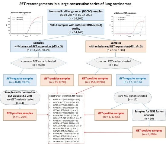
Cases with Unbalanced
5′/3′-End Expression of RET
Cases with Balanced
5′/3′-End Expression of RET
Total
Fusions detected via PCR for common variants152 (82.2%)33 (17.8%)185
Fusions detected via PCR for rare variants3 (75.0%)1 (25.0%)4
Fusions detected via NGS8 (100%)
(6–rare, 2–novel)
-8
Total RET-fusion-positive cases (n = 197)163 (82.7%)34 (17.3%)197
Table 2. Spectrum of identified RET fusions.
FusionNumber of Cases
KIF5B::RET (K15;R12)136 (69.1%)
CCDC6::RET (C1;R12)30 (15.1%)
KIF5B::RET (K16;R12)8 (4.1%)
KIF5B::RET (K22;R12)4 (2.0%)
NCOA4::RET (N8;R12)3 (1.5%)
KIF5B::RET (K23;R12)3 (1.5%)
RELCH::RET (R10;R12)2 (1.0%)
KIAA1217::RET (K13;R12)2 (1.0%)
KIF5B::RET (K24;R12)1 (0.5%)
KIF13A::RET (K18;R12)1 (0.5%)
KIAA1217::RET (K11;R12)1 (0.5%)
CCSER2::RET (C9;R12)1 (0.5%)
RBPMS::RET (R6;R12)1 (0.5%)
RUFY3::RET (R11;R12)1 (0.5%)
NCOA4::RET (N9;R12)1 (0.5%)
ZC3H7A::RET (Z3;R12)1 (0.5%)
HOOK1::RET (H21;R12)1 (0.5%)
Total197
Table 3. The proportion of RET-rearranged tumors among NSCLCs with different dCt values as assessed using the 5′/3′-end expression imbalance test.
dCt >/= 3dCt >/= 2dCt >/= 1dCT < 1Total
Cases tested for common variants16922437644734849
RET-positive (8 common variants)152/169 (89.9%)170/224 (75.9%)176/376 (46.8%)9/4473 (0.2%)185/4849 (3.8%)
Disclaimer/Publisher’s Note: The statements, opinions and data contained in all publications are solely those of the individual author(s) and contributor(s) and not of MDPI and/or the editor(s). MDPI and/or the editor(s) disclaim responsibility for any injury to people or property resulting from any ideas, methods, instructions or products referred to in the content.

