Review

Pain Biomarkers in Fibromyalgia Syndrome: Current Understanding and Future Directions

by
Martina Favretti
,
Cristina Iannuccelli
* and
Manuela Di Franco
Rheumatology Unit, Department of Internal Clinical, Anesthesiologic and Cardiovascular Sciences, Sapienza University of Rome, 00161 Rome, Italy
*
Author to whom correspondence should be addressed.
Int. J. Mol. Sci. 2023, 24(13), 10443; https://doi.org/10.3390/ijms241310443
Submission received: 16 May 2023 / Revised: 16 June 2023 / Accepted: 18 June 2023 / Published: 21 June 2023
(This article belongs to the Special Issue Chronic Pain and Fibromyalgia: Biomarkers and Pathophysiology)

Abstract

:
Fibromyalgia is a complex and heterogeneous clinical syndrome, mainly characterized by the presence of widespread pain, possibly associated with a variety of other symptoms. Fibromyalgia can have an extremely negative impact on the psychological, physical and social lives of people affected, sometimes causing patients to experience dramatically impaired quality of life. Nowadays, the diagnosis of fibromyalgia is still clinical, thus favoring diagnostic uncertainties and making its clear identification challenging to establish, especially in primary care centers. These difficulties lead patients to undergo innumerable clinical visits, investigations and specialist consultations, thus increasing their stress, frustration and even dissatisfaction. Unfortunately, research over the last 25 years regarding a specific biomarker for the diagnosis of fibromyalgia has been fruitless. The discovery of a reliable biomarker for fibromyalgia syndrome would be a critical step towards the early identification of this condition, not only reducing patient healthcare utilization and diagnostic test execution but also providing early intervention with guideline-based treatments. This narrative article reviews different metabolite alterations proposed as possible biomarkers for fibromyalgia, focusing on their associations with clinical evidence of pain, and highlights some new, promising areas of research in this context. Nevertheless, none of the analyzed metabolites emerge as sufficiently reliable to be validated as a diagnostic biomarker. Given the complexity of this syndrome, in the future, a panel of biomarkers, including subtype-specific biomarkers, could be considered as an interesting alternative research area.

1. Introduction

Fibromyalgia (FM) is a chronic clinical syndrome characterized by widespread pain (WP), fatigue and sleep disturbances. In addition, neuropsychiatric manifestations—such as cognitive impairment and mood disturbances—and somatic and dysautonomic symptoms can be present. The global prevalence of FM is estimated at 2.7%, with a higher frequency in women, patients over 50 years of age, those with a lower socio-economical level and those with obesity [1]. The accurate prevalence of FM is difficult to determine, considering the increasing evidence of frequent misdiagnosis, facilitated by the lack of reliable diagnostic biomarkers; furthermore, FM is rarely an isolated syndrome and its comorbidity with other chronic pain conditions or mental health disorders is frequent, leading to an increased likelihood of misdiagnosis [2,3]. The lack of reliable biomarkers is a central problem and barrier in FM diagnosis, not only because it is associated with an increased risk of misdiagnosis but also because it makes it difficult, if not impossible, to identify this syndrome in the earlier stages [4]. Moreover, diagnostic uncertainties linked to the absence of biomarkers increase patients’ frustration and dissatisfaction. These patients frequently seek even more medical attention and undergo numerous investigations and specialist consultations, favoring a rise in direct and indirect healthcare costs [5]. Since any serological, imaging or histological marker is currently available, the diagnosis of FM is still exclusively clinical; however, classification criteria may be used to confirm the diagnosis. The first set of classification criteria was developed in 1990 by the American College of Rheumatology (ACR) and required a history of WP and tenderness in 11 of 18 tender points [6]. In 2010, the ACR published its preliminary diagnostic criteria for FM [7], with the aim to overcome some practical and philosophical problems of the 1990 criteria; according to these criteria, a diagnosis of FM can be made in the presence of WP, evaluated by the Widespread Pain Index (WPI), and associated typical symptoms, assessed by the Symptom Severity Scale (SSS). These criteria were subsequently revised in 2016, adding the generalized pain criterion and stating that the diagnosis of FM does not exclude the presence of other clinically important illnesses [8]. Regardless of the differences between these criteria, one main feature remains unchanged: the presence of WP. Pain is a central symptom in FM; it is typically chronic, as it persists or recurs for longer than three months [9], and generalized, as it is localized in at least four out of five body regions [8]. It is associated with significant emotional distress and functional disability and cannot be better explained by other chronic pain conditions. According to the new International Association for the Study of Pain (IASP) classification of chronic pain [10], these features enable us to identify FM as a chronic primary pain (CPP) condition. CPP is a new diagnosis developed with the aim to define conditions characterized by a complex interaction of biological, psychological and social factors and therefore overcome the obsolete dichotomy of ‘physical’ and ‘psychological’ pain. Different chronic pain states fall within this new definition and at least some of them—including FM—can be defined using the pathophysiological descriptor of ‘nociplastic pain’ [11]. This new term refers to ‘pain that arises from altered nociception despite no clear evidence of actual or threatened tissue damage, causing the activation of peripheral nociceptors or evidence for disease or lesion of the somatosensory causing the pain’, and it is likely provoked by changes in nociceptive processing, probably due to central nervous system (CNS) activity modifications [11,12]. Nociceptive processing derives from the transmission of a stimulus from peripheral tissue to the brain and can be modulated in different ways. Changes in transmission mechanisms have been shown using different experimental pain tests and new imaging techniques such as functional magnetic resonance imaging (fMRI), proton spectroscopy or positron emission tomography (PET) [13]. Furthermore, these modifications of nociceptive perception, transduction and transmission could also be identified by the level modification of different metabolites. The aim of this narrative review was to collect metabolites likely involved in chronic pain perception whose alteration has been described in FM patients, in order to identify possible biomarkers of pain in FM syndrome.

2. Pathophysiology of Pain in FM Syndrome

Despite extensive studies, the exact pathogenesis of FM remains not fully understood, but different risk factors and pathophysiological mechanisms, potentially implicated in the development of the disease, have been identified. Pain in FM can be either described as dull, deep or aching—traditional descriptors of nociceptive pain—or burning or tingling—usually defining neuropathic pain [13]. However, as a hallmark of nociplastic pain, no clear evidence of primary tissue damage exists. In 2013, Üçeyler N. et al. [14] described the presence of small fiber neuropathy (SFN) in FM patients, supporting the hypothesis of small fiber damage as the first trigger of pain in FM syndrome. Since then, different studies have been published either supporting [15,16,17] or disproving [18,19] this hypothesis. The exact role of SFN in the pathogenesis of FM remains unclear. Thus far, it is assumed that at least some FM patients can develop SFN as a consequence of the same pathophysiological mechanism underlying the syndrome, but SFN cannot be considered the first pathogenetic event [20]. Typically, FM patients display enhanced sensitivity to painful and nonpainful stimuli, secondary to the development of hyperalgesia, allodynia or the abnormal wind-up of secondary pain [21]. These and other distinctive features are the consequences of functional and morphological changes in the CNS, primarily in pain processing brain structures [22]. Central sensitization (CS) is supposed to be the pathophysiological mechanism underlying these changes, possibly supported by glial activation due to neuroinflammation triggers. Moreover, additional aberrant systems could contribute to enhanced pain in FM patients: these include autonomic nervous system (ANS) abnormalities and hypothalamic–pituitary–adrenal (HPA) axis dysfunction [23].

2.1. Central Sensitization

According to the IASP, CS can be defined as the ‘increased responsiveness of nociceptive neurons in CNS to their normal or subthreshold afferent input’ [20]. CS is characterized by evidence of dysfunctional descending inhibitory pathways and increased facilitative activity, resulting in temporal summation with enlarged receptive fields, lower nociceptor thresholds, increased spontaneous neuronal activity and augmented stimulus responses [24]. Physiological nociceptive transmission strictly depends on the balance of excitatory and inhibitory stimuli [25]. The ascending system starts with primary afferent neurons—myelinated Aδ-fibers and unmyelinated C-fibers—conveying the noxious stimulus to the projection neurons in the dorsal horn (DH) of the spinal cord, a central network of integrating inputs [26]. Subsequently, these projection neurons transmit the noxious information either to the somatosensory cortex or to the cingulate and insular cortices, mainly via the spinothalamic tract [25]. A crucial nucleus of the descending system is the periaqueductal grey (PAG), which integrates information from higher structures of the brain—including the hypothalamus, amygdala and frontal lobe—and ascending nociceptive impulses and transmits inhibitory stimuli to the DH of the spinal cord [27]. Primary afferent fiber activation is physiologically associated with glutamate (Glu) release and the short post-synaptic depolarization of second neurons via alpha amino-3-hydroxy-5-methyl-4-isoxazolepropionic acid receptor (AMPA-R) activation. Indeed, the repetitive stimulation and co-release of other neuropeptides, such as substance P (SP), induce sustained post-synaptic depolarization with subsequent N-methyl-D-aspartate receptor (NMDA-R) unlock, due to Mg2+ removal [28]. NMDA-R activation results in Ca2+ influx and hyperexcitability of the DH neurons, favoring CS. In turn, hyperexcitability of the DH neurons facilitates NMDA-R activation, sustaining CS persistence [29]. Therefore, CS is the result of repetitive stimulation of nociceptors that activate several mechanisms of neuroplasticity, such as the reduction of pain thresholds, amplification of pain responses and spread of pain sensitivity to non-injured areas, leading to the enhanced excitability of the neurons of the DH of the spinal cord [30]. Both structural and functional modifications of the cortical regions implicated in pain processes and imbalanced levels of neurotransmitters involved in pain sensation have been associated with CS.
Several studies in FM patients have demonstrated that similar alterations in imaging and neurotransmitters could be present, corroborating the hypothesis of pain as a consequence of CS. Different noninvasive neuroimaging methods are currently available to enable a better understanding of pain processes. These methods include structural imaging techniques, such as voxel-based morphometry (VBM), and functional imaging techniques, such as fMRI and resting-state functional magnetic resonance imaging (rs-fMRI). A recent meta-analysis of VMB studies comparing CPP patients with healthy controls (HC) identified grey matter alterations in pain-processing brain regions, such as the cingulate, prefrontal and insular cortices, in patients affected by CPP [31]. Most of the studies conducted in FM patients indicate similar regional atrophies, probably secondary to chronic pain development, but none of the structural changes are specific to FM syndrome [20]. Conversely, different functional changes related to CS in FM patients have been described. Studies comparing noxious stimuli responses using fMRI demonstrate that, when an equal-pain-intensity stimulus is applied, FM patients display similar but more extensive patterns of brain activation than HC, particularly observed in the posterior insula and secondary somatosensory cortex [32,33]. Furthermore, fMRI studies of descending inhibitory pathways have identified modified PAG connectivity in FM patients, possibly associated with decreased activation of the anti-nociceptive system [20,34]. These findings are consistent with the hypothesis that a deficit in pain inhibition systems is specifically involved in chronic pain development in FM syndrome. Two principal descending inhibitory pathways exist: the opioidergic and 5-hydroxytryptaminergic-noradrenergic pathways. Endogenous opioid tone appears to be normal or even increased in FM patients [35], while endogenous serotoninergic and noradrenergic activity is apparently decreased, leading to reduced conditioned pain modulation (CPM) [36]. Finally, rs-fMRI studies in FM patients showed imbalanced resting-state connectivity in pain-processing regions with greater neuronal activation, suggesting that chronic pain could induce changes in brain processes activated even in the absence of an external stimulus [32,37].

2.2. Neuroinflammation

Neuroinflammation is a form of localized inflammation occurring in the peripheral nervous system (PNS) and CNS and it is characterized by vascular changes with increased permeability, glial cell activation, the infiltration and activation of leukocytes and the increased production of inflammatory mediators including cytokines and chemokines [38]. While acute inflammation can elicit acute pain sensations, neuroinflammation is supposed to be implicated in the chronification and persistence of pain. Compelling evidence exists proving that cytokines, chemokines and other glia-produced mediators could either induce or maintain CS, driving the development of WP [39]. Apparently, neuroinflammation is the result of a bidirectional crosstalk between nociceptors and different non-neuronal cells, particularly immune and glial cells. Cytokines and inflammatory mediators provided by immune and glial cells activate and sensitize nociceptors, while nociceptors themselves can secrete cytokines and chemokines, which are essential for immune modulation [39].
Regarding immune cells, mast cells (MCs) seem to play a central role in neuroinflammation development. MCs are resident immune cells located ubiquitously in vascularized tissue, predominantly at the interface with the external environment. These cells can be found close to small-caliber sensory nerve fibers in peripheral innervated tissue, on the endoneurial compartments of peripheral nerves and, in small quantities, in the brain [40]. In the brain, MCs can be found on the abluminal sides of blood vessels, where they interact with neurons and glial cells [41]. Different receptors—such as toll-like receptor (TLR) 2 [42], TLR4 [42] or P2X receptors [43]—induce MC degranulation with the consequent release of various mediators, such as biogenic amines, cytokines, neuropeptides, growth factors or ATP [42]. Mediators secreted by MCs promote nociceptor activation and hypersensitization, both directly—via pro-nociceptor mediators’ production [44]—and indirectly—inducing neuropeptide release [42]. Furthermore, there is substantial evidence that MCs can impact blood–brain barrier (BBB) permeability, disrupting its integrity [45]. Therefore, in the absence of tight control, MC–nociceptor interactions induce nociceptive receptors’ hyperactivity while lowering pain thresholds. Persistent nociceptive stimulation finally sensitizes the neurons of the DH, leading to CS [44]. Glial cells are CNS cells that, in addition to MCs, seem to play a role in neuroinflammation and the chronification of pain. Among these cells, microglia and astrocytes’ involvement is better understood, but there is evidence of an active and context-specific role of oligodendrocytes as well [38]. Local microglia represent the resident macrophage population of CNS and are able to undergo rapid activation in response to minor pathological changes in the CNS. Different mediators released by immune cells and nociceptors—such as adenosine triphosphate (ATP) and chemokine ligand (CXCL) 1—could trigger microglia’s production of other mediators, either cytokines—such as tumor necrosis factor (TNF), interleukin (IL) 1 and IL-18—or growth factors—such as brain-derived neurotrophic factor (BDNF) [46]. These mediators are consequently able to enhance spinal pain transmission. Neuronal hyperexcitability and augmented pain signal transmission are also induced by astrocyte activation. In the context of neuroinflammation, there is evidence that astrocytes lose their ability to maintain the homeostatic concentrations of Glu and K+ while achieving the capacity to secrete ATP, Glu and chemokines, thus contributing to chronic pain development [47]. The activation of microglia and astrocytes has been described in different neurodegenerative diseases [48,49] and neuropsychiatric illnesses [50,51] using imaging techniques, such as PET and proton magnetic resonance spectroscopy (1H-MRS) [52], but it is supposed to be present in FM syndrome too.
A recent study [53] described elevated choline levels, a marker of glial activation, in the anterior insula of FM patients compared to HC, and reduced connectivity between the anterior insula and putamen. Both these findings were associated with worse pain interference in FM patients. A previous study [54] showed higher choline levels in the right dorsolateral prefrontal cortex (PFC) in FM patients, also describing a positive correlation with pain. According to these findings, but also on the basis of evidence of neuropeptide and cytokine alterations in FM patients, it has been proposed that neuroinflammation, or at least neurogenic inflammation, could be involved in the development and maintenance of pain in FM syndrome. Neurogenic inflammation is the consequence of nociceptor activation—particularly C fibers—and the associated release of neuropeptides such as SP, calcitonin gene-related peptide (CGRP) and prostanoids [55]. This augmented neuronal activity could possibly trigger neuroinflammation in peripheral tissue [56].

2.3. Neuroendocrine and Autonomic Nervous System Dysfunction

Stress stimuli, which can be either real or perceived and arise from different life events, are known modulators of the pain system. Under physiological conditions, exposure to a stressor elicits the rapid activation of the ANS and the HPA axis, with the consequent release of catecholamines and cortisol, starting the so-called ‘fight or flight’ response [57]. The induction of anti-nociceptive and analgesic mechanisms is a hallmark of this response [58]. Otherwise, deregulation of the stress response, caused by exaggerated or maladaptive reactions, is associated with altered nociceptive responses, eventually leading to chronic pain development. Usually, in response to a stressor stimulus, the corticolimbic system, mostly represented by the prefrontal cortex, amygdala and hippocampus, sends disinhibitory impulses to the hypothalamus. The first quasi-instantaneous response to hypothalamus disinhibition is catecholamine release—both noradrenaline and adrenaline—via locus coeruleus stimulation. Catecholamines induce a hypervigilant state that is characterized, among other features, by augmented pain resistance secondary to descending inhibitory pathway activation [59]. Conversely, pathological hyperactivity of the sympathetic system is associated with primary afferent nociceptor activation and, subsequently, the development of chronic pain [60]. Hypothalamus activation results also in corticotropin releasing hormone (CRH) secretion, which induces the pituitary gland release of adrenocorticotropic hormone (ACTH), which, in turn, activates the cortical adrenal gland’s production of cortisol. Cortisol stimulates immediate—on the scale of minutes—and tardive—due to structural and genomic effects—systemic responses. The corticolimbic system and hypothalamus show high affinity for cortisol and, therefore, its production controls the stress response via a negative feedback loop [61]. There is evidence that chronic stress induces functional and morphological changes in key regulatory regions of the stress response, especially the corticolimbic system [62]. These changes are associated with numerous abnormalities, including an unbalanced ANS and dysfunctional HPA axis, which have been described in various chronic pain conditions. Interestingly, it seems that distinct chronic pain conditions arise from a complex and partly unique interaction not only between steroid production but also between the HPA axis, ANS and immune system, possibly explaining the pathophysiological and clinical discrepancies between them [63].
Regarding FM, hyporeactivity of the HPA axis, often associated with lower basal cortisol levels, and basal hyperactive sympathetic activity have been described. Despite discrepancies between studies, FM patients present plasma cortisol level alterations characterized by the flattening of the cortisol concentration during the day, due to the generally dysregulated circadian variation of this hormone [33]. Furthermore, the administration of different stress stimuli, such as IL-6 injection or hypoglycemia induction, has been associated with a delayed or reduced ACTH response, showing the hyporeactivity state of the HPA axis [64]. ANS activity has been investigated using power spectrum analysis of heart rate variability (HRV). These studies described reduced HRV in FM patients, as a consequence of hyperactivity of the sympathetic nervous system and decreased parasympathetic tone [60,63]. As well as dysregulation of the neuroendocrine axis, which is widely described in FM syndrome, different physical traumas and psychological triggers are associated with enhanced pain in FM patients [23]. Several types of stressors have been proposed as potential environmental triggers of the disease, possibly influencing gene expression and leading to HPA axis impairment [65]. These potential stressors include adverse events in neonatal or childhood life [66] or repeated physical or psychological stressors [23]. Environmental factors are believed to promote FM in genetically predisposed individuals, and the genetic predisposition is supported by evidence of strong familial aggregation among FM patients [67]. Single-nucleotide polymorphisms (SNPs) of several genes have been associated with FM and, among these, the catechol-O-methyl transferase (COMT) gene is the most widely investigated [68]. COMT is the major enzyme involved in monoamine metabolism, such as dopamine, adrenaline and noradrenaline, and alterations in COMT activity are associated with chronic pain development [69]. COMT gene polymorphism has been considered a possible predisposing factor for the development of FM but current reports remain contradictory [70,71].

3. Biomarkers of Pain in Fibromyalgia Syndrome

3.1. Glutamate

Glutamate (Glu) is the principal excitatory neurotransmitter of the nervous system, and it plays a significant role in nociceptive modulation: it regulates other neurotransmitters’ release from afferent nerve fibers, stimulates the activation of DH neurons and its neuroplasticity and induces cortical activation, facilitating the storage and generation of long-term memory processes [25]. Dysfunction in Glu and other neurotransmitters’ metabolism and the consequently altered availability have been implicated in different chronic pain conditions, especially through 1H-MRI studies. Interestingly, the results of these studies suggest the existence of a unique neurometabolite signature for each different pain condition [72].
FM patients demonstrate a general increase in Glu levels, possibly playing a significant role in the development and maintenance of chronic pain [73]. FM patients also demonstrate higher cerebrospinal fluid (CSF) Glu levels than chronic migraine patients [74], and HC and Glu levels positively correlate with neurotrophin production but not with clinical evidence of pain [75]. These findings suggest that, despite the difficulty to relate Glu levels to clinical evidence of CS, their involvement in FM syndrome is unquestionable [75]. Subsequently, 1H-MRI studies have revealed significantly higher Glu levels in FM patients compared to HC in different brain regions, such as the posterior insular cortex [76,77], posterior cingulate gyrus [78], right amygdala [79] and ventrolateral PFC [80] Table 1. These findings lead to the hypothesis that Glu hyperactivity in pain-processing brain regions—due to more Glu in synaptic vesicles, a higher number or density of glutamatergic synapses or less Glu reuptake—causes FM syndrome [76]. There is strong evidence that Glu excess can lead to neuronal dysfunction and cell loss, possibly explaining the brain regional atrophy described in FM patients [81]. An alternative hypothesis states that neuronal dysfunction could be the consequence of astrocyte deficits. Astrocytes are implicated in the uptake, metabolism and recycling of Glu and their alteration could start a cascade of metabolic events leading to abnormal neurotransmission [78]. Additionally, there is evidence that Glu levels in particular brain regions correlate with clinical pain experience in FM patients, even when these levels are not significantly higher than in HC. Significant correlations have been identified in the posterior cingulate gyrus [78], left thalamus [80] and insular cortex [76]. Regarding the insular cortex, Lee J. et al. [82] demonstrated that the anterior and posterior insular cortices’ Glu levels are associated with different dimensions of chronic pain: the cognitive affective and sensory physical dimensions, respectively. These findings are in line with the different roles that these two brain regions play in pain processing [83]. Furthermore, Harris R.E. et al. [77] demonstrated that changing levels of insular Glu correlate with changes in clinical pain experience in FM patients.
This evidence suggests that Glu level detection in particular brain regions, assessed by neuroimaging techniques, could be a useful biomarker of pain in FM patients. Unfortunately, several limitations exist. First of all, it is unlikely that Glu measurements reflect the synaptic levels of this neurotransmitter: not only is the 1H-MRS Glu signal often contaminated by glutamine levels, but, arising from grey and white matter signals, it ensembles multiple different cell types, including regions distant from synapses [84]. Secondly, there are no standardized 1H-MRS parameters for neurometabolite level estimation and there is little consensus on the best way to measure Glu levels [72]. Therefore, the assessment of CNS Glu levels as a biomarker of pain using 1H-MRS nowadays remains scarcely affordable and poorly reproducible.

3.2. Substance P

Substance P (SP) is a neuropeptide member of the tachykinin family, along with neurokinin (NK) A and NKB. SP is expressed throughout the nervous and immune systems, and it is involved not only in a large number of physiological processes but also in numerous pathological conditions [85]. The biological SP actions are mediated by neurokinin (NK) receptors. Three NK receptors exist—NK1-R, NK2-R and NK3-R—and although each receptor has moderate affinity for each tachykinin ligand, every single NK receptor is preferentially activated by one ligand. NK1-R is the primary receptor for SP [86]. SP plays a central role in pain transmission and compelling evidence suggests that it could be implicated in the induction and maintenance of CS, as well as the activation of neurogenic inflammation. Primary afferent sensory fibers express SP along with Glu and are able to release this neuropeptide both centrally in the DH neurons’ synapses and backward in the peripheral terminals [87]. SP released in the DH of the spinal cord binds NK1-R and induces the hyperexcitability of the spinal neurons, both directly by causing a slow excitatory postsynaptic potential and indirectly by facilitating the activation of NMDA-R, thus favoring initial CS development [88]. Spinal neurons’ hyperexcitability is then maintained by SP release. NK1-R activation is able to increase Ca2+ influx, subsequently activating C-fos and C-jun, two proto-oncogenes involved in the persistence of sensitization [89]. Additionally, there is evidence that CGRP, released together with SP, enhances NK1-R gene expression, thus favoring its activation and, therefore, CS persistence [88]. By contrast, SP released in the peripheral endings of primary sensory neurons induces arteriolar dilatation, plasma extravasation and leukocyte infiltration, thus generating neurogenic inflammation [90]. Green D.P. et al. [91] demonstrated that these effects are mediated by MrgprX2 activation, a receptor expressed exclusively on MCs’ surfaces.
Considering SP’s involvement in pain processes, it has been suggested that this neuropeptide could be implicated in the pathogenesis of FM syndrome, and the measurement of SP levels—either from CSF or peripheral samples—has been proposed as a possible diagnostic biomarker (Table 2). Different studies [92,93,94] demonstrate significantly higher SP levels in the CSF of FM patients compared to HC, but no relation [93] or, surprisingly, an inverse relation [92] with the clinical severity of pain was found. One study [95] did not report any differences in SP serum levels between FM patients and HC. Conversely, two more recent studies [96,97] demonstrated higher serum levels of SP in FM patients compared to HC. None of these studies compare serum SP levels with clinical pain scores. It has also been proposed that the beneficial effects of different complementary FM treatments could be explained by SP level changes after treatment administration. It has been shown that there is a significant SP serum level reduction after the administration of massage therapy [98], acupuncture [99] and dietary supplements containing primarily an extract of salmon’s milt [97]. All of these treatments have been demonstrated to improve the clinical evidence of pain but a direct correlation with SP levels has not been found. A previous study [100] showed an increase in SP serum levels after acupuncture treatment, despite a significant reduction in pain scores. However, unlike the other studies, this was not a randomized controlled trial. Another study [101] investigated the effect of different exercise and walking programs on pain and SP level changes in the CSF of FM patients, demonstrating a significant association between changes in pain threshold and changes in SP levels after exercise. According to these findings, it appears sufficiently clear that abnormal SP levels can be found in FM patients, both in CSF and peripheral samples, but the actual association of this marker with the clinical evidence of pain remains less clear.

3.3. Nerve Growth Factor

Nerve growth factor (NGF) is a member of the neurotrophin family and participates in the survival and growth of distinct neuronal populations, especially during development. In contrast, during adulthood, NGF changes its role and is involved in modulating nociceptive transmission [89]. Neurotrophins can bind two types of receptors: one is the tyrosine-kinase (Trk) receptor, which is highly selective—NGF binds preferentially to TrkA—and the other, named p75NTR, is much more promiscuous [102]. It has been demonstrated that the exogenous administration of NGF is able to induce both thermal—early onset—and mechanical—delayed—hyperalgesia in healthy volunteers, lasting up to 7 days [103,104,105], with pain sensations expanding both proximally and distally after NGF injections [106,107,108]. These findings suggest NGF’s ability to induce both peripheral and central sensitization. Rapidly after stimulation, TrkA expressed on the peripheral terminals of nociceptive fibers reduces the nociceptor threshold by inducing the sensitization of different ion channels, thus favoring early-onset peripheral sensitization [109]. Subsequently, the NGF/TrkA complex is retrogradely transported to the neuronal soma in the dorsal root ganglia (DRG), where it stimulates neuropeptide release—such as SP, CGRP and BDNF—either in peripheral or central terminals, and upregulates the gene expression of both ion channels and neuropeptides. These delayed effects contribute both to peripheral and central sensitization [102]. Finally, a third mechanism could be implicated in NGF-mediated sensitization: it might cause pain by promoting the sprouting of nociceptive neurons. There is evidence of pathological peripheral nerve sprouting and hyperinnervation in disease models of bone cancer pain [110] and it has been hypothesized that a similar effect could be induced also in central sites such as the DH of the spinal cord [111]. The increased fiber density could stimulate increased nociceptor responsiveness, leading to augmented pain transmission.
NGF appears to be little expressed in normal adult conditions but there is evidence of augmented NGF levels in multiple pathological states, especially those associated with inflammation. For this reason, it has been proposed that NGF could perform an immunomodulatory function in inflammatory response regulation, including MC differentiation, maturation and degranulation [112]. However, this feature remains controversial: although there is evidence of a possible NGF proinflammatory action on MCs [113,114], several recent reports demonstrate that NGF does not affect MC activation [115] and contradictory evidence exists regarding NGF receptor expression on MC surfaces [102]. In contrast with peripheral production, NGF expression in the CNS appears to be much more limited. In the CNS, NGF can be found primarily in the basal forebrain cholinergic neurons of limbic areas. The presence of NGF—and BDNF—in areas involved in neuroendocrine response regulation indicates that these neurotrophins could be involved in the modulation of the endocrine response to stress, and neurotrophin-mediated neuronal plasticity could increase the susceptibility to stress-related psychiatric disorders [116].
Considering the putative role of NGF in chronic pain, and according to the evidence of altered levels of NGF in different chronic pain conditions [117,118], it has been hypothesized that abnormal NGF levels could be observed in FM patients and contribute to the pain experience (Table 3). However, only two studies have measured NGF levels in the CSF of FM patients, and both detected significantly higher NGF levels in FM compared to HC [75,119]. Sarchielli P. et al. [75] also demonstrated a positive correlation between NGF levels and BDNF levels and both correlated with the duration of chronic pain but not with clinical evidence of pain. This evidence suggests that NGF favors CS in FM patients via BDNF upregulation, thus sustaining pain persistence. Two additional studies evaluated NGF levels from peripheral samples of FM patients, with conflicting results. Baumeister D. et al. [120] did not find differences in NGF levels between FM patients and HC, refuting the notion that peripheral growth factor levels contribute to the pathophysiology of FM syndrome and peripheral sensitization development. Jablochkova A. et al. [121] showed reduced peripheral NGF levels in FM patients compared to HC but did not find any relation with other serological biomarkers or clinical data. According to the authors, this finding could be explained by the emotional status of patients, considering that major depression disorder has been associated with low serum NGF levels. Unfortunately, the study did not demonstrate a significant relation of NGF levels with depressive symptoms. Furthermore, the authors speculatively proposed that low peripheral levels of NGF could explain the nociceptive fiber alterations described in FM patients, such as the reduction in distal intraepidermal nerve fiber density or C-fibers with smaller diameters. Nonetheless, considering the paucity and inconsistency of the current literature, it is difficult to consider NGF levels as a reliable biomarker of pain in FM syndrome.

3.4. Brain-Derived Neurotrophic Factor

BDNF is another member of the neurotrophin family and it takes part in the regulation of different processes, such as the development of brain circuits and the formation and maintenance of the neuronal morphology or brain architecture [122]. Moreover, it is well established that the altered expression of BDNF can initiate and maintain inflammatory, neuropathic and chronic pain, by mediating pain plasticity and sensitization processes [123]. Moreover, an anti-nociceptive role of BDNF has been occasionally described, suggesting that it can act in different ways depending on the specific intracellular pathway activated and the different cell types involved [124]. BDNF is expressed by different neuronal and non-neuronal cells. Among neuronal cells, BDNF expression has been demonstrated in small- and medium-sized sensory neurons [125] but also in several brain areas, such as the hippocampus, cortex, amygdala, striatum and hypothalamus [126]. Along with neuronal cells, BDNF is also synthetized by immune system cells, especially microglial cells, and platelets [127]. It has been demonstrated that various stimuli are able to induce BDNF release in different cells. Distinct electrical activity patterns, including Glu-mediated stimuli, are efficient to induce BDNF release in the CNS to mediate synaptic as well as network plasticity processes [126]. In the PNS, different pathological conditions associated with the enhanced retrograde transport of NGF to the DRG increase BDNF gene expression and central terminal release, thus favoring sensitization processes [128]. Lastly, various extracellular stimuli can induce the microglial release of BDNF and, among them, P2X4R activation, mediated by extracellular ATP, has been associated with central sensitization features such as pain hypersensitivity or allodynia [129]. BDNF’s activities are mediated by high-affinity TrkB and low-affinity p75NTR receptor binding [102]. BDNF released from nociceptors in the DH of the spinal cord binds TrkB receptors expressed by medium- to large-sized neurons in the spinal cord, including ascending projection neurons, and induces the pre- and post-synaptic potentiation of Glu transmission via NMDA-R plasticity [124]. Moreover, the spinal microglial secretion of BDNF binds neuronal TrkB receptors and contributes to pain hypersensitivity through the disinhibition of pain processes: it suppresses the intrinsic inhibitory circuits in the DH by decreasing the expression of potassium-chloride cotransporter 2 (KCC2) and weakening GABAergic inhibitory synapses [127]. BDNF is also a central factor in long-term potentiation (LTP) processes, a core mechanism of plasticity in the nervous system, and it has been shown to be sufficient to induce LTP in the DH neurons, thereby maintaining CS [130].
A potential role of BDNF in the pathophysiology of FM and the development of pain have been extensively studied in the last few years, with various results. Several studies demonstrated higher CSF [75], plasma [121,131] and serum [132,133,134] BDNF levels in FM patients compared to HC. These findings advocate for a key role of BDNF in FM pathophysiology, especially as a pain modulator. Given the lack of relation between serum BDNF levels and illness duration, Laske C. et al. [132] concluded that the increased BDNF serum concentration is rather due to peripheral pain modulation. Subsequently, Nugraha B. et al. [135] suggested that higher serum BDNF levels in FM patients may be part of an adaptation mechanism in chronic pain, or, considering its possible anti-nociceptive role, a long-term high concentration of BDNF may act as a defensive mechanism. However, the absence of differences in plasma [136] and serum [120,137] BDNF levels between FM patients and HC have also been described. Moreover, a study by Iannuccelli C. et al. [138] described significantly lower BDNF serum levels in FM patients compared to HC. Different explanations could be made for these findings. First, the heterogeneous nature of FM makes it difficult to generalize, and the mechanisms underlying CS are difficult to establish [120]. Secondly, although it has been demonstrated that circulating BDNF represents 70–80% of that produced in the CNS [139], BDNF levels could be influenced by other cellular sources, such as platelet degranulation in clotting processes—which could influence especially plasma BDNF levels [140]—or skeletal muscle production in response to contraction stimulation [141]. Lastly, it must be taken into account that the chronic administration of antidepressants modifies BDNF expression [142] and could therefore interfere with the levels of this mediator. Bidar A. et al. [143] actually showed a rapid reduction in serum BDNF levels in FM patients after one-month treatment with duloxetine, thus supporting the overall role of this metabolite in pain modulation. Since this study did not show any significant difference in BDNF levels between FM patients and patients affected by non-FM chronic nociceptive pain disorders, the authors also noted that their finding weakens the exclusive role of BDNF in nociplastic pain. Nonetheless, even studies that have described significantly higher BDNF levels in FM patients are not able to demonstrate any relation with clinical pain [75,121,131,132,134]. Only one study [144] demonstrated a significant inverse relation between BDNF levels and pressure pain thresholds, supporting its value as a potential biomarker of pain in FM patients. Recently, a possible association between BDNF levels and dysfunction in the descending pain modulatory system in FM syndrome has been studied. Caumo W. et al. [145] demonstrated that central sensitization syndromes (CSS) without a structural pathology, including FM, present higher BDNF serum levels than in patients affected by CSS with a structural pathology, such as osteoarthritis. The authors were also able to show an inverse relation between BDNF serum levels and CPM, favoring a higher propensity for pain. In addition, Soldatelli M.D. et al. [146] described an association between dysfunction of the descending modulatory system and the severity of FM symptoms related to psychological aspects, including pain catastrophizing and disability due to pain.
It is therefore reasonable to conclude that altered BDNF production is implicated in the pathophysiology of FM, most likely regulating neuroplasticity processes that favor CS (Table 4). On the other hand, BDNF’s association with clinical evidence of pain remains controversial. Further studies are needed not only to better investigate BDNF’s role in the development of FM and pain but also to clarify the most reliable sample, between serum and plasma, to measure BDNF levels of CNS origin.

3.5. Mu Opioid Receptor

Dysregulation of the descending anti-nociceptive system is a potential pathophysiological feature of chronic pain in FM syndrome. Compelling evidence regarding decreased endogenous noradrenergic and serotoninergic activity in FM syndrome exists [147], while the endogenous opioid system appears to affect CPM in a different way. It has been largely demonstrated that opioid treatment in FM patients is not effective [148,149], while there is promising evidence of low-dose opioid antagonism’s efficacy in the management of FM symptoms [150,151,152]. Endogenous enkephalin levels appear to be higher in the CSF of FM patients [35], while Mu opioid receptor (MOR) availability is reduced in different pain-processing brain regions [153,154,155] and shows a significant relation with the affective dimension of pain [153]. Authors have suggested that the reduced MOR availability in FM patients could be the consequence of an overactive opioidergic system, which could lead to MOR’s reduced affinity or the downregulation of GABAergic inhibitory interneurons [153]. As a result of the reduced MOR availability, phasic endogenous opioid release occurring during noxious stimulation is no longer able to produce the appropriate inhibition of GABAergic interneurons, leading to the development of pain [154].
Recently, Üçeyler N. et al. [155] proposed a possible neuroimmune connection linking IL-4 gene expression to cerebral opioid receptor availability in FM patients. Endogenous opioid system and immune system cells are known to engage in bidirectional and complex communication [156], and it has been proposed that opioid receptors, including MOR, expressed by immune cells could be implicated in pain modulation [157,158]. Two recently published studies demonstrated a lower percentage of B cells [159] and natural killer cells [160] expressing MOR in FM and osteoarthritis patients with a moderate/severe intensity of pain compared to HC, and an inverse relation between the percentage of MOR B lymphocytes and natural killer cells and pain intensity. The authors proposed the determination of the percentage of MOR cell expression as a possible biomarker of pain in FM syndrome but also in other chronic pain states. Altogether, these studies indicate MOR’s reduced expression as an interesting possible biomarker of pain in FM syndrome. However, further studies are needed, not only to confirm the possible use of this marker but also to investigate the potential relation between MOR availability in brain regions and peripheral immune cells’ MOR expression, in order to deepen our knowledge regarding endogenous opioid system dysfunction in FM.

3.6. Mast Cells and Cytokine Production

Several FM symptoms—such as hyperalgesia, sleepiness and fatigue—are remarkably similar to those that occur after contact with infectious agents, with the consequent stimulation of inflammatory cytokines, and accumulating evidence supports the hypothesis that FM patients could present the dysfunctional regulation of cytokine production [161]. FM symptoms can be worsened by stress, and stressful stimuli are able to trigger MC degranulation and the release of proinflammatory and neuro-sensitizing mediators activating both nociceptors and microglial cells [162]. As well as stressful stimuli, mediators released by nociceptive sensory neurons can regulate the maturation, recruitment and degranulation of MCs [40]. Starting from this assumption, it has been hypothesized that dysfunctional MCs and the associated altered cytokine production could be implicated in the pathogenesis of pain in FM. MCs appear to be overexpressed in the papillary dermis in FM patients [163,164]. It has been suggested that excessive SP release could trigger MC activation [163], and increased numbers of MCs’ dermal mediators could represent a source of repetitive peripheral stimuli reaching the CNS, which cause CS [164]. However, a clinical trial conducted in 2015 [165] failed to demonstrate a pain sensitivity reduction or clinical pain and symptom severity improvement after MC stabilizer (ketotifen) administration. In addition, cytokines’ gene expression, evaluated from skin biopsies of FM patients, reported opposing results: Salemi S. et al. [166] found TNF, IL-1β and IL-6 expression in the skin biopsies of FM patients, while no cytokines could be detected in HC, whereas Üçeyler N. et al. [167] did not find any differences in cytokine expression in the skin samples of FM patients compared to HC. Besides tissue gene expression, different cytokines and chemokines’ levels, either from serum or plasma samples, have been studied extensively, with extremely mixed results. The various results in the literature have been collected in three systematic reviews and meta-analyses [168,169,170].
According to these studies, FM patients present a peripheral blood cytokine profile that differs from that of HC: increased levels of TNF-α [169,170], IL-6 [168,169,170], IL-8 [168,169,170] and IL-10 [167] can be detected. It must be taken into account that cytokine research in FM syndrome remains poorly standardized and several factors—such as the circadian rhythmicity of secretion, environmental factors and medication—could influence cytokine assays and reduce the quality of calculated data [168]. The lack of standardized analytical methods to measure the levels of immune mediators considerably contribute to the increased heterogeneity of the results, thus making it even more difficult to draw any definitive conclusion [170]. However, FM patients may indeed present a particular cytokine signature as a result of complex features, involving potential compensatory anti-inflammatory cytokines and chemokines implicated in the modulation of neuronal plasticity [169]. The actual relation between this cytokine signature and the clinical evidence of pain remains more elusive and appears difficult to establish. Ang D.C. et al. [171] showed a longitudinal relation between chemokine levels, especially IL-8 and monocyte chemotactic protein (MCP-) 1, and the severity of pain, while Wang H. et al. [172] observed a concordant reduction in IL-8 levels and pain intensity during pain therapy but no significant relation was demonstrated, except after six months of treatment. Another plausible hypothesis is that the first cytokine source could be represented by non-neuronal cells in the CNS, rather than peripheral cells. Two studies [173,174] demonstrated IL-8 levels in the CSF of FM patients that were higher than those of HC and rheumatoid arthritis patients; in addition, in FM patients, IL-8 levels in CSF were higher than in peripheral blood samples, leading the authors to conclude that the first source of this chemokine is the CNS—in particular, astrocytes and microglial cells. It has also been hypothesized that thalamic MCs contribute to neuroinflammation and pain by releasing neurosensitizing mediators, which can stimulate diencephalon microglial cells, in the context of central cytokine production [175]. In conclusion, it appears that chronic cytokine production and neuroinflammation are part of the pathophysiology of FM, but different features, such as the existence of a specific cytokine signature, the actual first cytokine source and the clinical relation between cytokines and pain, remain poorly understood.
Thus far, neither peripheral nor CSF cytokine levels can be considered a reliable biomarker of pain in FM syndrome, and additional studies are warranted to increase our knowledge regarding the effective role of inflammation in the pathophysiology of pain in FM patients.

3.7. Pentraxin-3

Pentraxin-3 (PTX-3) is an acute-phase glycoprotein, a member of the long pentraxin family, which acts as a modulator of inflammatory processes during microbial or sterile inflammatory responses [176]. PTX-3 synthesis in stimulated by different molecules, such as lipopolysaccharides (LPS), IL-1β and TNF-α, while IL-6 does not influence PTX-3 production [177]. Both protective and detrimental effects of PTX-3 have been described. This glycoprotein has the ability to bind to a variety of microbes, acting as an opsonin and thus stimulating the immune response, but it is also able to protect the organism from infection-induced organ injury and sterile-inflammation-induced tissue damage, limiting excessive immune cell infiltration. Conversely, it has also been described that PTX-3 can worsen tissue injury in certain pathological conditions, such as post-ischemic renal injury, intestinal ischemia and reperfusion or ventilation-induced lung injury [178].
PTX-3 could be considered a plasmatic marker of immune system activation and it has been proposed that FM patients may present augmented PTX-3 levels as a consequence of cytokine dysregulation and increased TNF-α production (Table 5). Skare T.L. et al. [179] demonstrated higher PTX-3 plasmatic levels in FM women compared to HC, in the absence of significant differences between depressed patients and those without depression [180]. In addition, Garcia J.J. et al. [181] found that monocytes from FM women without depression released more PTX-3 than HC, both constitutively and after LPS stimulation, while no difference was found in neutrophils’ ability to release PTX-3. These findings support the hypothesis of increased PTX-3 production in FM patients, suggesting that activated monocytes may be a possible peripheral source, and allow us to include PTX-3 levels as a potential biomarker for FM syndrome. However, further studies are needed to better understand the role of PTX-3 in FM and possibly evaluate whether PTX-3 levels are associated with particular clinical features of FM, especially with clinical evidence of pain.

3.8. Neuropeptide Y

Neuropeptide Y (NPY) is a 36-amino-acid peptide widely distributed in the CNS and PNS but also expressed by a variety of immune cells and implicated in many physiological and pathological conditions [182]. NPY preferentially binds two major inhibitory receptors—Y1 and Y2—broadly expressed on the surfaces of different neuronal and non-neuronal cells, leading to the activation of different molecular pathways and consequent biological effects [183]. It is implicated in pain modulation at the DRG and DH levels, where it seems to mediate mostly anti-nociceptive effects. After hyperalgesia-induced excitatory signals, primary afferent neurons and DH inhibitory interneurons upregulate NPY expression and release it in the DH of the spinal cord [184]. Released NPY activates the Y1 and Y2 receptors. The Y1 receptor is expressed on excitatory interneurons localized in the superficial layer—lamina II—of the DH of the spinal cord, and its activation leads to the post-synaptic inhibition of the spinal interneurons and a net reduction in the pain excitatory:inhibitory ratio [185]. Indeed, the Y2 receptor is expressed on the central terminals of small- to medium-sized myelinated A fibers and its activation induces pre-synaptic nociceptive inhibition, especially via pronociceptive substances’—such as SP—reduction [184]. Besides mediating analgesia, NPY activity has also been related to the stress response and resilience-promoting properties. It has been demonstrated that NPY can be expressed in different brain areas, including the hypothalamic arcuate nucleus and excitatory interneurons located in the amygdala [186]. The hypothalamic arcuate nucleus is functionally associated with the paraventricular nucleus of the hypothalamus—the main brain source of CRH—and there is evidence that NPY activity counteracts the biological actions of CRH, favoring stress adaptation processes. On the other hand, NPY released in the amygdala mediates excitatory pyramidal neurons’ inhibition, and the reduced activity of this brain area is associated with anxiolytic effects and increased stress resilience [187]. Moreover, NPY is widely distributed throughout post-ganglionic sympathetic nerves and it is co-stored and co-secreted with noradrenaline, so that it can be considered a marker of noradrenergic function [188]. Lastly, NPY may also act as an immunoregulatory factor, acting on immune system function either directly through NPY receptors expressed on the surfaces of different immune cells or indirectly through the regulation of different physiological or pathological conditions [189].
Considering the known relationship between stressful stimuli exposure and FM symptom onset, as well as the evidence of a maladaptive response to these stimuli, authors have investigated the possible application of NPY peripheral levels as a biomarker of ANS dysfunction and its potential association with pain experience (Table 6). Different studies describe not only reduced plasma levels of NPY in FM patients compared to HC [190,191] but also low NPY levels after 30 min on a tilt table [191,192], thus suggesting the hypofunction as well as hyporeactivity of the sympathetic stress axis in FM patients. Conversely, subsequent studies demonstrated higher NPY plasma and serum levels in FM patients compared to HC [188,193,194] and lower HRV in FM patients, also showing a significant relation between HRV and NPY [193]. These findings suggest that FM patients could develop increased sympathetic–adrenal activity, possibly as a maladaptive response to high stress or chronic tension. In none of these studies [188,193,194] was a significant relationship between NPY levels and clinical evidence of pain present. These findings suggest the contribution of impaired ANS activation and altered NPY expression to the pathophysiology of FM, but the exact role of sympathetic dysfunction remains less understood, mostly due to the complexity of this system and its implications in a variety of biological conditions. To date, there is no evidence of a direct association of NPY levels with any FM symptoms; thus, so far, NPY cannot be considered a useful biomarker of pain in FM syndrome.

4. Potential Future Directions

4.1. Vitamin D

Recently, the possible involvement of hypovitaminosis D in the pathophysiology of chronic pain disorders has elicited increasing interest among the scientific community. Vitamin D, beyond its fundamental role in skeletal and calcium homeostasis, has been recognized to be implicated in a variety of other actions, including a potential regulatory function in pain pathways [195]. On a clinical level, vitamin D deficiency is associated with osteomalacia, a pathological condition characterized by the development of bone pain, proximal muscle weakness and generalized fatigue and commonly misdiagnosed as FM syndrome or other chronic pain conditions [196]. In addition, both vitamin D receptor (VDR) and 1α-hydroxylase—the enzyme that converts 25-hydroxyvitamin D (25(OH)D) by hydroxylation into the active 1,25 di-hydroxyvitamin D (1,25(OH)2D3)—have been found in the DRG, spinal cord and different pain-processing brain areas, expressed either by neuronal or glial cells [197], but the underlying molecular mechanisms by which vitamin D/VDR may modulate pain pathways remain scarcely understood [198]. However, a large number of studies have been conducted to explore the potential association of low levels of serum vitamin D with pain in FM syndrome. Different systematic reviews of the literature have been conducted, focusing on the possible association between vitamin D and FM, and, considering the highest-quality available studies, it would appear that FM patients are characterized by lower vitamin D levels than HC [199]. According to a meta-analysis conducted by Makrani A.H. et al. [200], FM patients present significantly lower vitamin D levels compared to HC, and a second meta-analysis [201] found a positive association between low serum vitamin D levels and chronic widespread pain conditions, including FM. However, since these results came from observational studies, no causal relationship can be inferred. Conversely, a third meta-analysis [202] did not find any difference in the circulating concentration of vitamin D or vitamin D deficiency between FM patients and HC. According to the authors, most of the considered studies presented poor-quality data and considerable heterogeneity, making it difficult to reach accurate final conclusions regarding the vitamin D status in FM patients. Unfortunately, the majority of the available studies present low statistical power and several types of bias, so that current evidence regarding an association between FM syndrome and vitamin D deficiency and their cause–effect relationship remains inconclusive [199,203,204]. In addition, methodological issues regarding vitamin D assays exist. First, different methodological approaches exist, with extreme variability in precision among them; secondly, there is still no consensus regarding the ideal serum level of vitamin D and, consequently, the most appropriate cut-off to define hypovitaminosis D in the general population [203].
To date, extremely discordant data exist regarding the potential role of hypovitaminosis D in FM patients and, even if a higher prevalence of hypovitaminosis D cannot be excluded in these patients, questions remain regarding the effective pathophysiological role of vitamin D. Possible alternative explanations are that hypovitaminosis D could be a consequence of limited exercise and sun exposure, both favored by chronic pain [205], or it could simply reflect a characteristic of the general local population considered [203]. Future research is warranted, focusing on prospective study designs that exhaustively account for confounders and potentially problematic methodological pitfalls, in order to ascertain any causative role of vitamin D in the development of FM [199]. In a similar way, evidence regarding the effect of the supplementation of vitamin D on FM symptoms, including pain and quality of life, remains inconclusive [204]. Safe clinical conclusions are hindered by several limitations of the studies conducted, such as the absence of a control group, small sample size and discrepancies in the type, regimen and dose of vitamin D supplements administered [197]. Nonetheless, authors agree that FM patients should be screened for hypovitaminosis D and, if it is present, supplementation should be administered not only to maintain bone health [204] but also for the possible positive impact on pain perception and quality of life [205].

4.2. Gut Microbiome

The gut microbiome is the most complex and populous micro-ecological system in the body, and it is highly variable among healthy individuals due to the influence of many factors, including genetic, physiological, psychological and environmental determinants [206]. It is now clear that homeostasis between the gut microbiome and the host is essential for the maintenance of a healthy status, given the regulating role of the gut microbiome in a variety of different conditions, such as metabolic pathways, gut barrier integrity, protection from pathogens, brain development and immune system function [207]. In addition, increasing evidence exists of bidirectional communication between the gut and brain, and the gut microbiome is nowadays considered a key gastrointestinal regulatory factor in the so-called gut–brain axis, thus participating in the development of different neurological disorders, including chronic pain syndromes [208]. The gastrointestinal microbiota can directly or indirectly modulate the peripheral sensitization underlying chronic pain through multiple gut-microbiota-derived mediators, including pathogen-associated molecular patterns (PAMPs), metabolites—such as short-chain fatty acids and bile acids—and neurotransmitters or neuromodulators’ release. It has also been proposed that the gut microbiome could be implicated in the direct and indirect regulation of neuroinflammation-mediated CS [209]. Recently, it has been suggested that changes in the gut microbiome could play a role in the pathogenesis of FM and associated symptoms’ severity. Clos-Garcia M. et al. [210] first described differences in the gut microbiome compositions of FM patients and HC, also identifying correlations with different serum metabolite alterations in FM patients. A subsequent interesting review [211] highlighted that some of the species identified as differentially abundant are microorganisms characterized by metabolic activity, especially short-chain-fatty-acid-producing bacteria and species implicated in bile acid metabolism, suggesting a possible correlation of these species with the clinical phenotype and symptom severity in FM patients. Thus, it appears that an association between the composition and metabolism of the gut microbiome and FM symptoms may exist. However, it must be taken into account that current data are limited, characterized by the variance of findings, heterogeneity of the techniques applied and the possible influence of confounding factors [212]. Therefore, further well-designed studies are warranted in order to confirm the possible association between the gut microbiome and FM syndrome and symptoms and to hopefully identify more specific possible biomarkers for the disease.

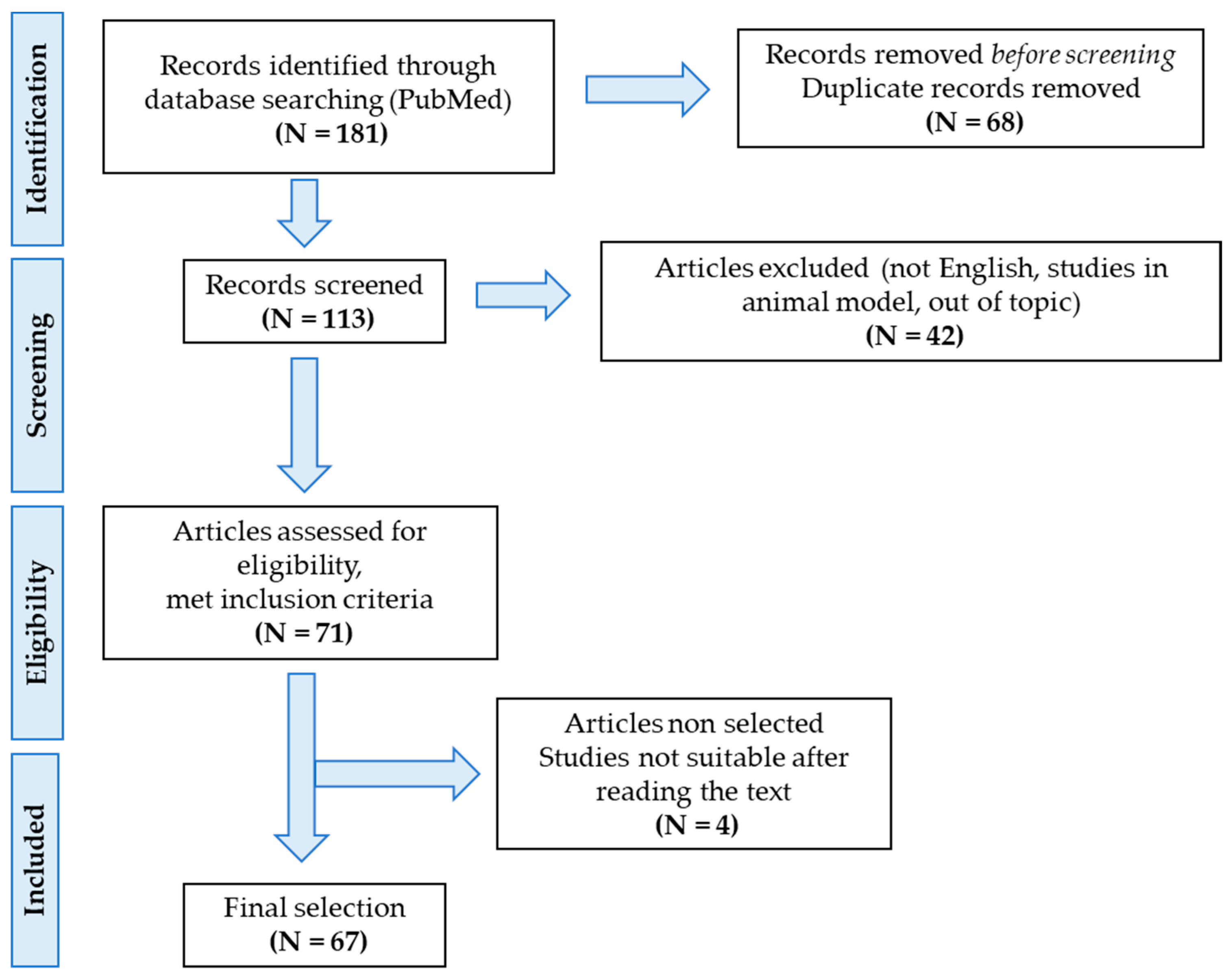
5. Materials and Methods

A bibliographic search strategy was conducted to identify all studies reporting metabolites possibly associated with pain in FM. An electronic database search of PubMed was performed. First, we searched for studies that investigated pathogenetic mechanisms associated with pain in FM, in order to define the most relevant metabolites to include. We first used key terms that included ‘fibromyalgia’, ‘pain’, ‘central sensitization’, ‘neuroinflammation’, ‘hypothalamic-pituitary-adrenal axis disfunction’. For specific biomarkers, we subsequently applied the following search: ‘fibromyalgia pain’ AND ‘glutamate’; ‘fibromyalgia pain’ AND ‘substance P’; ‘fibromyalgia pain’ AND ‘nerve growth factor’; ‘fibromyalgia pain’ AND ‘brain derived neurotrophic factor’; ‘fibromyalgia pain’ AND ‘mu opioid receptor’; ‘fibromyalgia pain’ AND ‘mast cells’ OR ‘cytokine levels’; ‘fibromyalgia pain’ AND ‘pentraxin-3′; ‘fibromyalgia pain’ AND ‘neuropeptide Y’; ‘fibromyalgia pain’ AND ‘vitamin D’; ‘fibromyalgia pain’ AND ‘gut microbiome composition’ OR ‘gut microbiome metabolite’. Only research performed in humans was considered, while studies conducted on animal models were excluded. Only full-text articles in English were considered. Finally, after reading abstracts for relevance, 67 papers were included. The process of the selection of the studies can be found in Figure 1.

6. Conclusions

In this narrative review, we summarized the actual knowledge regarding metabolites associated with chronic pain development, whose alteration has been linked to FM syndrome. We not only considered more extensively studied metabolites but also possible new areas of research. Among them, more consistent evidence is available regarding Glu, SP, BDNF and NPY level alterations. In particular, dysfunction in Glu metabolism is apparently implicated in chronic pain development in FM and, therefore, Glu level detection might be considered useful in these patients. Unfortunately, the strongest evidence is related to Glu level assessment in particular brain regions using functional neuroimaging techniques, which, currently, are difficult to apply in clinical practice. On the other hand, the altered production of SP, BDNF and NPY seems reasonably implicated in FM pathophysiology but the association of these metabolites with the clinical evidence of pain is still unclear. It must be noted that there is a large discrepancy between studies regarding the inclusion and exclusion criteria used, sources of samples considered and tests used to assess metabolite levels, thus making it even more difficult to compare results. Based on the results of the analyzed studies, it seems unlikely that one of these metabolites could be used as a biomarker for FM diagnosis in the near future. Research in this field has, however, been important since it has contributed to clarifying at least part of the pathogenetic mechanism underlying this complex syndrome.
Besides diagnostic porpoises, the identification of a biomarker for FM might be considered useful in the monitoring of treatment efficacy. As a matter of fact, some of the metabolites included in this review—in particular, SP, BDNF and cytokine levels—have been used to monitor different treatment strategies, reporting conflicting results. In our opinion, since there is still little evidence regarding the association of these markers with any clinical symptoms—especially pain—their actual usefulness in monitoring treatment efficacy remains unclear. Thus, more studies in this area should be conducted.
In summary, many research areas are currently under investigation for the identification of possible biomarkers for FM syndrome. These studies also enable us to deepen our knowledge regarding the pathophysiological mechanisms underlying this heterogenous condition. Since WP is the major symptom in FM patients, we decided to focus on biomarkers under investigation for their possible associations with the clinical evidence of pain. Thus far, none of the proposed metabolites is sufficiently reliable to be validated as a diagnostic biomarker, and the different methodological issues mostly remain unresolved. Considering the clinical complexity and variability of this syndrome, we can conclude that a single biomarker is not sufficiently reliable; however, in the future, a panel of biomarkers, including subtype-specific biomarkers, could aid in FM diagnosis.

Author Contributions

Conceptualization, M.D.F. and C.I.; visualization, M.F.; writing—original draft preparation, M.F.; writing—review and editing, M.F., M.D.F. and C.I.; supervision, C.I.; project administration, M.D.F. All authors have read and agreed to the published version of the manuscript.

Funding

This research received no external funding.

Institutional Review Board Statement

Not applicable.

Informed Consent Statement

Not applicable.

Data Availability Statement

Not applicable.

Conflicts of Interest

The authors declare no conflict of interest.

Abbreviations

ACTHAdrenocorticotropic hormone
ACRAmerican College of Rheumatology
AMPA-RAmino-3-hydroxy-5-methyl-4-isoxazolepropionic acid receptors
ANSAutonomic nervous system
ATPAdenosine triphosphate
BBBBlood–brain barrier
BDNFBrain-derived neurotrophic factor
CGRPCalcitonin gene-related peptide
COMTCatechol-O-methyl transferase
CNSCentral nervous system
CPMConditioned pain modulation
CPPChronic primary pain
CRHCorticotropin releasing hormone
CSCentral sensitization
CSFCerebrospinal fluid
CSSCentral sensitization syndromes
CXCLChemokine ligand
DHDorsal horn
DRGDorsal root ganglia
FM Fibromyalgia
fMRIFunctional magnetic resonance imaging
GluGlutamate
HCHealthy control
1H-MRSProton magnetic resonance spectroscopy
HPAHypothalamic–pituitary–adrenal
HRVHeart rate variability
IASPInternational Association for the Study of Pain
ILInterleukin
KCC2Potassium-chloride cotransporter 2
LTPLong-term potentiation
MCsMast cells
MCPMonocyte chemotactic protein
MORMu opioid receptor
NGFNerve growth factor
NKNeurokinin
NMDA-RN-methyl-D-aspartate receptor
NPYNeuropeptide Y
PAGPeriaqueductal grey
PETPositron emission tomography
PFCPrefrontal cortex
PNSPeripheral nervous system
PTX-3Pentraxin-3
rs-fMRIResting-state functional magnetic resonance imaging
SFNSmall fiber neuropathy
SNPSingle-nucleotide polymorphism
SPSubstance P
SSSSymptom Severity Scale
TLRToll-like receptor
TNFTumor necrosis factor
TrkTyrosine-kinase
VBMVoxel-based morphometry
VDRVitamin D receptor
WPWidespread pain
WPIWidespread Pain Index

References

  1. Queiroz, L.P. Worldwide Epidemiology of Fibromyalgia. Curr. Pain Headache Rep. 2013, 17, 356. [Google Scholar] [CrossRef] [PubMed]
  2. Di Franco, M.; Iannuccelli, C.; Bazzichi, L.; Atzeni, F.; Consensi, A.; Salaffi, F.; Pietropaolo, M.; Alessandri, C.; Basili, S.; Olivieri, M.; et al. Misdiagnosis in fibromyalgia: A multicentre study. Ann. Rheum. Dis. 2012, 29, S104–S108. [Google Scholar]
  3. Häuser, W.; Sarzi-Puttini, P.; Fitzcharles, M.A. Fibromyalgia syndrome: Under-, over- and misdiagnosis. Clin. Exp. Rheumatol. 2019, 37, 90–97. [Google Scholar]
  4. Giorgi, V.; Sirotti, S.; Romano, M.E.; Marotto, D.; Ablin, J.N.; Salaffi, F.; Sarzi-Puttini, P. Fibromyalgia: One year in review 2022. Clin. Exp. Rheumatol. 2022, 40, 1065–1072. [Google Scholar] [CrossRef] [PubMed]
  5. Sarzi-Puttini, P.; Giorgi, V.; Marotto, D.; Atzeni, F. Fibromyalgia: An update on clinical characteristics, aetiopathogenesis and treatment. Nat. Rev. Rheumatol. 2020, 16, 645–660. [Google Scholar] [CrossRef]
  6. Wolfe, F.; Smythe, H.A.; Yunus, M.B.; Bennett, R.M.; Bombardier, C.; Goldenberg, D.L.; Tugwell, P.; Campbell, S.M.; Abeles, M.; Clark, P.; et al. The american college of rheumatology 1990 Criteria for the classification of fibromyalgia. Report of the Multicenter Criteria Committee. Arthritis Rheumatol. 1990, 33, 160–172. [Google Scholar] [CrossRef]
  7. Wolfe, F.; Clauw, D.J.; Fitzcharles, M.-A.; Goldenberg, D.L.; Katz, R.S.; Mease, P.; Russell, A.S.; Russell, I.J.; Winfield, J.B.; Yunus, M.B. The American College of Rheumatology Preliminary Diagnostic Criteria for Fibromyalgia and Measurement of Symptom Severity. Arthritis Care Res. 2010, 62, 600–610. [Google Scholar] [CrossRef] [Green Version]
  8. Wolfe, F.; Clauw, D.J.; Fitzcharles, M.A.; Goldenberg, D.L.; Häuser, W.; Katz, R.L.; Mease, P.J.; Russell, A.S.; Russell, I.J.; Walitt, B. 2016 Revisions to the 2010/2011 fibromyalgia diagnostic criteria. Semin. Arthritis. Rheum. 2016, 46, 319–329. [Google Scholar] [CrossRef]
  9. Treede, R.-D.; Rief, W.; Barke, A.; Aziz, Q.; Bennett, M.I.; Benoliel, R.; Cohen, M.; Evers, S.; Finnerup, N.B.; First, M.B.; et al. Chronic pain as a symptom or a disease: The IASP Classification of Chronic Pain for the International Classification of Diseases (ICD-11). Pain 2019, 160, 19–27. [Google Scholar] [CrossRef] [Green Version]
  10. Nicholas, M.; Vlaeyen, J.W.; Rief, W.; Barke, A.; Aziz, Q.; Benoliel, R.; Cohen, M.; Evers, S.; Giamberardino, M.A.; Goebel, A.; et al. The IASP classification of chronic pain for ICD-11: Chronic primary pain. Pain 2019, 160, 28–37. [Google Scholar] [CrossRef]
  11. Kosek, E.; Cohen, M.; Baron, R.; Gebhart, G.F.; Mico, J.-A.; Rice, A.S.C.; Rief, W.; Sluka, A.K. Do we need a third mechanistic descriptor for chronic pain states? Pain 2016, 157, 1382–1386. [Google Scholar] [CrossRef]
  12. Trouvin, A.-P.; Perrot, S. New concepts of pain. Best Pract. Res. Clin. Rheumatol. 2019, 33, 101415. [Google Scholar] [CrossRef]
  13. Fitzcharles, M.-A.; Cohen, S.P.; Clauw, D.J.; Littlejohn, G.; Usui, C.; Häuser, W. Nociplastic pain: Towards an understanding of prevalent pain conditions. Lancet 2021, 397, 2098–2110. [Google Scholar] [CrossRef] [PubMed]
  14. Üçeyler, N.; Zeller, D.; Kahn, A.-K.; Kewenig, S.; Kittel-Schneider, S.; Schmid, A.; Casanova-Molla, J.; Reiners, K.; Sommer, C. Small fibre pathology in patients with fibromyalgia syndrome. Brain 2013, 136, 1857–1867. [Google Scholar] [CrossRef] [Green Version]
  15. Caro, X.J.; Winter, E.F. Evidence of Abnormal Epidermal Nerve Fiber Density in Fibromyalgia: Clinical and Immunologic Implications. Arthritis Rheumatol. 2014, 66, 1945–1954. [Google Scholar] [CrossRef]
  16. Giannoccaro, M.P.; Donadio, V.; Incensi, A.; Avoni, P.; Liguori, R. Small nerve fiber involvement in patients referred for fibromyalgia. Muscle Nerve 2014, 49, 757–759. [Google Scholar] [CrossRef] [PubMed]
  17. Lawson, V.H.; Grewal, J.; Hackshaw, K.V.; Mongiovi, P.C.; Stino, A.M. Fibromyalgia syndrome and small fiber, early or mild sensory polyneuropathy. Muscle Nerve 2018, 58, 625–630. [Google Scholar] [CrossRef] [PubMed]
  18. Van Assche, D.C.F.; Plaghki, L.; Masquelier, E.; Hatem, S.M. Fibromyalgia syndrome—A laser-evoked potentials study unsupportive of small nerve fibre involvement. Eur. J. Pain 2019, 24, 448–456. [Google Scholar] [CrossRef] [Green Version]
  19. Fasolino, A.; Di Stefano, G.; Leone, C.; Galosi, E.; Gioia, C.; Lucchino, B.; Terracciano, A.; Di Franco, M.; Cruccu, G.; Truini, A. Small-fibre pathology has no impact on somatosensory system function in patients with fibromyalgia. Pain 2020, 161, 2385–2393. [Google Scholar] [CrossRef]
  20. de Tommaso, M.; Vecchio, E.; Nolano, M. The puzzle of fibromyalgia between central sensitization syndrome and small fiber neuropathy: A narrative review on neurophysiological and morphological evidence. Neurol. Sci. 2022, 43, 1667–1684. [Google Scholar] [CrossRef]
  21. Staud, R.; Rodriguez, M.E. Mechanisms of Disease: Pain in fibromyalgia syndrome. Nat. Clin. Pract. Rheumatol. 2006, 2, 90–98. [Google Scholar] [CrossRef]
  22. Schmidt-Wilcke, T.; Clauw, D.J. Fibromyalgia: From pathophysiology to therapy. Nat. Rev. Rheumatol. 2011, 7, 518–527. [Google Scholar] [CrossRef] [PubMed]
  23. Bradley, L.A. Pathophysiology of Fibromyalgia. Am. J. Med. 2009, 122 (Suppl. 12), S22–S30. [Google Scholar] [CrossRef] [Green Version]
  24. Staud, R.; Smitherman, M.L. Peripheral and central sensitization in fibromyalgia: Pathogenetic role. Curr. Pain Headache Rep. 2002, 6, 259–266. [Google Scholar] [CrossRef] [PubMed]
  25. Yam, M.F.; Loh, Y.C.; Tan, C.S.; Adam, S.K.; Manan, N.A.; Basir, R. General Pathways of Pain Sensation and the Major Neurotransmitters Involved in Pain Regulation. Int. J. Mol. Sci. 2018, 19, 2164. [Google Scholar] [CrossRef] [PubMed] [Green Version]
  26. Todd, A.J. Neuronal circuitry for pain processing in the dorsal horn. Nat. Rev. Neurosci. 2010, 11, 823–836. [Google Scholar] [CrossRef] [Green Version]
  27. Heinricher, M.; Tavares, I.; Leith, J.; Lumb, B. Descending control of nociception: Specificity, recruitment and plasticity. Brain Res. Rev. 2009, 60, 214–225. [Google Scholar] [CrossRef] [Green Version]
  28. Di Maio, V.; Ventriglia, F.; Santillo, S. A model of cooperative effect of AMPA and NMDA receptors in glutamatergic synapses. Cogn. Neurodyn. 2016, 10, 315–325. [Google Scholar] [CrossRef] [Green Version]
  29. Liu, X.J.; Salter, M.W. Glutamate receptor phosphorylation and trafficking in pain plasticity in spinal cord dorsal horn. Eur. J. Neurosci. 2010, 32, 278–289. [Google Scholar] [CrossRef]
  30. Latremoliere, A.; Woolf, C.J. Central Sensitization: A Generator of Pain Hypersensitivity by Central Neural Plasticity. J. Pain 2009, 10, 895–926. [Google Scholar] [CrossRef] [Green Version]
  31. Wang, Z.; Yuan, M.; Xiao, J. Gray Matter Abnormalities in Patients with Chronic Primary Pain: A Coordinate-Based Meta-Analysis. Pain Physician 2022, 25, 1–13. [Google Scholar]
  32. Cagnie, B.; Coppieters, I.; Denecker, S.; Six, J.; Danneels, L.; Meeus, M. Central sensitization in fibromyalgia? A systematic review on structural and functional brain MRI. Semin. Arthritis Rheum. 2014, 44, 68–75. [Google Scholar] [CrossRef] [PubMed]
  33. Siracusa, R.; Di Paola, R.; Cuzzocrea, S.; Impellizzeri, D. Fibromyalgia: Pathogenesis, Mechanisms, Diagnosis and Treatment Options Update. Int. J. Mol. Sci. 2021, 22, 3891. [Google Scholar] [CrossRef] [PubMed]
  34. Truini, A.; Tinelli, E.; Gerardi, M.C.; Calistri, V.; Iannuccelli, C.; La Cesa, S.; Tarsitani, L.; Mainero, C.; Sarzi-Puttini, P.; Cruccu, G.; et al. Abnormal resting state functional connectivity of the periaqueductal grey in patients with fibromyalgia. Clin. Exp. Rheumatol. 2016, 34 (Suppl. 36), S129–S133. [Google Scholar]
  35. Baraniuk, J.N.; Whalen, G.; Cunningham, J.; Clauw, D.J. Cerebrospinal fluid levels of opioid peptides in fibromyalgia and chronic low back pain. BMC Musculoskelet. Disord. 2004, 5, 48. [Google Scholar] [CrossRef] [PubMed] [Green Version]
  36. Sluka, K.A.; Clauw, D.J. Neurobiology of fibromyalgia and chronic widespread pain. Neuroscience 2016, 338, 114–129. [Google Scholar] [CrossRef] [Green Version]
  37. Cifre, I.; Sitges, C.; Fraiman, D.; Muñoz, M.Á.; Balenzuela, P.; González-Roldán, A.; Martínez-Jauand, M.; Birbaumer, N.; Chialvo, D.R.; Montoya, P. Disrupted Functional Connectivity of the Pain Network in Fibromyalgia. Psychosom. Med. 2012, 74, 55–62. [Google Scholar] [CrossRef] [Green Version]
  38. Ji, R.-R.; Chamessian, A.; Zhang, Y.-Q. Pain regulation by non-neuronal cells and inflammation. Science 2016, 354, 572–577. [Google Scholar] [CrossRef] [Green Version]
  39. Ji, R.-R.; Nackley, A.; Huh, B.Y.; Terrando, N.; Maixner, D.W. Neuroinflammation and Central Sensitization in Chronic and Widespread Pain. Anesthesiology 2018, 129, 343–366. [Google Scholar] [CrossRef]
  40. Mai, L.; Liu, Q.; Huang, F.; He, H.; Fan, W. Involvement of Mast Cells in the Pathophysiology of Pain. Front. Cell. Neurosci. 2021, 15, 665066. [Google Scholar] [CrossRef]
  41. Traina, G. Mast Cells in Gut and Brain and Their Potential Role as an Emerging Therapeutic Target for Neural Diseases. Front. Cell. Neurosci. 2019, 13, 345. [Google Scholar] [CrossRef]
  42. Skaper, S.D.; Facci, L. Mast cell–glia axis in neuroinflammation and therapeutic potential of the anandamide congener palmitoylethanolamide. Philos. Trans. R. Soc. B Biol. Sci. 2012, 367, 3312–3325. [Google Scholar] [CrossRef]
  43. Salcman, B.; Affleck, K.; Bulfone-Paus, S. P2X Receptor-Dependent Modulation of Mast Cell and Glial Cell Activities in Neuroinflammation. Cells 2021, 10, 2282. [Google Scholar] [CrossRef] [PubMed]
  44. Skaper, S.D.; Facci, L.; Zusso, M.; Giusti, P. Neuroinflammation, Mast Cells, and Glia: Dangerous Liaisons. Neuroscientist 2017, 23, 478–498. [Google Scholar] [CrossRef] [PubMed]
  45. Qian, Y.; Dong, H.; Zhang, X. Mast Cells and Neuroinflammation. Med. Sci. Monit. Basic Res. 2014, 20, 200–206. [Google Scholar] [CrossRef] [Green Version]
  46. Grace, P.M.; Hutchinson, M.R.; Maier, S.F.; Watkins, L.R. Pathological pain and the neuroimmune interface. Nat. Rev. Immunol. 2014, 14, 217–231. [Google Scholar] [CrossRef] [Green Version]
  47. Ji, R.-R.; Berta, T.; Nedergaard, M. Glia and pain: Is chronic pain a gliopathy? Pain 2013, 154, S10–S28. [Google Scholar] [CrossRef] [Green Version]
  48. Zhou, R.; Ji, B.; Kong, Y.; Qin, L.; Ren, W.; Guan, Y.; Ni, R. PET Imaging of Neuroinflammation in Alzheimer’s Disease. Front. Immunol. 2021, 12, 739130. [Google Scholar] [CrossRef]
  49. Zhang, P.-F.; Gao, F. Neuroinflammation in Parkinson’s disease: A meta-analysis of PET imaging studies. J. Neurol. 2021, 269, 2304–2314. [Google Scholar] [CrossRef]
  50. Meyer, J.H.; Cervenka, S.; Kim, M.-J.; Kreisl, W.C.; Henter, I.D.; Innis, R.B. Neuroinflammation in psychiatric disorders: PET imaging and promising new targets. Lancet Psychiatry 2020, 7, 1064–1074. [Google Scholar] [CrossRef]
  51. Gritti, D.; Delvecchio, G.; Ferro, A.; Bressi, C.; Brambilla, P. Neuroinflammation in Major Depressive Disorder: A Review of PET Imaging Studies Examining the 18-kDa Translocator Protein. J. Affect. Disord. 2021, 292, 642–651. [Google Scholar] [CrossRef] [PubMed]
  52. Albrecht, D.S.; Granziera, C.; Hooker, J.M.; Loggia, M.L. In Vivo Imaging of Human Neuroinflammation. ACS Chem. Neurosci. 2016, 7, 470–483. [Google Scholar] [CrossRef] [PubMed] [Green Version]
  53. Jung, C.; Ichesco, E.; Ratai, E.-M.; Gonzalez, R.G.; Burdo, T.; Loggia, M.L.; Harris, R.E.; Napadow, V. Magnetic resonance imaging of neuroinflammation in chronic pain: A role for astrogliosis? Pain 2020, 161, 1555–1564. [Google Scholar] [CrossRef]
  54. Petrou, M.; Harris, R.; Foerster, B.; McLean, S.; Sen, A.; Clauw, D.; Sundgren, P. Proton MR Spectroscopy in the Evaluation of Cerebral Metabolism in Patients with Fibromyalgia: Comparison with Healthy Controls and Correlation with Symptom Severity. Am. J. Neuroradiol. 2008, 29, 913–918. [Google Scholar] [CrossRef] [PubMed] [Green Version]
  55. Matsuda, M.; Huh, Y.; Ji, R.-R. Roles of inflammation, neurogenic inflammation, and neuroinflammation in pain. J. Anesth. 2018, 33, 131–139. [Google Scholar] [CrossRef]
  56. Littlejohn, G. Neurogenic neuroinflammation in fibromyalgia and complex regional pain syndrome. Nat. Rev. Rheumatol. 2015, 11, 639–648. [Google Scholar] [CrossRef]
  57. Deussing, J.M.; Chen, A. The Corticotropin-Releasing Factor Family: Physiology of the Stress Response. Physiol. Rev. 2018, 98, 2225–2286. [Google Scholar] [CrossRef]
  58. Butler, R.K.; Finn, D.P. Stress-induced analgesia. Prog. Neurobiol. 2009, 88, 184–202. [Google Scholar] [CrossRef] [Green Version]
  59. Schlereth, T.; Birklein, F. The Sympathetic Nervous System and Pain. NeuroMol. Med. 2007, 10, 141–147. [Google Scholar] [CrossRef]
  60. Di Franco, M.; Iannuccelli, C.; Valesini, G. Neuroendocrine immunology of fibromyalgia. Ann. N. Y. Acad. Sci. 2010, 1193, 84–90. [Google Scholar] [CrossRef]
  61. Vachon-Presseau, E. Effects of stress on the corticolimbic system: Implications for chronic pain. Prog. Neuro-Psychopharmacol. Biol. Psychiatry 2018, 87, 216–223. [Google Scholar] [CrossRef]
  62. Timmers, I.; Quaedflieg, C.W.; Hsu, C.; Heathcote, L.C.; Rovnaghi, C.R.; Simons, L.E. The interaction between stress and chronic pain through the lens of threat learning. Neurosci. Biobehav. Rev. 2019, 107, 641–655. [Google Scholar] [CrossRef] [PubMed]
  63. Woda, A.; Picard, P.; Dutheil, F. Dysfunctional stress responses in chronic pain. Psychoneuroendocrinology 2016, 71, 127–135. [Google Scholar] [CrossRef]
  64. Singh, L.; Kaur, A.; Bhatti, M.S.; Bhatti, R. Possible Molecular Mediators Involved and Mechanistic Insight into Fibromyalgia and Associated Co-morbidities. Neurochem. Res. 2019, 44, 1517–1532. [Google Scholar] [CrossRef]
  65. D’agnelli, S.; Arendt-Nielsen, L.; Gerra, M.C.; Zatorri, K.; Boggiani, L.; Baciarello, M.; Bignami, E. Fibromyalgia: Genetics and epigenetics insights may provide the basis for the development of diagnostic biomarkers. Mol. Pain 2018, 15, 1744806918819944. [Google Scholar] [CrossRef]
  66. Zouikr, I.; Karshikoff, B. Lifetime Modulation of the Pain System via Neuroimmune and Neuroendocrine Interactions. Front. Immunol. 2017, 8, 276. [Google Scholar] [CrossRef] [PubMed] [Green Version]
  67. Park, D.-J.; Kang, J.-H.; Yim, Y.-R.; Kim, J.-E.; Lee, J.-W.; Lee, K.-E.; Wen, L.; Kim, T.-J.; Park, Y.-W.; Lee, S.-S. Exploring Genetic Susceptibility to Fibromyalgia. Chonnam Med. J. 2015, 51, 58–65. [Google Scholar] [CrossRef] [PubMed] [Green Version]
  68. Park, D.-J.; Lee, S.-S. New insights into the genetics of fibromyalgia. Korean J. Intern. Med. 2017, 32, 984–995. [Google Scholar] [CrossRef] [PubMed] [Green Version]
  69. Tammimäki, A.; Männistö, P. Catechol-O-methyltransferase gene polymorphism and chronic human pain: A systematic re-view and meta-analysis. Pharmacogenet. Genom. 2012, 22, 673–691. [Google Scholar] [CrossRef] [Green Version]
  70. Lee, Y.H.; Kim, J.-H.; Song, G.G. Association between the COMT Val158Met polymorphism and fibromyalgia susceptibility and fibromyalgia impact questionnaire score: A meta-analysis. Rheumatol. Int. 2014, 35, 159–166. [Google Scholar] [CrossRef]
  71. Zhang, L.; Zhu, J.; Chen, Y.; Zhao, J. Meta-analysis reveals a lack of association between a common catechol-O-methyltransferase (COMT) polymorphism val¹⁵⁸met and fibromyalgia. Int. J. Clin. Exp. Pathol. 2014, 7, 8489–8497. [Google Scholar] [PubMed]
  72. Peek, A.L.; Rebbeck, T.; Puts, N.A.; Watson, J.; Aguila, M.-E.R.; Leaver, A.M. Brain GABA and glutamate levels across pain conditions: A systematic literature review and meta-analysis of 1H-MRS studies using the MRS-Q quality assessment tool. Neuroimage 2020, 210, 116532. [Google Scholar] [CrossRef] [PubMed]
  73. Lesnak, J.; Sluka, K.A. Chronic non-inflammatory muscle pain: Central and peripheral mediators. Curr. Opin. Physiol. 2019, 11, 67–74. [Google Scholar] [CrossRef] [PubMed]
  74. Peres, M.; Zukerman, E.; Soares, C.S.; Alonso, E.; Santos, B.; Faulhaber, M. Cerebrospinal Fluid Glutamate Levels in Chronic Migraine. Cephalalgia 2004, 24, 735–739. [Google Scholar] [CrossRef] [PubMed]
  75. Sarchielli, P.; Mancini, M.L.; Floridi, A.; Coppola, F.; Rossi, C.; Nardi, K.; Acciarresi, M.; Pini, L.A.; Calabresi, P. Increased Levels of Neurotrophins Are Not Specific for Chronic Migraine: Evidence From Primary Fibromyalgia Syndrome. J. Pain 2007, 8, 737–745. [Google Scholar] [CrossRef]
  76. Harris, R.E.; Sundgren, P.; Craig, A.; Kirshenbaum, E.; Sen, A.; Napadow, V.; Clauw, D.J. Elevated insular glutamate in fibromyalgia is associated with experimental pain. Arthritis Rheum. 2009, 60, 3146–3152. [Google Scholar] [CrossRef] [Green Version]
  77. Harris, R.E.; Sundgren, P.; Pang, Y.; Hsu, M.; Petrou, M.; Kim, S.-H.; McLean, S.; Gracely, R.H.; Clauw, D.J. Dynamic levels of glutamate within the insula are associated with improvements in multiple pain domains in fibromyalgia. Arthritis Rheum. 2008, 58, 903–907. [Google Scholar] [CrossRef] [Green Version]
  78. Fayed, N.; Garcia-Campayo, J.; Magallón, R.; Andrés-Bergareche, H.; Luciano, J.V.; Andres, E.; Beltrán, J. Localized 1H-NMR spectroscopy in patients with fibromyalgia: A controlled study of changes in cerebral glutamate/glutamine, inositol, choline, and N-acetylaspartate. Thromb. Haemost. 2010, 12, R134. [Google Scholar] [CrossRef] [Green Version]
  79. Valdés, M.; Collado, A.; Bargalló, N.; Vázquez, M.; Rami, L.; Gómez, E.; Salamero, M. Increased glutamate/glutamine compounds in the brains of patients with fibromyalgia: A magnetic resonance spectroscopy study. Arthritis Rheum. 2010, 62, 1829–1836. [Google Scholar] [CrossRef]
  80. Feraco, P.; Bacci, A.; Pedrabissi, F.; Passamonti, L.; Zampogna, G.; Pedrabissi, F.; Malavolta, N.; Leonardi, M. Metabolic Abnormalities in Pain-Processing Regions of Patients with Fibromyalgia: A 3T MR Spectroscopy Study. Am. J. Neuroradiol. 2011, 32, 1585–1590. [Google Scholar] [CrossRef] [PubMed] [Green Version]
  81. Feraco, P.; Nigro, S.; Passamonti, L.; Grecucci, A.; Caligiuri, M.E.; Gagliardo, C.; Bacci, A. Neurochemical Correlates of Brain Atrophy in Fibromyalgia Syndrome: A Magnetic Resonance Spectroscopy and Cortical Thickness Study. Brain Sci. 2020, 10, 395. [Google Scholar] [CrossRef] [PubMed]
  82. Lee, J.; Andronesi, O.C.; Torrado-Carvajal, A.; Ratai, E.; Loggia, M.L.; Weerasekera, A.; Berry, M.P.; Ellingsen, D.; Isaro, L.; Lazaridou, A.; et al. 3D magnetic resonance spectroscopic imaging reveals links between brain metabolites and multidimensional pain features in fibromyalgia. Eur. J. Pain 2021, 25, 2050–2064. [Google Scholar] [CrossRef] [PubMed]
  83. Lu, C.; Yang, T.; Zhao, H.; Zhang, M.; Meng, F.; Fu, H.; Xie, Y.; Xu, H. Insular Cortex is Critical for the Perception, Modulation, and Chronification of Pain. Neurosci. Bull. 2016, 32, 191–201. [Google Scholar] [CrossRef] [Green Version]
  84. Harris, R.E. Elevated excitatory neurotransmitter levels in the fibromyalgia brain. Arthritis Res. Ther. 2010, 12, 141. [Google Scholar] [CrossRef] [Green Version]
  85. Steinhoff, M.S.; von Mentzer, B.; Geppetti, P.; Pothoulakis, C.; Bunnett, N.W.; Bakirtzi, K.; Law, I.K.M.; Fang, K.; Iliopoulos, D.; Dér, B.; et al. Tachykinins and Their Receptors: Contributions to Physiological Control and the Mechanisms of Disease. Physiol. Rev. 2014, 94, 265–301. [Google Scholar] [CrossRef] [Green Version]
  86. Almeida, T.A.; Rojo, J.; Nieto, P.M.; Pinto, F.M.; Hernandez, M.; Martín, J.D.; Candenas, M.L. Tachykinins and Tachykinin Receptors: Structure and Activity Relationships. Curr. Med. Chem. 2004, 11, 2045–2081. [Google Scholar] [CrossRef] [PubMed]
  87. Schank, J.R.; Heilig, M. Substance P and the Neurokinin-1 Receptor: The New CRF. Int. Rev. Neurobiol. 2017, 136, 151–175. [Google Scholar] [CrossRef] [PubMed]
  88. Seybold, V. The Role of Peptides in Central Sensitization. Handb. Exp. Pharmacol. 2009, 194, 451–491. [Google Scholar] [CrossRef]
  89. Pace, M.C.; Passavanti, M.B.; De Nardis, L.; Bosco, F.; Sansone, P.; Pota, V.; Barbarisi, M.; Palagiano, A.; Iannotti, F.A.; Panza, E.; et al. Nociceptor plasticity: A closer look. J. Cell. Physiol. 2017, 233, 2824–2838. [Google Scholar] [CrossRef]
  90. Navratilova, E.; Porreca, F. Substance P and Inflammatory Pain: Getting It Wrong and Right Simultaneously. Neuron 2019, 101, 353–355. [Google Scholar] [CrossRef]
  91. Green, D.P.; Limjunyawong, N.; Gour, N.; Pundir, P.; Dong, X. A Mast-Cell-Specific Receptor Mediates Neurogenic Inflammation and Pain. Neuron 2019, 101, 412–420.e3. [Google Scholar] [CrossRef] [Green Version]
  92. Vaerøy, H.; Helle, R.; Førre, Ø.; Kåss, E.; Terenius, L. Elevated CSF levels of substance P and high incidence of Raynaud phenomenon in patients with fibromyalgia: New features for diagnosis. Pain 1988, 32, 21–26. [Google Scholar] [CrossRef]
  93. Russell, I.J.; Orr, M.D.; Littman, B.; Vipraio, G.A.; Alboukrek, D.; Michalek, J.E.; Lopez, Y.; Mackillip, F. Elevated cerebrospinal fluid levels of substance p in patients with the fibromyalgia syndrome. Arthritis Rheum. 1994, 37, 1593–1601. [Google Scholar] [CrossRef]
  94. Russell, I.J. Neurochemical pathogenesis of fibromyalgia. Z. Rheumatol. 1998, 57, S63–S66. [Google Scholar] [CrossRef]
  95. Reynolds, W.J.; Chiu, B.; Inman, R.D. Plasma substance P levels in fibrositis. J. Rheumatol. 1988, 15, 1802–1803. [Google Scholar]
  96. Tsilioni, I.; Russell, I.J.; Stewart, J.M.; Gleason, R.M.; Theoharides, T.C. Neuropeptides CRH, SP, HK-1, and Inflammatory Cytokines IL-6 and TNF Are Increased in Serum of Patients with Fibromyalgia Syndrome, Implicating Mast Cells. J. Pharmacol. Exp. Ther. 2016, 356, 664–672. [Google Scholar] [CrossRef] [PubMed] [Green Version]
  97. Tsilioni, I.; Pipis, H.; Freitag, M.S.C.; Izquierdo, M.D.C.; Freitag, K.; Theoharides, T.C. Effects of an Extract of Salmon Milt on Symptoms and Serum TNF and Substance P in Patients With Fibromyalgia Syndrome. Clin. Ther. 2019, 41, 1564–1574.e2. [Google Scholar] [CrossRef] [PubMed]
  98. Field, T.; Diego, M.; Cullen, C.; Hernandez-Reif, M.; Sunshine, W.; Douglas, S. Fibromyalgia Pain and Substance P Decrease and Sleep Improves After Massage Therapy. Am. J. Clin. Oncol. 2002, 8, 72–76. [Google Scholar] [CrossRef] [PubMed]
  99. Karatay, S.; Okur, S.C.; Uzkeser, H.; Yildirim, K.; Akcay, F. Effects of Acupuncture Treatment on Fibromyalgia Symptoms, Serotonin, and Substance P Levels: A Randomized Sham and Placebo-Controlled Clinical Trial. Pain Med. 2017, 19, 615–628. [Google Scholar] [CrossRef] [Green Version]
  100. Sprott, H.; Franke, S.; Kluge, H.; Hein, G. Pain treatment of fibromyalgia by acupuncture. Rheumatol. Int. 1998, 18, 35–36. [Google Scholar] [CrossRef] [PubMed]
  101. Bjersing, J.L.; Dehlin, M.; Erlandsson, M.; Bokarewa, M.I.; Mannerkorpi, K. Changes in pain and insulin-like growth factor 1 in fibromyalgia during exercise: The involvement of cerebrospinal inflammatory factors and neuropeptides. Arthritis Res. Ther. 2012, 14, R162. [Google Scholar] [CrossRef] [PubMed] [Green Version]
  102. Denk, F.; Bennett, D.L.; McMahon, S.B. Nerve Growth Factor and Pain Mechanisms. Annu. Rev. Neurosci. 2017, 40, 307–325. [Google Scholar] [CrossRef]
  103. Svensson, P.; Cairns, B.E.; Wang, K.; Arendt-Nielsen, L. Injection of nerve growth factor into human masseter muscle evokes long-lasting mechanical allodynia and hyperalgesia. Pain 2003, 104, 241–247. [Google Scholar] [CrossRef] [PubMed]
  104. Rukwied, R.; Mayer, A.; Kluschina, O.; Obreja, O.; Schley, M.; Schmelz, M. NGF induces non-inflammatory localized and lasting mechanical and thermal hypersensitivity in human skin. Pain 2010, 148, 407–413. [Google Scholar] [CrossRef]
  105. Weinkauf, B.; Obreja, O.; Schmelz, M.; Rukwied, R. Differential time course of NGF-induced hyperalgesia to heat versus mechanical and electrical stimulation in human skin. Eur. J. Pain 2014, 19, 789–796. [Google Scholar] [CrossRef]
  106. Andersen, H.; Arendt-Nielsen, L.; Svensson, P.; Danneskiold-Samsøe, B.; Graven-Nielsen, T. Spatial and temporal aspects of muscle hyperalgesia induced by nerve growth factor in humans. Exp. Brain Res. 2008, 191, 371–382. [Google Scholar] [CrossRef]
  107. Gerber, R.K.H.; Nie, H.; Arendt-Nielsen, L.; Curatolo, M.; Graven-Nielsen, T. Local Pain and Spreading Hyperalgesia Induced by Intramuscular Injection of Nerve Growth Factor Are Not Reduced by Local Anesthesia of the Muscle. Clin. J. Pain 2011, 27, 240–247. [Google Scholar] [CrossRef]
  108. Sørensen, L.B.; Boudreau, S.A.; Gazerani, P.; Graven-Nielsen, T. Enlarged Areas of Pain and Pressure Hypersensitivityby Spatially Distributed Intramuscular Injections ofLow-Dose Nerve Growth Factor. J. Pain 2018, 20, 566–576. [Google Scholar] [CrossRef] [PubMed]
  109. Kelleher, J.H.; Tewari, D.; McMahon, S.B. Neurotrophic factors and their inhibitors in chronic pain treatment. Neurobiol. Dis. 2017, 97, 127–138. [Google Scholar] [CrossRef] [Green Version]
  110. Bloom, A.P.; Jimenez-Andrade, J.M.; Taylor, R.N.; Castañeda-Corral, G.; Kaczmarska, M.J.; Freeman, K.T.; Coughlin, K.A.; Ghilardi, J.R.; Kuskowski, M.A.; Mantyh, P.W. Breast Cancer-Induced Bone Remodeling, Skeletal Pain, and Sprouting of Sensory Nerve Fibers. J. Pain 2011, 12, 698–711. [Google Scholar] [CrossRef] [Green Version]
  111. Barker, P.A.; Mantyh, P.; Arendt-Nielsen, L.; Viktrup, L.; Tive, L. Nerve Growth Factor Signaling and Its Contribution to Pain. J. Pain Res. 2020, 13, 1223–1241. [Google Scholar] [CrossRef]
  112. Skaper, S.D. Nerve growth factor: A neuroimmune crosstalk mediator for all seasons. Immunology 2017, 151, 1–15. [Google Scholar] [CrossRef] [PubMed] [Green Version]
  113. Redegeld, F.A.; Yu, Y.; Kumari, S.; Charles, N.; Blank, U. Non-IgE mediated mast cell activation. Immunol. Rev. 2018, 282, 87–113. [Google Scholar] [CrossRef] [PubMed]
  114. Kritas, S.; Caraffa, A.; Antinolfi, P.; Saggini, A.; Pantalone, A.; Rosati, M.; Tei, M.; Speziali, A.; Pandolfi, F.; Cerulli, G.; et al. Nerve Growth Factor Interactions with Mast Cells. Int. J. Immunopathol. Pharmacol. 2014, 27, 15–19. [Google Scholar] [CrossRef] [PubMed] [Green Version]
  115. Xu, H.; Shi, X.; Li, X.; Zou, J.; Zhou, C.; Liu, W.; Shao, H.; Chen, H.; Shi, L. Neurotransmitter and neuropeptide regulation of mast cell function: A systematic review. J. Neuroinflamm. 2020, 17, 356. [Google Scholar] [CrossRef] [PubMed]
  116. Cirulli, F.; Alleva, E. The NGF saga: From animal models of psychosocial stress to stress-related psychopathology. Front. Neuroendocr. 2009, 30, 379–395. [Google Scholar] [CrossRef] [PubMed]
  117. Mozafarihashjin, M.; Togha, M.; Ghorbani, Z.; Farbod, A.; Rafiee, P.; Martami, F. Assessment of peripheral biomarkers potentially involved in episodic and chronic migraine: A case-control study with a focus on NGF, BDNF, VEGF, and PGE2. J. Headache Pain 2022, 23, 3. [Google Scholar] [CrossRef]
  118. Montagnoli, C.; Tiribuzi, R.; Crispoltoni, L.; Pistilli, A.; Stabile, A.M.; Manfreda, F.; Placella, G.; Rende, M.; Cerulli, G.G. β-NGF and β-NGF receptor upregulation in blood and synovial fluid in osteoarthritis. Biol. Chem. 2017, 398, 1045–1054. [Google Scholar] [CrossRef]
  119. Giovengo, S.L.; Russell, I.J.; Larson, A.A. Increased concentrations of nerve growth factor in cerebrospinal fluid of patients with fibromyalgia. J. Rheumatol. 1999, 26, 1564–1569. [Google Scholar]
  120. Baumeister, D.; Eich, W.; Saft, S.; Geisel, O.; Hellweg, R.; Finn, A.; Svensson, C.I.; Tesarz, J. No evidence for altered plasma NGF and BDNF levels in fibromyalgia patients. Sci. Rep. 2019, 9, 13667. [Google Scholar] [CrossRef] [Green Version]
  121. Jablochkova, A.; Bäckryd, E.; Kosek, E.; Mannerkorpi, K.; Ernberg, M.; Gerdle, B.; Ghafouri, B. Unaltered low nerve growth factor and high brain-derived neurotrophic factor levels in plasma from patients with fibromyalgia after a 15-week progressive resistance exercise. J. Rehabil. Med. 2019, 51, 779–787. [Google Scholar] [CrossRef] [Green Version]
  122. Binder, D.K.; Scharfman, H.E. Brain-derived Neurotrophic Factor. Growth Factors 2004, 22, 123–131. [Google Scholar] [CrossRef] [PubMed] [Green Version]
  123. Malfait, A.-M.; Miller, R.E.; Block, J.A. Targeting neurotrophic factors: Novel approaches to musculoskeletal pain. Pharmacol. Ther. 2020, 211, 107553. [Google Scholar] [CrossRef]
  124. Ferrini, F.; Salio, C.; Boggio, E.M.; Merighi, A. Interplay of BDNF and GDNF in the Mature Spinal Somatosensory System and Its Potential Therapeutic Relevance. Curr. Neuropharmacol. 2021, 19, 1225–1245. [Google Scholar] [CrossRef]
  125. Garraway, S.M.; Huie, J.R. Spinal Plasticity and Behavior: BDNF-Induced Neuromodulation in Uninjured and Injured Spinal Cord. Neural Plast. 2016, 2016, 9857201. [Google Scholar] [CrossRef] [Green Version]
  126. Brigadski, T.; Leßmann, V. The physiology of regulated BDNF release. Cell Tissue Res. 2020, 382, 15–45. [Google Scholar] [CrossRef]
  127. Taves, S.; Berta, T.; Chen, G.; Ji, R.-R. Microglia and Spinal Cord Synaptic Plasticity in Persistent Pain. Neural Plast. 2013, 2013, 753656. [Google Scholar] [CrossRef] [Green Version]
  128. Obata, K.; Noguchi, K. BDNF in sensory neurons and chronic pain. Neurosci. Res. 2006, 55, 1–10. [Google Scholar] [CrossRef] [PubMed]
  129. Beggs, S.; Salter, M.W. The known knowns of microglia–neuronal signalling in neuropathic pain. Neurosci. Lett. 2013, 557, 37–42. [Google Scholar] [CrossRef]
  130. Price, T.J.; Inyang, K.E. Commonalities Between Pain and Memory Mechanisms and Their Meaning for Understanding Chronic Pain. Prog. Mol. Biol. Transl. Sci. 2015, 131, 409–434. [Google Scholar] [CrossRef] [PubMed] [Green Version]
  131. Haas, L.; Portela, L.V.C.; Böhmer, A.E.; Oses, J.P.; Lara, D.R. Increased Plasma Levels of Brain Derived Neurotrophic Factor (BDNF) in Patients with Fibromyalgia. Neurochem. Res. 2010, 35, 830–834. [Google Scholar] [CrossRef]
  132. Laske, C.; Stransky, E.; Eschweiler, G.W.; Klein, R.; Wittorf, A.; Leyhe, T.; Richartz, E.; Köhler, N.; Bartels, M.; Buchkremer, G.; et al. Increased BDNF serum concentration in fibromyalgia with or without depression or antidepressants. J. Psychiatr. Res. 2007, 41, 600–605. [Google Scholar] [CrossRef] [PubMed]
  133. Stefani, L.C.; Leite, F.M.; Tarragó, M.D.G.L.; Zanette, S.A.; de Souza, A.; Castro, S.M.; Caumo, W. BDNF and serum S100B levels according the spectrum of structural pathology in chronic pain patients. Neurosci. Lett. 2019, 706, 105–109. [Google Scholar] [CrossRef] [PubMed] [Green Version]
  134. Nugraha, B.; Korallus, C.; Gutenbrunner, C. Serum level of brain-derived neurotrophic factor in fibromyalgia syndrome correlates with depression but not anxiety. Neurochem. Int. 2013, 62, 281–286. [Google Scholar] [CrossRef] [PubMed]
  135. Nugraha, B.; Karst, M.; Engeli, S.; Gutenbrunner, C. Brain-derived neurotrophic factor and exercise in fibromyalgia syndrome patients: A mini review. Rheumatol. Int. 2012, 32, 2593–2599. [Google Scholar] [CrossRef]
  136. Ribeiro, V.; Mendonça, V.; Souza, A.; Fonseca, S.; Camargos, A.; Lage, V.; Neves, C.; Santos, J.; Teixeira, A.L.; Vieira, E.; et al. Inflammatory biomarkers responses after acute whole body vibration in fibromyalgia. Braz. J. Med. Biol. Res. 2018, 51, e6775. [Google Scholar] [CrossRef]
  137. Ranzolin, A.; Duarte, A.L.B.P.; Bredemeier, M.; Neto, C.A.D.C.; Ascoli, B.M.; Wollenhaupt-Aguiar, B.; Kapczinski, F.; Xavier, R.M. Evaluation of cytokines, oxidative stress markers and brain-derived neurotrophic factor in patients with fibromyalgia—A controlled cross-sectional study. Cytokine 2016, 84, 25–28. [Google Scholar] [CrossRef]
  138. Iannuccelli, C.; Lucchino, B.; Gioia, C.; Dolcini, G.; Rabasco, J.; Venditto, T.; Ioppolo, F.; Santilli, V.; Conti, F.; Di Franco, M. Gender influence on clinical manifestations, depressive symptoms and brain-derived neurotrophic factor (BDNF) serum levels in patients affected by fibromyalgia. Clin. Rheumatol. 2022, 41, 2171–2178. [Google Scholar] [CrossRef]
  139. Rasmussen, P.; Brassard, P.; Adser, H.; Pedersen, M.V.; Leick, L.; Hart, E.; Secher, N.H.; Pedersen, B.K.; Pilegaard, H. Evidence for a release of brain-derived neurotrophic factor from the brain during exercise. Exp. Physiol. 2009, 94, 1062–1069. [Google Scholar] [CrossRef]
  140. Fujimura, H.; Chen, R.; Nakamura, T.; Nakahashi, T.; Kambayashi, J.-I.; Sun, B.; Altar, C.; Tandon, N.N. Brain-derived Neurotrophic Factor Is Stored in Human Platelets and Released by Agonist Stimulation. Thromb. Haemost. 2002, 87, 728–734. [Google Scholar] [CrossRef] [Green Version]
  141. Matthews, V.B.; Åström, M.-B.; Chan, M.H.S.; Bruce, C.R.; Krabbe, K.S.; Prelovsek, O.; Åkerström, T.; Yfanti, C.; Broholm, C.; Mortensen, O.H.; et al. Brain-derived neurotrophic factor is produced by skeletal muscle cells in response to contraction and enhances fat oxidation via activation of AMP-activated protein kinase. Diabetologia 2009, 52, 1409–1418. [Google Scholar] [CrossRef] [PubMed] [Green Version]
  142. Björkholm, C.; Monteggia, L.M. BDNF—A Key Transducer of Antidepressant Effects. Neuropharmacology 2016, 102, 72–79. [Google Scholar] [CrossRef] [Green Version]
  143. Bidari, A.; Ghavidel-Parsa, B.; Gharibpoor, F. Comparison of the serum brain-derived neurotrophic factor (BDNF) between fibromyalgia and nociceptive pain groups; and effect of duloxetine on the BDNF level. BMC Musculoskelet. Disord. 2022, 23, 411. [Google Scholar] [CrossRef] [PubMed]
  144. Zanette, S.A.; Dussan-Sarria, J.A.; Souza, A.; Deitos, A.; Torres, I.L.S.; Caumo, W. Higher Serum S100B and BDNF Levels are Correlated with a Lower Pressure-Pain Threshold in Fibromyalgia. Mol. Pain 2014, 10, 46. [Google Scholar] [CrossRef] [Green Version]
  145. Caumo, W.; Deitos, A.; Carvalho, S.; Leite, J.; Carvalho, F.; Dussán-Sarria, J.A.; Tarragó, M.D.G.L.; Souza, A.; Torres, I.L.D.S.; Fregni, F. Motor Cortex Excitability and BDNF Levels in Chronic Musculoskeletal Pain According to Structural Pathology. Front. Hum. Neurosci. 2016, 10, 357. [Google Scholar] [CrossRef] [PubMed] [Green Version]
  146. Soldatelli, M.D.; Siepmann, T.; Illigens, B.M.-W.; dos Santos, V.S.; Torres, I.L.d.S.; Fregni, F.; Caumo, W. Mapping of predictors of the disengagement of the descending inhibitory pain modulation system in fibromyalgia: An exploratory study. Br. J. Pain 2020, 15, 221–233. [Google Scholar] [CrossRef]
  147. Tzadok, R.; Ablin, J.N. Current and Emerging Pharmacotherapy for Fibromyalgia. Pain Res. Manag. 2020, 2020, 6541798. [Google Scholar] [CrossRef]
  148. Peng, X.; Robinson, R.L.; Mease, P.; Kroenke, K.; Williams, D.A.; Chen, Y.; Faries, D.; Wohlreich, M.; McCarberg, B.; Hann, D. Long-term Evaluation of Opioid Treatment in Fibromyalgia. Clin. J. Pain 2015, 31, 7–13. [Google Scholar] [CrossRef] [Green Version]
  149. Hwang, J.-M.; Lee, B.-J.; Oh, T.H.; Park, D.; Kim, C.-H. Association between initial opioid use and response to a brief interdisciplinary treatment program in fibromyalgia. Medicine 2019, 98, e13913. [Google Scholar] [CrossRef]
  150. Younger, J.; Mackey, S. Fibromyalgia Symptoms Are Reduced by Low-Dose Naltrexone: A Pilot Study. Pain Med. 2009, 10, 663–672. [Google Scholar] [CrossRef] [Green Version]
  151. Younger, J.; Noor, N.; McCue, R.; Mackey, S. Low-dose naltrexone for the treatment of fibromyalgia: Findings of a small, randomized, double-blind, placebo-controlled, counterbalanced, crossover trial assessing daily pain levels. Arthritis Rheum. 2013, 65, 529–538. [Google Scholar] [CrossRef]
  152. Parkitny, L.; Younger, J. Reduced Pro-Inflammatory Cytokines after Eight Weeks of Low-Dose Naltrexone for Fibromyalgia. Biomedicines 2017, 5, 16. [Google Scholar] [CrossRef] [PubMed]
  153. Harris, R.E.; Clauw, D.J.; Scott, D.J.; McLean, S.A.; Gracely, R.H.; Zubieta, J.-K. Decreased Central μ-Opioid Receptor Availability in Fibromyalgia. J. Neurosci. 2007, 27, 10000–10006. [Google Scholar] [CrossRef] [PubMed] [Green Version]
  154. Schrepf, A.; Harper, D.E.; Harte, S.E.; Wang, H.; Ichesco, E.; Hampson, J.P.; Zubieta, J.-K.; Clauw, D.J.; Harris, R.E. Endogenous opioidergic dysregulation of pain in fibromyalgia: A PET and fMRI study. Pain 2016, 157, 2217–2225. [Google Scholar] [CrossRef] [PubMed] [Green Version]
  155. Üçeyler, N.; Buchholz, H.-G.; Kewenig, S.; Ament, S.-J.; Birklein, F.; Schreckenberger, M.; Sommer, C. Cortical Binding Potential of Opioid Receptors in Patients With Fibromyalgia Syndrome and Reduced Systemic Interleukin-4 Levels—A Pilot Study. Front. Neurosci. 2020, 14, 512. [Google Scholar] [CrossRef]
  156. Brejchova, J.; Holan, V.; Svoboda, P. Expression of Opioid Receptors in Cells of the Immune System. Int. J. Mol. Sci. 2020, 22, 315. [Google Scholar] [CrossRef]
  157. Celik, M.Ö.; Labuz, D.; Henning, K.; Busch-Dienstfertig, M.; Gaveriaux-Ruff, C.; Kieffer, B.L.; Zimmer, A.; Machelska, H. Leukocyte opioid receptors mediate analgesia via Ca2+-regulated release of opioid peptides. Brain Behav. Immun. 2016, 57, 227–242. [Google Scholar] [CrossRef] [Green Version]
  158. Machelska, H.; Celik, M.Ö. Opioid Receptors in Immune and Glial Cells—Implications for Pain Control. Front. Immunol. 2020, 11, 300. [Google Scholar] [CrossRef] [Green Version]
  159. Raffaeli, W.; Malafoglia, V.; Bonci, A.; Tenti, M.; Ilari, S.; Gremigni, P.; Iannuccelli, C.; Gioia, C.; Di Franco, M.; Mollace, V.; et al. Identification of MOR-Positive B Cell as Possible Innovative Biomarker (Mu Lympho-Marker) for Chronic Pain Diagnosis in Patients with Fibromyalgia and Osteoarthritis Diseases. Int. J. Mol. Sci. 2020, 21, 1499. [Google Scholar] [CrossRef] [Green Version]
  160. Malafoglia, V.; Ilari, S.; Gioia, C.; Vitiello, L.; Tenti, M.; Iannuccelli, C.; Cristiani, C.M.; Garofalo, C.; Passacatini, L.C.; Viglietto, G.; et al. An Observational Study on Chronic Pain Biomarkers in Fibromyalgia and Osteoarthritis Patients: Which Role for Mu Opioid Receptor’s Expression on NK Cells? Biomedicines 2023, 11, 931. [Google Scholar] [CrossRef]
  161. Conti, P.; Gallenga, C.E.; Caraffa, A.; Ronconi, G.; Kritas, S.K. Impact of mast cells in fibromyalgia and low-grade chronic inflammation: Can IL-37 play a role? Dermatol. Ther. 2020, 33, e13191. [Google Scholar] [CrossRef] [Green Version]
  162. Eller-Smith, O.C.; Nicol, A.L.; Christianson, J.A. Potential Mechanisms Underlying Centralized Pain and Emerging Therapeutic Interventions. Front. Cell. Neurosci. 2018, 12, 35. [Google Scholar] [CrossRef] [Green Version]
  163. Enestrbm, S.; Bengtsson, A.; Frddin, T. Dermal IgG Deposits and Increase of Mast Cells in Patients with Fibromyalgia—Relevant Findings or Epiphenomena? Scand. J. Rheumatol. 1997, 26, 308–313. [Google Scholar] [CrossRef]
  164. Blanco, I.; Béritze, N.; Argüelles, M.; Cárcaba, V.; Fernández, F.; Janciauskiene, S.; Oikonomopoulou, K.; de Serres, F.J.; Fernández-Bustillo, E.; Hollenberg, M.D. Abnormal overexpression of mastocytes in skin biopsies of fibromyalgia patients. Clin. Rheumatol. 2010, 29, 1403–1412. [Google Scholar] [CrossRef]
  165. Ang, D.C.; Hilligoss, J.; Stump, T. Mast Cell Stabilizer (Ketotifen) in Fibromyalgia: Phase 1 Randomized Controlled Clinical Trial. Clin. J. Pain 2015, 31, 836–842. [Google Scholar] [CrossRef]
  166. Salemi, S.; Rethage, J.; Wollina, U.; Michel, B.A.; Gay, R.E.; Gay, S.; Sprott, H. Detection of interleukin 1beta (IL-1beta), IL-6, and tumor necrosis factor-alpha in skin of patients with fibromyalgia. J. Rheumatol. 2003, 30, 146–150. [Google Scholar]
  167. Üçeyler, N.; Kewenig, S.; Kafke, W.; Kittel-Schneider, S.; Sommer, C. Skin cytokine expression in patients with fibromyalgia syndrome is not different from controls. BMC Neurol. 2014, 14, 185. [Google Scholar] [CrossRef] [Green Version]
  168. Üçeyler, N.; Häuser, W.; Sommer, C. Systematic review with meta-analysis: Cytokines in fibromyalgia syndrome. BMC Musculoskelet. Disord. 2011, 12, 245. [Google Scholar] [CrossRef] [PubMed] [Green Version]
  169. O’mahony, L.F.; Srivastava, A.; Mehta, P.; Ciurtin, C. Is fibromyalgia associated with a unique cytokine profile? A systematic review and meta-analysis. Rheumatology 2021, 60, 2602–2614. [Google Scholar] [CrossRef] [PubMed]
  170. Kumbhare, D.; Hassan, S.; Diep, D.; Duarte, F.C.K.; Hung, J.; Damodara, S.; West, D.W.; Selvaganapathy, P.R. Potential role of blood biomarkers in patients with fibromyalgia: A systematic review with meta-analysis. Pain 2021, 163, 1232–1253. [Google Scholar] [CrossRef] [PubMed]
  171. Ang, D.C.; Moore, M.N.; Hilligoss, J.; Tabbey, R. MCP-1 and IL-8 as Pain Biomarkers in Fibromyalgia: A Pilot Study. Pain Med. 2011, 12, 1154–1161. [Google Scholar] [CrossRef] [PubMed] [Green Version]
  172. Wang, H.; Buchner, M.; Moser, M.T.; Daniel, V.; Schiltenwolf, M. The Role of IL-8 in Patients With Fibromyalgia: A prospective longitudinal study of 6 months. Clin. J. Pain 2009, 25, 1–4. [Google Scholar] [CrossRef]
  173. Kadetoff, D.; Lampa, J.; Westman, M.; Andersson, M.; Kosek, E. Evidence of central inflammation in fibromyalgia—Increased cerebrospinal fluid interleukin-8 levels. J. Neuroimmunol. 2012, 242, 33–38. [Google Scholar] [CrossRef] [Green Version]
  174. Kosek, E.; Altawil, R.; Kadetoff, D.; Finn, A.; Westman, M.; Le Maître, E.; Andersson, M.; Jensen-Urstad, M.; Lampa, J. Evidence of different mediators of central inflammation in dysfunctional and inflammatory pain—Interleukin-8 in fibromyalgia and interleukin-1 β in rheumatoid arthritis. J. Neuroimmunol. 2015, 280, 49–55. [Google Scholar] [CrossRef] [Green Version]
  175. Theoharides, T.C.; Tsilioni, I.; Bawazeer, M. Mast Cells, Neuroinflammation and Pain in Fibromyalgia Syndrome. Front. Cell. Neurosci. 2019, 13, 353. [Google Scholar] [CrossRef]
  176. Kunes, P.; Holubcova, Z.; Kolackova, M.; Krejsek, J. Pentraxin 3(PTX 3): An Endogenous Modulator of the Inflammatory Response. Mediat. Inflamm. 2012, 2012, 920517. [Google Scholar] [CrossRef] [Green Version]
  177. CieŚlik, P.; Hrycek, A. Long pentraxin 3 (PTX3) in the light of its structure, mechanism of action and clinical implications. Autoimmunity 2012, 45, 119–128. [Google Scholar] [CrossRef]
  178. Daigo, K.; Mantovani, A.; Bottazzi, B. The yin-yang of long pentraxin PTX3 in inflammation and immunity. Immunol. Lett. 2014, 161, 38–43. [Google Scholar] [CrossRef]
  179. Skare, T.L.; Plawiak, A.C.; Veronese, G.; Paiva, E.S.; Messias-Reason, I.; Nisihara, R. Pentraxin 3 levels in women with fibromyalgia. Clin. Exp. Rheumatol. 2015, 33, S142. [Google Scholar]
  180. Skare, T.L.; Plawiak, A.C.; Veronese, G.; Paiva, E.S.; Messias-Reason, I.; Nisihara, R. Reply to ‘PTX-3 release is increased by monocytes from patients with primary fibromyalgia without major depression’ by J.J. Garcia et al. Ann. Rheum. Dis. 2016, 34, S151. [Google Scholar]
  181. García, J.J.; Herrero-Oléa, A.; Carvajal-Gil, J.; Gómez-Galán, R. PTX-3 release is increased by monocytes from patients with primary fibromyalgia without major depression. Ann. Rheum. Dis. 2015, 34, S150. [Google Scholar]
  182. Shende, P.; Desai, D. Physiological and Therapeutic Roles of Neuropeptide Y on Biological Functions. Adv. Exp. Med. Biol. 2020, 1237, 37–47. [Google Scholar] [CrossRef]
  183. Brothers, S.P.; Wahlestedt, C. Therapeutic potential of neuropeptide Y (NPY) receptor ligands. EMBO Mol. Med. 2010, 2, 429–439. [Google Scholar] [CrossRef]
  184. Diaz-Delcastillo, M.; Woldbye, D.P.; Heegaard, A.M. Neuropeptide Y and its Involvement in Chronic Pain. Neuroscience 2018, 387, 162–169. [Google Scholar] [CrossRef] [Green Version]
  185. Nelson, T.S.; Taylor, B.K. Targeting spinal neuropeptide Y1 receptor-expressing interneurons to alleviate chronic pain and itch. Prog. Neurobiol. 2020, 196, 101894. [Google Scholar] [CrossRef]
  186. Kask, A.; Harro, J.; von Hörsten, S.; Redrobe, J.P.; Dumont, Y.; Quirion, R. The neurocircuitry and receptor subtypes mediating anxiolytic-like effects of neuropeptide Y. Neurosci. Biobehav. Rev. 2002, 26, 259–283. [Google Scholar] [CrossRef]
  187. Reichmann, F.; Holzer, P. Neuropeptide Y: A stressful review. Neuropeptides 2016, 55, 99–109. [Google Scholar] [CrossRef] [Green Version]
  188. Iannuccelli, C.; Di Franco, M.; Alessandri, C.; Guzzo, M.; Croia, C.; Di Sabato, F.; Foti, M.; Valesini, G. Pathophysiology of fibromyalgia: A comparison with the tension-type headache, a localized pain syndrome. Ann. N. Y. Acad. Sci. 2010, 1193, 78–83. [Google Scholar] [CrossRef]
  189. Chen, W.-C.; Liu, Y.-B.; Liu, W.-F.; Zhou, Y.-Y.; He, H.-F.; Lin, S. Neuropeptide Y Is an Immunomodulatory Factor: Direct and Indirect. Front. Immunol. 2020, 11, 580378. [Google Scholar] [CrossRef]
  190. Crofford, L.J.; Pillemer, S.R.; Kalogeras, K.T.; Cash, J.M.; Michelson, D.; Kling, M.A.; Sternberg, E.M.; Gold, P.W.; Chrousos, G.P.; Wilder, R.L. Hypothalamic–pituitary–adrenal axis perturbations in patients with fibromyalgia. Arthritis Rheum. 1994, 37, 1583–1592. [Google Scholar] [CrossRef]
  191. Claw, D.J.; Sabol, M. Serum neuropeptides in patients with both fibromyalgia (FMS) and chronic fatigue syndrome (CFS). J. Musculoskelet. Pain 1995, 79, R28. [Google Scholar]
  192. Eisinger, J. Dysautonomia, fibromyalgia and reflex dystrophy. Arthritis Res. Ther. 2007, 9, 105. [Google Scholar] [CrossRef] [Green Version]
  193. Di Franco, M.; Iannuccelli, C.; Alessandri, C.; Paradiso, M.; Riccieri, V.; Libri, F.; Valesini, G. Autonomic dysfunction and neuropeptide Y in fibromyalgia. Ann. Rheum. Dis. 2010, 27, S75–S78. [Google Scholar]
  194. Anderberg, U.M.; Liu, Z.; Berglund, L.; Nyberg, F. Elevated plasma levels of neuropeptide Y in female fibromyalgia patients. Eur. J. Pain 1999, 3, 19–30. [Google Scholar] [CrossRef]
  195. Shipton, E.A.; Shipton, E.E. Vitamin D and Pain: Vitamin D and Its Role in the Aetiology and Maintenance of Chronic Pain States and Associated Comorbidities. Pain Res. Treat. 2015, 2015, 904967. [Google Scholar] [CrossRef] [PubMed] [Green Version]
  196. Charoenngam, N.; Shirvani, A.; Holick, M.F. Vitamin D for skeletal and non-skeletal health: What we should know. J. Clin. Orthop. Trauma 2019, 10, 1082–1093. [Google Scholar] [CrossRef] [PubMed]
  197. Karras, S.; Rapti, E.; Matsoukas, S.; Kotsa, K. Vitamin D in Fibromyalgia: A Causative or Confounding Biological Interplay? Nutrients 2016, 8, 343. [Google Scholar] [CrossRef] [Green Version]
  198. Habib, A.M.; Nagi, K.; Thillaiappan, N.B.; Sukumaran, V.; Akhtar, S. Vitamin D and Its Potential Interplay With Pain Signaling Pathways. Front. Immunol. 2020, 11, 820. [Google Scholar] [CrossRef]
  199. Ellis, S.D.; Kelly, S.T.; Shurlock, J.H.; Hepburn, A.L.N. The role of vitamin D testing and replacement in fibromyalgia: A systematic literature review. BMC Rheumatol. 2018, 2, 28. [Google Scholar] [CrossRef] [PubMed] [Green Version]
  200. Makrani, A.H.; Afshari, M.; Ghajar, M.; Foroughi, Z.; Moosazadeh, M. Vitamin D and fibromyalgia: A meta-analysis. Korean J. Pain 2017, 30, 250–257. [Google Scholar] [CrossRef]
  201. Hsiao, M.-Y.; Hung, C.-Y.; Chang, K.-V.; Han, D.-S.; Wang, T.-G. Is Serum Hypovitaminosis D Associated with Chronic Widespread Pain Including Fibromyalgia? A Meta-analysis of Observational Studies. Pain Physician 2015, 18, E877–E887. [Google Scholar] [PubMed]
  202. Joustra, M.L.; Minovic, I.; Janssens, K.A.M.; Bakker, S.J.L.; Rosmalen, J.G.M. Vitamin and mineral status in chronic fatigue syndrome and fibromyalgia syndrome: A systematic review and meta-analysis. PLoS ONE 2017, 12, e0176631. [Google Scholar] [CrossRef] [PubMed] [Green Version]
  203. Martins, Y.A.; Cardinali, C.A.E.; Ravanelli, M.I.; Brunaldi, K. Is hypovitaminosis D associated with fibromyalgia? A systematic review. Nutr. Rev. 2020, 78, 115–133. [Google Scholar] [CrossRef]
  204. Ali, O.M.E. Prevalence of Vitamin D Deficiency and Its Relationship with Clinical Outcomes in Patients with Fibromyalgia: A Systematic Review of the Literature. SN Compr. Clin. Med. 2022, 4, 38. [Google Scholar] [CrossRef]
  205. Haddad, H.W.; Jumonville, A.C.; Stark, K.J.; Temple, S.N.; Dike, C.C.; Cornett, E.M.; Kaye, A.D. The Role of Vitamin D in the Management of Chronic Pain in Fibromyalgia: A Narrative Review. Health Psychol. Res. 2021, 9, 25208. [Google Scholar] [CrossRef]
  206. Mohajeri, M.H.; La Fata, G.; Steinert, R.E.; Weber, P. Relationship between the gut microbiome and brain function. Nutr. Rev. 2018, 76, 481–496. [Google Scholar] [CrossRef] [Green Version]
  207. Ghaisas, S.; Maher, J.; Kanthasamy, A. Gut microbiome in health and disease: Linking the microbiome–gut–brain axis and environmental factors in the pathogenesis of systemic and neurodegenerative diseases. Pharmacol. Ther. 2015, 158, 52–62. [Google Scholar] [CrossRef] [Green Version]
  208. Cryan, J.F.; O’Riordan, K.J.; Cowan, C.S.M.; Sandhu, K.V.; Bastiaanssen, T.F.S.; Boehme, M.; Codagnone, M.G.; Cussotto, S.; Fulling, C.; Golubeva, A.V.; et al. The Microbiota-Gut-Brain Axis. Physiol. Rev. 2019, 99, 1877–2013. [Google Scholar] [CrossRef]
  209. Guo, R.; Chen, L.-H.; Xing, C.; Liu, T. Pain regulation by gut microbiota: Molecular mechanisms and therapeutic potential. Br. J. Anaesth. 2019, 123, 637–654. [Google Scholar] [CrossRef] [Green Version]
  210. Clos-Garcia, M.; Andrés-Marin, N.; Fernández-Eulate, G.; Abecia, L.; Lavín, J.L.; van Liempd, S.; Cabrera, D.; Royo, F.; Valero, A.; Errazquin, N.; et al. Gut microbiome and serum metabolome analyses identify molecular biomarkers and altered glutamate metabolism in fibromyalgia. Ebiomedicine 2019, 46, 499–511. [Google Scholar] [CrossRef] [PubMed] [Green Version]
  211. Minerbi, A.; Fitzcharles, M.-A. Gut microbiome: Pertinence in fibromyalgia. Ann. Rheum. Dis. 2020, 38, 99–104. [Google Scholar]
  212. Erdrich, S.; Hawrelak, J.A.; Myers, S.P.; Harnett, J.E. Determining the association between fibromyalgia, the gut microbiome and its biomarkers: A systematic review. BMC Musculoskelet. Disord. 2020, 21, 181. [Google Scholar] [CrossRef] [PubMed] [Green Version]
Figure 1. Flow chart showing the study selection process.
Ijms 24 10443 g001
Table 1. Glutamate levels and association with pain in fibromyalgia patients.
Population SampleSpecimenResultsCorrelation with Pain
Peres MF et al., 2004 [74]8 CM pts, 12 FM pts and 20 HCCSF Glu levels measured by HPLCHigher Glu levels in FM than CM (p < 0.04) and HC (p < 0.001)Positive correlation of mean pain scores with Glu levels (r 0.551 p < 0.012)
Sarchielli P et al., 2007 [75]20 FM pts, 20 CM pts and 20 HCCSF Glu levels measured by HPLC Higher Glu levels in FM (p < 0.003) and CM (p < 0.001) than HCAbsence of correlation between Glu levels and VAS values and pain intensity, pressure pain threshold and TPC
Harris RE et al., 2009 [76]19 FM pts and 14 HCPosterior right insula Glu levels assessed by H-MRSHigher Glu levels in FM pts (p < 0.009) than HC Inverse correlation of low (r −0.53 p < 0.002) medium (r −0.43 p < 0.012) and high (r −0.38 p < 0.03) pressure pain thresholds with posterior insula Glu levels
Fayed N et al., 2010 [78]10 FM pts and 10 HCPosterior cingulate gyrus Glx and Glx/Cr levels assessed by
MRS
Higher Glx (p < 0.049) and Glx/Cr levels in FM pts (p < 0.034) than HCInverse correlation of pain threshold assessed by sphygmomanometer with posterior cingulate Glx (r −0.45 p < 0.047) and Glx/Cr levels (r −0.50 p < 0.024)
Valdés et al., 2010 [79]28 FM pts and 24 HCRight amygdala Glx and Glx/Cr levels assessed by H-MRSHigher Glx (p < 0.03) and Glx/Cr levels in FM pts (p < 0.04) than HCAbsence of correlation with pain
Feraco T et al., 2011 [80]12 FM pts and 12 HCRight VLPC and left thalamus Glu/Cr levels assessed by MRSHigher Glu/Cr levels in FM pts (p < 0.01) than HCPositive correlation of VAS pain (r 0.73 p < 0.007) with left thalamus Glu/Cr levels
CM, chronic migraine; pts, patients; FM, fibromyalgia; HC, healthy control; CSF, cerebrospinal fluid; Glu, glutamate; HPLC, high-performance liquid chromatography; VAS, visual analog scale; TPC, tender point count; 1H-MRS, magnetic resonance spectroscopy; Glx, glutamate + glutamine; Cr, creatinine.
Table 2. Substance P levels and association with pain in fibromyalgia patients.
Population SampleSpecimenResultsCorrelation with Pain
Vaerøy H, et al., 1988 [92]30 FM pts CSF SP levels measured by RIA techniqueHigher SP levels in FM pts (p < 0.001) compared to normal valueNot evaluated
Reynolds WJ et al., 1988 [95]32 FM pts and 26 HCPlasma SP levels measured by RIA techniqueNo significant difference between FM pts and HC Not evaluated
Russel IJ et al., 1994 [93]32 FM pts and 30 HCCSF SP levels measured by RIA techniqueHigher SP levels in FM pts (p < 0.001) than HCAbsence of correlation between SP levels and pain
Tsilioni I et al., 2016 [96]84 FM pts and 20 HCSerum SP levels measured by ELISA techniqueHigher SP levels in FM pts (p < 0.0001) than HC Not evaluated
FM, fibromyalgia; pts, patients; CSF, cerebrospinal fluid; SP, substance P; HC, healthy control; RIA, radioimmunoassay; ELISA, enzyme-linked immunosorbent assay.
Table 3. Nerve growth factor levels and association with pain in fibromyalgia patients.
Population SampleSpecimenResultsCorrelation with Pain
Sarchielli P et al., 2007 [75]20 FM patients, 20 CM patients and 20 HCCSF NGF levels measured by ELISA techniqueHigher NGF levels in FM pts (p < 0.001) and CM pts (p < 0.0005) than HC Positive correlation of NGF levels with duration of chronic pain (r 0.66 p < 0.003); absence of correlation between NGF levels and VAS values and pain intensity, pressure pain threshold and TPC
Baumeister D et al., 2019 [120]97 FM patients and 35 HCSerum NGF levels measured by ECL assay techniqueNo significant difference between FM pts and HCNot evaluated
Jablochkova A et al., 2019 [121]75 FM patients and 25 HCPlasma NGF levels measured by ECL assay techniqueLower NGF levels in FM pts (p < 0.001) than HCAbsence of correlation between NGF levels and global pain intensity, pressure pain threshold and TPC
FM, fibromyalgia; CM, chronic migraine; HC, healthy control; CSF, cerebrospinal fluid; NGF, nerve growth factor; ELISA, enzyme-linked immunosorbent assay; VAS, visual analog scale; TPC, tender point count; ECL, electrochemiluminescence.
Table 4. Brain-derived neurotrophic factor levels and association with pain in fibromyalgia patients.
Population SampleSpecimenResultsCorrelation with Pain
Sarchielli P et al., 2007 [75]20 FM pts, 20 CM pts and 20 HCCSF BDNF levels measured by ELISA techniqueHigher BDNF levels in FM pts (p < 0.001) and CM pts (p < 0.0001) than HC Positive correlation of BDNF levels with duration of chronic pain (r 0.57 p < 0.01); absence of correlation between BDNF levels and VAS values and pain intensity, pressure pain threshold and TPC
Laske C et al., 2007 [132]41 FM pts and 45 HCPlasma BDNF levels measured by ELISA techniqueHigher BDNF levels in FM pts (p < 0.0001) than HCNot evaluated
Haas L et al., 2010 [131]30 FM pts and 30 HCPlasma BDNF levels measured by ELISA techniqueHigher BDNF levels in FM pts (p < 0.049) than HCAbsence of correlation between BDNF levels and VAS values and TPC
Nugraha B et al., 2013 [134] 28 FM pts and 27 HCSerum BDNF levels measured by ELISA techniqueHigher BDNF levels in FM pts (p < 0.05) than HCAbsence of correlation between BDNF levels and pain and TPC
Ranzolin A et al., 2016 [137]69 FM pts and 61 HCSerum BDNF levels measured by ELISA techniqueNo significant difference between FM pts and HCNot evaluated
Baumeister D et al., 2019 [121]97 FM pts and 35 HCSerum BDNF levels measured by ELISA techniqueNo significant difference between FM pts and HCNot evaluated
Jablochkova A et al., 2019 [121]75 FM pts and 25 HCPlasma BDNF levels measured by ECL assay techniqueHigher BDNF levels in FM pts (p < 0.001) than HCAbsence of correlation between BDNF levels and global pain intensity, pressure pain threshold and TPC
Iannuccelli C et al., 2022 [138]40 FM pts and 40 HCSerum BDNF levels measured by ELISA techniqueLower BDNF levels in FM pts (p < 0.0001) than in HCAbsence of correlation between BDNF levels and TPC
Bidari A et al., 2022 [143]53 FM pts and 23 non-FM chronic nociceptive pain pts Serum BDNF levels measured by ELISA techniqueNo significant difference between FM pts and non-FM chronic nociceptive pain ptsNegative correlation of BDNF levels with VAS pain (r −0.32, p < 0.05)
FM, fibromyalgia; pts, patients; CM, chronic migraine; HC, healthy control; CSF, cerebrospinal fluid; BDNF, brain-derived neurotrophic factor; ELISA, enzyme-linked immunosorbent assay; VAS, visual analog scale; TPC, tender point count; ECL, electrochemiluminescence.
Table 5. Pentraxin-3 levels and association with pain in fibromyalgia patients.
Population SampleSpecimenResultsCorrelation with Pain
Skare TL et al., 2015 [176]94 FM pts and 94 HCPlasma PTX-3 levels measured by ELISA techniqueHigher PTX-3 levels in FM pts (p < 0.005) than HC Absence of correlation between PTX-3 levels and pain
Garcia JJ et al., 2016 [178]10 FM pts and 10 HCPTX-3 release by phagocytes, measured by ELISA techniqueHigher PTX-3 constitutive release in FM pts (p < 0.05) than HCNot evaluated
FM, fibromyalgia; pts, patients; HC, healthy control; PTX-3, pentraxin-3; ELISA, enzyme-linked immunosorbent assay.
Table 6. Neuropeptide Y levels and association with pain in fibromyalgia patients.
Population SampleSpecimenResultsCorrelation with Pain
Crofford LJ et al., 1994 [187]12 FM pts and 10 HCPlasma NPY levels measured by RIA techniqueLower NPY levels in FM pts (p < 0.001) than HCNot evaluated
Anderberg UM et al., 1999 [191] 24 FM pts and 17 HCPlasma NPY levels measured by RIA techniqueHigher NPY levels in FM pts (p < 0.002) than HCAbsence of correlation between NPY levels and pain
Di Franco M et al., 2009 [190]51 FM pts, 25 SSc pts and 15 HCSerum NPY levels measured by immunoenzymatic assay techniqueHigher NPY levels in FM pts (p < 0.0001) than SSc patients and HC Absence of correlation between NPY levels and pain
Iannuccelli C et al., 2010 [185]51 FM pts, 25 TTH patients and 15 HCSerum NPY levels measured by immunoenzymatic assay techniqueHigher NPY levels in FM pts (p < 0.0001) than HC Absence of correlation between NPY levels and pain
FM, fibromyalgia; pts, patients; TTH, tension-type headache; HC, healthy control; NPY, neuropeptide Y; RIA, radioimmunoassay; SSc, systemic sclerosis.
Disclaimer/Publisher’s Note: The statements, opinions and data contained in all publications are solely those of the individual author(s) and contributor(s) and not of MDPI and/or the editor(s). MDPI and/or the editor(s) disclaim responsibility for any injury to people or property resulting from any ideas, methods, instructions or products referred to in the content.

