Article

Genome Assembly of a Relict Arabian Species of Daphnia O. F. Müller (Crustacea: Cladocera) Adapted to the Desert Life

1
Biology Department, College of Science, United Arab Emirates University, Al Ain P.O. Box 15551, United Arab Emirates
2
Khalifa Center for Genetic Engineering and Biotechnology, United Arab Emirates University, Al Ain P.O. Box 15551, United Arab Emirates
3
A.N. Severtsov Institute of Ecology and Evolution, Russian Academy of Sciences, 119071 Moscow, Russia
*
Authors to whom correspondence should be addressed.
These authors contributed equally to this work.
Int. J. Mol. Sci. 2023, 24(1), 889; https://doi.org/10.3390/ijms24010889
Submission received: 7 December 2022 / Revised: 29 December 2022 / Accepted: 2 January 2023 / Published: 3 January 2023
(This article belongs to the Section Molecular Genetics and Genomics)

Abstract

:
The water flea Daphnia O.F. Müller 1776 (Crustacea: Cladocera) is an important model of recent evolutionary biology. Here, we report a complete genome of Daphnia (Ctenodaphnia) arabica (Crustacea: Cladocera), recently described species endemic to deserts of the United Arab Emirates. In this study, genome analysis of D. arabica was carried out to investigate its genomic differences, complexity as well as its historical origins within the subgenus Daphnia (Ctenodaphnia). Hybrid genome assembly of D. arabica resulted in ~116 Mb of the assembled genome, with an N50 of ~1.13 Mb (BUSCO score of 99.2%). From the assembled genome, in total protein coding, 5374 tRNA and 643 rRNA genes were annotated. We found that the D. arabica complete genome differed from those of other Daphnia species deposited in the NCBI database but was close to that of D. cf. similoides. However, its divergence time estimate sets D. arabica in the Mesozoic, and our demographic analysis showed a great reduction in its genetic diversity compared to other Daphnia species. Interestingly, the population expansion in its diversity occurred during the megadrought climate around 100 Ka ago, reflecting the adaptive feature of the species to arid and drought-affected environments. Moreover, the PFAM comparative analysis highlights the presence of the important domain SOSS complex subunit C in D. arabica, which is missing in all other studied species of Daphnia. This complex consists of a few subunits (A, B, C) working together to maintain the genome stability (i.e., promoting the reparation of DNA under stress). We propose that this domain could play a role in maintaining the fitness and survival of this species in the desert environment. The present study will pave the way for future research to identify the genes that were gained or lost in this species and identify which of these were key factors to its adaptation to the harsh desert environment.

1. Introduction

In recent years, water fleas (Crustacea: Cladocera) have become important models for geneticists and ecologists. These organisms are commonly used in studies that test ecological and evolutionary theories due to easy culturing, short generation time, and clonal reproduction [1,2]. However, despite the long history of cladoceran investigations, many aspects of their taxonomy, evolutionary history, and even biology are not adequately known.
A genomic approach can deal with the above problems, and a species of the genus Daphnia O.F. Müller, 1776, was among the first organisms to be subjected to such studies. The species was “D. (Daphnia) pulex Leydig, 1860” [3,4], although it was another taxon with dubious status. Following this, the genomes were studied in D. (D.) galeata Sars, 1864 [5], D. (Ctenodaphnia) magna Straus, 1820 [6,7], and other species of this genus, along with other members of the family Daphniidae [8]. Genomic methods have become much more accessible over the past five years. This has allowed geneticists to expand their studies from the most studied genus of the Cladocera—Daphnia to other families: Bosminidae [9], Chydoridae [10], and Sididae [11,12,13]. Full-genome phylogenies of the cladocerans have been proposed recently [8,14,15]. However, we are still very far from understanding the principles of the whole genome structure in cladocerans, and the accumulation of species-specific genome data is a very important step in this work.
We still lack adequate data on the species composition of the cladocerans inhabiting areas with extreme natural conditions such as deserts, which cover huge areas of the Earth’s surface. Cladocerans from such regions were objects of some morphological studies in the past [16,17,18,19], but up to now, biology and genomic adaptations to hard conditions of such creatures have been inadequately studied by comparison with other animals (e.g., mammals) [20].
The Arabian Peninsula is desert terrain in the Middle East. It has a vast land area covering around 2,590,000 km2. The Arabian Peninsula is an arid desert region that receives precipitation of less than 100 mm/year [21], while evaporation is 10 times greater than precipitation, leading freshwater scarcity [22,23,24]. The United Arab Emirates (UAE), in particular, has no permanent streams or regularly accumulating bodies of surface freshwater. Flash flooding is one of the characteristics of the area. This mostly occurs in the eastern UAE, and is usually accompanied by violent, short-lived rainstorms. The flash floods surge from the mountain toward the proximal ends of the watersheds, along valleys, and thence toward the Gulf of Oman in the east, or toward the desert in the west. A few previous studies of cladocerans have been conducted in this region using morphological identification [25,26,27].
Recently, we established a program of cladoceran studies using genetic methods, in which we demonstrated the pre-Pleistocene relict status of some taxa [28] and found a very specific species of Daphnia (Ctenodaphnia) Dybowski et Grochowski, 1895, namely, D. (C.) arabica, known to derive from a single shallow water body that completely dries up in summer [29]. The aim of this article was to present the complete genomic analyses of this Daphnia species and reveal its differences from other species at the genomic level. The genomic adaptations of this species to extreme conditions will be explored.

2. Results

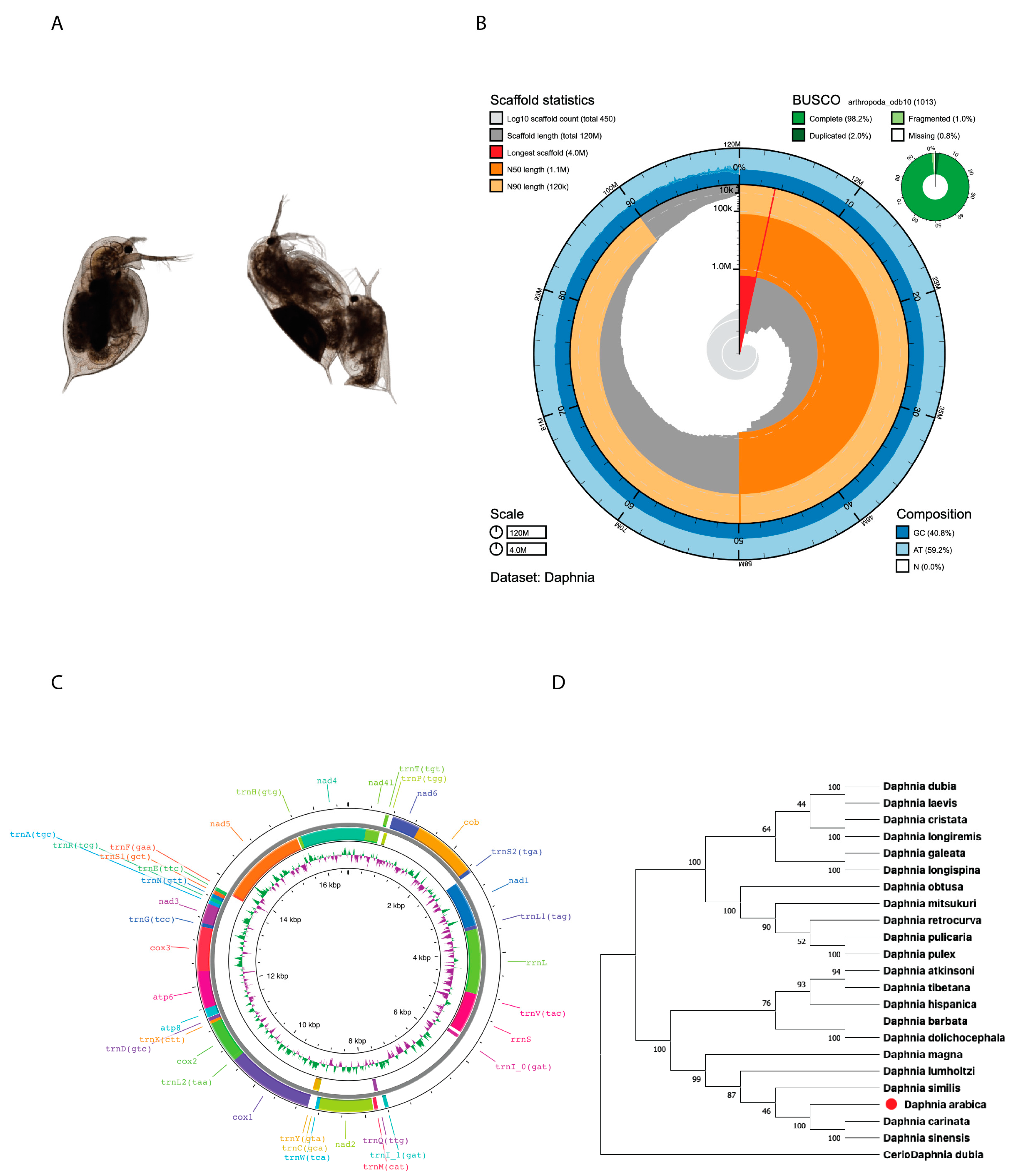
2.1. Genome Assembly and Characterization

In this study, we generated 60.6 Gb (523 X) of D. arabica whole genome sequencing (WGS) data (Table S1) for whole genome assembly. The hybrid de novo genome assembly resulted in a draft genome with the size of ~116 Mb. The assembled genome size was more than ~18% of the theoretically estimated haploid genome size (~98 MB; without repeats) (Figure S1). In total, 454 contigs were obtained from the assembly with an N50 value of ~1.13 Mb and GC% of ~40.8 (Figure 1B). Furthermore, genome completeness was confirmed by BUSCO analysis, which found 99.2% of arthropod orthologous genes (single copy: 96.2%, duplicated: 2%, and fragmented: 1%) from the assembly (Figure 1B). The finally assembled draft genome with N50 (1.13 Mb) was comparable to the finished published genomes of other Daphnia species (Table S2). From the final assembly, mitochondrial genome-related contig (size 16,588 bp) was separated. The assembly statistics of both the nuclear and mitochondrial genome are given in Table 1. From the mitogenome, we annotated 13 proteins coding 23 tRNA and 2 rRNA genes (Table S3 and Figure 1C). Our original mitogenome-based phylogenetic analysis showed that the assembled Daphnia arabica was evolutionarily closely related to subgenus Daphnia (Ctenodaphnia): Daphnia carinata, D. magna, D. similis, and D. sinensis) (Figure 1D).
Our repeat analysis identified 13.33% of the genome repeats (Figure 2A,B). We observed an abundance of long terminal repeats (LTRs) as well as tRNA/SINEs and LINEs. From the repeat masked genome, we annotated 24,041 proteins, coding the 5374 tRNA and 643 rRNA genes (Table 1). Based on the similarity search against NCBI-NR and the Uniprot-trEMBL Protein Database using BlastP program (e-value: 0.000001), ~89% of the predicted genes were functionally annotated (Tables S4 and S5). Furthermore, we annotated 13,823 protein sequences using InterProScan and obtained protein domain-related information (Table S6). Based on KEGG pathway analysis, 6411 metabolic pathway-related proteins were identified (Table S7). Among the revealed genes, the C subunit of SOSS (sensor of ssDNA) was detected, missing in all of the other studied taxa of Daphnia. Many possible fragments of a viral origin were detected, previously found in other daphniids [30], but were not discussed here.
Based on our data, we concluded that D. arabica shares the highest homology with D. sinensis, which was expected, as the former belongs to the D. sinensis species complex sensu Hamza et al. [29]. Our whole genome synteny analysis showed the same results with more similarity to D. sinensis (80%), with the similarity dropping to 25% when compared to D. pulex (Figure 1D and Figure 3A,B). Note that the set of studied species was somewhat different in Figure 1D and Figure 3A as full genomes are known for a smaller number of taxa compared to mitogenomes.

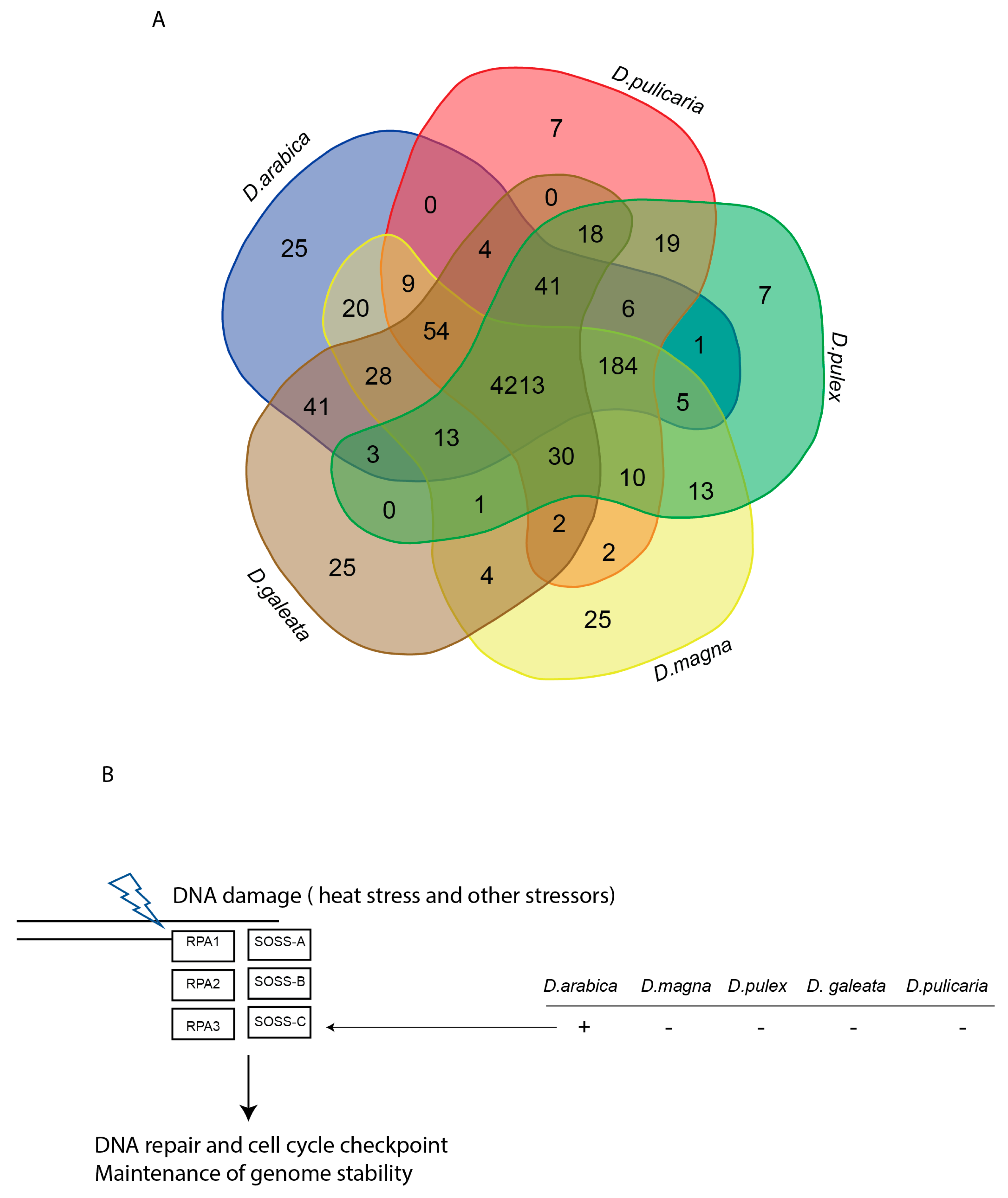
2.2. Diversity and Comparative Genome Analysis

Our diversity analysis results showed a reduction in diversity (Pi=) in D. arabica compared to other Daphnia species (Figure 4A). PFAM comparative genome analysis identified 4213 shared domains among the compared Daphnia species, and interestingly, 25 domains that are unique to D. arabica (Figure 4A). We conducted a manual curation to make sure that the unique identified PFAM were all real by performing a blast against the nr database. Our results showed that there was only one PFAM unique to D. arabica but missing in the other Daphnia species (Figure 4B), which belonged to the subunit C of the SOSS complex.

2.3. Evolution and Demographic History

Our phylogenetic results confirm that the newly isolated species of D. (C.) arabica is an old species that is closer to D. (C.) cf. similoides and D. (C.) sinensis than the other D. (Ctenodaphnia) species from the D. (C.) sinensis group sensu Hamza et al. [29] (Figure 3A). Our rough estimation of the differentiation timing led to the conclusion of a Paleogene (c.a. 60 MYA) differentiation of the D. similis-complex (D. similis + D. cf. similoides + D. arabica), and approximately the same differentiation time of the D. arabica clade. In contrast, the demographic history analysis showed a relatively “recent” bottleneck reflected by the reduction in the effective population size, then an expansion of this species took place around 100,000 years ago (Figure 5).

3. Discussion

In the study, we present the first complete genome of a relict microscopic crustacean, Daphnia (Ctenodaphnia) arabica, recently discovered in the desert of the United Arab Emirates. It is a very old lineage; a divergence time estimate using whole genome unassembled data for phylogenetic analysis dates the species divergence at the Paleogene. This is consistent with the estimations by Hamza et al. [29] based only on three mitochondrial genes. Moreover, the entire Arid Belt of Eurasia could be particularly rich in pre-Pleistocene freshwater relicts [28], but such a hypothesis needs statistically accurate confirmation based on several cladoceran and non-cladoceran taxa.
Note that the results of our previous mtDNA analysis were by chance dependent on a part of the mitochondrial genome used in that study. It has been mentioned that molecular techniques such as mtDNA sequence and barcoding have been introduced as supporting tools capable of shedding light on genetic differences between morphologically similar species [31,32]. However, molecular analyses have experienced many difficulties, especially in the consequent use of different software to analyze the resulting DNA barcodes [33]. Moreover, the mtDNA has substantial limitations, since it only describes the history of a single locus and it shows discrepancies between individual genes and the underlying species tree [8,34,35]. Alternatively, the complete analyses of mitochondrial genomes offer a wealth of high-resolution input and can resolve problems related to taxonomic conflicts and the history of such D. (Ctenodaphnia) species [36]. Additionally, a combination between traditional morphological taxonomy with molecular and genetic tools are essential for better phylogeny of faunistic studies [37].
Even among the water fleas—being a very old group [38]—D. arabica represents a relict lineage, differentiated much earlier from the Gondwanan ancestor [29], so we were not surprised to find its divergence from other daphniids, even at the genomic level.
At the same time, the demographic analysis with a bottleneck effect is consistent with a very strong reduction in the genetic diversity in the species compared to other Daphnia species that had already occurred in the Pleistocene. Interestingly, the subsequent expansion happened around 100 Ka, during Marine Isotope Stage 5 (MIS5), with a great fluctuation in the humidity in the Arabian Peninsula including extra-dry episodes [39,40]. Most probably at this time, other species of Daphnia in the Arabian Peninsula had passed through a mass extinction due to unstable conditions including the periods of extremely high temperatures and extremely low humidity. Such extinction occurred in the Late Pleistocene in different regions of Eurasia [41,42] and North Africa [43]. These times are also very important as “opportunities for modern human dispersal” through the Arabian Peninsula [40].
Yampolsky et al. [44] studied the functional genomics of acclimation and adaptation in response to thermal stress in Daphnia pulex and concluded that “a large number of genes responded to temperature, and many demonstrated a significant genotype-by-environment (GxE) interaction”. Here, in the genome of D. arabica, we found some traces of a special adaptation to desert conditions. Specifically, its genome contains an important domain, the C subunit of SOSS (sensor of ssDNA), which was missing in all of the other studied taxa of Daphnia. This complex, which consists of a few subunits (A, B and C), contributed to the maintenance of the genome stability (i.e., DNA reparation under stress that creates its breakage) [45,46,47]. Since it is present in D. arabica and given the environmental stress that the isolated species faced (mainly the high temperatures), we propose that the SOSS-C subunit could play a role in maintaining the fitness and survival of this species to adapt to the desert environment [45]. The SOSS-C was previously recorded in different animals [48]. There are many sequences in the GenBank to date, but the SOSS-C subunit function has never been discussed in the context of desert animals.
The absence of a critical subunit of a multicomponent protein complex often destabilizes the complex [49], but we need to conclude that missing the SOSS-C in most Daphnia taxa was not critical for them. In contrast, this genus came to be an example of a greatly successful animal in continental waters. Moreover, bearing in mind that the separation of subgenera took place before the D. arabica differentiation, we need to hypothesize that SOSS-C was independently lost in different lineages of Daphnia, as its secondary “appearance” in a single taxon seems to be a less realistic scenario. Unfortunately, no information of the SOSS-complex in other cladocerans and branchiopod crustaceans is available to date.
In conclusion, the sequenced genome of the newly discovered Daphnia will pave the way for future research to identify positively selected genes that are gained or lost in the species and are able to underpin key genes involved in the adaptation of the species to this harsh environment. In addition, our findings will assist in the generation of the crisps of freshwater water fleas, to which we have added this gene, that will be able to tolerate higher global temperatures that are an imminent threat to different ecosystems including diverse freshwater bodies. We believe that it is possible to generate a modified freshwater Daphnia using the D. arabica SOSS subunit C and subject the modified species to a range of temperatures, followed by viability and genome stability measures.

4. Materials and Methods

4.1. D. arabica Isolation

Parthenogenetic females of D. arabica were hatched from the ephippia (modified molting exuvia containing resting eggs) found in the sediment core collected from its type locality: a dry basin behind Al Shuwaib Dam, which is located near Al Ain City, Abu Dhabi, UAE (24°46′18.8″ N and 55°48′15.2″ E) [26]. The core sediments were poured into a 2 L beaker and rinsed with desalinated bottled commercial drinking water at room temperature (20 ± 1 °C), under a 12:12 h light/dark condition for about 2 weeks. In the third week, a few drops of freshly harvested unicellular monoclonal culture of Chlorella sp. were added to the surface water that covered the sediments. A few days later, juveniles were observed on the sediment–water interface. These were transferred to a Petri dish of clear drinking water. The moving juveniles were picked out using a plastic dropper and placed in a 500 mL beaker that contained desalinated commercial drinking water. They were fed every other day at the above-mentioned laboratory conditions.
Under a stereomicroscope, single parthenogenetic females of D. arabica were isolated and reared in a 250 mL glass beaker under laboratory conditions. Newborns were isolated in a larger (500 mL) beaker and left to grow. The third generation produced from the grown adults were then reared in a 2-L beaker and reared under lab conditions, until maturation. For molecular analyses, >60 mature females were isolated and preserved in ethyl alcohol (96%) in an Eppendorf cuvette.
Prior to the species being formally identified, its parthenogenetic female, and gamogenetic females and males (Figure 1A) were described morphologically. A few Sanger sequences were deposited in GenBank, and a preliminary phylogenetic analysis was made based on the mitochondrial 12S, 16S, and COI fragments [29].

4.2. Genomic DNA Isolation and QC

From the 96% ethanol fixed Daphnia sample, high-quality genomic DNA was isolated using a QIAamp DNA Mini Kit (Qiagen, Valencia, California, USA; Cat no. 51306) using the tissue protocol. Isolated genomic DNA quality was confirmed using agarose gel electrophoresis and quantitated on a NanoDrop 2000 spectrophotometer (ThermoFisher Scientific™, Waltham, MA, USA) and Qubit Fluorometer (QubitdsDNA HS Assay Kits, Cat no. Q32851; ThermoFisher Scientific™, Waltham, MA, USA).

4.3. Whole Genome Sequencing Library Preparation

For this study, we generated high-depth Illumina shotgun data and Nanopore (MinIon) based long read data. Illumina compatible whole genome shotgun library for the Daphnia sample was prepared using the NEBNext® Ultra™ II DNA Library Preparation Kit and sequenced using Illumina NovaSeq 6000 (150 bp paired end (PE) sequencing chemistry). Long read whole genome sequencing (WGS) was carried out using the Oxford Nanopore platform. Oxford Nanopore WGS libraries were prepared using the ligation sequencing kit (SQK-LSK 109; Oxford Nanopore, Oxford, UK) and WGS sequencing was performed on an Oxford Nanopore MinION system (flow cell, FLO-MIN106D R9.4 revision D chip; Oxford Nanopore).

4.4. Transcriptome Sequencing

From the sample containing parthenogenetic females of D. arabica, the total RNA was isolated using Maxwell (R) RSC simply RNA Tissue Kit. The quality and quantity of isolated RNA were confirmed by agarose gel, NanoDrop2000, and Qubit. The RNA-Seq library was prepared using the directional lib (Ribo-Zero™ rRNA Removal Kits and NEB Next UltraTM Directional RNA Library PrepKit, New England Biolabs, MA, USA) kit and sequenced in an Illumina NovaSeq machine. The generated transcriptome was used for the downstream gene prediction process.

4.5. Sequencing Data Quality Check and Trimming

The raw Illumina data (both WGS and transcriptome) quality were confirmed using the FastQC tool [50] and the low-quality, adapter, and N-regions present in the reads were trimmed using Trimmomatic v.0.39 software [51]. The sequencing errors found in the Nanopore-MinION reads were corrected and trimmed using CANU v.1.8 [52] software.

4.6. Genome Size Estimation Using Shot Gun Data

We estimated the theoretical genome size of the isolated D. arabica using Illumina shot-gun data by the k-mer based approach. All the k-mers (21-mer) present in Illumina PE reads were mined and a k-mer based histogram file was generated using Jellyfish v.2.3.0 software [53]. The theoretical haploid genome size of the D. arabica was estimated from the k-mer histogram file using thee GenomeScope v.1 tool [54].

4.7. Genome Assembly and QC

We carried out hybrid de novo genome assembly of D. arabica using both shot-gun and long reads in MaSuRCA v.4.0.4 software [55] for whole-genome assembly that included both the Illumina and Nanopore trimmed reads. The sequencing read error found in the assembled genome was corrected using the Pilon v.1.23 program [56]. From the final genome assembly, the genome size, number of contigs, N50 value, and GC content were calculated and the genome assembly completeness was confirmed by the BUSCO v.4.1.4 tool (using arthropoda_odb10 db) [57]. Furthermore, the genome assembly quality was confirmed by aligning the Illumina WGS reads against the assembled genome. Similarly, the transcriptome reads generated for this study were aligned against the assembled genome and confirmed the assembled genome quality based on the read alignment percentage.

4.8. Mitogenome Annotation and Phylogenetic Tree Construction

From the final whole genome assembly, we separated the mitochondrial genome. Mitogenome annotation (CDS, rRNA, and tRNA annotation) was performed using the MITOS tool [58] and the mitogenome map was generated using the Proksee tool (https://proksee.ca/, accessed on 1 December 2022). For phylogenetic tree construction, 22 already published mitogenomes of Daphnia (Table S8) were retrieved from the NCBI database and the coding regions were annotated using the MITOS tool. Furthermore, all coding regions were aligned using the MUSCLE program [59] and a coding region based phylogenetic tree was constructed by the MEGA v.X tool [60] using the ML method (bootstrap value 1000).

4.9. Gene Prediction and Annotation

After genome assembly, we masked the repeat regions found in the Daphnia genome using RepeatsModular v.2.0.1 [61] and the RepeatMasker v.4.1 tool [62]. For the genome annotation, we used both homology-based and ab initio-based gene prediction methods. Generated transcript reads were aligned against the assembled genome using the HiSat v.2.1.0 tool [63] and possible expressed portions (exons or transcripts) of the genome were assembled using StringTie v.2.1.3 tools [64]. These identified transcripts were used as evidence for the gene prediction. Additionally, we retrieved proteins from closely related species and used them for the homology-based gene prediction. Initial gene prediction was carried using the BRAKER v.2.1.5 [65] pipeline (using Augustus v.3.3.3 [66], GeneMark v.4.61 [67], and EVM v.1.1.1 [68] and the final gene prediction was obtained using the MAKER v.3.01 pipeline using Augustus, GeneMark, EVM, and SNAP [69]. Both tRNA and rRNA genes found in the genome were predicted using tRNscan-SE v.2.0.6 [70] and RNAmmer v.1.2 [71]. Predicted proteins were similarity searched against the NCBI-NR and Uniprot-trEMBL Protein Database using thee BlastP program (e-value: 0.000001) [72]. Furthermore, the predicted proteins were functionally annotated using InterProScan v.5.51.85 [73]. Metabolic pathway genes were annotated from the predicted genes using KEGG-KAAS [74], while for pathway analysis, Daphnia pulex and Penaeus vannamei were considered as reference organisms.

4.10. Diversity and Comparative Genomic Analysis

We used our InterProScan results for our isolated D. arabica and compared it with the available annotated genome of Daphnia from the NCBI (D. pulex, D. magna, D. galatea, and D. pulicaria), and identified shared PFAM domains among the different species as well as the unique PFAM for each species. We estimated the nucleotide diversity of ANGSD [75] for each of the species using Illumina shot gun reads.

4.11. Evolutionary and Demographic History

We applied an assembly and alignment-free (AAF) method (https://sourceforge.net/projects/aaf-phylogeny, accessed on 7 November 2022) [76] using K = 25 to construct the phylogeny of unassembled genomic sequences of Daphnia species available on the NCBI short archive (SRA). The divergence time estimate was carried out by running the tool r8s [77] to convert the newick tree generated using the AAF method [76] into the ultrametric tree, where we used a known calibrated adjusted divergence time from TimeTree (http://www.timetree.org, accessed on 10 November 2022) between Daphnia pulex and Daphnia magna and found it to be 131 Mya. Note that this estimation is somewhat younger compared to a widely used 145 Mya by Kotov and Taylor [78], but also could be applied to such analysis. The whole genome synteny plots between Daphnia arabica and the available genomes of D. sinensis and D. pulex were generated using D-genie [79]. Effective population size history was estimated using the pairwise sequentially Markovian coalescent (PSMC) model, following the pipeline by Li et al. [80]. BAM alignments from D. arabica were used to create a consensus sequence using samtools, vcfutils, and bcftools [80]. We performed PSMC analysis using the default parameters recommended by the authors of this method, and we chose an average mutation rate of 8.9 10−9 as well as the generation time of 1 year following Eddie et al. [81].

Supplementary Materials

The following supporting information can be downloaded at: https://www.mdpi.com/article/10.3390/ijms24010889/s1.

Author Contributions

Conceptualization, W.H., K.M.H. and A.A.K.; Methodology, S.E.S.A.N., N.S., K.M.H., K.M.A.A. and A.N.N.; Software, N.S. and K.M.H.; Formal analysis, K.M.H., N.S., A.A.K. and W.H.; Investigation, W.H., S.E.S.A.N., A.N.N., A.A.K. and K.M.H.; Resources, K.M.A.A., W.H. and A.A.K.; Data curation, N.S., K.M.H., W.H. and A.A.K.; Writing—original draft preparation, W.H., K.M.H., S.E.S.A.N., A.N.N. and A.A.K.; Writing—review and editing, W.H. and A.A.K.; Visualization, N.S., A.N.N. and K.M.H.; Supervision, W.H.; Project administration, W.H., K.M.A.A. and A.A.K.; Funding acquisition, W.H. and A.N.N. All authors have read and agreed to the published version of the manuscript.

Funding

The genome sequencing work was supported by the Genome sequencing unit, UAE University and KCGEB, UAE University (Internal research fund: #21R007). The phylogenetic reconstruction was supported by the Russian Science Foundation (grant 22-14-00258 for ANN).

Institutional Review Board Statement

Not applicable.

Informed Consent Statement

Not applicable.

Data Availability Statement

The sequencing data generated during this study were submitted to the NCBI-SRA database under the project id: PRJNA904511. The assembled genome of D. arabica along with the annotation was deposited at the Zenodo repository. URL: https://doi.org/10.5281/zenodo.7408613.

Acknowledgments

All genomic analyses were carried out at the Khalifa Center for Genetic Engineering and Biotechnology, United Arab Emirates University (KCGEB-UAEU). Many thanks to I.B. Metrsalov for the consultations on the SOSS genes and to J. Fowler for the English editing and linguistic corrections of the manuscript.

Conflicts of Interest

The authors declare no conflict of interest.

References

  1. Forró, L.; Korovchinsky, N.P.M.; Kotov, A.A.; Petrusek, A. Global diversity of cladocerans (Cladocera; Crustacea) in freshwater. Hydrobiologia 2007, 595, 177–184. [Google Scholar] [CrossRef]
  2. Dumont, H. Introduction to the class Branchiopoda. Guides Identif. Microinvertebr. Cont. Waters World 2002, 19, 1–398. [Google Scholar]
  3. Crease, T.J. The complete sequence of the mitochondrial genome of Daphnia pulex (Cladocera: Crustacea). Gene 1999, 233, 89–99. [Google Scholar] [CrossRef] [PubMed]
  4. Colbourne, J.K.; Pfrender, M.E.; Gilbert, D.; Thomas, W.K.; Tucker, A.; Oakley, T.H.; Tokishita, S.; Aerts, A.; Arnold, G.J.; Basu, M.K. The ecoresponsive genome of Daphnia pulex. Science 2011, 331, 555–561. [Google Scholar] [CrossRef] [Green Version]
  5. Tokishita, S.-i.; Shibuya, H.; Kobayashi, T.; Sakamoto, M.; Ha, J.-Y.; Yokobori, S.-i.; Yamagata, H.; Hanazato, T. Diversification of mitochondrial genome of Daphnia galeata (Cladocera, Crustacea): Comparison with phylogenetic consideration of the complete sequences of clones isolated from five lakes in Japan. Gene 2017, 611, 38–46. [Google Scholar] [CrossRef]
  6. Routtu, J.; Jansen, B.; Colson, I.; De Meester, L.; Ebert, D. The first-generation Daphnia magna linkage map. BMC Genom. 2010, 11, 1–7. [Google Scholar] [CrossRef]
  7. Lee, J.-S.; Kim, D.-H.; Choi, B.-S.; Kato, Y.; Watanabe, H.; Lee, J.-S. Complete mitochondrial genome of the freshwater water flea Daphnia magna NIES strain (Cladocera, Daphniidae): Rearrangement of two ribosomal RNA genes. Mitochondrial DNA Part B 2020, 5, 1822–1823. [Google Scholar] [CrossRef] [Green Version]
  8. Cornetti, L.; Fields, P.D.; Van Damme, K.; Ebert, D. A fossil-calibrated phylogenomic analysis of Daphnia and the Daphniidae. Mol. Phylogenet. Evol. 2019, 137, 250–262. [Google Scholar] [CrossRef]
  9. Wei, W.; Zhang, K.; Shi, Q. Complete mitochondrial genome of Bosmina fatalis (Cladocera: Bosminidae) and its phylogenetic analysis. Mitochondrial DNA Part B 2021, 6, 2567–2568. [Google Scholar] [CrossRef]
  10. Gu, Y.-L.; Sun, C.-H.; Liu, P.; Zhang, X.; Sinev, A.Y.; Dumont, H.J.; Han, B.-P. Complete mitochondrial genome of Ovalona pulchella (Branchiopoda, Cladocera) as the first representative in the family Chydoridae: Gene rearrangements and phylogenetic analysis of Cladocera. Gene 2022, 818, 146230. [Google Scholar] [CrossRef]
  11. Liu, P.; Xu, S.; Huang, Q.; Dumont, H.J.; Lin, Q.; Han, B.-P. The mitochondrial genome of Diaphanosoma dubium with comparison with Daphnia magna. Mitochondrial DNA Part B 2017, 2, 926–927. [Google Scholar] [CrossRef] [PubMed]
  12. Choi, B.-S.; Lee, Y.H.; Kim, H.-J.; Hagiwara, A.; Lee, J.-S. Complete mitochondrial DNA of the marine water flea Diaphanosoma celebensis (Cladocera, Sididae). Mitochondrial DNA Part B 2020, 5, 2254–2255. [Google Scholar] [CrossRef] [PubMed]
  13. Pan, J.; Liu, P.; Pajk, F.; Dumont, H.J.; Han, B.-P. The mitochondrial genome of Diaphanosoma excisum Sars, 1885 (Crustacea: Branchiopoda: Cladocera) from Hainan Island, China. Mitochondrial DNA Part B 2021, 6, 1279–1280. [Google Scholar] [CrossRef] [PubMed]
  14. Xu, S.-L.; Han, B.-P.; Martínez, A.; Schwentner, M.; Fontaneto, D.; Dumont, H.J.; Kotov, A.A. Mitogenomics of Cladocera (Branchiopoda): Marked gene order rearrangements and independent predation roots. Mol. Phylogenet. Evol. 2021, 164, 107275. [Google Scholar] [CrossRef] [PubMed]
  15. Van Damme, K.; Cornetti, L.; Fields, P.D.; Ebert, D. Whole-genome phylogenetic reconstruction as a powerful tool to reveal homoplasy and ancient rapid radiation in waterflea evolution. Syst. Biol. 2022, 71, 777–787. [Google Scholar] [CrossRef] [PubMed]
  16. Gurney, R.X. On the Fresh-water Crustacea of Algeria and Tunisia. J. R. Microsc. Soc. 1909, 29, 273–305. [Google Scholar] [CrossRef]
  17. Gurney, R. List of Entomostraca collected in Seistan and the Baluch Desert. Rec. Zool. Surv. India 1920, 18, 145–146. [Google Scholar] [CrossRef]
  18. Harding, J. The Armstrong College Zoological Expedition to Siwa Oasis (Libyan Desert) 1935. Crustacea: Branchiopoda and Ostracoda. Proc. Egypti. Acad. Sci. 1955, 10, 58–68. [Google Scholar]
  19. Dumont, H.J.; Laureys, P.; Pensaert, J. Anostraca, Conchostraca, Cladocera and Copepoda from Tunisia. Hydrobiologia 1979, 66, 259–274. [Google Scholar] [CrossRef]
  20. Rocha, J.L.; Godinho, R.; Brito, J.C.; Nielsen, R. Life in deserts: The genetic basis of mammalian desert adaptation. Trends Ecol. Evol. 2021, 36, 637–650. [Google Scholar] [CrossRef]
  21. Tourenq, C.; Brook, M.; Knuteson, S.; Shuriqi, M.K.; Sawaf, M.; Perry, L. Hydrogeology of Wadi Wurayah, United Arab Emirates, and its importance for biodiversity and local communities. Hydrol. Sci. J. 2011, 56, 1407–1422. [Google Scholar] [CrossRef]
  22. Hamza, W.; Munawar, M. Protecting and managing the Arabian Gulf: Past, present and future. Aquat. Ecosyst. Health Manag. 2009, 12, 429–439. [Google Scholar] [CrossRef]
  23. Neubert, E.; Amr, Z.; Van Damme, D. The status and distribution of freshwater molluscs in the Arabian Peninsula. Status Distrib. Freshw. Biodivers. Arab. Penins. 2015, 30, 30–38. [Google Scholar]
  24. Odhiambo, G.O. Water scarcity in the Arabian Peninsula and socio-economic implications. Appl. Water Sci. 2017, 7, 2479–2492. [Google Scholar] [CrossRef] [Green Version]
  25. Van Damme, K.; Dumont, H.J. Further division of Alona Baird, 1843: Separation and position of Coronatella Dybowski & Grochowski and Ovalona gen. n.(Crustacea: Cladocera). Zootaxa 2008, 1960, 1–44. [Google Scholar]
  26. Hamza, W.; Ramadan, G.; AlKaabi, M. Morphological and molecular identification of first recorded Cladoceran organisms in the desert of Abu Dhabi, UAE. MOJ Eco. Environ. Sci. 2018, 3, 220–224. [Google Scholar] [CrossRef] [Green Version]
  27. Soesbergen, M. A preliminary investigation of plankton organisms of fresh and brackish inland waters in the northern United Arab Emirates. Tribulus 2018, 26, 47. [Google Scholar]
  28. Kotov, A.A.; Neretina, A.N.; Al Neyadi, S.E.S.; Karabanov, D.P.; Hamza, W. Cladocera (Crustacea: Branchiopoda) of Man-Made Lakes at the Northeast Part of the United Arab Emirates with a Hypothesis on Their Origin. Diversity 2022, 14, 688. [Google Scholar] [CrossRef]
  29. Hamza, W.; Neretina, A.N.; Al Neyadi, S.E.S.; Amiri, K.; Karabanov, D.P.; Kotov, A.A. Discovery of a New Species of Daphnia (Crustacea: Cladocera) from the Arabian Peninsula Revealed a Southern Origin of a Common Northern Eurasian Species Group. Water 2022, 14, 2350. [Google Scholar] [CrossRef]
  30. Ballinger, M.J.; Bruenn, J.A.; Kotov, A.A.; Taylor, D.J. Selectively maintained paleoviruses in Holarctic water fleas reveal an ancient origin for phleboviruses. Virology 2013, 446, 276–282. [Google Scholar] [CrossRef] [Green Version]
  31. Hebert, P.D.; Gregory, T.R. The promise of DNA barcoding for taxonomy. Syst. Biol. 2005, 54, 852–859. [Google Scholar] [CrossRef] [PubMed]
  32. Elías-Gutiérrez, M.; Hubert, N.; Collins, R.A.; Andrade-Sossa, C. Aquatic Organisms Research with DNA Barcodes. Diversity 2021, 13, 306. [Google Scholar] [CrossRef]
  33. Garibian, P.G.; Neretina, A.N.; Taylor, D.J.; Kotov, A.A. Partial revision of the neustonic genus Scapholeberis Schoedler, 1858 (Crustacea: Cladocera): Decoding of the barcoding results. PeerJ 2020, 8, e10410. [Google Scholar] [CrossRef] [PubMed]
  34. Rubinoff, D.; Holland, B.S. Between two extremes: Mitochondrial DNA is neither the panacea nor the nemesis of phylogenetic and taxonomic inference. Syst. Biol. 2005, 54, 952–961. [Google Scholar] [CrossRef] [PubMed]
  35. Galtier, N.; Nabholz, B.; Glémin, S.; Hurst, G. Mitochondrial DNA as a marker of molecular diversity: A reappraisal. Mol. Ecol. 2009, 18, 4541–4550. [Google Scholar] [CrossRef] [PubMed]
  36. Sullivan, K.A.; Platt, R.N.; Bradley, R.D.; Ray, D.A. Whole mitochondrial genomes provide increased resolution and indicate paraphyly in deer mice. BMC Zool. 2017, 2, 1–6. [Google Scholar] [CrossRef]
  37. Ebach, M.C.; Holdrege, C. DNA barcoding is no substitute for taxonomy. Nature 2005, 434, 697-697. [Google Scholar] [CrossRef] [PubMed] [Green Version]
  38. Van Damme, K.; Kotov, A.A. The fossil record of the Cladocera (Crustacea: Branchiopoda): Evidence and hypotheses. Earth-Sci. Rev. 2016, 163, 162–189. [Google Scholar] [CrossRef]
  39. Parker, A.G. Pleistocene climate change in Arabia: Developing a framework for hominin dispersal over the last 350 ka. In The Evolution of Human Populations in Arabia; Springer: Dordrecht, The Netherlands, 2010; pp. 39–49. [Google Scholar]
  40. Rosenberg, T.; Preusser, F.; Fleitmann, D.; Schwalb, A.; Penkman, K.; Schmid, T.; Al-Shanti, M.; Kadi, K.; Matter, A. Humid periods in southern Arabia: Windows of opportunity for modern human dispersal. Geology 2011, 39, 1115–1118. [Google Scholar] [CrossRef]
  41. Kotov, A.A.; Garibian, P.G.; Bekker, E.I.; Taylor, D.J.; Karabanov, D.P. A new species group from the Daphnia curvirostris species complex (Cladocera: Anomopoda) from the eastern Palaearctic: Taxonomy, phylogeny and phylogeography. Zool. J. Linn. Soc. 2021, 191, 772–822. [Google Scholar] [CrossRef]
  42. Korovchinsky, N. The Cladocera (Crustacea: Branchiopoda) as a relict group. Zool. J. Linn. Soc. 2006, 147, 109–124. [Google Scholar] [CrossRef] [Green Version]
  43. Dumont, H.J. Relict distribution patterns of aquatic animals: Another tool in evaluating late Pleistocene climate changes in the Sahara and Sahel. In Palaeoecology of Africa and the Surrounding Islands; Routledge: Oxfordshire, UK, 1982; pp. 1–24. [Google Scholar]
  44. Yampolsky, L.Y.; Zeng, E.; Lopez, J.; Williams, P.J.; Dick, K.B.; Colbourne, J.K.; Pfrender, M.E. Functional genomics of acclimation and adaptation in response to thermal stress in Daphnia. BMC Genom. 2014, 15, 1–12. [Google Scholar] [CrossRef] [PubMed] [Green Version]
  45. Huang, J.; Gong, Z.; Ghosal, G.; Chen, J. SOSS complexes participate in the maintenance of genomic stability. Mol. Cell 2009, 35, 384–393. [Google Scholar] [CrossRef] [Green Version]
  46. Nam, E.A.; Cortez, D. SOSS1/2: Sensors of single-stranded DNA at a break. Mol. Cell 2009, 35, 258–259. [Google Scholar] [CrossRef] [Green Version]
  47. Pfleiderer, M.M.; Galej, W.P. Emerging insights into the function and structure of the Integrator complex. Transcription 2021, 12, 251–265. [Google Scholar] [CrossRef] [PubMed]
  48. Partridge, L.; Barrie, B.; Fowler, K.; French, V. Evolution and development of body size and cell size in Drosophila melanogaster in response to temperature. Evolution 1994, 48, 1269–1276. [Google Scholar] [CrossRef] [PubMed]
  49. Yin, J.; Sobeck, A.; Xu, C.; Meetei, A.R.; Hoatlin, M.; Li, L.; Wang, W. BLAP75, an essential component of Bloom’s syndrome protein complexes that maintain genome integrity. EMBO J. 2005, 24, 1465–1476. [Google Scholar] [CrossRef] [Green Version]
  50. Andrews, S. FastQC: A Quality Control Tool for High Throughput Sequence Data; Babraham Bioinformatics, Babraham Institute: Cambridge, UK, 2010. [Google Scholar]
  51. Bolger, A.M.; Lohse, M.; Usadel, B. Trimmomatic: A flexible trimmer for Illumina sequence data. Bioinformatics 2014, 30, 2114–2120. [Google Scholar] [CrossRef] [Green Version]
  52. Koren, S.; Walenz, B.P.; Berlin, K.; Miller, J.R.; Bergman, N.H.; Phillippy, A.M. Canu: Scalable and accurate long-read assembly via adaptive k-mer weighting and repeat separation. Genome Res. 2017, 27, 722–736. [Google Scholar] [CrossRef] [Green Version]
  53. Marcais, G.; Kingsford, C. Jellyfish: A fast k-mer counter. Tutor. Manuais 2012, 1, 1–8. [Google Scholar]
  54. Vurture, G.W.; Sedlazeck, F.J.; Nattestad, M.; Underwood, C.J.; Fang, H.; Gurtowski, J.; Schatz, M.C. GenomeScope: Fast reference-free genome profiling from short reads. Bioinformatics 2017, 33, 2202–2204. [Google Scholar] [CrossRef] [PubMed] [Green Version]
  55. Zimin, A.V.; Marçais, G.; Puiu, D.; Roberts, M.; Salzberg, S.L.; Yorke, J.A. The MaSuRCA genome assembler. Bioinformatics 2013, 29, 2669–2677. [Google Scholar] [CrossRef] [PubMed] [Green Version]
  56. Walker, B.J.; Abeel, T.; Shea, T.; Priest, M.; Abouelliel, A.; Sakthikumar, S.; Cuomo, C.A.; Zeng, Q.; Wortman, J.; Young, S.K. Pilon: An integrated tool for comprehensive microbial variant detection and genome assembly improvement. PloS ONE 2014, 9, e112963. [Google Scholar] [CrossRef]
  57. Seppey, M.; Manni, M.; Zdobnov, E.M. BUSCO: Assessing genome assembly and annotation completeness. In Gene Prediction; Springer: New York, NY, USA, 2019; pp. 227–245. [Google Scholar]
  58. Bernt, M.; Donath, A.; Jühling, F.; Externbrink, F.; Florentz, C.; Fritzsch, G.; Pütz, J.; Middendorf, M.; Stadler, P.F. MITOS: Improved de novo metazoan mitochondrial genome annotation. Mol. Phylogenet. Evol. 2013, 69, 313–319. [Google Scholar] [CrossRef]
  59. Edgar, R.C. MUSCLE: Multiple sequence alignment with high accuracy and high throughput. Nucleic Acids Res. 2004, 32, 1792–1797. [Google Scholar] [CrossRef] [PubMed] [Green Version]
  60. Kumar, S.; Stecher, G.; Li, M.; Knyaz, C.; Tamura, K. MEGA X: Molecular evolutionary genetics analysis across computing platforms. Mol. Biol. Evol. 2018, 35, 1547. [Google Scholar] [CrossRef]
  61. Flynn, J.M.; Hubley, R.; Goubert, C.; Rosen, J.; Clark, A.G.; Feschotte, C.; Smit, A.F. RepeatModeler2 for automated genomic discovery of transposable element families. Proc. Natl. Acad. Sci. USA 2020, 117, 9451–9457. [Google Scholar] [CrossRef]
  62. Chen, N. Using Repeat Masker to identify repetitive elements in genomic sequences. Curr. Protoc. Bioinform. 2004, 5, 4–10. [Google Scholar] [CrossRef]
  63. Kim, D.; Paggi, J.M.; Park, C.; Bennett, C.; Salzberg, S.L. Graph-based genome alignment and genotyping with HISAT2 and HISAT-genotype. Nat. Biotechnol. 2019, 37, 907–915. [Google Scholar] [CrossRef]
  64. Pertea, M.; Kim, D.; Pertea, G.M.; Leek, J.T.; Salzberg, S.L. Transcript-level expression analysis of RNA-seq experiments with HISAT, StringTie and Ballgown. Nat. Protoc. 2016, 11, 1650–1667. [Google Scholar] [CrossRef]
  65. Hoff, K.J.; Lomsadze, A.; Borodovsky, M.; Stanke, M. Whole-genome annotation with BRAKER. In Gene Prediction; Springer: New York, NY, USA, 2019; pp. 65–95. [Google Scholar]
  66. Stanke, M.; Keller, O.; Gunduz, I.; Hayes, A.; Waack, S.; Morgenstern, B. AUGUSTUS: Ab initio prediction of alternative transcripts. Nucleic Acids Res. 2006, 34, W435–W439. [Google Scholar] [CrossRef] [PubMed] [Green Version]
  67. Brůna, T.; Lomsadze, A.; Borodovsky, M. GeneMark-EP+: Eukaryotic gene prediction with self-training in the space of genes and proteins. NAR Genom. Bioinform. 2020, 2, lqaa026. [Google Scholar] [CrossRef] [PubMed]
  68. Haas, B.J.; Salzberg, S.L.; Zhu, W.; Pertea, M.; Allen, J.E.; Orvis, J.; White, O.; Buell, C.R.; Wortman, J.R. Automated eukaryotic gene structure annotation using EVidenceModeler and the Program to Assemble Spliced Alignments. Genome Biol. 2008, 9, 1–22. [Google Scholar] [CrossRef] [Green Version]
  69. Korf, I. Gene finding in novel genomes. BMC Bioinform. 2004, 5, 1–9. [Google Scholar] [CrossRef] [PubMed] [Green Version]
  70. Chan, P.P.; Lin, B.Y.; Mak, A.J.; Lowe, T.M. tRNAscan-SE 2.0: Improved detection and functional classification of transfer RNA genes. Nucleic Acids Res. 2021, 49, 9077–9096. [Google Scholar] [CrossRef]
  71. Lagesen, K.; Hallin, P.; Rødland, E.A.; Stærfeldt, H.-H.; Rognes, T.; Ussery, D.W. RNAmmer: Consistent and rapid annotation of ribosomal RNA genes. Nucleic Acids Res. 2007, 35, 3100–3108. [Google Scholar] [CrossRef]
  72. Altschul, S.F.; Gish, W.; Miller, W.; Myers, E.W.; Lipman, D.J. Basic local alignment search tool. J. Mol. Biol. 1990, 215, 403–410. [Google Scholar] [CrossRef]
  73. Jones, P.; Binns, D.; Chang, H.-Y.; Fraser, M.; Li, W.; McAnulla, C.; McWilliam, H.; Maslen, J.; Mitchell, A.; Nuka, G. InterProScan 5: Genome-scale protein function classification. Bioinformatics 2014, 30, 1236–1240. [Google Scholar] [CrossRef] [Green Version]
  74. Moriya, Y.; Itoh, M.; Okuda, S.; Yoshizawa, A.C.; Kanehisa, M. KAAS: An automatic genome annotation and pathway reconstruction server. Nucleic Acids Res. 2007, 35, W182–W185. [Google Scholar] [CrossRef] [Green Version]
  75. Korneliussen, T.S.; Albrechtsen, A.; Nielsen, R. ANGSD: Analysis of next generation sequencing data. BMC Bioinform. 2014, 15, 1–13. [Google Scholar] [CrossRef] [Green Version]
  76. Fan, H.; Ives, A.R.; Surget-Groba, Y.; Cannon, C.H. An assembly and alignment-free method of phylogeny reconstruction from next-generation sequencing data. BMC Genom. 2015, 16, 1–18. [Google Scholar] [CrossRef] [PubMed] [Green Version]
  77. Sanderson, M.J. r8s: Inferring absolute rates of molecular evolution and divergence times in the absence of a molecular clock. Bioinformatics 2003, 19, 301–302. [Google Scholar] [CrossRef] [Green Version]
  78. Kotov, A.A.; Taylor, D.J. Mesozoic fossils (>145 Mya) suggest the antiquity of the subgenera of Daphnia and their coevolution with chaoborid predators. BMC Evol. Biol. 2011, 11, 1–9. [Google Scholar] [CrossRef] [Green Version]
  79. Cabanettes, F.; Klopp, C. D-GENIES: Dot plot large genomes in an interactive, efficient and simple way. PeerJ 2018, 6, e4958. [Google Scholar] [CrossRef] [PubMed]
  80. Li, H.; Handsaker, B.; Wysoker, A.; Fennell, T.; Ruan, J.; Homer, N.; Marth, G.; Abecasis, G.; Durbin, R. 1000 genome project data processing subgroup. The sequence alignment/map format and SAMtools. Bioinformatics 2009, 25, 2078–2079. [Google Scholar] [CrossRef] [PubMed] [Green Version]
  81. Ho, E.K.; Macrae, F.; Latta, L.C., 4th; McIlroy, P.; Ebert, D.; Fields, P.D.; Benner, M.J.; Schaack, S. High and highly variable spontaneous mutation rates in Daphnia. Mol. Biol. Evol. 2020, 37, 3258–3266. [Google Scholar] [CrossRef] [PubMed]
Figure 1. (A) General views of Daphnia arabica: parthenogenetic female and ephippial female and male at copulation. (B) D. arabica whole genome assembly statistics and assembly quality in snail plot view. (C) D. arabica mitogenome map. (D) Mitogenome based phylogenetic tree of D. arabica.
Ijms 24 00889 g001
Figure 2. (A) Repeat landscape annotation in D. arabica genome. (B) D. arabica whole genome characterization with black color of the pie chart reflecting the total genome not occupied with repeats.
Ijms 24 00889 g002
Figure 3. (A) Whole genome shot-gun sequence-based divergence time estimation of D. arabica. (B) Pairwise genome comparison D. arabica vs. D. pulex (left) and D. arabica vs. D. sinensis (right).
Ijms 24 00889 g003
Figure 4. Comparative analysis based on the Daphnia gene families. (A) Venn diagram shows the distribution of PFam gene families between five Daphnia species (D. arabica, D. pulicaria, D. pulex, D. magna and D. galeata). (B) Comparison of SOSS-C in D. arabica, D. pulicaria, D. pulex, D. magna, and D. galeata.
Ijms 24 00889 g004
Figure 5. D. arabica effective population size reduction estimation.
Ijms 24 00889 g005
Table 1. D. arabica whole genome assembly statistics.
D. arabica GenomeD. arabica Mitogenome
Total sequences4531
Genome size116,021,02416,588
A + T%~59.2~69.7
G + C%~40.7~30.2
n %0.00010
Minimum sequence length117916,588
Maximum sequence length4,005,66116,588
N50 length (bp)1,139,06816,588
L50 number301
Length 1001–3000 bp90
Length 3001–5000 bp100
Length 5001–7000 bp70
Length 7001–10,000 bp00
Length 10,001–0.1 Mb bp2491
Length 100,001–1 Mb bp1340
Length > 1 Mb bp350
Protein coding genes24,04113
tRNA genes537423
rRNA genes6432
Disclaimer/Publisher’s Note: The statements, opinions and data contained in all publications are solely those of the individual author(s) and contributor(s) and not of MDPI and/or the editor(s). MDPI and/or the editor(s) disclaim responsibility for any injury to people or property resulting from any ideas, methods, instructions or products referred to in the content.

