Review

Mucopolysaccharidoses: Cellular Consequences of Glycosaminoglycans Accumulation and Potential Targets

by
Andrés Felipe Leal
1,2,
Eliana Benincore-Flórez
2,
Estera Rintz
2,3,
Angélica María Herreño-Pachón
2,4,
Betul Celik
2,4,
Yasuhiko Ago
2,
Carlos Javier Alméciga-Díaz
1 and
Shunji Tomatsu
2,4,5,6,*
1
Institute for the Study of Inborn Errors of Metabolism, Faculty of Science, Pontificia Universidad Javeriana, Bogotá 110231, Colombia
2
Nemours Children’s Health, Wilmington, DE 19803, USA
3
Department of Molecular Biology, Faculty of Biology, University of Gdansk, 80-308 Gdansk, Poland
4
Faculty of Arts and Sciences, University of Delaware, Newark, DE 19716, USA
5
Department of Pediatrics, Graduate School of Medicine, Gifu University, Gifu 501-1193, Japan
6
Department of Pediatrics, Thomas Jefferson University, Philadelphia, PA 19144, USA
*
Author to whom correspondence should be addressed.
Int. J. Mol. Sci. 2023, 24(1), 477; https://doi.org/10.3390/ijms24010477
Submission received: 25 October 2022 / Revised: 9 December 2022 / Accepted: 24 December 2022 / Published: 28 December 2022
(This article belongs to the Special Issue Mucopolysaccharidoses: Diagnosis, Treatment, and Management 3.0)

Abstract

:
Mucopolysaccharidoses (MPSs) constitute a heterogeneous group of lysosomal storage disorders characterized by the lysosomal accumulation of glycosaminoglycans (GAGs). Although lysosomal dysfunction is mainly affected, several cellular organelles such as mitochondria, endoplasmic reticulum, Golgi apparatus, and their related process are also impaired, leading to the activation of pathophysiological cascades. While supplying missing enzymes is the mainstream for the treatment of MPS, including enzyme replacement therapy (ERT), hematopoietic stem cell transplantation (HSCT), or gene therapy (GT), the use of modulators available to restore affected organelles for recovering cell homeostasis may be a simultaneous approach. This review summarizes the current knowledge about the cellular consequences of the lysosomal GAGs accumulation and discusses the use of potential modulators that can reestablish normal cell function beyond ERT-, HSCT-, or GT-based alternatives.

1. Introduction

The mucopolysaccharidoses (MPSs) are a group of rare genetic diseases caused by mutations in the genes encoding for the enzymes involved in the glycosaminoglycans (GAGs) catabolism, leading to their accumulation within the lysosome. GAGs are complex lineal carbohydrates composed of repeating disaccharide units of hexamine and uronic acid. Five GAGs have been described: heparan sulfate (HS), dermatan sulfate (DS), chondroitin sulfate (CS), keratan sulfate (KS), and hyaluronic acid (HA) [1].
The lysosomal accumulation of partially degraded GAGs was seen as the only cause of the pathophysiology in these diseases. Nevertheless, currently, it is well known that the pathophysiology of MPS results from the impairment of several cellular systems [2]. In fact, it has been described that GAG accumulation may trigger the storage of secondary substrates (e.g., glycosphingolipids, phospholipids, cholesterol, aggregated-prone proteins, and alpha-synuclein), inflammation, impairment of autophagy, changes in the intracellular trafficking of vesicles, mitochondrial dysfunction, oxidative stress, and dysregulation of signaling pathways [2,3,4,5,6].
Clinical therapeutic options include enzyme replacement therapy (ERT) and hematopoietic stem cell therapy (HSCT) for most MPS diseases [7]. Moreover, experimental therapies involving gene therapy [8,9,10], substrate reduction therapy, anti-inflammatory therapy, and pharmacological chaperone therapy [11,12,13] are under evaluation and could be potential alternatives shortly.
Approaches focusing on replacing missing enzymes are supposed to be the most promising alternatives; however, it has been shown that recovery of altered organelles and their related cellular processes positively impacts the whole cell homeostasis [2,14,15,16,17,18,19]. Thus, recognizing intracellular changes due to primary GAG accumulation is critical for attempting future holistic treatments. Herein, we review key aspects of GAGs, including the cellular consequences of their lysosomal accumulation and the potential use of modulators to recover the organelle/signal pathway affected.

2. Glycosaminoglycans: Structure, Biosynthesis, and Catabolism

GAGs are the most abundant biomolecules in the extracellular matrix (ECM). GAGs are a diverse class of long, linear, and heterogeneous polysaccharides characterized by disaccharide repeats composed of alternating units of uronic acid and amino sugar (Table 1), forming chains that range from 1 to 25,000 disaccharide units [20,21]. They are involved in critical biological processes, including ECM hydration, cell signaling, and regulation of growth factors [22]. Heterogeneity of the GAGs is based not only on the disaccharide composition but also on the polysaccharide chain length, the glycosidic linkage binding these units, either α or β, and the sulfation, acetylation, and epimerization patterns of the glycan chains. GAGs exist as free polysaccharide chains or are covalently linked to core proteins forming proteoglycans (PGs) [23].

2.1. Glycosaminoglycans Biosynthesis

The biosynthesis of GAGs (except for KS) begins in the cytoplasm with the formation of a tetrasaccharide linkage to the core proteins, catalyzed by the sequential actions of four glycosyltransferases, which adds one xylose, two galactose, and one glucuronic acid residues [24,25]. This tetrasaccharide block is a common precursor for the biosynthesis of HS and CS/DS polysaccharide chains, while three types of KS occur, depending on its core structure, and are detailed in the next section. These blocks are then transported into the Golgi lumen, where the polymerization occurs. The step that differentiates the formation of HS or CS/DS remains in the addition of the N-Acetylglucosamine (GlcNAc)- or N-Acetylgalactosamine (GalNAc)-units to the nonreducing end of the tetrasaccharide, respectively [24]. The polymerization continues in the case of HS by the alternating addition of GlcNAc and glucuronic acid (GlcA) residues by HS polymerases. In contrast, for CS/DS, the addition of GalNAc to the linker tetrasaccharide is required for the elongation [26]. After that, the backbones are subjected to modifications by epimerases or acetylases, increasing the structural heterogeneity of these molecules [27]. For KS, the only GAG composed of alternating β 1,3 and β 1,4 linkages between galactose and N-acetylglucosamine [28], biosynthesis begins with the building of the region binding with the core protein. Depending upon that linkage to the PG, three types of KS exist: KS I, found mainly in the cornea, is N-linked via asparagine in the core protein in a mannose-containing linkage oligosaccharide; KS II, frequently found in cartilage, is O-linked to serine/threonine via galactosamine; and KS III, mainly expressed in nervous tissue, is O-linked to serine/threonine via mannose and is the most sulfated of the three types [29].

2.1.1. Keratan Sulfate

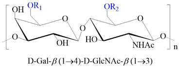
KS is the only GAG containing galactose (Gal) instead of uronic acids. Hence, it comprises alternating repeating units of β1,3-Gal and β1,4-GlcNAc [30]. Both structures may have 6-O sulfation. The molecular weight ranges from 6 KDa to >50 Kda; however, the degree of sulfation and size depends on the tissue from which it is found. For instance, KS I, found mainly in the cornea, is N-linked via asparagine in the core protein in a mannose-containing linkage oligosaccharide; KS II, frequently found in cartilage, is O-linked to serine/threonine via galactosamine; and KS III, mainly expressed in nervous tissue, is O-linked to serine/threonine via mannose and is the most sulfated of the three types [29].

2.1.2. Heparan Sulfate (HS)

HS is synthesized as repeating disaccharides (GlcA and GlcNAc), suffering from N-deacetylations, N-sulfation, and epimerization of glucuronic to iduronic acid (IdoA). Both GlcA and IdoA can be sulfated at the C2 position, and glucosamine can be sulfated at the C3 and C6 positions [31]. The sulfation and acetylation patterns vary depending on the cell type. The molecular weight ranges from 5 to 50 Kda with 10 to 100 disaccharide units. HS is O-linked to serine residues of diverse core proteins, forming different HS proteoglycans (PG) types, including glypican and syndecans expressed on endothelial cell surface and leukocytes; perlecan, agrin, and collagen type XVIII are components of the subendothelial basement membrane [32]. Perlecan also regulates the development of blood vessels, cartilage, and endochondral ossification [33,34]. HS has the most ubiquitous expression and is the most diverse type of GAG, present in the mammalian cell surface of all tissues and the ECM. HS promotes hydration and participates in water and tissue homeostasis, organization of the ECM, and cellular migration; HS also interacts with collagen, growth factors, cytokines, chemokines, and interleukins, and hence plays a role in regulating inflammatory response, receptor-mediated signaling, phagocytosis, morphogenesis, and organogenesis [35,36]. HS storage disorders have been broadly correlated with central nervous system (CNS) impairment because of the involvement of HS in neuronal development as an essential component of the vascular basement membrane in the brain [37,38].

2.1.3. Chondroitin (CS) and Dermatan Sulfate (DS)

CS and DS are synthesized as alternating linkages β1,3 and β1,4 between galactosamine and glucuronic acid (GalNAcβ1,4GlcAβ1,3)n in CS, or between galactosamine and mainly iduronic acid (GalNAcβ1,4IdoAβ1,3)n, in DS [26]. In CS, the GalNAc residues may be sulfated at C4 and/or C6, while GlcA can be sulfated at C2. Hence, based on sulfo group substitution and the type of uronic acid that each contains, multiple forms of CS have been described: CS-A is GlcA-GalNAc(4S); CS-B is also known as DS; CS-C is GlcA-GalNAc(6S); CS-D is GlcA(2S)-GalNAc(6S); CS-E is GlcA-GalNAc(4S,6S). Since the hybrid nature of DS structure is due to epimerization at C5 of GlcA to form IdoA, the sulfation at the C4 position is mainly found in DS. However, it can also be present at the C6 of GalNAc and C2 IdoA residues. The molecular weights of the CS/DS family of GAGs range from 2 to 50 kDa, and diverse biological functions have been described thanks to their high structural heterogeneity. CS/DS PGs are the major components of the CNS, participating in the axon growth tans synapse formation [39].
CS is one of the most critical constituents of bones due to its essential role in cartilage and other connective tissues. Moreover, it has been found to participate in the prevention of inflammation, immune modulation, maintenance of the structure and function of cartilage, and regulation of cell adhesion to the ECM [30]. For DS, the biological process described includes collagen organization, regulation of transforming growth factor (TGF)-β activity, stabilization of the basement membrane, and regulation of the cell–cell and cell–matrix interactions [26]. On the other hand, since DS shares structural similarities with lipopolysaccharide, DS activates the Toll-like receptor 4 (TLR4) signaling pathway, initiating the release of pro-inflammatory cytokines, and promoting the articular chondrocyte apoptosis. Decorin and biglycan are DS-/CS-PG that act as structural constituents of skin, cartilage, intervertebral discs, tendons, cornea, kidney, and muscle, promoting proper collagen fiber organization and endochondral ossification [40]. Biglycan is also present in bone and is vital in regulating skeletal growth by interacting with growth factors such as TGF-β, bone morphogenic protein 4 (BMP-4), and the Wnt signaling pathway [41].

2.2. Glycosaminoglycans Degradation

The stepwise degradation of the GAGs occurs within the lysosomes, where acid hydrolases degrade DS, HS, KS, and CS through the sequential removal of monosaccharides followed by the removal of sulfate groups, resulting in the complete degradation of the polysaccharide to its fundamental constituents’ components [42]. Misfunctioning of any of the 12 enzymes participating in the GAGs degradation results in eight different types of disorders known as MPS, characterized by the lysosomal accumulation of undegraded or partially degraded substrates in most cells, tissues, and organs, which ultimately results in multi-systemic failure and death (Table 2) [43,44].
Depending on GAG stored and the residual enzyme activity, MPS patients may present with hepatosplenomegaly, cardiac and pulmonary disease, skeletal deformities, earing and ophthalmological abnormalities, and central or peripheral nervous system impairment [45,46].
Although for many years, the pathogenesis and the clinical outcomes of the MPSs were considered a simple consequence of the cellular accumulation of GAGs. However, it is clear that GAGs are molecules biologically active, either free polysaccharide chains or attached to diverse proteins, playing essential roles in several biological activities, acting as modulators of proper organization of collagen fibrils and elastin fibers, immunomodulators, anticoagulants, antioxidants, anti-inflammatories, and neuroprotectors [47,48]. Consequently, any impairment in the degradation pathway can trigger intracellular pathological cascade activation.

3. Intracellular Organelle Impairment

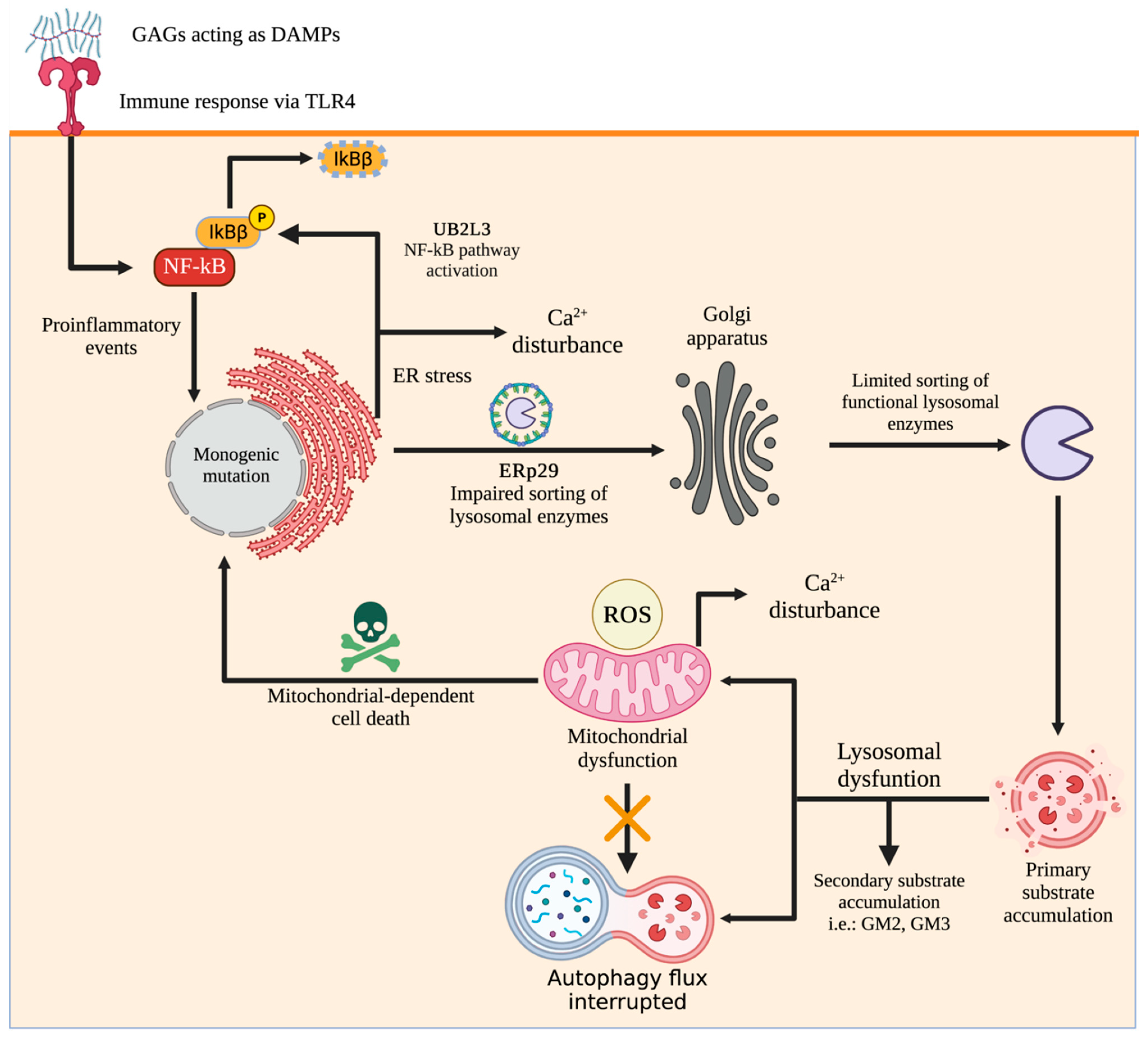
In MPS, the lysosome and many other organelles are affected. It is well known that a global cell homeostasis impairment occurs, leading to the subsequent alteration at the tissue, organs, and systems levels. In the following sections, we will cover critical aspects of organelles impairment (summarized in Figure 1) and potential interventions for their recovery beyond classical approaches related to correcting the primary enzyme deficiency.

3.1. Lysosomal Impairment

Lysosomes process a broad range of materials as protein and nutrients products of endocytosis, phagocytosis, and autophagia, as well as sense environmental conditions to integrate a quick response through complexes signaling that allow homeostatic regulation, metabolic energy regulation, immune responses, and membrane sealing, among other regulatory pathways [2,49,50]. Because of their pivotal functions, GAG accumulation within the lysosome induces lysosomal stress, which is characterized by changes in lysosome biogenesis, increased intraluminal pH, membrane permeability, and crosstalk between organelles, leading to the activation of several pathways [49,50,51,52], with catastrophic consequences for the cell homeostasis. As stated by Simonaro, “the lysosomal system shuts down, leading to abnormalities in endocytosis, autophagy, inflammation, and ultimately causing cell death” [53].
Upon lysosomal GAG accumulation, the cell increases the lysosome biogenesis by activating the transcription factor EB (TFEB), a member of the microphthalmia-transcription factor E (MiT/TFE) transcription factor family. Members of MiT/TFE are responsible for the activation and regulation of several stress response pathways [49,51,52,54]. Dephosphorylation of TFEB by phosphatases calcineurin or protein phosphatase 2A (PP2A) allows a fast translocation to the nucleus to bind directly to the promoter region in the binding element called coordinated lysosomal expression and regulation (CLEAR), activating the expression of numerous genes involved in autophagy and lysosome biogenesis pathways [49,51,55,56]. Furthermore, an elevation of the calcium levels in the cytosol allows activation and translocation to the nucleus of TFEB by activating calcineurin [51,55].
Moreover, during lysosomal stress, an increased intraluminal pH is observed in MPS and other LSDs. The dramatic increase in the pH is caused by the dysregulation of membrane-resident ion channels or vacuolar-ATPases, given that the integrity of the membrane is affected by undegraded materials accumulated into the lysosome [2,51,57]. Since endolysosomes store ions like calcium and iron, their de-acidification induces efflux of those ions to the cytoplasm, impairing the activity of several organelles such as mitochondria and ER. Moreover, it has been described that de-acidification leads to changes in size and cytosolic mislocalization of endolysosomes [51].
Since the integrity of the lysosome membrane is affected by accumulating undegraded materials, such as GAGs, the lysosome releases several intraluminal molecules to the cytosol, such as galectins and cathepsins [49]. Galectins are a group of proteins involved in different physiological processes [49,58], and a role in regulating unfunctional lysosomes through autophagy has been reported [49] through the activation of AMP-activated protein kinase (AMPK), which results in the activation of the autophagy [49,59,60,61]. On the other hand, cathepsins are a group of proteases that participate in various physiological functions [62]. The secretion of cathepsins to the cytosol after lysosomal membrane impairment has been related to caspase-dependent and -independent cell death pathways [63].

3.2. Autophagy Impairment

Autophagy is an essential evolutionary cellular process that degrades cytoplasm components assisted by the lysosome [18,64]. Classically, three autophagy classes have been described, chaperone-mediated autophagy, microautophagy, and macroautophagy. Although these types of autophagy differ in the stepped mechanism to recognize and catch the cytoplasm cargo, all of them deliver the cargo to the lysosome for their degradation. A complete review of molecular mechanisms in autophagy was previously published by Pierzynowska et al. (2018) [18], and Figure 2 shows the molecular mechanisms of the autophagy process.
Since lysosomal fusion is critical to degrade the cargo inside autophagosomes, during MPS, the autophagosome–lysosome (AL) pathway is also affected [2,65,66]. Early reports showed that autophagosomes are accumulated because of the impaired AL fusion in MPS IIIA and multiple sulfatase deficiency [67], emphasizing the close relationship between the lysosome and autophagic pathways. In concordance, LC3 increased in the cerebral cortex and stratum from MPS IIIB [68]. Interestingly, by treating animals with CLR01, a molecular tweezer, significant neuroinflammatory findings such as astrogliosis and microgliosis were significantly decreased in untreated animals, with improvement in memory tests [68]. Elevated LC3-II levels were also reported in MPS IIIC mice [69].
Previously, Maeda et al. (2019) found abnormal autophagy in MPS II mice [70]. Immunostaining of p62 showed an increase over time in MPS II mice, compared to wild type, as well as in the cytoplasm vacuoles in neurons, microglia, and pericytes, suggesting a lack of AL fusion. Remarkably, by using chloroquine, an autophagy-suppressing drug [71], neurodegeneration was prevented, while microglia and pericytes remained vacuolated [70].
Increased phosphorylation levels on P70S6K and ULK1 were reported in MPS II, VI, and VII chondrocytes. Furthermore, MPS VII chondrocytes have shown nuclear localization of TFEB and TFE3, suggesting an mTOR-independent nuclear accumulation, although the autophagic vacuole biogenesis is not primarily affected. Autophagosome maturation was significantly involved in MPS VII chondrocytes [72]. For MPS IVA, a significant decrease in p62 and LC3B was found in MPS IVA patients, suggesting a blocked autophagosome formation [73,74].
Although AL impairment has been widely recognized in MPS, some reports contrast those findings. For example, LC3-I conversion to LC3-II was found unaltered in MPS IIIB mouse cortical neurons, suggesting that macroautophagy was unaffected in this model [75]. Similar findings were reported in peritoneal MPS I mouse fibroblasts [76].
Figure 1. Global cellular changes occur during GAG accumulation. Note that primary GAG accumulation affects not only lysosomal function but also any intracellular organelles contributing to altered cell homeostasis. Since GAGs act in cell–cell communication, their extracellular accumulation, due to lack of plasma turnover [77], promotes inflammation-related activation pathways since they are recognized as damage-associated molecular patterns (DAMPs). For instance, soluble HS fragments can act as Toll-like receptor 4 antagonists supporting the pro-inflammatory events of MPS III [78,79]. This figure was created with BioRender.com.
Ijms 24 00477 g001

3.3. Mitochondrial Impairment

Primary lysosomal dysfunction occurring during MPS can affect mitochondrial functions. Mitochondria is the primary energy producer in eukaryotes through oxidative phosphorylation (OXPHOS). It is also a critical organelle involved in vital cellular processes such as calcium homeostasis, lipid metabolism, viral response, and programmed cell death [80]. OXPHOS is a predominant source of reactive oxygen species (ROS). Therefore, mitochondrial impairment results in imbalanced ROS levels [81]. ROS are oxygen-containing molecules with a high affinity to react with macromolecules, including DNA, proteins, and lipids [82].
Figure 2. Schematic representation of the autophagy pathway. After extra- or intra-cellular autophagy stimulus such as nutrient starvation or oxidative stress [83], autophagy is triggered by the interaction of several effectors such as ULK1, ATG13, FIP200, and ATG101. In a nutrient-rich environment, the mTOR kinase hyperphosphorylates ATG13, avoiding binding to ULK1 and FIP200 [83]. During elongation and maturation, proteins and lipids are recruited to the phagophore biogenesis. In this process, several steps take place, such as the proteolytic cleavage of LC3 to LC3 I and its later conjugation with phosphatidylethanolamine to form LC3 II. LC3 II arrives at the autophagosome membrane, which has selection cargo engulfment functions [84]. Cytosolic structures that will be degraded via AL undergoes p62 labeling. According to the cargo, autophagy can be termed mitophagy or ribophagy [85]. Then, the autophagosome is fused to the lysosome in a SNARE-mediated process [86]. Several proteins, such as LAMP-2, Rubicon, and monomeric GTP-ases, contribute to the AL fusion [18]. Finally, the acid hydrolases inside the lysosome are released to the phagosome to digest the loaded cytosolic components. New macromolecules such as amino acids, fatty and nucleic acids are now available for cellular needs [18,83]. This figure was created with BioRender.com.
Ijms 24 00477 g002
Recent studies have shown the critical role of ROS in the pathophysiology of MPS. For MPS II, oxidative stress has been involved in the pathogenesis. In this regard, Filippon et al. (2011) tested 12 MPS II patients aged from one to seven years old concerning oxidative stress before and during ERT treatment [87]. Blood samples were collected from MPS II patients before starting treatment and during each session of ERT. The results revealed clearly that malondialdehyde (a biomarker of lipid peroxidation), carbonyl groups, and erythrocyte catalase activity were higher in plasma before ERT than in the healthy control group. Moreover, total antioxidant levels and levels of the protein-bound sulfhydryl group were lower than control groups before ERT, suggesting an imbalance between anti- and pro-oxidant molecules in MPS II. Despite ERT leading to a decrease in the malondialdehyde levels, an increase in the sulfhydryl groups was observed compared to findings before treatment. Furthermore, ERT does not significantly impact plasma antioxidant levels, carbonyl groups, catalase, or superoxide dismutase activities, suggesting a limited effect of the ERT on the pro-oxidant profile observed in MPS II patients [87].
Oxidative stress was also evaluated in MPS IIIA mouse cerebral tissues in an early study conducted by Arfi et al. (2011) [88]. For instance, oxidative stress-related genes such as the inducible nitric oxide synthase (fold change: 2.1), NADPH oxidase subunit (fold change: 2.6), superoxide dismutase 2 (fold change: 1.9), glutathione peroxidase (fold change: 2.1) were found overexpressed in the brain of MPS IIIA mice, compared with healthy animals [88], supporting the link between a pro-oxidative profile and the CNS impairment in the MPS IIIA. Interestingly, by using an antioxidant treatment based on vitamin C administration, animals failed to show any improvement in oxidant profile [88], contrary to other LSDs, such as Sandhoff (SD), where the same treatment led to an improvement in the pro-oxidant findings in SD mice [89].
In MPS IVA, the analysis of blood and urine samples from patients showed lower levels of defensive antioxidants, such as glutathione, than healthy control groups, suggesting an oxidative homeostasis impairment [90]. Furthermore, superoxide dismutase levels increased while glutathione levels decreased in red blood cells [90,91]. Free radicals may oxidize lipids and proteins, resulting in isoprostanes and dityrosine yields, which can be used as biomarkers of oxidative stress [92,93]. Increased isoprostane and dityrosine levels in urine samples from MPS IVA patients were reported, which suggested lipid and protein damage, respectively [90].

3.4. Endoplasmic Reticulum Stress

Since most mutations in MPS can induce misfolding of lysosomal enzymes [13], ER stress can also be present. For example, by using transcriptomic approaches, it has been found that IER3IP1, a gene encoding for a resident ER membrane protein, is significantly downregulated in almost all MPSs except in MPSs IIID and VI, concerning unaffected human dermal fibroblast [94]. Although IER3IP1 has been associated with normal brain development [95], which could explain the symptoms of neurodegenerative MPS, it is well known that some of them, like MPS IVA, do not elicit such impairment [96,97]. In this sense, new approaches have attempted to elucidate IER3IP1′s functions. For instance, it was observed that IER3IP1 decreases the unfolding protein response (UPR) by shutting down two key activators, IR1α and PERK, and preventing caspase-mediated cell death [98].
These results suggest a new role for IER3IP1 in the context of MPS, at least when mutation cause misfolding. In this scenario, Osaki et al. (2019) recently demonstrated that attenuated (A85T)- and severe (R468Q)-associated IDS mutations are attached by calnexin (CNX) to perform refolding inside ER [99]. Interestingly, the authors reported an increase in the IDS band in Hela cells expressing mutated IDS (R468Q) upon siRNA transfection against HDR1, an E3 ligase involved in ER-associated degradation (ERAD) [100], suggesting that mutant IDS is degraded by the ERAD pathway because of the unsuccessful refolding process by CNX. In contrast, A85T seems susceptible to CNX refolding, and the later IDS sorting to the lysosome could exert its canonical function [100]. Furthermore, it was previously shown that knocking down HDR1 and ERdj3 (an HSP40 co-chaperone family member) leads to an increase in the translocation ratio of mutant IDS carrying out A85T to the lysosome [101,102], evidencing potential therapeutic targets for MPS II [103]. ER stress was also recognized by the impairment of endoplasmic calcium concentration in MPS I mice, although no apoptosis-related cell death was observed [76].
In concordance, proteomic analysis has shown that UB2L3 and ERP29 remain downregulated in MPS IVA leukocytes [104]. UB2L3 is a ubiquitin-conjugating E2 enzyme promoting the translocation of NF-κB to the nucleus when IL-1 and TNF-receptors are activated, suggesting a role in the pro-inflammatory events which take place in MPS IVA [90]. Moreover, UB2L3 has been found in the glycogen synthase kinase 3b (GSK3β)/p65 signaling pathway by decreasing the p65 levels and the apoptosis ratio [105]. Since p65 is part of the NF-κB structure [106], a direct relation between UB2L3 and the NF-κB regulation by the GSK3β ubiquitin-dependent degradation process was established [105]. On the other hand, ERP29 is a well-characterized UPR-related protein that facilitates the transport of proteins from ER to the Golgi apparatus [107]. Its dysregulation could be associated with impairment traffic of the GALNS enzyme between both organelles.

3.5. Apoptosis Activation

Despite several cell death mechanisms described in physiological and pathological scenarios [108], apoptosis is the most documented in MPS. Apoptosis can occur by several stimuli coming from intra- and extracellular environments, and it seems to be the last cellular decision under irreversible vital pathways dysregulation [109]. One is the calcium homeostasis imbalance which can trigger intrinsic apoptotic cells by accumulating inside the mitochondria. Calcium is primarily stored in the ER, where resident antiapoptotic proteins such as Bcl-2 control its ratio, decreasing the potential risk of harm upon calcium release to the cytoplasm [110]. Although this relationship was observed clearly for splenic lymphocytes and macrophages from MPS I mice in early studies conducted by Pereira et al., 2010 [111] and Viana et al., 2017 [76], later reports showed the opposite behavior in fibroblasts. MPS I fibroblasts seem more resistant to staurosporine-mediated cell death than normal cells [76], suggesting that apoptosis activation could be cell-specific.
Apoptosis was also identified as a critical finding in neuronal cells from MPS II-specific induced pluripotent stem cells (iPSC) [112], MPS III [88,113,114], MPS IV [115], and MPS VI rats [116,117,118]. However, dissecting mechanisms remain to be elucidated.

3.6. Immune Response Activation

Accumulating GAGs and secondary storage materials trigger innate immune responses via the TLR4 pathway, leading to inflammation in various organs [119,120,121,122,123]. The most detailed molecular pathway of how GAGs excite inflammation has been elucidated for HS. Many chemokines (e.g., IL-1) utilize HS as a coreceptor for binding with cognate ligands; the sulfation state of HS is essential for regulating this interaction [20,121,124]. Some of these chemokines appear to bind preferentially to the 2-O sulfated site of uronic acid, which constitutes HS. Among these chemokines is stromal cell-derived factor-1 (SDF-1 or CXCL12), associated with calcium release and chemotaxis of neutrophils, monocytes, and T lymphocytes [125,126]. In addition, interleukin 8, a chemokine known as a neutrophil chemotactic factor, also prefers 2-O sulfated sites [127]. The chain length of HS is also crucial for facilitating chemokine binding to HS. Moreover, it has been shown that at least a hexameric to a 20-mer disaccharide is required to promote binding [20]. The binding of chemokines to HS chains is essential in immune responses because some chemokines are activated by attachment to HS chains to form oligomers on those chains [128,129].
In addition, HS appears to act as a DAMP able to bind TLR4 directly and work as an agonist to stimulate this receptor [130,131]. Brennan et al. (2012) showed that HS activates TLR4 on dendritic cells in vitro, causing accelerated maturation of dendritic cells and alloreactive T-cell responses [130]. They also performed in vivo experiments in allogeneic HSCT in mice. They showed a positive correlation between serum HS concentration and the severity of graft-versus-host disease (GVHD), suggesting a strong association between HS and immune responses [130]. Goodall et al. (2014) demonstrated that soluble HS induces the release of pro-inflammatory cytokines from human peripheral blood mononuclear cells and mouse splenocytes in vitro [131]. Furthermore, treatment of splenocytes from MyD88 or TLR4-deficient mice with β-D-endoglucuronidase heparanase (HPSE) to increase the concentration of soluble HS revealed that a TLR4-mediated pathway is involved in this phenomenon [131].
HS binds with many extracellular proteins, not only cytokines and chemokines, but also matrix components, enzymes, coagulation factors, complements, growth factors, and morphogens [129,132,133]. Because the sulfated domain of HS typically allows for growth factor binding, excessive or abnormal sulfation can prevent or even dysregulate HS-dependent processes like neuronal proliferation and survival, synapse formation, and maintenance of function [125,134]. An early study on MPS I showed that HS accumulation upregulates HS sulfation via positive feedback [135]. This positive feedback of sulfation may further exacerbate neurologic symptoms through increased binding of HS to growth factors and excessive immune activation. In fact, highly sulfated disaccharides have been recognized as potential endogenous DAMPs, and it has been hypothesized that they could activate TLR-4 similar to the mechanism described for bacteria [78].
As mentioned above, activation of innate immunity is deeply related to the symptoms and progression of MPS. However, the relationship between adaptive immunity and MPS signs remains unclear in clinical settings. The lysosome pathways should be more affected. In adaptive immunity, lysosomes are essential organelles for professional antigen-presenting cells because antigens taken up by these cells need to be processed by lysosomes before they can present epitopes via Class II major histocompatibility complex (MHC) molecules [135], which are critical for CD4+ helper T cells to recognize the invasion of pathogenic microbes. Using an MPS VII mouse model, Daly et al. (2000) demonstrated a blunted T cell proliferative response to protein antigens and decreased antibody production [136].
Interestingly, similar responses were not seen in peptide antigens [136], which supports the inference that proteolysis in lysosomes is impaired in MPS. On the other hand, Class I MHC molecules do not require lysosomes. Instead, they need proteolysis of the antigen by proteasomes in the cytoplasm [137]. DiRosario et al. (2009) found elevated expression of CD8+, but not CD4+, using gene expression microarrays and real-time quantitative reverse transcriptase-polymerase chain reaction with brains from MPS IIIB model mice [138]. Class I MHC antigens were also upregulated, but not Class II [138]. In conclusion, humoral immunity is affected to a greater extent than cell-mediated immunity in patients with MPS.

4. Modulation of Altered Intracellular Pathways as a Potential Therapeutic Approach

Lysosomal enzymes contain mannose 6-phosphate (M6P) residues, which are recognized by the mannose 6-phosphate receptor (M6PR) and transported into the lysosome (Figure 3). The M6PR receptors on the cell membrane recognize lysosomal enzymes outside the cell, resulting in the internalization of the enzyme into the lysosome in a cross-correction mechanism [9]. Cross-correction is the rationale for classical strategies focused on recovering enzymes, such as ERT [139,140], HSCT [97,141,142], or GT [12,143,144], and are well reviewed elsewhere.

4.1. Substrate Reduction Therapy (SRT)

SRT aims to limit substrate synthesis by interfering with metabolic pathways related to GAG biogenesis [145]. For instance, small molecules such as genistein, a non-toxic isoflavone, induce the GAG reduction in many organs with behavioral correction in MPS II and IIIB mice [146,147]. Genistein acts by impeding the expression of genes encoding GAG synthetases, which leads to a decrease in the GAG levels in cells through negative regulation of epidermal growth factor receptor (EGFR) [148,149]. Additionally, genistein has been described as an mTOR inhibitor inducing dephosphorylation of TFEB and its translocation to the nucleus, where it positively regulates the expression of lysosomal biogenesis-related genes, ultimately leading to lysosomal degradation of GAGs [54]. A study of MPS II patients treated with genistein thought 26 weeks improved joint mobility [150]. Even with the promising results, clinical trials have not confirmed genistein’s pre-clinical success [151]. Another inhibitor of GAG biosynthesis is rhodamine B (RhoB), which was assessed in MPS IIIA mice with a significant reduction toward normal levels upon treatment with RhoB [152]. A slight improvement in urinary GAGs levels and cognitive tests were also reported for MPS I mice after treatment with RhoB [153].
Synthetic xylosides such as 5-thio-β-d-xyloside odiparcil, which compete with endogenous xyloside-containing core proteins for GAG assembly [154], result in the excretion of GAGs through urine instead of classical lysosomal accumulation [155]. Odiparcil is an orally active compound that allows the synthesis of soluble GAGs, mainly CS and DS. The neosynthesized solubles GAGs are then excreted in urine. By diverting endogenous GAG synthesis to the synthesis of soluble odiparcil-linked GAGs, odiparcil should decrease the intracellular pool of GAGs and consequently decrease the lysosomal GAG accumulation. A clinical trial of odiparcil demonstrated it to be safe in MPS VI patients [156], suggesting a novel treatment not only for MPS VI but also for other types of MPS.

4.2. Autophagy Recovery

As discussed in Section 3.2, autophagy can be impaired in some MPSs because of lysosomal homeostasis disruption; thus, stimulation of autophagy might be a potential target for treating patients. Autophagy modulators such as resveratrol and genistein, which were reviewed by Rintz et al. (2020) [19] and Pierzynowska et al. (2018) [18], respectively, which are recognized to restore autophagy flux and could be included in the management of MPS patients. Likewise, chloroquine, an autophagy inhibitor, was administered orally to MPS II mice. Chloroquine reduced neuronal vacuolation and eliminated neuronal cells with abnormal inclusions without suppressing microglia and pericyte damage [70]. Similarly, fluoxetine, an autophagy promoter, improves the function of lysosomes and activates autophagy through TFEB activation in a RagC-dependent mechanism [16]. In embryonic MPS IIIA mice fibroblasts, fluoxetine reduced HS accumulation through increasing lysosomal exocytosis. Likewise, MPS IIIA mice treated with fluoxetine showed a decrease in GAG and aggregated autophagic substrates accumulation, which led to inflammation and improved behavior [16].

4.3. Mitochondria and Oxidative Stress

Reduction of mitochondrial impairment and oxidative species appears to be one of the potential treatment options for MPS patients [157]. Coenzyme Q10 (CoQ) is a lipid that acts in the mitochondrial respiratory chain as an electron transporter essential for adenosine triphosphate (ATP) synthesis and a lipophilic antioxidant, among other functions. Secondary CoQ10 deficiencies result from gene mutations unrelated to the CoQ10 biosynthetic pathway or non-genetic factors. Secondary CoQ10 deficiencies have been linked with many disorders, including primary mitochondrial respiratory chain disorder, cardiovascular disease, chronic kidney disease, type II diabetes, and metabolic syndrome. The presence of low plasma CoQ value was reported in MPS and phenylketonuria [156]. A deficit of CoQ10 status has been reported in MPS III A and B patients [158]. However, there was no detectable impairment in the CoQ10 biosynthetic pathway. Evidence for secondary CoQ10 deficiencies has also been reported in association with HMG-CoA reductase inhibitors, ‘statins’, and Amitriptyline therapy. Treatment with CoQ10 in non-MPS LSDs mouse models reduced the level of ROS and cell death and restored mitochondrial membrane potential [159]. CoQ10 was decreased significantly in plasma from MPS patients compared with healthy individuals [160]; thus, implementing CoQ10 as a treatment could have a positive outcome.
Additionally, modulation of sirtuin activation, such as sirtuin 1 (SIRT1), could be another strategy for restoring global cell homeostasis. For instance, SIRT1 increases mitochondrial biogenesis through PGC1α transcription factor activation [161]. Treatment with resveratrol activates SIRT1 and increases the lifespan of mouse models of LSD (infantile neuronal ceroid lipofuscinosis) through mitochondrial function recovery [157] and could be attempted in several MPS models.

4.4. Immune Response Modulation

Due to substantial inflammation induced by cell pathology progress over MPS, several immune modulators have been investigated in both clinical and preclinical stages [162,163]. So far, immune modulators have only been studied in combination with ERT [14,118].
As mentioned above (Section 3.6), the TLR4 receptor is overly stimulated in MPS. Thus, increased inflammation, mainly by TNF-α, appears in MPS patients. Promising strategies to target this mechanism have been tested, including adalimumab, a monoclonal antibody that inhibits TNF-α. Adalimumab was confirmed to be safe and effective (improve pain and physical and neurogenic function) in the clinical trial for both MPS I and II (NCT02437253; NCT03153319) [163]. Another monoclonal antibody—alemtuzumab—targeting CD52 resulted in a profound but transient peripheral immune depletion [164]. Alemtuzumab was tested in a clinical trial for different types of MPS patients I and II as a polylactic regimen before HSCT [14]. Although cognitive skills were improved, a mild developmental delay was also noticed [165].
Due to their size, most cytokines produced in the periphery can cross the blood-brain barrier (BBB), affecting both CNS and systematic disease during MPS. Thus, lowering peripheral inflammation would improve the neurological outcomes of the disease [14]. Even if therapeutical antibodies do not cross BBB, their use in the combined treatment context might secondarily improve CNS inflammation. Abatacept, which blocks the co-stimulatory signal mediated by CD28–CD80/86 engagement, which is required for T-cell activation, is currently tested in children undergoing unrelated HSC transplant for serious non-malignant diseases—MPS I patients will be recruited as well (NCT01917708). It is administered in combination with cyclosporine and mycophenolate mofetil as GVHD prophylaxis. Tolerability and immunological effects will be assessed in the trial.
Pentosan polysulfate (PPS) is a well-known molecule that also suppresses the activation of TLR4 in in vitro and in vivo MPS models. For instance, decreased neuroinflammation and neurodegeneration in the MPS IIIA mouse brain [166] and enhanced delivery to the bone in the MPS VI mouse model [15,167] are observed upon PPS administration. PPS reached clinical trials with MPS I patients. PPS was well tolerated and led to a decrease in urine GAG levels and bone pathology improvement [168]. Likewise, MPS II patients treated with PPS showed decreased levels of TNF-α [162], supporting its use as a potential immune modulator.
Furthermore, GAG accumulation, impaired autophagy, mitochondrial dysfunction, and oxidative stress ultimately lead to NLRP3 inflammasome activation with IL-1β release [78]. Anakinra, a human antagonist of IL-1 receptor (IL-1R), is currently being tested in a clinical trial for MPS III patients (NCT04018755). Anakinra can cross BBB [169], which may increase its therapeutic efficacy for CNS symptoms in MPS patients.

5. Perspectives

During decades, MPS pathophysiology was only associated to the GAGs built-up within the lysosome. Nevertheless, in recent years, a significant body of evidence has shown that several cellular pathways are also affected in these disorders. In this sense, new treatment alternatives, such as stand-alone or co-administration, could be implemented. For instance, while the existing therapies only focus on the specific deficient enzyme supply, the use of modulators to restore affected organelles for recovering cell homeostasis is an alternative therapeutic approach to several MPS. Cell-homeostatic modulators as small molecules may have several advantages, including: (1) applicable to different types of MPS (broad application), (2) less expensive compared to ERT (low cost), (3) the possibility of oral administration and/or existing drug (feasibility), and (4) impact to hard-to-reach tissues including cornea, bone, brain, and/or heart valves as a small molecule (high penetration). The design of potential modulators of the ER–Golgi–lysosome pathway should be a major research focus in the near future. Through the recovery of this critical pathway, pathological events such as aberrant immune response activation could be mitigated. The use of combined approaches, based on organelles modulators and enzyme supply strategies, may positively impact the natural history of systemic diseases like MPS.

Author Contributions

A.F.L. conceived the original idea for the review. All the authors were involved in the draft writing. A.F.L., C.J.A.-D. and S.T. reviewed and edited the entire manuscript. All authors have read and agreed to the published version of the manuscript.

Funding

A.F.L. received a doctoral scholarship from Pontificia Universidad Javeriana. C.J.A.-D. is supported by the Ministerio de Ciencia, Tecnología e Innovación, Colombia (Contract 120380763212, ID 8352), Pontificia Universidad Javeriana (ID 20289, 20567, and 20300), the National MPS Society (ID 9509), and the Institute for the Study of Inborn Errors of Metabolism (activity 120289301011ZZ). This work was also supported by grants from the Austrian MPS society, A Cure for Robert, 736 Inc, The Carol Ann Foundation, Angelo R. Cali & Mary V. Cali Family Foundation, Inc., The 737 Vain and Harry Fish Foundation, Inc., The Bennett Foundation, Jacob Randall Foundation, and 738 Nemours Funds. S.T. was supported by an Institutional Development Award from the Eunice 739 Kennedy Shriver National Institute of Child Health & Human Development of the National 740 Institutes of Health (NICHD) (1R01HD102545-01A1).

Institutional Review Board Statement

Not applicable.

Informed Consent Statement

Not applicable.

Data Availability Statement

Not applicable.

Conflicts of Interest

The authors declare no competing interests.

References

  1. Khan, S.A.; Mason, R.W.; Kobayashi, H.; Yamaguchi, S.; Tomatsu, S. Advances in glycosaminoglycan detection. Mol. Genet. Metab. 2020, 130, 101–109. [Google Scholar] [CrossRef] [PubMed]
  2. Fecarotta, S.; Tarallo, A.; Damiano, C.; Minopoli, N.; Parenti, G. Pathogenesis of Mucopolysaccharidoses, an Update. Int. J. Mol. Sci. 2020, 21, 2515. [Google Scholar] [CrossRef] [PubMed] [Green Version]
  3. Salazar, D.A.; Rodríguez-López, A.; Herreño, A.; Barbosa, H.; Herrera, J.; Ardila, A.; Barreto, G.E.; González, J.; Alméciga-Díaz, C.J. Systems biology study of mucopolysaccharidosis using a human metabolic reconstruction network. Mol. Genet. Metab. 2016, 117, 129–139. [Google Scholar] [CrossRef]
  4. De Pasquale, V.; Pavone, L.M. Heparan sulfate proteoglycans: The sweet side of development turns sour in mucopolysaccharidoses. Biochim. Biophys. Acta (BBA)-Mol. Basis Dis. 2019, 1865, 165539. [Google Scholar] [CrossRef]
  5. Stepien, K.M.; Roncaroli, F.; Turton, N.; Hendriksz, C.J.; Roberts, M.; Heaton, R.A.; Hargreaves, I. Mechanisms of Mitochondrial Dysfunction in Lysosomal Storage Disorders: A Review. J. Clin. Med. 2020, 9, 2596. [Google Scholar] [CrossRef] [PubMed]
  6. Pierzynowska, K.; Gaffke, L.; Cyske, Z.; Węgrzyn, G.; Buttari, B.; Profumo, E.; Saso, L. Oxidative Stress in Mucopolysaccharidoses: Pharmacological Implications. Molecules 2021, 26, 5616. [Google Scholar] [CrossRef] [PubMed]
  7. Sawamoto, K.; Stapleton, M.; Alméciga-Díaz, C.J.; Espejo-Mojica, A.J.; Losada, J.C.; Suarez, D.A.; Tomatsu, S. Therapeutic Options for Mucopolysaccharidoses: Current and Emerging Treatments. Drugs 2019, 79, 1103–1134. [Google Scholar] [CrossRef]
  8. Poletto, E.; Baldo, G.; Gomez-Ospina, N. Genome Editing for Mucopolysaccharidoses. Int. J. Mol. Sci. 2020, 21, 500. [Google Scholar] [CrossRef] [Green Version]
  9. Leal, A.F.; Espejo-Mojica, A.J.; Sánchez, O.F.; Ramírez, C.M.; Reyes, L.H.; Cruz, J.C.; Alméciga-Díaz, C.J. Lysosomal storage diseases: Current therapies and future alternatives. J. Mol. Med. 2020, 98, 931–946. [Google Scholar] [CrossRef]
  10. Leal, A.F.; Alméciga-Díaz, C.J. Efficient CRISPR/Cas9 nickase-mediated genome editing in an in vitro model of mucopolysaccharidosis IVA. Gene Ther. 2022, 1–8. [Google Scholar] [CrossRef]
  11. Alméciga-Diaz, C.; Guevara, J.; Espejo, A.; Rodriguez, A.; Echeverri, O.; Barrera, L. Therapies for Mucopolysaccharidoses: An Overview. In Mucopolysaccharidoses Update (2 Volume Set); Tomatsu, S., Lavery, C., Giugliani, R., Harmatz, P., Scarpa, M., Węgrzyn, G., Orii, T., Eds.; Nova Science Publishers, Inc.: Hauppauge, NY, USA, 2018. [Google Scholar]
  12. Sawamoto, K.; Chen, H.-H.; Alméciga-Díaz, C.; Mason, R.W.; Tomatsu, S. Gene therapy for Mucopolysaccharidoses. Mol. Genet. Metab. 2017, 123, 59–68. [Google Scholar] [CrossRef] [PubMed]
  13. Díaz, J.C.L.; del Castillo, J.C.; Rodriguez-López, E.A.; Alméciga-Díaz, C.J. Advances in the Development of Pharmacological Chaperones for the Mucopolysaccharidoses. Int. J. Mol. Sci. 2020, 21, 232. [Google Scholar] [CrossRef] [PubMed] [Green Version]
  14. Wiesinger, A.-M.; Bigger, B.; Giugliani, R.; Scarpa, M.; Moser, T.; Lampe, C.; Kampmann, C.; Lagler, F.B. The Inflammation in the Cytopathology of Patients With Mucopolysaccharidoses-Immunomodulatory Drugs as an Approach to Therapy. Front. Pharmacol. 2022, 13, 863667. [Google Scholar] [CrossRef] [PubMed]
  15. Frohbergh, M.; Ge, Y.; Meng, F.; Karabul, N.; Solyom, A.; Lai, A.; Iatridis, J.; Schuchman, E.H.; Simonaro, C.M. Dose Responsive Effects of Subcutaneous Pentosan Polysulfate Injection in Mucopolysaccharidosis Type VI Rats and Comparison to Oral Treatment. PLoS ONE 2014, 9, e100882. [Google Scholar] [CrossRef] [Green Version]
  16. Capuozzo, A.; Montefusco, S.; Cacace, V.; Sofia, M.; Esposito, A.; Napolitano, G.; Nusco, E.; Polishchuk, E.; Pizzo, M.T.; De Risi, M.; et al. Fluoxetine ameliorates mucopolysaccharidosis type IIIA. Mol. Ther. 2022, 30, 1432–1450. [Google Scholar] [CrossRef]
  17. Argüello, G.; Balboa, E.; Tapia, P.; Castro, J.; Yañez, M.; Mattar, P.; Pulgar, R.; Zanlungo, S. Genistein Activates Transcription Factor EB and Corrects Niemann–Pick C Phenotype. Int. J. Mol. Sci. 2021, 22, 4220. [Google Scholar] [CrossRef]
  18. Pierzynowska, K.; Gaffke, L.; Cyske, Z.; Puchalski, M.; Rintz, E.; Bartkowski, M.; Osiadły, M.; Pierzynowski, M.; Mantej, J.; Piotrowska, E.; et al. Autophagy stimulation as a promising approach in treatment of neurodegenerative diseases. Metab. Brain Dis. 2018, 33, 989–1008. [Google Scholar] [CrossRef]
  19. Rintz, E.; Pierzynowska, K.; Podlacha, M.; Węgrzyn, G. Has resveratrol a potential for mucopolysaccharidosis treatment? Eur. J. Pharm. 2020, 888, 173534. [Google Scholar] [CrossRef]
  20. Gandhi, N.S.; Mancera, R.L. The Structure of Glycosaminoglycans and their Interactions with Proteins. Chem. Biol. Drug Des. 2008, 72, 455–482. [Google Scholar] [CrossRef]
  21. Solakyildirim, K. Recent advances in glycosaminoglycan analysis by various mass spectrometry techniques. Anal. Bioanal. Chem. 2019, 411, 3731–3741. [Google Scholar] [CrossRef]
  22. Nikitovic, D.; Pérez, S. Preface for the Special Issue on the Exploration of the Multifaceted Roles of Glycosaminoglycans: GAGs. Biomolecules 2021, 11, 1630. [Google Scholar] [CrossRef] [PubMed]
  23. Pomin, V.H.; Mulloy, B. Glycosaminoglycans and Proteoglycans. Pharmaceuticals 2018, 11, 27. [Google Scholar] [CrossRef] [PubMed] [Green Version]
  24. Varki, A.; Cummings, R.D.; Esko, J.D.; Stanley, P.; Hart, G.W.; Aebi, M.; Darvill, A.G.; Kinoshita, T.; Packer, N.H.; Prestegard, J.H.; et al. Essentials of Glycobiology; Cold Spring Harbor Laboratory Press: Cold Spring Harbor, NY, USA, 2022. [Google Scholar]
  25. Haouari, W.; Dubail, J.; Poüs, C.; Cormier-Daire, V.; Bruneel, A. Inherited Proteoglycan Biosynthesis Defects—Current Laboratory Tools and Bikunin as a Promising Blood Biomarker. Genes 2021, 12, 1654. [Google Scholar] [CrossRef] [PubMed]
  26. Schwartz, N.B.; Domowicz, M.S. Chemistry and Function of Glycosaminoglycans in the Nervous System. Adv. Neurobiol. 2014, 9, 89–115. [Google Scholar] [CrossRef] [PubMed]
  27. Prydz, K.; Dalen, K.T. Synthesis and sorting of proteoglycans. J. Cell Sci. 2000, 113 (Pt 2), 193–205. [Google Scholar] [CrossRef]
  28. Funderburgh, J.L. MINI REVIEW Keratan sulfate: Structure, biosynthesis, and function. Glycobiology 2000, 10, 951–958. [Google Scholar] [CrossRef] [Green Version]
  29. Caterson, B.; Melrose, J. Keratan sulfate, a complex glycosaminoglycan with unique functional capability. Glycobiology 2018, 28, 182–206. [Google Scholar] [CrossRef] [Green Version]
  30. Smith, P.D.; Coulson-Thomas, V.J.; Foscarin, S.; Kwok, J.C.; Fawcett, J.W. “GAG-ing with the neuron”: The role of glycosaminoglycan patterning in the central nervous system. Exp. Neurol. 2015, 274, 100–114. [Google Scholar] [CrossRef]
  31. Mochizuki, H.; Yoshida, K.; Shibata, Y.; Kimata, K. Tetrasulfated disaccharide unit in heparan sulfate: Enzymatic formation and tissue distribution. J. Biol. Chem. 2008, 283, 31237–31245. [Google Scholar] [CrossRef] [Green Version]
  32. Farrugia, B.L.; Lord, M.S.; Melrose, J.; Whitelock, J.M. The Role of Heparan Sulfate in Inflammation, and the Development of Biomimetics as Anti-Inflammatory Strategies. J. Histochem. Cytochem. 2018, 66, 321–336. [Google Scholar] [CrossRef]
  33. Wilusz, R.E.; Sanchez-Adams, J.; Guilak, F. The structure and function of the pericellular matrix of articular cartilage. Matrix Biol. 2014, 39, 25–32. [Google Scholar] [CrossRef] [PubMed]
  34. Hayes, A.J.; Farrugia, B.L.; Biose, I.J.; Bix, G.J.; Melrose, J. Perlecan, A Multi-Functional, Cell-Instructive, Matrix-Stabilizing Proteoglycan With Roles in Tissue Development Has Relevance to Connective Tissue Repair and Regeneration. Front. Cell Dev. Biol. 2022, 10, 856261. [Google Scholar] [CrossRef] [PubMed]
  35. Pichert, A.; Schlorke, D.; Franz, S.; Arnhold, J. Functional aspects of the interaction between interleukin-8 and sulfated glycosaminoglycans. Biomatter 2012, 2, 142–148. [Google Scholar] [CrossRef] [PubMed] [Green Version]
  36. Crijns, H.; Vanheule, V.; Proost, P. Targeting Chemokine—Glycosaminoglycan Interactions to Inhibit Inflammation. Front. Immunol. 2020, 11, 483. [Google Scholar] [CrossRef]
  37. Bosch, M.E.; Kielian, T. Neuroinflammatory paradigms in lysosomal storage diseases. Front. Neurosci. 2015, 9, 417. [Google Scholar] [CrossRef] [PubMed] [Green Version]
  38. Thomsen, M.S.; Routhe, L.J.; Moos, T. The vascular basement membrane in the healthy and pathological brain. J. Cereb. Blood Flow Metab. 2017, 37, 3300–3317. [Google Scholar] [CrossRef] [PubMed] [Green Version]
  39. Amendum, P.C.; Khan, S.; Yamaguchi, S.; Kobayashi, H.; Ago, Y.; Suzuki, Y.; Celik, B.; Rintz, E.; Hossain, J.; Xiao, W.; et al. Glycosaminoglycans as Biomarkers for Mucopolysaccharidoses and Other Disorders. Diagnostics 2021, 11, 1563. [Google Scholar] [CrossRef] [PubMed]
  40. Robinson, K.A.; Sun, M.; Barnum, C.E.; Weiss, S.N.; Huegel, J.; Shetye, S.S.; Lin, L.; Saez, D.; Adams, S.M.; Iozzo, R.V.; et al. Decorin and biglycan are necessary for maintaining collagen fibril structure, fiber realignment, and mechanical properties of mature tendons. Matrix Biol. 2017, 64, 81–93. [Google Scholar] [CrossRef]
  41. Koosha, E.; Eames, B.F. Two Modulators of Skeletal Development: BMPs and Proteoglycans. J. Dev. Biol. 2022, 10, 15. [Google Scholar] [CrossRef]
  42. Filocamo, M.; Tomanin, R.; Bertola, F.; Morrone, A. Biochemical and molecular analysis in mucopolysaccharidoses: What a paediatrician must know. Ital. J. Pediatr. 2018, 44, 35–45. [Google Scholar] [CrossRef]
  43. Celik, B.; Tomatsu, S.C.; Tomatsu, S.; Khan, S.A. Epidemiology of Mucopolysaccharidoses Update. Diagnostics 2021, 11, 273. [Google Scholar] [CrossRef] [PubMed]
  44. Khan, S.A.; Peracha, H.; Ballhausen, D.; Wiesbauer, A.; Rohrbach, M.; Gautschi, M.; Mason, R.W.; Giugliani, R.; Suzuki, Y.; Orii, K.E.; et al. Epidemiology of mucopolysaccharidoses. Mol. Genet. Metab. 2017, 121, 227–240. [Google Scholar] [CrossRef] [PubMed]
  45. Verheyen, S.; Blatterer, J.; Speicher, M.R.; Bhavani, G.S.; Boons, G.-J.; Ilse, M.-B.; Andrae, D.; Sproß, J.; Vaz, F.M.; Kircher, S.G.; et al. Novel subtype of mucopolysaccharidosis caused by arylsulfatase K (ARSK) deficiency. J. Med. Genet. 2021, 59, 957–964. [Google Scholar] [CrossRef] [PubMed]
  46. Giugliani, R.; Tomatsu, S.; Lavery, C.; Harmatz, P.; Scarpa, M.; Węgrzyn, G.; Orii, T. Overview of Mucopolysaccharidoses: Diagnosis, Natural History, Clinical pictures. In Mucopolysaccharidoses Update (2 Volume Set); Tomatsu, S., Lavery, C., Giugliani, R., Harmatz, P., Scarpa, M., Węgrzyn, G., Orii, T., Eds.; Nova Science Publishers, Inc.: Hauppauge, NY, USA, 2018; pp. 17–19. [Google Scholar]
  47. Parini, R.; Biondi, A. The new frame for Mucopolysaccharidoses. Ital. J. Pediatr. 2018, 44, 117. [Google Scholar] [CrossRef]
  48. Lawrence, R.; Brown, J.R.; Lorey, F.; Dickson, P.I.; Crawford, B.E.; Esko, J.D. Glycan-based biomarkers for mucopolysaccharidoses. Mol. Genet. Metab. 2014, 111, 73–83. [Google Scholar] [CrossRef] [Green Version]
  49. Saftig, P.; Puertollano, R. How Lysosomes Sense, Integrate, and Cope with Stress. Trends Biochem. Sci. 2021, 46, 97–112. [Google Scholar] [CrossRef]
  50. Gaffke, L.; Pierzynowska, K.; Podlacha, M.; Brokowska, J.; Węgrzyn, G. Changes in cellular processes occurring in mucopolysaccharidoses as underestimated pathomechanisms of these diseases. Cell Biol. Int. 2021, 45, 498–506. [Google Scholar] [CrossRef]
  51. Lakpa, K.L.; Khan, N.; Afghah, Z.; Chen, X.; Geiger, J.D. Lysosomal Stress Response (LSR): Physiological Importance and Pathological Relevance. J. Neuroimmune Pharmacol. 2021, 16, 219–237. [Google Scholar] [CrossRef]
  52. Lawrence, R.E.; Zoncu, R. The lysosome as a cellular centre for signalling, metabolism and quality control. Nat. Cell Biol. 2019, 21, 133–142. [Google Scholar] [CrossRef]
  53. Simonaro, C.M. Lysosomes, Lysosomal Storage Diseases, and Inflammation. J. Inborn Errors Metab. Screen. 2019, 4, 1–8. [Google Scholar] [CrossRef]
  54. Moskot, M.; Montefusco, S.; Jakóbkiewicz-Banecka, J.; Mozolewski, P.; Węgrzyn, A.; Di Bernardo, D.; Węgrzyn, G.; Medina, D.L.; Ballabio, A.; Gabig-Cimińska, M. The Phytoestrogen Genistein Modulates Lysosomal Metabolism and Transcription Factor EB (TFEB) Activation. J. Biol. Chem. 2014, 289, 17054–17069. [Google Scholar] [CrossRef] [PubMed] [Green Version]
  55. Medina, D.L.; Di Paola, S.; Peluso, I.; Armani, A.; De Stefani, D.; Venditti, R.; Montefusco, S.; Scotto-Rosato, A.; Prezioso, C.; Forrester, A.; et al. Lysosomal calcium signalling regulates autophagy through calcineurin and TFEB. Nat. Cell Biol. 2015, 17, 288–299. [Google Scholar] [CrossRef] [Green Version]
  56. Martina, J.A.; Puertollano, R. Protein phosphatase 2A stimulates activation of TFEB and TFE3 transcription factors in response to oxidative stress. J. Biol. Chem. 2018, 293, 12525–12534. [Google Scholar] [CrossRef] [PubMed] [Green Version]
  57. Trivedi, P.C.; Bartlett, J.J.; Pulinilkunnil, T. Lysosomal Biology and Function: Modern View of Cellular Debris Bin. Cells 2020, 9, 1131. [Google Scholar] [CrossRef] [PubMed]
  58. Johannes, L.; Jacob, R.; Leffler, H. Galectins at a glance. J. Cell Sci. 2018, 131, jcs208884. [Google Scholar] [CrossRef] [Green Version]
  59. Chauhan, S.; Kumar, S.; Jain, A.; Ponpuak, M.; Mudd, M.H.; Kimura, T.; Choi, S.W.; Peters, R.; Mandell, M.; Bruun, J.-A.; et al. TRIMs and Galectins Globally Cooperate and TRIM16 and Galectin-3 Co-direct Autophagy in Endomembrane Damage Homeostasis. Dev. Cell 2016, 39, 13–27. [Google Scholar] [CrossRef] [Green Version]
  60. Jia, J.; Bissa, B.; Brecht, L.; Allers, L.; Choi, S.W.; Gu, Y.; Zbinden, M.; Burge, M.R.; Timmins, G.; Hallows, K.; et al. AMPK, a Regulator of Metabolism and Autophagy, Is Activated by Lysosomal Damage via a Novel Galectin-Directed Ubiquitin Signal Transduction System. Mol. Cell 2020, 77, 951–969.e9. [Google Scholar] [CrossRef]
  61. Li, F.-Y.; Wang, S.-F.; Bernardes, E.S.; Liu, F.-T. Galectins in Host Defense Against Microbial Infections. Adv. Exp. Med. Biol. 2020, 1204, 141–167. [Google Scholar] [CrossRef]
  62. Patel, S.; Homaei, A.; El-Seedi, H.R.; Akhtar, N. Cathepsins: Proteases that are vital for survival but can also be fatal. Biomed. Pharmacother. 2018, 105, 526–532. [Google Scholar] [CrossRef]
  63. Qi, X.; Man, S.M.; Malireddi, R.S.; Karki, R.; Lupfer, C.; Gurung, P.; Neale, G.; Guy, C.S.; Lamkanfi, M.; Kanneganti, T.-D. Cathepsin B modulates lysosomal biogenesis and host defense against Francisella novicida infection. J. Exp. Med. 2016, 213, 2081–2097. [Google Scholar] [CrossRef]
  64. Levine, B.; Kroemer, G. Biological Functions of Autophagy Genes: A Disease Perspective. Cell 2019, 176, 11–42. [Google Scholar] [CrossRef] [PubMed] [Green Version]
  65. Seranova, E.; Connolly, K.J.; Zatyka, M.; Rosenstock, T.; Barrett, T.; Tuxworth, R.I.; Sarkar, S. Dysregulation of autophagy as a common mechanism in lysosomal storage diseases. Essays Biochem. 2017, 61, 733–749. [Google Scholar] [CrossRef] [PubMed] [Green Version]
  66. Monaco, A.; Fraldi, A. Protein aggregation and autophagy dysfunction: New lessons from mucopolysaccharidoses. Autophagy 2021, 17, 3875–3876. [Google Scholar] [CrossRef] [PubMed]
  67. Settembre, C.; Fraldi, A.; Jahreiss, L.; Spampanato, C.; Venturi, C.; Medina, D.L.; De Pablo, R.; Tacchetti, C.; Rubinsztein, D.C.; Ballabio, A. A block of autophagy in lysosomal storage disorders. Hum. Mol. Genet. 2008, 17, 119–129. [Google Scholar] [CrossRef]
  68. Monaco, A.; Maffia, V.; Sorrentino, N.C.; Sambri, I.; Ezhova, Y.; Giuliano, T.; Cacace, V.; Nusco, E.; De Risi, M.; De Leonibus, E.; et al. The Amyloid Inhibitor CLR01 Relieves Autophagy and Ameliorates Neuropathology in a Severe Lysosomal Storage Disease. Mol. Ther. 2020, 28, 1167–1176. [Google Scholar] [CrossRef]
  69. Pshezhetsky, A.V. Lysosomal storage of heparan sulfate causes mitochondrial defects, altered autophagy, and neuronal death in the mouse model of mucopolysaccharidosis III type C. Autophagy 2016, 12, 1059–1060. [Google Scholar] [CrossRef] [Green Version]
  70. Maeda, M.; Seto, T.; Kadono, C.; Morimoto, H.; Kida, S.; Suga, M.; Nakamura, M.; Kataoka, Y.; Hamazaki, T.; Shintaku, H. Autophagy in the Central Nervous System and Effects of Chloroquine in Mucopolysaccharidosis Type II Mice. Int. J. Mol. Sci. 2019, 20, 5829. [Google Scholar] [CrossRef] [Green Version]
  71. Fedele, A.O.; Proud, C.G. Chloroquine and bafilomycin A mimic lysosomal storage disorders and impair mTORC1 signalling. Biosci. Rep. 2020, 40, BSR20200905. [Google Scholar] [CrossRef] [Green Version]
  72. Bartolomeo, R.; Cinque, L.; De Leonibus, C.; Forrester, A.; Salzano, A.C.; Monfregola, J.; De Gennaro, E.; Nusco, E.; Azario, I.; Lanzara, C.; et al. mTORC1 hyperactivation arrests bone growth in lysosomal storage disorders by suppressing autophagy. J. Clin. Investig. 2017, 127, 3717–3729. [Google Scholar] [CrossRef] [Green Version]
  73. Puentes-Tellez, M.A.; Sánchez, O.F.; Rojas-Rodriguez, F.; Benincore-Flórez, E.; Barbosa, H.; Díaz, C.J.A. Evaluation of HIV–1 derived lentiviral vectors as transductors of Mucopolysaccharidosis type IV a fibroblasts. Gene 2021, 780, 145527. [Google Scholar] [CrossRef]
  74. Alméciga-Diaz, C.J.; Hidalgo, O.A.; Olarte-Avellaneda, S.; Rodríguez-López, A.; Guzman, E.; Garzón, R.; Pimentel-Vera, L.N.; Puentes-Tellez, M.A.; Rojas-Rodriguez, A.F.; Gorshkov, K.; et al. Identification of Ezetimibe and Pranlukast as Pharmacological Chaperones for the Treatment of the Rare Disease Mucopolysaccharidosis Type IVA. J. Med. Chem. 2019, 62, 6175–6189. [Google Scholar] [CrossRef] [PubMed]
  75. Vitry, S.; Bruyère, J.; Hocquemiller, M.; Bigou, S.; Ausseil, J.; Colle, M.-A.; Prévost, M.-C.; Heard, J.M. Storage Vesicles in Neurons Are Related to Golgi Complex Alterations in Mucopolysaccharidosis IIIB. Am. J. Pathol. 2010, 177, 2984–2999. [Google Scholar] [CrossRef] [PubMed]
  76. Viana, G.M.; Nascimento, C.C.D.; Paredes-Gamero, E.J.; D’Almeida, V. Altered Cellular Homeostasis in Murine MPS I Fibroblasts: Evidence of Cell-Specific Physiopathology. JIMD Rep. 2017, 36, 109–116. [Google Scholar] [CrossRef] [PubMed] [Green Version]
  77. Hampe, C.; Yund, B.; Orchard, P.; Lund, T.; Wesley, J.; McIvor, R. Differences in MPS I and MPS II Disease Manifestations. Int. J. Mol. Sci. 2021, 22, 7888. [Google Scholar] [CrossRef] [PubMed]
  78. Parker, H.; Bigger, B.W. The role of innate immunity in mucopolysaccharide diseases. J. Neurochem. 2019, 148, 639–651. [Google Scholar] [CrossRef] [PubMed] [Green Version]
  79. Johnson, G.B.; Brunn, G.J.; Kodaira, Y.; Platt, J.L. Receptor-Mediated Monitoring of Tissue Well-Being Via Detection of Soluble Heparan Sulfate by Toll-Like Receptor 4. J. Immunol. 2002, 168, 5233–5239. [Google Scholar] [CrossRef] [Green Version]
  80. Balderas, P.M.D.O. Mitochondria–plasma membrane interactions and communication. J. Biol. Chem. 2021, 297, 101164. [Google Scholar] [CrossRef]
  81. Peoples, J.N.; Saraf, A.; Ghazal, N.; Pham, T.T.; Kwong, J.Q. Mitochondrial dysfunction and oxidative stress in heart disease. Exp. Mol. Med. 2019, 51, 1–13. [Google Scholar] [CrossRef]
  82. Juan, C.; de la Lastra, J.P.; Plou, F.; Pérez-Lebeña, E. The Chemistry of Reactive Oxygen Species (ROS) Revisited: Outlining Their Role in Biological Macromolecules (DNA, Lipids and Proteins) and Induced Pathologies. Int. J. Mol. Sci. 2021, 22, 4642. [Google Scholar] [CrossRef]
  83. Li, X.; He, S.; Ma, B. Autophagy and autophagy-related proteins in cancer. Mol. Cancer 2020, 19, 12. [Google Scholar] [CrossRef]
  84. Runwal, G.; Stamatakou, E.; Siddiqi, F.H.; Puri, C.; Zhu, Y.; Rubinsztein, D.C. LC3-positive structures are prominent in autophagy-deficient cells. Sci. Rep. 2019, 9, 10147. [Google Scholar] [CrossRef] [PubMed] [Green Version]
  85. Beese, C.J.; Brynjólfsdóttir, S.H.; Frankel, L.B. Selective Autophagy of the Protein Homeostasis Machinery: Ribophagy, Proteaphagy and ER-Phagy. Front. Cell Dev. Biol. 2020, 7, 373. [Google Scholar] [CrossRef] [PubMed] [Green Version]
  86. Wang, Y.; Li, L.; Hou, C.; Lai, Y.; Long, J.; Liu, J.; Zhong, Q.; Diao, J. SNARE-mediated membrane fusion in autophagy. Semin. Cell Dev. Biol. 2016, 60, 97–104. [Google Scholar] [CrossRef] [PubMed] [Green Version]
  87. Filippon, L.; Vanzin, C.S.; Biancini, G.B.; Pereira, I.N.; Manfredini, V.; Sitta, A.; Peralba, M.d.C.R.; Schwartz, I.V.D.; Giugliani, R.; Vargas, C.R. Oxidative stress in patients with mucopolysaccharidosis type II before and during enzyme replacement therapy. Mol. Genet. Metab. 2011, 103, 121–127. [Google Scholar] [CrossRef]
  88. Arfi, A.; Richard, M.; Gandolphe, C.; Bonnefont-Rousselot, D.; Thérond, P.; Scherman, D. Neuroinflammatory and oxidative stress phenomena in MPS IIIA mouse model: The positive effect of long-term aspirin treatment. Mol. Genet. Metab. 2011, 103, 18–25. [Google Scholar] [CrossRef]
  89. Jeyakumar, M.; Smith, D.A.; Williams, I.M.; Borja, M.C.; Neville, D.C.A.; Butters, T.D.; Dwek, R.A.; Platt, F. NSAIDs increase survival in the Sandhoff disease mouse: Synergy withN-butyldeoxynojirimycin. Ann. Neurol. 2004, 56, 642–649. [Google Scholar] [CrossRef]
  90. Donida, B.; Marchetti, D.P.; Biancini, G.B.; Deon, M.; Manini, P.R.; da Rosa, H.T.; Moura, D.J.; Saffi, J.; Bender, F.; Burin, M.G.; et al. Oxidative stress and inflammation in mucopolysaccharidosis type IVA patients treated with enzyme replacement therapy. Biochim. Biophys. Acta BBA Mol. Basis Dis. 2015, 1852, 1012–1019. [Google Scholar] [CrossRef] [Green Version]
  91. Donida, B.; Marchetti, D.P.; Jacques, C.E.D.; Ribas, G.; Deon, M.; Manini, P.; da Rosa, H.T.; Moura, D.J.; Saffi, J.; Giugliani, R.; et al. Oxidative profile exhibited by Mucopolysaccharidosis type IVA patients at diagnosis: Increased keratan urinary levels. Mol. Genet. Metab. Rep. 2017, 11, 46–53. [Google Scholar] [CrossRef]
  92. Orhan, H.; Coolen, S.; Meerman, J.H. Quantification of urinary o,o′-dityrosine, a biomarker for oxidative damage to proteins, by high performance liquid chromatography with triple quadrupole tandem mass spectrometry: A comparison with ion-trap tandem mass spectrometry. J. Chromatogr. B 2005, 827, 104–108. [Google Scholar] [CrossRef]
  93. Graille, M.; Wild, P.; Sauvain, J.-J.; Hemmendinger, M.; Canu, I.G.; Hopf, N. Urinary 8-isoprostane as a biomarker for oxidative stress. A systematic review and meta-analysis. Toxicol. Lett. 2020, 328, 19–27. [Google Scholar] [CrossRef]
  94. Gaffke, L.; Pierzynowska, K.; Krzelowska, K.; Piotrowska, E.; Węgrzyn, G. Changes in expressions of genes involved in the regulation of cellular processes in mucopolysaccharidoses as assessed by fibroblast culture-based transcriptomic analyses. Metab. Brain Dis. 2020, 35, 1353–1360. [Google Scholar] [CrossRef] [PubMed]
  95. Stone, S.I.; Abreu, D.; McGill, J.B.; Urano, F. Monogenic and syndromic diabetes due to endoplasmic reticulum stress. J. Diabetes Complicat. 2020, 35, 107618. [Google Scholar] [CrossRef] [PubMed]
  96. Quijada-Fraile, P.; Canales, E.A.; Martín-Hernández, E.; Ballesta-Martínez, M.J.; Guillén-Navarro, E.; Pintos-Morell, G.; Moltó-Abad, M.; Moreno-Martínez, D.; Morillo, S.G.; Blasco-Alonso, J.; et al. Clinical features and health-related quality of life in adult patients with mucopolysaccharidosis IVA: The Spanish experience. Orphanet J. Rare Dis. 2021, 16, 464. [Google Scholar] [CrossRef] [PubMed]
  97. Sawamoto, K.; González, J.V.; Piechnik, M.; Otero, F.J.; Couce, M.L.; Suzuki, Y.; Tomatsu, S. Mucopolysaccharidosis IVA: Diagnosis, Treatment, and Management. Int. J. Mol. Sci. 2020, 21, 1517. [Google Scholar] [CrossRef] [PubMed] [Green Version]
  98. Sun, J.; Ren, D. IER3IP1 deficiency leads to increased β-cell death and decreased β-cell proliferation. Oncotarget 2017, 8, 56768–56779. [Google Scholar] [CrossRef] [PubMed] [Green Version]
  99. Osaki, Y.; Matsuhisa, K.; Che, W.; Kaneko, M.; Asada, R.; Masaki, T.; Imaizumi, K.; Saito, A. Calnexin promotes the folding of mutant iduronate 2-sulfatase related to mucopolysaccharidosis type II. Biochem. Biophys. Res. Commun. 2019, 514, 217–223. [Google Scholar] [CrossRef]
  100. Xu, Y.; Fang, D. Endoplasmic reticulum-associated degradation and beyond: The multitasking roles for HRD1 in immune regulation and autoimmunity. J. Autoimmun. 2020, 109, 102423. [Google Scholar] [CrossRef]
  101. Osaki, Y.; Saito, A.; Kanemoto, S.; Kaneko, M.; Matsuhisa, K.; Asada, R.; Masaki, T.; Orii, K.; Fukao, T.; Tomatsu, S.; et al. Shutdown of ER-associated degradation pathway rescues functions of mutant iduronate 2-sulfatase linked to mucopolysaccharidosis type II. Cell Death Dis. 2018, 9, 808. [Google Scholar] [CrossRef] [Green Version]
  102. Chen, K.C.; Qu, S.; Chowdhury, S.; Noxon, I.C.; Schonhoft, J.D.; Plate, L.; Powers, E.T.; Kelly, J.W.; Lander, G.C.; Wiseman, R.L. The endoplasmic reticulum HSP40 co-chaperone ERdj3/DNAJB11 assembles and functions as a tetramer. EMBO J. 2017, 36, 2296–2309. [Google Scholar] [CrossRef]
  103. Matsuhisa, K.; Imaizumi, K. Loss of Function of Mutant IDS Due to Endoplasmic Reticulum-Associated Degradation: New Therapeutic Opportunities for Mucopolysaccharidosis Type II. Int. J. Mol. Sci. 2021, 22, 12227. [Google Scholar] [CrossRef]
  104. Álvarez, V.J.; Bravo, S.B.; Chantada-Vazquez, M.P.; Colón, C.; De Castro, M.J.; Morales, M.; Vitoria, I.; Tomatsu, S.; Otero-Espinar, F.J.; Couce, M.L. Characterization of New Proteomic Biomarker Candidates in Mucopolysaccharidosis Type IVA. Int. J. Mol. Sci. 2020, 22, 226. [Google Scholar] [CrossRef] [PubMed]
  105. Zhang, X.; Huo, C.; Liu, Y.; Su, R.; Zhao, Y.; Li, Y. Mechanism and Disease Association With a Ubiquitin Conjugating E2 Enzyme: UBE2L3. Front. Immunol. 2022, 13, 793610. [Google Scholar] [CrossRef] [PubMed]
  106. Liu, T.; Zhang, L.; Joo, D.; Sun, S.C. NF-κB signaling in inflammation. Signal Transduct. Target. Ther. 2017, 2, 17023. [Google Scholar] [CrossRef] [PubMed] [Green Version]
  107. Chen, S.; Zhang, D. Friend or foe: Endoplasmic reticulum protein 29 (ERp29) in epithelial cancer. FEBS Openbio 2015, 5, 91–98. [Google Scholar] [CrossRef] [Green Version]
  108. Bertheloot, D.; Latz, E.; Franklin, B.S. Necroptosis, pyroptosis and apoptosis: An intricate game of cell death. Cell. Mol. Immunol. 2021, 18, 1106–1121. [Google Scholar] [CrossRef]
  109. Obeng, E. Apoptosis (programmed cell death) and its signals—A review. Braz. J. Biol. 2021, 81, 1133–1143. [Google Scholar] [CrossRef]
  110. Patergnani, S.; Danese, A.; Bouhamida, E.; Aguiari, G.; Previati, M.; Pinton, P.; Giorgi, C. Various Aspects of Calcium Signaling in the Regulation of Apoptosis, Autophagy, Cell Proliferation, and Cancer. Int. J. Mol. Sci. 2020, 21, 8323. [Google Scholar] [CrossRef]
  111. Pereira, V.G.; Gazarini, M.L.; Rodrigues, L.C.; da Silva, F.H.; Han, S.W.; Martins, A.M.; Tersariol, I.L.; D’Almeida, V. Evidence of lysosomal membrane permeabilization in mucopolysaccharidosis type I: Rupture of calcium and proton homeostasis. J. Cell. Physiol. 2010, 223, 335–342. [Google Scholar] [CrossRef]
  112. Kobolák, J.; Molnar, K.; Varga, E.; Bock, I.; Jezsó, B.; Téglási, A.; Zhou, S.; Giudice, M.L.; Hoogeveen-Westerveld, M.; Pijnappel, W.P.; et al. Modelling the neuropathology of lysosomal storage disorders through disease-specific human induced pluripotent stem cells. Exp. Cell Res. 2019, 380, 216–233. [Google Scholar] [CrossRef]
  113. Wu, Y.-P.; Proia, R.L. Deletion of macrophage-inflammatory protein 1α retards neurodegeneration in Sandhoff disease mice. Proc. Natl. Acad. Sci. USA 2004, 101, 8425–8430. [Google Scholar] [CrossRef]
  114. Villani, G.R.D.; Gargiulo, N.; Faraonio, R.; Castaldo, S.; Gonzalez y Reyero, E.; Di Natale, P. Cytokines, neurotrophins, and oxidative stress in brain disease from mucopolysaccharidosis IIIB. J. Neurosci. Res. 2007, 85, 612–622. [Google Scholar] [CrossRef] [PubMed]
  115. Martell, L.; Lau, K.; Mei, M.; Burnett, V.; Decker, C.; Foehr, E.D. Biomarker analysis of Morquio syndrome: Identification of disease state and drug responsive markers. Orphanet J. Rare Dis. 2011, 6, 84. [Google Scholar] [CrossRef] [PubMed] [Green Version]
  116. Simonaro, C.M.; Haskins, M.E.; Schuchman, E.H. Articular Chondrocytes from Animals with a Dermatan Sulfate Storage Disease Undergo a High Rate of Apoptosis and Release Nitric Oxide and Inflammatory Cytokines: A Possible Mechanism Underlying Degenerative Joint Disease in the Mucopolysaccharidoses. Lab. Investig. 2001, 81, 1319–1328. [Google Scholar] [CrossRef] [PubMed] [Green Version]
  117. Tessitore, A.; Pirozzi, M.; Auricchio, A. Abnormal autophagy, ubiquitination, inflammation and apoptosis are dependent upon lysosomal storage and are useful biomarkers of mucopolysaccharidosis VI. Pathogenetics 2009, 2, 4. [Google Scholar] [CrossRef] [PubMed] [Green Version]
  118. Eliyahu, E.; Wolfson, T.; Ge, Y.; Jepsen, K.J.; Schuchman, E.H.; Simonaro, C.M. Anti-TNF-Alpha Therapy Enhances the Effects of Enzyme Replacement Therapy in Rats with Mucopolysaccharidosis Type VI. PLoS ONE 2011, 6, e22447. [Google Scholar] [CrossRef] [Green Version]
  119. Mandolfo, O.; Parker, H.; Bigger, B. Innate Immunity in Mucopolysaccharide Diseases. Int. J. Mol. Sci. 2022, 23, 1999. [Google Scholar] [CrossRef]
  120. Simonaro, C.M.; Ge, Y.; Eliyahu, E.; He, X.; Jepsen, K.J.; Schuchman, E.H. Involvement of the Toll-like receptor 4 pathway and use of TNF-alpha antagonists for treatment of the mucopolysaccharidoses. Proc. Natl. Acad. Sci. USA 2010, 107, 222–227. [Google Scholar] [CrossRef] [Green Version]
  121. Parker, H.; Ellison, S.M.; Holley, R.J.; O’Leary, C.; Liao, A.; Asadi, J.; Glover, E.; Ghosh, A.; Jones, S.; Wilkinson, F.L.; et al. Haematopoietic stem cell gene therapy with IL -1Ra rescues cognitive loss in mucopolysaccharidosis IIIA. EMBO Mol. Med. 2020, 12, e11185. [Google Scholar] [CrossRef]
  122. Jeyakumar, M.; Thomas, R.; Elliot-Smith, E.; Smith, D.A.; Van Der Spoel, A.C.; D’Azzo, A.; Perry, V.H.; Butters, T.D.; Dwek, R.A.; Platt, F. Central nervous system inflammation is a hallmark of pathogenesis in mouse models of GM1 and GM2 gangliosidosis. Brain 2003, 126 (Pt 4), 974–987. [Google Scholar] [CrossRef] [Green Version]
  123. Vitner, E.B.; Platt, F.M.; Futerman, A.H. Common and Uncommon Pathogenic Cascades in Lysosomal Storage Diseases. J. Biol. Chem. 2010, 285, 20423–20427. [Google Scholar] [CrossRef]
  124. Watson, H.A.; Holley, R.J.; Langford-Smith, K.J.; Wilkinson, F.L.; van Kuppevelt, T.H.; Wynn, R.F.; Wraith, J.E.; Merry, C.L.R.; Bigger, B.W. Heparan Sulfate Inhibits Hematopoietic Stem and Progenitor Cell Migration and Engraftment in Mucopolysaccharidosis I. J. Biol. Chem. 2014, 289, 36194–36203. [Google Scholar] [CrossRef] [Green Version]
  125. Wilkinson, F.L.; Holley, R.J.; Langford-Smith, K.J.; Badrinath, S.; Liao, A.; Langford-Smith, A.; Cooper, J.D.; Jones, S.A.; Wraith, J.E.; Wynn, R.F.; et al. Neuropathology in Mouse Models of Mucopolysaccharidosis Type I, IIIA and IIIB. PLoS ONE 2012, 7, e35787. [Google Scholar] [CrossRef]
  126. Amara, A.; Lorthioir, O.; Valenzuela, A.; Magerus, A.; Thelen, M.; Montes, M.; Virelizier, J.-L.; Delepierre, M.; Baleux, F.; Lortat-Jacob, H.; et al. Stromal cell-derived factor-1alpha associates with heparan sulfates through the first beta-strand of the chemokine. J. Biol. Chem. 1999, 274, 23916–23925. [Google Scholar] [CrossRef] [Green Version]
  127. Spillmann, D.; Witt, D.; Lindahl, U. Defining the Interleukin-8-binding Domain of Heparan Sulfate. J. Biol. Chem. 1998, 273, 15487–15493. [Google Scholar] [CrossRef] [Green Version]
  128. O’Callaghan, P.; Zhang, X.; Li, J.-P. Heparan Sulfate Proteoglycans as Relays of Neuroinflammation. J. Histochem. Cytochem. 2018, 66, 305–319. [Google Scholar] [CrossRef] [Green Version]
  129. Proudfoot, A.E.I.; Handel, T.M.; Johnson, Z.; Lau, E.K.; LiWang, P.; Clark-Lewis, I.; Borlat, F.; Wells, T.N.C.; Kosco-Vilbois, M.H. Glycosaminoglycan binding and oligomerization are essential for the in vivo activity of certain chemokines. Proc. Natl. Acad. Sci. USA 2003, 100, 1885–1890. [Google Scholar] [CrossRef] [Green Version]
  130. Brennan, T.V.; Lin, L.; Huang, X.; Cardona, D.; Li, Z.; Dredge, K.; Chao, N.J.; Yang, Y. Heparan sulfate, an endogenous TLR4 agonist, promotes acute GVHD after allogeneic stem cell transplantation. Blood 2012, 120, 2899–2908. [Google Scholar] [CrossRef] [Green Version]
  131. Goodall, K.; Poon, I.; Phipps, S.; Hulett, M.D. Soluble Heparan Sulfate Fragments Generated by Heparanase Trigger the Release of Pro-Inflammatory Cytokines through TLR-4. PLoS ONE 2014, 9, e109596. [Google Scholar] [CrossRef]
  132. Ori, A.; Wilkinson, M.C.; Fernig, D.G. The heparanome and regulation of cell function: Structures, functions and challenges. Front. Biosci. 2008, 13, 4309–4338. [Google Scholar] [CrossRef] [Green Version]
  133. Lau, E.K.; Paavola, C.D.; Johnson, Z.; Gaudry, J.-P.; Geretti, E.; Borlat, F.; Kungl, A.J.; Proudfoot, A.E.; Handel, T.M. Identification of the Glycosaminoglycan Binding Site of the CC Chemokine, MCP-1. J. Biol. Chem. 2004, 279, 22294–22305. [Google Scholar] [CrossRef]
  134. Dwyer, C.A.; Scudder, S.L.; Lin, Y.; Dozier, L.; Phan, D.; Allen, N.J.; Patrick, G.N.; Esko, J.D. Neurodevelopmental Changes in Excitatory Synaptic Structure and Function in the Cerebral Cortex of Sanfilippo Syndrome IIIA Mice. Sci. Rep. 2017, 7, 46576. [Google Scholar] [CrossRef] [Green Version]
  135. Holley, R.J.; Deligny, A.; Wei, W.; Watson, H.A.; Niñonuevo, M.R.; Dagälv, A.; Leary, J.A.; Bigger, B.W.; Kjellén, L.; Merry, C.L.; et al. Mucopolysaccharidosis type I, unique structure of accumulated heparan sulfate and increased N-sulfotransferase activity in mice lacking α-l-iduronidase. J. Biol. Chem. 2011, 286, 37515–37524. [Google Scholar] [CrossRef] [Green Version]
  136. Daly, T.M.; Lorenz, R.G.; Sands, M.S. Abnormal Immune Function In Vivo in a Murine Model of Lysosomal Storage Disease. Pediatr. Res. 2000, 47, 757–762. [Google Scholar] [CrossRef] [Green Version]
  137. Blum, J.S.; Wearsch, P.A.; Cresswell, P. Pathways of Antigen Processing. Annu. Rev. Immunol. 2013, 31, 443–473. [Google Scholar] [CrossRef] [Green Version]
  138. DiRosario, J.; Divers, E.; Wang, C.; Etter, J.; Charrier, A.; Jukkola, P.; Auer, H.; Best, V.; Newsom, D.L.; McCarty, D.M.; et al. Innate and adaptive immune activation in the brain of MPS IIIB mouse model. J. Neurosci. Res. 2009, 87, 978–990. [Google Scholar] [CrossRef]
  139. Chen, H.H.; Sawamoto, K.; Mason, R.W.; Kobayashi, H.; Yamaguchi, S.; Suzuki, Y.; Orii, K.; Orii, T.; Tomatsu, S. Enzyme replacement therapy for mucopolysaccharidoses; past, present, and future. J. Hum. Genet. 2019, 64, 1153–1171. [Google Scholar] [CrossRef]
  140. Concolino, D.; Deodato, F.; Parini, R. Enzyme replacement therapy: Efficacy and limitations. Ital. J. Pediatr. 2018, 44 (Suppl. S2), 117–126. [Google Scholar] [CrossRef]
  141. Taylor, M.; Khan, S.; Stapleton, M.; Wang, J.; Chen, J.; Wynn, R.; Yabe, H.; Chinen, Y.; Boelens, J.J.; Mason, R.W.; et al. Hematopoietic Stem Cell Transplantation for Mucopolysaccharidoses: Past, Present, and Future. Biol. Blood Marrow Transplant. 2019, 25, e226–e246. [Google Scholar] [CrossRef]
  142. Yabe, H.; Tanaka, A.; Chinen, Y.; Kato, S.; Sawamoto, K.; Yasuda, E.; Shintaku, H.; Suzuki, Y.; Orii, T.; Tomatsu, S. Hematopoietic stem cell transplantation for Morquio A syndrome. Mol. Genet. Metab. 2016, 117, 84–94. [Google Scholar] [CrossRef] [Green Version]
  143. Rintz, E.; Higuchi, T.; Kobayashi, H.; Galileo, D.S.; Wegrzyn, G.; Tomatsu, S. Promoter considerations in the design of lentiviral vectors for use in treating lysosomal storage diseases. Mol. Ther.-Methods Clin. Dev. 2022, 24, 71–87. [Google Scholar] [CrossRef]
  144. Leal, A.F.; Fnu, N.; Benincore-Flórez, E.; Pachón, A.M.H.; Echeverri-Peña, O.Y.; Alméciga-Díaz, C.J.; Tomatsu, S. The landscape of CRISPR/Cas9 for inborn errors of metabolism. Mol. Genet. Metab. 2022, 138, 106968. [Google Scholar] [CrossRef]
  145. Gambello, M.J.; Li, H. Current strategies for the treatment of inborn errors of metabolism. J. Genet. Genom. 2018, 45, 61–70. [Google Scholar] [CrossRef]
  146. Friso, A.; Tomanin, R.; Salvalaio, M.; Scarpa, M. Genistein reduces glycosaminoglycan levels in a mouse model of mucopolysaccharidosis type II. Br. J. Pharmacol. 2010, 159, 1082–1091. [Google Scholar] [CrossRef] [Green Version]
  147. Malinowska, M.; Wilkinson, F.L.; Langford-Smith, K.J.; Langford-Smith, A.; Brown, J.R.; Crawford, B.E.; Vanier, M.T.; Grynkiewicz, G.; Wynn, R.F.; Wraith, J.E.; et al. Genistein Improves Neuropathology and Corrects Behaviour in a Mouse Model of Neurodegenerative Metabolic Disease. PLoS ONE 2010, 5, e14192. [Google Scholar] [CrossRef] [Green Version]
  148. Jakóbkiewicz-Banecka, J.; Piotrowska, E.; Narajczyk, M.; Barańska, S.; Węgrzyn, G. Genistein-mediated inhibition of glycosaminoglycan synthesis, which corrects storage in cells of patients suffering from mucopolysaccharidoses, acts by influencing an epidermal growth factor-dependent pathway. J. Biomed. Sci. 2009, 16, 26. [Google Scholar] [CrossRef] [Green Version]
  149. Piotrowska, E.; Jakóbkiewicz-Banecka, J.; Baranska, S.; Tylki-Szymanska, A.; Czartoryska, B.; Wegrzyn, A.; Wegrzyn, G. Genistein-mediated inhibition of glycosaminoglycan synthesis as a basis for gene expression-targeted isoflavone therapy for mucopolysaccharidoses. Eur. J. Hum. Genet. 2006, 14, 846–852. [Google Scholar] [CrossRef] [Green Version]
  150. Marucha, J.; Tylki-Szymańska, A.; Jakóbkiewicz-Banecka, J.; Piotrowska, E.; Kloska, A.; Czartoryska, B.; Węgrzyn, G. Improvement in the range of joint motion in seven patients with mucopolysaccharidosis type II during experimental gene expression-targeted isoflavone therapy (GET IT). Am. J. Med. Genet. Part A 2011, 155, 2257–2262. [Google Scholar] [CrossRef]
  151. Ghosh, A.; Rust, S.; Langford-Smith, K.; Weisberg, D.; Canal, M.; Breen, C.; Hepburn, M.; Tylee, K.; Vaz, F.M.; Vail, A.; et al. High dose genistein in Sanfilippo syndrome: A randomised controlled trial. J. Inherit. Metab. Dis. 2021, 44, 1248–1262. [Google Scholar] [CrossRef]
  152. Roberts, A.L.K.; Thomas, B.J.; Wilkinson, A.S.; Fletcher, J.M.; Byers, S. Inhibition of Glycosaminoglycan Synthesis Using Rhodamine B in a Mouse Model of Mucopolysaccharidosis Type IIIA. Pediatr. Res. 2006, 60, 309–314. [Google Scholar] [CrossRef] [Green Version]
  153. Derrick-Roberts, A.L.K.; Jackson, M.R.; Pyragius, C.E.; Byers, S. Substrate Deprivation Therapy to Reduce Glycosaminoglycan Synthesis Improves Aspects of Neurological and Skeletal Pathology in MPS I Mice. Diseases 2017, 5, 5. [Google Scholar] [CrossRef]
  154. Chua, J.S.; Kuberan, B. Synthetic Xylosides: Probing the Glycosaminoglycan Biosynthetic Machinery for Biomedical Applications. Accounts Chem. Res. 2017, 50, 2693–2705. [Google Scholar] [CrossRef] [PubMed]
  155. Mohamed, S.; He, Q.Q.; Singh, A.A.; Ferro, V. Mucopolysaccharidosis type II (Hunter syndrome): Clinical and biochemical aspects of the disease and approaches to its diagnosis and treatment. Adv. Carbohydr. Chem. Biochem. 2020, 77, 71–117. [Google Scholar] [CrossRef] [PubMed]
  156. Guffon, N.; Chowdary, P.; Teles, E.L.; Hughes, D.; Hennermann, J.B.; Huot-Marchand, P.; Faudot-Vernier, E.; Lacombe, O.; Fiquet, A.; Richard, M.; et al. Oral treatment for mucopolysaccharidosis VI: Outcomes of the first phase IIa study with odiparcil. J. Inherit. Metab. Dis. 2022, 45, 340–352. [Google Scholar] [CrossRef] [PubMed]
  157. Stepien, K.M.; Cufflin, N.; Donald, A.; Jones, S.; Church, H.; Hargreaves, I.P. Secondary Mitochondrial Dysfunction as a Cause of Neurodegenerative Dysfunction in Lysosomal Storage Diseases and an Overview of Potential Therapies. Int. J. Mol. Sci. 2022, 23, 10573. [Google Scholar] [CrossRef]
  158. Yubero, D.; Montero, R.; O’Callaghan, M.; Pineda, M.; Meavilla, S.; Delgadillo, V.; Sierra, C.; Altimira, L.; Navas, P.; Pope, S.; et al. Coenzyme Q10 and Pyridoxal Phosphate Deficiency Is a Common Feature in Mucopolysaccharidosis Type III. JIMD Rep. 2016, 25, 1–7. [Google Scholar]
  159. Osellame, L.D.; Rahim, A.A.; Hargreaves, I.P.; Gegg, M.E.; Richard-Londt, A.; Brandner, S.; Waddington, S.N.; Schapira, A.H.; Duchen, M.R. Mitochondria and Quality Control Defects in a Mouse Model of Gaucher Disease—Links to Parkinson’s Disease. Cell Metab. 2013, 17, 941–953. [Google Scholar] [CrossRef] [PubMed] [Green Version]
  160. Montero, R.; Yubero, D.; Salgado, M.C.; González, M.J.; Campistol, J.; O’Callaghan, M.D.M.; Pineda, M.; Delgadillo, V.; Maynou, J.; Fernandez, G.; et al. Plasma coenzyme Q10 status is impaired in selected genetic conditions. Sci. Rep. 2019, 9, 793. [Google Scholar] [CrossRef] [Green Version]
  161. Zhou, Y.; Wang, S.; Li, Y.; Yu, S.; Zhao, Y. SIRT1/PGC-1α Signaling Promotes Mitochondrial Functional Recovery and Reduces Apoptosis after Intracerebral Hemorrhage in Rats. Front. Mol. Neurosci. 2017, 10, 443. [Google Scholar] [CrossRef] [Green Version]
  162. Orii, K.; Lim, A.; Tomatsu, S.; Stapleton, M.; Suzuki, Y.; Simonaro, C.M.; Schuchman, E.H.; Fukao, T.; Matsumoto, T. Safety Study of Sodium Pentosan Polysulfate for Adult Patients with Mucopolysaccharidosis Type II. Diagnostics 2019, 9, 226. [Google Scholar] [CrossRef] [Green Version]
  163. Polgreen, L.E.; Kunin-Batson, A.; Rudser, K.; Vehe, R.K.; Utz, J.J.; Whitley, C.B.; Dickson, P. Pilot study of the safety and effect of adalimumab on pain, physical function, and musculoskeletal disease in mucopolysaccharidosis types I and II. Mol. Genet. Metab. Rep. 2017, 10, 75–80. [Google Scholar] [CrossRef]
  164. Moser, T.; Akgün, K.; Proschmann, U.; Sellner, J.; Ziemssen, T. The role of TH17 cells in multiple sclerosis: Therapeutic implications. Autoimmun. Rev. 2020, 19, 102647. [Google Scholar] [CrossRef] [PubMed]
  165. Lugt, M.T.V.; Chen, X.; Escolar, M.L.; Carella, B.A.; Barnum, J.L.; Windreich, R.M.; Hill, M.J.; Poe, M.; Marsh, R.A.; Stanczak, H.; et al. Reduced-intensity single-unit unrelated cord blood transplant with optional immune boost for nonmalignant disorders. Blood Adv. 2020, 4, 3041–3052. [Google Scholar] [CrossRef]
  166. Guo, N.; DeAngelis, V.; Zhu, C.; Schuchman, E.H.; Simonaro, C.M. Pentosan Polysulfate Treatment of Mucopolysaccharidosis Type IIIA Mice. JIMD Rep. 2019, 43, 37–52. [Google Scholar] [CrossRef] [PubMed]
  167. Simonaro, C.M.; Tomatsu, S.; Sikora, T.; Kubaski, F.; Frohbergh, M.; Guevara, J.M.; Wang, R.Y.; Vera, M.; Kang, J.L.; Smith, L.J.; et al. Pentosan Polysulfate: Oral Versus Subcutaneous Injection in Mucopolysaccharidosis Type I Dogs. PLoS ONE 2016, 11, e0153136. [Google Scholar] [CrossRef] [PubMed] [Green Version]
  168. Hennermann, J.B.; Gökce, S.; Solyom, A.; Mengel, E.; Schuchman, E.H.; Simonaro, C.M. Treatment with pentosan polysulphate in patients with MPS I: Results from an open label, randomized, monocentric phase II study. J. Inherit. Metab. Dis. 2016, 39, 831–837. [Google Scholar] [CrossRef]
  169. Sjöström, E.O.; Culot, M.; Leickt, L.; Åstrand, M.; Nordling, E.; Gosselet, F.; Kaiser, C. Transport study of interleukin-1 inhibitors using a human in vitro model of the blood-brain barrier. Brain Behav. Immun.-Health 2021, 16, 100307. [Google Scholar] [CrossRef]
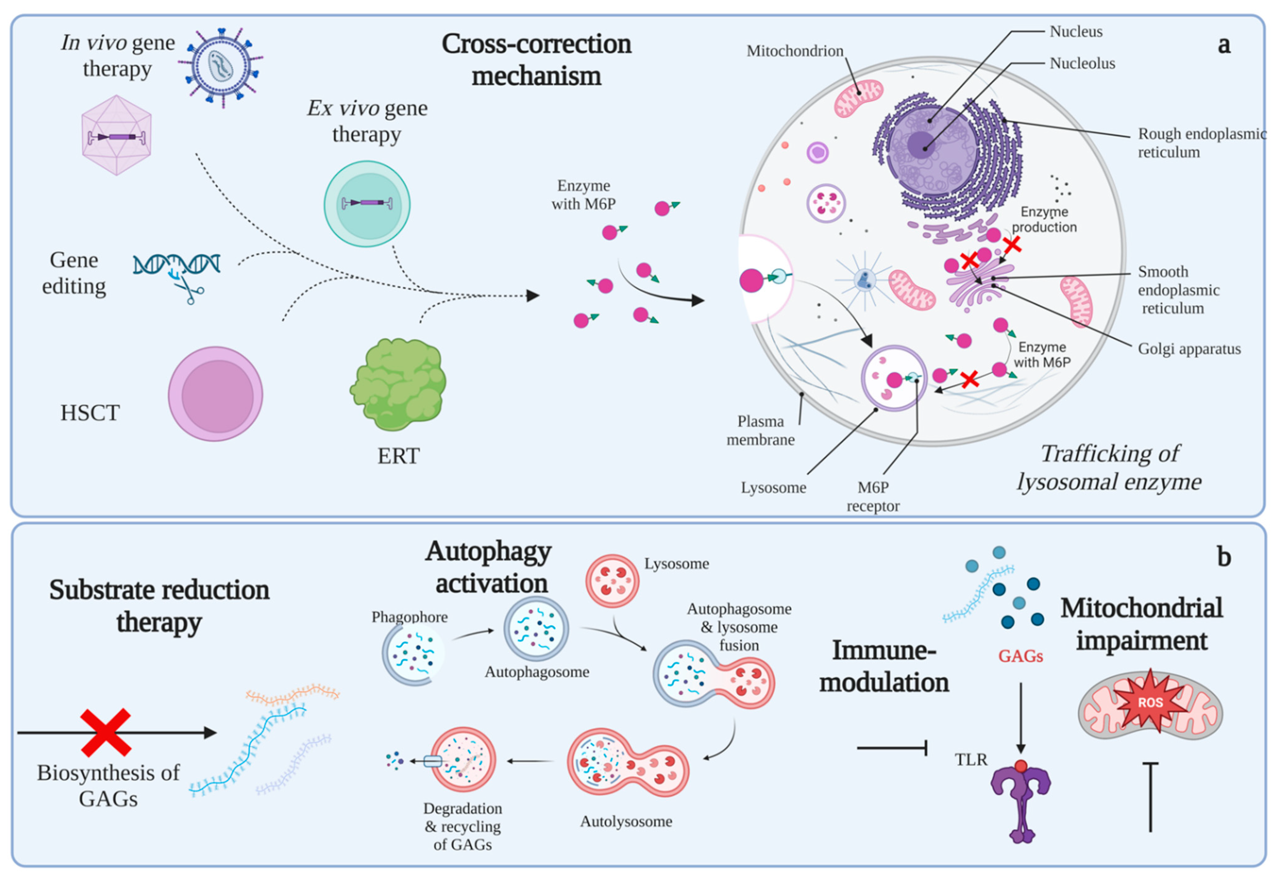
Figure 3. Potential therapies for MPS. Panel (a) shows possible treatments based on the cross-correction mechanism, including gene therapy, ERT, and HSCT. Panel (b) shows potential therapies not based on cross-correction mechanisms, such as substrate reduction therapy, autophagy activation, immune modulation, and mitochondrial impairment. Note that every organelle affected in MPS can be susceptible to intervention to recover cell homeostasis, together with current strategies to supply missing enzymes. This figure was created with BioRender.com.
Ijms 24 00477 g003
Table 1. GAGs structures.
GAGMonosaccharides ConstituentsChemical Structure
Keratan sulfateGalactose
N-acetylglucosamine
Ijms 24 00477 i001
Heparan sulfateGlucuronic acid
N-acetylglucosamine
Ijms 24 00477 i002
Chondroitin sulfateGlucuronic acid
N-acetylgalactosamine
Ijms 24 00477 i003
Dermatan sulfateIduronic acid
N-acetylgalactosamine
Ijms 24 00477 i004
HyaluronanGlucuronic acid
N-acetylglucosamine
Ijms 24 00477 i005
R1,2,3: -H or -SO3. R*: -H, -SO3, or -COCH3.
Table 2. Classification of MPS and major characteristics.
Type
(Eponym)
OMIMAffected GeneClinical CharacteristicsGAGs
MPS I
(Hurler)
607014
(Hurler)
IDUACorneal clouding, multiple dysostoses, organomegaly, cardiac disease, CNS impairmentDS and HS
607015
(Scheie)
Corneal clouding, joint stiffness, normal CNSDS and HS
607016
(Hurler-Scheie)
Intermediate phenotype between Hurler and Scheie phenotypesDS and HS
MPS II
(Hunter)
309900IDSMultiple dysostoses, organomegaly, CNS impairmentDS and HS
MPS IIIA
(Sanfilippo A)
252900SGSHCNS impairment, hyperactivity, mild somatic manifestationsHS
MPS IIIB
(Sanfilippo B)
252920NAGLULike MPS IIIAHS
MPS III C
(Sanfilippo C)
252930HGSNATLike MPS IIIAHS
MPS IIID
(Sanfilippo D)
252940GNSLike MPS IIIAHS
MPS IVA
(Morquio A)
253000GALNSShort stature, skeletal dysplasia, cornealKS and CS
MPS IVB
(Morquio B)
253010GLB1Like MPS IVAKS
MPS VI
(Maroteaux-Lamy)
253200ARSBMultiple dysostoses, corneal clouding, cardiac diseaseDS
MPS VII
(Sly)
253220GUSBMultiple dysostoses, hepatosplenomegaly, mild to severe central nervous system involvementHS, DS, and CS
MPS IX
(Natowicz)
601492HYAL1Short stature, soft-tissue massesHA
MPS X619698ARSKShort stature, coarse facial features, dysostosis multiplex, cardiac and ophthalmological abnormalities.DS
IDUA: α-L-iduronidase. IDS: Iduronate-2-sulfatase. SGSH: Heparan N-sulfatase (sulfamidase). NAGLU: α-N-acetylglucosaminidase. HGSNAT: Acetyl CoA: α-glucosamine N-acetyl transferase. GNS: N-acetylglucosamine-sulfate-6-sulfatase. GALNS: N-acetylgalactosamine-6-sulfate sulfatase. GLB1: β-galactosidase. ARSB: N-acetylgalactosamine-4-sulfatase (arylsulfatase B). GUSB: β -glucuronidase. HYAL1: Hyaluronidase. ARSK: Arylsulfatase K. DS: Dermatan sulfate. HS: Heparan sulfate. KS: Keratan sulfate. CS: Chondroitin sulfate. HA: Hyaluronic acid.
Disclaimer/Publisher’s Note: The statements, opinions and data contained in all publications are solely those of the individual author(s) and contributor(s) and not of MDPI and/or the editor(s). MDPI and/or the editor(s) disclaim responsibility for any injury to people or property resulting from any ideas, methods, instructions or products referred to in the content.