Article

Anti-Inflammatory Activities of an Anti-Histamine Drug, Loratadine, by Suppressing TAK1 in AP-1 Pathway

1
Department of Integrative Biotechnology, Sungkyunkwan University, Suwon 16419, Korea
2
Biomedical Institute for Convergence at SKKU (BICS), Sungkyunkwan University, Suwon 16419, Korea
*
Authors to whom correspondence should be addressed.
These authors contributed equally to this work.
Int. J. Mol. Sci. 2022, 23(7), 3986; https://doi.org/10.3390/ijms23073986
Submission received: 24 January 2022 / Revised: 1 April 2022 / Accepted: 1 April 2022 / Published: 3 April 2022
(This article belongs to the Special Issue Bioactive Compounds: Potential New Anti-inflammatory Drugs)

Abstract

:
Loratadine is an anti-histamine routinely used for treating allergies. However, recent findings have shown that Loratadine may also have anti-inflammatory functions, while their exact mechanisms have not yet been fully uncovered. In this paper, we investigated whether Loratadine can be utilized as an anti-inflammatory drug through a series of in vitro and in vivo experiments using a murine macrophage cell line and an acute gastritis mouse model. Loratadine was found to dramatically reduce the expression of pro-inflammatory genes, including MMP1, MMP3, and MMP9, and inhibit AP-1 transcriptional activation, as demonstrated by the luciferase assay. Therefore, we decided to further explore its role in the AP-1 signaling pathway. The expression of c-Jun and c-Fos, AP-1 subunits, was repressed by Loratadine and, correspondingly, the expression of p-JNK, p-MKK7, and p-TAK1 was also inhibited. In addition, Loratadine was able to reduce gastric bleeding in acute gastritis-induced mice; Western blotting using the stomach samples showed reduced p-c-Fos protein levels. Loratadine was shown to effectively suppress inflammation by specifically targeting TAK1 and suppressing consequent AP-1 signaling pathway activation and inflammatory cytokine production.

1. Introduction

Inflammation is an immunological response to the intrusion of foreign substances into the body or tissue repair failure. It protects our bodies from such harmful external or internal stimuli by recruiting immune cells, which can attach to or eliminate foreign elements or injured tissue [1,2,3]. However, when the inflammatory response outstrips the level of infection, it becomes a critical factor in driving other diseases, including diabetes, arthritis, and cancer [4,5,6,7]. Immune cells express pattern recognition receptors (PRRs), which are responsible for recognizing damage/pathogen-associated molecular patterns (DAMPs/PAMPs) expressed by pathogens [8,9,10]. Some of the most widely studied mammalian PRRs are toll-like receptors (TLRs), which are responsible for inflammatory pathway activation. Among the 10 TLRs discovered in humans, TLR4 is responsible for lipopolysaccharide (LPS) recognition to identify representative disease-causing PAMPs expressed by gram-negative bacteria [11,12,13]. Once an LPS is bound to TLR4, sequential intracellular inflammatory signaling processes are initiated by myeloid differentiation factor 88 (MyD88) and TIR-domain-containing adaptor-inducing interferon-β (TRIF), two adaptor proteins attached to the cytosolic toll-interleukin-1 receptor (TIR) domain of TLR4. Nuclear factor-kappa B (NF-κB), interferon regulatory factor 3 (IRF3), and activator protein-1 (AP-1) signaling pathways are the three representative intracellular inflammatory pathways activated by MyD88 and TRIF [14,15,16].
Among them, the AP-1 pathway is regulated by the transcription factor AP-1, which is responsible for regulating cell differentiation, cell cycle progression, and apoptosis. The AP-1 pathway consists of c-Fos and c-Jun families, and its transcriptional activation is mediated through the dimerization of these two subunits [17,18,19,20,21]. Originally located in the cytosol, c-Fos and c-Jun translocate into the nucleus and dimerize after they are activated by their upstream kinases, mitogen-activated protein kinases (MAPKs). MAPKs comprise a family of serine/threonine protein kinases, notably including extracellular signal-regulated kinase (ERK), c-Jun N-terminal kinase (JNK), and p38. MAPKs are activated by upstream MAPK kinases (MAPKKs), such as MAPK/ERK kinase (MEK) and MAPK kinase (MKK). Activation of MAPKKs is mediated by MAPKK kinases (MAPKKKs), notably including transforming growth factor-β-activated kinase 1 (TAK-1), mixed lineage kinase 3 (MLK3), and apoptosis signal-regulating kinase 1 (ASK1) [22,23,24,25]. The activation of the above kinases is regulated by sequential phosphorylation from upstream kinases to downstream kinases. Activation of AP-1 allows the transcription of inflammation-related enzymes, including matrix metallopeptidases (MMPs) and cyclooxygenase 2 (COX-2) [26,27,28,29,30].
Histamine is a primary mediator of the allergic response. It is kept in mast cells or basophils until the cells receive certain stimuli allowing it to be released out of the cells [31,32]. When histamine interacts with histamine receptors H1, H2, H3, or H4, an allergic reaction occurs. Among them, drugs targeting the H1 receptor are most commonly used for the treatment of allergies [33,34]. Meanwhile, Loratadine is an H1 histamine receptor antagonist; therefore, it is widely used as allergy medication. More importantly, it is within the second generation of non-sedating anti-histamine drugs [35,36].
However, there have been several reports presenting the H1 receptor-independent anti-inflammatory effects of H1 receptor antagonists by inhibiting NF-κB and AP-1 activities. Fexofenadine, a widely-used anti-histamine drug, was also shown to have inhibitory effects with COX-2 in an H1-receptor-independent manner [37,38]. In the meantime, various studies have revealed that Loratadine may be used as a novel anti-inflammatory drug [39,40,41]. Similarly, our previous study of Loratadine showed that it possesses inflammation-suppressive activities in the NF-κB signaling pathway [42].
Nevertheless, its inflammation-regulatory activities in another important inflammatory response pathway, the AP-1 signaling pathway, remain undetermined. Therefore, in this paper, we investigate the exact mechanism of the anti-inflammatory effects of Loratadine in the AP-1 signaling pathway.

2. Results

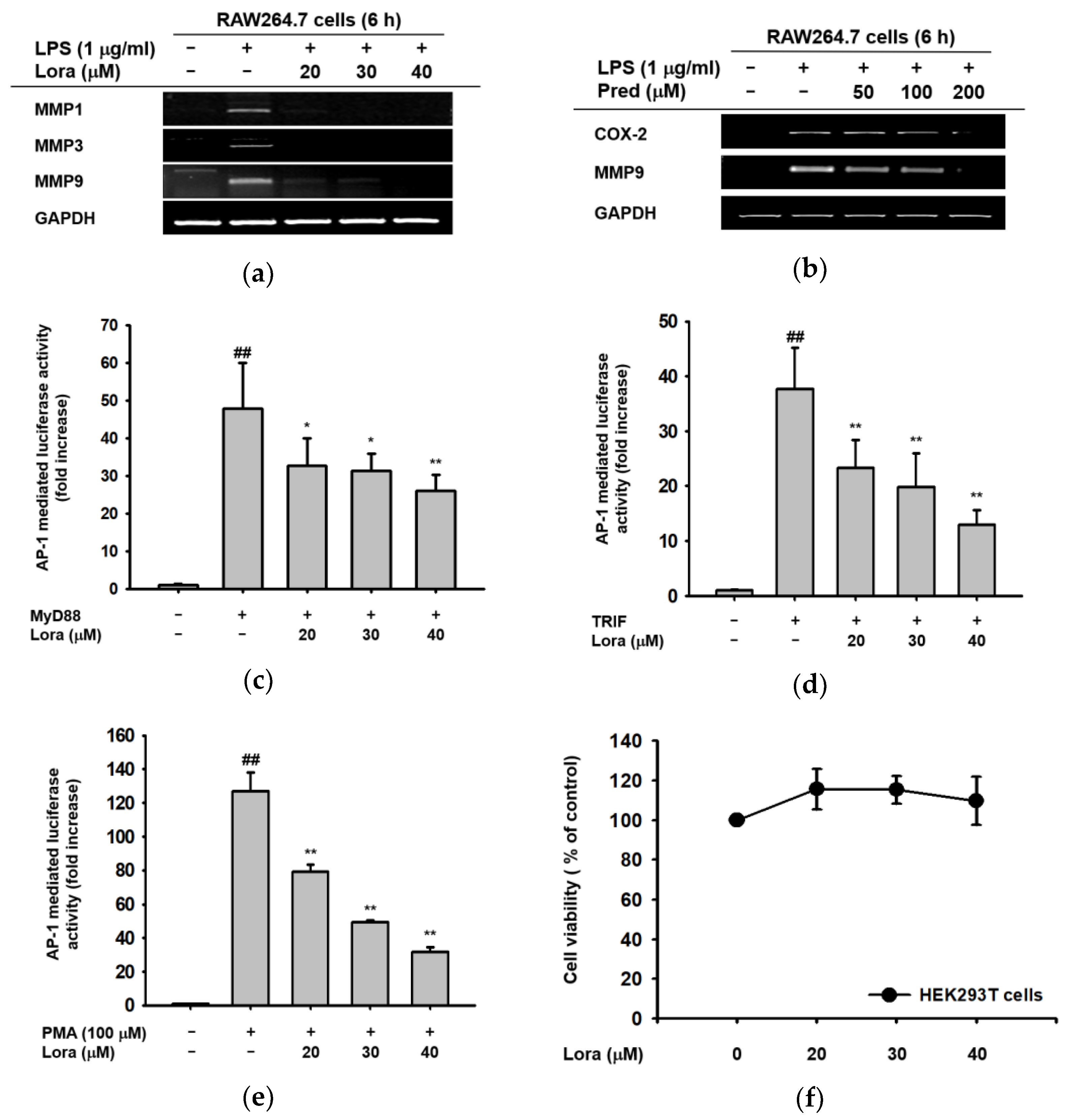
2.1. Anti-Inflammatory Effects of Loratadine Are Mediated by Its Transcriptional Regulation of Pro-Inflammatory Genes

First, in order to determine whether or not Loratadine has anti-inflammatory effects on the transcriptional level, we examined the mRNA expression levels of pro-inflammatory genes as well as the activation of transcription factor AP-1 under inflammation-triggered conditions. In RAW264.7 cells treated with LPS (1 μM), Loratadine (20–40 μM) effectively suppressed the mRNA expression of MMP1, MMP3, and MMP9 (Figure 1a), which are representative pro-inflammatory factors expressed via AP-1 pathway activation [43,44]. In addition, for further rationalization of such an effect, we also conducted the same experiment with prednisolone, an FDA-proved anti-allergic and anti-inflammatory drug [45], as a positive control. As shown in Figure 1b, prednisolone also suppressed the expression of COX-2 and MMP9 in a dose-dependent manner, supporting the validity of Loratadine’s anti-inflammatory effects indicated by MMP-reducing behavior. Therefore, we decided to further investigate whether such reduction in pro-inflammatory gene expression was a consequence of direct AP-1 inhibition by Loratadine.
In MyD88- and TRIF-overexpressed or PMA (100 μM)-treated HEK293T cells, AP-1-luc activation levels were significantly reduced in a concentration-dependent manner by Loratadine treatment (20–40 μM), as indicated in Figure 1c–e. The figures clearly indicate that AP-1 promoter activity, in other words, AP-1 transcription, is suppressed by Loratadine treatment. Furthermore, the cell viability assay showed that Loratadine treatment (20–40 μM) did not have any cytotoxicity at investigated concentrations (Figure 1f), implying that the reduction in AP-1 transcription was not due to cell death. Therefore, we decided to set the target concentration of Loratadine as 20–40 μM since these three concentrations showed a dose-dependent mode of action, and none of them showed cell toxicity. In conclusion, these data suggest that Loratadine suppresses pro-inflammatory factors via transcriptional regulation, and such effects are not outcomes of cell death.

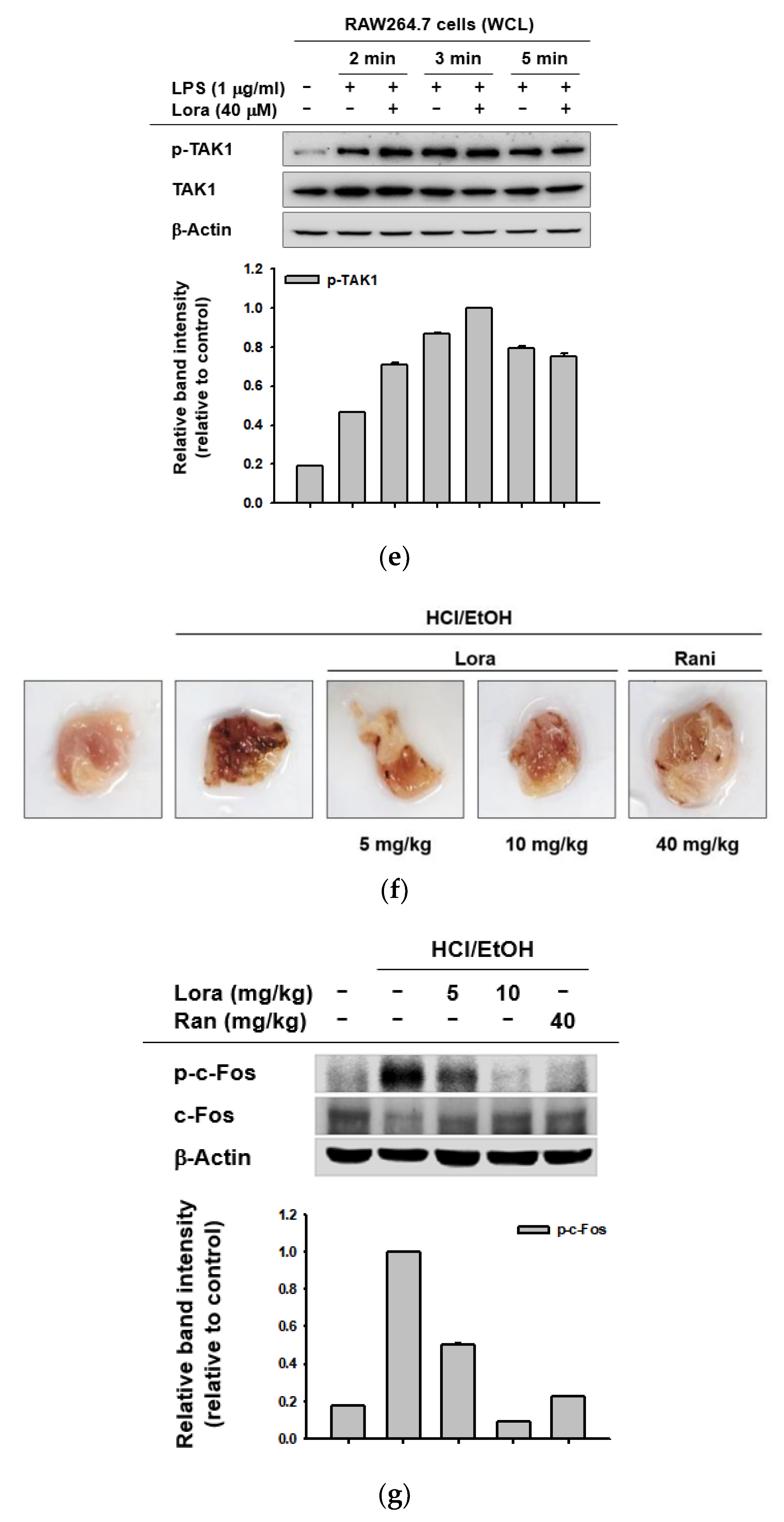
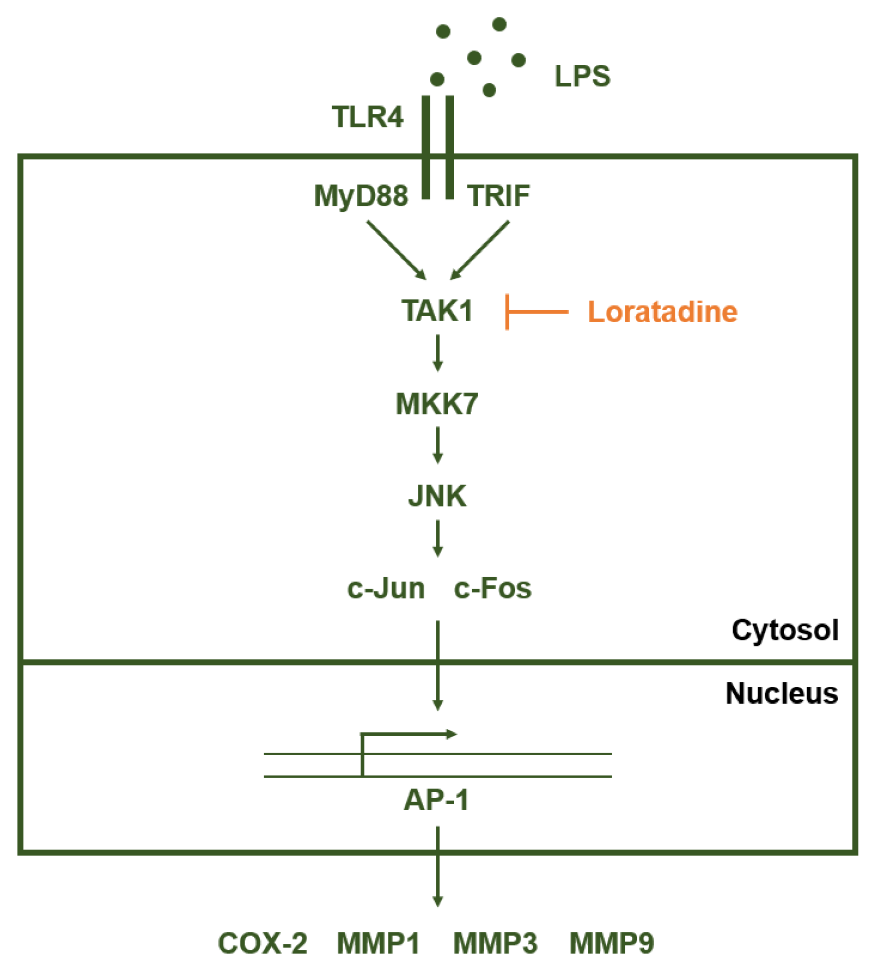
2.2. Loratadine Targets the AP-1 Signaling Pathway to Reduce Inflammatory Responses Both In Vitro and In Vivo

Since we discovered suppressive effects of Loratadine on AP-1-mediated gene expression and promoter activity of AP-1 (Figure 1), we further investigated whether such anti-inflammatory effects could also affect the constituent molecules of the intracellular AP-1 signaling pathway. Based on the concept that two subunits of AP-1, c-Jun, and c-Fos, translocate from the cytosol to the nucleus upon activation [46], the expression levels of c-Jun and c-Fos were determined within the nuclear fraction. In RAW264.7 cells, the nuclear expression levels of both c-Jun and c-Fos were clearly decreased following Loratadine treatment (40 μM) 15 min after LPS induction (Figure 2a). On the other hand, total cell lysate also showed decreased phosphorylation levels of c-Jun at 30 min and c-Fos at 30 and 45 min after LPS treatment (Figure 2b). It clearly indicates that Loratadine inhibits the activity of c-Jun and c-Fos. In addition, decreased levels of total c-Jun and c-Fos by Loratadine imply that Loratadine may also be involved in the degradation of the transcription factors.
Subsequently, we evaluated the cytosolic levels of AP-1 upstream kinases in LPS-induced RAW264.7 cells. Among the three different MAPKs, ERK, JNK, and p38, p-JNK (phosphorylated JNK) levels were clearly decreased 5 and 15 min post-inflammation induction by Loratadine treatment (40 μM), as shown in Figure 2c. Consistently, we also found the expression levels of p-MKK7 (phosphorylated MKK7), an upstream molecule of JNK, to be decreased by the same concentration of Loratadine 3 and 5 min post-induction (Figure 2d). Other MAPKKs, including MEK1/2 and MKK4, did not show such a clear reduction (Figure 2d). Finally, the expression levels of TAK1, which is a member of the MAPKKK family and an upstream activator of MKK7, were examined. LPS induction and Loratadine treatment (40 μM) were not found to reduce p-TAK1 (phosphorylated TAK1) levels (Figure 2e). Under inflammation-triggering conditions, a reduction in molecules downstream from TAK1 but not p-TAK1 itself indicates that TAK1, among other molecules of the AP-1 signaling pathway, might be the primary target of Loratadine.
In addition, in order to demonstrate the anti-inflammatory effects of Loratadine in vivo, we investigated a hydrochloride (HCl)-induced acute gastritis mouse model. Ethanol-induced gastritis leads to increased gastric acid secretion, which results in sequential activation of c-AMP, H2R, and H+/K+ ATPase, eventually causing gastric mucosal damage [47,48,49]. On the other hand, Ranitidine is known to be a potential H2R antagonist, inhibiting H2R-mediated gastric damage [50,51]. Therefore, we decided to use Ranitidine as a control drug in the experiment. HCl/EtOH-induced mice showed the typical phenotype of acute gastritis, bleeding of the stomach. However, Loratadine (5, 10 mg/kg) injection significantly reduced gastric bleeding, and its effect was comparable to Ranitidine (40 mg/kg) (Figure 2f). Moreover, phosphorylated c-Fos levels were dose-dependently decreased in stomach samples from Loratadine-treated mice compared to those of the vehicle-treated group (Figure 2g). These results suggest that the anti-inflammatory effects of Loratadine are mediated by its suppressive activity in the AP-1 signaling pathway.

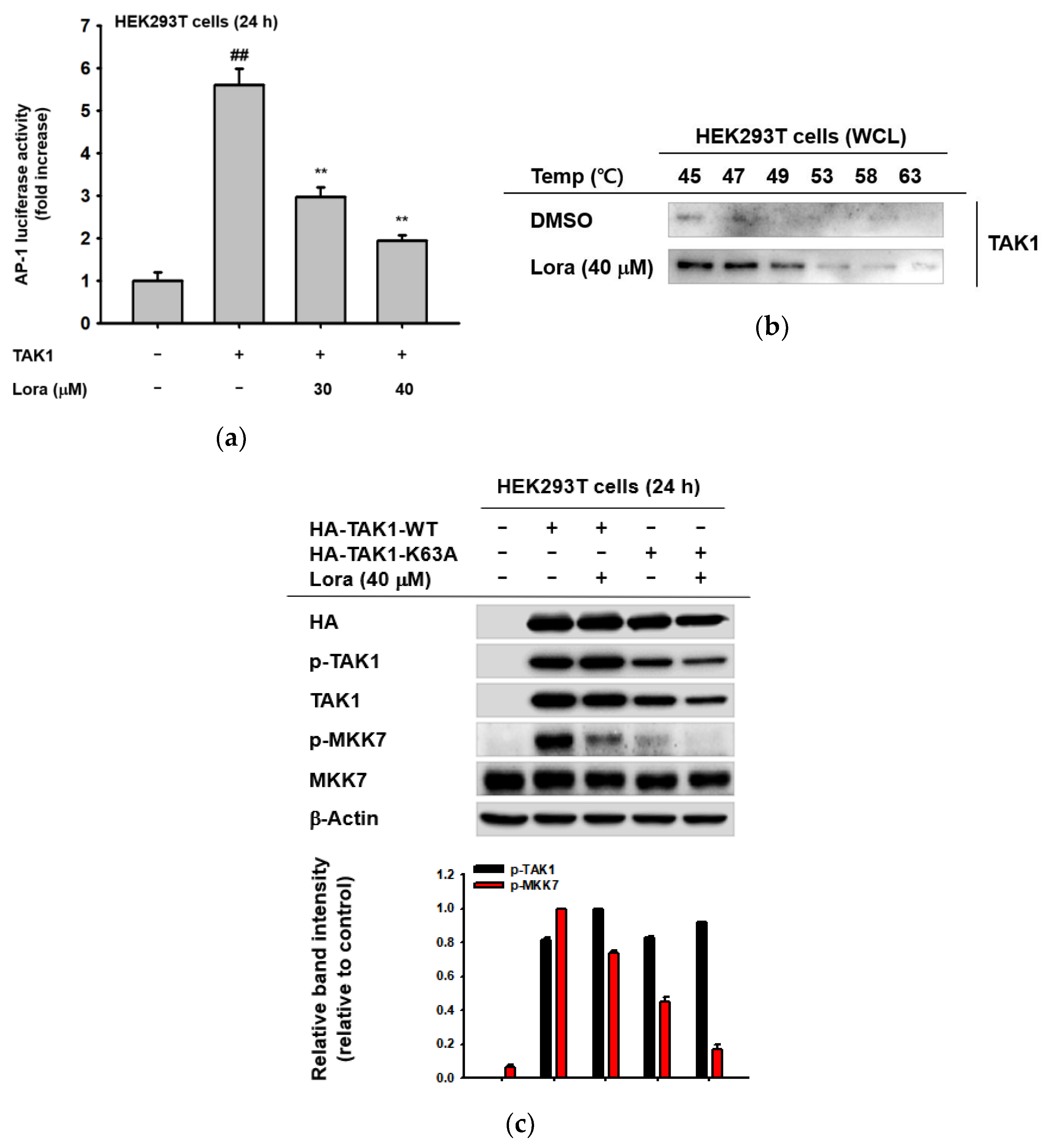
2.3. TAK1 Is the Prime Target Molecule of Loratadine

In order to determine whether TAK1 is the primary target of Loratadine, we further examined the drug’s anti-inflammatory activity in TAK1-overexpressing cells. Overexpression of TAK1 can induce and activate its downstream molecules, resulting in levels of inflammation similar to cells treated with LPS [52,53]. First, we decided to check the expression of pro-inflammatory cytokines after TAK1 overexpression. When TAK1 was overexpressed in RAW264.7 cells, COX-2 expression was significantly reduced following Loratadine treatment (40 μM) (data not shown). Furthermore, TAK1 overexpression in HEK293T cells induced AP-1 promoter activation; however, it was dose-dependently suppressed by Loratadine (30 and 40 μM) (Figure 3a). These results suggest that TAK1-induced inflammation can be suppressed by Loratadine treatment, implying that Loratadine may directly interact with TAK1. Therefore, we performed the cellular thermal shift assay (CETSA) to find out whether or not Loratadine directly targets and binds TAK1 to suppress the AP-1 signaling pathway during inflammation. Compared to the unbound state, the thermal stability of TAK1 is higher when it is bound with other molecules. Within 45 to 63 degrees Celsius, the TAK1 band intensity of DMSO-treated cells was clearly lower than that of Loratadine (40 μM)-treated cells, showing a reducing pattern as the temperature rose (Figure 3b). This indicates that Loratadine and TAK1 interact and may directly bind. In addition, we decided to further investigate the target binding site of TAK1 for Loratadine. TAK1 has an ATP binding site, K63, which enables it to transform into an active state upon ATP binding [54,55]. In order to find out whether K63 of TAK1 is the target binding site for Loratadine, we point-mutated K63 into A63. If K63 was the binding site, mutated TAK1 with A63 would show no change in expression of both p-TAK1 and p-MKK7. However, we found that there was still a reducing pattern of p-MKK7 in K63A TAK1-transfected HEK293T cells after Loratadine treatment (40 μM) (Figure 3c), indicating that Loratadine does not appear to bind to K63. These results suggest that Loratadine targets TAK1 within the AP-1 signaling pathway to exert anti-inflammatory effects, and the ATP-binding site of TAK1 is not the target site for Loratadine.

3. Discussion

Inflammation is a fundamental process for maintaining immunologic homeostasis; however, an excessive inflammatory response can lead to severe disorders, including autoimmune diseases and cancer [56,57,58]. In this paper, we demonstrate the anti-inflammatory activity of a commercialized anti-histamine drug, Loratadine, which specifically inhibits TAK1 activation and, therefore, suppresses the AP-1 signaling pathway and consequent MMP production. We have previously reported that Loratadine can reduce several pro-inflammatory cytokines, including iNOS, IL-1β, TNF-α, IL-6, and COX-2, by suppressing the NF-κB signaling pathway [42]. These findings suggest that Loratadine is able to counteract exaggerated inflammatory responses, thereby maintaining immunological balance while preventing the uncontrolled progression of inflammation.
Recently, drugs targeting the AP-1 pathway have emerged as next-generation treatments for both cancer and autoimmune disease. The AP-1 pathway governs major inflammatory responses by regulating cell differentiation, proliferation, apoptosis, angiogenesis, and invasion [59,60,61,62,63]. Meanwhile, AP-1 is composed of different combinations of the Fos and Jun families. The Fos family includes c-Fos, FosB, and Fra-1 and -2, whereas the Jun family includes c-Jun, JunB, and JunD [61,64,65]. Each of them controls different cellular responses by up- or down-regulating related gene products. C-Fos and c-Jun up-regulate factors that are responsible for proliferation, such as EGFR, GM-CSF, and cyclin D1 [66,67,68,69]. On the other hand, c-Jun also down-regulates proteins that inhibit proliferation and stimulate apoptosis, such as p53 and FASL [70,71,72,73]. In addition, c-Fos can up-regulate genes that promote cellular invasions, such as MMP1, MMP3, and CD44 [74,75,76,77]. Therefore, AP-1 has been a foremost target for cancer and inflammatory disease treatment. For instance, T-5224, a selective inhibitor of c-Fos/AP-1, has been reported to have preventive effects on cartilage destruction and osteophyte formation [78]. Furthermore, a selective small-molecule inhibitor of c-Fos/AP-1 has been shown to resolve arthritis in pre-clinical models [79].
TAK1, a constituent molecule of the AP-1 signaling pathway, is also a chief regulator of cell survival and apoptosis, which has made itself a promising target for a wide set of diseases. For instance, Takinib, a TAK1-specific inhibitor, was reported to mediate cell death in rheumatoid arthritis and breast cancer by suppressing TNF-α production [80,81]. Other TAK1 inhibitors, such as LYTAK1 and (5Z)-7-Oxozeaenol, also showed suppressive effects on various types of cancer, including ovarian cancer, cervical cancer, and neuroblastoma [82,83,84,85].
Loratadine is one of the most frequently used anti-allergic drugs, whose safety and efficacy have already been approved by the FDA. Together with the findings that outline AP-1 and TAK1 as being promising drug targets, our paper suggests the potential of Loratadine as an operative anti-inflammatory agent, with known and manageable side effects and proven safety and stability. Furthermore, such findings will also contribute to future research on drug repositioning by providing proper methods and workflows for discovering new effects of pre-existing commercial drugs on different diseases.

4. Materials and Methods

4.1. Materials

Loratadine was purchased from Tokyo Chemical Industry Co., LTD. (Tokyo, Japan). Carboxymethylcellulose (CMC), dimethylsulfoxide (DMSO), L-NAME, lipopolysaccharide, 3-(4,5-dimethylthiazol-2yl)-2,5-diphenyltetrazolium bromide (MTT), polyethyleneimine (PEI), Ranitidine, and stillen were obtained from Sigma Aldrich (St. Louis, MO, USA). DMEM, fetal bovine serum (FBS), penicillin/streptomycin, phosphate-buffered saline (PBS), RPMI 1640, trypsin, and TRIzol were acquired from GIBCO (Grand Island, NY, USA). HEK293T (CRL-1573) and RAW264.7 cells (CRL-2278) were products from American Type Culture Collection (Rockville, MD, USA). Luciferase constructs with AP-1 binding sites and CFP-TRIF, FLAG-MYD88, pcDNA, and Tag-2 constructs were used as previously reported [86]. Real-time and semiquantitative reverse transcriptase-polymerase chain reaction primers were bought from Macrogen Inc. (Seoul, Korea). All other chemicals were purchased from Sigma. Total and phospho antibodies for the MAPK family were obtained from Cell Signaling Technology (Beverly, MA, USA) and Santa Cruz Biotechnology (Santa Cruz, CA, USA).

4.2. Compound Preparation, Animals, and Cell Culture

First, 40 mM of Loratadine stock solution was prepared using DMSO as a carrier. The stock solution was further diluted and treated at final concentrations of 20–40 μM using cell culture media for in vitro experiments. For in vivo procedures, 5–10 mg/kg using 0.5% sodium carboxymethyl cellulose (CMC) solution as a carrier. Male ICR mice (5–6 weeks old) were purchased from Daehan Biolink (Osong, Korea) and were housed in autoclaved plastic cages under standard housing conditions. All studies were conducted according to the guidelines of the Institutional Animal Care and Use Committee at Sungkyunkwan University (Suwon, Korea; approval ID: 2019-12-03-1). Murine macrophage RAW264.7 cells and human embryonic kidney HEK293T cells were cultured in RPMI1640 and DMEM media, respectively, containing 5 or 10% inactivated FBS, penicillin (100 IU/mL), and streptomycin (100 μg/mL). Cells were cultured in an incubator that maintained a 5% CO2 level and a temperature of 37 °C [87,88].

4.3. Gastritis Mouse Model

Negative and positive control groups, 5 ICR mice per group, received oral administration of 0.5% CMC solution, while the two Loratadine-treated groups were orally injected with 5 mg/kg or 10 mg/kg Loratadine solution. The control group was injected with 40 mg/kg Ranitidine. The injection was performed twice a day for 3 days. For acute gastritis induction, mice received orally administered 150 mM hydrochloric acid (HCl) in 60% ethanol (EtOH) 1 h before sacrifice. The stomach of each mouse was extracted to measure the area of inflammatory lesions. Quantification of the lesion area was performed using ImageJ [88].

4.4. mRNA Expression Analysis Using Reverse-Transcription Polymerase Chain Reaction

RAW264.7 cells (1 × 10⁶ cells/mL) were seeded in 12-well culture plates and incubated for 24 h. Loratadine (20–40 μM) or DMSO (as a control) was administered to cells 30 min before LPS induction (1 μg/mL). After 6 h of incubation, cells were harvested using cold PBS, and total RNA was isolated from the cells using TRIzol reagent following the manufacturer’s guidelines. In order to increase sample stability, cDNA was synthesized from the extracted mRNA using Thermo Fisher Scientific’s cDNA synthesis kit (Waltham, MA, USA); corresponding primers of COX-2, MMP1, MMP3, and MMP9 were applied for general PCR amplification. mRNA expressions of samples were analyzed by agarose gel electrophoresis; PCR band intensities were quantified using ImageJ. Primer sequences are listed in Table 1 [89].

4.5. Luciferase Reporter Assay

HEK293T cells (1.2 × 10⁵ cells/mL) were seeded in 24-well culture plates and incubated for 24 h. Target gene constructs (Tag2-MyD88, Tag2, CFP-TRIF, CFP, HA-TAK1, HA, AP-1-luc, and β-gal) were then transfected into the cells using PEI. After 24, 40, or 48 h incubation, the original culture media was completely removed, and Loratadine (20–40 μM) or DMSO were introduced to the cells with fresh media. After incubating for 3 h, the media was completely removed, and the cells were treated with luciferase lysis buffer. The plate was frozen at −70 °C for 3 h. Luciferin and β-gal assay buffers were added for luciferase and β-gal analysis, respectively. Luciferase activity was assessed by detecting luminescence levels, and β-gal activity was measured through OD values at 405 nm [90].

4.6. Cell Viability Assay

HEK293T cells (1 × 10⁵ cells/mL) were seeded in 96-well culture plates and incubated for 24 h. Loratadine (20–40 μM) or DMSO (as a control) were administered, and the cells were incubated for 24 h. MTT solution was added to the cell-containing medium, which underwent incubation for 3 h. MTT stopping solution was added thereafter. After 24 h of incubation, the cell viability was assessed through OD values at 570 nm [91].

4.7. Total Cell Lysate Preparation

RAW264.7 cells (2.5 × 10⁶ cells/mL) were seeded in 3 cm culture plates and incubated for 24 h. The cells were pre-treated with Loratadine (20–40 μM) or DMSO (as a control) for 30 min prior to inflammation induction. The cells were then treated with LPS (1 μg/mL) for specific periods (2–60 min), and the cells were then harvested using cold PBS. Total cell lysates were prepared using cold cell lysis buffer (20 mM of Tris-HCl, pH: 7.4; including 2 mM of EDTA, 2 mM of EGTA, 50 mM glycerol phosphate, 1 mM of DTT, 2 μg/mL of aprotinin, 2 μg/mL of leupeptin, 1 μg/mL of pepstatin, 50 μM of PMSF, 1 mM of benzamide, 2% Triton X-100, 10% glycerol, 0.1 mM of sodium vanadate, 1.6 mM of pervanadate, and 20 mM of NaF). Cell debris was removed by centrifugation at 12,000 RPM for 1 min. Total cell lysates were kept at −70 °C until use [87,92].

4.8. Western Blotting

Protein concentrations of total cell lysate samples were quantified for size-dependent separation. Proteins were separated by SDS-polyacrylamide gel electrophoresis in SDS-PAGE running buffer (10% SDS, Tris-base, glycine). Subsequently, proteins were transferred to PVDF membranes by electrophoresis in a transfer buffer solution (10% SDS, Tris-base, glycine, methanol). Membrane blocking was performed by rolling the tubes on a roller with 5% BSA solution applied for 1 h at room temperature. Primary antibodies specific to the target proteins (total or phosphorylated c-Jun, c-Fos, Lamin A/C, ERK, JNK, p38, MEK1/2, MKK4, MKK7, ASK, MLK3, TAK-1, and β-actin) were added, and the solutions were incubated with 5% BSA solution for 1–2 h at room temperature or overnight at 4 °C. After washing primary antibodies 3 times with 0.1% TBST buffer (Tris-base, NaCl; 0.1% Tween 20; pH: 7.6), specific HRP-linked secondary antibodies (anti-rabbit or anti-mouse) were added, and the solutions were incubated with 5% BSA solution for 1–2 h at room temperature or overnight at 4 °C. ECL reagent was applied for protein visualization, and signals were detected by ChemiDoc XRS (Bio-Rad, Hercules, CA, USA.). The relative intensity of each band was analyzed through the ImageJ program [93].

4.9. Gene Overexpression

HEK293T cells (2 × 10⁵ cells/mL) were seeded in 12-well culture plates and incubated for 24 h. Target gene construct and its carrier vector (HA-TAK1 and HA) were then transfected into cells using PEI. After 24 h, the original media was completely removed, and Loratadine (40 μM) was introduced to the cells in addition to fresh media. After further incubation for 3 h, media containing Loratadine was completely removed, the cells were harvested, and the total cell lysates were prepared. Protein expression levels were assessed by Western blotting using ECL reagents [94].

4.10. Cellular Thermal Shift Assay

HEK293T cells (1.2 × 10⁶ cells/mL) were seeded in 6 cm culture plates and incubated for 24 h. The target gene construct (HA-TAK1) was transfected into cells using PEI. After 24 h of incubation, Loratadine (40 μM) was introduced to the cells after the original media was replaced by fresh media, and the cells were thereafter incubated for 1 h. After 24 h of incubation, the cells were counted, and each group was adjusted to have an identical number of cells. Cells were lysed in a real-time PCR machine (Bio-Rad) within a temperature range of 45 to 63 °C. After a temperature shift, the protein level of TAK1 was analyzed by Western blotting [95].

5. Conclusions

Loratadine exhibits anti-inflammatory activity in murine macrophage cells by specifically inhibiting the AP-1 signaling pathway. During this process, Loratadine targets TAK1 to suppress AP-1 transcriptional activity, thereby reducing pro-inflammatory cytokine expression, including that of MMP1, MMP3, and MMP9. Loratadine can also exert such inflammation-suppressing effects in an acute gastritis mouse model, implying its potential as an effective anti-inflammatory drug (Figure 4).

Author Contributions

J.J., S.T.H., H.G.K. and J.Y.C. conceived and designed the experiments; J.J., S.T.H., H.G.K. and J.Y.C. analyzed the data; J.J., S.T.H., J.W.K. and H.P.L. performed the experiments; J.J., S.T.H., H.G.K. and J.Y.C. wrote the manuscript. All authors have read and agreed to the published version of the manuscript.

Funding

This research was supported by grants from the Basic Science Research Program through the National Research Foundation of Korea (NRF), funded by the Korean Ministry of Education (Grant no.: 2017R1A6A1A03015642).

Institutional Review Board Statement

The in vivo experiments were performed in agreement with the guidelines of the Institutional Animal Care and Use Committee of Sungkyunkwan University (Suwon, Korea; approval ID: SKKUIACUC2021-03-07-2).

Informed Consent Statement

Not applicable.

Data Availability Statement

The data used to support the findings of this study are available from the corresponding authors upon request.

Conflicts of Interest

The authors have no conflicts of interest to declare.

References

  1. Hawiger, J. Innate immunity and inflammation: A transcriptional paradigm. Immunol. Res. 2001, 23, 99–110. [Google Scholar] [CrossRef]
  2. Medzhitov, R. Origin and physiological roles of inflammation. Nature 2008, 454, 428–435. [Google Scholar] [CrossRef]
  3. Larsen, G.L.; Henson, P.M. Mediators of inflammation. Annu. Rev. Immunol. 1983, 1, 335–359. [Google Scholar] [CrossRef]
  4. Gupta, S.C.; Kunnumakkara, A.B.; Aggarwal, S.; Aggarwal, B.B. Inflammation, a double-edge sword for cancer and other age-related diseases. Front. Immunol. 2018, 9, 2160. [Google Scholar] [CrossRef]
  5. Chen, L.; Deng, H.; Cui, H.; Fang, J.; Zuo, Z.; Deng, J.; Li, Y.; Wang, X.; Zhao, L. Inflammatory responses and inflammation-associated diseases in organs. Oncotarget 2018, 9, 7204–7218. [Google Scholar] [CrossRef] [Green Version]
  6. Kotas, M.E.; Medzhitov, R. Homeostasis, inflammation, and disease susceptibility. Cell 2015, 160, 816–827. [Google Scholar] [CrossRef] [Green Version]
  7. Robinson, M.; Harmon, C.; O’Farrelly, C. Liver immunology and its role in inflammation and homeostasis. Cell. Mol. Immunol. 2016, 13, 267–276. [Google Scholar] [CrossRef] [Green Version]
  8. Ratan, Z.A.; Youn, S.H.; Kwak, Y.-S.; Han, C.-K.; Haidere, M.F.; Kim, J.K.; Min, H.; Jung, Y.-J.; Hosseinzadeh, H.; Hyun, S.H.; et al. Adaptogenic effects of Panax ginseng on modulation of immune functions. J. Ginseng Res. 2021, 45, 32–40. [Google Scholar] [CrossRef]
  9. Tang, D.; Kang, R.; Coyne, C.B.; Zeh, H.J.; Lotze, M.T. PAMPs and DAMPs: Signals that spur autophagy and immunity. Immunol. Rev. 2012, 249, 158–175. [Google Scholar] [CrossRef]
  10. Zindel, J.; Kubes, P. DAMPs, PAMPs, and LAMPs in immunity and sterile inflammation. Annu. Rev. Pathol. Mech. Dis. 2020, 15, 493–518. [Google Scholar] [CrossRef] [Green Version]
  11. Kim, J.H.; Yi, Y.-S.; Kim, M.-Y.; Cho, J.Y. Role of ginsenosides, the main active components of Panax ginseng, in inflammatory responses and diseases. J. Ginseng Res. 2017, 41, 435–443. [Google Scholar] [CrossRef] [Green Version]
  12. Ratan, Z.A.; Haidere, M.F.; Hong, Y.H.; Park, S.H.; Lee, J.-O.; Lee, J.; Cho, J.Y. Pharmacological potential of ginseng and its major component ginsenosides. J. Ginseng Res. 2021, 45, 199–210. [Google Scholar] [CrossRef]
  13. Yang, W.S.; Kim, H.G.; Kim, E.; Han, S.Y.; Aziz, N.; Yi, Y.-S.; Kim, S.; Lee, Y.; Yoo, B.C.; Han, J.-W.; et al. Isoprenylcysteine carboxyl methyltransferase and its substrate Ras are critical players regulating TLR-mediated inflammatory responses. Cells 2020, 9, 1216. [Google Scholar] [CrossRef]
  14. Yamamoto, M.; Sato, S.; Hemmi, H.; Hoshino, K.; Kaisho, T.; Sanjo, H.; Takeuchi, O.; Sugiyama, M.; Okabe, M.; Takeda, K.; et al. Role of adaptor TRIF in the MyD88-independent Toll-like receptor signaling pathway. Science 2003, 301, 640–643. [Google Scholar] [CrossRef]
  15. Kim, H.G.; Kim, J.H.; Yang, W.S.; Kim, E.; Jeong, D.; Park, J.G.; Aziz, N.; Kim, S.; Parameswaran, N.; Choy, J.Y. Syk-MyD88 axis Is a critical determinant of inflammatory-response in activated macrophages. Front. Immunol. 2021, 12, 767366. [Google Scholar] [CrossRef]
  16. Fitzgerald, K.; Rowe, D.C.; Barnes, B.J.; Caffrey, D.R.; Visintin, A.; Latz, E.; Monks, B.; Pitha, P.M.; Golenbock, D.T. LPS-TLR4 signaling to IRF-3/7 and NF-κB involves the Toll adapters TRAM and TRIF. J. Exp. Med. 2003, 198, 1043–1055. [Google Scholar] [CrossRef] [Green Version]
  17. Xue, Q.; He, N.; Wang, Z.; Fu, X.; Aung, L.H.H.; Liu, Y.; Li, M.; Cho, J.Y.; Yang, Y.; Yu, T. Functional roles and mechanisms of ginsenosides from Panax ginseng in atherosclerosis. J. Ginseng Res. 2021, 45, 22–31. [Google Scholar] [CrossRef]
  18. Ha, A.T.; Rahmawati, L.; You, L.; Hossain, M.A.; Kim, J.-H.; Cho, J.Y. Anti-inflammatory, antioxidant, moisturizing, and antimelanogenesis effects of quercetin 3-O-β-D-glucuronide in human keratinocytes and melanoma cells via activation of NF-κB and AP-1 pathways. Int. J. Mol. Sci. 2021, 23, 433. [Google Scholar] [CrossRef]
  19. Kim, E.; Ahuja, A.; Kim, M.-Y.; Cho, J.Y. DNA or protein methylation-dependent regulation of activator protein-1 function. Cells 2021, 10, 461. [Google Scholar] [CrossRef]
  20. Schonthaler, H.B.; Guinea-Viniegra, J.; Wagner, E.F. Targeting inflammation by modulating the Jun/AP-1 pathway. Ann. Rheum. 2011, 70, i109–i112. [Google Scholar] [CrossRef]
  21. Liu, X.; Yin, S.; Chen, Y.; Wu, Y.; Zheng, W.; Dong, H.; Bai, Y.; Qin, Y.; Li, J.; Feng, S.J.; et al. LPS-induced proinflammatory cytokine expression in human airway epithelial cells and macrophages via NF-κB, STAT3 or AP-1 activation. Mol. Med. Rep. 2018, 17, 5484–5491. [Google Scholar] [CrossRef] [Green Version]
  22. Whitmarsh, A.J.; Davis, R.J. Transcription factor AP-1 regulation by mitogen-activated protein kinase signal transduction pathways. J. Mol. Med. 1996, 74, 589–607. [Google Scholar] [CrossRef]
  23. Silvers, A.L.; Bachelor, M.A.; Bowden, G.T. The Role of JNK and p38 MAPK Activities in UVA-induced signaling pathways leading to AP-1 activation and c-Fos expression. Neoplasia 2003, 5, 319–329. [Google Scholar] [CrossRef] [Green Version]
  24. Davies, M.; Robinson, M.; Smith, E.; Huntley, S.; Prime, S.; Paterson, I. Induction of an epithelial to mesenchymal transition in human immortal and malignant keratinocytes by TGF-β1 involves MAPK, Smad and AP-1 signalling pathways. J. Cell. Biochem. 2005, 95, 918–931. [Google Scholar] [CrossRef]
  25. Morse, D.; Pischke, S.E.; Zhou, Z.; Davis, R.J.; Flavell, R.A.; Loop, T.; Otterbein, S.L.; Otterbein, L.E.; Choi, A.M.K. Suppression of inflammatory cytokine production by carbon monoxide involves the JNK pathway and AP-1. J. Biol. Chem. 2003, 278, 36993–36998. [Google Scholar] [CrossRef] [Green Version]
  26. Kajanne, R.; Miettinen, P.; Mehlem, A.; Leivonen, S.-K.; Birrer, M.; Foschi, M.; Kähäri, V.-M.; Leppä, S. EGF-R regulates MMP function in fibroblasts through MAPK and AP-1 pathways. J. Cell. Physiol. 2007, 212, 489–497. [Google Scholar] [CrossRef]
  27. Bergman, M.R.; Cheng, S.; Honbo, N.; Piacentini, L.; Karliner, J.S.; Lovett, D.H. A functional activating protein 1 (AP-1) site regulates matrix metalloproteinase 2 (MMP-2) transcription by cardiac cells through interactions with JunB-Fra1 and JunB-FosB heterodimers. Biochem. J. 2003, 369, 485–496. [Google Scholar] [CrossRef] [Green Version]
  28. Barchowsky, A.; Frleta, D.; Vincenti, M.P. Integration of the NF-κB and mitogen-activated protein kinase/AP-1 pathways at the collagenase-1 promoter: Divergence of IL-1 and TNF-dependent signal transduction in rabbit primary synovial fibroblasts. Cytokine 2000, 12, 1469–1479. [Google Scholar] [CrossRef]
  29. Subbaramaiah, K.; Norton, L.; Gerald, W.; Dannenberg, A.J. Cyclooxygenase-2 is overexpressed in HER-2/neu-positive breast cancer: Evidence for involvement of AP-1 and PEA3. J. Biol. Chem. 2002, 277, 18649–18657. [Google Scholar] [CrossRef] [Green Version]
  30. Cho, J.-W.; Park, K.; Kweon, G.R.; Jang, B.-C.; Baek, W.-K.; Suh, M.-H.; Kim, C.-W.; Lee, K.-S.; Suh, S.-I. Curcumin inhibits the expression of COX-2 in UVB-irradiated human keratinocytes (HaCaT) by inhibiting activation of AP-1: p38 MAP kinase and JNK as potential upstream targets. Exp. Mol. Med. 2005, 37, 186–192. [Google Scholar] [CrossRef] [Green Version]
  31. Arrang, J.-M.; Garbarg, M.; Schwartz, J.-C. Auto-inhibition of brain histamine release mediated by a novel class (H3) of histamine receptor. Nature 1983, 302, 832–837. [Google Scholar] [CrossRef]
  32. Lichtenstein, L.M.; Gillespie, E. Inhibition of histamine release by histamine controlled by H2 receptor. Nature 1973, 244, 278–288. [Google Scholar] [CrossRef]
  33. Thangam, E.B.; Jemima, E.A.; Singh, H.; Baig, M.S.; Khan, M.; Mathias, C.B.; Church, M.K.; Saluja, R. The role of histamine and histamine receptors in mast cell-mediated allergy and inflammation: The hunt for new therapeutic targets. Front. Immunol. 2018, 9, 1873. [Google Scholar] [CrossRef] [Green Version]
  34. Branco, A.C.C.C.; Yoshikawa, F.S.Y.; Pietrobon, A.J.; Sato, M.N. Role of histamine in modulating the immune response and inflammation. Mediat. Inflamm. 2018, 2018, 9524075. [Google Scholar] [CrossRef]
  35. Kreutner, W.; Chapman, R.W.; Gulbenkian, A.; Siegel, M.I. Antiallergic activity of loratadine, a non-sedating antihistamine. Allergy 1987, 42, 57–63. [Google Scholar] [CrossRef]
  36. Kay, G.; Harris, A.G. Loratadine: A non-sedating antihistamine. Review of its effects on cognition, psychomotor performance, mood and sedation. Clin. Exp. Allergy 1999, 29, 147–150. [Google Scholar] [CrossRef]
  37. Roumestan, C.; Henriquet, C.; Gougat, C.; Michel, A.; Bichon, F.; Portet, K.; Jaffuel, D.; Mathieu, M. Histamine H1-receptor antagonists inhibit nuclear factor-kappaB and activator protein-1 activities via H1-receptor-dependent and-independent mechanisms. Clin. Exp. Allergy 2008, 38, 947–956. [Google Scholar] [CrossRef]
  38. Juergens, U.R.; Gillissen, A.; Uen, S.; Racké, K.; Stöber, M.; Darlath, W.; Vetter, H. New evidence of H1-receptor independent COX-2 inhibition by fexofenadine HCl in vitro. Pharmacology 2006, 78, 129–135. [Google Scholar] [CrossRef]
  39. Zhou, Y.; Gao, C.; Wang, H.; Liu, L.; Huang, Z.; Fa, X. Histamine H1 type receptor antagonist loratadine ameliorates oxidized LDL induced endothelial dysfunction. Biomed. Pharmacother. 2018, 106, 1448–1453. [Google Scholar] [CrossRef]
  40. Mitrea, D.-R.; Clichici, S.; Filip, A.; Olteanu, D.; Bâldea, I.; Moldovan, R.; Decea, N.; Hoteiuc, O.-A. Resveratrol and loratadine effects on oxidative stress induced by experimental inflammation. Studia UBB Chemia 2017, 62, 89–100. [Google Scholar] [CrossRef]
  41. Traidl-Hoffmann, C.; Münster, I.; Ring, J.; Behrendt, H. Impact of desloratadine and loratadine on the crosstalk between human keratinocytes and leukocytes: Implications for anti-inflammatory activity of antihistamines. Int. Arch. Allergy Immunol. 2006, 140, 315–320. [Google Scholar] [CrossRef]
  42. Hunto, S.T.; Kim, H.G.; Baek, K.-S.; Jeong, D.; Kim, E.; Kim, J.H.; Cho, J.Y. Loratadine, an antihistamine drug, exhibits anti-inflammatory activity through suppression of the NF-kB pathway. Biochem. Pharmacol. 2020, 177, 113949. [Google Scholar] [CrossRef]
  43. Benbow, U.; Brinckerhoff, C.E. The AP-1 site and MMP gene regulation: What is all the fuss about? Matrix Biol. 1997, 15, 519–526. [Google Scholar] [CrossRef]
  44. Liacini, A.; Sylvester, J.; Li, W.Q.; Zafarullah, M. Inhibition of interleukin-1-stimulated MAP kinases, activating protein-1 (AP-1) and nuclear factor kappa B (NF-κB) transcription factors down-regulates matrix metalloproteinase gene expression in articular chondrocytes. Matrix Biol. 2002, 21, 251–262. [Google Scholar] [CrossRef]
  45. Sakurai, H.; Shigemori, N.; Hisada, Y.; Ishizuka, T.; Kawashima, K.; Sugita, T. Suppression of NF-κB and AP-1 activation by glucocorticoids in experimental glomerulonephritis in rats: Molecular mechanisms of anti-nephritic action. Biochim. Biophys. Acta (BBA)—Mol. Basis Dis. 1997, 1362, 252–262. [Google Scholar] [CrossRef] [Green Version]
  46. Angel, P.; Karin, M. The role of Jun, Fos and the AP-1 complex in cell-proliferation and transformation. Biochim. Biophys. Acta 1991, 1072, 129–157. [Google Scholar] [CrossRef]
  47. Boby, N.; Abbas, M.A.; Lee, E.-B.; Im, Z.-E.; Hsu, W.H.; Park, S.-C. Protective effect of Pyrus ussuriensis Maxim. extract against ethanol-induced gastritis in rats. Antioxidants 2021, 10, 439. [Google Scholar] [CrossRef]
  48. Hernández-Muñoz, R.; Montiel-Ruíz, F. Reversion by histamine H2-receptor antagonists of plasma membrane alterations in ethanol-induced gastritis. Am. J. Dig. Dis. Sci. 1996, 41, 2156–2165. [Google Scholar] [CrossRef]
  49. Kim, J.Y.; Park, S.-D.; Nam, W.; Nam, B.; Bae, C.H.; Kim, H.J.; Kim, J.; Lee, J.-L.; Sim, J.-H. Gastroprotective effects of Cudrania tricuspidata leaf extracts by suppressing gastric cAMP and increasing gastric mucins. Prev. Nutr. Food Sci. 2020, 25, 158–165. [Google Scholar] [CrossRef]
  50. Bradshaw, J.; Brittain, R.T.; Clitherow, J.W.; Daly, M.J.; Jack, D.; Price, B.J.; Stables, R. Ranitidine (AH 19065): A new potent, selective histamine H2-receptor antagonist [proceedings]. Br. J. Pharmacol. 1979, 66, 464P. [Google Scholar]
  51. Koch-Weser, J.; Zeldis, J.B.; Friedman, L.S.; Isselbacher, K.J. Ranitidine: A new H2-receptor antagonist. N. Engl. J. Med. 1983, 309, 1368–1373. [Google Scholar] [CrossRef]
  52. Adhikari, A.; Xu, M.; Chen, Z.J. Ubiquitin-mediated activation of TAK1 and IKK. Oncogene 2007, 26, 3214–3226. [Google Scholar] [CrossRef] [Green Version]
  53. Irie, T.; Muta, T.; Takeshige, K. TAK1 mediates an activation signal from toll-like receptor (s) to nuclear factor-κB in lipopolysaccharide-stimulated macrophages. FEBS Lett. 2000, 467, 160–164. [Google Scholar] [CrossRef] [Green Version]
  54. Brown, K.; Vial, S.C.; Dedi, N.; Long, J.M.; Dunster, N.J.; Cheetham, G.M. Structural basis for the interaction of TAK1 kinase with its activating protein TAB1. J. Mol. Biol. 2005, 354, 1013–1020. [Google Scholar] [CrossRef]
  55. Sato, S.; Sanjo, H.; Takeda, K.; Ninomiya-Tsuji, J.; Yamamoto, M.; Kawai, T.; Matsumoto, K.; Takeuchi, O.; Akira, S. Essential function for the kinase TAK1 in innate and adaptive immune responses. Nat. Immunol. 2005, 6, 1087–1095. [Google Scholar] [CrossRef]
  56. Peterson, L.W.; Artis, D. Intestinal epithelial cells: Regulators of barrier function and immune homeostasis. Nat. Rev. Immunol. 2014, 14, 141–153. [Google Scholar] [CrossRef]
  57. Vyse, T.J.; Todd, J.A. Genetic analysis of autoimmune disease. Cell 1996, 85, 311–318. [Google Scholar] [CrossRef] [Green Version]
  58. Davies, M.; Hagen, P. Systemic inflammatory response syndrome. J. Br. Surg. 1997, 84, 920–935. [Google Scholar] [CrossRef]
  59. Shaulian, E.; Karin, M. AP-1 as a regulator of cell life and death. Nat. Cell Biol. 2002, 4, E131–E136. [Google Scholar] [CrossRef]
  60. Shaulian, E.; Karin, M. AP-1 in cell proliferation and survival. Oncogene 2001, 20, 2390–2400. [Google Scholar] [CrossRef] [Green Version]
  61. Hess, J.; Angel, P.; Schorpp-Kistner, M. AP-1 subunits: Quarrel and harmony among siblings. J. Cell Sci. 2004, 117, 5965–5973. [Google Scholar] [CrossRef] [PubMed] [Green Version]
  62. Milde-Langosch, K.; Röder, H.; Andritzky, B.; Aslan, B.; Hemminger, G.; Brinkmann, A.; Bamberger, C.M.; Löning, T.; Bamberger, A.-M. The role of the AP-1 transcription factors c-Fos, FosB, Fra-1 and Fra-2 in the invasion process of mammary carcinomas. Breast Cancer Res. Treat. 2004, 86, 139–152. [Google Scholar] [CrossRef] [PubMed]
  63. Jones, M.K.; Tsugawa, K.; Tarnawski, A.S.; Baatar, D. Dual actions of nitric oxide on angiogenesis: Possible roles of PKC, ERK, and AP-1. Biochem. Biophys. Res. Commun. 2004, 318, 520–528. [Google Scholar] [CrossRef] [PubMed]
  64. Chiu, R.; Boyle, W.J.; Meek, J.; Smeal, T.; Hunter, T.; Karin, M. The c-fos protein interacts with c-JunAP-1 to stimulate transcription of AP-1 responsive genes. Cell 1988, 54, 541–552. [Google Scholar] [CrossRef]
  65. Liebermann, D.A.; Gregory, B.; Hoffman, B. AP-1 (Fos/Jun) transcription factors in hematopoietic differentiation and apoptosis. Int. J. Oncol. 1998, 12, 685–1385. [Google Scholar] [CrossRef]
  66. Li, J.; Stickel, S.L.; Bouton-Verville, H.; Burgin, K.E.; Yu, X.; Wong, D.K.; Wagner, T.E.; Wei, Y. Fermented Noni exudate (fNE): A mediator between immune system and anti-tumor activity. Oncol. Rep. 2008, 20, 1505–1509. [Google Scholar] [CrossRef] [Green Version]
  67. Oltmanns, U.; Issa, R.; Sukkar, M.B.; John, M.; Chung, K.F. Role of c-jun N-terminal kinase in the induced release of GM-CSF, RANTES and IL-8 from human airway smooth muscle cells. Br. J. Pharmacol. 2003, 139, 1228–1234. [Google Scholar] [CrossRef] [Green Version]
  68. Schwabe, R.F.; Bradham, C.A.; Uehara, T.; Hatano, E.; Bennett, B.L.; Schoonhoven, R.; Brenner, D.A. c-Jun-N-terminal kinase drives cyclin D1 expression and proliferation during liver regeneration. Hepatology 2003, 37, 824–832. [Google Scholar] [CrossRef]
  69. Wulf, G.M.; Ryo, A.; Wulf, G.G.; Lee, S.W.; Niu, T.; Petkova, V.; Lu, K.P. Pin1 is overexpressed in breast cancer and cooperates with Ras signaling in increasing the transcriptional activity of c-Jun towards cyclin D1. EMBO J. 2001, 20, 3459–3472. [Google Scholar] [CrossRef] [Green Version]
  70. Eferl, R.; Ricci, R.; Kenner, L.; Zenz, R.; David, J.-P.; Rath, M.; Wagner, E.F. Liver tumor development: C-Jun antagonizes the proapoptotic activity of p53. Cell 2003, 112, 181–192. [Google Scholar] [CrossRef] [Green Version]
  71. Stepniak, E.; Ricci, R.; Eferl, R.; Sumara, G.; Sumara, I.; Rath, M.; Hui, L.; Wagner, E.F. c-Jun/AP-1 controls liver regeneration by repressing p53/p21 and p38 MAPK activity. Genes Dev. 2006, 20, 2306–2314. [Google Scholar] [CrossRef] [PubMed] [Green Version]
  72. Chen, X.; Shen, J.; Wang, Y.; Chen, X.; Yu, S.; Shi, H.; Huo, K. Up-regulation of c-Fos associated with neuronal apoptosis following intracerebral hemorrhage. Cell. Mol. Neurobiol. 2015, 35, 363–376. [Google Scholar] [CrossRef] [PubMed]
  73. Wang, W.-H.; Grégori, G.; Hullinger, R.L.; Andrisani, O.M. Sustained activation of p38 mitogen-activated protein kinase and c-Jun N-terminal kinase pathways by hepatitis B virus X protein mediates apoptosis via induction of Fas/FasL and tumor necrosis factor (TNF) receptor 1/TNF-α expression. Mol. Cell. Biol. 2004, 24, 10352–10365. [Google Scholar] [CrossRef] [PubMed] [Green Version]
  74. Litherland, G.J.; Elias, M.S.; Hui, W.; Macdonald, C.D.; Catterall, J.B.; Barter, M.J.; Farren, M.J.; Jefferson, M.; Rowan, A.D. Protein kinase C isoforms ζ and ι mediate collagenase expression and cartilage destruction via STAT3- and ERK-dependent c-fos induction. J. Biol. Chem. 2010, 285, 22414–22425. [Google Scholar] [CrossRef] [PubMed] [Green Version]
  75. Makino, H.; Seki, S.; Yahara, Y.; Shiozawa, S.; Aikawa, Y.; Motomura, H.; Nogami, M.; Watanabe, K.; Sainoh, T.; Ito, H.; et al. A selective inhibition of c-Fos/activator protein-1 as a potential therapeutic target for intervertebral disc degeneration and associated pain. Sci. Rep. 2017, 7, 16983. [Google Scholar] [CrossRef] [Green Version]
  76. Dong, C.; Ye, D.-X.; Zhang, W.-B.; Pan, H.-Y.; Zhang, Z.-Y.; Zhang, L. Overexpression of c-fos promotes cell invasion and migration via CD44 pathway in oral squamous cell carcinoma. J. Oral Pathol. Med. 2015, 44, 353–360. [Google Scholar] [CrossRef]
  77. Lamb, R.F.; Hennigan, R.F.; Turnbull, K.; Katsanakis, K.D.; MacKenzie, E.D.; Birnie, G.D.; Ozanne, B.W. AP-1-mediated invasion requires increased expression of the hyaluronan receptor CD44. Mol. Cell. Biol. 1997, 17, 963–976. [Google Scholar] [CrossRef] [Green Version]
  78. Motomura, H.; Seki, S.; Shiozawa, S.; Aikawa, Y.; Nogami, M.; Kimura, T. A selective c-Fos/AP-1 inhibitor prevents cartilage destruction and subsequent osteophyte formation. Biochem. Biophys. Res. Commun. 2018, 497, 756–761. [Google Scholar] [CrossRef]
  79. Aikawa, Y.; Morimoto, K.; Yamamoto, T.; Chaki, H.; Hashiramoto, A.; Narita, H.; Hirono, S.; Shiozawa, S. Treatment of arthritis with a selective inhibitor of c-Fos/activator protein-1. Nat. Biotechnol. 2008, 26, 817–823. [Google Scholar] [CrossRef]
  80. Totzke, J.; Gurbani, D.; Raphemot, R.; Hughes, P.F.; Bodoor, K.; Carlson, D.A.; Loiselle, D.R.; Bera, A.K.; Eibschutz, L.S.; Perkins, M.M. Takinib, a selective TAK1 inhibitor, broadens the therapeutic efficacy of TNF-α inhibition for cancer and autoimmune disease. Cell Chem. Biol. 2017, 24, 1029–1039.e1027. [Google Scholar] [CrossRef] [Green Version]
  81. Scarneo, S.A.; Eibschutz, L.S.; Bendele, P.J.; Yang, K.W.; Totzke, J.; Hughes, P.; Fox, D.A.; Haystead, T.A.J. Pharmacological inhibition of TAK1, with the selective inhibitor takinib, alleviates clinical manifestation of arthritis in CIA mice. Arthritis Res. Ther. 2019, 21, 292. [Google Scholar] [CrossRef] [PubMed] [Green Version]
  82. Ying, L.; Chunxia, Y.; Wei, L. Inhibition of ovarian cancer cell growth by a novel TAK1 inhibitor LYTAK1. Cancer Chemother. Pharmacol. 2015, 76, 641–650. [Google Scholar] [CrossRef] [PubMed]
  83. Guan, S.; Lu, J.; Zhao, Y.; Woodfield, S.E.; Zhang, H.; Xu, X.; Yu, Y.; Zhao, J.; Bieerkehazhi, S.; Liang, H.; et al. TAK1 inhibitor 5Z-7-oxozeaenol sensitizes cervical cancer to doxorubicin-induced apoptosis. Oncotarget 2017, 8, 33666–33675. [Google Scholar] [CrossRef] [PubMed] [Green Version]
  84. Wu, J.; Powell, F.; Larsen, N.A.; Lai, Z.; Byth, K.F.; Read, J.; Gu, R.-F.; Roth, M.; Toader, D.; Saeh, J.C.; et al. Mechanism and in vitro pharmacology of TAK1 inhibition by (5Z)-7-oxozeaenol. ACS Chem. Biol. 2013, 8, 643–650. [Google Scholar] [CrossRef] [PubMed]
  85. Fan, Y.; Cheng, J.; Vasudevan, S.A.; Patel, R.H.; Liang, L.; Xu, X.; Zhao, Y.; Jia, W.; Lu, F.; Zhang, H.; et al. TAK1 inhibitor 5Z-7-oxozeaenol sensitizes neuroblastoma to chemotherapy. Apoptosis 2013, 18, 1224–1234. [Google Scholar] [CrossRef] [Green Version]
  86. Lee, J.-O.; Hwang, S.-H.; Shen, T.; Kim, J.H.; You, L.; Hu, W.; Cho, J.Y. Enhancement of skin barrier and hydration-related molecules by protopanaxatriol in human keratinocytes. J. Ginseng Res. 2021, 45, 354–360. [Google Scholar] [CrossRef]
  87. Jang, J.; Lee, J.S.; Jang, Y.-J.; Choung, E.S.; Li, W.Y.; Lee, S.W.; Kim, E.; Kim, J.-H.; Cho, J.Y. Sorbaria kirilowii ethanol extract exerts anti-inflammatory effects in vitro and in vivo by targeting Src/nuclear factor (NF)-κB. Biomolecules 2020, 10, 741. [Google Scholar] [CrossRef]
  88. Kim, S.H.; Park, J.G.; Sung, G.-H.; Yang, S.; Yang, W.S.; Kim, E.; Kim, J.H.; Ha, V.T.; Kim, H.G.; Yi, Y.-S.; et al. Kaempferol, a dietary flavonoid, ameliorates acute inflammatory and nociceptive symptoms in gastritis, pancreatitis, and abdominal pain. Mol. Nutr. Food Res. 2015, 59, 1400–1405. [Google Scholar] [CrossRef]
  89. Lee, J.-O.; Kim, J.H.; Kim, S.; Kim, M.-Y.; Hong, Y.H.; Kim, H.G.; Cho, J.Y. Gastroprotective effects of the nonsaponin fraction of Korean Red Ginseng through cyclooxygenase-1 upregulation. J. Ginseng Res. 2020, 44, 655–663. [Google Scholar] [CrossRef]
  90. Yu, T.; Wang, Z.; Jie, W.; Fu, X.; Li, B.; Xu, H.; Liu, Y.; Li, M.; Kim, E.; Yang, Y.; et al. The kinase inhibitor BX795 suppresses the inflammatory response via multiple kinases. Biochem. Pharmacol. 2020, 174, 113797. [Google Scholar] [CrossRef]
  91. Song, C.; Lorz, L.R.; Lee, J.; Cho, J.Y. In vitro photoprotective, anti-inflammatory, moisturizing, and antimelanogenic effects of a methanolic extract of Chrysophyllum lucentifolium Cronquist. Plants 2021, 11, 94. [Google Scholar] [CrossRef]
  92. Ahuja, A.; Kim, M.-Y.; Cho, J.Y. Protium javanicum Burm. methanol extract attenuates LPS-induced inflammatory activities in macrophage-like RAW264.7 cells. Evidence-Based Complement. Altern. Med. 2019, 2019, 2910278. [Google Scholar] [CrossRef] [PubMed] [Green Version]
  93. Hwang, S.-H.; Lorz, L.R.; Yi, D.-K.; Noh, J.K.; Yi, Y.-S.; Cho, J.Y. Viburnum pichinchense methanol extract exerts anti-inflammatory effects via targeting the NF-κB and caspase-11 non-canonical inflammasome pathways in macrophages. J. Ethnopharmacol. 2019, 245, 112161. [Google Scholar] [CrossRef] [PubMed]
  94. Aziz, N.; Hong, Y.H.; Jo, M.K.; Kim, J.K.; Kim, K.-H.; Ashktorab, H.; Smoot, D.T.; Hur, H.; Yoo, B.C.; Cho, J.Y. Molecular signatures of JMJD10/MINA53 in gastric cancer. Cancers 2020, 12, 1141. [Google Scholar] [CrossRef] [PubMed]
  95. Kim, J.K.; Choi, E.; Hong, Y.H.; Kim, H.; Jang, Y.-J.; Lee, J.S.; Choung, E.S.; Woo, B.Y.; Hong, Y.D.; Lee, S. Syk/NF-κB-targeted anti-inflammatory activity of Melicope accedens (Blume) TG Hartley methanol extract. J. Ethnopharmacol. 2021, 271, 113887. [Google Scholar] [CrossRef] [PubMed]
Figure 1. Inhibitory effects of Loratadine on the mRNA expression of pro-inflammatory genes and the transcriptional activation of inflammatory transcription factors. (a,b) RAW264.7 cells were pre-treated with the indicated concentrations of Loratadine (20–40 μM) or prednisolone (50–200 μM) for 30 min. The cells were treated with LPS (1 μg/mL) for 6 h, and the mRNA expression levels of MMP1, MMP3, MMP9, and GAPDH (control) were measured by reverse-transcription polymerase chain reaction (RT-PCR) and agarose gel electrophoresis. Visualization of DNA bands was achieved by exposing the gel to UV irradiation. (cf) HEK293T cells were transfected with AP-1-luc, MyD88, TRIF, and β-galactosidase (control) using PEI for 24 h. Cells were treated with the indicated concentrations of Loratadine (20–40 μM) for 6 h before cell harvesting. The expression of AP-1 was measured by luciferase activity. (f) HEK293T cells were treated with the indicated doses of Loratadine (20–40 μM) for 24 h, and viability was measured using the MTT cell viability assay. ## p < 0.01 compared to normal group; * p < 0.05 and ** p < 0.01 compared to control group. All data are presented (af) as mean ± standard deviation of the experiments performed with 4–6 samples.
Ijms 23 03986 g001
Figure 2. Anti-inflammatory effects of Loratadine on the constituent kinases of the AP-1 signaling pathway. (a) RAW264.7 cells were pre-treated with Loratadine (40 μM) for 30 min following LPS treatment (1 μg/mL) for the indicated time points (15–45 min). The expression levels of c-Jun, c-Fos, and Lamin A/C (control) within the nuclear fraction were assessed by Western blotting analysis. (be) RAW264.7 cells were pre-treated with Loratadine (40 μM) for 30 min following LPS treatment (1 μg/mL) for the indicated time points (5–60 min). The levels of total or phosphorylated c-Jun, c-Fos, ERK, JNK, p-38, MEK1/2, MKK4, MKK7, TAK-1, and β-actin (control) within the whole lysate were measured by Western blotting analysis. NF: nuclear fraction; WCL: whole cell lysate. (f) Acute gastritis was induced using HCl/EtOH after vehicle, Loratadine, or ranitidine injection. Stomach samples were obtained and cut open after sacrifice to assess stomach bleeding levels. (g) Stomach samples were ground in liquid nitrogen and lysed using cell lysis buffer. Western blotting was performed to assess the expression levels of total and phosphorylated c-Fos and β-Actin (control). NF: nuclear fraction; WCL: whole cell lysate; Ran: Ranitidine. (Bottom panels of ae,g). Relative intensity of these proteins was calculated by ImageJ.
Ijms 23 03986 g002aIjms 23 03986 g002b
Figure 3. Loratadine specifically targets TAK-1 to exert anti-inflammatory activity. (a) HEK293T cells were transfected with AP-1-luc, HA-TAK-1, and β-galactosidase (control) using PEI for 24 h. Cells were treated with the indicated doses of Loratadine (30–40 μM) for 1 h. The expression level of AP-1 was assessed by measuring luciferase activity. (b) HEK293T cells were transfected with TAK1 using PEI for 24 h and treated with Loratadine (40 μM) for 1 h after transfection. The binding affinity of TAK-1 and Loratadine was evaluated using the cellular thermal shift assay at the indicated temperatures. (c) HEK293T cells were transfected with HA-TAK-1-WT or HA-TAK-1-K63A constructs using PEI for 24 h and treated with Loratadine (40 μM) for 1 h and whole-cell lysates were prepared. The expression levels of total or phosphorylated TAK-1, MKK7, HA, and β-actin (control) were measured through Western blotting analysis. (Bottom panel of c) Relative intensity of these proteins was calculated by ImageJ. ## p < 0.01 compared to the normal group; ** p < 0.01 compared to the control group. Data are presented as mean ± the standard deviation of experiments performed with 4–6 samples. WCL: whole cell lysate.
Ijms 23 03986 g003
Figure 4. Schematic representation of Loratadine targeting the AP-1 signaling pathway. Loratadine specifically inhibits TAK1 in the AP-1 signaling pathway upon LPS induction, thereby suppressing the constituent kinases of the AP-1 pathway and eventually reducing the production of pro-inflammatory cytokines.
Ijms 23 03986 g004
Table 1. Primer sequences used in this study.
Gene Name Sequence (5′ to 3′)
MMP-1FGCCTGCGTCCATCAACACT
RCCCTCCTCGTCCACCTCAA
MMP-3FACTCCCTGGGACTCTACCAC
RTTCTTCACGGTTGCAGGGAG
MMP-9FTCTTCCCCAAAGACCTGAAA
RTGATGTTATGATGGTCCCAC
COX-2FGGGAGTCTGGAACATTGTGAA
RGCACATTGTAAGTAGGTGGACTGT
GAPDHFCAATGAATACGGCRACAGCA
RAGGGAGATGCTGGTTGG
F: Forward, R: Reverse.
Publisher’s Note: MDPI stays neutral with regard to jurisdictional claims in published maps and institutional affiliations.

