Abstract
The uterus is essential for embryo implantation and fetal development. During the estrous cycle, the uterine endometrium undergoes dramatic remodeling to prepare for pregnancy. Angiogenesis is an essential biological process in endometrial remodeling. Steroid hormones regulate the series of events that occur during such remodeling. Researchers have investigated the potential factors, including angiofactors, involved in endometrial remodeling. The Hippo signaling pathway discovered in the 21st century, plays important roles in various cellular functions, including cell proliferation and cell death. However, its role in the endometrium remains unclear. In this review, we describe the female reproductive system and its association with the Hippo signaling pathway, as well as novel Hippo pathway genes and potential target genes.
1. Introduction
Cellular differentiation, proliferation, and tissue size control have long been mysteries in developmental biology. Molecular mechanisms regulate tissue growth during development and regeneration. In such a growth control system, tissue size must be communicated to each cell to regulate cell proliferation. Several genes that control both cell proliferation and apoptosis have been identified. The discovery of the Hippo signaling pathway helps us to understand and solve these riddles and mechanisms. The Hippo pathway was first discovered [1,2,3,4,5]. It is an evolutionarily conserved signaling cascade that regulates numerous biological processes, including cell growth, fate, and determination, tissue size control and regeneration, as a major regulator of tissue growth [6,7]. Various biochemical, physical, and structural signals and, stressors, including mechanical signals, cell polarity, cell-cell adhesion, hormones, and biologically active chemicals influence this pathway. Such a part of the Hippo signaling cascade is related to the plasma membrane, and substances in the extracellular environment or cell-to-cell interaction can trigger a complex network of regulatory mechanisms [8,9,10].
The uterus is an important part of the reproductive system and is precisely regulated by female hormones for the initiation and maintenance of pregnancy [11]. Estrogen plays a role in regulation of proliferation of the uterine endometrium. The estrogen receptor (ER) acts as a transcription factor that directs target gene expression, and it exerts this via a membrane bound ER. The latter is involved in regulating gene expression using intracellular signaling systems such as the mitogen-activated protein kinase (MAPK/ERK), phosphoinositide 3-kinase (PI3K) and RAS signaling pathways [11]. Many signaling pathways are linked to each other in endometrial dynamics.
Recently, several studies have reported on the Hippo signaling pathway in the uterus. This review summarizes the implications of the Hippo pathway as a new signaling system in the physiology and pathology of the uterus.
2. Hippo Signaling in Various Cellular Functions
2.1. Kinase Reaction Related to Hippo Signaling Pathway in Cellular Functions
In the standard Hippo kinase cascade, the STK3/4-SAV1 complex phosphorylates and activates the LATS1/2-MOB1A/B complex, which in turn phosphorylates and inactivates YAP/TAZ [12]. Recent work has not only described detailed molecular mechanisms in the standard Hippo kinase cascade but has also identified kinases and phosphatases that participate in the cascade (Figure 1). Activation of STK3/4 requires the phosphorylation of threonine 180/183 within the activation loop. STK3/4 can be activated by TAOK1-3 [13,14,15]. STK3/4 phosphorylates LATS1/2, which is promoted by SAV1, MOB1A/B, and NF2. Phosphorylated LATS1/2 phosphorylates YAP at serine 127 (YAP[S127]) and induces its sequestration in the cytoplasm. Cytoplasmic isolation of YAP through interaction with 14-3-3 proteins inhibits cell proliferation and tissue growth by inhibiting anti-apoptotic programs [16]. Mutation of a key component of the Hippo pathway or overexpression of YAP increases the level of functional YAP in the nucleus, thereby maintaining proliferation and anti-apoptotic transcription programs, and promoting cancer development by overcoming normal cell-cell contact inhibition and tissue size control [17,18]. The Hippo signaling component is essential for regulating YAP phosphorylation and activity in mammalian cells. However, STK3/4 knockout mouse embryonic fibroblasts (MEF) exhibit normal LATS1/2 phosphorylation. Cell-cell contact induces YAP(S127) phosphorylation and its maintenance in the cytoplasm and is important for contact inhibition of cell proliferation. The phosphorylation of cell contact-induced YAP(S127) in STK3/4 null MEF is not impaired [19,20]. Like YAP(S127) phosphorylation, LATS1/2 activation loop phosphorylation is induced at high cell density regardless of the state of STK3/4 [21]. As an alternative to LATS1/2 activation by STK3/4, P190A phosphorylates LATS1/2. The major GTPase activating protein (GAP) of the Rho family, P190A RhoGAP, is encoded by ARHGAP35 [22]. Rho GTPases have roles in signaling pathways that regulate various cellular activities, including gene expression, cell cycle progression, counting and skeletal reorganization and are essential regulators of tissue morphogenesis during embryonic development (Figure 2). They also play an important role in cancer development and progression, including unlimited proliferation, apoptosis avoidance and survival, and tissue metastasis and invasion [23]. p190 RhoGAP knockout mice were found to be an important regulator of cell size in developing mice as body size decreases and cell size also decreases [24]. In another study, P190A promoted the contact inhibition of proliferation (CIP) in cells, a fundamental characteristic of cancer cells in Madin-Darby Canine Kidney (MDCK) epithelial cells [25]. YAP is a well-established regulator of CIP. P190A induces CIP by inhibiting the expression of a gene cassette controlled by YAP through the activation of LATS1/2 in MDCK epithelial cells in a cell density-dependent manner [25,26].
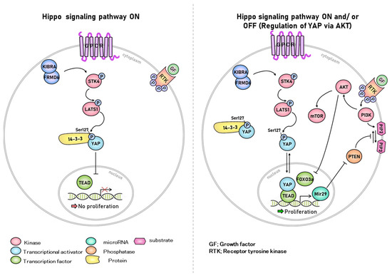
Figure 1.
Hippo signaling pathway. YAP, a downstream target of the Hippo signaling pathway, activates the mammalian target of rapamycin (mTOR), a major regulator of cell growth. This suggests that the Hippo pathway, PTEN, and AKT are related. The stimulation of AKT and inhibition of the Hippo signaling pathway affect ovarian follicle growth. AKT activates dormant primordial follicles and promotes follicle growth by interfering with the Hippo signaling pathway by ovarian fragments. YAP regulates carcinogenesis in endometrial adenocarcinoma, and YAP expression correlated with the type of endometrial adenocarcinoma. In addition, the Hippo signaling pathway is a key downstream signaling branch of the G protein-coupled estrogen receptor (GPER) and plays a crucial role in breast tumorigenesis.
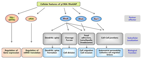
Figure 2.
Role of P190A RhoGAP in various cellular processes. The interaction between P190A and p120-catenin at the cell-cell junction inhibits RhoA activity. CRAD (catenin-RhoGAP-association domain), a C-terminus domain of p120-catenin, interacts with P190A. CRAD depletion in p120-catenin of endothelial cells affects the transmembrane transduction of P190A and regulates endothelial permeability by increasing RhoA and decreasing the Rac1 signal. The crossover between cell-cell contact and cell-matrix adhesion is produced by the P190/P120-catenin complex to spatially regulate RhoA activity. The transient decrease in P190A levels during late mitosis in the cell cycle finely regulates RhoA activity required for cell division; P190A is also required to regulate spindle formation [23].
2.2. Cell-to-Cell Contact Interaction
The idea that signaling pathways mediate CIP was hypothesized because of the observation that YAP is localized in the nucleus when cell cultures reach confluence [27]. Unlike single-celled organisms, cell growth and division in metazoans with multicellular systems are inhibited by contact inhibition and the interaction between growth factors. The culture of human cells limits cell proliferation and division when cells contact each other, regardless of active cellular metabolism or the presence of growth factors. Contact inhibition is overcome in rapidly growing tissues during embryonic development, morning regeneration, wound healing, and uncontrolled proliferation and growth due to the loss of contact inhibition, which are also characteristics of solid tumors. E-cadherin is an example of such a contact inhibitory factor [7]. Cadherins inhibit cell contact-dependent proliferation by altering receptor tyrosine kinase signaling through interactions with the neurofibromatosis 2 (NF2) tumor suppressor Merlin. Additionally, the expression of E-cadherin in cells deficient in cadherin induces transportation of YAP into the cytoplasm from the nucleus. Thus, E-cadherin inhibits the nuclear localization of YAP [8,28]. Merlin also regulates the Hippo signaling pathway through its interaction with Kibra [29,30,31]. Merlin and Kibra act upstream of STK3/4, form complexes in cultured cells, and coexist in vivo [29,32]. These observations suggested that the Hippo signaling pathway can be regulated both in cultured cells and in vivo systems.
2.3. Hippo Signaling in Regulation of Cell Density and Cancer
Normal cell growth is restricted through cell–cell contact inhibition, regulating proliferation, whereas cancer cells continue to grow, by passing cell–cell contact inhibition [33]. However, cancer cells do sense the contact with surrounding cells and respond through signaling pathways. Deregulation of YAP signaling, which promotes cell growth and cell survival, was found to be upregulated in several cancers, and the activation or inactivation of YAP was correlated with poor prognosis of cancers [34,35,36]. At low cell densities, YAP showed decreased phosphorylation, increased nuclear localization, and upregulation of YAP target genes in cells. Reduced metastasis is consistent with YAP localization in the nucleus, and is representative of the invasive phenotype of cancer cells [37]. The loss of E-cadherin, Ca2+-dependent mediators of cell adhesion, and the formation of the cell junction promote tumor progression and metastasis [38,39]. The Hippo pathway was shown to be associated with E-cadherin-dependent contact proliferation inhibition. Depletion of LATS1/2 leads to E-cadherin ligation, thus suppressing E-cadherin-induced reduction in cell proliferation [7]. Deletion of LATS1 turns non-invasive cancer cells grown at high densities (trans-endothelial invasion), similar to those grown at low densities [37].
2.4. STK4 in Apoptosis
Apoptosis is a pathway centered on caspases [40]. STK3/4, which is crucial to the Hippo pathway, is cleaved and activated by caspases in response to apoptotic signals. The overexpression of STK4 induces caspase activity and activates the p38 MAPK and stress-activated protein kinase (SAPK) pathways, which are associated with apoptosis [41]. Therefore, STK4 acts as a positive regulator of the apoptotic response. Additionally, the basophilic kinase STK4 with pro-apoptotic activity is phosphorylated by the eosinophilic casein kinase 2 (CK2, also called CSNK2A1), which is anti-apoptotic [42,43]. Inhibition of CK2 activity induces apoptosis in cancer cells [44]. Cleavage of substrates by caspases is regulated by phosphorylation near the shear site. Serine 320 of STK4 is located close to the site cleaved by caspase, and CK2 phosphorylates serine 320 [45]. CK2 binds to STK4, including full-length STK4 located in the cytoplasm, and the N-terminal fragment of STK4 is located in the nucleus [43]. The interaction between the cytoplasm and nucleus is expected to open new avenues for downstream target phosphorylation.
STK3/4 regulates cell proliferation and apoptosis by targeting the cell cycle regulator cyclin E and apoptosis inhibitor DIAP1 [3,14,46,47,48]. In Drosophila, Hippo (Hpo, a homolog of mammalian STK3/4) induces apoptosis during development while promoting the proper termination of cell proliferation [14]. Hippo mutation leads to unregulated proliferation, resulting in enlarged tissue size as well as delayed cell cycle termination and resistance to pro-apoptotic signals, resulting in increased expression levels of cyclin E and DIAP1 [3].
STK4 is poorly expressed in human thyroid cancer cells; STK4 silencing promotes the proliferation of these cancer cells, inhibits apoptosis and autophagy, and thereby enhances tumor growth [49]. In addition, the overexpression of STK4 in MDA-T32 cells, a thyroid cancer cell line, induces cancer cell death by increasing mitochondrial stress, similar to that observed in YAP knockdown. The synergy between STK4 upregulation and YAP inhibition damages mitochondria, owing to the excess ROS production, and further increases apoptosis by activating the c-Jun N-terminal kinase (JNK) pathway [50]. Natural killer/T-cell lymphomas, a type of malignancy, exhibit downregulated expression of STK4, markedly upregulated expression of YAP, and suppressed phosphorylation of YAP. STK4 overexpression or YAP knockdown in natural killer/T-cell lymphomas is consistent with increased BCL2 and BAX expression levels, decreased cyclin D1 and CTGF expression levels, inhibition of cell proliferation and cell cycle progression, and promotion of apoptosis [51]. This suggests that the activation of the Hippo pathway can inhibit tumorigenesis.
2.5. Rules of Hippo Signaling in an In Vitro System
As described previously, the expression of nuclear-localized YAP is prevalent in cells cultured at low density and the activation of the Hippo pathway and cytoplasmic localization of YAP are prevalent in cells cultured at high density [18]. However, regardless of the density of the cultured cells, when serum was removed from the medium, YAP remained in the cytoplasm, and YAP(S127) phosphorylation dramatically increased. The absence of serum in the medium induced YAP(S127) phosphorylation and cytoplasmic maintenance independent of cell-cell contact. In contrast, the addition of serum to the medium induced YAP dephosphorylation and nuclear localization, ignoring cell-cell contact-mediated YAP inhibition. Serum substances that cause these responses include sphingosine-1-phosphate (S1P) and lysophosphatidic acid (LPA) [52]. S1P is a component of follicular fluid that regulates ovarian angiogenesis [53,54].
LPA is a simple phospholipid with various physiological and pathological functions, such as cell proliferation and differentiation [55,56], intercellular interactions [57], and tumorigenesis [58], and can be produced by endometrial and ovarian cells [59,60,61,62,63]. S1P activates YAP/TAZ and promotes cell proliferation by inhibiting LATS1/2 through the S1P receptor (S1PR) [52,64]. In zebrafish progenitor cells, YAP and CTGF are involved in the regulation of endoderm formation as downstream targets of S1P [65]. In mammals, nuclear localization of YAP is induced by S1P, increasing the CTGF transcript [66,67,68,69,70]. In addition to S1PR, S1P and LPA activate YAP/TAZ by inhibiting LATS1/2 via G protein-coupled receptors (GPCRs). LPA has six receptors capable of binding to GPCRs and can induce or inhibit YAP phosphorylation depending on which GPCR receptors are expressed and bind to the cell type [10].
3. Hippo Signaling Factors in the Uterine Endometrium
3.1. STK3/4 in the Uterine Endometrium
The Hippo signaling pathway cascade is triggered in the uterine epithelium to regulate downstream targets that affect dynamics during the estrous cycle. Interestingly, we found that the expression levels of STK3/4 change dynamically in the endometrium during the estrous cycle, demonstrating that STK3/4 is regulated by estrogen and estrogen receptors. STK3/4 is rapidly expressed via estrogen receptor-dependent signaling in both the endometrial glandular epithelium and luminal epithelium. STK3/4 mRNA and protein levels in the mouse endometrium were highest at 6 h of estrogen treatment and decreased thereafter. The regulation of STK3/4 and its downstream targets is mediated by p38-MAPK, an estrogen receptor-dependent intracellular signaling pathway. Our previous study showed that the phosphorylation of LATS1/2 was faster than that of STK3/4, which may occur through an unknown signaling pathway for the phosphorylation of LATS1/2 in uterine dynamics during the estrous cycle [71].
3.2. LATS1/2 in the Uterine Endometrium
Since its discovery in Drosophila, LATS1/2 has been considered a tumor suppressor. LATS1/2 plays an important role in controlling tumor development and the cell cycle through several mechanisms, including p53 [30,72,73,74].
However, there are few studies on the role of LATS1/2 in the uterus, among other Hippo signaling factors. In 2010, Strakova et al. reported an abstract describing that significant upregulation of LATS1/2 occurs during the secretory phase of the menstrual cycle in baboon (unpublished data). Additionally a recent study showed that Scrib knockout mice showed weak LATS1 expression in the uterus [75]. In addition, various mutations in LATS1/2 were found in human cancers, including uterine endometrial carcinoma [76].
The direct association between LATS1/2 and uterine endometrial cancer requires further research, but recent data implied that LATS1/2, like other Hippo factors, plays a role in regulating the uterine environment.
3.3. YAP in the Uterine Endometrium
YAP has been studied in endometrial stromal cells and deciduous membranes [77]. The desiccation of endometrial stromal cells is important for successful transplantation and maintenance of pregnancy, and it involves extensive cell proliferation and differentiation. YAP is upregulated in human decidual tissue, and YAP expression level in decidual cells is higher than that in the endometrial stromal cells of non-pregnant women [77]. The expression level of YAP mRNA is higher in the endometrial stromal cells in women with endometriosis compared with that in women without endometriosis [78,79,80]. mTOR is also overexpressed in eutopic endometrial stromal cells, indicating decreased autophagy. YAP expression influences cellular autophagy by mediating the mTOR-autophagy pathway in eutopic endometrial stromal cells. Treatment of isotopic endometrial stromal cells with the mTOR inhibitor rapamycin improved autophagy and significantly reduced YAP expression. This suggested a negative regulatory relationship between YAP and autophagy in eutopic endometrial stromal cells [78]. Thus, YAP may be considered as a biomarker for the treatment of endometriosis.
3.4. P190A in the Uterine Endometrium
Recent studies have reported a correlation between P190A and YAP in endometrial cancer [81]. Endometrial cancer is the sixth most common cancer in women worldwide and causes approximately 30% of deaths annually [82]. ARHGAP35, the gene encoding P190A, is mutated in approximately 15% of endometrial cancers. Inactivation of P190A due to mutations causes abnormal activation of YAP, a target gene of the Hippo signaling pathway, leading to the malignant transformation of cancer. When P190A is depleted in endometrial cancer cells, epithelial-mesenchymal metastasis (EMT) occurs, conferring metastatic function by enhancing resistance to cancer cell migration, invasion, and apoptosis. In P190A-depleted cells, the levels of CYR61, CTGF, THBS1, and ANKRD1 mRNA, the transcriptional target genes for EMT-related genes, and YAP were significantly increased. Moreover, a decrease in LATS1 and YAP phosphorylation was observed, suggesting that P190A negatively regulates YAP activity [81,82]. Therefore, depletion of P190A in endometrial cancer cells indicates that EMT is partially induced through the transcription of YAP in the Hippo pathway (Figure 3).
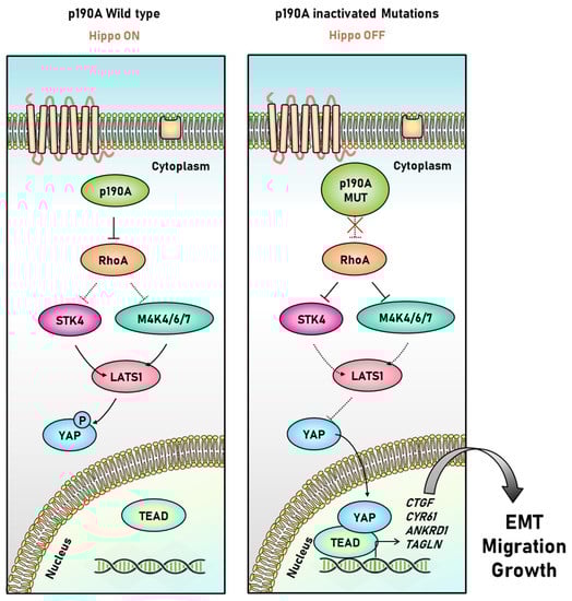
Figure 3.
Interaction between P190A and Hippo signaling pathway in endometrial cancer cells. Under normal conditions, P190A induces YAP phosphorylation by crossing the Hippo signaling pathway through RhoA inhibition. When P190A is mutated, RhoA is abnormally activated, interfering with the activity of STK4 and MAP4K4/6/7, preventing the phosphorylation of LATS1/2, and triggering the transcriptional activation of YAP, resulting in the transcription of EMT-related target genes [81].
4. Downstream Targets of Hippo Signaling in the Endometrium
When LATS1/2 is inactive, YAP translocates to the nucleus without being phosphorylated. As YAP does not contain a DNA-binding domain, it binds to a transcription factor of the TEAD family, such as TEAD1-4, and induces the expression of target genes such as CTGF and CYR61 [83] that promote cell growth, proliferation, cell migration, and survival [35,84,85,86,87]. Other YAP target genes included AMOT, AMOTL1, AMOTL2 [88,89], CTNNA1 [90], CTNNB1 [91,92,93], ADAMTS1 [94,95], ANKRD1 [96,97], AXL [98], MCL1 [99], and THBS1 [100].
CYR61 is a marker of estrogen activity in normal and abnormal endometrial cells. In endometrial cancer, overexpression of CYR61 increases apoptosis and is suggested to be involved in growth arrest or growth stimulation depending on cell conditions [101,102]. AMOT, AMOTL1, and AMOTL2 are angiogenesis-related proteins regulated by estrogen in the luminal epithelium and progesterone in the endometrial matrix [88,89]. CTNNB1 is abnormally expressed in uterine fibroids and has been suggested to play a role in menstruation and implantation in the uterus. It has also been associated with endometrial hyperplasia and endometrial cancer; in low-grade endometrial cancer, CTNNB1 mutations are associated with recurrence-free survival [91,92]. ADAMTS1 is involved in various biological functions, including ovulation and embryo receptivity, through cell-matrix remodeling, developmental processes, or both, and is involved in bovine endometrial remodeling, which is required in conjunction with ovarian steroid hormones, for implantation and placental development [94,95].
4.1. Connective Tissue Growth Factor (CTGF)
CTGF, also known as cellular communication network factor 2 (CCN2),is a multifunctional growth factor that exhibits biological and immunological activities similar to that of platelet-derived growth factor (PDGF) and is strongly expressed in wound healing, angiogenesis, and connective tissue formation sites [103,104]. Therefore, CTGF may play a role in endometrial repair. CTGF expression was found to be consistent with endometrial repair and regeneration. CTGF is localized in glandular epithelial cells and macrophages in the stromal cell compartment, and CTGF mRNA and protein expression levels increase considerably in endometrial cells treated with progesterone and with hypoxia [105]. A marked increase in adhesion molecules, including CTGF, in the functional layer of the endometrium, suggests that CTGF influences the healing of the endometrial surface after menstruation. Restoring the epithelial surface without blood vessel repair in the endometrial epithelium will only result in the collection of a large amount of epithelial blood. Although the role of macrophages in endometrial epithelial recovery is not fully understood, they have been suggested to play an important role in endometrial growth and blood vessel formation [106]. Endometrial macrophages respond to estrogen and progesterone and are thought to contribute to the increased expression level of CTGF in stromal cells during menstruation when estrogen and progesterone levels are low. In ovariectomized mice, CTGF levels were increased by estrogen and decreased by progesterone. CTGF mRNA levels and protein localization in the uterine tissues of mice and pigs during the estrous cycle and early pregnancy have been studied along with spatiotemporal changes [107,108,109]. In the uterine fluids of pregnant pigs, CTGF concentration reached its highest level at the time of the generation of betrayal kidney and estrogen [103]. These data suggests that CTGF plays an important role in the estrous cycle, menstrual cycle, and pregnancy.
CTGF is located within the endometrial and decidual membranes, in a cell- and stage-specific manner, and in human endometrial tissue [101,105,110,111]. CTGF potentially regulates various normal cellular processes in uterine tissues. The localization of CTGF in mice differs depending on the stage of gestation. About 2.5 d after gestation, CTGF colocalized with TGFβ in uterine epithelial cells, but after about 4.5 d, there was a decrease in epithelial cells and an increase in stromal and endothelial cells. These changes occurred at different stages of extracellular matrix remodeling and angiogenesis in the endometrium. CTGF and TGFβ expression levels increased in the uterine epithelial cells due to estrogen induction and in the stroma. These effects were antagonized by progesterone; however, progesterone also appeared to temporarily induce CTGF production [112]. Therefore, CTGF in the mouse uterus is regulated by estrogen and progesterone via a TGFβ-dependent or independent mechanism.
4.2. Cysteine-Rich Angiogenesis Inducer 61 (CYR61)
CYR61, also known as CCN1, is an extracellular matrix (ECM)-related signaling protein from the CCN protein family. CYR61 is a multifunctional protein with a variety of functions, including cell adhesion and migration, cell proliferation and differentiation, and cell activation and apoptosis. It has conflicting roles as it both promotes tumor growth and inhibits tumor formation under various conditions. Additionally, it contributes to inflammation control, wound healing, and tissue repair [113,114,115,116]. CYR61 expression is downregulated in uterine leiomyosarcoma cells compared to normal uterine smooth muscle cells. Investigation of the factors affecting CYR61 in myometrial smooth muscle cells revealed that basal fibroblast growth factor (bFGF) and estrogen elevate CYR61 levels to heal uterine smooth muscle cells; progesterone has no effects on CYR61 expression [117]. In addition, the expression level of CYR61 is increased by EGF (epidermal growth factor) in endometrial epithelial cells. CYR61 was expected to be upregulated by the MAPK/ERK pathway triggered by the epidermal growth factor receptor (EGFR); however, MAPK/ERK showed no effect on CYR61 expression. JAK2/STAT3 pathway as an EGF sub-pathway, affects CYR61 expression. Blockage of the JAK2/STAT3 pathway leads to decreased CYR61 expression level [118].
CYR61 expression in the human endometrium promotes the proliferative phase of the menstrual cycle, in endometrial epithelial cells and the synovial endothelium [119,120]. It increases equally with VEGFA, an angiogenic factor upregulated during the menstrual cycle, suggesting a similar role in human endometrial vascular repair, growth, and maturation after menstruation [101]. CYR61 occurs downstream of the ERK pathway and is involved in angiogenesis induction [121]. CYR61 expression level was reduced in the endometrial adenocarcinoma cell line, Ishikawa compared to in normal endometrial cells, which is known to promote apoptosis of endometrial cancer cells and increase the expression levels of apoptosis-related genes. Treatment of Ishikawa cells with siCYR61 increased the cell count [122,123,124]. CYR61 was shown to have a progressive decline in expression level with the progression of endometrial adenocarcinoma from low-grade to high-grade. Therefore, CYR61 may be important for the development of endometrial cancer and survival prognosis [116,125].
4.3. Thrombospondin-1 (THBS1)
THBS1, also known as TSP or TSP-1, affects the structure of the extracellular matrix. It is an important factor in angiogenesis and tissue remodeling. Mutations in oncogenes and tumor suppressor genes in tumor cells are associated with decreased THBS1 levels [126]. As tumors grow, they require angiogenesis [127], which is induced by angiogenesis regulators such as VEGF. THBS1 is an endogenous angiogenesis inhibitor that inhibits tumor growth by inhibiting tumor angiogenesis [128]. THBS1 inhibits tumor cell growth by activating TGFβ in TGFβ-responsive tumor cells [126]. Tumor growth and angiogenesis were considerably increased in THBS1 cKO mice and in dilated capillaries. Conversely, in THBS1 overexpressing mice, tumor growth is delayed or underdeveloped and capillaries in the tumor are constricted [129].
The expression level of THBS1 in the human endometrium is significantly higher during the secretory phase than during the proliferative phase of the menstrual cycle. As in the mouse model, the number of capillaries is higher in endometrial cancer with high THBS1 expression levels, suggesting that elevated THBS1 expression is associated with an angiogenic phenotype in endometrial cancer [130]. As previously described, the potential effects of THBS1 and endometrial estrogen on inducing vascular destruction were evaluated. Estrogen treatment on the endometrium of baboons considerably decreased THBS1 expression levels in the endometrial glandular epithelial cells and stromal cells 6 h after estrogen injection. However, the expression level of angiopoietin, which promotes angiogenesis, increased in endometrial gland epithelial cells 3 h after estrogen treatment, and did not change even with decreased THBS1 expression level [131]. This finding suggested that estrogen induces rapid and cell-specific changes in the expression of angiogenesis-stimulating and angiogenesis-inhibiting factors in the endometrium.
4.4. Cyclin D1 (CCND1)
CCND1, also called BCL1, is a cell cycle regulator that can alter cell cycle progression and is essential for G1 phase progression [132]. CCND1 regulates proliferation by transducing extracellular signals [133]. It participates in the detachment (differentiation) of endometrial stromal cells via signal transduction [134]. CCND1 is a proto-oncogene with oncogenic characteristics in various carcinomas [135,136,137,138]. CCND1 is highly expressed in the proliferative and secretory phases of the menstrual cycle in the uterus [139], and its high expression level in endometrial cancer is observed in metastatic rather than non-metastatic carcinomas. This frequency of expression is significantly correlated with the stage of endometrial cancer; the higher the expression level, the poorer the prognosis of patients with endometrial cancer [140]. CCND1 expression is positively correlated with that of Ki67, which is a widely known marker of endometrial proliferation [141] and can be used as a marker for endometrial cancer cell proliferation [142]. The overexpression of CCND1 in endometrial cancer is associated with the loss of PTEN [143]. Loss of PTEN is commonly found in endometrial cancer and hyperplasia with cell dysplasia [144,145,146,147], which reinforces that CCND1 overexpression can be used as a marker for the early development of endometrial cancer.
5. Conclusions
The uterus undergoes dynamic changes during the reproductive cycle, including endometrial cell proliferation and decidualization induced by estrogen and progesterone. An imbalance in the levels of these hormones can lead to endometrial cancer or an abnormal uterine environment. It is becoming increasingly clear that Hippo signaling pathway, which regulates angiogenesis, cell proliferation, differentiation, and apoptosis, is involved in the changes caused by the uterine cycle. Our review elucidates the dynamic changes in the endometrium influenced by the Hippo signaling pathway.
Author Contributions
Conceptualization, Y.C.; Data curation, S.M. and S.H.; Funding acquisition, Y.C.; Investigation, Y.C.; Methodology, S.M. and S.H.; Supervision, Y.C.; Writing—original draft, S.M., S.H. and Y.C.; Writing—review and editing, B.K., S.L., H.K., G.L., K.H. and H.S. All authors have read and agreed to the published version of the manuscript.
Funding
This paper was supported by Konkuk University in 2021.
Informed Consent Statement
Not applicable.
Data Availability Statement
Not applicable.
Acknowledgments
I would like to thank Sun Han Shim and Hoon Jang for valuable discussion and comments for the manuscript.
Conflicts of Interest
The authors declare no conflict of interest.
References
- Baumgartner, R.; Poernbacher, I.; Buser, N.; Hafen, E.; Stocker, H. The WW domain protein Kibra acts upstream of Hippo in Drosophila. Dev. Cell 2010, 18, 309–316. [Google Scholar] [CrossRef] [PubMed]
- Genevet, A.; Wehr, M.C.; Brain, R.; Thompson, B.J.; Tapon, N. Kibra is a regulator of the Salvador/Warts/Hippo signaling network. Dev. Cell 2010, 18, 300–308. [Google Scholar] [CrossRef] [PubMed]
- Harvey, K.F.; Pfleger, C.M.; Hariharan, I.K. The Drosophila Mst ortholog, hippo, restricts growth and cell proliferation and promotes apoptosis. Cell 2003, 114, 457–467. [Google Scholar] [CrossRef]
- Huang, J.; Wu, S.; Barrera, J.; Matthews, K.; Pan, D. The Hippo signaling pathway coordinately regulates cell proliferation and apoptosis by inactivating Yorkie, the Drosophila Homolog of YAP. Cell 2005, 122, 421–434. [Google Scholar] [CrossRef] [PubMed]
- Yu, J.; Zheng, Y.; Dong, J.; Klusza, S.; Deng, W.M.; Pan, D. Kibra functions as a tumor suppressor protein that regulates Hippo signaling in conjunction with Merlin and Expanded. Dev. Cell 2010, 18, 288–299. [Google Scholar] [CrossRef] [PubMed]
- Halder, G.; Johnson, R.L. Hippo signaling: Growth control and beyond. Development 2011, 138, 9–22. [Google Scholar] [CrossRef] [PubMed]
- Kim, N.G.; Koh, E.; Chen, X.; Gumbiner, B.M. E-cadherin mediates contact inhibition of proliferation through Hippo signaling-pathway components. Proc. Natl. Acad. Sci. USA 2011, 108, 11930–11935. [Google Scholar] [CrossRef]
- Gumbiner, B.M.; Kim, N.G. The Hippo-YAP signaling pathway and contact inhibition of growth. J. Cell Sci. 2014, 127, 709–717. [Google Scholar] [CrossRef]
- Nishioka, N.; Inoue, K.; Adachi, K.; Kiyonari, H.; Ota, M.; Ralston, A.; Yabuta, N.; Hirahara, S.; Stephenson, R.O.; Ogonuki, N.; et al. The Hippo signaling pathway components Lats and Yap pattern Tead4 activity to distinguish mouse trophectoderm from inner cell mass. Dev. Cell 2009, 16, 398–410. [Google Scholar] [CrossRef]
- Yu, F.X.; Zhao, B.; Panupinthu, N.; Jewell, J.L.; Lian, I.; Wang, L.H.; Zhao, J.; Yuan, H.; Tumaneng, K.; Li, H.; et al. Regulation of the Hippo-YAP pathway by G-protein-coupled receptor signaling. Cell 2012, 150, 780–791. [Google Scholar] [CrossRef]
- Hong, K.; Choi, Y. Role of estrogen and RAS signaling in repeated implantation failure. BMB Rep. 2018, 51, 225–229. [Google Scholar] [CrossRef] [PubMed]
- Mauviel, A.; Nallet-Staub, F.; Varelas, X. Integrating developmental signals: A Hippo in the (path)way. Oncogene 2012, 31, 1743–1756. [Google Scholar] [CrossRef] [PubMed]
- Boggiano, J.C.; Vanderzalm, P.J.; Fehon, R.G. Tao-1 phosphorylates Hippo/MST kinases to regulate the Hippo-Salvador-Warts tumor suppressor pathway. Dev. Cell 2011, 21, 888–895. [Google Scholar] [CrossRef] [PubMed]
- Udan, R.S.; Kango-Singh, M.; Nolo, R.; Tao, C.; Halder, G. Hippo promotes proliferation arrest and apoptosis in the Salvador/Warts pathway. Nat. Cell Biol. 2003, 5, 914–920. [Google Scholar] [CrossRef]
- Xiang, C.; Li, J.; Hu, L.; Huang, J.; Luo, T.; Zhong, Z.; Zheng, Y.; Zheng, L. Hippo signaling pathway reveals a spatio-temporal correlation with the size of primordial follicle pool in mice. Cell. Physiol. Biochem. Int. J. Exp. Cell. Physiol. Biochem. Pharmacol. 2015, 35, 957–968. [Google Scholar] [CrossRef]
- Avruch, J.; Zhou, D.; Fitamant, J.; Bardeesy, N.; Mou, F.; Barrufet, L.R. Protein kinases of the Hippo pathway: Regulation and substrates. Semin. Cell Dev. Biol. 2012, 23, 770–784. [Google Scholar] [CrossRef]
- Johnson, R.; Halder, G. The two faces of Hippo: Targeting the Hippo pathway for regenerative medicine and cancer treatment. Nat. Rev. Drug Discov. 2014, 13, 63–79. [Google Scholar] [CrossRef]
- Zhao, B.; Wei, X.; Li, W.; Udan, R.S.; Yang, Q.; Kim, J.; Xie, J.; Ikenoue, T.; Yu, J.; Li, L.; et al. Inactivation of YAP oncoprotein by the Hippo pathway is involved in cell contact inhibition and tissue growth control. Genes Dev. 2007, 21, 2747–2761. [Google Scholar] [CrossRef]
- Hansen, C.G.; Moroishi, T.; Guan, K.L. YAP and TAZ: A nexus for Hippo signaling and beyond. Trends Cell Biol. 2015, 25, 499–513. [Google Scholar] [CrossRef]
- Zhou, D.; Conrad, C.; Xia, F.; Park, J.S.; Payer, B.; Yin, Y.; Lauwers, G.Y.; Thasler, W.; Lee, J.T.; Avruch, J.; et al. Mst1 and Mst2 maintain hepatocyte quiescence and suppress hepatocellular carcinoma development through inactivation of the Yap1 oncogene. Cancer Cell 2009, 16, 425–438. [Google Scholar] [CrossRef]
- Kim, W.; Jho, E.H. The history and regulatory mechanism of the Hippo pathway. BMB Rep. 2018, 51, 106–118. [Google Scholar] [CrossRef] [PubMed]
- Settleman, J. Rho GTPases in development. Prog. Mol. Subcell. Biol. 1999, 22, 201–229. [Google Scholar] [PubMed]
- Heraud, C.; Pinault, M.; Lagree, V.; Moreau, V. p190RhoGAPs, the ARHGAP35- and ARHGAP5-Encoded Proteins, in Health and Disease. Cells 2019, 8, 351. [Google Scholar] [CrossRef] [PubMed]
- Sordella, R.; Classon, M.; Hu, K.Q.; Matheson, S.F.; Brouns, M.R.; Fine, B.; Zhang, L.; Takami, H.; Yamada, Y.; Settleman, J. Modulation of CREB activity by the Rho GTPase regulates cell and organism size during mouse embryonic development. Dev. Cell 2002, 2, 553–565. [Google Scholar] [CrossRef]
- Frank, S.R.; Kollmann, C.P.; Luong, P.; Galli, G.G.; Zou, L.; Bernards, A.; Getz, G.; Calogero, R.A.; Frodin, M.; Hansen, S.H. p190 RhoGAP promotes contact inhibition in epithelial cells by repressing YAP activity. J. Cell Biol. 2018, 217, 3183–3201. [Google Scholar] [CrossRef]
- Ouyang, H.; Luong, P.; Frodin, M.; Hansen, S.H. p190A RhoGAP induces CDH1 expression and cooperates with E-cadherin to activate LATS kinases and suppress tumor cell growth. Oncogene 2020, 39, 5570–5587. [Google Scholar] [CrossRef]
- Zeng, Q.; Hong, W. The emerging role of the hippo pathway in cell contact inhibition, organ size control, and cancer development in mammals. Cancer Cell 2008, 13, 188–192. [Google Scholar] [CrossRef]
- Mayor, R.; Carmona-Fontaine, C. Keeping in touch with contact inhibition of locomotion. Trends Cell Biol. 2010, 20, 319–328. [Google Scholar] [CrossRef]
- Li, W.; Cooper, J.; Zhou, L.; Yang, C.; Erdjument-Bromage, H.; Zagzag, D.; Snuderl, M.; Ladanyi, M.; Hanemann, C.O.; Zhou, P.; et al. Merlin/NF2 loss-driven tumorigenesis linked to CRL4(DCAF1)-mediated inhibition of the hippo pathway kinases Lats1 and 2 in the nucleus. Cancer Cell 2014, 26, 48–60. [Google Scholar] [CrossRef]
- Lin, X.Y.; Zhang, X.P.; Wu, J.H.; Qiu, X.S.; Wang, E.H. Expression of LATS1 contributes to good prognosis and can negatively regulate YAP oncoprotein in non-small-cell lung cancer. Tumour Biol. J. Int. Soc. Oncodevel. Biol. Med. 2014, 35, 6435–6443. [Google Scholar] [CrossRef]
- McClatchey, A.I.; Fehon, R.G. Merlin and the ERM proteins—Regulators of receptor distribution and signaling at the cell cortex. Trends Cell Biol. 2009, 19, 198–206. [Google Scholar] [CrossRef] [PubMed]
- Gladden, A.B.; Hebert, A.M.; Schneeberger, E.E.; McClatchey, A.I. The NF2 tumor suppressor, Merlin, regulates epidermal development through the establishment of a junctional polarity complex. Dev. Cell 2010, 19, 727–739. [Google Scholar] [CrossRef] [PubMed]
- Hanahan, D.; Weinberg, R.A. The hallmarks of cancer. Cell 2000, 100, 57–70. [Google Scholar] [CrossRef]
- Chen, Q.; Zhang, N.; Gray, R.S.; Li, H.; Ewald, A.J.; Zahnow, C.A.; Pan, D. A temporal requirement for Hippo signaling in mammary gland differentiation, growth, and tumorigenesis. Genes Dev. 2014, 28, 432–437. [Google Scholar] [CrossRef] [PubMed]
- Lamar, J.M.; Stern, P.; Liu, H.; Schindler, J.W.; Jiang, Z.G.; Hynes, R.O. The Hippo pathway target, YAP, promotes metastasis through its TEAD-interaction domain. Proc. Natl. Acad. Sci. USA 2012, 109, E2441–E2450. [Google Scholar] [CrossRef] [PubMed]
- Nallet-Staub, F.; Marsaud, V.; Li, L.; Gilbert, C.; Dodier, S.; Bataille, V.; Sudol, M.; Herlyn, M.; Mauviel, A. Pro-invasive activity of the Hippo pathway effectors YAP and TAZ in cutaneous melanoma. J. Investig. Dermatol. 2014, 134, 123–132. [Google Scholar] [CrossRef] [PubMed]
- Sharif, G.M.; Schmidt, M.O.; Yi, C.; Hu, Z.; Haddad, B.R.; Glasgow, E.; Riegel, A.T.; Wellstein, A. Cell growth density modulates cancer cell vascular invasion via Hippo pathway activity and CXCR2 signaling. Oncogene 2015, 34, 5879–5889. [Google Scholar] [CrossRef]
- Hajra, K.M.; Fearon, E.R. Cadherin and catenin alterations in human cancer. Genes Chromosomes Cancer 2002, 34, 255–268. [Google Scholar] [CrossRef]
- Jeanes, A.; Gottardi, C.; Yap, A. Cadherins and cancer: How does cadherin dysfunction promote tumor progression? Oncogene 2008, 27, 6920–6929. [Google Scholar] [CrossRef]
- Henkart, P.A. ICE family proteases: Mediators of all apoptotic cell death? Immunity 1998, 4, 195–201. [Google Scholar] [CrossRef]
- Graves, J.D.; Gotoh, Y.; Draves, K.E.; Ambrose, D.; Han, D.K.; Wright, M.; Chernoff, J.; Clark, E.A.; Krebs, E.G. Caspase-mediated activation and induction of apoptosis by the mammalian Ste20-like kinase Mst1. EMBO J. 1998, 17, 2224–2234. [Google Scholar] [CrossRef] [PubMed]
- Montenarh, M.; Gotz, C. Protein kinase CK2 and ion channels (Review). Biomed. Rep. 2020, 13, 55. [Google Scholar] [CrossRef] [PubMed]
- Servas, C.; Kiehlmeier, S.; Hach, J.; Gross, R.; Gotz, C.; Montenarh, M. The mammalian STE20-like kinase 1 (MST1) is a substrate for the apoptosis inhibiting protein kinase CK2. Cell. Signal. 2017, 36, 163–175. [Google Scholar] [CrossRef]
- Intemann, J.; Saidu, N.E.B.; Schwind, L.; Montenarh, M. ER stress signaling in ARPE-19 cells after inhibition of protein kinase CK2 by CX-4945. Cell. Signal. 2014, 26, 1567–1575. [Google Scholar] [CrossRef] [PubMed]
- Turowec, J.P.; Duncan, J.S.; Gloor, G.B.; Litchfield, D.W. Regulation of caspase pathways by protein kinase CK2: Identification of proteins with overlapping CK2 and caspase consensus motifs. Mol. Cell. Biochem. 2011, 356, 159–167. [Google Scholar] [CrossRef] [PubMed]
- Jia, J.; Zhang, W.; Wang, B.; Trinko, R.; Jiang, J. The Drosophila Ste20 family kinase dMST functions as a tumor suppressor by restricting cell proliferation and promoting apoptosis. Genes Dev. 2003, 17, 2514–2519. [Google Scholar] [CrossRef] [PubMed]
- Kango-Singh, M.; Nolo, R.; Tao, C.; Verstreken, P.; Hiesinger, P.R.; Bellen, H.J.; Halder, G. Shar-pei mediates cell proliferation arrest during imaginal disc growth in Drosophila. Development 2002, 129, 5719–5730. [Google Scholar] [CrossRef]
- Wu, S.; Huang, J.; Dong, J.; Pan, D. hippo encodes a Ste-20 family protein kinase that restricts cell proliferation and promotes apoptosis in conjunction with salvador and warts. Cell 2003, 114, 445–456. [Google Scholar] [CrossRef]
- Peng, X.; Ji, C.; Tan, L.; Lin, S.; Zhu, Y.; Long, M.; Luo, D.; Li, H. Long non-coding RNA TNRC6C-AS1 promotes methylation of STK4 to inhibit thyroid carcinoma cell apoptosis and autophagy via Hippo signalling pathway. J. Cell. Mol. Med. 2020, 24, 304–316. [Google Scholar] [CrossRef]
- Zhang, X.; Li, F.; Cui, Y.; Liu, S.; Sun, H. Mst1 overexpression combined with Yap knockdown augments thyroid carcinoma apoptosis via promoting MIEF1-related mitochondrial fission and activating the JNK pathway. Cancer Cell Int. 2019, 19, 143. [Google Scholar] [CrossRef]
- Chang, Y.; Fu, X.R.; Cui, M.; Li, W.M.; Zhang, L.; Li, X.; Li, L.; Sun, Z.C.; Zhang, X.D.; Li, Z.M.; et al. Activated hippo signal pathway inhibits cell proliferation and promotes apoptosis in NK/T cell lymphoma cells. Cancer Med. 2019, 8, 3892–3904. [Google Scholar] [CrossRef] [PubMed]
- Miller, E.; Yang, J.; DeRan, M.; Wu, C.; Su, A.I.; Bonamy, G.M.; Liu, J.; Peters, E.C.; Wu, X. Identification of serum-derived sphingosine-1-phosphate as a small molecule regulator of YAP. Chem. Biol. 2012, 19, 955–962. [Google Scholar] [CrossRef] [PubMed]
- Becker, S.; von Otte, S.; Robenek, H.; Diedrich, K.; Nofer, J.-R. Follicular fluid high-density lipoprotein-associated sphingosine 1-phosphate (S1P) promotes human granulosa lutein cell migration via S1P receptor type 3 and small G-protein RAC1. Biol. Reprod. 2011, 84, 604–612. [Google Scholar] [CrossRef] [PubMed]
- Spiegel, S.; Milstien, S. Sphingosine-1-phosphate: An enigmatic signalling lipid. Nat. Rev. Mol. Cell Biol. 2003, 4, 397–407. [Google Scholar] [CrossRef]
- Moolenaar, W.H. Lysophosphatidic Acid, a Multifunctional Phospholipid Messenger. J. Biol. Chem. 1995, 270, 12949–12952. [Google Scholar] [CrossRef]
- Pustilnik, T.B.; Estrella, V.; Wiener, J.R.; Mao, M.; Eder, A.; Watt, M.-A.V.; Bast, R.C.; Mills, G.B. Lysophosphatidic acid induces urokinase secretion by ovarian cancer cells. Clin. Cancer Res. 1999, 5, 3704–3710. [Google Scholar]
- Fukushima, N.; Weiner, J.A.; Kaushal, D.; Contos, J.J.; Rehen, S.K.; Kingsbury, M.A.; Kim, K.Y.; Chun, J. Lysophosphatidic acid influences the morphology and motility of young, postmitotic cortical neurons. Mol. Cell. Neurosci. 2002, 20, 271–282. [Google Scholar] [CrossRef]
- Kim, K.-S.; Sengupta, S.; Berk, M.; Kwak, Y.-G.; Escobar, P.F.; Belinson, J.; Mok, S.C.; Xu, Y. Hypoxia enhances lysophosphatidic acid responsiveness in ovarian cancer cells and lysophosphatidic acid induces ovarian tumor metastasis in vivo. Cancer Res. 2006, 66, 7983–7990. [Google Scholar] [CrossRef]
- Eder, A.M.; Sasagawa, T.; Mao, M.; Aoki, J.; Mills, G.B. Constitutive and lysophosphatidic acid (LPA)-induced LPA production: Role of phospholipase D and phospholipase A2. Clin. Cancer Res. 2000, 6, 2482–2491. [Google Scholar]
- Kowalczyk-Zieba, I.; Boruszewska, D.; Saulnier-Blache, J.S.; Da Costa, L.L.; Jankowska, K.; Skarzynski, D.J.; Woclawek-Potocka, I. Lysophosphatidic acid action in the bovine corpus luteum—An in vitro study. J. Reprod. Dev. 2012, 58, 661–671. [Google Scholar] [CrossRef]
- Luquain, C.; Singh, A.; Wang, L.; Natarajan, V.; Morris, A.J. Role of phospholipase D in agonist-stimulated lysophosphatidic acid synthesis by ovarian cancer cells. J. Lipid Res. 2003, 44, 1963–1975. [Google Scholar] [CrossRef] [PubMed]
- Shen, Z.; Belinson, J.; Morton, R.E.; Xu, Y. Phorbol 12-myristate 13-acetate stimulates lysophosphatidic acid secretion from ovarian and cervical cancer cells but not from breast or leukemia cells. Gynecol. Oncol. 1998, 71, 364–368. [Google Scholar] [CrossRef] [PubMed]
- Woclawek-Potocka, I.; Brzezicka, E.; Skarzynski, D.J. Lysophosphatic acid modulates prostaglandin secretion in the bovine endometrial cells differently on days 8–10 of the estrous cycle and early pregnancy. J. Reprod. Dev. 2009, 55, 393–399. [Google Scholar] [CrossRef] [PubMed][Green Version]
- Cheng, J.-C.; Wang, E.Y.; Yi, Y.; Thakur, A.; Tsai, S.-H.; Hoodless, P.A. S1P stimulates proliferation by upregulating CTGF expression through S1PR2-mediated YAP activation. Mol. Cancer Res. 2018, 16, 1543–1555. [Google Scholar] [CrossRef] [PubMed]
- Fukui, H.; Terai, K.; Nakajima, H.; Chiba, A.; Fukuhara, S.; Mochizuki, N. S1P-Yap1 signaling regulates endoderm formation required for cardiac precursor cell migration in zebrafish. Dev. Cell 2014, 31, 128–136. [Google Scholar] [CrossRef] [PubMed]
- Cheng, Y.; Feng, Y.; Jansson, L.; Sato, Y.; Deguchi, M.; Kawamura, K.; Hsueh, A.J. Actin polymerization-enhancing drugs promote ovarian follicle growth mediated by the Hippo signaling effector YAP. FASEB J. 2015, 29, 2423–2430. [Google Scholar] [CrossRef]
- Chowdhury, I.; Chaqour, B. Regulation of connective tissue growth factor (CTGF/CCN2) gene transcription and mRNA stability in smooth muscle cells: Involvement of RhoA GTPase and p38 MAP kinase and sensitivity to actin dynamics. Eur. J. Biochem. 2004, 271, 4436–4450. [Google Scholar] [CrossRef]
- Fan, Q.; Cheng, Y.; Chang, H.-M.; Deguchi, M.; Hsueh, A.J.; Leung, P.C. Sphingosine-1-phosphate promotes ovarian cancer cell proliferation by disrupting Hippo signaling. Oncotarget 2017, 8, 27166. [Google Scholar] [CrossRef]
- Li, M.-H.; Sanchez, T.; Pappalardo, A.; Lynch, K.R.; Hla, T.; Ferrer, F. Induction of antiproliferative connective tissue growth factor expression in Wilms’ tumor cells by sphingosine-1-phosphate receptor 2. Mol. Cancer Res. 2008, 6, 1649–1656. [Google Scholar] [CrossRef]
- Young, N.; Pearl, D.K.; Van Brocklyn, J.R. Sphingosine-1-phosphate regulates glioblastoma cell invasiveness through the urokinase plasminogen activator system and CCN1/Cyr61. Mol. Cancer Res. 2009, 7, 23–32. [Google Scholar] [CrossRef]
- Moon, S.; Lee, O.-H.; Lee, S.; Lee, J.; Park, H.; Park, M.; Chang, E.M.; Park, K.-H.; Choi, Y. STK3/4 Expression Is Regulated in Uterine Endometrial Cells during the Estrous Cycle. Cells 2019, 8, 1643. [Google Scholar] [CrossRef] [PubMed]
- Britschgi, A.; Duss, S.; Kim, S.; Couto, J.P.; Brinkhaus, H.; Koren, S.; De Silva, D.; Mertz, K.D.; Kaup, D.; Varga, Z.; et al. The Hippo kinases LATS1 and 2 control human breast cell fate via crosstalk with ERalpha. Nature 2017, 541, 541–545. [Google Scholar] [CrossRef]
- St John, M.A.; Tao, W.; Fei, X.; Fukumoto, R.; Carcangiu, M.L.; Brownstein, D.G.; Parlow, A.F.; McGrath, J.; Xu, T. Mice deficient of Lats1 develop soft-tissue sarcomas, ovarian tumours and pituitary dysfunction. Nat. Genet. 1999, 21, 182–186. [Google Scholar] [CrossRef] [PubMed]
- Sun, T.; Pepling, M.E.; Diaz, F.J. Lats1 Deletion Causes Increased Germ Cell Apoptosis and Follicular Cysts in Mouse Ovaries. Biol. Reprod. 2015, 93, 22. [Google Scholar] [CrossRef] [PubMed]
- Yuan, J.; Aikawa, S.; Deng, W.; Bartos, A.; Walz, G.; Grahammer, F.; Huber, T.B.; Sun, X.; Dey, S.K. Primary decidual zone formation requires Scribble for pregnancy success in mice. Nat. Commun. 2019, 10, 5425. [Google Scholar] [CrossRef]
- Yu, T.; Bachman, J.; Lai, Z.C. Mutation analysis of large tumor suppressor genes LATS1 and LATS2 supports a tumor suppressor role in human cancer. Protein Cell 2015, 6, 6–11. [Google Scholar] [CrossRef]
- Chen, H.; Song, Y.; Yang, S.; Fu, J.; Feng, X.; Huang, W. YAP mediates human decidualization of the uterine endometrial stromal cells. Placenta 2017, 53, 30–35. [Google Scholar] [CrossRef]
- Pei, T.; Huang, X.; Long, Y.; Duan, C.; Liu, T.; Li, Y.; Huang, W. Increased expression of YAP is associated with decreased cell autophagy in the eutopic endometrial stromal cells of endometriosis. Mol. Cell. Endocrinol. 2019, 491, 110432. [Google Scholar] [CrossRef]
- Song, Y.; Fu, J.; Zhou, M.; Xiao, L.; Feng, X.; Chen, H.; Huang, W. Activated hippo/yes-associated protein pathway promotes cell proliferation and anti-apoptosis in endometrial stromal cells of endometriosis. J. Clin. Endocrinol. Metab. 2016, 101, 1552–1561. [Google Scholar] [CrossRef]
- Wang, Y.; Yu, M.; Yang, J.-X.; Cao, D.-Y.; Zhang, Y.; Zhou, H.-M.; Yuan, Z.; Shen, K. Genomic comparison of endometrioid endometrial carcinoma and its precancerous lesions in Chinese patients by high-depth next generation sequencing. Front. Oncol. 2019, 9, 123. [Google Scholar] [CrossRef]
- Wen, X.; Wan, J.; He, Q.; Wang, M.; Li, S.; Jiang, M.; Qian, Z.; Liu, B.; Lu, W.; Wang, K.; et al. p190A inactivating mutations cause aberrant RhoA activation and promote malignant transformation via the Hippo-YAP pathway in endometrial cancer. Signal Transduct. Target. Ther. 2020, 5, 81. [Google Scholar] [CrossRef] [PubMed]
- Amant, F.; Moerman, P.; Neven, P.; Timmerman, D.; Van Limbergen, E.; Vergote, I. Endometrial cancer. Lancet 2005, 366, 491–505. [Google Scholar] [CrossRef]
- Shome, D.; von Woedtke, T.; Riedel, K.; Masur, K. The HIPPO Transducer YAP and Its Targets CTGF and Cyr61 Drive a Paracrine Signalling in Cold Atmospheric Plasma-Mediated Wound Healing. Oxidative Med. Cell. Longev. 2020, 2020, 4910280. [Google Scholar] [CrossRef] [PubMed]
- Haskins, J.W.; Nguyen, D.X.; Stern, D.F. Neuregulin 1-activated ERBB4 interacts with YAP to induce Hippo pathway target genes and promote cell migration. Sci. Signal. 2014, 7, ra116. [Google Scholar] [CrossRef] [PubMed]
- Yu, F.X.; Zhao, B.; Guan, K.L. Hippo Pathway in Organ Size Control, Tissue Homeostasis, and Cancer. Cell 2015, 163, 811–828. [Google Scholar] [CrossRef] [PubMed]
- Zhao, B.; Li, L.; Lei, Q.; Guan, K.L. The Hippo-YAP pathway in organ size control and tumorigenesis: An updated version. Genes Dev. 2010, 24, 862–874. [Google Scholar] [CrossRef] [PubMed]
- Zhao, B.; Ye, X.; Yu, J.; Li, L.; Li, W.; Li, S.; Yu, J.; Lin, J.D.; Wang, C.Y.; Chinnaiyan, A.M.; et al. TEAD mediates YAP-dependent gene induction and growth control. Genes Dev. 2008, 22, 1962–1971. [Google Scholar] [CrossRef] [PubMed]
- Chan, S.W.; Lim, C.J.; Chong, Y.F.; Pobbati, A.V.; Huang, C.; Hong, W. Hippo pathway-independent restriction of TAZ and YAP by angiomotin. J. Biol. Chem. 2011, 286, 7018–7026. [Google Scholar] [CrossRef]
- Wang, W.; Huang, J.; Chen, J. Angiomotin-like proteins associate with and negatively regulate YAP1. J. Biol. Chem. 2011, 286, 4364–4370. [Google Scholar] [CrossRef]
- Reardon, S.N.; King, M.L.; MacLean, J.A., 2nd; Mann, J.L.; DeMayo, F.J.; Lydon, J.P.; Hayashi, K. CDH1 is essential for endometrial differentiation, gland development, and adult function in the mouse uterus. Biol. Reprod. 2012, 86, 141-1–141-10. [Google Scholar] [CrossRef]
- Herington, J.L.; Bi, J.; Martin, J.D.; Bany, B.M. Beta-catenin (CTNNB1) in the mouse uterus during decidualization and the potential role of two pathways in regulating its degradation. J. Histochem. Cytochem. 2007, 55, 963–974. [Google Scholar] [CrossRef] [PubMed]
- Kurnit, K.C.; Kim, G.N.; Fellman, B.M.; Urbauer, D.L.; Mills, G.B.; Zhang, W.; Broaddus, R.R. CTNNB1 (beta-catenin) mutation identifies low grade, early stage endometrial cancer patients at increased risk of recurrence. Mod. Pathol. 2017, 30, 1032–1041. [Google Scholar] [CrossRef] [PubMed]
- Zaitseva, M.; Holdsworth-Carson, S.J.; Waldrip, L.; Nevzorova, J.; Martelotto, L.; Vollenhoven, B.J.; Rogers, P.A. Aberrant expression and regulation of NR2F2 and CTNNB1 in uterine fibroids. Reproduction 2013, 146, 91–102. [Google Scholar] [CrossRef] [PubMed]
- Keightley, M.C.; Sales, K.J.; Jabbour, H.N. PGF2alpha-F-prostanoid receptor signalling via ADAMTS1 modulates epithelial cell invasion and endothelial cell function in endometrial cancer. BMC Cancer 2010, 10, 488. [Google Scholar] [CrossRef] [PubMed]
- Mishra, B.; Koshi, K.; Kizaki, K.; Ushizawa, K.; Takahashi, T.; Hosoe, M.; Sato, T.; Ito, A.; Hashizume, K. Expression of ADAMTS1 mRNA in bovine endometrium and placenta during gestation. Domest. Anim. Endocrinol. 2013, 45, 43–48. [Google Scholar] [CrossRef]
- Almodovar-Garcia, K.; Kwon, M.; Samaras, S.E.; Davidson, J.M. ANKRD1 acts as a transcriptional repressor of MMP13 via the AP-1 site. Mol. Cell. Biol. 2014, 34, 1500–1511. [Google Scholar] [CrossRef]
- Wu, Y.; Ruggiero, C.L.; Bauman, W.A.; Cardozo, C. Ankrd1 is a transcriptional repressor for the androgen receptor that is downregulated by testosterone. Biochem. Biophys. Res. Commun. 2013, 437, 355–360. [Google Scholar] [CrossRef]
- Honda, H.; Barrueto, F.F.; Gogusev, J.; Im, D.D.; Morin, P.J. Serial analysis of gene expression reveals differential expression between endometriosis and normal endometrium. Possible roles for AXL and SHC1 in the pathogenesis of endometriosis. Reprod. Biol. Endocrinol. 2008, 6, 59. [Google Scholar] [CrossRef]
- Findakly, D.; Wang, J. Molecular Profiling of Benign Metastasizing Leiomyoma of the Uterus Revealing Unique Novel Therapeutic Targets. Cureus 2020, 12, e7701. [Google Scholar] [CrossRef]
- Zhang, K.; Ge, L.; Dong, S.; Liu, Y.; Wang, D.; Zhou, C.; Ma, C.; Wang, Y.; Su, F.; Jiang, Y. Global miRNA, lncRNA, and mRNA Transcriptome Profiling of Endometrial Epithelial Cells Reveals Genes Related to Porcine Reproductive Failure Caused by Porcine Reproductive and Respiratory Syndrome Virus. Front. Immunol. 2019, 10, 1221. [Google Scholar] [CrossRef]
- Gashaw, I.; Stiller, S.; Boing, C.; Kimmig, R.; Winterhager, E. Premenstrual regulation of the pro-angiogenic factor CYR61 in human endometrium. Endocrinology 2008, 149, 2261–2269. [Google Scholar] [CrossRef] [PubMed]
- MacLaughlan, S.D.; Palomino, W.A.; Mo, B.; Lewis, T.D.; Lininger, R.A.; Lessey, B.A. Endometrial expression of Cyr61: A marker of estrogenic activity in normal and abnormal endometrium. Obstet. Gynecol. 2007, 110, 146–154. [Google Scholar] [CrossRef] [PubMed]
- Ball, D.K.; Surveyor, G.A.; Diehl, J.R.; Steffen, C.L.; Uzumcu, M.; Mirando, M.A.; Brigstock, D.R. Characterization of 16- to 20-kilodalton (kDa) connective tissue growth factors (CTGFs) and demonstration of proteolytic activity for 38-kDa CTGF in pig uterine luminal flushings. Biol. Reprod. 1998, 59, 828–835. [Google Scholar] [CrossRef] [PubMed]
- Brigstock, D.R.; Steffen, C.L.; Kim, G.Y.; Vegunta, R.K.; Diehl, J.R.; Harding, P.A. Purification and characterization of novel heparin-binding growth factors in uterine secretory fluids. Identification as heparin-regulated Mr 10,000 forms of connective tissue growth factor. J. Biol. Chem. 1997, 272, 20275–20282. [Google Scholar] [CrossRef]
- Maybin, J.A.; Barcroft, J.; Thiruchelvam, U.; Hirani, N.; Jabbour, H.N.; Critchley, H.O. The presence and regulation of connective tissue growth factor in the human endometrium. Hum. Reprod. 2012, 27, 1112–1121. [Google Scholar] [CrossRef]
- Moon, S.; Lee, S.; Caesar, J.A.; Pruchenko, S.; Leask, A.; Knowles, J.A.; Sinon, J.; Chaqour, B. A CTGF-YAP regulatory pathway is essential for angiogenesis and barriergenesis in the retina. Iscience 2020, 23, 101184. [Google Scholar] [CrossRef]
- Harding, P.A.; Surveyor, G.A.; Brigstock, D.R. Characterization of pig connective tissue growth factor (CTGF) cDNA, mRNA and protein from uterine tissue. DNA Seq. J. DNA Seq. Mapp. 1998, 8, 385–390. [Google Scholar] [CrossRef]
- Moussad, E.E.; Rageh, M.A.; Wilson, A.K.; Geisert, R.D.; Brigstock, D.R. Temporal and spatial expression of connective tissue growth factor (CCN2; CTGF) and transforming growth factor beta type 1 (TGF-beta1) at the utero-placental interface during early pregnancy in the pig. Mol. Pathol. MP 2002, 55, 186–192. [Google Scholar] [CrossRef]
- Surveyor, G.A.; Wilson, A.K.; Brigstock, D.R. Localization of connective tissue growth factor during the period of embryo implantation in the mouse. Biol. Reprod. 1998, 59, 1207–1213. [Google Scholar] [CrossRef]
- Uzumcu, M.; Homsi, M.F.; Ball, D.K.; Coskun, S.; Jaroudi, K.; Hollanders, J.M.; Brigstock, D.R. Localization of connective tissue growth factor in human uterine tissues. Mol. Hum. Reprod. 2000, 6, 1093–1098. [Google Scholar] [CrossRef]
- Winterhager, E.; Gellhaus, A. The role of the CCN family of proteins in female reproduction. Cell. Mol. Life Sci. CMLS 2014, 71, 2299–2311. [Google Scholar] [CrossRef] [PubMed]
- Rageh, M.A.; Moussad, E.E.; Wilson, A.K.; Brigstock, D.R. Steroidal regulation of connective tissue growth factor (CCN2; CTGF) synthesis in the mouse uterus. Mol. Pathol. MP 2001, 54, 338–346. [Google Scholar] [CrossRef] [PubMed][Green Version]
- Chiodoni, C.; Colombo, M.P.; Sangaletti, S. Matricellular proteins: From homeostasis to inflammation, cancer, and metastasis. Cancer Metastasis Rev. 2010, 29, 295–307. [Google Scholar] [CrossRef] [PubMed]
- Jun, J.-I.; Lau, L.F. The matricellular protein CCN1 induces fibroblast senescence and restricts fibrosis in cutaneous wound healing. Nat. Cell Biol. 2010, 12, 676–685. [Google Scholar] [CrossRef]
- Kyriakides, T.R.; Bornstein, P. Matricellular proteins as modulators of wound healing and the foreign body response. Thromb. Haemost. 2003, 90, 986–992. [Google Scholar] [CrossRef]
- Lau, L.F. CCN1/CYR61: The very model of a modern matricellular protein. Cell. Mol. Life Sci. 2011, 68, 3149–3163. [Google Scholar] [CrossRef]
- Sampath, D.; Zhu, Y.; Winneker, R.C.; Zhang, Z. Aberrant expression of Cyr61, a member of the CCN (CTGF/Cyr61/Cef10/NOVH) family, and dysregulation by 17β-estradiol and basic fibroblast growth factor in human uterine leiomyomas. J. Clin. Endocrinol. Metab. 2001, 86, 1707–1715. [Google Scholar] [CrossRef][Green Version]
- Klein, R.; Stiller, S.; Gashaw, I. Epidermal growth factor upregulates endometrial CYR61 expression via activation of the JAK2/STAT3 pathway. Reprod. Fertil. Dev. 2012, 24, 482–489. [Google Scholar] [CrossRef]
- Li, Z.; Yan, G.; Diao, Q.; Yu, F.; Sheng, X.; Liu, Y.; Dai, Y.; Zhou, H.; Zhen, X.; Hu, Y. Transplantation of human endometrial perivascular cells with elevated CYR61 expression induces angiogenesis and promotes repair of a full-thickness uterine injury in rat. Stem Cell Res. Ther. 2019, 10, 1–16. [Google Scholar] [CrossRef]
- Park, M.-H.; Kim, A.k.; Manandhar, S.; Oh, S.-Y.; Jang, G.-H.; Kang, L.; Lee, D.-W.; Lee, S.-H.; Lee, H.E.; Huh, T.-L. CCN1 interlinks integrin and hippo pathway to autoregulate tip cell activity. Elife 2019, 8, e46012. [Google Scholar] [CrossRef]
- Hilfiker-Kleiner, D.; Kaminski, K.; Kaminska, A.; Fuchs, M.; Klein, G.; Podewski, E.; Grote, K.; Kiian, I.; Wollert, K.C.; Hilfiker, A. Regulation of proangiogenic factor CCN1 in cardiac muscle: Impact of ischemia, pressure overload, and neurohumoral activation. Circulation 2004, 109, 2227–2233. [Google Scholar] [CrossRef] [PubMed]
- Absenger, Y.; Hess-Stumpp, H.; Kreft, B.; Kratzschmar, J.; Haendler, B.; Schutze, N.; Regidor, P.A.; Winterhager, E. Cyr61, a deregulated gene in endometriosis. Mol. Hum. Reprod. 2004, 10, 399–407. [Google Scholar] [CrossRef] [PubMed]
- Chien, W.; Kumagai, T.; Miller, C.W.; Desmond, J.C.; Frank, J.M.; Said, J.W.; Koeffler, H.P. Cyr61 suppresses growth of human endometrial cancer cells. J. Biol. Chem. 2004, 279, 53087–53096. [Google Scholar] [CrossRef] [PubMed]
- Gashaw, I.; Hastings, J.M.; Jackson, K.S.; Winterhager, E.; Fazleabas, A.T. Induced endometriosis in the baboon (Papio anubis) increases the expression of the proangiogenic factor CYR61 (CCN1) in eutopic and ectopic endometria. Biol. Reprod. 2006, 74, 1060–1066. [Google Scholar] [CrossRef] [PubMed]
- Witek, L.; Janikowski, T.; Bodzek, P.; Olejek, A.; Mazurek, U. Expression of tumor suppressor genes related to the cell cycle in endometrial cancer patients. Adv. Med. Sci. 2016, 61, 317–324. [Google Scholar] [CrossRef] [PubMed]
- Lawler, J. Thrombospondin-1 as an endogenous inhibitor of angiogenesis and tumor growth. J. Cell. Mol. Med. 2002, 6, 1–12. [Google Scholar] [CrossRef]
- Folkman, J. Tumor angiogenesis: Therapeutic implications. N. Engl. J. Med. 1971, 285, 1182–1186. [Google Scholar]
- Zaslavsky, A.; Baek, K.-H.; Lynch, R.C.; Short, S.; Grillo, J.; Folkman, J.; Italiano, J.E., Jr.; Ryeom, S. Platelet-derived thrombospondin-1 is a critical negative regulator and potential biomarker of angiogenesis. Blood J. Am. Soc. Hematol. 2010, 115, 4605–4613. [Google Scholar] [CrossRef]
- Rodríguez-Manzaneque, J.C.; Lane, T.F.; Ortega, M.A.; Hynes, R.O.; Lawler, J.; Iruela-Arispe, M.L. Thrombospondin-1 suppresses spontaneous tumor growth and inhibits activation of matrix metalloproteinase-9 and mobilization of vascular endothelial growth factor. Proc. Natl. Acad. Sci. USA 2001, 98, 12485–12490. [Google Scholar] [CrossRef]
- Seki, N.; Kodama, J.; Hashimoto, I.; Hongo, A.; Yoshinouchi, M.; Kudo, T. Thrombospondin-1 and-2 messenger RNA expression in normal and neoplastic endometrial tissues: Correlation with angiogenesis and prognosis. Int. J. Oncol. 2001, 19, 305–310. [Google Scholar] [CrossRef]
- Bonagura, T.W.; Aberdeen, G.W.; Babischkin, J.S.; Koos, R.D.; Pepe, G.J.; Albrecht, E.D. Divergent regulation of angiopoietin-1 and-2, Tie-2, and thrombospondin-1 expression by estrogen in the baboon endometrium. Mol. Reprod. Dev. Inc. Gamete Res. 2010, 77, 430–438. [Google Scholar] [CrossRef] [PubMed]
- Lukas, J.; Müller, H.; Bartkova, J.; Spitkovsky, D.; Kjerulff, A.A.; Jansen-Dürr, P.; Strauss, M.; Bartek, J. DNA tumor virus oncoproteins and retinoblastoma gene mutations share the ability to relieve the cell’s requirement for cyclin D1 function in G1. J. Cell Biol. 1994, 125, 625–638. [Google Scholar] [CrossRef] [PubMed]
- Sherr, C.J. D-type cyclins. Trends Biochem. Sci. 1995, 20, 187–190. [Google Scholar] [CrossRef]
- Wang, L.; Yang, H.; Hu, L.; Hu, D.; Ma, S.; Sun, X.; Jiang, L.; Song, J.; Ji, L.; Masau, J.F. CDKN1C (P57): One of the determinants of human endometrial stromal cell decidualization. Biol. Reprod. 2018, 98, 277–285. [Google Scholar] [CrossRef]
- Wang, Y.; Zhu, J.-f.; Liu, Y.-y.; Han, G.-p. An analysis of cyclin D1, cytokeratin 5/6 and cytokeratin 8/18 expression in breast papillomas and papillary carcinomas. Diagn. Pathol. 2013, 8, 1–7. [Google Scholar] [CrossRef]
- Horvai, A.E.; Kramer, M.J.; O’Donnell, R. β-catenin nuclear expression correlates with cyclin D1 expression in primary and metastatic synovial sarcoma: A tissue microarray study. Arch. Pathol. Lab. Med. 2006, 130, 792–798. [Google Scholar] [CrossRef]
- Kim, J.K.; Diehl, J.A. Nuclear cyclin D1: An oncogenic driver in human cancer. J. Cell. Physiol. 2009, 220, 292–296. [Google Scholar] [CrossRef]
- Lin, L.; Hicks, D.; Xu, B.; Sigel, J.E.; Bergfeld, W.F.; Montgomery, E.; Fisher, C.; Hartke, M.; Tubbs, R.; Goldblum, J.R. Expression profile and molecular genetic regulation of cyclin D1 expression in epithelioid sarcoma. Mod. Pathol. 2005, 18, 705–709. [Google Scholar] [CrossRef][Green Version]
- Khabaz, M.N.; Abdelrahman, A.S.; Butt, N.S.; Al-Maghrabi, B.; Al-Maghrabi, J. Cyclin D1 is significantly associated with stage of tumor and predicts poor survival in endometrial carcinoma patients. Ann. Diagn. Pathol. 2017, 30, 47–51. [Google Scholar] [CrossRef]
- Liang, S.; Mu, K.; Wang, Y.; Zhou, Z.; Zhang, J.; Sheng, Y.; Zhang, T. CyclinD1, a prominent prognostic marker for endometrial diseases. Diagn. Pathol. 2013, 8, 1–8. [Google Scholar] [CrossRef]
- McCormick, D.; Chong, H.; Hobbs, C.; Datta, C.; Hall, P. Detection of the Ki-67 antigen in fixed and wax-embedded sections with the monoclonal antibody MIB1. Histopathology 1993, 22, 355–360. [Google Scholar] [CrossRef] [PubMed]
- Shevra, C.; Ghosh, A.; Kumar, M. Cyclin D1 and Ki-67 expression in normal, hyperplastic and neoplastic endometrium. J. Postgrad. Med. 2015, 61, 15. [Google Scholar] [PubMed]
- Shawana, S.; Kehar, S.I.; Masood, S.; Aamir, I. Immunoexpression of cyclin D1 and PTEN in various endometrial pathologies. J. Coll. Physicians Surg. Pak. 2016, 26, 277–282. [Google Scholar] [PubMed]
- Cirpan, T.; Terek, M.; Mgoyi, L.; Zekioglu, O.; Iscan, O.; Ozsaran, A. Immunohistochemical evaluation of PTEN protein in patients with endometrial intraepithelial neoplasia compared to endometrial adenocarcinoma and proliferative phase endometrium. Eur. J. Gynaecol. Oncol. 2006, 27, 389–392. [Google Scholar]
- Lee, K.-K.; Yonehara, S. Identification of mechanism that couples multisite phosphorylation of Yes-associated protein (YAP) with transcriptional coactivation and regulation of apoptosis. J. Biol. Chem. 2012, 287, 9568–9578. [Google Scholar] [CrossRef]
- Lee, H.; Choi, H.J.; Kang, C.S.; Lee, H.J.; Lee, W.S.; Park, C.S. Expression of miRNAs and PTEN in endometrial specimens ranging from histologically normal to hyperplasia and endometrial adenocarcinoma. Mod. Pathol. 2012, 25, 1508–1515. [Google Scholar] [CrossRef]
- Tantbirojn, P.; Triratanachat, S.; Trivijitsilp, P.; Niruthisard, S. Detection of PTEN Immunoreactivity in Endmetrial Hyperplasia and Adenocarcinoma. Med. J. Med. Assoc. Thail. 2008, 91, 1161–1165. [Google Scholar]
Publisher’s Note: MDPI stays neutral with regard to jurisdictional claims in published maps and institutional affiliations. |
© 2022 by the authors. Licensee MDPI, Basel, Switzerland. This article is an open access article distributed under the terms and conditions of the Creative Commons Attribution (CC BY) license (https://creativecommons.org/licenses/by/4.0/).