Article

GABAA Receptor Autoantibodies Decrease GABAergic Synaptic Transmission in the Hippocampal CA3 Network

1
Department of Neurology, Institute of Translational Neurology, University of Münster, 48149 Münster, Germany
2
Department of Neurology, University Hospital Knappschaftskrankenhaus Bochum, Ruhr University Bochum, 44892 Bochum, Germany
3
Institute of Clinical Neuroimmunology, Biomedical Center and Hospital of the Ludwig-Maximilians-Universität München, 82152 Martinsried, Germany
4
Munich Cluster for Systems Neurology (SyNergy), Ludwig-Maximilians-Universität München, 81377 München, Germany
5
Department of Neurology, Heinrich-Heine University of Düsseldorf, 40225 Düsseldorf, Germany
*
Author to whom correspondence should be addressed.
These authors contributed equally to this work.
Int. J. Mol. Sci. 2022, 23(7), 3707; https://doi.org/10.3390/ijms23073707
Submission received: 27 February 2022 / Revised: 19 March 2022 / Accepted: 23 March 2022 / Published: 28 March 2022
(This article belongs to the Collection State-of-the-Art Molecular Neurobiology in Germany)

Abstract

:
Autoimmune encephalitis associated with antibodies (Abs) against α1, β3, and γ2 subunits of γ-aminobutyric acid receptor A (GABAAR) represents a severe form of encephalitis with refractory seizures and status epilepticus. Reduction in inhibitory GABAergic synaptic activity is linked to dysfunction of neuronal networks, hyperexcitability, and seizures. The aim in this study was to investigate the direct pathogenic effect of a recombinant GABAAR autoantibody (rAb-IP2), derived from the cerebrospinal fluid (CSF) of a patient with autoimmune GABAAR encephalitis, on hippocampal CA1 and CA3 networks. Acute brain slices from C57BL/6 mice were incubated with rAb-IP2. The spontaneous synaptic GABAergic transmission was measured using electrophysiological recordings in voltage-clamp mode. The GABAAR autoantibody rAb-IP2 reduced inhibitory postsynaptic signaling in the hippocampal CA1 pyramidal neurons with regard to the number of spontaneous inhibitory postsynaptic currents (sIPSCs) but did not affect their amplitude. In the hippocampal CA3 network, decreased number and amplitude of sIPSCs were detected, leading to decreased GABAergic synaptic transmission. Immunohistochemical staining confirmed the rAb-IP2 bound to hippocampal tissue. These findings suggest that GABAAR autoantibodies exert direct functional effects on both hippocampal CA1 and CA3 pyramidal neurons and play a crucial role in seizure generation in GABAAR autoimmune encephalitis.

1. Introduction

The γ-aminobutyric acid receptor A (GABAAR) is a pentameric ligand-gated chloride channel consisting of different subunits (α1–6, β1–3, γ1–3, δ, ε, π, θ, and σ1–3). GABAAR mediates phasic synaptic and tonic extrasynaptic inhibition [1,2,3]. Epilepsy and epileptic encephalopathies, as well as other neuropsychiatric disorders such as anxiety disorders, schizophrenia, and depression, have been related to dysfunctional GABAAR, e.g., by mutations within α1 or β3 subunits [1,4]. Moreover, a severe form of autoimmune encephalitis with refractory seizures, status epilepticus, and antibodies (Abs) against α1, β3, and γ2 subunits of GABAAR has been reported as a new form of central nervous system (CNS) autoimmunity [5,6,7,8]. Several studies have demonstrated the pathogenic effects of these antibodies on GABAAR function [5,9,10,11,12,13]. These Abs led to the reduction in synaptic GABAAR complexes, presumably via cross-linking and the subsequent internalization of the Ab-receptor complex [5,9,10]. This caused selective reduction in the postsynaptic GABAAR clusters at inhibitory GABAergic synapses that was linked to the hyperexcitability and dysfunction of neuronal networks [5,9,10]. In contrast, a recent study showed that recombinant human monoclonal Abs (mAbs) with GABAAR reactivity, which were derived from a patient with GABAAR encephalitis and generated using single-cell cloning, reduced inhibitory postsynaptic signaling in neuronal cultures without causing receptor internalization [12]. Subsequently, a severe clinical phenotype with epileptic seizures was induced by cerebroventricular infusion of GABAAR mAbs into rodents [12]. In another study, a recombinantly expressed pathogenic antibody from the cerebrospinal fluid (CSF) B cells of a patient with autoimmune GABAAR encephalitis, previously described and termed “index patient 2” (IP2), led to the reduction in phasic GABAergic inhibitory synaptic activity and the increase in excitability in hippocampal CA1 pyramidal neurons, most probably contributing to clinical disease symptoms [13]. The CA1 pyramidal cells of the hippocampus received input from other hippocampal fields such as CA3 [14]. There is evidence that the epileptiform-discharges-like sharp waves are correlated with the synchronous discharges of pyramidal cells in CA1 and CA3 fields, of dentate granule cells, and of interneurons [15]. In mesial temporal lobe epilepsy, it has been supposed that interictal spikes are initiated in the CA3 field of the hippocampus by pacemaker pyramidal cells and propagated as population bursts throughout the CA3 subfield to the CA1 subfield via the Schaffer collaterals [15,16,17,18,19]. The CA1 subfield is responsible for further propagation and spread of the interictal spikes to the subcortical brain structures outside the hippocampus via the subiculum and the entorhinal cortex [15,20].
Studies of the influence of GABAAR Ab on signal transduction in hippocampal CA3 pyramidal neurons are missing. Therefore, our aim in this study was to investigate the direct pathogenic effect of the recombinant GABAAR antibody (rAb-IP2), derived from the CSF of a patient with GABAAR encephalitis, on the hippocampal CA3 network in addition to CA1.

2. Results

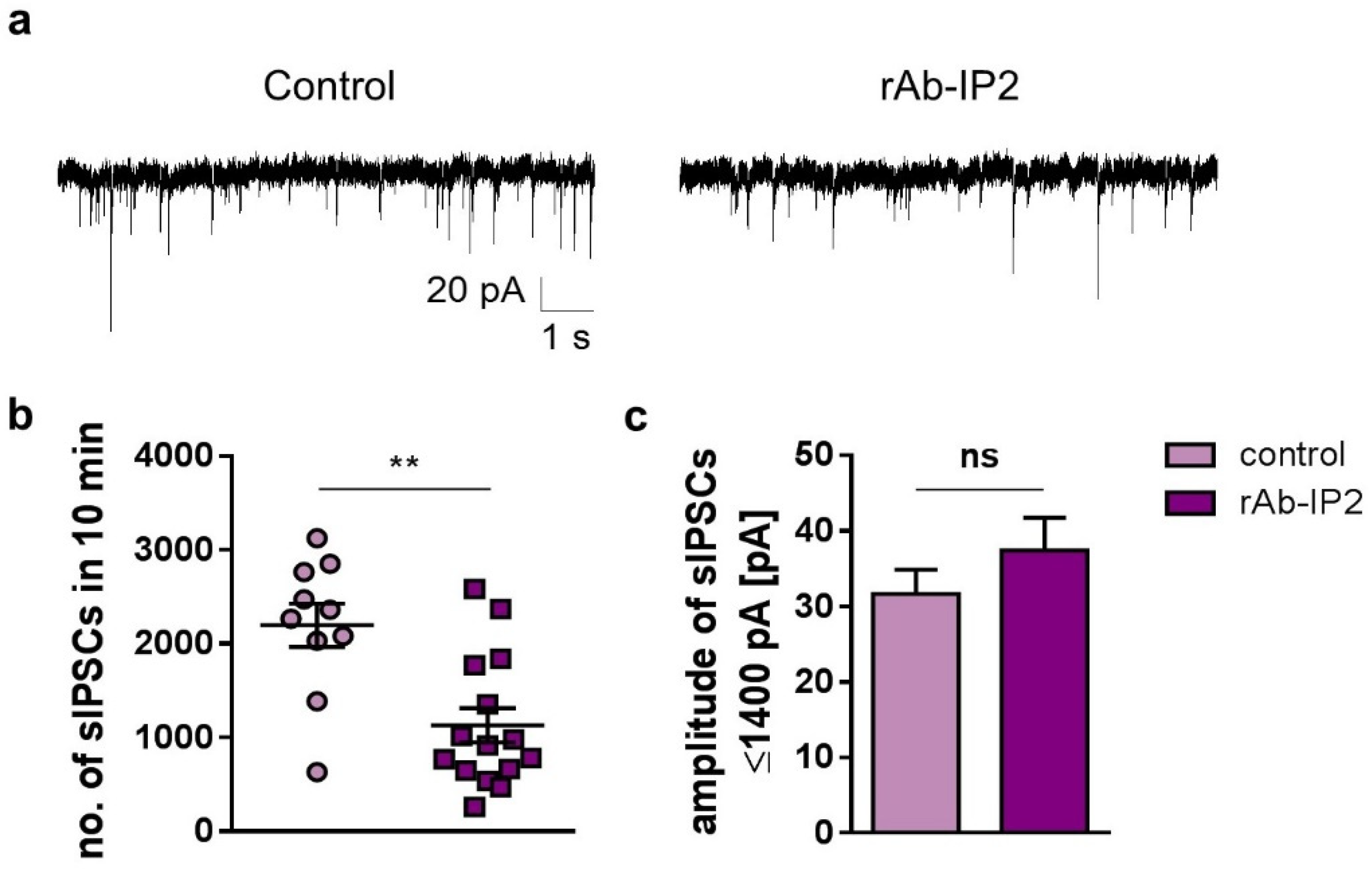
2.1. GABAA Receptor Autoantibodies (rAb-IP2) Reduce Inhibitory Postsynaptic Signaling in Hippocampal CA1 Pyramidal Cells

To characterize the effect of the recombinant antibody rAb-IP2 on GABAAR function in different hippocampal regions, we performed electrophysiological experiments using acute murine brain slices. The GABAergic activity of pyramidal neurons was recorded in hippocampal CA1 and CA3, in which the α1 and β3 subunits are frequently expressed on GABAARs [21,22]. In CA1, the incubation of brain slices with rAb-IP2 for 2 h led to a significant decrease in the number of spontaneous inhibitory postsynaptic currents (sIPSCs) recorded in a period of 10 min, compared with the control group without antibody application during the incubation period (rAb-IP2: 1130 ± 181.8, n = 10; control: 2198 ± 232.7, n = 15; unpaired Student’s t-test: t = 3.65, df = 23, ** p = 0.001; Figure 1a,b). The amplitude of GABAergic-mediated currents was not altered upon rAb-IP2 incubation, compared with the control (rAb-IP2: 59.71 ± 10.05 pA; control: 127.8 ± 42.89 pA; unpaired Student’s t-test: t = 1.85, df = 23, p = 0.08). Remarkably, high-amplitude ion currents were present during recordings in CA1 and CA3 pyramidal neurons, which were previously not commonly reported in these hippocampal areas. The currents were measured to have an amplitude > 1400 pA in contrast with sIPSCs with an amplitude ≤ 1400 pA. These high-amplitude currents were very likely contaminated with Na+ currents; therefore, they were not considered in the following steps. After excluding these recorded high-amplitude currents with an amplitude > 1400 pA which were not GABA-mediated, it appeared that the amplitude of sIPSCs < 1400 pA was not reduced by rAb-IP2 incubation (rAb-IP2: 37.39 ± 4.35 pA; control: 31.67 ± 3.21 pA; unpaired Student’s t-test: t = 0.96; df = 23, p = 0.35; Figure 1c). In summary, rAb-IP2 decreased GABAergic signaling in CA1 pyramidal neurons with regard to the number of sIPSCs, but not with regard to the amplitude.

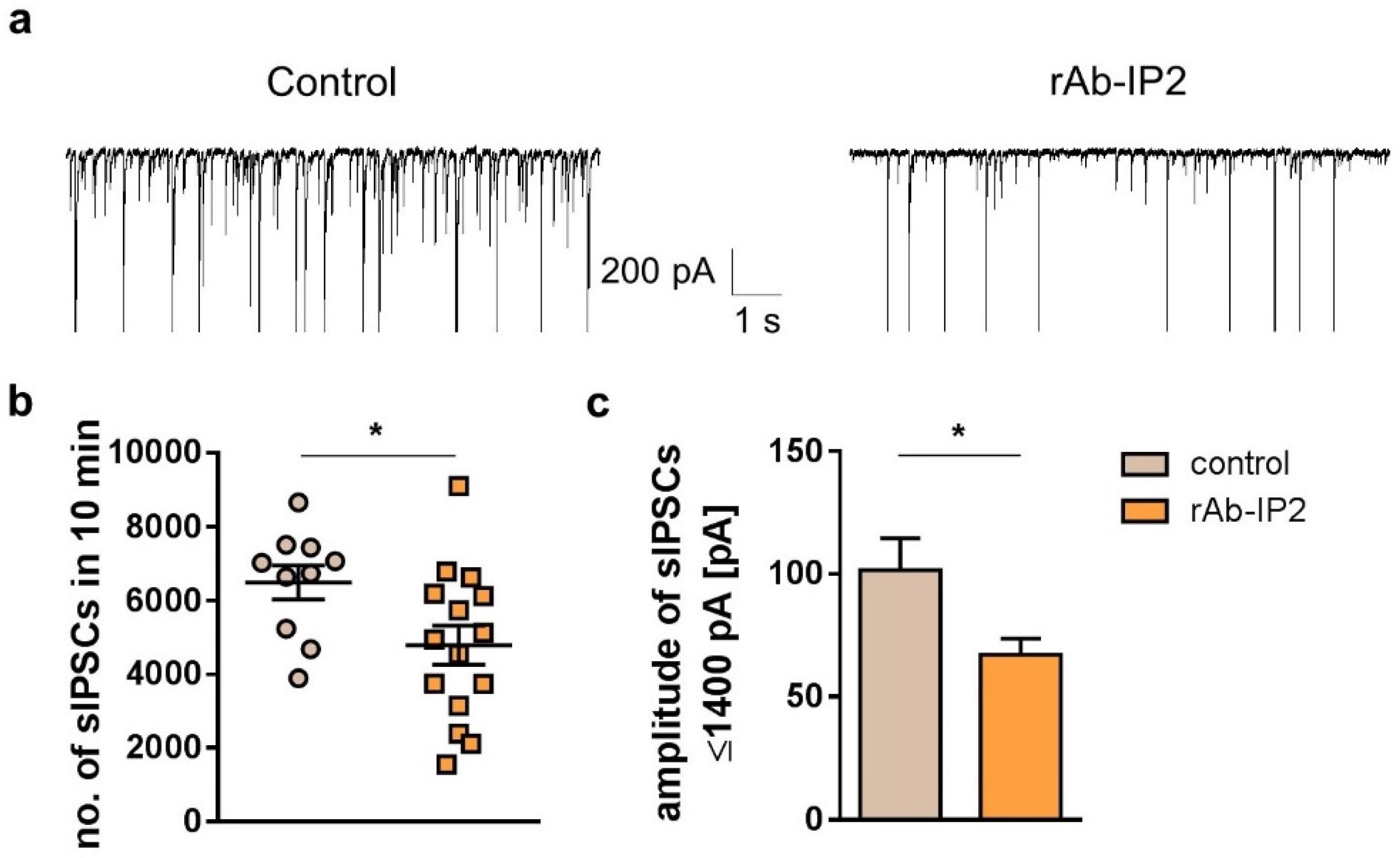
2.2. GABAA Receptor Autoantibodies Decrease GABAergic Synaptic Transmission in Hippocampal CA3 Network

In the next step, the results of CA1 were compared with those of the CA3 hippocampal region, which plays a critical role in the initiation of epileptic activity. The CA3 pyramidal neurons showed a significantly reduced number of sIPSCs upon rAb-IP2 incubation (rAb-IP2: 4788 ± 530.5, n = 10; control: 6487 ± 458.2, n = 15; unpaired Student’s t-test: t = 2.259, df = 23, * p = 0.03; Figure 2a,b). The amplitude of sIPSCs tended to be reduced after rAb-IP2 incubation (rAb-IP2: 227.7 ± 40.68 pA; control: 374.9 ± 73.89 pA, n = 11; unpaired Student’s t-test: t = 1.867, df = 24, p = 0.07, n = 15). Focusing on sIPSCs ≤ 1400 pA, the amplitude of GABAergic postsynaptic currents was significantly reduced after rAb-IP2 incubation (rAb-IP2: 67.14 ± 6.51 pA; control: 101.6 ± 12.94 pA; unpaired Student’s t-test: t = 2.577, df = 24, * p = 0.02; Figure 2c). In summary, the recombinant antibody rAb-IP2 led to both decreased number and amplitude of sIPSCs in CA3 pyramidal neurons.

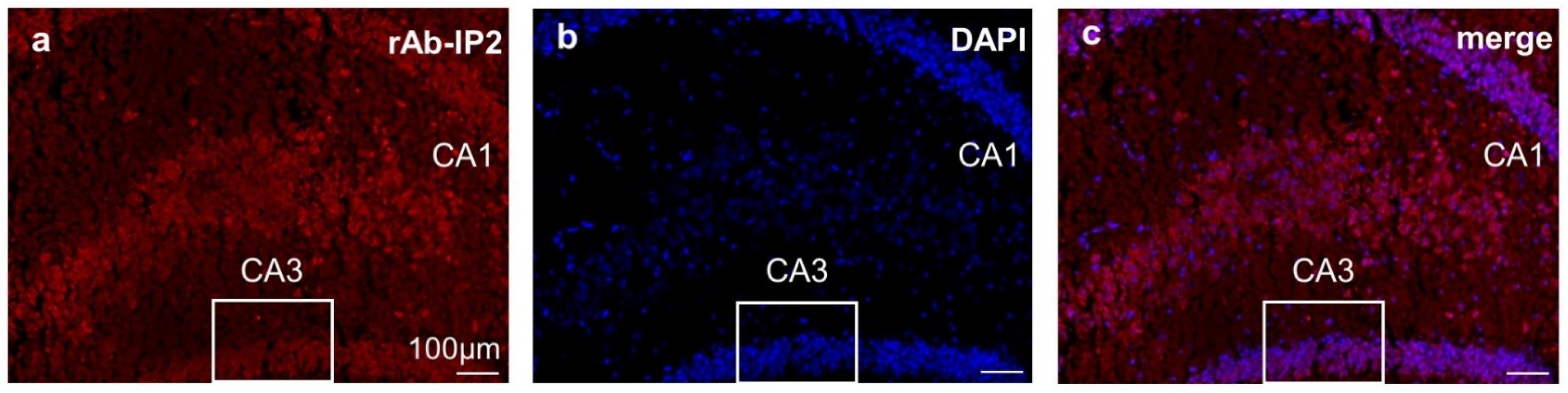
2.3. Immunohistochemical Staining Confirmed GABAA Receptor Autoantibody Binding to Hippocampal Tissue

The hippocampal slices showed strong immunoreactivity to rAb-IP2 compared with the negative controls (Figure 3a–c). The staining confirmed that the human autoantibody cross-reacts with rodent brain structures.

3. Discussion

In this study, the recombinant GABAA receptor autoantibody (rAb-IP2) reduced inhibitory postsynaptic signaling in hippocampal CA1 and CA3 pyramidal cells. Our results confirmed previous findings showing rAb-IP2-induced reduction in spontaneous postsynaptic GABAergic events in hippocampal CA1 pyramidal neurons [13]. Furthermore, this was linked to increased excitability in hippocampal CA1 pyramidal neurons. All these alterations in GABAergic synaptic transmission were assumed to cause the clinical symptoms of patients with GABAAR encephalitis [5,7,8]. Additionally, in our study, the GABAergic synaptic transmission was also decreased in CA3 pyramidal neurons. This finding is important because CA3 neurons are involved in the generation of epileptic discharges [15,16,17,18,19]. In another study, the cerebroventricular infusion of GABAAR mAb in Wistar rats caused spontaneous seizures in vivo [12]. Moreover, increased spontaneous epileptic activity was detected from electrodes placed in the hippocampal areas CA1 and CA3 of the rats ex vivo [12]. In line with these findings, incubation of cultured autaptic neurons with GABAAR mAb reduced inhibitory postsynaptic signaling in vitro [12]. These effects were observed independent of receptor internalization, indicating that GABAAR autoantibodies exert direct functional effects on the CA1 and CA3 pyramidal neurons and play a crucial role in seizure generation in GABAAR autoimmune encephalitis. The exact pathomechanism for Ab-mediated effects is yet not known. Multiple possible mechanisms were suggested, including receptor modulation, e.g., by desensitized conformation, redistribution, or network effects [12]. Further studies revealing the exact Ab-mediated pathomechanisms are needed. The target epitope of the mAb involved mainly the α1 and γ2 subunits [12]. In addition, in a complementary assay using flow cytometry, the mAb bound to GABAARs, coexpressing the α1, γ2, and β3 subunits [12]. It is important to consider that GABAAR subunit expression and composition significantly vary among different brain regions and subcellular locations [22,23,24]. This large heterogeneity of possible subunit combinations implies differences in the electrophysiological properties of the GABAAR [25,26]. For example, the presence of the α1 subunit is responsible for the fast decay time kinetics in IPSCs [9]. The varying expression of α1 and β3 subunits in the stratum pyramidale of CA1 and CA3 might explain why the amplitude and quantity of sIPSCs are affected in different ways by rAb-IP2. Moreover, it was shown that decreased expression of the α1-subunit mRNA correlated with altered GABAAR function and neuronal excitability in single dentate granule cells in an animal model of temporal lobe epilepsy [27]. The mutations in the GABAAR α1 and γ2 subunits have been related to different idiopathic generalized epilepsy syndromes [28,29]. These results confirmed that aberrant GABAAR expression and function play an essential role during epileptogenesis. Patients with GABAAR encephalitis develop frequent seizures, including refractory status epilepticus or epilepsia partialis continua [5,6]. Consistent with these clinical data, epileptiform activity was detected using wireless electroencephalography (EEG) in living animals that received GABAAR mAb as an infusion [12]. A peak of ictal events was achieved under the GABAAR mAb infusion, and the events persisted until 14 d after termination of the infusion [12]. In contrast, in animal models of N-methyl-D-aspartate receptor (NMDAR) encephalitis, memory deficits remained 4 d after the infusion was stopped and then resolved within the next 7 d [30]. GABAAR mAb led to higher EEG coastline length in the infused animals and to significantly higher power in all the power band ranges, especially in the lower frequency range (1–4 Hz), which is in line with EEG features in NMDAR encephalitis [12,31]. Similar findings with increases in the theta and delta powers were detected in the kainate-induced status epilepticus in mice [32]. A recent study demonstrated that the intracerebroventricular injection of NMDAR Abs in rats led to a higher number of interictal events in the CA3 hippocampal region compared with the CA1, and to a spontaneous epileptic activity, highest in the CA3 region [33]. Additionally, whole-cell patch-clamp recordings from hippocampal CA3 pyramidal cells after the injection of NMDAR Abs showed a reduction in excitatory, but not in inhibitory, synaptic neurotransmission, or intrinsic hyperexcitability. The reduced synaptic excitatory neurotransmission is assumed to underlay seizures in this rat model of NMDAR Ab-mediated encephalitis [33]. In our study, GABAAR Abs led to decreased number and amplitude of sIPSCs in CA3 pyramidal neurons, the injection of NMDAR Abs caused no changes in the frequency or amplitude of sIPSCS. Following this, each CNS autoantibody may contribute in different ways to excitatory–inhibitory imbalance, which plays a critical role in the pathophysiology of seizures. The neuronal circuits are highly complex, and changes in synaptic transmission can have extensive impacts on the stability of neuronal networks.
It can be summarized that GABAAR autoantibodies exert direct functional effects on the hippocampal CA1 and CA3 pyramidal neurons and play a crucial role in seizure generation in GABAAR autoimmune encephalitis.

4. Materials and Methods

4.1. Clinical Samples

In our study, the recombinant GABAAR autoantibody (named rAb-IP2) from the CSF of patient IP2, who was suffering from anti-GABAAR encephalitis, was cloned, expressed, purified, and characterized according to a previous study [13]. The clinical data of patient IP2 were previously described [5,13]. It was demonstrated that rAb-IP2 specifically binds to the α1 and β3 subunit of the GABAAR [13].
This study was approved by the ethics committee of Münster, Germany (AZ 2013-682-b-S). A written informed consent was obtained prior to study conduct, according to the principles of the Declaration of Helsinki.

4.2. Animals

All C57BL/6 mice were kept under pathogen-free conditions and had access to food and water ad libitum. All experiments were conducted according to German law and were approved by the local authorities (Landesamt für Natur, Umwelt und Verbraucherschutz Nordrhein-Westfalen).
Immunohistochemical studies were performed on brains obtained from 10–17-week- old mice, whereas for the electrophysiological analysis, younger mice (2–7 weeks old) were used because the GABAergic system in mice is robust and no significant age-related differences in the GABAAR α1, α2, α3, α5, β3, and γ2 subunit expression levels were found between the young- and old-age groups in different regions of the mouse hippocampus [34]. Moreover, one additional reason for the age difference between the mice used for the two different experiments was that our laboratory follows the 3R principles, which aim to refine and reduce the number of animals used in a given experiment.

4.3. Preparation of Acute Murine Brain Slices

Brains were collected from C57BL/6 mice (2–7 weeks old) after anesthesia with 4% isoflurane in O2 and fast decapitation. Acute brain slices were obtained by cutting 260 μm thick slices using a vibratome (Leica, Wetzlar, Germany). For investigation of the hippocampal CA1 and CA3 regions, transversal slices were collected. Slices were placed in a submersion chamber and continuously perfused with an extracellular solution (artificial CSF) containing 120 mM NaCl, 2.5 mM KCl, 1.25 mM NaH2PO4, 22 mM NaHCO3, 20 mM C6H12O6, 2 mM CaCl2, and 2 mM MgSO4 (Merck, Darmstadt, Germany), set to pH of 7.35 with carbogen.

4.4. Incubation of Acute Murine Brain Slices with the GABAAR Autoantibody rAb-IP2

Acute brain slices were incubated with the human antibody rAb-IP2 using an incubation chamber. The bottom of the chamber was filled with extracellular solution (artificial CSF), which was kept at room temperature and purged with carbogen. After centrifugation and vortexing of a batch containing rAb-IP2, the Ab was added at a final concentration of 4.84 µg/mL. The brain slices were incubated with rAb-IP2 for 2 h. Control slices were kept under the same conditions in the absence of rAb-IP2.

4.5. Electrophysiological Recordings by Patch-Clamp Technique

After the incubation step, the brain slices were placed in a custom-built recording chamber, which was continuously perfused with the external solution. Glass pipettes for recording were pulled from borosilicate glass (GC150TF-10; Harvard Apparatus, Holliston, MA, USA) capillaries by using a vertical pipette puller. For the voltage-clamp mode, the pipettes were filled with a KCl-based, high-chloride intracellular solution containing 10 mM NaCl, 110 mM KCl, 11 mM EDTA, 10 mM HEPES, 1 mM MgCl2, 0.5 mM CaCl2, 15 mM phosphocreatine, 3 mM Mg-ATP, and 0.5 mM Na-GTP, set to pH 7.25 with KOH and an osmolality of 295 mOsmol/kg (Sigma-Aldrich, Schnelldorf, Germany; Merck, Darmstadt, Germany). The liquid-filled glass pipette was connected to an EPC-10 amplifier (HEKA Elektronik, Lamprecht, Germany) for generating and measuring ionic currents. The electrode resistance varied between 4 and 7 MΩ, and the series resistance was 5–15 MΩ (compensation ≥ 25%). A bright-light microscopy was used for visual identification of the pyramidal neurons in the hippocampal regions. The recordings were governed by Patchmaster software (HEKA Elektronik, Lamprecht, Germany).

Voltage-Clamp Analysis

In order to analyze the effects of the rAb-IP2 on GABAAR function, the spontaneous synaptic GABAergic transmission was measured in voltage-clamp mode. As is well-known, inhibitory postsynaptic currents (IPSCs) are induced by release of presynaptic GABA, which binds to postsynaptic GABAARs, resulting in hyperpolarization of the postsynaptic membrane. In this experimental setup, an intracellular solution with a higher concentration of Cl (117 mM) and physiological intracellular concentrations of other ions was prepared. Following this, we investigated the cells in the range of their resting membrane potential (RMP) at −65 mV and measured outwardly directed Cl- currents. To evaluate only GABA-triggered ion currents, the competitive AMPA- and kainate-receptor antagonist 6,7-dinitroquinoxaline-2,3-dione (DNQX, 10 µM) and the NMDA-receptor blocker (2R)-amino-5-phosphonopentanoate (AP5, 10 µM) were added to the extracellular solution. The GABAergic activity was recorded for 10 min. The analysis of sIPSCs was performed semiautomatically using MiniAnalysis (Synaptosoft Inc., Fort Lee, NJ, USA) and FitMaster (HEKA Elektronik, Lamprecht, Germany). The number and the amplitude of sIPSCs were used as read-outs.

4.6. Immunohistochemistry

After deep anesthesia of the C57BL/6 mice (10–17 weeks old), the brains were removed, placed into a cryo-protective fixative buffer (Tissue-Tek, Sakura Finetek, Alphen aan den Rijn, The Netherlands), and frozen at −20 °C. Afterward, coronal slices of 10 µm including the hippocampal region were cut using a cryotome. The slices were fixed with 10% of paraformaldehyde (PFA) (Merck, Darmstadt, Germany) for 10 min and washed three times with phosphate-buffered saline (PBS) (Sigma-Aldrich, Schnelldorf, Germany) for 5 min. Next, a blocking solution containing 1% goat serum (PAA, Pasching, Austria), 10% bovine serum albumin (Sigma-Aldrich, Schnelldorf, Germany), and 1% Triton X-100 (Sigma-Aldrich, Schnelldorf, Germany) was applied for 2 h. Afterward, the slices were treated with the primary antibody rAb-IP2 at a concentration of 1:250 at 4 °C overnight. After washing with PBS, Cy3-conjugated goat antihuman IgG was used as a secondary antibody (emission at a wavelength of 565 nm) (Dianova, Hamburg, Germany) at a concentration of 1:300 (incubation period 2 h at room temperature). The slices were stained with 5–10 µL Fluoromount G containing 4′,6-diamidino-2-phenylindole (DAPI) (Thermo Fisher Scientific, Waltham, Massachusetts, USA) for visualization of the cell nuclei. Hippocampal slices without rAb-IP2 incubation were prepared as negative controls without the detection of any fluorescent signal. For image acquisition and conducting the analysis, an Axio Scope A1 fluorescence microscope (Carl Zeiss GmbH, Jena, Germany) was used.

4.7. Statistics

Grubb’s test was used for testing significant outliers in the data sets. Gaussian-distributed data sets were analyzed using Student’s t-test for comparisons of two groups. GraphPad Prism 6 (GraphPad Software, San Diego, CA, USA) was applied to analyze and present the data in graphs. Representative traces were generated with OriginPro 2018 software (OriginLab, Friedrichsdorf, Germany). The number of experiments was reported as the number of recordings. All results are reported as mean value ± standard error of the mean (SEM). Level of significance was set as p < 0.05.

5. Conclusions

This study confirmed previous findings that GABAAR autoantibodies (rAb-IP2) led to reduced inhibitory postsynaptic signaling and increased excitability of pyramidal neurons in hippocampal CA1. We demonstrated further disruption of GABAergic signaling in hippocampal CA3, which is known to be a critical region for seizure generation in mesial temporal lobe epilepsy and can be one of the crucial points in the pathophysiology of GABAAR encephalitis. Our results support previous study data showing that the infusion of monoclonal GABAAR autoantibodies in rodents caused increased spontaneous epileptic activity in the hippocampal CA1 and CA3 areas ex vivo and spontaneous seizures in vivo.
Taken together, these findings strongly indicate the pathogenic effects of GABAAR autoantibodies on neuronal function as dampened GABAergic transmission and increased neuronal excitability, significantly contributing to seizures and status epilepticus.

Author Contributions

Conceptualization, N.M., S.G.M. and M.C.; methodology, N.M., S.G.M., A.F.M. and M.C.; software, A.F.M.; formal analysis, A.F.M.; investigation, A.F.M.; providing recombinant antibodies, K.D.; writing—original draft preparation, F.S.I. and A.F.M.; writing—review and editing, N.M., S.G.M., M.C. and K.D.; supervision, N.M., S.G.M. and M.C.; project administration, N.M. and S.G.M. All authors have agreed to be accountable for all aspects of the work in ensuring that questions related to the accuracy or integrity of any part of the work are appropriately investigated and resolved. All authors have read and agreed to the published version of the manuscript.

Funding

This project was supported partly by the German Federal Ministry of Science and Education (Comprehensive, Orchestrated, National Network to Explain, Categorize, and Treat Autoimmune Encephalitis and Allied Diseases within the German Network for Research on Autoimmune Encephalitis—CONNECT GENERATE; 01GM1908) and the German Research Council through grant Munich Cluster for Systems Neurology (SyNergy, grant EXC 2145, project ID 390857198).

Institutional Review Board Statement

The study was conducted according to the guidelines of the Declaration of Helsinki and approved by the Ethics Committee of Münster, Germany (AZ 2013-682-b-S).

Informed Consent Statement

Informed consent was obtained from all subjects involved in the study.

Data Availability Statement

The datasets used or analyzed during this study are available from the corresponding author upon reasonable request.

Conflicts of Interest

The authors declare no conflict of interest.

References

  1. Jacob, T.C.; Moss, S.J.; Jurd, R. GABA(A) receptor trafficking and its role in the dynamic modulation of neuronal inhibition. Nat. Rev. Neurosci. 2008, 9, 331–343. [Google Scholar] [CrossRef] [PubMed] [Green Version]
  2. Olsen, R.W.; Sieghart, W. GABA A receptors: Subtypes provide diversity of function and pharmacology. Neuropharmacology 2009, 56, 141–148. [Google Scholar] [CrossRef] [PubMed] [Green Version]
  3. Farrant, M.; Nusser, Z. Variations on an inhibitory theme: Phasic and tonic activation of GABA(A) receptors. Nat. Rev. Neurosci. 2005, 6, 215–229. [Google Scholar] [CrossRef] [PubMed]
  4. Rudolph, U.; Knoflach, F. Beyond classical benzodiazepines: Novel therapeutic potential of GABAA receptor subtypes. Nat. Rev. Drug Discov. 2011, 10, 685–697. [Google Scholar] [CrossRef] [Green Version]
  5. Petit-Pedrol, M.; Armangue, T.; Peng, X.; Bataller, L.; Cellucci, T.; Davis, R.; McCracken, L.; Martinez-Hernandez, E.; Mason, W.P.; Kruer, M.C.; et al. Encephalitis with refractory seizures, status epilepticus, and antibodies to the GABAA receptor: A case series, characterisation of the antigen, and analysis of the effects of antibodies. Lancet Neurol. 2014, 13, 276–286. [Google Scholar] [CrossRef] [Green Version]
  6. Spatola, M.; Petit-Pedrol, M.; Simabukuro, M.M.; Armangue, T.; Castro, F.J.; Barcelo Artigues, M.I.; Julià Benique, M.R.; Benson, L.; Gorman, M.; Felipe, A.; et al. Investigations in GABAA receptor antibody-associated encephalitis. Neurology 2017, 88, 1012–1020. [Google Scholar] [CrossRef] [Green Version]
  7. Pettingill, P.; Kramer, H.B.; Coebergh, J.A.; Pettingill, R.; Maxwell, S.; Nibber, A.; Malaspina, A.; Jacob, A.; Irani, S.R.; Buckley, C.; et al. Antibodies to GABAA receptor α1 and γ2 subunits: Clinical and serologic characterization. Neurology 2015, 84, 1233–1241. [Google Scholar] [CrossRef] [Green Version]
  8. Ohkawa, T.; Satake, S.; Yokoi, N.; Miyazaki, Y.; Ohshita, T.; Sobue, G.; Takashima, H.; Watanabe, O.; Fukata, Y.; Fukata, M. Identification and characterization of GABA(A) receptor autoantibodies in autoimmune encephalitis. J. Neurosci. 2014, 34, 8151–8163. [Google Scholar] [CrossRef] [Green Version]
  9. Zhou, C.; Huang, Z.; Ding, L.; Deel, M.E.; Arain, F.M.; Murray, C.R.; Patel, R.S.; Flanagan, C.D.; Gallagher, M.J. Altered cortical GABAA receptor composition, physiology, and endocytosis in a mouse model of a human genetic absence epilepsy syndrome. J. Biol. Chem. 2013, 288, 21458–21472. [Google Scholar] [CrossRef] [Green Version]
  10. Dalmau, J.; Graus, F. Antibody-Mediated Encephalitis. N. Engl. J. Med. 2018, 378, 840–851. [Google Scholar] [CrossRef] [Green Version]
  11. Bracher, A.; Alcalá, C.; Ferrer, J.; Melzer, N.; Hohlfeld, R.; Casanova, B.; Beltrán, E.; Dornmair, K. An expanded parenchymal CD8+ T cell clone in GABAA receptor encephalitis. Ann. Clin. Transl. Neurol. 2020, 7, 239–244. [Google Scholar] [CrossRef] [PubMed] [Green Version]
  12. Kreye, J.; Wright, S.K.; van Casteren, A.; Stöffler, L.; Machule, M.L.; Reincke, S.M.; Nikolaus, M.; van Hoof, S.; Sanchez-Sendin, E.; Homeyer, M.A.; et al. Encephalitis patient-derived monoclonal GABAA receptor antibodies cause epileptic seizures. J. Exp. Med. 2021, 218, e20210012. [Google Scholar] [CrossRef] [PubMed]
  13. Brändle, S.M.; Cerina, M.; Weber, S.; Held, K.; Menke, A.F.; Alcalá, C.; Gebert, D.; Herrmann, A.M.; Pellkofer, H.; Gerdes, L.A.; et al. Cross-reactivity of a pathogenic autoantibody to a tumor antigen in GABAA receptor encephalitis. Proc. Natl. Acad. Sci. USA 2021, 118, e1916337118. [Google Scholar] [CrossRef] [PubMed]
  14. Spruston, N. Pyramidal neurons: Dendritic structure and synaptic integration. Nat. Rev. Neurosci. 2008, 9, 206–221. [Google Scholar] [CrossRef]
  15. Lopes da Silva, F.H.; Witter, M.P.; Boeijinga, P.H.; Lohman, A.H. Anatomic organization and physiology of the limbic cortex. Physiol. Rev. 1990, 70, 453–511. [Google Scholar] [CrossRef]
  16. Le Duigou, C.; Simonnet, J.; Teleñczuk, M.T.; Fricker, D.; Miles, R. Recurrent synapses and circuits in the CA3 region of the hippocampus: An associative network. Front. Cell Neurosci. 2014, 7, 262. [Google Scholar] [CrossRef]
  17. Jefferys, J.G. Basic mechanisms of focal epilepsies. Exp. Physiol. 1990, 75, 127–162. [Google Scholar] [CrossRef] [Green Version]
  18. Wittner, L.; Miles, R. Factors defining a pacemaker region for synchrony in the hippocampus. J. Physiol. 2007, 584, 867–883. [Google Scholar] [CrossRef]
  19. Stoop, R.; Pralong, E. Functional connections and epileptic spread between hippocampus, entorhinal cortex and amygdala in a modified horizontal slice preparation of the rat brain. Eur. J. Neurosci. 2000, 12, 3651–3663. [Google Scholar] [CrossRef]
  20. van Groen, T.; Wyss, J.M. Extrinsic projections from area CA1 of the rat hippocampus: Olfactory, cortical, subcortical, and bilateral hippocampal formation projections. J. Comp. Neurol. 1990, 302, 515–528. [Google Scholar] [CrossRef]
  21. Heldt, S.A.; Ressler, K.J. Forebrain and midbrain distribution of major benzodiazepine-sensitive GABAA receptor subunits in the adult C57 mouse as assessed with in situ hybridization. Neuroscience 2007, 150, 370–385. [Google Scholar] [CrossRef] [PubMed] [Green Version]
  22. Hörtnagl, H.; Tasan, R.O.; Wieselthaler, A.; Kirchmair, E.; Sieghart, W.; Sperk, G. Patterns of mRNA and protein expression for 12 GABAA receptor subunits in the mouse brain. Neuroscience 2013, 236, 345–372. [Google Scholar] [CrossRef] [PubMed] [Green Version]
  23. Wisden, W.; Seeburg, P.H. GABAA receptor channels: From subunits to functional entities. Curr. Opin. Neurobiol. 1992, 2, 263–269. [Google Scholar] [CrossRef]
  24. Gao, Y.; Heldt, S.A. Enrichment of GABAA Receptor α-Subunits on the Axonal Initial Segment Shows Regional Differences. Front. Cell Neurosci. 2016, 10, 39. [Google Scholar] [CrossRef] [PubMed] [Green Version]
  25. Minier, F.; Sigel, E. Positioning of the alpha-subunit isoforms confers a functional signature to gamma-aminobutyric acid type A receptors. Proc. Natl. Acad. Sci. USA 2004, 101, 7769–7774. [Google Scholar] [CrossRef] [PubMed] [Green Version]
  26. Tia, S.; Wang, J.F.; Kotchabhakdi, N.; Vicini, S. Distinct deactivation and desensitization kinetics of recombinant GABAA receptors. Neuropharmacology 1996, 35, 1375–1382. [Google Scholar] [CrossRef]
  27. Brooks-Kayal, A.R.; Shumate, M.D.; Jin, H.; Rikhter, T.Y.; Coulter, D.A. Selective changes in single cell GABA(A) receptor subunit expression and function in temporal lobe epilepsy. Nat. Med. 1998, 4, 1166–1172, Erratum in Nat. Med. 1999, 5, 590. [Google Scholar] [CrossRef]
  28. Maljevic, S.; Krampfl, K.; Cobilanschi, J.; Tilgen, N.; Beyer, S.; Weber, Y.G.; Schlesinger, F.; Ursu, D.; Melzer, W.; Cossette, P.; et al. A mutation in the GABA(A) receptor alpha(1)-subunit is associated with absence epilepsy. Ann. Neurol. 2006, 59, 983–987. [Google Scholar] [CrossRef]
  29. Macdonald, R.L.; Gallagher, M.J.; Feng, H.J.; Kang, J. GABA(A) receptor epilepsy mutations. Biochem. Pharmacol. 2004, 68, 1497–1506. [Google Scholar] [CrossRef]
  30. Planagumà, J.; Leypoldt, F.; Mannara, F.; Gutiérrez-Cuesta, J.; Martín-García, E.; Aguilar, E.; Titulaer, M.J.; Petit-Pedrol, M.; Jain, A.; Balice-Gordon, R.; et al. Human N-methyl D-aspartate receptor antibodies alter memory and behaviour in mice. Brain 2015, 138, 94–109. [Google Scholar] [CrossRef]
  31. Symmonds, M.; Moran, C.H.; Leite, M.I.; Buckley, C.; Irani, S.R.; Stephan, K.E.; Friston, K.J.; Moran, R.J. Ion channels in EEG: Isolating channel dysfunction in NMDA receptor antibody encephalitis. Brain 2018, 141, 1691–1702. [Google Scholar] [CrossRef] [PubMed] [Green Version]
  32. Puttachary, S.; Sharma, S.; Tse, K.; Beamer, E.; Sexton, A.; Crutison, J.; Thippeswamy, T. Immediate Epileptogenesis after Kainate-Induced Status Epilepticus in C57BL/6J Mice: Evidence from Long Term Continuous Video-EEG Telemetry. PLoS ONE 2015, 10, e0131705. [Google Scholar] [CrossRef] [PubMed]
  33. Wright, S.K.; Rosch, R.E.; Wilson, M.A.; Upadhya, M.A.; Dhangar, D.R.; Clarke-Bland, C.; Wahid, T.T.; Barman, S.; Goebels, N.; Kreye, J.; et al. Multimodal electrophysiological analyses reveal that reduced synaptic excitatory neurotransmission underlies seizures in a model of NMDAR antibody-mediated encephalitis. Commun. Biol. 2021, 4, 1106. [Google Scholar] [CrossRef] [PubMed]
  34. Palpagama, T.H.; Sagniez, M.; Kim, S.; Waldvogel, H.J.; Faull, R.L.; Kwakowsky, A. GABAA Receptors Are Well Preserved in the Hippocampus of Aged Mice. eNeuro 2019, 6, ENEURO.0496-18.2019. [Google Scholar] [CrossRef] [Green Version]
Figure 1. The recombinant GABAA receptor autoantibody rAb-IP2 reduced the number of spontaneous inhibitory postsynaptic currents (sIPSCs) generated in CA1 pyramidal neurons. (a) Exemplary traces depict the GABAergic synaptic transmission in rAb-IP2-incubated pyramidal neurons and controls. Notably, the exemplary traces are cropped at 600 pA to simplify illustration and do not represent the full high-amplitude currents with an amplitude > 1400 pA, which were very likely contaminated with Na+ currents and were not GABA-mediated. (b) Scatter plot showing that the number of sIPSCs recorded in 10 min decreased upon rAb-IP2 incubation in comparison with the control. (c) Bar graphs showing the amplitude of sIPSCs in rAb-IP2-incubated neurons and controls. The amplitude of sIPSCs ≤ 1400 pA was not altered by rAb-IP2 incubation. ** p < 0.01, ns: not significant.
Ijms 23 03707 g001
Figure 2. Incubation with the recombinant GABAA receptor autoantibody rAb-IP2 led to both decreased number and amplitude of spontaneous inhibitory postsynaptic currents (sIPSCs) in pyramidal CA3 neurons. (a) Exemplary traces show GABA-mediated synaptic currents in rAb-IP2-incubated pyramidal neurons and controls. For illustration purposes, the amplitude of the high-amplitude currents is cut at 800 pA. (b) rAb-IP2-incubated neurons showed a significantly reduced number of sIPSCs in comparison with controls. (c) rAb-IP2 led to a significant reduction in the amplitude of sIPSCs when sIPSCs ≤ 1400 pA were analyzed. * p < 0.05, ns: not significant.
Ijms 23 03707 g002
Figure 3. Immunohistochemical staining of a formalin-fixed, paraffin-embedded murine hippocampus with the recombinant human GABAA receptor autoantibody rAb-IP2. Murine-fixed hippocampal tissue shows immunoreactivity to the recombinant human antibody rAb-IP2. (a) Representative coronal hippocampal slices with immunoreactivity to rAb-IP2 (red). The box marks the hippocampal region CA3, showing the area used for the in vitro electrophysiological recordings as shown in Figure 1 and Figure 2. For more visibility of CA1, we also referred to our previous study [13]. (b) DAPI (blue) was used as a marker for the cell nuclei. (c) Merged picture. Scale bars represent 100 μm.
Ijms 23 03707 g003
Publisher’s Note: MDPI stays neutral with regard to jurisdictional claims in published maps and institutional affiliations.

